forensic
TRANSCRIPT


Simpson’s Forensic Medicine
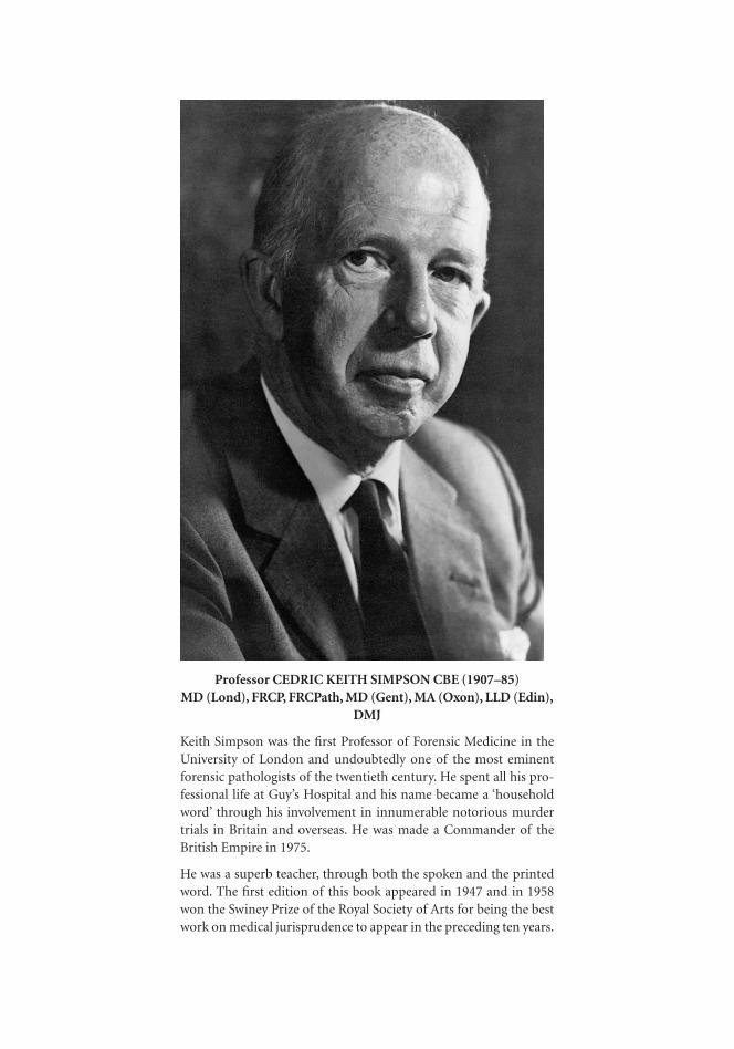
Professor CEDRIC KEITH SIMPSON CBE (1907–85)MD (Lond), FRCP, FRCPath, MD (Gent), MA (Oxon), LLD (Edin),
DMJ
Keith Simpson was the first Professor of Forensic Medicine in theUniversity of London and undoubtedly one of the most eminentforensic pathologists of the twentieth century. He spent all his pro-fessional life at Guy’s Hospital and his name became a ‘householdword’ through his involvement in innumerable notorious murdertrials in Britain and overseas. He was made a Commander of theBritish Empire in 1975.
He was a superb teacher, through both the spoken and the printedword. The first edition of this book appeared in 1947 and in 1958won the Swiney Prize of the Royal Society of Arts for being the bestwork on medical jurisprudence to appear in the preceding ten years.
Simpson’s Forensic MedicineTwelfth Edition
Richard ShepherdSenior Lecturer in Forensic Medicine
Forensic Medicine Unit
St George’s Medical and Dental School
Tooting, London, UK
A member of the Hodder Headline Group
LONDON
First published in Great Britain in 1947Twelfth edition published in 2003 byArnold, a member of the Hodder Headline Group,338 Euston Road, London NW1 3BH
http://www.arnoldpublishers.com
Distributed in the United States of America byOxford University Press Inc.,198 Madison Avenue, New York, NY10016Oxford is a registered trademark of Oxford University Press
© 2003 Arnold
All rights reserved. No part of this publication may bereproduced or transmitted in any form or by any means,electronically or mechanically, including photocopying,recording or any information storage or retrieval system,without either prior permission in writing from thepublisher or a licence permitting restricted copying.In the United Kingdom such licences are issued by theCopyright Licensing Agency: 90 Tottenham Court Road,London W1T 4LP.
Whilst the advice and information in this book are believed to be true and accurate at the date of going topress, neither the authors nor the publisher can accept anylegal responsibility or liability for any errors or omissionsthat may be made. In particular (but without limiting thegenerality of the preceding disclaimer) every effort has beenmade to check drug dosages; however it is still possible thaterrors have been missed. Furthermore, dosage schedules areconstantly being revised and new side-effects recognized.For these reasons the reader is strongly urged to consult thedrug companies’ printed instructions before administeringany of the drugs recommended in this book.
British Library Cataloguing in Publication DataA catalogue record for this book is available from the British Library
Library of Congress Cataloging-in-Publication DataA catalog record for this book is available from the Library of Congress
ISBN 0 340 76422 8
ISBN 0 340 81059 9 (International Students’ Edition –restricted territorial availability)
1 2 3 4 5 6 7 8 9 10
Commissioning Editor: Serena BureauDevelopment Editor: Layla VandenberghProject Editor: James RabsonProduction Controller: Deborah SmithCover Design: Stewart Larking
Typeset in 9.5/12 pt Minion by Charon Tec Pvt. Ltd,Chennai, India
Printed and bound in India
What do you think about this book? Or any other Arnold title?Please send your comments to [email protected]
Preface ixAuthor’s Note xAcknowledgements xi
1 The Doctor and the Law 1
The legal system 2
Doctors and the law 2
Doctor in court 3
The behaviour of a doctor in court 5
Preparation of medical reports 5
Structure of a report 6
2 The Ethics of Medical Practice 8
International Code of Medical Ethics 9
Medical ethics in practice 10
Medical confidentiality 11
Consent to medical treatment 13
3 Medical Malpractice 15
Medical negligence 15
Systems of compensation 17
Compensation and damages 17
Types of medical negligence 17
Professional misconduct 19
The General Medical Council 20
4 The Medico-legal Aspects of Mental Disease 22
Normal and abnormal behaviour 22
Types of abnormal mental condition 22
Mental health legislation and the criminal justice system 23
Criminal responsibility: age and mental capacity 24
The effect of drink or drugs on responsibility 25
Testamentary capacity 25
5 The Medical Aspects of Death 27
Definition of death 27
Resuscitation 28
Persistent vegetative state 29
Tissue and organ transplantation 29
Death certification 30
Medico-legal investigation of death 32
The autopsy 34
Exhumation 35
6 Changes after Death 37
Early changes 37
Rigor mortis 38
Cadaveric rigidity 38
Post-mortem hypostasis 39
Cooling of the body after death 40
Estimation of the time of death 41
7 Identification of the Living and the Dead 49
Morphological characteristics 49
Fingerprints 50
Identity from teeth 50
Identification of the origin of tissue or samples 51
Contents
The individuality of cells 51
Identification by DNA profiling 52
Tattoos and body piercing 53
Identity of decomposed or skeletalized remains 54
Facial reconstruction from skulls 55
8 Blood Stains 57
Blood-stain patterns 57
Tests for blood 58
Species specificity 58
9 The Examination of Wounds 59
Law on wounding 59
Reports 60
Terminology 60
Wounds 60
Patterns of injury 66
Survival 68
Self-inflicted injuries 68
10 Regional Injuries 70
Head injuries 70
Neck injuries 75
Spinal injuries 75
Chest injuries 76
Abdomen 77
11 Firearm and Explosive Injuries 79
Types of firearms 79
Gunshot wounds 81
Air weapons, unusual projectiles and other weapons 84
Accident, suicide or murder? 85
The doctor’s duty in firearm injuries and deaths 85
Explosives 86
12 Transportation Injuries 87
Road traffic injuries 87
The medical examination of victims of road traffic accidents 90
Railway injuries 91
Aircraft fatalities 92
Mass disasters and the doctor 92
13 Asphyxia 94
Suffocation 95
Smothering 96
Gagging 96
Choking 96
Pressure on the neck 97
‘Vagal inhibition’ or reflex cardiac arrest 97
Manual strangulation 98
Ligature strangulation 98
Hanging 99
The sexual asphyxias 101
Traumatic asphyxia 101
14 Immersion and Drowning 103
Signs of immersion 103
Drowning 104
Laboratory tests for drowning 105
15 Injury due to Heat, Cold and Electricity 107
Injury due to heat 107
Cold injury (hypothermia) 110
Electrical injury 111
Death from lightning 113
16 Effects of Injuries 115
Haemorrhage 115
Infection 116
Embolism 116
Disseminated intravascular coagulation 118
Adult respiratory distress syndrome 118
Suprarenal haemorrhage 118
Subendocardial haemorrhages 119
17 Unexpected and Sudden Death from Natural Causes 120
Causes of sudden and unexpected death 120
Cardiovascular system 121
vi Contents

Contents vii
Respiratory system 126
Gastrointestinal system 126
Gynaecological conditions 126
Deaths from asthma and epilepsy 126
18 Sexual Offences 128
Types of sexual offence 128
The genuineness of allegations of sexual assault 131
Forensic examination of victims of sexual offences 131
Examination of an alleged assailant 132
19 Pregnancy and Abortion 134
Conception: artificial insemination, in-vitrofertilization and embryo research 134
Pregnancy 134
Abortion 135
20 Deaths and Injury in Infancy 141
Stillbirths 141
Infanticide 142
The estimation of maturity of a newborn baby or fetus 143
Sudden infant death syndrome 144
Child abuse 145
21 Neglect, Starvation and Abuse of Human Rights 150
Physical abuse of human rights: torture 150
Neglect and starvation 152
22 General Aspects of Poisoning 154
The toxic and fatal dose 155
Tolerance and idiosyncrasy 156
The doctor’s duty in a case of suspected poisoning 156
Samples required for toxicological analysis 157
23 Alcohol 159
Sources of alcohol 159
Absorption of alcohol 160
Elimination of alcohol 160
The measurement of alcohol 160
The effects of alcohol 160
Dangers of drunkenness 161
Drinking and driving 162
24 Drugs of Dependence and Abuse 164
Tolerance and synergy 164
Dependence and withdrawal symptoms 165
The dangers of drug dependence 165
Shared syringes 165
Solid drugs 165
Overdosage and hypersensitivity 166
Heroin, morphine and other opiates 166
Barbiturates and other hypnotics 167
Amphetamines 167
Cocaine 167
Cannabis 168
Lysergic acid diethylamide (LSD) 168
Solvent abuse 168
25 Medicinal Poisons 170
Analgesics 171
Antidepressant and sedative drugs 172
Barbiturates 172
Chloral 173
Phenacetin 173
Lithium 173
Insulin 173
26 Corrosive and Metallic Poisons 175
Corrosive poisons 175
Heavy-metal poisoning 176
27 Agrochemical Poisons 179
Pesticides and insecticides 179
Herbicides (weed killers) 179
28 Gaseous Poisons 181
Carbon monoxide 181
Carbon dioxide 182
Ammonia 183
Cyanogen gas and cyanides 183

viii Contents
29 Miscellaneous Poisons 184
Strychnine 184
Halogenated hydrocarbons 184
Gasoline and kerosene 185
The glycols 185
Nicotine 185
Appendix 1 Guidelines for an Autopsy andExhumation 186
Guidelines for a medico-legal autopsy 186
The autopsy 187
Appendix 2 Preparation of the Reagent for theKastle–Meyer Test 189
Recommended Reading 190
Autopsy procedures and anthropology 190
Forensic medicine and pathology 190
Medical ethics 190
Toxicology 191
Websites 191
Index 193

The increasing interest in Forensic Medicine throughoutthe world is no doubt a result of the global rise in bothcrime and litigation. The advancement of the academicas well as the popular aspects of the subject have led tothe continuing success of Simpson’s Forensic Medicine.
The causes and effects of homicides, suicides andaccidents and the abuse of drugs and poisons are broadlythe same wherever a Forensic Practitioner works. Whileno single textbook can be expected to record and reportall of the possible legal permutations, it is hoped that thistwelfth edition of Simpson’s Forensic Medicine, writtenfrom a broad perspective but with a firm attachment toBritish and European law, will serve as a useful basis forForensic Practitioners working within any legal system.To this end, the book has been completely re-written, andnew photographs and diagrams have been included toelucidate and expand the text, and, particularly, to clarifysignificant forensic points.
Improved techniques for the examination of boththe living and the dead are continually being devel-oped, often in response to particular events, and theyare commonly associated with major advances in theForensic Sciences. As a result, some aspects of ForensicMedicine originally described by Keith Simpson in theearly editions of this textbook are now outdated. Twoexamples are toxicology and human identification,both of which have developed into specialities in theirown right.
Toxicology has become something of a ‘black box’science to Forensic Practitioners: they do not need toknow the minutiae of the analytical processes. How-ever, they do still need to know some of the funda-mentals that underpin them, they must understandthe effects of natural or man-made drugs and poisons,
and they must be able to interpret accurately theresults provided by the toxicologist. In the field ofhuman identification, DNA technology has all butobliterated the study of serology that was so importantto Keith Simpson and his contemporaries.
As our own specialist knowledge develops and pro-gresses we must also ensure that our basic skills con-tinue to be reviewed and that advances in our specialityare debated, tested and validated by our forensic peersbefore they are presented to the courts as reliable evi-dence. We must never allow ‘good enough’ to be accept-able, since we are dealing not only with the lives of theinjured or killed, and but also with the lives and thefreedom of the accused. A Forensic Practitioner wholacks knowledge, skill or impartiality has no role what-soever in today’s local, national or international prac-tice of Forensic Medicine.
Other professionals in the legal systems – the police,the lawyers and the forensic scientists – need an under-standing of our skills and the limits of our knowledge sothat together we can strive to improve the quality of ouradvice and the standard of the evidence we give to thecourts. Simpson’s has been popular with students, doc-tors, scientists, police officers and lawyers for manyyears and, it would seem, has furthered that under-standing of the role of the Forensic Practitioner. It ishoped that this edition will continue that long tradition.
Whatever the future of Forensic Medicine andScience, the author’s aim is that Simpson’s ForensicMedicine will continue to provide a firm foundationfor all those requiring accurate and clear information,whether in the field, the laboratory or the courtroom.
Richard Shepherd
Preface

Author’s Note
Throughout this book, the words ‘he’ and ‘she’ areused at random and where words denoting the gender are encountered, the opposite sex is equally
applicable, except where the context makes it obvi-ously inappropriate.

The first 35 years and eight editions of this great ‘little’textbook were written by Professor Keith Simpsonalone. He was joined for the 9th edition, which was published in 1985, by Professor Bernard Knight. WhenProfessor Knight assumed sole editorship of Simpson’she amended and brought it up to date, and for nearly 20 years thereafter he ensured, through his scholarship,experience and writing, that Simpson’s Forensic Medicineremained at the forefront of forensic publishing.
It was somewhat daunted when asked to assumeresponsibility for such an institution but, with theguiding hand of Professor Knight to assist, I havereviewed and updated this wonderful textbook, discov-ering as I did so the many pearls of knowledge, com-mon sense and simple wisdom left within its covers
by the previous editor. I have prepared the latest edi-tion of ‘Simpson’s’ to reflect the advantages of the lastyears of the old century, and to anticipate the expectedadvances of this new millennium. I owe a deep debt ofgratitude to Professor Knight, whose excellent steward-ship of this book has made my job far easier and farmore stimulating.
I would also like to thank my family and friendswho may have noticed a degree of introspection andpreoccupation during the inception, development anddelivery of this book.
It was the third edition of this textbook, shown tome while still at school, that inspired my own interestand subsequent career in Forensic Medicine. I hopethat this edition will inspire others in turn.
Acknowledgements

This page intentionally left blank
Most countries in the world have established rules andcodes that govern the behaviour of the populationwithin that country. By and large, the rules have beenestablished over many hundreds of years and are gen-erally accepted because they are for the mutual benefitof the population – they are the framework which prevents anarchy. Whereas there are some funda-mental rules (for instance concerning the casual takingof life) that are to be found in every country, there arealso considerable variations from country to countryin many of the other codes or rules. The laws of acountry are usually established by an elected politicalinstitution, the population accepts them and they areenforced by the imposition of penalties on those whoare found guilty of breaking them.
Members of the medical profession are bound bythe same general laws as the population as a whole,but they are also bound by additional laws specific tothe practice of medicine. The training, qualification andregistration of doctors, the use of drugs and medicines,the registration of births and deaths, and the organiza-tion of the health service may all be regarded as parts ofmore general medical legislation, and individual lawsmay be passed to deal with specific issues such as abor-tion, transplantation, in-vitro fertilization etc.
There was a time when the practice of medicinewas more paternalistic and a relatively low level oflegal awareness was probably acceptable. However, the
increasingly litigious nature of current medical practice,especially in the Western world, suggests that it isessential for doctors to be aware of the specific lawsrelating to medicine.
The great diversity of the legal systems around theworld poses a number of problems to the author whengiving details of the law in a book such as this. Laws onthe same aspect commonly differ widely from countryto country, and some medical procedures (e.g. abortion)that are considered standard practice in some countriesare considered to be a crime in others. Even within theBritish Isles, there are three main legal systems with con-siderable medico-legal variations: England and Wales,Scotland, and Northern Ireland. There are also smallerjurisdictions with their own individual variations in theIsle of Man and the Channel Isles. Over all of these areasthere is now European legislation and with it the possi-bility of final appeals to the European Court. Whileaccepting the variations between continents and coun-tries, this book will try to present best current practiceas viewed from the UK, but with the other medico-legalareas in mind.
It is important also to establish the differencebetween legal and ethical responsibilities. Ethicalresponsibilities are discussed in greater detail inChapter 2, but, put simply, ethics are a self-imposedcode of the national or international medical com-munity which are not fixed in legislation but which
C h a p t e r o n e
The Doctor and the Law
The legal systemThe criminal systemCivil courtsDoctors and the lawProfessional witnessExpert witness
Doctor in courtStatementRequest or order to attend courtAttendance at courtGiving evidenceDoctor for the defence
Medical reports and statementsThe behaviour of a doctor in courtPreparation of medical reportsStructure of a report

are assumed or adopted voluntarily by the medicalprofession.
There are many national variations but the basic patternis very similar. The exact structure is often rooted deepwithin the history or the religious beliefs of the country.
The criminal system
Criminal law deals with disputes between the state andthe individual. Criminal trials involve offences that are‘against public interest’; these include offences againstthe person, property, public safety, security of the stateetc. The dispute is between the state and the individualand in these matters the state acts as the voice or theagent of the people.
In continental Europe, a form of law derived fromthe Napoleonic era applies. Napoleonic law is inquisi-torial and both the prosecution and the defence haveto make their cases to the court, which then chooseswhich is the more credible. Evidence is often taken inwritten form as depositions, sometimes referred to as‘documentary evidence’.
The Anglo-Saxon model applies in the UK and inmany, if not most, of the countries that it has influ-enced in the past. It is an adversarial system and so it is for the prosecution to prove their case to the jury or the magistrates ‘beyond reasonable doubt’. Thedefence does not have to prove innocence because anyindividual is presumed innocent until found guilty.However, it is most unusual for the defence lawyerssimply to remain silent, and they will usually attackthe weaknesses of the case presented by the prosecu-tion lawyers and also present their own evidence.
The penalties that can be imposed in the criminalsystem commonly include monetary charges (fines)and loss of liberty (imprisonment). Some countriesallow for corporal punishment (beatings), mutilation(amputation of parts of the body) and capital punish-ment (execution).
Civil courts
These courts exist to resolve disputes between individ-uals caused by some private wrong or disadvantage
that is not the concern of the state. The dispute may bebased upon alleged negligence, contractual failure,debt, libel/slander etc. The state accepts that humaninteractions are fallible but that differences are notnecessarily criminal. The civil courts can be viewed asa mechanism set up by the state that allows for the fairresolution of disputes in a structured way.
The penalty that can be imposed by these courts is designed to restore the position of the successfulclaimant to that which he had before the event, and is generally financial compensation (damages). In theUSA there may also be a punitive part to these damages.
In both civil and criminal trials, the person againstwhom the action is being taken is called the defendant;the accuser in criminal trials is the state and in civil trials it is the plaintiff.
There are situations in which both types of pro-ceeding may follow a single incident. An example is a road traffic accident following which the driver may be charged through the criminal court with trafficoffences (such as dangerous driving) and sued throughthe civil court for the injuries he has caused to anotherperson involved.
Doctors may become involved with the law in thesame way as any other citizen: they may be chargedwith a criminal offence or they may be sued throughthe civil court. A doctor may also be witness to a crim-inal act and may be required to give evidence about itin court.
There are circumstances in which doctors becomeinvolved with the law simply because they have profes-sional skills or experience. In these cases, the doctormay have one of two roles, which are sometimes overlapping.
Professional witness
This role is equivalent to a simple witness of an event,but occurs when the doctor is providing factual medical evidence. For instance, a casualty doctor mayconfirm that a leg was broken or that a laceration waspresent and may report on the treatment given. A general practitioner may confirm that an individualhas been diagnosed as having epilepsy or angina. No
DOCTORS AND THE LAW
THE LEGAL SYSTEM
2 The doctor and the law

comment or opinion is given and any report dealssolely with medical facts.
Expert witness
An expert witness is one who expresses an opinionabout medical facts. An expert will form an opinion,for instance about the cause of the fractured leg or thelaceration. An expert will express an opinion about thecause of the epilepsy or the ability of an individualwith angina to drive a passenger service vehicle. Beforeforming an opinion, an expert witness will ensure thatthe relevant facts about a case are made available tothem and they may also wish to examine the patient.
There are often situations of overlap between thesetwo witness roles: the dermatologist may diagnose an allergic dermatitis (professional aspect) and thencomment on the role that exposure to particularchemicals may have played in the development of thatdermatitis (expert aspect). Forensic pathologists willproduce a report on their post-mortem examination(professional aspect) and then form conclusions basedupon their findings (expert aspect).
The role of the expert in the civil courts hasrecently changed in the UK and the court now expectsexperts to report on all relevant aspects of a case andnot just those aspects that are of importance to theparty who has instructed them. The civil courts maynow request experts from opposing sides to meet andproduce a joint report. The aims of these new rules are to enable the court to identify and deal morespeedily and fairly with the medical points at issue in a case.
There are many different courts in the UK: Coroner,Magistrate, Crown and the Courts of Appeal etc.Court structure in other jurisdictions will have similarcomplexity and, although the exact process doctorsmay experience when attending court will depend tosome extent upon which court in which jurisdictionthey attend, there are a number of general rules thatcan be made about giving evidence. If there is anydoubt in a doctor’s mind about what will happen whenhe attends a particular court, he should ask either theperson or group (solicitor, police, state prosecution
service etc.) that is requesting him to give evidence orhe can ask at the court itself.
Statement
A statement in a criminal case is a report that is pre-pared in a particular form so that it can be used as evi-dence. There is an initial declaration that ensures thatthe person preparing the statement is aware that hemust not only tell the truth but must also ensure thatthere is nothing within the report that he knows to befalse. The effect of this declaration is to render the indi-vidual liable for criminal prosecution if he has lied.
In civil proceedings a different official style isadopted. In these cases a sworn statement (an affi-davit) is made before a lawyer who administers anoath or other formal declaration at the time of sign-ing. This makes the document acceptable to the court.
In many countries, a statement in official form or a sworn affidavit is commonly acceptable alone andpersonal appearances in court are unusual. However,in the system of law based on Anglo-Saxon principles,personal appearances are common and it is the verbalevidence – tested by the defence – that is important.
If a case comes to trial, any statement made for theprosecution will be made available to all interested par-ties at the court; at present the same does not apply toall reports prepared for the defence in a criminal trial.
Request or order to attend court
If summoned to appear as a witness for the court, it isthe duty of every citizen to comply, and attendance atcourt is generally presumed without the need to resortto a written order. In general, a doctor is sent a ‘witnessorder’, which is a letter informing them of the name ofthe accused and possibly the nature of the case,together with the time, date and place they shouldattend to give evidence.
In a few cases – usually when a witness has failed toattend after the usual witness order or if it is thoughtthat a witness may be reluctant to attend the court – aformal subpoena may be issued. A subpoena is a courtorder signed by a judge or other court official thatmust be obeyed or the individual will be in contemptof court and a fine or imprisonment may result. It israre for a doctor to be subpoened, but occasionallydoctors may request such an order to demonstrate to a
DOCTOR IN COURT
Doctor in court 3

patient that they are unwilling to divulge the medicalfacts that the court is going to require them to give.
Attendance at court
Before going to court, doctors should ensure that theyhave all of the relevant notes, x-rays, reports etc. Thenotes should be organized so that the relevant partscan be easily found.
It is imperative for a witness to attend at the timestated on the witness order or subpoena, because onecan never be faulted for being on time, but it is likelythat witnesses will have to wait to give their evidence.Courts are usually conscious of the pressures on pro-fessional witnesses such as doctors and try hard not tokeep them waiting any longer than necessary.
Giving evidence
When called into court, every witness will, almostinvariably, undergo some formality to ensure that theytell the truth. This is colloquially known as ‘taking theoath’ or ‘swearing in’. The oath may be taken usingsome acceptable religious text (the Bible, Koran etc.)or by making a public declaration in a standard formwithout the need to touch a religious artefact. This latter process is sometimes referred to as ‘affirming’.However it is done, the effect of the words is the same:once the oath has been taken, the witness is liable forthe penalties of perjury.
Whether a doctor is called as a witness of fact, aprofessional witness of fact or an expert witness, theprocess of giving evidence is the same. However, beforedescribing this process it is important to rememberthat the doctor’s overriding duty is to give evidence toassist the court.
Whoever has ‘called’ the witness will be the first toexamine him under oath; this is called the ‘examin-ation in chief ’ and the witness will be asked to confirmthe truth of the facts in his statement(s). This exam-ination may take the form of one catch-all question asto whether the whole of the statement is true, or thetruth of individual facts may be dealt with one at atime. If the witness is not an expert, there may bequestions to ascertain how the facts were obtained andthe results of any examinations or ancillary tests per-formed. If the witness is an expert, the questioning
may be expanded into the opinions that have beenexpressed and other opinions may be sought.
When this questioning is finished, the otherlawyers will have the opportunity to question the wit-ness; this is commonly called ‘cross-examination’. Thisquestioning will test the evidence that has been givenand will concentrate on those parts of the evidencethat are damaging to the lawyer’s case. It is likely thatboth the facts and any opinions given will be tested.
The final part of giving evidence is the ‘re-examination’. Here, the original lawyer has theopportunity to clarify anything that has been raised incross-examination but cannot introduce new topics.
The judge may ask questions at any time if he feelsthat by doing so he may clarify a point or clear a pointof contention. However, most judges will refrain fromasking questions until the end of each of the three sec-tions noted above.
Doctor for the defence
The defence commonly needs specialist expert med-ical advice too. Doctors may be asked to examine living victims of crime or the accused, to consider witness statements, photographs or medical notes etc.They may also be asked to comment on ‘normal’ or‘standard’ protocols. All of these areas have their ownparticular aspects and it is a foolhardy doctor who istempted to stray outside his own area of expertise.
The initial form of advice to the solicitor acting forthe defendant is a letter or a report. There may followa conference with the solicitor or with counsel, provi-sion of additional information and then the prepar-ation of a final report. This is a privileged document,which does not have to be released to other parties ineither a criminal or civil case. Whether or not hisreport is released to the court, the doctor may berequested to attend the court to listen to the evidence,in particular the medical evidence given by others. Thedoctor will be able to advise counsel about the ques-tions that can be asked of the medical and other wit-nesses and may also be called to give evidence.
Medical reports and statements
Apart from slight differences in emphasis, there will be no essential difference between medical reports
4 The doctor and the law

produced for legal purposes – whether for the police,the lawyers acting for the defence, an insurance com-pany or any other instructing authority. Before agree-ing to write a report, a doctor must be certain that hehas the necessary training, skill and experience and,whether because of medical secrecy or confidentiality,that he is legally entitled to do so.
Any medico-legal report must be prepared and writ-ten with care because it will either constitute the med-ical evidence on that aspect of a case or it will be thebasis of any oral evidence that may be given in thefuture. Any doctor who does not, or cannot, sustainthe comments and conclusions made in the originalreport while giving evidence will have a difficult timeduring cross-examination. However, any comments orconclusions within the report are based upon a set offacts that surround that particular case. If other factsor hypotheses are suggested by the lawyers in courtduring their examination, a doctor should reconsiderthe medical evidence in the light of these new facts orhypotheses and, if necessary, should accept that, inview of the different basis, his conclusions may be dif-ferent. A doctor clinging to the flotsam of his report inthe face of all evidence to the contrary is as absurd asthe doctor who, at the first hint of a squall, changes hisview to match the direction of the wind.
Any doctor appearing before any court in eitherrole should ensure that his or her dress and demeanourare compatible with the role of an authoritative pro-fessional. It is imperative that doctors retain a profes-sional demeanour and give their evidence in a clear,balanced and dispassionate manner.
The oath or affirmation should be taken in a clearvoice. In some courts, witnesses will be invited to sit,whereas in others they will be required to stand. Manyexpert witnesses prefer to stand as they feel that it addsto their professionalism, but this decision must bematter of personal preference. Whether standing orsitting, the doctor should remain alert to the proceed-ings and should not lounge or slouch. The doctorshould look at the person asking the questions and, ifthere is one, at the jury when giving his answers, andshould remain business-like and polite at all times.
Evidence should also be given in a clear voice that is loud enough to reach across the court room. It is
extremely irritating for all those in the court who needto hear what is said if a witness has to be constantlyreminded to speak up. A muttering witness also givesthe impression that his evidence is not of value or thathe is not comfortable with what he is saying.
When replying to questions, it is important to keepthe answers to the point of the question and as short aspossible: an over-talkative witness who loses the factsin a welter of words is as bad as a monosyllabic wit-ness. Questions should be answered fully and then thewitness should stop and wait for the next question. Onno account should a witness try to fill the silence withan explanation or expansion of the answer. If thelawyers want an explanation or expansion of anyanswer, they will, no doubt, ask for it. Clear, conciseand complete should be the watchwords when answer-ing questions.
A witness, particularly a professional one, shouldnever become hostile, angry, rude or sarcastic whilegiving evidence. It is important to remember that it isthe lawyers who are in control in the courtroom; theywill very quickly take advantage of any witness whoshows such emotions. No matter how you behave as awitness, you will remain giving evidence until the courtsays that you are released; it is not possible to bluff,boast or bombast a way out of this situation – and everywitness must remember that they are under oath.A judge will normally intervene if he feels that thequestioning is unreasonable or unfair.
A witness must be alert to attempts by lawyersunreasonably to circumscribe answers: ‘yes’ or ‘no’may be adequate for simple questions but they aresimply not sufficient for most questions and, if told toanswer a complex question ‘with a simple “yes” or “no”doctor’, they should decline to do so and, if necessary,explain to the judge that it is not possible to answersuch a complex question in that way.
The old forensic adage of ‘dress up, stand up, speakup, and shut up’ is still applicable and it is a fool whoignores such simple advice.
The diversity of uses of a report is reflected in the indi-viduals or groups that may request a report: the police,prosecutors, coroners, judges, medical administrators,government departments, city authorities and lawyersof all types. The most important question that doctors
PREPARATION OF MEDICAL REPORTS
THE BEHAVIOUR OF A DOCTOR IN COURT
Preparation of medical reports 5
must ask themselves before agreeing to write a reportis whether they are entitled to write such a report –they may be limited by confidentiality, medical secrecyor, of course, by lack of knowledge or expertise. Thefact of a request, even from a court, does not meanthat a doctor can necessarily ignore the rules of med-ical confidentiality; however, a direct order from acourt is a different matter and should, if valid, beobeyed. If there are any doubts, contact your medicaldefence society or a lawyer.
Medical confidentiality is dealt with in greaterdetail in Chapter 2, but in general terms the consent ofa living patient is required and, if at all possible, thisshould be given in writing to the doctor. However, insome countries the law rides roughshod over individ-ual patients’ rights and a doctor may be forced to write reports without any reference to the wishes ofthe patient. If no consent was provided, this should be stated in the report, as should the basis on whichthe report was written. In other countries, for exampleBelgium, the protection of medical secrecy is verystrict and even the patient’s written consent may notbe sufficient to allow for the disclosure of medical factsby a doctor.
In most advanced, democratic countries with estab-lished civil and human rights, the police have no particu-lar power to order a doctor to provide confidentialinformation against the wishes of the patient, althoughwhere a serious crime has been committed the doctorhas a public duty to assist the law enforcement system.It is usual for the victim of an assault to be entirelyhappy to give permission for the release of medical factsso that the perpetrator can be brought to justice. It isimportant to remember that a doctor cannot simplyassume this consent, especially if the alleged perpetratoris the husband, wife or other member of the family. It isalso important to remember that consent to disclose theeffects of an alleged assault does not imply consent todisclose all the medical details of the victim, and a doc-tor must limit his report to relevant details only.
If a victim refuses to give consent or for some reasonthe doctor is of the opinion that he cannot make areport, there are commonly laws available to the courtsto force the doctor to divulge medical information. Thelaws may be very specific: for instance in NorthernIreland, where terrorist shootings and explosions werecommon for the last quarter of the twentieth century,emergency powers make it compulsory for doctors toreport any injuries due to guns or explosives. More gen-erally, there is a duty to report some infectious diseases.
The basis of most reports lies in the notes made at thetime of an examination and it is important to remem-ber that these notes may be required in court. A reportshould be headed with the details of the patient,including their name, date of birth and address. Thedoctor’s address and qualifications should follow. Thedate of the report is clearly essential and the date(s)and place(s) of any examination(s) should be listed,as should the details of any other person who waspresent during the examination(s). The details of whorequested the report, the reasons for requesting it andany special instructions should be documented. A briefaccount of the circumstances as reported to the doctorshould follow. The fact of consent of the patient mustbe included, although the patient’s signature will remainin the doctor’s notes of the examination(s).
What follows next are the details of the physicalexamination and then the details of any treatment given.If information other than observation during a physicalexamination (medical records, x-rays etc.) forms partof the basis of the report, it too must be recorded. Thisis the end of the factual, professional report where noopinions are given. A more senior doctor or an expert in
STRUCTURE OF A REPORT
6 The doctor and the law
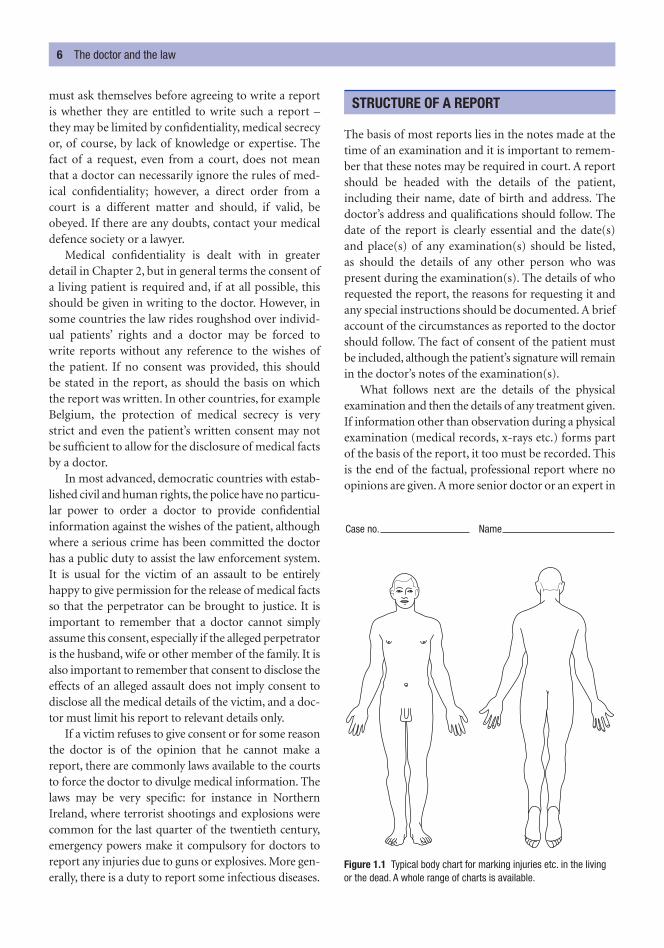
Case no. Name
Figure 1.1 Typical body chart for marking injuries etc. in the livingor the dead. A whole range of charts is available.
a particular field may well be asked to express opinionsabout aspects of the case and those opinions will followthe factual part of the report.
There is no great trick to writing medical reportsand, to make the process simpler, they can be con-structed along the same lines as the clinical notes in thatthey need to be structured, detailed and accurate. Donot include every single aspect of a medical historyunless it is relevant.A court does not need to know everydetail, but it does need to know every relevant detail,and a good report will give the relevant facts clearly,concisely and completely and in a way that an intelligentperson without medical training can understand.
Medical abbreviations should be used with careand highly technical terms, especially those relating tocomplex pieces of equipment or techniques, should beexplained in simple, but not condescending, terms. Onthe other hand, the courts are not medically illiterateand abbreviations in common usage such as ECG cansafely be used without explanation.
It goes without saying that the contents of each andevery report must be true. A report should be typed onstandard-sized (A4 or foolscap) paper and not scrib-bled on paper torn from a drug company’s advertisingpad; remember it will be you who is trying to read thescribble 6 or 12 months later while under oath andunder stress in the witness box. Counsel will have hadweeks to decipher your writing and if even you cannotmake sense of your report, it is unlikely that the courtwill take much notice of it. On the other hand, a clear,concise and complete report may just save you from
having to attend court at all, and if you do have to giveevidence, it is so much easier to do so from a reportthat is legible.
Autopsy reports are a specialist type of report andmay be commissioned by the coroner, the police orany other legally competent person or body. Theauthority to perform the examination will replace theconsent given by a live patient, and is equally import-ant. The history and background to the death will beobtained by the police or the coroner’s officer, but thedoctor should seek any additional details that appearto be relevant, including speaking to any cliniciansinvolved in the care of the deceased and reviewing thehospital notes. A visit to the scene of death in non-suspicious deaths, especially if there are any unusualor unexplained aspects, is to be encouraged.
An autopsy report is confidential and should only bedisclosed to the legal authority who commissioned theexamination. Disclosure to others, who must be inter-ested parties, may only be made with the specific per-mission of the commissioning authority and, in generalterms, it would be sensible to allow that authority todeal with any requests for copies of the report.
Doctors should resist any attempt to change ordelete any parts of their report by lawyers who mayfeel those parts are detrimental to their case; anyrequests to rewrite and resubmit a report with alter-ations for these reasons should be refused. A doctor isa witness to and for the court; he should give his evi-dence without fear or favour because it is for the courtto decide upon the facts and not the witnesses.
Structure of a report 7

There has been a proliferation of the types of ‘medicalpractice’ that are available around the world. There is the science-based ‘Western medicine’, traditionalChinese medicine, Ayurvedic medicine in India, themany native systems from Africa and Asia and the rapidly proliferating modes of ‘fringe medicine’ inWesternized countries. These alternative forms of medi-cine may have their own traditions, conventions andvariably active codes of conduct but we are only con-cerned in this book with the ethics of the science-basedmedical practice, ‘Western medicine’.
To describe modern, science-based medicine as‘Western medicine’ is historically inaccurate becauseits origins can be traced through ancient Greece to asynthesis of Asian, North African and European medi-cine. The Greek tradition of medical practice was epit-omized by the Hippocratic School on the island ofCos around 400 BC. It was there that the foundations of both modern medicine and the ethical facets ofthe practice of that medicine were laid. A form ofwords universally known as the Hippocratic Oath was developed at and for those times, but the fact that itremains the basis of ethical medical behaviour, eventhough some of the detail is now obsolete, is a testa-ment to its simple common sense and universal accept-ance. A generally accepted translation runs as follows:
I swear by Apollo the physician and Aesculapius andHealth and All-heal and all the gods and goddesses,that according to my ability and judgement, I willkeep this Oath and this stipulation – to hold himwho taught me this art, equally dear to me as my
own parents, to make him partner in my livelihood:when he is in need of money, to share mine withhim; to consider his family as my own brothers andto teach them this art, if they want to learn it, with-out fee or indenture. To impart precept, oral instruc-tion and all other instruction to my own sons, thesons of my teacher and to those who have taken thedisciples oath, but to no-one else. I will use treat-ment to help the sick according to my ability andjudgement, but never with a view to injury orwrong-doing. Neither will I administer a poison toanybody when asked to do so nor will I suggest sucha course. Similarly, I will not give a woman a pessaryto produce abortion. But I will keep pure and holyboth my life and my art. I will not use the knife, noteven sufferers with the stone, but leave this to bedone by men who are practitioners of this work. Intowhatsoever houses I enter, I will go into them for thebenefit of the sick and will abstain from every volun-tary act of mischief or corruption: and further, fromthe seduction of females or males, of freeman orslaves. And whatever I shall see or hear in the courseof my profession or not in connection with it, whichought not to be spoken of abroad, I will not divulge,reckoning that all such should be kept secret. WhileI carry out this oath, and not break it, may it begranted to me to enjoy life and the practice of theart, respected by all men: but if I should transgress it,may the reverse be my lot.
What is now broadly called ‘medical ethics’ hasdeveloped over several thousand years and is constantly
C h a p t e r t w o
The Ethics of Medical Practice
International Code of Medical EthicsDuties of physicians in generalDuties of physicians to the sickDuties of physicians to each other
Medical ethics in practiceMedical confidentialityConsent to medical treatmentImplied consent
Express consentThe concept of informed consent

The ethics of medical practice 9
being modified by changing circumstances. The lawsgoverning the practice of medicine vary from countryto country, but the broad principles of medical ethicsare universal and are formulated not only by nationalmedical associations, but by international organiza-tions such as the World Medical Association.
Following the serious violations of medical ethicsby fascist doctors in Germany and Japan during the1939–45 war, when horrific experiments were carriedout in concentration camps, the international medicalcommunity re-stated the Hippocratic Oath in a mod-ern form in the Declaration of Geneva in 1948. Thiswas amended by the World Medical Association in1968 and again in 1983 and the most recent versionwas approved in 1994.
This declaration, made at the time of being admit-ted as a member of the medical profession, states that:
I solemnly pledge myself to consecrate my life tothe service of humanity.
I will give to my teachers the respect and gratitudewhich is their due.
I will practice my profession with conscience anddignity.
The health of my patients will be my first consideration.
I will respect the secrets which are confided in me,even after the patient has died.
I will maintain by all the means in my power,the honour and noble traditions of the medicalprofession.
My colleagues will be my brothers and sisters.
I will not permit considerations of age, disease ordisability, creed, ethnic origin, gender, nationality,political affiliation, race, sexual orientation orsocial standing to intervene between my duty andmy patients.
I will maintain the utmost respect for human lifefrom its beginning and even under threat I will notuse my medical knowledge contrary to the laws ofhumanity.
I make these promises solemnly, freely and uponmy honour.
An International Code of Medical Ethics (derivedfrom the Declaration of Geneva) was originally adopted
by the World Medical Association in 1949. The codewas amended in 1968 and in 1983 and currently reads:
Duties of physicians in general
A physician shall always maintain the highest stand-ards of professional conduct.
A physician shall not permit motives of profit toinfluence the free and independent exercise of pro-fessional judgement on behalf of patients.
A physician shall, in all types of medical practice,be dedicated to providing competent medical ser-vice in full technical and moral independence, withcompassion and respect for human dignity.
A physician shall deal honestly with patients andcolleagues and strive to expose those physiciansdeficient in character or competence or whoengage in fraud or deception.
The following practices are deemed to be unethicalconduct:
a. Self-advertising by physicians, unless permittedby the laws of the country and the Code ofEthics of the National Medical Association.
b. Paying or receiving any fee or other consider-ation solely to procure the referral of a patient or for prescribing or referring a patient to anysource.
A physician shall respect the rights of patients, ofcolleagues and of other health professionals andshall safeguard patient confidences.
A physician shall act only in the patient’s interestwhen providing medical care which might have theeffect of weakening the physical and mental condi-tion of the patient.
A physician shall use great caution in divulging dis-coveries or new techniques or treatment throughnon-professional channels.
A physician shall certify only that which he has per-sonally verified.
Duties of physicians to the sick
A physician shall always bear in mind the obliga-tion of preserving human life.
INTERNATIONAL CODE OF MEDICAL ETHICS

A physician shall owe his patients complete loyaltyand all the resources of his science. Whenever anexamination or treatment is beyond the physician’scapacity, he should summon another physicianwho has the necessary ability.
A physician shall observe absolute confidentialityon all he knows about his patient even after thepatient has died.
A physician shall give emergency care as a humani-tarian duty unless he is assured that others are will-ing and able to give such care.
Duties of physicians to each other
A physician shall behave towards his colleagues ashe would have them behave towards him.
A physician shall not entice patients from his colleagues.
A physician shall observe the principles of theDeclaration of Geneva approved by the WorldMedical Association.
The fact that both the Declaration of Geneva andthe International Code of Medical Ethics have had tobe amended during the past 50 years serves to remindus that medical ethics are not static. Both the practiceof medicine and the societies in which doctors workchange, and medical ethics must alter to reflect thesechanges. The World Medical Association has adopteda number of other important declarations over theyears, providing international guidance, and some-times support, for doctors everywhere (Table 2.1).
There are many aspects of medical ethics and the sub-ject has blossomed to the point where there are nowInstitutes of Medical Ethics and full-time specialistscalled medical ethicists.
It is hard to find any medical activity that does nothave some ethical considerations, varying from researchon patients to medical confidentiality, from informedconsent to doctor–doctor relationships. Many olderethical considerations have progressed into law, whilenew concerns have arisen. But despite all this change,the basic nature of ethical behaviour remains the sameand all medical ethics can be said to rest on the principlethat ‘The patient is the centre of the medical universearound which all the efforts of doctors revolve’.
The doctor exists for the patient, not the other wayaround. The doctor must never do anything to or for apatient that is not in the best interests of that patientand all other considerations are irrelevant in that doctor–patient relationship. From this one simplestatement spring all other aspects of ethical behaviour,
MEDICAL ETHICS IN PRACTICE
10 The ethics of medical practice
Date Name Subject
1970 The Declaration of Oslo Therapeutic abortion
1973 The Declaration of Munich Racial, political discrimination etc. in medicine
1975 The Declaration of Tokyo Torture and other cruel and degrading treatment or punishment
1975 The Declaration of Human experimentation Helsinki and clinical trials
1981 The Declaration of Lisbon Rights of the patient
1983 The Declaration of Venice Terminal illness
1983 The Declaration of Oslo Therapeutic abortion
1984 The Declaration of PollutionSan Paulo
1987 The Declaration of Madrid Professional autonomy and self-regulation
1987 The Declaration of Medical educationRancho Mirage
1989 The Declaration of The abuse of the elderlyHong Kong
1995 The Declaration of Lisbon The rights of the patient
1996 The Declaration of Helsinki Biomedical research involving human subjects
1997 The Declaration of Support for doctors Hamburg refusing to participate in
torture or other forms of cruel inhuman or degrading treatment
1998 The Declaration of Ottawa The right of the child to health care
Table 2.1 Declarations of the World Medical Association 1970–2001

including the interaction of doctor with doctor and ofdoctor with society and with government.
The general principle which guides ethical behav-iour is ‘peer conduct’, in that, even if some action is notstrictly illegal in terms of the national laws, that actionshould not be carried out if it is against the acceptedbehaviour of medical colleagues. In other words, evenif a doctor thinks he can ‘get away with it’ under thenational criminal or civil laws, the disapproval of hisfellow doctors – often reinforced by professional discipl-inary procedures – should deter them from acting inthat way.
International codes are quite clear and virtually allnational medical associations subscribe to them intheory, if, regrettably, less strictly in practice. The dis-ciplinary process and the sanctions that can be appliedagainst the doctor found guilty of unethical practicesby the General Medical Council in the UK are describedin more detail in Chapter 3 and range from a publicadmonishment to permanent erasure from the register.
Though the spectrum of unethical conduct is wide,certain universally relevant subjects are recognized.The seriousness with which each is viewed may varyconsiderably in different parts of the world.
• A doctor’s over-riding consideration is to thepatient, while also accepting that doctors have aduty to their medical colleagues and to the com-munity at large. The patient is the reason for thedoctor’s existence and all other matters must besubservient to this fact. A doctor cannot abandonhis patient without ensuring that medical care ishanded over to someone equally competent. A doc-tor cannot simply leave a patient because it is theend of his shift for that day nor can he refuse tocontinue long-term treatment without ensuringthat some other doctor takes that person into care.
• The doctor must always do what he thinks is bestfor the patient’s physical and mental health withoutconsideration of race, wealth, religion, nationalityetc. The doctor must act independently, or withother doctors, free of political or administrativedoctrines or pressure to establish a diagnosis and tocarry out treatment. However, financial resourcescan be limited and health ‘priorities’ may be dic-tated by administrators and politicians and so doctors may find themselves in situations whereresources are limited and treatment options aredenied. This may pose significant ethical dilemmasfor the treating doctor.
• Doctors must act reasonably and courteously toeach other for the patient’s benefit as the best regi-men of treatment cannot be provided by doctorssplit by professional or personal disputes or jeal-ousy. A doctor should not interfere in the treat-ment of a patient except in an emergency whendiscussion with the treating doctor is not possible.If emergency treatment is provided, the patient’susual doctor should be informed as soon as pos-sible about the nature and extent of this emergencytreatment. A doctor should not criticize anotherdoctor’s judgement or treatment directly to thepatient except in extreme and unusual situations,but should instead confront the other doctordirectly if it is thought that the maximum benefit isnot being offered to the patient. If a doctor is con-cerned about the professional skills or the health ofa colleague, he is morally obliged to draw thoseconcerns to the attention of the authorities.
Secrecy is now termed ‘confidentiality’, but whatever itis called it is as vital now as when the HippocraticOath was written. It is a fundamental tenet that what-ever a doctor sees or hears in the life of his patient mustbe treated as totally confidential. The British MedicalAssociation (BMA) defines confidentiality as ‘the prin-ciple of keeping secure and secret from others, informa-tion given by or about an individual in the course of aprofessional relationship’. There are, however, exceptionsto this fundamental rule, which are discussed later.
The concept of medical confidentiality is alsodirected at the well-being of the patient and assumesthat if people cannot be confident that what they telltheir doctor will stay secret, they are much less likely toreveal everything during a consultation, especially inintimate matters concerning their sex life, social andmoral behaviour, use or abuse of drugs or alcohol andeven their excretory functions. As a result, the clinicalhistory may be deficient or even misleading and thebest diagnosis and hence the best treatment may notbe provided.
The doctor must therefore keep everything he hearsto himself and it must be appreciated that the ‘secrecy’belongs to the patient, not the doctor. The latter ismerely the guardian of the patient’s confidential mat-ters, which does not cease on the death of the patient.
MEDICAL CONFIDENTIALITY
Medical confidentiality 11
Giving health information that can be identified asbelonging to a particular individual is termed ‘disclos-ure’. Healthcare information may only be disclosed inthe following situations, although different countriesmay have variations of this list.
• With the consent of the patient. If an adult patientgives consent for disclosure of information, in mostcountries the doctor is free so to do. The crucialfeature in this process is the consent given by thepatient, which is defined by the BMA as ‘a decisionfreely made in appreciation of its consequences’.However, as already mentioned in Chapter 1, somecountries have much stricter laws about the disclos-ure of medical information.
An individual with ‘parental responsibility’ foran immature minor may consent to the disclosureof their medical information, but the situationregarding mentally incapacitated adults is slightlymore complicated.
• To other doctors. The keeping of medical notes andrecords is universal, indeed a doctor would be neg-ligent not to keep such records. These records areused to assist in the provision of health care to thepatient and, in the absence of evidence to the con-trary, it is assumed that patients have given ‘impliedconsent’ for the sharing of their health informationon a ‘need to know’ basis with other healthcare pro-fessionals, who should be under the same obliga-tion of secrecy as the doctor. However, it must beadmitted that as multidisciplinary teams grow everbigger, it is becoming increasingly difficult to con-trol information, which now reaches an ever-widening circle of people.
• To relatives. In most circumstances, close relativesare told of the nature of the patient’s illness, espe-cially if they live together and have to care for thepatient at home. However, this disclosure is by nomeans automatic and, if the patient requests that arelative is not told, the doctor must abide by thatwish. If there is a medical reason why the relativeshould be told, this can be discussed with thepatient, but the doctor cannot disclose the infor-mation in the face of refusal by the patient unlessnot to do so would place the relatives at risk. Thisdilemma is now faced, for example, when one fam-ily member has been diagnosed as having activepulmonary tuberculosis.
Particular caution is required over the disclos-ure of sexual matters, such as pregnancy, abortion
or venereal disease, as disclosure might cause severeconflict between close relatives such as husbandand wife. In some societies the senior male relativemay play a dominant role in the family and maywell insist on the doctor providing him with med-ical information on anyone in the family, irrespect-ive of the wishes of the individual. While remainingaware of the various ethnic and religious factors,a doctor must resist if patients themselves will notgive informed consent to the release of the infor-mation. Where immature children are concerned,it is obvious that all possible information must begiven to the parents or those with parental respon-sibility. Mature children pose different problemsand, if a doctor considers them to be sufficientlymature, they may make their own decisions, whichmust be followed by the doctor. Such a child mayalso deny his parents access to his medical records.
• Statutory (legal) requirements. The absolute duty ofmedical confidentiality has, in reality, been consid-erably diminished. Many national laws now forcethe doctor to reveal what are essentially medicalsecrets and many are so commonplace that they arenot even thought about and the whole communityaccepts them without question, for example officialnotification of births, deaths and stillbirths. Inaddition, statutory notification is required of manyinfectious diseases and occupational diseases, as arethe details of therapeutic abortions, drug addictionetc. Doctors are citizens and have to obey the law ofthe land and so they have to submit to these regula-tions and patients cannot complain about theirdoctor revealing these types of personal informa-tion. The patient has no right of refusal, but shouldbe notified about what information will be pro-vided and to whom.
• In courts of law. Where a doctor is a witness beforea court or tribunal, the magistrate, judge, coroneretc. has the power to force the doctor to discloseany relevant medical facts. The doctor may protestor ask if he can write down the confidential facts sothat the public and press in court do not hear theanswer. However, if the judge so directs, the doctormust answer, on pain of a fine or even imprison-ment for ‘contempt of court’. In such circum-stances, the evidence given by the doctor is totallyprivileged and thus the patient cannot bring a legalaction for breach of confidence.
• The police. In most Western countries the policehave no greater power to demand the disclosure of
12 The ethics of medical practice
medical information by a doctor than anyone else.There are a few well-defined circumstances, notspecifically related to medical information or tomedical practice, in which the police can requiredisclosure of information by any citizen; theseinvolve terrorist activity and information that mayidentify a driver alleged to have committed a trafficoffence.
The police usually require information con-cerning an assault on a patient, but where assaultoccurs within a family, such as between spouses orclose relatives, the victim may not wish to bringcriminal charges and so the doctor must not auto-matically assume that consent for disclosure willhave be given.
• Disclosure by police surgeons (forensic medicalexaminers). A doctor examining a patient, usually avictim or alleged perpetrator, at the request ofthe police owes the same duty of confidentiality tothat patient as any other doctor, and any informa-tion that is not relevant to any criminal proceed-ings must be given the same protection as any othermedical information. The doctor in this situationmay, however, disclose medical facts that are rele-vant to a crime, but the patient should be madeaware of this before the examination begins. Ifordered by a court to disclose other informationgained from this examination, the doctor must, ofcourse, comply.
• In the public good. This is a most difficult issue andit must be left to the doctor’s own consciencewhether he should reveal matters which affect peo-ple other than the patient. For instance, if a doctorlearns of a serious crime (e.g. by treating wounds ofan assailant that he knows must have originated in aserious assault or rape), then the issue of confiden-tiality clashes with the need to protect some indi-vidual or the public at large from possible furtherdanger. The same issue may arise where a doctorsuspects that a child patient is being physically ormentally abused, but here the over-riding consider-ation is the safety of the child.
More commonly, the dilemma for a doctorarises from disease rather than injuries. If a seriousillness in a patient poses a potential threat of‘serious harm’ to the safety or health of either thepatient or the public, the doctor must decidewhether to break silence about the condition, forexample in the case of a bus driver with serioushypertension or a teacher with tuberculosis or
some other infective disease. Usually, people inpositions of public responsibility are required todisclose significant illness to their employers and tohave occupational medical checks performed onbehalf of the employer or licensing authority.
The proper course is for the doctor to explain therisks to the patient and to persuade him to allowthe doctor to report the problem to his employers.The patient may, of course, refuse. It is always wiseto seek the advice of senior colleagues or of a pro-fessional insurance organization or national med-ical association before making any disclosure.
• Disclosure to lawyers. Lawyers have no automaticright to obtain medical information and recordswithout the patient’s consent, but in general, if alawyer in a civil case wishes to obtain medicalrecords and these are denied to him, he may applyto the court for ‘disclosure’. In the UK the Access toHealth Records Act 1990 means that this requestwill be granted if a lawyer can show that his clienthas reasonable grounds for wishing to see medicalrecords; these grounds may be either to discover ifthere are grounds for a legal action or to obtain evi-dence for an action already commenced.
When asked by a lawyer for a medical report, adoctor should always insist on seeing written per-mission for the disclosure of information signed bythe individual about whom the report is to be writ-ten. If a doctor releases confidential information toanother party without the consent of the patient,he may be sued or face disciplinary action by the regulatory medical authorities for unethicalbehaviour.
No adult person need accept medical treatment unlessthey wish to do so. However, if they do desire medicalattention, they must give valid consent. Permission fordiagnosis and treatment is essential as otherwise thedoctor may be guilty of assault if he touches or evenattempts to touch an unwilling person. In Britain, youngpersons over the age of 16 can choose their own doc-tor and children of this age are presumed to be com-petent to give permission for any treatment. Below theage of 16 there is no presumption of competency, butif the doctor thinks they are mature enough to under-stand, they can still give valid consent. There is no
CONSENT TO MEDICAL TREATMENT
Consent to medical treatment 13

lower age limit to this competency, the crucial testbeing the child’s ability to comprehend and to make arational decision. Interestingly, a decision by a childunder 18 to refuse treatment is not necessarily bindingupon a doctor and may be overridden by those withparental responsibility or by a court.
The situation in which an adult lacks the capacity,for whatever reason, to make an informed decision issomewhat confused. Where a patient is suffering froma mental condition, and is detained in hospital undermental health legislation, he may be given treatmentfor his mental condition without his consent. However,the legislation does not extend to other types of med-ical treatment. The Adults with Incapacity Act appliesin Scotland only, and allows people over 16 years toappoint a proxy decision maker to whom they delegatethe power to consent to medical treatment, but only ifthe patient has lost the capacity to do so.
In an emergency, such as an accident where the vic-tim is in extremis, unconscious or shocked, no permis-sion is necessary and doctors must do as they thinkbest for the patient in those urgent circumstances. Aslong as medical intervention was made in good faithfor the benefit of the victim, no subsequent legalaction based on lack of consent is likely to succeed.
Consent to medical treatment is of two types.
Implied consent
Most medical practice is conducted under the principleof ‘implied consent’, where the very fact that a personhas presented at a doctor’s surgery to be examined,or asks the doctor to visit him, implies that he is will-ing to undergo the basic clinical methods of examina-tion, such as history taking, observation, palpationand auscultation etc. It does not extend to intimateexaminations such as vaginal and rectal examinations or to invasive examinations such as venepuncture.These intimate and invasive tests should be discussedwith patients and their express consent specificallyobtained after explaining what is to be done and why.Refusal of consent for the procedure precludes the testor examination.
Express consent
Where complex medical procedures are concerned,more specific permission must be obtained from thepatient, this being called ‘express consent’, and if thesame procedure is repeated on another occasion, fur-ther express consent must again be obtained.
Express consent may often be obtained in writing,but this is not a legal requirement and written consentis not more valid than verbal consent. However, writ-ten consent is much easier to prove at a later dateshould any dispute ever arise. Ideally, either verbal orwritten consent should be witnessed by another per-son, who should also sign any document.
Consent only extends to what was explained to thepatient beforehand and nothing extra should be doneduring the operation for which express consent hasnot been obtained. This can pose a dilemma for a sur-geon if something unexpected is found at operationthat necessitates a change of procedure.
The concept of informed consent
Consent is not legally valid unless the patient under-stands what he or she is giving the doctor permissionto do and why the doctor wants to do it, and it maywell be that the patient, having weighed up the risks,the pain and discomfort and many other factors, maydecline the operation and it is their right to be able todo this. There is some room for clinical judgement anda doctor may withhold some information he believesmay cause mental anguish that would adversely affectthe patient’s health or recovery. However, any with-holding of facts may need to be justified at a later dateand careful notes should be kept of any matters dis-cussed, or specifically not discussed, during theprocess of obtaining express consent.
The question of consent has usually been con-sidered by the medical and surgical teams at hospitalsand as long as junior doctors conform to the protocolslaid down by those teams they will not be personallyresponsible for any failings or omissions later dis-covered in the process as a whole.
14 The ethics of medical practice
The term medical malpractice covers all failures in theconduct of doctors but only where it impinges upontheir professional skills, ability and relationships.Malpractice can be conveniently divided into twobroad types:
1 Medical negligence – where the standard of medicalcare given to a patient is considered to be inadequate.
2 Professional misconduct – where the personal, pro-fessional behaviour falls below that which is expectedof a doctor.
Medical treatment is not provided with an absoluteguarantee of complete success. Improvements in med-ical science and techniques have markedly reduced therate of complications and unexpected outcomes fromall types of treatment, but they will never disappearcompletely. However, all patients have the legal rightto expect a satisfactory standard of medical care fromtheir doctor even though it is accepted that this cannever mean that the doctor can guarantee a satisfac-tory outcome to the treatment.
Most legal actions for negligence in countries withan Anglo-Saxon system of law remain within the civil
law, in which a patient brings a personal action againstthe doctor or hospital, and to understand the con-cept of medical negligence certain principles must beconsidered.
Before a patient can succeed in a civil action fornegligence against a doctor, it must be established:
1 that the doctor had a duty of care towards thepatient; (and)
2 that there was a failure in that duty of care; (which)3 resulted in physical or mental damage.
1 Once it is established that there is a duty of care, thedoctor must then provide both diagnosis and treat-ment at a reasonable ‘standard of care’ – that is,consistent with the doctor’s own experience andtraining. A junior doctor is not expected to have asmuch expertise as a specialist but is expected to pos-sess at least the minimum skills tested by the quali-fying examinations and, in addition, is expected toapply the level of experience consistent with his orher postgraduate training. It is accepted that doc-tors cannot be expected to know the details of everysingle recent advance in all areas of medicine, butthe patient can expect a doctor to have kept up todate with major developments in his own and inclosely related fields, now often referred to asContinuing Professional Development (CPD).
MEDICAL NEGLIGENCE
C h a p t e r t h r e e
Medical Malpractice
Medical negligenceSystems of compensationCompensation and damagesTypes of medical negligenceObstetrics and gynaecology
Orthopaedics and accident surgeryGeneral surgeryGeneral medical practiceAnaesthesiologyGeneral errors
Professional misconductThe General Medical CouncilConduct procedures

2 For negligence to be established, there must be a‘breach’ of this standard of care, either by omission(failing to do something) or by commission (doingsomething wrong). It is accepted that the circum-stances under which a doctor treats a patient mayhave a considerable bearing on the reasonable stand-ard of care that the patient may expect; for exampletreatment in an acute emergency when there is neither the time nor the facilities may legitimatelybe less ideal than that given for the same conditionin a non-urgent situation. The test of negligence thatis applied relies upon the response of the averagedoctor with the same medical background, placedin identical circumstances.
3 Even if a patient can prove the presence of a duty ofcare and a breach of the standard of care, he cannotsucceed in a legal action unless he can also showthat he has suffered physical or mental damage. If adoctor prescribes some obviously inappropriate oreven harmful medicine but the patient refuses totake the medicine, the patient cannot then recovercompensation from the doctor because he has suf-fered no damage.
It is important to note that ‘damage’, in the sense of injury or harm, is quite different from ‘damages’,which is the financial compensation awarded to a suc-cessful litigant.
There is rarely any dispute over whether the doctorowed the patient a duty of care; the major problem isusually proof of a breach of that duty and the onus lieson the plaintiff to show that a breach occurred and noton the defendant to prove that it did not. The onlyexception occurs when the facts are so glaringly obvi-ous that they need no explanation (legally res ipsaloquiter, or ‘the facts speak for themselves’); in thissituation the doctor is forced, if he can, to provide an explanation for his actions. If a patient goes into anoperating theatre to have the right leg amputated andthe left leg is removed instead, there is no dispute thatthe treatment is incorrect and the responsibility shiftsto the defending doctor to explain the error.
The great problem of alleged medical negligence liesin the continuum of ‘standard of care’ between actionsthat are accepted medical practice and those that con-stitute a lack of care. At the junction of these twoextremes is a grey area of debatable clinical judgementwhere some doctors would act in one way whereasothers would act, quite legitimately, in a different way.
To complicate matters further, errors of clinicaljudgement which lead to a bad result are not always
negligent. If the error results from decisions made ingood faith, based on all the information that couldreasonably be expected to be available at the time butwhich are recognized, in retrospect, to be an error,they cannot be considered to be a breach of either theduty or the standard of care.
The only way to resolve the problem of whether anact is truly negligent is by ‘peer judgement’, and this isthe means by which most medical disputes are settled,at least in the UK. The facts of the case are placedbefore experts in that particular specialty and theirviews sought. It is sufficient in this context to showonly that a substantial number of doctors agree withthe actions of the defendant; there is no need for unan-imity of either condemnation or support.
Most allegations of medical negligence never comebefore a court of law for decision. Some cases cannotbe defended as far as the doctor is concerned and thesewill be settled by financial negotiation, either directlyor, more commonly, through the medical insurers,without further argument. Other cases cannot be substantiated by the plaintiff and are eventually abandoned, often on legal advice. Of the remainder,less than half go through the full process of investiga-tion in which each ‘side’ obtains expert opinions fromindependent medical specialists. The strength of theseopinions is then reviewed and often the experts andlawyers meet to try to reach common ground and asettlement, and only if there is a wide gap between the expert opinions is the case likely to go to court fora judge to decide on the relative merits of each argu-ment. Recent changes in the process of civil litigationin the UK have resulted in greater collaborationbetween the experts who, amongst other things, nowhave to state clearly in their reports that they under-stand that their primary duty is to the court and not tothe party who instructs them; this has greatly reduced,if not prevented entirely, the ‘hired gun’ expert.
There is a common legal principle in employmentthat ‘the master is responsible for the acts of his servants’and this principle applies to all of the employees –technicians, nurses etc. as well as to the medical staff –and so hospitals are now responsible for the actions of their staff. The cost of assuming these risks has to be met out of annual health budgets and so the con-tinued escalation of negligence actions has resulted ina significant, and increasing, cost to the employers. It isvery important to emphasize that medical insuranceprovided by an employing hospital or health authoritydoes not extend to any work outside that establishmentand all private specialists, general practitioners and all
16 Medical malpractice
doctors undertaking emergency, locum or casual workcontinue to need personal insurance for their practiceof medicine.
In many countries, commercial insurance com-panies provide indemnity for doctors, but other coun-tries have mutual non-profit schemes, often run by themedical profession. These mutual insurance organiza-tions will often take a more robust view of their mem-bers’ interest and professional standing and are lesslikely to settle a case simply for the sake of financialexpediency than either commercial firms or anemploying health authority.
The situation described above is the ‘fault’ system,whereby the patient has to prove that the doctor was in breach of a duty of care before he can get a singlepenny in damages. The system relies upon the lowerlevel of proof available in law – the balance of prob-ability. As a result, it is an ‘all or nothing’ system, for if the balance of probability of fault is assessed at 51per cent, the patient may get a large award of damages,whereas in the same case, if the probability of fault isonly 49 per cent, he gets nothing at all.
In addition, the legal process can be very slow andit will often take a number of years to settle quite asimple claim. A recent change in the law regarding thepayment of lawyers now means that they can work foran agreed percentage of the damages their clientsreceive if they are successful; the balance to thatmethod of payment is that they will have worked fornothing if the case does not succeed. This simple ideahas been clouded by the interposition of a number ofcomplex insurance schemes and possible litigantsshould study very carefully the terms and conditionsof any contract that they sign to get ‘free’ legal assist-ance for a civil medical claim.
It is a socially illogical and wholly unjust situationthat a patient injured by an act deemed by a court tobe negligent will receive damages, whereas anotherpatient with the same injuries resulting from an actthat is not deemed negligent will receive nothing.Some countries, such as Sweden, Finland, Norway andNew Zealand, with high levels of state-funded socialsecurity have introduced ‘no-fault’ systems wherebythe test for compensation is not the presence orabsence of the doctor’s negligence, but rather thepatient’s needs. This concept is by no means new in
non-medical fields; in Britain, industrial injuries com-pensation has been in existence for over 40 years, therequisite funding coming from employers, employeesand the state, and compensation for injuries due tocriminal acts has been paid entirely by the state throughthe Criminal Injuries Compensation Authority forover a decade.
If a plaintiff is successful, either in a court action or bysettlement between the lawyers for the parties out ofcourt, damages will be paid. The object of this award ofmoney is to try to restore the patient to the financialstate that he was in before the incident and in additionthere may be additional sums to compensate for pain,suffering and loss of quality of life. If the individual isnot able to continue with his chosen work, a large part ofany damages will relate to this loss of earnings, and theamount paid will clearly depend upon the nature of thework and the remuneration that the individual had beenreceiving or might reasonably be expected to receive.
Other damages may be based on the need for long-term nursing and special care in the future. A brain-damaged child or young person may require 24-hourattention for the rest of his life, which accounts for thehuge damages awarded to infant victims of cerebralhypoxia. Similarly, the costs of future treatment toremedy some negligent damage may be calculated,though where free state medicine is available, themoney will be correspondingly reduced. Pain, suffer-ing and loss or reduction in the quality of life are usu-ally compensated on a modest scale.
Where death has been caused, the dependent rela-tives (if there are any) will receive compensation forthe loss of salary of the family breadwinner. The deathof a child is poorly compensated as it can have nodependants and no forecast as to its level of futureearnings can be made.
It is impossible to give a complete list of negligent situ-ations in medical practice. An English judge oncestated that ‘the categories of negligence are never closed’,which must be correct as every new technique that isdeveloped provides more opportunity for something
TYPES OF MEDICAL NEGLIGENCE
COMPENSATION AND DAMAGES
SYSTEMS OF COMPENSATION
Types of medical negligence 17
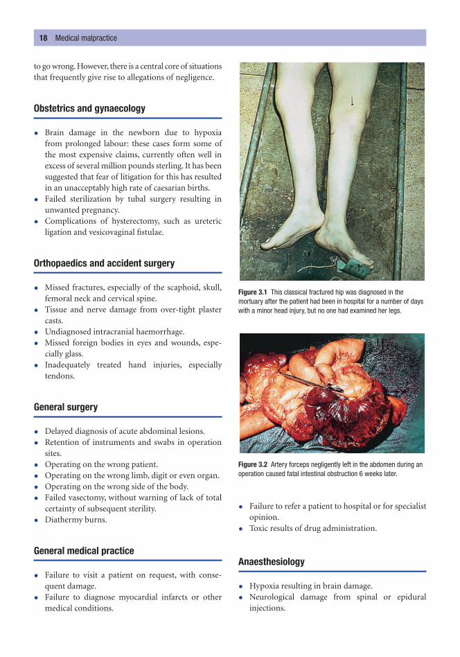
to go wrong. However, there is a central core of situationsthat frequently give rise to allegations of negligence.
Obstetrics and gynaecology
• Brain damage in the newborn due to hypoxia from prolonged labour: these cases form some ofthe most expensive claims, currently often well inexcess of several million pounds sterling. It has beensuggested that fear of litigation for this has resultedin an unacceptably high rate of caesarian births.
• Failed sterilization by tubal surgery resulting inunwanted pregnancy.
• Complications of hysterectomy, such as uretericligation and vesicovaginal fistulae.
Orthopaedics and accident surgery
• Missed fractures, especially of the scaphoid, skull,femoral neck and cervical spine.
• Tissue and nerve damage from over-tight plastercasts.
• Undiagnosed intracranial haemorrhage.
• Missed foreign bodies in eyes and wounds, espe-cially glass.
• Inadequately treated hand injuries, especially tendons.
General surgery
• Delayed diagnosis of acute abdominal lesions.
• Retention of instruments and swabs in operationsites.
• Operating on the wrong patient.
• Operating on the wrong limb, digit or even organ.
• Operating on the wrong side of the body.
• Failed vasectomy, without warning of lack of totalcertainty of subsequent sterility.
• Diathermy burns.
General medical practice
• Failure to visit a patient on request, with conse-quent damage.
• Failure to diagnose myocardial infarcts or othermedical conditions.
• Failure to refer a patient to hospital or for specialistopinion.
• Toxic results of drug administration.
Anaesthesiology
• Hypoxia resulting in brain damage.
• Neurological damage from spinal or epidural injections.
18 Medical malpractice
Figure 3.1 This classical fractured hip was diagnosed in themortuary after the patient had been in hospital for a number of dayswith a minor head injury, but no one had examined her legs.
Figure 3.2 Artery forceps negligently left in the abdomen during anoperation caused fatal intestinal obstruction 6 weeks later.

• Peripheral nerve damage from splinting duringinfusion.
• Incompatible blood transfusion.
• Incorrect or excessive anaesthetic agents.
• Allowing awareness of pain during anaesthesia.
General errors
• Failure to act on radiological or laboratory reports.
• Inadequate clinical records and failure to commu-nicate with other doctors involved in the treatmentof a patient.
• Failure to admit to hospital when necessary.
• Failure to obtain informed consent to any procedure.
• Administration of incorrect type or quantity ofdrugs, especially by injection.
In general, all doctors need to be constantly awareof the risks of their professional duties but not soobsessed with the possibility of legal consequences thatthey avoid using a potentially risky treatment that mayoffer considerable benefit to the patient. The practiceof ‘defensive medicine’ may lead to the withholding ofbeneficial treatment to the majority because of a stat-istical risk to the minority.
The professional behaviour of a doctor, either in con-nection with his treatment of patients or in other areasof his behaviour, may lead to allegations of misconduct
that are separate from the civil actions for negligencediscussed in the previous section. Where the personalor professional conduct of a doctor is seriously criti-cized, his worthiness to continue as a recognized mem-ber of the medical profession may be at stake. Thisaspect is dealt with by various tribunals of the officialauthority responsible in that particular country forgranting registration or a licence to practise medicine.These tribunals can examine the fitness of any doctorto remain an accredited physician and this mechanismof referral and review is designed primarily to protectthe public from unsuitable or even dangerous doctors.
Because national systems of licensing and registra-tion vary so widely, it is impossible to describe anyuniversal rules. Nevertheless, there is a general level of ethical behaviour, morality and competence thatshould be subscribed to by doctors all over the world.These high standards are not born of snobbery or elitism but of practical necessity, for if patients are toderive the maximum benefit from diagnosis and treat-ment, they must be confident that their physician isresponsible, diligent, honest and discreet. It is believedthat patients are less likely to reveal intimate details oftheir medical history or to cooperate in treatmentwithout the necessary ingredient of faith and confi-dence in the treating doctor. Thus doctors must actu-ally possess, and be seen to possess, all the betterqualities that will befit them to manage life-and-deathissues.
Every country has a system for admitting new doc-tors to a regulated list of competent practitioners; thislist is usually limited to those who have passed thefinal examinations of a university medical school orother accrediting institution. This initial registrationor licensing will allow newly qualified doctors to com-mence their postgraduate training with the ‘houseyear’ or internship and this will be followed by a widerange of postgraduate training schemes and qualifica-tions. The doctor’s professional career is dependentupon remaining registered or licensed until retirementor death.
Each country may have widely different criteriathat define professional misconduct; some states arevery strict about the behaviour of their doctors,whereas others, unfortunately, have far too lenient anattitude. The regulatory system for professional con-duct also varies greatly from place to place and, in gen-eral terms, it is most organized where the criteria forprofessional conduct are most strict and applied withmost diligence. In many countries, the regulation and
PROFESSIONAL MISCONDUCT
Professional misconduct 19
Figure 3.3 Necrosis of the spinal cord following excessiveintrathecal injection of penicillin. Thirty times the proper dose wasgiven, due to misreading the label on the box.
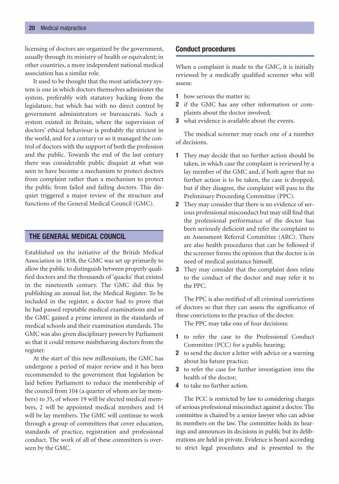
licensing of doctors are organized by the government,usually through its ministry of health or equivalent; inother countries, a more independent national medicalassociation has a similar role.
It used to be thought that the most satisfactory sys-tem is one in which doctors themselves administer thesystem, preferably with statutory backing from thelegislature, but which has with no direct control bygovernment administrators or bureaucrats. Such asystem existed in Britain, where the supervision ofdoctors’ ethical behaviour is probably the strictest inthe world, and for a century or so it managed the con-trol of doctors with the support of both the professionand the public. Towards the end of the last centurythere was considerable public disquiet at what wasseen to have become a mechanism to protect doctorsfrom complaint rather than a mechanism to protectthe public from failed and failing doctors. This dis-quiet triggered a major review of the structure andfunctions of the General Medical Council (GMC).
Established on the initiative of the British MedicalAssociation in 1858, the GMC was set up primarily toallow the public to distinguish between properly quali-fied doctors and the thousands of ‘quacks’ that existedin the nineteenth century. The GMC did this by publishing an annual list, the Medical Register. To beincluded in the register, a doctor had to prove that he had passed reputable medical examinations and sothe GMC gained a prime interest in the standards ofmedical schools and their examination standards. TheGMC was also given disciplinary powers by Parliamentso that it could remove misbehaving doctors from theregister.
At the start of this new millennium, the GMC hasundergone a period of major review and it has beenrecommended to the government that legislation belaid before Parliament to reduce the membership ofthe council from 104 (a quarter of whom are lay mem-bers) to 35, of whom 19 will be elected medical mem-bers, 2 will be appointed medical members and 14 will be lay members. The GMC will continue to workthrough a group of committees that cover education,standards of practice, registration and professionalconduct. The work of all of these committees is over-seen by the GMC.
Conduct procedures
When a complaint is made to the GMC, it is initiallyreviewed by a medically qualified screener who willassess:
1 how serious the matter is;2 if the GMC has any other information or com-
plaints about the doctor involved;3 what evidence is available about the events.
The medical screener may reach one of a numberof decisions.
1 They may decide that no further action should betaken, in which case the complaint is reviewed by alay member of the GMC and, if both agree that nofurther action is to be taken, the case is dropped,but if they disagree, the complaint will pass to thePreliminary Proceeding Committee (PPC).
2 They may consider that there is no evidence of ser-ious professional misconduct but may still find thatthe professional performance of the doctor hasbeen seriously deficient and refer the complaint toan Assessment Referral Committee (ARC). Thereare also health procedures that can be followed ifthe screener forms the opinion that the doctor is inneed of medical assistance himself.
3 They may consider that the complaint does relateto the conduct of the doctor and may refer it to the PPC.
The PPC is also notified of all criminal convictionsof doctors so that they can assess the significance ofthese convictions to the practice of the doctor.
The PPC may take one of four decisions:
1 to refer the case to the Professional ConductCommittee (PCC) for a public hearing;
2 to send the doctor a letter with advice or a warningabout his future practice;
3 to refer the case for further investigation into thehealth of the doctor;
4 to take no further action.
The PCC is restricted by law to considering chargesof serious professional misconduct against a doctor. Thecommittee is chaired by a senior lawyer who can adviseits members on the law. The committee holds its hear-ings and announces its decisions in public but its delib-erations are held in private. Evidence is heard accordingto strict legal procedures and is presented to the
THE GENERAL MEDICAL COUNCIL
20 Medical malpractice

committee by lawyers acting for the GMC and the doc-tor. Witnesses are summonsed to appear and evidence isgiven under oath and the witnesses are questioned byboth the lawyers and the members of the committee.
After hearing the evidence, the committee mustdecide whether or not the doctor is guilty of ‘seriousprofessional misconduct’. The committee may reachone of five conclusions:
1 to admonish the doctor;
2 to postpone a decision in order to collect more evidence;
3 to place conditions on the registration of the doctorfor up to 3 years;
4 to suspend the registration of the doctor for up to a year; or
5 to erase the registration of the doctor.
There is a right of appeal if the doctor considers theconclusion to be incorrect.
The general medical council 21

The interaction between mental health and criminalbehaviour and the resulting contacts with the police, thecriminal justice system and the prisons are extremelycomplex and most of this work lies within the specialistfield of forensic psychiatry, but a general understandingof the law relating to mental health and criminal behav-iour is essential for any medical practitioner.
The spectrum of behaviour that can be considered to be‘normal’ depends on so many factors – national, cul-tural, social, ethical, religious etc. – that any definitionof ‘normality’ must be extremely wide for any givenpopulation. A further complication is that ‘abnormalbehaviour’ and behaviour due to ‘disorders of the mind’are not one and the same thing, although the latter maybe associated with the former. Not everyone whosebehaviour is ‘abnormal’ is suffering from a ‘disorder ofthe mind’ and it is certainly untrue that everyone with a‘disorder of the mind’ will behave ‘abnormally’.
Mental abnormality can be congenital (mental defect,impairment, handicap or subnormality) or acquired
(mental disease or illness). These categories are notsharply divided, as diseases such as schizophrenia mayhave a congenital origin even if the manifestations aredelayed and not all antisocial psychological traits canbe classified as a disease.
The term mental subnormality covers a wide rangeof severity; for instance, individuals with Down’s syndrome may, at one end of the spectrum, be able toattend normal school and go on to live an independ-ent adult life. Mental subnormality per se generally hasfew criminal medico-legal aspects apart from for thoseindividuals who may drift into crime: theft, such asshop-lifting, is most likely but arson and some sexualoffences may be related, although there is no directcorrelation between the mental state and these moreserious offences. The old idea of a homicidal ‘villageidiot’ is part of the mythology that used to be directedagainst those with mental subnormality.
The psychopathic personality is difficult to categor-ize into congenital or acquired, and the effects ofnature and nurture are closely intertwined. In manyways, a psychopathic personality is not a mental defectat all but simply one extreme end of the spectrum ofvariation in individual personality. This trait is associ-ated with a failure of maturation of the personality andthe retention of a child-like selfishness. Frustration ofany whim is not tolerated and may be met with imme-diate violence, including murder. The response to theseacts of violence is completely devoid of any regret or
TYPES OF ABNORMAL MENTAL CONDITION
NORMAL AND ABNORMAL BEHAVIOUR
C h a p t e r f o u r
The Medico-legal Aspects of Mental Disease
Normal and abnormal behaviourTypes of abnormal mental conditionMental health legislation and the
criminal justice system
Criminal responsibility: age and mental capacity
The effect of drink or drugs on responsibility
Testamentary capacity

Mental health legislation and the criminal justice system 23
remorse. However, these individuals may have noabnormality of thought, mood or intelligence.
True mental illnesses acquired or appearing afterchildhood comprise several major groups. Those thatarise from some structural brain damage may be calledorganic psychoses, whereas those that appear to haveno neurological basis are called functional psychoses.
The organic psychoses comprise the various demen-tias which may present with a bewildering constellationof signs and symptoms, of which lability of mood, fail-ure of memory, deterioration of intellect, irritability,irrational anger, confusion and loss of social inhibitionsare most likely to lead to some medico-legal conse-quences. The dementing or demented individual maywell be arrested for unacceptable behaviour or even forsudden, inexplicable and uncharacteristic acts of vio-lence. The aetiology of these dementias is very wide:multi-infarct dementia, infective, degenerative andmetabolic disorders, direct physical trauma, the acuteeffects of toxic substances (e.g. carbon monoxide) andthe effects of chronic ingestion of toxic substances(especially alcohol). Cerebral tumours, either primaryor secondary, may also lead to rapid personality changes.
A few epileptics may occasionally have an identifi-able organic basis for their disease and some may havea concomitant mental abnormality. Of greater medico-legal interest are the rare episodes of post-ictal automa-tism and some forms of pre-fit aura, which may, rarely,give rise to dangerous behaviour.
The functional psychoses are characterized by dis-orders of thought for which no discernible physicalbasis can be identified and which can be seen as severeexaggerations of the normal variations of mood experi-enced by everyone. The functional psychoses are fur-ther subdivided into the affective psychoses, in whichdisorders of mood (affect) are the prime feature, andschizophrenic psychoses, in which disorders of thethought process are dominant.
A severe example of an affective disorder is theindividual with manic–depressive psychosis who showswide swings of mood from euphoric elation and hyper-activity to deepest depression and stupor. These changesin mood are quite out of proportion or often totallyunrelated to external circumstances; however, for mostof the time the individual will remain in some sort ofequilibrium between the two extremes.
In the schizophrenic psychoses there are commonlydelusions, mistaken beliefs held by the patient that can-not be altered by logical argument, and also hallucina-tions, which are false sensory experiences without true
sensory stimuli. Patients may also have illusions, whichare real sensory perceptions that are misinterpreted assomething quite different. Commonly, these individ-uals have lost contact with reality.
The forensic aspects of these severe mental illnessesdepend on the type and severity of the disease. On theone hand are the increased suicides that are associatedwith all forms of psychosis, and the confusion inherentin a manic–depressive psychosis may also apparentlymanifest itself in petty theft, especially shop-lifting.
Schizophrenics may sometimes be even more dan-gerous to their family or their immediate circle as theycannot see the real world clearly, if at all, because theirthought processes are distorted by delusions, illusionsand hallucinations. For instance, they may decide to‘rescue’ someone from the harm or dangers that theyfalsely perceive by a ‘mercy killing’. These fantasies mayobstruct their normal living activities, often monop-olizing their attention to the exclusion of real practi-calities. In paranoid schizophrenia, the delusions andhallucinations are of a persecutory nature and the vic-tims spend their lives in suspicious analysis of everyevent around them, and these feelings can be aggra-vated by hallucinations of voices and whispering andpseudo-scientific fears of malign rays and electronicsurveillance.
In another clinical manifestation, the schizo-phrenic may become withdrawn, apathetic and some-times neglectful and dirty, which may reach extremesat which the individual is catatonic. These individualsseldom cause harm to others, but may need protectionfrom themselves and their illness.
Most commonly, schizophrenics have difficulty insurviving unaided in modern society; they may beunemployable, adopt a vagrant lifestyle and resort topetty theft and deceptions, but only very few will com-mit more serious crimes, including assaults and homi-cide. That most of the notorious multiple murderersare claimed to be schizophrenic or to have ‘schizo-phrenic tendencies’ possibly owes more to theirdefence strategy than to a strictly applied and univer-sally accepted clinical diagnosis.
Most advanced countries have legislation that allowsfor the detention and treatment of individuals, against
MENTAL HEALTH LEGISLATION AND THECRIMINAL JUSTICE SYSTEM

their will, if they are diagnosed to have such severemental disorders that they are a danger to themselves orto the public. The individual with mental health prob-lems may also come into contact with doctors throughvarious stages of the criminal justice system.
In the UK, if the police officer in charge of detaineesin the custody suite is concerned about the physical ormental health of an individual, he may request thatthey are assessed by a doctor with expertise in clinicalforensic medicine – known in the UK as a ForensicMedical Examiner (FME) or a police surgeon – at thepolice station. The FME will be asked to confirm thatthe detainee is ‘fit to be detained’ within the police sta-tion and also that he is ‘fit to be interviewed’. Duringthese examinations, the mental health of the individualwill come under scrutiny. Rarely, it will be apparentthat the detainee has a serious mental illness thatrequires urgent treatment in a secure hospital environ-ment; more commonly, some less serious disease isidentified that may require treatment either in hospitalor while the individual is detained at the police station.
An individual may be considered to be unfit forinterview for a number of reasons, amongst which ismental deficiency or illness. If an individual is notconsidered to be ‘fit to be interviewed’, for whateverreason, the police cannot interview him and this pro-vides a very important point of protection for whatmight be a vulnerable individual. In some cases ofmental deficiency or illness, an ‘appropriate adult’(social worker etc.) may be appointed to be presentduring the interviews to ensure that no undue pres-sure or influence is brought to bear on an individualwith mental health problems.
All advanced countries accept that children under acertain age should not be held criminally responsiblefor their actions. This age of responsibility varies indifferent countries but in England, for example, nochild under 10 years can be held responsible for anyact, even if it is obviously criminal. The law holds thatthe child below 10 years is too immature to have themens rea or ‘guilty mind’ (meaning intent) that lays a person open to account for his antisocial acts, and no attempt may be made to show that the child knewthe evil nature of his behaviour. Children aged between
10 and 13 are now considered to be capable of distin-guishing between serious wrong and simple naughti-ness and children of this age are now treated in thesame way as juveniles aged 14 to 17 years.
Similarly, most legal systems have provision forexcusing an adult who, by reason of mental defect or abnormality, commits what would otherwise be acriminal act. The problem lies in determining what is a sufficient degree of mental abnormality to qualify forthis exclusion from the retribution of the law. Everycountry has its own criteria for evaluating the degree ofmental impairment that will excuse criminal responsi-bility: the English legal system has been used as thebasis for the legal systems in many parts of the world,although it must be noted that the concept of ‘dimin-ished responsibility’, introduced into English law as lateas 1957, was copied from Scotland, where it had alreadybeen in use for a very long period.
Unfitness to plead indicates a severe degree ofmental disease, the type being immaterial, whichwould cause the accused person to be unfit to appearat a criminal trial. It does not matter if the person hadbeen perfectly normal at the time of committing theoffence – it is his condition at the time of trial that isrelevant. It would be against natural justice to stand aperson in court if he was incapable of appreciatingwhat was happening to him. For a person to be ‘unfitto plead’ it must be shown that he would be unable byreason of his mental illness:
• to understand the nature of the court proceedings;
• to understand what he was being accused of andwhy he was there;
• to challenge any of the jurors, as is his normal right;
• to instruct the lawyers acting in his defence.
The degree of abnormality has to be quite pro-found for people to be unfit to plead; they are oftendeluded, confused or in profound depression border-ing on stupor. If this plea is accepted, the defendantcan then only be committed to a secure mental hos-pital for an indefinite period, though should he recoverhe could theoretically be brought back for trial.
When the defendant is fit to plead, the only way inwhich criminal responsibility can be evaded is by prov-ing the existence of a ‘sufficient’ mental abnormality.Until the concept of diminished responsibility wasintroduced, the term ‘sufficient’ meant that the mentaldisorder had to be severe enough for the verdict at thetrial to be that the accused was ‘guilty but insane’ at thetime of the offence. Such a verdict automatically led
CRIMINAL RESPONSIBILITY: AGE AND MENTAL CAPACITY
24 The medico-legal aspects of mental disease
to committal to a criminal mental institution for anindefinite period and so it was only of assistance to thedefendant in cases of murder.
In 1957 the Homicide Act introduced the conceptof ‘diminished responsibility’ which, in part, wasintended as a means of avoiding the death sentence forpeople with a lesser degree of mental abnormalitythan could be brought within the McNaghten Rules,which were the former test of ‘sufficient’ mental dis-ease. However, as capital punishment was abolished in Britain soon after the introduction of the Act, thischange lost much of its force, as a sentence of lifeimprisonment (nominally 15 years but in reality onlyabout 10 years in prison) would be less than an indef-inite stay in a criminal mental hospital.
The Homicide Act states that:
Where a person kills another, he shall not be con-victed of murder if he was suffering from suchabnormality of mind (whether arising from a condi-tion of arrested development or any inherent causesor induced by disease or injury) as substantiallyimpaired his mental responsibility for his acts andomissions in doing or being a party to the killing.
The wording of the Act makes no effort to quantifyor describe the ‘abnormality of mind’, this being left tothe expert medical witnesses involved in the case. TheHomicide Act also allows extenuation for provocation,‘whether by things done or things said or by bothtogether’, that may cause a person to lose his self-control.This assessment is not a matter for experts and is leftto the jury.
In practice, the defendant will usually be examinedby a specialist forensic psychiatrist on behalf of thestate and may also be examined by specialist psych-iatrists retained by the defence. In some murder trials,when the actual facts of the killing are not in dispute,the only evidence offered may be the psychiatric med-ical evidence.
In general, the effect of alcohol or of any drugs on a per-son is no excuse for his criminal actions. If a person vol-untarily gets drunk or ‘high’ on drugs, any subsequentcriminal act is his responsibility as he is supposed to beaware that drink or drugs have the potential to affect his
behaviour. However, if he can prove that these were nottaken voluntarily, for example that someone else hasslipped strong drink into a coffee or a beer, there is apossible defence.
In more serious crimes, usually homicide, it hasbeen pleaded that the state of intoxication was sosevere that the accused was rendered incapable offorming any intent to kill and therefore could not havethe mens rea or ‘guilty mind’ which is the essence ofculpability in a normal adult. This defence is acceptedwith great reluctance, as is amnesia, the claim that norecollection of the events remains.
Mental illness may affect matters of civil as well ascriminal law. In Britain the law defines the capacity tomake a will as the ‘possession of a sound disposingmind’. It is not concerned with whether the testator(the person making the will) is suffering from somemental illness, but merely with whether, at the timethey are about to make their will:
• they know of what property they possess and howthey are able to dispose of it – that is, they knowwhat a will is;
• they know to whom they may reasonably give theirpossessions and the nature of the formality they areabout to carry out;
• they have some good reason for their actions – notbeing obsessed with some unreasonable dislike oraffected by delusions which prevent a sense of rightor wrong.
If a person is unable to speak, he may signify hisapproval or otherwise of questions put to him by anod or shake of his head and so is not prevented bythis disability from making a will. He must appear tounderstand the purport of the questions dealing withthe disposition of his property and, if this is so, he willbe considered legally to possess a sound testamentarycapacity. Under English law, if people cannot fulfilthese criteria, a judge can authorize another person tomake a will on their behalf.
That there must be no undue influence by any otherperson on a testator is self-evident, but unfortunatelyin the past there have been some substantiated alle-gations that medical attendants around a sick or dying person have influenced him in their favour in
TESTAMENTARY CAPACITY
THE EFFECT OF DRINK OR DRUGS ONRESPONSIBILITY
Testamentary capacity 25

26 The medico-legal aspects of mental disease
the disposition of his property after death. To establishundue influence, it is necessary to prove coercion orsome kind of fraud.
The will must be signed at the end by two witnesseswho are present at the same time and who saw the tes-tator actually sign his name. They do not have to beaware of the contents of the will, only the fact of sign-ing. Even a mark by the testator, if he is too ill to write,is sufficient and he may direct that some other personsigns for him; clearly, the witnesses must confirm thatthis request has been made when they witness thesigning of the will.
A doctor is often called upon to be one of the twowitnesses of the signing of a will and he may later be avaluable witness if the will is subsequently contestedby dissatisfied relatives who may wish to establish thatit was made while the testator was of unsound mind orthat undue influence was exerted on him. Death maytake place years after the will was made and the doctorwho witnessed the signature can be called upon to giveevidence as to the capacity of the testator if the matterproceeds to court for trial. Neither an attesting witnessnor his or her spouse can receive a gift from the will.
Death is such a common feature of medical practicethat all doctors will have come into contact with it atsome time in their medical career.
It is only organisms that have experienced life that candie, because death is the cessation of life in a previ-ously living organism. A rock cannot die because it hasnever lived, but the fossil contained within it has livedand has died. Medically and scientifically, death is notan event, it is a process, and this is particularly so inthe higher animals, including humans, in which themore complex and more specialized internal organshave different functions with different cellular meta-bolic processes which cease to function at differentrates.
This differential rate of cellular death has resultedin much debate – ethical, religious and moral – as towhen ‘death’ actually occurs. The practical solution to this argument is to consider the death of a single cell (cellular death) and the cessation of the integratedfunctioning of an individual (somatic death) as twoseparate aspects.
Cellular death
Cellular death means the cessation of respiration (theutilization of oxygen) and the normal metabolic activ-ity in the body tissues and cells. Cessation of respir-ation is soon followed by autolysis and decay, which, if itaffects the whole body, is indisputable evidence of truedeath. The differences in cellular metabolism deter-mine the rate with which cells die and this can be veryvariable – except, perhaps, in the synchronous death ofall of the cells following a nearby nuclear explosion.
Skin and bone will remain metabolically active and thus ‘alive’ for many hours and these cells can besuccessfully cultured days after somatic death. Whiteblood cells are capable of movement for up to 12hours after cardiac arrest – a fact that makes the con-cept of microscopic identification of a ‘vital reaction’to injury of doubtful reliability. The cortical neuron,on the other hand, will die after only 3–7 minutes ofcomplete oxygen deprivation. A body dies cell by celland the complete process may take many hours.
Somatic death
Somatic death means that the individual will neveragain communicate or deliberately interact with the
DEFINITION OF DEATH
C h a p t e r f i v e
The Medical Aspects of Death
Definition of deathCellular deathSomatic deathResuscitationPersistent vegetative state
Tissue and organ transplantationHomologous transplantationLive donationCadaveric donationXenografts
CloningDeath certificationMedico-legal investigation of deathThe autopsyExhumation

environment. The individual is irreversibly uncon-scious and unaware of both the world and his ownexistence. The key word in this definition is ‘irre-versible’, as lack of communication and interactionwith the environment may also occur during deepsleep, under anaesthesia and as a result of a tempor-ary coma. Even if irreversible unconsciousness hasoccurred, if there continues to be spontaneous respira-tory movements and the heart continues to beat, it isdoubtful if this would be accepted as fulfilling the cri-teria of ‘true death’.
Advances in resuscitation techniques, in ventilationand in the support of the unconscious patient haveresulted in the survival of patients who would other-wise have died as a result of direct cerebral trauma or of cerebral hypoxia from whatever cause. There is a spectrum of survival: some will recover both spontaneous respiration and consciousness, otherswill never regain consciousness but will regain theability to breathe on their own, and some will regainneither consciousness nor the ability to breathe andwill require permanent artificial ventilation to remain‘alive’.
The Department of Health in the UK, acting onadvice from a Conference of the Royal Colleges, pub-lished a definitive Code of Practice in the 1970s con-cerning the diagnosis of brain death and this code isnow accepted both legally and medically as being reli-able. The main points are:
• The patient must be in deep coma and treatablecauses such as depressant drugs, metabolic orendocrine disorders (diabetic or myxoedema coma)or hypothermia must be excluded.
• The patient must be on mechanical ventilationbecause of absent or inadequate spontaneous res-piration. Neuromuscular blocking agents and anycurare-like drugs must be excluded as a possiblecause of the respiratory failure.
• A firm diagnosis of the basic pathology must beavailable and must be known to be due to irremed-iable brain damage. The most common causes arehead injury and intracerebral haemorrhage from aruptured cerebral aneurysm.
• Diagnostic tests for brainstem death must beunequivocally positive. These tests should be determined by two doctors, preferably one of whomshould be the physician in charge of the patient. Thisphysician should have been registered for at least 5 years and should have had experience of suchcases. The second, independent doctor should havesimilar experience. If there has been a request fororgan donation, none of the doctors involved in thecare of the patient or in the final diagnosis of brain-stem death may be part of the transplant team.
• A checklist of the diagnostic tests and their resultsshould be kept in the patient’s notes and the testsshould be repeated at least once, the intervalbetween the tests depending on the opinion of thedoctors.
In order to establish the points of the Code ofPractice in the UK, a series of clinical tests must beperformed and these are set out below. In other juris-dictions, additional tests, including electroencephalo-graphy and even cerebral angiography or cerebral bloodflow measurements, are required.
• All brainstem reflexes are absent, with fixed dilatedand unreactive pupils. Corneal reflexes are absent.It should be noted that persistence of spinalreflexes are irrelevant in the diagnosis of brainstemdeath.
• Vestibulo-ocular reflexes are negative when icedwater is introduced into the ears.
• There are no motor responses to painful stimuli inany of the cranial nerves.
• There is no gag reflex to a catheter placed in the lar-ynx and trachea.
• There are no respiratory movements when thepatient is disconnected from the ventilator with anarterial PCO2 level in excess of 50 mmHg as a stimu-lus to breathing.
• Testing must be performed with a body tempera-ture not less then 35°C to avoid hypothermia simu-lating brainstem damage.
Advances in resuscitation and ventilation techniquesnow prevent the immediate death of many individ-uals. Previously, brainstem death would lead inexorablyto respiratory arrest and this would cause myocardialhypoxia and cardiac arrest. Artificial ventilation breaksthat chain and, while ventilation is continued, myocar-dial hypoxia and cardiac arrest are prevented.
RESUSCITATION
28 The medical aspects of death

Developments in the care of the long-term uncon-scious patient, particularly their nutrition, led to thelong-term survival of the unconscious but spontan-eously respiring patient. The term persistent vegeta-tive state (PVS) was coined to describe this condition,in which it is assumed there is some functioningbrainstem activity but no higher cerebral function canbe detected. It has long been accepted that medicaltreatment in terms of drug therapy, antibiotics etc. canbe withdrawn if there is no chance of survival, thisusually being done after discussion with the relatives.The position with regard to PVS patients is slightlydifferent: they are not in need of life-sustaining treat-ment and, unless they develop a chest infection, theydo not require antibiotics. What they do require isnutrition and hydration.
This situation was considered by the English courtsin 1993 when they reviewed the case of Tony Bland, ayoung man who suffered crush (traumatic) asphyxiacausing cerebral hypoxia at the Hillsborough footballcrowd disaster some 2 years earlier. His parents askedfor nutrition be withdrawn, allowing their son to diewith dignity. The court gave consent to the withdrawalof nutrition and Tony Bland died a week or so later.Other cases have followed and the courts have con-sidered each on its own merits. If, however, the rela-tives wish to withdraw treatment and nutrition in theknowledge that this will lead to death, the court ismost unlikely to refuse their request, providing a diag-nosis of PVS is confirmed.
The laws relating to tissue and organ donation andtransplantation are dependent upon the religious and ethical views of the country in which they apply. The laws vary in both extent and detail around the world, but there are very few countrieswhere transplantation is expressly forbidden and fewreligions that forbid it – Jehovah’s Witnesses are onesuch group, who also reject transfusion of donatedblood.
The organs and tissues to be transplanted maycome from one of several sources.
Homologous transplantation
Tissue is moved between sites on the same body. Forinstance, skin is taken from the thigh to graft onto aburn site or bone chips from the pelvis may be taken toassist in the healing of the fragmented fracture site of along bone. Homologous blood transfusion can beused where there is a religious objection to the use ofanonymously donated blood.
Live donation
In this process, tissue is taken from a living donor whosetissues have been matched to or are compatible withthose of the recipient. The most common example isblood transfusion but marrow transplantation is nowalso very common. Other live donations usually involvethe kidneys as these are paired organs and donors can, ifthe remaining kidney is healthy, maintain their elec-trolyte and water balance with only one kidney.
Most kidneys for transplant are derived fromcadaveric donation (see below), but live donation isalso possible and this, associated with a high demandfor kidneys, especially in Western countries, has resultedin a few surgeons seeking donors (in particular poorpeople from developing countries) who would be will-ing to sell one of their kidneys. This practice is illegalin many countries and, if not specifically illegal, it is certainly unethical.
With increasing surgical skill, the transplantationof a part of a singleton organ with large physiologicalreserve (such as the liver) has been attempted. Thesetransplants are not as successful as the whole organtransplants and the risks to the donor are considerablyhigher.
Cadaveric donation
In many countries, cadaveric donation is the majorsource of all tissues for transplantation. The surgicaltechniques to harvest the organs are improving, as arethe storage and transportation techniques, but thebest results are still obtained if the organs are obtainedwhile circulation is present or immediately after cessa-tion of the circulation. The aim being to minimize the‘warm ischaemic time’, kidneys are more resilient to
TISSUE AND ORGAN TRANSPLANTATION
PERSISTENT VEGETATIVE STATE
Tissue and organ transplantation 29

anoxia than some other organs and can survive up to30 minutes after cardiac stoppage.
Cadaveric donation is now so well established thatmost developed countries have sophisticated laws toregulate it. However, these laws vary greatly: somecountries allow the removal of organs no matter whatthe wishes of the relatives, other countries allow for an‘opting-out’ process in which organs can be taken fortransplantation unless there is an objection from rela-tives. The converse of that system is the one practisedin the UK, which requires ‘opting in’. In this system,the transplant team must ensure that the donor eithergave active permission during life or at least did notobject and also that no close relative objects afterdeath. The Human Tissue Act (1961, and in NorthernIreland 1962) allows the person ‘in lawful possession’of a body to authorize the donation of tissues ororgans only if he has no reason to believe that (a) thedeceased had indicated during life that he objected todonation and that (b) the surviving spouse or relativeof the deceased objects.
If an autopsy will be required by law for any reason,the permission of the Coroner, Procurator Fiscal orother legal officer investigating the death must beobtained before harvesting of tissue or organs isundertaken. In general, there is seldom any reason forthe legal officer investigating the death to object toorgan or tissue donation because it is self-evident thatinjured, diseased or damaged organs are unlikely to beharvested and certainly will not be transplanted and sowill be available for examination. In what is almostalways a tragic unexpected death, the donation oforgans may be the one positive feature and can oftenbe of great assistance to the relatives.
Xenografts
Grafting of animal tissue into humans has alwaysseemed tempting and clinical trials have been per-formed with limited success. There is considerable difficulty with cross-matching the tissues and consider-able concern about the possibility of transfer of animalviruses to an immunocompromised human host.Strains of donor animals, usually pigs, are being bredin clinically clean conditions to prevent viral contamin-ation, but there is still no guarantee of a close or idealtissue match. Also, the complexity of their breedingand rearing means that these animals are expensive.
Cloning
A potentially cheaper solution involves the cloning ofanimals for use as transplant donors. This researchtook a step forward with the successful cloning ofDolly the sheep, but other advances have been slow toappear and although cloning remains a theoreticalcourse of action, much research is still to be done.
Exactly how a doctor determines death is a matter ofindividual, local, national and international choice.Some simply palpate for pulse and respiration, somerequire absence of heart sounds and breath sounds onauscultation, some require a flat trace on an ECG.
In general, if a doctor knows the cause of death andthe cause of death is natural and without any suspi-cious or unusual features, he may issue a certificate ofthe medical cause of death – commonly called a deathcertificate. Which doctor may do this varies: in somecountries the doctor must have seen and treated thepatient before death, whereas in other countries anydoctor who has seen the body after death may issue acertificate.
The format of certifying the cause of death is nowdefined by the World Health Organization (WHO)and is an international standard that is now used inmost countries. The system divides the cause of deathinto two parts: the first part (Part I) describes the con-dition(s) that led directly to death; Part II is for otherconditions, not related to those listed in Part I, thathave also contributed to death.
Part I is divided into subsections and generallythree – (a), (b) and (c) – are printed on the certificate.These subsections are for disease processes that haveled directly to death and that are causally related toone another, (a) being due to or consequent on (b),which in turn is due to or consequent on (c). It isimportant to realize that, in this system of death certi-fication, it is the disease lowest in the Part I list that isthe most important, as it is the primary pathologicalcondition, the start of the events leading to death. It isthis disease that is most important statistically and isused to compile national and international mortalitystatistics.
It is not necessary to complete parts Ib, Ic or II ifthere are no predisposing conditions; it is sufficient to
DEATH CERTIFICATION
30 The medical aspects of death

Figure 5.1 Reproduction of death certificate (doctor’s counterfoil omitted).

record the cause of death, for example as:
Ia Intracerebral haemorrhage
Alternatively, if the same patient survived for a numberof days or weeks before succumbing to a chest infec-tion, the death certificate should record both processes:
Ia BronchopneumoniaIb Intracerebral haemorrhage
Statistically, both certificates would record the pri-mary disease as intracerebral haemorrhage.
Doctors should resist including the mode of death(coma, heart failure etc.) on the death certificate; theseterms are not prohibited but they are not useful. Thus:
Ia Heart failureIb Hypertrophic cardiomyopathy
or:
Ia ComaIb Subarachnoid haemorrhageIc Ruptured congenital aneurysm
are not incorrect, they are just overly cumbersome.All too often, doctors give only a mode of death
without documenting the underlying pathology or spe-cific disease process; this may represent the main clinicalsymptoms but has no statistical significance whatever.
Some jurisdictions will allow specific causes ofdeath that would not be acceptable elsewhere. In theUK it is acceptable, if the patient is over 70 years ofage, to record ‘Ia: Old age’.
Some doctors would limit this conclusion to thosepatients who died after a slow and inexorable declinebut, in the absence of any specific diagnosis, othersmay be tempted to use it for any individual over theminimum age limit, even in the face of an establisheddiagnosis of a lethal disease.
At the other end of the age range, the diagnosis ofsudden infant death syndrome is now well established;unfortunately, the diagnostic criteria are seldom aswell known and even less frequently are they appliedto the letter.
The second part of the death certificate is often moreproblematical and is commonly used as something ofa ‘dustbin’ to record all, many or some of the diseasesafflicting the patient at the time of death. Part II ismost often used for the elderly in whom multiplepathologies may well have contributed to death.
Death certificates are important documents but theinformation on them may be unreliable for the reasons
discussed above or they may be incomplete or incorrect because the doctor chooses not to completethem in full deliberately to give false information. Theformer may occur when the doctor does not wantmembers of the family to discover the disease(s) fromwhich the patient died. This is most common in sexu-ally transmitted diseases – once it was syphilis now it isAIDS. The latter occurs when a doctor is dishonest; hemay be hiding his clinical incompetence or, worse, hemay be concealing a homicide.
International classifications of disease are now wellestablished and the WHO produces a book, Inter-national Classification of Disease (ICD), which can beused for both clinical diagnoses and death certificates.In this classification, each condition is given a four-digitICD code, which simplifies both data recording anddata analysis and allows information from manynational and international sources to be compared.
In some countries, doctors also have to record themanner of death (homicide, suicide etc.) on the deathcertificate, as advocated by the WHO; however, inmost Western countries with an efficient medico-legalinvestigative system, the conclusion about the mannerof death is delegated to a legal officer – this may be theCoroner in England and Wales, the Procurator Fiscalin Scotland, the Medical Examiner in some of thestates of the USA, a magistrate etc.
If a death is natural and a doctor can sign a death cer-tificate, this allows the relatives to continue with theprocess of disposal of the body, whether by burial orcremation. If the death is not natural or if no doctorcan complete a death certificate, some other methodof investigating and certifying the death must be pre-sent. In England and Wales there are approximately560 000 deaths each year, of which about 435 000 arecertified by doctors, but some 55 000 of these cases areonly certified after discussion with the coroner’s office.The coroners themselves certify some 122 000 deaths ayear and most usually require an autopsy examinationbefore doing so.
The deaths that cannot be certified by a doctor areexamined by a variety of legal officers in other coun-tries: coroners, procurators fiscal, medical examiners,magistrates, judges and even police officers. The exactsystems of referral, responsibility and investigation
MEDICO-LEGAL INVESTIGATION OF DEATH
32 The medical aspects of death

differ widely, but the general framework is much thesame. The systems are arranged to identify and inves-tigate deaths that are, or might be, unnatural and thatmight be overtly criminal, suspicious, traumatic ordue to poisoning or that might simply be deaths thatare unexpected or unexplained.
There is no common law duty for a doctor to reportan unnatural death to the coroner, but it would per-haps be a foolish or foolhardy doctor who did not doso. Conversely, the Registrar of Deaths does have a dutyto inform the coroner about any death that appears tobe unnatural or where the rules about completion ofthe death certificate have not been complied with.
Following the death of a person who has not beenreceiving medical supervision and where no doctor wasin attendance, the fact of death can be confirmed bynurses, paramedics and other healthcare professionalsas well as by doctors. The police will usually investigatethe scene and the circumstances of the death andreport their findings to the coroner or other legalauthority. The coroner, through his officers, willattempt to find a family practitioner to obtain medicaldetails. That family practitioner, if found, may be able
to complete the death certificate if he is aware of suffi-cient natural disease and if the scene and circum-stances of the death are not suspicious.
If no family practitioner can be found, or if thepractitioner is unwilling to issue a death certificate, thecoroner will usually exercise his right to request anautopsy, but in Scotland the ability to perform only an external examination of the body on cases such asthis – the so-called ‘view and grant’– is well established.
This all-embracing coronial power to order autop-sies is not found in other countries, where autopsiesare often much more restricted. It is not surprising,therefore, that the autopsy rate varies widely fromjurisdiction to jurisdiction; in some cases it is nearly100 per cent but it may fall as low as 5–10 per cent.Some jurisdictions with low autopsy rates insist on the external examination of the body by a doctor withmedico-legal training. Autopsy examinations are notthe complete and final answer to every death, butwithout an internal examination it can be impossibleto be certain about the cause and the mechanism ofdeath. It should be remembered that at least 50 per cent of the causes of death given by doctors havebeen shown to be incorrect by a subsequent autopsy.
In England and Wales, the coroner may take aninterest in any body lying within his jurisdiction,whether referred or not. However, most cases arereferred to the coroner by doctors, police and mem-bers of the public. Deaths may also be referred by theRegistrar of Deaths if a death certificate issued by adoctor is unacceptable, which it may be for the follow-ing reasons:
• the deceased was not attended in his last illness bythe doctor completing the certificate;
• the deceased had not been seen by a doctor eitherafter death or within 14 days prior to death;
• where the cause of death is unknown;
• where death appears to be due to poisoning or toindustrial disease;
• where death may have been unnatural or where itmay have been caused by violence or neglect orabortion or where it is associated with suspiciouscircumstances;
• where death occurred during a surgical operationor before recovery from an anaesthetic.
Once a death is reported the coroner, if he is satis-fied that it is due to natural causes, he can decide notto pursue any further enquiries and to ask the doctorto issue a death certificate. Alternatively, and more
Medico-legal investigation of death 33
Figure 5.2 The pathologist, forensic medical examiner and seniorpolice officer at the scene of a sudden death. Face masks wouldnow be worn to prevent DNA contamination of the scene.

commonly, he may order an autopsy and, if thisreveals that death was due to natural causes, may issuea certificate to allow for disposal of the body. If theautopsy cannot establish that death was due to naturalcauses or if there is a public interest in the death, thecoroner may hold an inquest – a public inquiry intothe death. The modern inquest is severely restricted inits functions and the verdicts it may return. An inquestseeks to answer four questions: who the person is,when and where they died and how they died. The‘who’, ‘when’ and ‘where’ questions seldom pose aproblem; it is the answer to the fourth question – the‘how’ – that is often the most difficult.
The coroner can sit with or without a jury, exceptin some specific cases (e.g. deaths on a rail-track or ina prison) when they must sit with a jury. The coroner’scourt cannot form any view about either criminal orcivil blame for the death. Indeed, some would say thatthe coroners’ system is long overdue for the reviewthat is currently taking place.
A coroner or the jury has a prescribed list ofpossible verdicts and, although riders or commentsmay be attached to these verdicts, they must not indi-cate or imply blame. The commonly used verdictsinclude:
• Unlawful killing (which includes murder,manslaughter, infanticide, death by dangerousdriving etc.);
• Lawful killing (legal use of lethal force by a policeofficer);
• Accident (misadventure);
• Killed himself/herself (suicide);
• Natural causes;
• Industrial disease;
• Abuse of drugs (dependent or non-dependent);
• Open verdict (where the evidence is insufficient toreach any other verdict).
The words autopsy, necropsy and post-mortem exam-ination are synonymous, although post-mortemexamination can have a broader meaning encompass-ing any examination made after death, including asimple external examination. In general terms, autop-sies can be performed for two reasons: clinical interestand medico-legal purposes.
The clinical autopsy is performed in a hospitalmortuary after consent for the examination has beensought from and granted by the relatives of thedeceased. The doctors treating the patient shouldknow why their patient has died and be able to com-plete a death certificate even in the absence of anautopsy. These examinations have been used in thepast for the teaching of medical students etc. and forresearch.
The medico-legal autopsy is performed on behalfof the state. The aims of these examinations are muchbroader than those of the clinical autopsy and include:
• to identity of the body;
• to estimate the time of death;
• to identify and document the nature and numberof injuries;
• to interpret the significance and effect of the injuries;
• to identify the presence of any natural disease;
• to interpret the significance and effect of the nat-ural disease present;
• to identify the presence of poisons; and
• to interpret the effect of any medical or surgicaltreatment.
Taken at its broadest, autopsies can be performedby any doctor, but ideally they should be performed bya properly trained pathologist. Medico-legal autopsiesare a specialized version of the standard autopsy andshould be performed by pathologists who have hadthe necessary training and experience in forensicpathology. The autopsy should be performed in amortuary with adequate facilities. (Guidelines for anautopsy are contained in Appendix 1.)
THE AUTOPSY
34 The medical aspects of death
Figure 5.3 A body found in suspicious circumstances must beprotected during the journey from scene to mortuary to preserve anytrace evidence. Body bags are commonly used, but here the bodyhas been wrapped in plastic sheeting.

However, where there are no trained staff or noadequate facilities – which can occur not only in somedeveloping countries but also in some so-called developed countries that do not adequately fund theirmedico-legal systems – non-specialist doctors mayoccasionally have to perform autopsies and histo-pathologists may have to perform medico-legal autop-sies. A poorly performed autopsy may be considerablyworse than no autopsy at all; it is certainly worse than
an autopsy delayed for a short while to await thearrival of a specialist.
The first crucial part of any autopsy is observationand documentation and these skills should lie withinthe competence of almost every doctor. All documen-tation should be in writing, and diagrams, drawingsand annotations must be signed and dated at the endof the examination. Photographs are extremely usefuland, if used, should always include a scale and someanatomical reference point.
It is rare for a body to be removed from its grave forfurther examination; the most common reasons forexhumation are personal, for example if a familychooses to move the body or if a cemetery is to be closed or altered. Various legal formalities must beobserved before permission to exhume a body can begiven, but these lie outside the scope of this book. Oncelegal permission has been given, the correct site of thegrave must be determined from plans and records ofthe cemetery as well as inscriptions on headstones.
In some countries with a low autopsy rate, forexample Belgium, exhumations are more common, aslegal arguments about an accident or an insuranceclaim etc. require an examination of the body to estab-lish the medical facts.
The mechanics of removal of a coffin require somethought and the practice of actually lifting the coffinbefore dawn owes more to Hollywood than to prac-tical needs. Because the body may need to be exam-ined as quickly as possible due to decomposition, themortuary, the pathologist, the police and everyone elsewith a legitimate interest must be aware of the timearranged.
An examination of a body after exhumation is sel-dom as good as the examination of a fresh body, but itis surprising how well preserved a body may remainand how useful such an examination often is. It isalmost impossible to predict how well preserved abody might be, as there are so many confounding factors. The autopsy that follows an exhumation isbasically the same as that performed at any other time,although decomposition may lead to some modifi-cation of the basic technique. Exhumations are theprovince of the skilled and experienced forensicpathologist.
EXHUMATION
Exhumation 35
Figure 5.4 Where multiple wounds are present, numbering theindividual injuries will assist in relating them to the autopsy report.Note the variation in the appearances of these stab wounds, allinflicted by the same knife.
Figure 5.5 Radiology is essential in some autopsies.

If there is a possibility that poisoning may haveplayed a part in the death, samples of soil should betaken from above, below and to the sides of the coffinand submitted for toxicological examination. A con-trol sample should also be taken from a distant part of
the cemetery. Additional samples should be taken ofany fluid or solid material within the coffin. In fact,this is only of significance in arsenic poisoning, butthese control samples may prove useful if any sugges-tion of contamination is raised at a later date.
36 The medical aspects of death

It is accepted by everyone that if irreversible cardiacarrest has occurred, the individual has died and eventu-ally all of the cells of the body will cease their normalmetabolic functions and the changes of decompositionwill begin. Initially these changes can only be detectedbiochemically as the metabolism in the cells alters toautolytic pathways. Eventually the changes become vis-ible and these visible changes are important for two rea-sons: firstly, because a doctor needs to know the normalprogress of decomposition so that he does not misinter-pret these normal changes for signs of an unnaturaldeath and, secondly, because they can be used in deter-mining how long the individual has been dead.
When the heart stops and breathing ceases, there is animmediate fall in blood pressure and the supply ofoxygen to the cells of the body ceases. Initially the cellsthat can use anoxic pathways will do so until their meta-bolic reserves are exhausted, and then their metabol-ism will begin to fail. With loss of neuronal activity,all nervous activity ceases, the reflexes are lost andbreathing stops. In the eye the corneal reflex ceases andthe pupils stop reacting to light. The retinal vessels,
viewed with an ophthalmoscope, show the break up or fragmentation of the columns of blood, which iscalled ‘trucking’ or ‘shunting’ as the appearances havesuggested the movement of railway wagons. The eyesthemselves lose their intraocular tension.
The muscles rapidly become flaccid (primary flac-cidity), with complete loss of tone, but they may retaintheir reactivity and may respond to touch and otherforms of stimulation for some hours after cardiacarrest. Discharges of the dying motor neurons maystimulate small groups of muscle cells and lead to focaltwitching, although these decrease with time.
The fall in blood pressure and cessation of circula-tion of the blood usually render the skin, conjunctivaeand mucous membranes pale. The skin of the face and the lips may remain red or blue in colour inhypoxic/congestive deaths. The hair follicles die at thesame time as the rest of the skin and there is no truthin the belief that hair continues to grow after death,although the beard may appear more prominentagainst a pale skin.
Loss of muscle tone in the sphincters may result invoiding of urine; this is such a common finding thatno relationship with deaths from epilepsy or asphyxiacan be established. Emission of semen is also found insome deaths; the presence of semen cannot be used asan indicator of sexual activity shortly before death.
EARLY CHANGES
C h a p t e r s i x
Changes after Death
Early changesRigor mortisCadaveric rigidityPost-mortem hypostasisCooling of the body after deathEstimation of the time of death
Body temperature
Other methods of estimating the time of death
Decomposition
PutrefactionMummification
AdipocereImmersion and burialBurialSkeletalizationPost-mortem injuries

Regurgitation is a very common feature of terminalcollapse and it is a common complication of resuscita-tion. Gastric contents are identified in the mouth orairways in up to 25 per cent of all autopsies. The pres-ence of this material cannot be used to indicate thatinhalation was the cause of death unless it is sup-ported by eyewitness accounts or by the microscopicidentification of food debris in the peripheral airways.
Rigor mortis is, at its simplest, a temperature-dependentphysicochemical change that occurs within muscle cellsas a result of lack of oxygen. The lack of oxygen meansthat energy cannot be obtained from glycogen via glu-cose using oxidative phosphorylation and so adenosinetriphosphate (ATP) production from this process ceasesand the secondary anoxic process takes over for a shorttime but, as lactic acid is a by-product of anoxic respira-tion, the cell cytoplasm becomes increasingly acidic.In the face of low ATP and high acidity, the actin andmyosin fibres bind together and form a gel. The out-ward result of these complex cellular metabolic changesis that the muscles become stiff. However, they do notshorten unless they are under tension.
It is clear from the short discussion above that ifmuscle glycogen levels are low or if the muscle cells areacidic at the time of death as a result of exercise, theprocess of rigor will develop faster. Electrocution isalso associated with rapidly developing rigor and thismay be due to the repeated stimulation of the muscles.Conversely, in the young, the old or the emaciated,rigor may be extremely hard to detect because of thelow muscle bulk.
Rigor develops uniformly throughout the body butit is first detectable in the smaller muscle groups suchas those around the eyes and mouth, the jaw and thefingers. It appears to ‘spread’ down the body from thehead to the legs as larger and larger muscle groups are rendered stiff.
The only use of assessing the presence or absence ofrigor lies in the estimation of the time of death, andthe key word here is ‘estimation’, as rigor is such a vari-able process that it can never provide an accurateassessment of the time of death and in practice shouldnever be used alone. The chemical processes that resultin the stiffening of the muscles, in common with allchemical processes, are affected by temperature: the
colder the temperature the slower the reactions andvice versa. In a cold body, the onset of rigor will bedelayed and the length of time that its effects on themuscles can be detected will be prolonged, whereas ina body lying in a warm environment, the onset of rigorand its duration will be short. It is important thatforensic pathologists are aware of the normal timecourse of rigor for the country in which they practise.
It is also important to be aware of the microenvi-ronment around the body when assessing rigor: abody lying in front of a fire or in a bath of hot waterwill develop rigor quickly, whereas rigor will progressslowly in a body lying outside in winter. When thepost-mortem cooling of a body is extreme, the stiffen-ing of the body may be due to the physical effects ofcooling rather than rigor. This will become apparentwhen the body is moved to a warmer environment(usually the mortuary) and the stiffening due to cold is seen to disappear as the body warms. Continuedobservation may reveal that true rigor then developsas the cellular chemical processes recommence.
In temperate conditions rigor can be first detectedin the face between 1 and 4 hours and in the limbsbetween 4 and 6 hours after death, and the strength ofthe rigor increases for the next 6–12 hours. Once estab-lished, rigor will remain static until decomposition of the cellular contents begins and the muscle cells lose their cohesion. The secondary flaccidity becomesapparent from 24 to 50 hours after death.
It is best to test for rigor across a joint using verygentle pressure from one or two fingers only; the aimis to detect the presence and extent of the stiffness,not to ‘break’ it. If rigor is broken by applying toomuch force, those muscle groups cannot reliably betested again.
A crude but useful aide-memoire is:
Body feels warm and flaccid – dead less than 3 hours.Body feels warm and stiff – dead 3–8 hours.Body feels cold and stiff – dead 8–36 hours.Body feels cold and flaccid – dead more than 36 hours.
This is a forensic rarity that is always quoted in text-books – and in court when rigor is being discussed!Cadaveric rigidity is the stiffness of muscles that is saidto have its onset immediately at death, and the basis
CADAVERIC RIGIDITY
RIGOR MORTIS
38 Changes after death
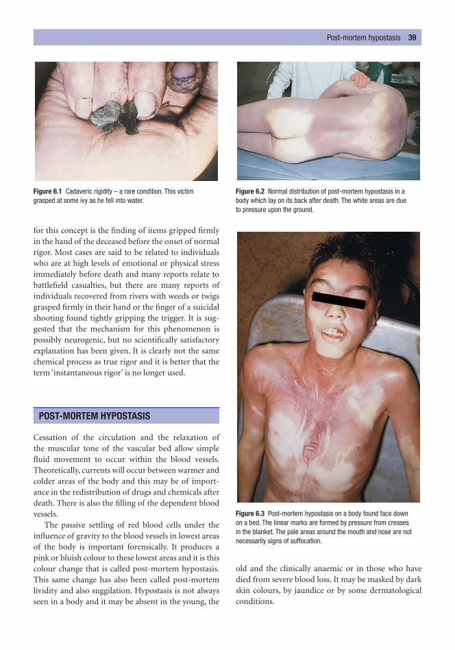
for this concept is the finding of items gripped firmlyin the hand of the deceased before the onset of normalrigor. Most cases are said to be related to individualswho are at high levels of emotional or physical stressimmediately before death and many reports relate tobattlefield casualties, but there are many reports ofindividuals recovered from rivers with weeds or twigsgrasped firmly in their hand or the finger of a suicidalshooting found tightly gripping the trigger. It is sug-gested that the mechanism for this phenomenon ispossibly neurogenic, but no scientifically satisfactoryexplanation has been given. It is clearly not the samechemical process as true rigor and it is better that theterm ‘instantaneous rigor’ is no longer used.
Cessation of the circulation and the relaxation ofthe muscular tone of the vascular bed allow simplefluid movement to occur within the blood vessels.Theoretically, currents will occur between warmer andcolder areas of the body and this may be of import-ance in the redistribution of drugs and chemicals afterdeath. There is also the filling of the dependent bloodvessels.
The passive settling of red blood cells under theinfluence of gravity to the blood vessels in lowest areasof the body is important forensically. It produces apink or bluish colour to these lowest areas and it is thiscolour change that is called post-mortem hypostasis.This same change has also been called post-mortemlividity and also suggilation. Hypostasis is not alwaysseen in a body and it may be absent in the young, the
old and the clinically anaemic or in those who havedied from severe blood loss. It may be masked by darkskin colours, by jaundice or by some dermatologicalconditions.
POST-MORTEM HYPOSTASIS
Post-mortem hypostasis 39
Figure 6.1 Cadaveric rigidity – a rare condition. This victim grasped at some ivy as he fell into water.
Figure 6.2 Normal distribution of post-mortem hypostasis in a body which lay on its back after death. The white areas are due to pressure upon the ground.
Figure 6.3 Post-mortem hypostasis on a body found face down on a bed. The linear marks are formed by pressure from creases in the blanket. The pale areas around the mouth and nose are notnecessarily signs of suffocation.
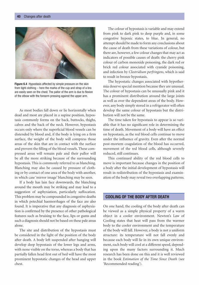
As most bodies fall down or lie horizontally whendead and most are placed in a supine position, hypos-tasis commonly forms on the back, buttocks, thighs,calves and the back of the neck. However, hypostasisoccurs only where the superficial blood vessels can bedistended by blood and, if the body is lying on a firmsurface, the weight of the body will compress thoseareas of the skin that are in contact with the surfaceand prevent the filling of the blood vessels. These com-pressed areas will remain pale and their pallor will be all the more striking because of the surroundinghypostasis. This is commonly referred to as blanching.Blanching may also be caused by pressure of cloth-ing or by contact of one area of the body with another,in which case ‘mirror image’ blanching may be seen.
If a body has lain face downwards, the blanchingaround the mouth may be striking and may lead to asuggestion of asphyxiation, particularly suffocation.This problem may be compounded in congestive deathsin which petechial haemorrhages of the face are alsofound. It is imperative that any diagnosis of asphyxia-tion is confirmed by the presence of other pathologicalfeatures such as bruising to the face, lips or gums andsuch a diagnosis should not be based on these pale areasalone.
The site and distribution of the hypostasis must be considered in the light of the position of the bodyafter death. A body left suspended after hanging willdevelop deep hypostasis of the lower legs and arms,with none visible on the torso, whereas a body that haspartially fallen head first out of bed will have the mostprominent hypostatic changes of the head and upperchest.
The colour of hypostasis is variable and may extendfrom pink to dark pink to deep purple and, in somecongestive hypoxic states, to blue. In general, noattempt should be made to form any conclusions aboutthe cause of death from these variations of colour, butthere are, however, a few colour changes that may act asindicators of possible causes of death: the cherry pinkcolour of carbon monoxide poisoning, the dark red orbrick red colour associated with cyanide poisoning,and infection by Clostridium perfringens, which is saidto result in bronze hypostasis.
The hypostatic changes associated with hypother-mia deserve special mention because they are unusual.The colour of hypostasis can be unusually pink and ithas a prominent distribution around the large joints as well as over the dependent areas of the body. How-ever, any body simply stored in a refrigerator will oftendevelop the same colour of hypostasis but the distri-bution will not be the same.
The time taken for hypostasis to appear is so vari-able that it has no significant role in determining thetime of death. Movement of a body will have an effecton hypostasis, as the red blood cells continue to moveunder the influence of gravity. Even after the normalpost-mortem coagulation of the blood has occurred,movement of the red blood cells, although severelyreduced, still continues.
This continued ability of the red blood cells tomove is important because changes in the position ofa body after the initial development of hypostasis willresult in redistribution of the hypostasis and examin-ation of the body may reveal two overlapping patterns.
On one hand, the cooling of the body after death canbe viewed as a simple physical property of a warmobject in a cooler environment. Newton’s Law ofCooling states that heat will pass from the warmerbody to the cooler environment and the temperatureof the body will fall. However, a body is not a uniformstructure: its temperature will not fall evenly andbecause each body will lie in its own unique environ-ment, each body will cool at a different speed, depend-ing upon the many factors surrounding it. Muchresearch has been done on this and it is well reviewedin the book Estimation of the Time Since Death (see‘Recommended reading’).
COOLING OF THE BODY AFTER DEATH
40 Changes after death
Figure 6.4 Hypostasis affected by simple pressure on the skin from tight clothing – here the marks of the cup and strap of a bra are easily seen on the chest. The pallor of the arm is due to flexion of the elbow with the forearm pressing against the upper arm.

In order to use body temperature as an indicator ofthe time of death, we must make three basic forensicassumptions.
1 The first forensic assumption is that the body tem-perature was 37°C at the time of death. However,humans are very variable and their temperaturewill vary during the day (diurnal variation) andwill also be altered by exercise, by illness, in womenof reproductive age by the stage of the menstrualcycle and by a host of other factors.
2 The second forensic assumption is that it is pos-sible to take one, or maybe a few, temperature read-ings and extrapolate a temperature to apply to thisnon-uniform body as a whole. Clearly, the morereadings that are taken, the more likely it is that themathematical formulae will be useful.
3 The third forensic assumption is that the body haslain in a thermally static environment so that thiscomplex cooling is not further complicated bychanges in the environment. Unfortunately, this isnot the case and even bodies lying in a confineddomestic environment may be subject to the dailyvariations of the central heating system, while thevariations imposed on a body lying outside arepotentially so great that no sensible ‘average’ can beachieved.
Other variables and factors also affect the rate ofcooling of a body (listed below) and together theyshow why the sensible forensic pathologist will be
reluctant to make any pronouncement on the time ofdeath based on the body temperature alone.
• Mass of the body.
• Mass/surface area.
• Body temperature at the time of death.
• Site of reading of body temperature(s).
• Posture of the body – extended or curled into afetal position.
• Clothing – type of material, position on the body –or lack of it.
• Obesity – because fat is a good insulator.
• Emaciation – lack of muscle bulk allows a body tocool faster.
• Environmental temperature.
• Winds, draughts, rain, humidity etc.
Despite all of these problems, the pathologist maystill be asked by the police or by the court for an esti-mate of the time of death. There would appear to betwo forms of this question. The simpler question asksthe pathologist to consider whether the time of deathgiven by a witness is, or is not, consistent with thepathological findings. The second form of the ques-tion is the wider one, commonly asked in the absenceof any other evidence, and it is for an estimate of thetime of death based on the pathological findingsalone. Great care must be taken in answering bothquestions, and the reliability or otherwise of the opin-ion must be stated.
Of the three historic pathological features that wereused to estimate death, only the fall in body tempera-ture has withstood the test of science; the others –rigor mortis and hypostasis – have now been shown to be utterly unreliable and will not be considered further.
Body temperature
Traditionally, the temperature of the body was takenrectally using a long, low-reading thermometer(0–50°C was considered to be adequate). However,there are problems with using this site because anyinterference with the anus or rectum before a fullforensic examination of the area in a good light may
ESTIMATION OF THE TIME OF DEATH
Estimation of the time of death 41
Plateau of variableduration
DecompositionTemperature°
Rigor
Temperature ofenvironment
Hours after death0 6 12 18 24 30 36 42 48 54
36.9°C(98.4°F)
feelscold
Figure 6.5 The sequence of major changes after death in atemperate environment. Note that the core body temperature does not show a fall for the first hour or so.

The nomogram is related to the chosen standard;that is, naked body extended lying in still air. Coolingconditions differing from the chosen standard maybe proportionally adjusted by corrective factors ofthe real body weight, giving the corrected bodyweight by which the death-time is to be read off.Factors above 1.0 may correct thermal isolationconditions, and factors below 1.0 may correct con-ditions accelerating the heat loss of a body.
How to read off the time of death
1 Connect the points of the scales by a straight line according to the rectal and the ambient
temperature. It crosses the diagonal of thenomogram at a special point.
2 Draw a second straight line going through thecentre of the circle, below left of nomogram, andthe intersection of the first line and the diagonal.The second line crosses the semi-circle of thebody weight: the time of death can be read off.The second line touches a segment of the outer-most semi-circle. Here can be seen the permis-sible variation of 95%.
Example: temperature of the rectum 26.4°C; ambi-ent temperature 12°C; body weight 90 kg.
Result: time of death 16 � 1.8 hours. Statement: thedeath occurred within 13.2 and 18.8 (13 and 19)
The temperature–time of death relating nomogram (for ambient temperatures up to 25°C). Permissible variation of 95% (�h).The nomogram expresses the death-time (t) by:
T T T
1.25 exp (B t) .25 exp (5Bt); B 1.2815 (kg .0284rectum ambient
ambient
.625�
�� � � � ��
37 2.)

confuse or contaminate later investigations into thepresence of semen, blood, hairs etc.
The development of small electronic temperatureprobes with rapid response times and digital readoutshas revolutionized the taking of body temperatures.
These probes have allowed the use of other orifices,including the nose and ear, although it must beremembered that these areas are unlikely to registerthe same temperature as the deep rectum or the liver.
Estimation of the time of death 43
hours before the time of measurement (with a relia-bility of 95%).
Note: If the values of the ambient temperatureand/or the body weight (see ‘corrective factors’) arecalled into question, repeat the procedure withother values which might be possible. The range ofdeath-time can be seen in this way.
Requirements for the use
• No strong radiation (e.g. sun, heater, cooling system).
• No strong fever or general hypothermia.
• No uncertain (�) severe changes of the coolingconditions during the period between the timeof death and examination (e.g. the place of deathmust be the same as where the body was found).
• No high thermal conductivity of the surfacebeneath the body (��).
� Known changes can be taken into account: achange of the ambient temperature can oftenbe evaluated (e.g. contact the weather station);use the mean ambient temperature of the
period in question. Changes by the operationsof the investigators (e.g. take any cover off)since finding the body are negligible: take theconditions before into account!
�� Measure the temperature of the surface beneaththe body too. If there is a significant differencebetween the temperature of the air and the surface temperature, use the mean.
Note: for the selection of the corrective factor of anycase, only the clothing or covering of the lowertrunk is relevant! Personal experience is needed,nevertheless this is quickly achieved by the consist-ent use of the method.
Marshall, T.K., Hoare, F.E. 1962: Estimating the Time ofDeath. J Forensic Sci 7, 56–81, 189–210, 211–12.
Henssge, D. 1979: Precision of Estimating the Time ofDeath by Mathematical Expression of Rectal Body Cooling. Z Rechtsmed 83, 49–67.
Henssge, C. 1981: Estimation of Death-Time by Computing the Rectal Body Cooling Under Various Cooling Conditions. Z Rechtsmed 87, 147–8.
Henssge, C., Brinkmann, B., Püschel, K. 1984:Determination of Time of Death by Measuring the Rectal Temperature in Corpses Suspended in Water.Z Rechtsmed 92, 255–76.
Empiric corrective factors of the body weight
Dry clothing/covering In air Corrective Wet through clothing/ In air/waterfactor covering wet body surface
3.5 naked flowing.5 naked still.7 naked moving.7 1–2 thin layers moving
naked moving .751–2 thin layers moving .9 2 or more thicker movingnaked still 1.01–2 thin layers still 1.1 2 thicker layers still2–3 thin layers 1.2 more than 2 thicker layers still1–2 thicker layers moving or still 1.23–4 thin layers 1.3more thin/thicker layers without influence 1.45thick bedspread � clothing 1.8combined 2.4

Currently, the most useful method of estimatingthe time of death is Henssge’s Nomogram (which isexplained in some detail on the preceeding pages).Crucially, the 95 per cent accuracy claimed for thismethod is, at best, only 2.8 hours on either side of themost likely time – a total spread of over 5.5 hours.Henssge’s Normogram relies on three measurements –body temperature, ambient temperature and bodyweight – and lack of accuracy in any one of these willdegrade the final result. In addition, there is the appli-cation of empirical corrective factors to allow forclothing, air movement and/or water and it should benoted that application of these empirical factors cansignificantly lengthen the time spans that lie withinthe 95 per cent confidence limits.
The need to record the ambient temperature posessome problems because pathologists are seldom in aposition to do this at the time of discovery and, as theirarrival at the scene is often delayed by some tens ofminutes or hours, it is most unlikely that the tempera-ture at that time will still be of relevance. Therefore, thefirst police officers or scientists at the scene should beencouraged to take the ambient temperature adjacentto the body and to record the time that they made theirmeasurement. An alteration of 5°C in the ambienttemperature may lead to, at least, a 1-hour alteration inthe most likely time of death.
Many pathologists have in the past used various‘rules of thumb’ to calculate the time of death from thebody temperature but these are, overall, so unreliablethat they should not now be used.
Sometimes the perceived warmth of the body totouch is mentioned in court as an indicator of the timeof death; this assessment is so unreliable as to be use-less and is even more so if the pathologist is asked tocomment upon the reported perceptions of anotherperson. If the pathologists themselves have made theobservation, it has slightly more validity and, in con-junction with a proper assessment of rigor, may beapplied in the broad framework of the aide-memoirenoted earlier.
Other methods of estimating the time of death
Various other methods have been researched in a vain attempt to find the hands of the post-mortemclock. Biochemical methods, including vitreous humor
potassium levels and changes in enzyme and electrolytelevels elsewhere in the body, have been researched;some remain as interesting research tools but none hasbeen successful in routine work.
Examination of gastric contents may confirm theconstituents of the final meal but it will not determinehow long before death the meal was eaten. Gastricemptying has been completely discredited as a meansof estimating the time of death because there is greatindividual variation even in normal conditions anddigestion of food and gastric emptying may cease ifthe individual is under physical or emotional stress.
Of considerably more value is the work of theforensic entomologist, who can determine a probabletime of death – in the region of days to months – fromexamination of the populations and stages of develop-ment of the various insects that invade a body.Initially, the sarcophagous flies – the bluebottle(Callphorae), the greenbottle (Lucilla) and houseflies(Musca) – lay their eggs on moist areas, particularlythe eyes, nose, mouth and, if exposed, the anus andgenitalia. The eggs hatch into larvae, which grow andshed their skins a number of times, each moult beingcalled an instar. Finally, they pupate and a new wingedinsect emerges. The time from egg laying through theinstars to pupation depends on the species and on the ambient temperature, but in general it is about21–24 days.
Other animals, both large and small, will arrive tofeed on the body, the species and the rapidity of theirarrival depending on the time of year and the environ-ment. The examination of buried bodies or skeletalremains will usually require the combined specialistskills of the forensic pathologist, an anthropologistand an entomologist.
Decomposition
In the cycle of life, dead bodies are usually returned,through reduction into their various components, tothe chemical pool that is the earth. Some componentswill do this by entering the food chain at almost anylevel – from ant to tiger – whereas others will bereduced to simple chemicals by the autolytic enzymicprocesses built into the lysosomes of each cell.
The early changes of decomposition are importantbecause they may be confused by the police or mem-bers of the public with the signs of violence or trauma.
44 Changes after death

Putrefaction
This is by far the commonest route of decompositiontaken by any body after death and this route will befollowed unless some unusual conditions apply. Thisform of decomposition results in liquefaction of thesoft tissues over a period of time. The time of visibleonset and the speed with which this process proceedsare, of course, functions of the ambient temperature:the warmer the temperature, the earlier the processstarts and the faster it progresses.
In temperate climates the process is usually firstvisible to the naked eye at about 3–4 days as an area ofgreen discoloration of the right iliac fossa of the anter-ior abdominal wall. This change is the result of theextension of the commensal gut bacteria through thebowel wall and into the skin, where they decomposehaemoglobin, resulting in the green colour. The rightiliac fossa is the usual origin as the caecum lies close tothe abdominal wall at this site. This green colour is butan external mark of the profound changes that areoccurring in the body as the gut bacteria move out ofthe bowel lumen into the abdominal cavity and theblood vessels.
The blood vessels provide an excellent channelthrough which the bacteria can spread with some easethroughout the body. Their passage is marked by thedecomposition of haemoglobin which, when presentin the superficial vessels, results in linear branchingpatterns of brown discoloration of the skin that iscalled ‘marbling’. The colour changes gradually affectmost of the skin and, as the superficial layers of theskin lose cohesions, blisters full of red or brown fluidform in many areas. When the blisters burst, the skinsloughs off.
Considerable gas formation is common and thebody begins to swell, with bloating of the face,abdomen, breasts and genitals. The increased internalpressure causes the eyes and tongue to protrude andforces bloody fluid up from the lungs and it will oftenleak out of the mouth and nose as ‘purge’.
The role of flies and maggots and other animals isdiscussed above; domestic animals are not excludedfrom this process.
The exact time scale for this process cannot be pre-dicted, as the vagaries of the ambient temperature andmany other aspects of the environment generally andthe microenvironment specifically may have signifi-cant effects to either extend or shorten the process.
However, in general terms, within a week or so thebody cavities will burst and the tissues will liquefy anddrain away into the underlying ground. The prostateand the uterus are relatively resistant to putrefactionand they may survive for months, as may the tendonsand ligaments. Eventually, skeletalization will be com-plete and unless the bones are destroyed by larger ani-mals, they may remain for years.
Mummification
A body lying in dry conditions, either climatic or in the microenvironment, may desiccate instead ofputrefying – a process known as mummification.Mummified tissue is dry and leathery and often brownin colour. It is most commonly seen in warm or hot environments such as the dessert and led to the
Estimation of the time of death 45
Figure 6.6 Early decomposition showing marbling of skin due tobacterial growth spreading through the venous system.
Figure 6.7 Putrefaction after about a week in temperate summerconditions. The skin is discoloured and there is gaseous distensionof the face, abdomen and scrotum.
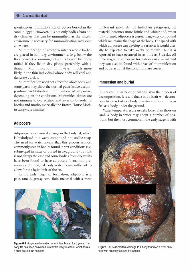
spontaneous mummification of bodies buried in thesand in Egypt. However, it is not only bodies from hotdry climates that can be mummified, as the micro-environment necessary for mummification may existanywhere.
Mummification of newborn infants whose bodiesare placed in cool dry environments, (e.g. below thefloor boards) is common, but adults too can be mum-mified if they lie in dry places, preferably with adraught. Mummification is, however, much morelikely in the thin individual whose body will cool anddesiccate quickly.
Mummification need not affect the whole body, andsome parts may show the normal putrefactive decom-position, skeletalization or formation of adipocere,depending on the conditions. Mummified tissues arenot immune to degradation and invasion by rodents,beetles and moths, especially the Brown House Moth,in temperate climates.
Adipocere
Adipocere is a chemical change in the body fat, whichis hydrolysed to a waxy compound not unlike soap.The need for water means that this process is mostcommonly seen in bodies found in wet conditions (i.e.submerged in water or buried in wet ground) but thisis not always the case and some bodies from dry vaultshave been found to have adipocere formation, pre-sumably the original body water being sufficient toallow for the hydrolysis of the fat.
In the early stages of formation, adipocere is a pale, rancid, greasy semi-fluid material with a most
unpleasant smell. As the hydrolysis progresses, thematerial becomes more brittle and whiter and, whenfully formed, adipocere is a grey, firm, waxy compoundwhich maintains the shape of the body. The speed withwhich adipocere can develop is variable; it would usu-ally be expected to take weeks or months, but it isreported to have occurred in as little as 3 weeks. Allthree stages of adipocere formation can co-exist andthey can also be found with areas of mummificationand putrefaction if the conditions are correct.
Immersion and burial
Immersion in water or burial will slow the process ofdecomposition. It is said that a body in air will decom-pose twice as fast as a body in water and four times asfast as a body under the ground.
Water temperatures are usually lower than those onland. A body in water may adopt a number of pos-itions, but the most common in the early stage is with
46 Changes after death
Figure 6.8 Adipocere formation in an infant buried for 3 years. Thebody fat has been converted into brittle waxy material, which formsa shell around the skeleton.
Figure 6.9 Post-mortem damage to a body found on a river bankthat was probably caused by rodents.

the air-containing chest floating uppermost and thehead and limbs hanging downwards. Hypostasis fol-lows the usual pattern and affects the head and limbs,and these areas may also be damaged by contact withthe bottom if the water is shallow.
The first change that affects the body in water is theloss of epidermis. Gaseous decomposition progressesand the bloated body is often, but not always, lifted tothe surface by these gases, most commonly at about 1 week but this time is extremely variable. Marinepredators will replace the animals found on land andthey can cause extensive damage. The exposure towater can, in some cases, predispose to the formationof adipocere, but this is unusual unless a body liesunderwater for many weeks.
Burial
The effects and the time scale of the changes followingburial are so variable that little specific can be saidother than buried bodies generally decay more slowly,especially if they are buried deep within the ground.The level of moisture in the surrounding soil and acid-ity of the soil will both significantly alter the speed ofdecomposition.
Skeletalization
The speed of skeletalization will depend on many fac-tors, including the climate and the microenvironmentaround the body. It will occur much more quickly in abody on the surface of the ground than in one that isburied. Generally speaking, in a formally buried body,the soft tissues will be absent by 2 years. Tendons, liga-ments hair and nails will be identifiable for some timeafter that.
At about 5 years, the bones will be bare and disar-ticulated, although fragments of articular cartilagemay be identified for many years and for several yearsthe bones will feel slightly greasy and, if cut with a saw,a wisp of smoke and a smell of burning organic mater-ial may be present. Examination of the bone marrowspace may reveal residual organic material that cansometimes be suitable for specialist DNA analysis.
Dating bones, as with all post-mortem dating, isfraught with difficulty. The microenvironment inwhich the bone has lain is of crucial importance and
the examination and dating of bones are now a spe-cialist subject and, if in doubt, the forensic pathologistshould enlist the assistance of anthropologists orarchaeologists who have the specialist skills and tech-niques to manage this type of material.
In the UK, the medico-legal interest in bones fadesrapidly if a bone has been dead for more than 70–80years, because even if it was from a criminal death, it ismost unlikely that the killer would still be alive.Carbon-14 dating is of no use in this short time scale,but examination of the bones for levels of strontium-90, which was released into the atmosphere in highlevels only after the detonation of the nuclear bombsin the 1940s, allows for the differentiation of bonesfrom before and after that time.
Post-mortem injuries
Dead bodies are not immune to injuries and can beexposed to a wide range of trauma and it is importantto bear this possibility in mind when examining anybody so that these injuries are not confused withinjuries sustained during life.
The injuries may be caused during an initial col-lapse and these will usually comprise areas of lacer-ation to the head and scalp, which can be extremelyhard to interpret. Predation by land animals andinsects has been mentioned earlier and can cause ser-ious damage to the body: if there is any doubt aboutbite marks, an odontologist should be consulted. Inwater, fish, crustaceans and larger animals can alsocause severe damage, but there is the added damagecaused by the water logging of the skin and the move-ment of the body across the bottom or against the
Estimation of the time of death 47
Figure 6.10 Post-mortem injury to the knee, with no evidence ofbleeding or reddening of the margins of the laceration.

banks. Contact with boats and propellers will gener-ally lead to patterned injuries that should be easily rec-ognizable.
It is not true to say that post-mortem injuries donot bleed because many do leak blood, especially thoseon the scalp and in bodies recovered from water. Theconfirmation that a wound is post mortem in origin
may be extremely difficult because injuries inflicted inthe last few minutes of life and those that were causedafter death may appear exactly the same. In general,post-mortem injuries do not have a rim of an earlyinflammatory response in the wound edges, but thelack of this response does not exclude an injuryinflicted in the last moments of life.
48 Changes after death

There are numerous medico-legal reasons why it isimportant to be able to establish the identity of a livingindividual or of a dead body. The living may not beable to identify themselves for a variety of reasons:coma, amnesia, infancy or mental defect and possiblyeven severe language barriers may prevent people fromgiving information about themselves. Alternatively, anindividual may simply chose to conceal his true iden-tity by providing false information. Even if the identityof the individual is known, other personal details, forinstance their exact age, may need to be confirmed forthe purposes of immigration or inheritance.
The identity of a corpse is of paramount importancein the investigation of any death and forms the first partof a coroner’s inquisition. Where the body is decom-posed or skeletalized, it will not be possible to obtainvisual identification and so identification can be moredifficult. In some homicides the body may be so injuredthat visual identification is not reliable or it may havebeen deliberately mutilated or concealed until post-mortem changes make visual recognition impossible.Mass disasters, either natural such as earthquakes orman-made such as air crashes, also require the identifi-cation of the dead.
The detailed examination of the dead for evidenceof identity is a specialized task for the forensic patholo-gist, forensic odontologist, anthropologist and otherexperts, but every doctor should have some idea of the
features that can be used to establish the identity of aperson, dead or alive.
Identity cannot be established by the simple measure-ment of a set of parameters of an individual or a body. Itcan only be established by matching the parameters thatcan be measured or seen on an individual with the sameparameters that were known to apply to or to be presenton a named individual. Identification is established bymatching an array of general observations made aboutthe body to an array of general information known to betrue about that particular individual. The finding of aspecific feature, or a combination of specific features,that is known to be possessed by that individual alonewill add considerable weight to the conclusions.
In both the living and dead, the height, weight andgeneral physique need to be recorded and compared.Hair colour and length, including beaching or dying,the presence of a beard or moustache, the amount anddistribution of other body hair, including sites that arecommonly shaved, all need to be established. Skin pig-mentation, racial and ethnic facial appearances, suchas flared nostrils, epicanthic folds etc., are also obviousfeatures which need to recorded in a standard fashion.Eye colour is useful in the Caucasian race but of little
MORPHOLOGICAL CHARACTERISTICS
C h a p t e r s e v e n
Identification of the Living and the Dead
Morphological characteristicsFingerprintsIdentity from teethIdentification of the origin of tissue
or samples
The individuality of cellsIdentification by DNA profilingContaminationPaternity/maternity testingSamples required for DNA analysis
Tattoos and body piercingIdentity of decomposed or
skeletalized remainsFacial reconstruction from skulls
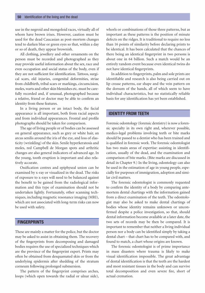
use in the negroid and mongoloid races, virtually all ofwhom have brown irises. However, caution must beused for the dead Caucasian as post-mortem changestend to darken blue or green eyes so that, within a dayor so of death, they appear brownish.
All clothing, jewellery and other ornaments on theperson must be recorded and photographed as theymay provide useful information about the sex, race andeven occupation and social status of the body, even ifthey are not sufficient for identification. Tattoos, surgi-cal scars, old injuries, congenital deformities, striaefrom childbirth, tribal scars or markings, circumcision,moles, warts and other skin blemishes etc. must be care-fully recorded and, if unusual, photographed because a relative, friend or doctor may be able to confirm anidentity from these features.
In a living person or an intact body, the facialappearance is all important, both from racial aspectsand from individual appearances. Frontal and profilephotographs should be taken for comparison.
The age of living people or of bodies can be assessedon general appearance, such as grey or white hair, anarcus senilis around the iris of the eye, and loss of elas-ticity (wrinkling) of the skin. Senile hyperkeratosis andmoles, red Campbell de Morgan spots and arthriticchanges are also general indicators of advanced age. Inthe young, tooth eruption is important and also rela-tively accurate.
Ossification centres and epiphyseal union can beexamined by x-ray or visualized in the dead. The risksof exposure to x-rays will need to be balanced againstthe benefit to be gained from the radiological infor-mation and this type of examination should not beundertaken lightly. Fortunately, other scanning tech-niques, including magnetic resonance imaging (MRI),which are not associated with long-term risks can nowbe used with safety.
These are mainly a matter for the police, but the doctormay be asked to assist in obtaining them. The recoveryof the fingerprints from decomposing and damagedbodies requires the use of specialized techniques whichare the province of the fingerprint expert. Prints mayoften be obtained from desquamated skin or from theunderlying epidermis after shedding of the stratumcorneum following prolonged submersion.
The pattern of the fingerprint comprises arches,loops (which open towards the radial or ulnar side),
whorls or combinations of those three patterns, but asimportant as these patterns is the position of minutedefects on the ridges. It is traditional to require no lessthan 16 points of similarity before declaring prints tobe identical. It has been calculated that the chances ofthere being an identical fingerprint in two persons isabout one in 64 billion. Such a match would be anentirely random event because even identical twins donot have identical fingerprints.
In addition to fingerprints, palm and sole prints areidentifiable and research is also being carried out onlip crease patterns, ear shape and the vein pattern onthe dorsum of the hands, all of which seem to haveindividual characteristics, but no statistically reliablebasis for any identification has yet been established.
Forensic odontology (forensic dentistry) is now a foren-sic specialty in its own right and, wherever possible,medico-legal problems involving teeth or bite marksshould be passed to a dentist who has been trained andis qualified in forensic work. The forensic odontologisthas two main areas of expertise: assisting in identifi-cation, usually of the dead, and the examination andcomparison of bite marks. (Bite marks are discussed indetail in Chapter 9.) In the living, odontology can alsobe used in the estimation of age in young people, espe-cially for purposes of immigration, adoption and simi-lar civil matters.
The forensic odontologist is commonly requestedto confirm the identity of a body by comparing ante-mortem dental chartings with the information gainedfrom a direct examination of the teeth. The odontolo-gist may also be asked to make dental chartings ofbodies whose identity remains unknown or uncon-firmed despite a police investigation, so that, shoulddental information become available at a later date, thetwo sets of records may be then be compared. It isimportant to remember that neither a living individualperson nor a body can be identified simply by taking adental chart – that chart has to be compared with, andfound to match, a chart whose origins are known.
The forensic odontologist is of prime importancein mass disasters where trauma is likely to make visual identification impossible. The great advantageof dental identification is that the teeth are the hardestand most resistant tissues in the body and can survivetotal decomposition and even severe fire, short ofactual cremation.
IDENTITY FROM TEETH
FINGERPRINTS
50 Identification of the living and the dead
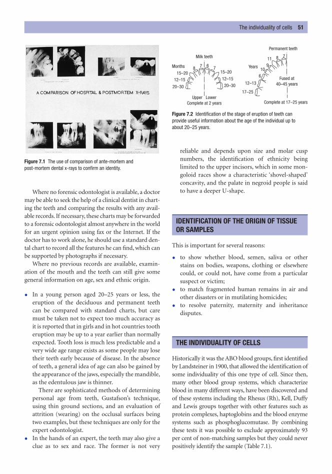
Where no forensic odontologist is available, a doctormay be able to seek the help of a clinical dentist in chart-ing the teeth and comparing the results with any avail-able records. If necessary, these charts may be forwardedto a forensic odontologist almost anywhere in the worldfor an urgent opinion using fax or the Internet. If thedoctor has to work alone, he should use a standard den-tal chart to record all the features he can find, which canbe supported by photographs if necessary.
Where no previous records are available, examin-ation of the mouth and the teeth can still give somegeneral information on age, sex and ethnic origin.
• In a young person aged 20–25 years or less, theeruption of the deciduous and permanent teethcan be compared with standard charts, but caremust be taken not to expect too much accuracy asit is reported that in girls and in hot countries tootheruption may be up to a year earlier than normallyexpected. Tooth loss is much less predictable and avery wide age range exists as some people may losetheir teeth early because of disease. In the absenceof teeth, a general idea of age can also be gained bythe appearance of the jaws, especially the mandible,as the edentulous jaw is thinner.
There are sophisticated methods of determiningpersonal age from teeth, Gustafson’s technique,using thin ground sections, and an evaluation ofattrition (wearing) on the occlusal surfaces beingtwo examples, but these techniques are only for theexpert odontologist.
• In the hands of an expert, the teeth may also give aclue as to sex and race. The former is not very
reliable and depends upon size and molar cuspnumbers, the identification of ethnicity being limited to the upper incisors, which in some mon-goloid races show a characteristic ‘shovel-shaped’concavity, and the palate in negroid people is saidto have a deeper U-shape.
This is important for several reasons:
• to show whether blood, semen, saliva or otherstains on bodies, weapons, clothing or elsewherecould, or could not, have come from a particularsuspect or victim;
• to match fragmented human remains in air andother disasters or in mutilating homicides;
• to resolve paternity, maternity and inheritance disputes.
Historically it was the ABO blood groups, first identifiedby Landsteiner in 1900, that allowed the identification ofsome individuality of this one type of cell. Since then,many other blood group systems, which characterizeblood in many different ways, have been discovered andof these systems including the Rhesus (Rh), Kell, Duffyand Lewis groups together with other features such asprotein complexes, haptoglobins and the blood enzymesystems such as phosphoglucomutase. By combiningthese tests it was possible to exclude approximately 93per cent of non-matching samples but they could neverpositively identify the sample (Table 7.1).
THE INDIVIDUALITY OF CELLS
IDENTIFICATION OF THE ORIGIN OF TISSUE OR SAMPLES
The individuality of cells 51
Figure 7.1 The use of comparison of ante-mortem and post-mortem dental x-rays to confirm an identity.
Milk teeth
Permanent teeth
Months Years
Upper LowerComplete at 2 years Complete at 17–25 years
Fused at40–45 years20–30 20–30
17–25
12–15 12–1512–13
7 7
7
106
119
8
6
815–20 15–20
Figure 7.2 Identification of the stage of eruption of teeth canprovide useful information about the age of the individual up to about 20–25 years.

This bank of tests was used for the forensic identi-fication of blood and other body fluids but has nowbeen replaced by DNA analysis of human tissue andfluid, which is claimed to be nearly 100 per cent accur-ate, both in exclusion and in identification.
Following the discoveries of Sir Alec Jeffreys, a Britishgeneticist, this powerful new tool has radically alteredforensic investigations. The specificity of deoxyribo-nucleic acid (DNA) profiles is so great in statisticalterms that it can be reliably specific to any individual.
The molecule of DNA has two strands of sugar andphosphate molecules that are linked by combinations offour bases – adenine, thymine, cytosine and guanine –forming the double helix of DNA. Only about 10 percent of the molecule is used for genetic coding (theactive genes), the remainder being ‘silent’. In these silentzones, there are between 200 and 14 000 repeats of iden-tical sequences of the four bases. Jeffreys found that
adjacent sequences were constant for a given individualand that they were transmitted, like blood groups, fromthe DNA of each parent. Only uni-ovular twins have thesame sequences; otherwise, the chances of two unre-lated individuals sharing the same sequence is one in abillion or higher. The statistical analysis of DNA identi-fication is extremely complex and it is important thatany calculations are based upon the DNA characteris-tics of a relevant population and not upon the charac-teristics of a ‘standard’ population somewhere else inthe world.
The technique of determining the sequences isextremely complex, relying on cutting the DNA strandsat predetermined points by the use of restrictionenzymes. The fragments of DNA are separated usingelectrophoresis and the different fragments are thenidentified using a radioactive probe. Autoradiographyproduces a picture of a series of bars of varying densityand spacing, which bears a marked resemblance to the‘bar code’ used for pricing articles in a supermarket.From the presence of different bars in given positions,comparisons may be made with other samples, knownor unknown – the classical forensic ‘comparison technique’.
Crucially, there is now no need to match bloodwith blood and semen with semen, as all the DNA inone individual’s body must of necessity be identical.A perpetrator leaving any of his cells or body fluids ata scene is leaving proof of his presence.
Contamination
As the scientists become more and more proficient atextracting and amplifying smaller and smaller amountsof genetic material, so the risks of contaminationincrease. Scene examination must now be approachedwith great care and with sufficient protective clothing toprevent the investigators obscuring any relevant DNAby their own material inadvertently shed from exposedskin, sneezing etc. There is now the need for investiga-tors to provide exclusion DNA samples in the same wayas fingerprints were once provided.
Paternity/maternity testing
Another medico-legal use for DNA is in paternity test-ing, in which, for the first time, a positive identity can be
IDENTIFICATION BY DNA PROFILING
52 Identification of the living and the dead
Percentage exclusion
Red cell antigensMNSs 32.1Rh 28.0ABO 17.6Duffy 4.8Kidd 4.5Kell 3.3Luthran 3.3
Serum proteinsHaptoglobin (Hp) 17.5Gc 14.5C3 14.2Gm 6.5
Red cell enzymesErythrocyte acid phosphatase (EAP) 21.0Glutamate pyruvate transaminase (GPT) 19.0Glycoxalase (Glo) 18.4Phosphoglucomutase (PGM) 14.5Esterase D (EsD) 9.0Adenylate kinase (AK) 4.5Adenosine deaminase (ADA) 4.56-Phosphogluconate dehydrogenase (6-GPD) 2.5
Total combined chance using all systems 93.0
Table 7.1 Percentage Chance of Exclusion by Individual Systems

made rather than relying on the exclusions possiblewith blood group methods. Every bar in the ‘bar code’must have come from either the father or mother, halffrom each. In testing, the coding is made from themother, the child and the putative father. The bars inthe child’s profile are first matched with the mother’s, sothe remaining bars must all have come from the fatherand complete matching of the bars confirms paternity,whereas any discrepancy will exclude it. Occasionally,similar testing must be performed to establish who theparents are following disputes in maternity units.
Samples required for DNA analysis
Samples suitable for DNA analysis are derived fromany nucleated cellular material such as white bloodcells, hair root bulb cells, spermatozoa etc. Initially,relatively large samples were required for this analysis,but the development of the polymerase chain reaction(PCR), which can ‘grow up’ or ‘amplify’ small amountsof DNA, means that positive DNA identification canbe made from the smallest of samples.
When taking samples either at post mortem orfrom a living victim or from the accused, there is sel-dom any problem with the size of the sample that canbe taken. Samples from areas of staining of the scene,the deceased, the living victim or the accused may posedifferent problems as these may be very small areas ofblood, semen staining etc., and these samples must becollected with great care to ensure that contaminationis kept to a minimum.
Blood and hair with roots are used in the living andthe dead, samples of spleen can be taken at post mortemand buccal smears can be taken from the living. If thereis the possibility of a sexual assault, or perhaps in allcases of suspicious death, vaginal, anal and penile swabsshould be taken. If there is to be any delay in gettingsamples to the laboratory, they should be frozen solidat �20°C or as instructed by the laboratory.
The main use of tattoos in forensic medicine is in theidentification of the bodies of unknown persons. Onceagain, the simple presence of a tattoo does not consti-tute identification and there has to be a comparisonwith the tattoos of a known individual. Photographs
should be taken or drawings made which can be used ifa visual identification is not possible or so that they canbe circulated when the identity is not known.
There is a huge diversity of skin tattoos in Westernsociety, some of which may have specific meaning orsignificance within certain subgroups of society, butthere is also much ‘folklore’ about the significance oftattoos, a large amount of which is fiction. Nameswithin tattoos may be useful but, as partners maychange rapidly, they may be of little value. It is saidthat girls more often have their boyfriend’s name tat-tooed than vice versa. Tattoos inside the lips, such asthe number 13, may still identify a drug dealer but aremore likely to indicate time in prison. Tattoos fromthe concentration camps and ghettos of the SecondWorld War are still occasionally seen.
Decomposing bodies should be examined carefullyfor tattoos, which may be rendered more visible whenthe superficial desquamated stratum corneum isremoved.
TATTOOS AND BODY PIERCING
Tattoos and body piercing 53
Figure 7.3 The identification of this body was considerably assistedby the tattoos, the surgical scar and the absence of two fingers.
Figure 7.4 This unusual tattoo aided identification of this body.

Body piercing has gained fashionable status in theWest, and the site and type of piercing should be notedand the rings (as with any other jewellery) can be usedas part of visual identification or can be recovered foridentification by relatives if visual identification is notpossible.
This is usually the task of the forensic pathologist, nowmost commonly with the aid of an anthropologist andother specialists if necessary. The examining doctorhas to carefully document his findings with drawingsand, if possible, with photographs. Wherever possible,the remains, or at least certain key parts, should beretained for later examination by experts.
When presumed skeletal remains are discovered,the investigators wish to know:
• Are the remains actually bones? Sometimes stonesor even pieces of wood are mistaken by the publicor police for bones: the anatomical shape and tex-ture should be obvious to a doctor.
• Are the remains human? A doctor should be able toidentify most intact long bones and a forensicpathologist almost all of the human skeleton,although phalanges, carpal and tarsal bones can beextremely difficult to positively identify as humanbecause some animals, such as the bear, have pawbones almost identical to the human hand. It maybe extremely difficult to identify fragmented bones,and the greater skills and experience of an experi-enced anthropologist or anatomist are invaluable.Cremated bones pose particular difficulties andthese should only be examined by specialists.
• Am I dealing with one or more bodies? This ques-tion can be answered very simply if there are twoskulls or two left femurs. However, if there are noobvious duplications, it is important to examineeach bone carefully to assess whether the sizes andappearances match. It can be extremely difficult toexclude the possibility of co-mingling of skeletalremains, and expert anthropological advice shouldbe sought if there is any doubt.
• What sex are the bones? The skull and the pelvisoffer the best information on sexing; although the femur and sternum can provide assistance, theyare of less direct value. It is important to attempt to
IDENTITY OF DECOMPOSED OR SKELETALIZED REMAINS
54 Identification of the living and the dead
Figure 7.5 Sex differences in the pelvis and femoral head.Left to right: male, female and unknown whose characteristics are obviously female. Note also the different sizes of the femoral heads.
Figure 7.6 This innominate bone reveals both age and sex. Thenarrow greater sciatic notch is definitely male, and the clear line ofthe incompletely fused epiphysis at the iliac crest places the subjectbetween 20 and 23 years of age.
Figure 7.7 Lateral view of a skull. The forehead is vertical and there are no supraorbital ridges. The muscle attachments are not prominent and the mastoid is small – features of a female skull.

determine the sex of each of these bones and not to rely on the assessment of just one. If most ofthe skeleton is available for examination, sexingcan be carried out with over 90 per cent certainty provided that the doctor has sufficient anatomi-cal knowledge. Specialist reference books such asKrogman and Iscan or the larger forensic textbookscan offer much information on all aspects of skeletalidentification. However, when the issues are serious,such as in homicide, it is emphasized that expertexamination by an anthropologist or anatomist isvital.
• What is the age of the person? Up to 20–25 or so,age can be estimated to within a couple of years bya combination of the eruption of the teeth and bythe appearance and fusion of ossification centresand epiphyses, which can be assessed either by eyeor radiologically. Epiphyseal fusion, like the erup-tion of the teeth, tends to be earlier in females andin tropical countries. Ageing can be more accuratein an infant or in fetal material because the rapidchanges taking place at that stage of developmentcan be used as time markers.
After about 25, ageing becomes much more dif-ficult as all bone growth has ceased. Despite the factthat changes do still occur in the skeleton, none isof practical use. Additional data can be obtained byradiology of the trabecular pattern in the head ofthe humerus and femur, which re-models with age.The pubic symphysis also changes with age, but greatexperience is needed for its interpretation. Arthriticdamage, osteoporosis and osteophytes are indicatorsof age, but these are extremely inexact. Specialistexamination of teeth for wear and degenerative
changes can provide valuable information, but theseare tests best left to an expert in the field.
• What is the height (stature) of the person? Thehead–heel measurement of even a completely freshdead body is seldom the same as the person’s stand-ing height in life, due to a combination of factors,including muscle relaxation and shrinkage of inter-vertebral discs. If a whole skeleton is present, anapproximate height can be obtained by direct meas-urement but, due to variable joint spaces, articularcartilage etc., this can only be, at best, an approxi-mation. If only some bones are available, calcula-tions can be made from the measured lengths of thefemur, tibia and fibula by using a table such as thatfrom Trotter and Gleser (Table 7.2).
• What is the race? The ‘shovel-shaped’ mongoloidincisor teeth have already been mentioned (page51). There are some differences in the facial skele-ton of various races, but this is very much the fieldof trained anthropologists and should not beattempted without their assistance.
• Can a personal identity be discovered? The previouscriteria can allot bones to various groups of age, sexetc., but putting a name to the individual depends,as does all identification, upon having reliable ante-mortem data. Occasionally, foreign bodies such asbullets or other metallic fragments may be foundembedded in the skeleton; these may either relate tothe cause of death or may simply be an incidentalfinding. Metal prostheses are now common andmay bear a unique reference number, which cansometimes be traced to an individual patient.
Where pre-mortem clinical radiographs are avail-able, the comparison of these with the post-mortemfilms may give a definite identity. If a skull x-ray is avail-able, comparison of the frontal sinus patterns is incon-trovertible as no two people have the same frontal sinusoutline. Expert comparisons of the lateral skull x-rayand radiographs of the upper ribs, humeri and femurscan provide useful information based partly, in thelimbs, on the pattern of the cancellous bone.
In recent years, much interest has been shown in thereconstruction of the facial appearances from a bareskull. The pioneer was Gerasimov in Moscow, whomeasured the average soft-tissue thickness at many
FACIAL RECONSTRUCTION FROM SKULLS
Facial reconstruction from skulls 55
Figure 7.8 Lateral view of a skull with a sloping forehead,prominent supraorbital ridges, rough muscle attachments and alarge mastoid process – features of a male skull.

points on the skull and rebuilt this layer with plasticineon unknown skulls. Some research has now been per-formed on computerized reconstructions in which theskull is scanned into the computer and the ‘tissue’ isplaced onto the scanned skull image electronically,with the completed image being displayed on thescreen. This system has the advantage that it can berotated electronically to obtain different views.
Whatever method is used to obtain the recon-structed face, a number of significant problems remain.One problem lies in the application of ‘standard’ soft-tissue thicknesses to the facial skeleton when it is thevariability of the thickness of this tissue that, in part,gives individuality to a face. Another major problem isthat the crucial features such as the eyes, lips and facialhair cannot be known and, as they are not dependentupon the underlying bone structure, their colour,shape, length etc. provide visual clues for identification.
An older technique, used for more than 50 years, isphotosuperimposition, in which a photograph of thepossible individual is overlaid with a photographictransparency of the skull, which has been scaled to sizeand orientated to match the angle of the head in theportrait. If they do not fit, the lack of correspondenceis usually obvious and any exclusion obtained can beas valuable as a positive identification. If the twoimages do fit together, by correspondence of anatom-ical landmarks such as chin, nasion, teeth, supraorbitalridges, external meatus etc., it is possible to say that thetwo images could have come from the same person,although identity cannot be fully established by theuse of this method alone. Recently, this procedure hasbeen refined by merging the two images on a televi-sion screen, which has the advantage that the images,particularly the portrait, can be manipulated prior toattempts at superimposition.
56 Identification of the living and the dead
Male White Male African American
Stature � 63.05 � 1.31 (femur � fibula) � 3.63 cm Stature � 63.05 � 1.31 (femur � fibula) � 3.63 cmStature � 67.09 � 1.26 (femur � tibia) � 3.74 cm Stature � 71.75 � 1.15 (femur � tibia) � 3.68 cmStature � 75.50 � 2.60 fibula � 3.86 cm Stature � 72.22 � 2.10 femur � 3.91 cmStature � 65.53 � 2.32 femur � 3.95 cm Stature � 85.36 � 2.19 tibia � 3.96 cmStature � 81.93 � 2.42 tibia � 4.00 cm Stature � 80.07 � 2.34 fibula � 4.02 cmStature � 67.97 � 1.82 (humerus � radius) � 4.31 cm Stature � 73.08 � 1.66 (humerus � radius) � 4.18 cmStature � 66.98 � (humerus � ulna) � 4.37 cm Stature � 70.67 � 1.65 (humerus � ulna) � 4.23 cmStature � 78.10 � 2.89 humerus � 4.57 cm Stature � 75.48 � 2.88 humerus � 4.23 cmStature � 79.42 � 3.79 radius � 4.66 cm Stature � 85.43 � 3.32 radius � 4.57 cmStature � 75.55 � 3.76 ulna � 4.72 cm Stature � 82.77 � 3.20 ulna � 4.74 cm
Female White Female African American
Stature � 50.12 � 0.68 humerus � 1.17 femur Stature � 56.33 � 0.44 humerus � 0.20 radius� 1.15 tibia � 3.61 cm � 1.46 femur � 0.86 tibia � 3.22 cm
Stature � 53.20 � 1.39 (femur � tibia) � 3.55 cm Stature � 58.54 � 1.53 femur � 0.96 tibia � 3.23 cmStature � 53.07 � 1.48 femur � 1.28 tibia � 3.55 cm Stature � 59.72 � 1.26 (femur � tibia) � 3.28 cmStature � 69.61 � 2.93 fibula � 3.57 cm Stature � 59.76 � 2.28 femur � 3.41 cmStature � 61.53 � 2.90 tibia � 3.66 cm Stature � 62.80 � 1.08 humerus � 1.79 tibia � 3.58 cmStature � 52.77 � 1.35 humerus � 1.95 tibia � 3.67 cm Stature � 72.65 � 2.45 tibia � 3.70 cmStature � 54.10 � 2.47 femur � 3.72 cm Stature � 70.90 � 2.49 fibula � 3.80 cmStature � 54.93 � 4.74 radius � 4.24 cm Stature � 64.67 � 3.08 humerus � 4.25 cmStature � 57.76 � 4.27 ulna � 4.20 cm Stature � 75.38 � 3.31 ulna � 4.83 cmStature � 57.97 � 3.36 humerus � 4.45 cm Stature � 94.51 � 2.75 radius � 5.05 cm
Table 7.2 Stature from Bones. Trotter and Gleser’s General Formulae for Reconstruction of Stature from Lengths of Dry Long Bones WithoutCartilage (Constant Terms in Metric and Adapted to Imperial System)
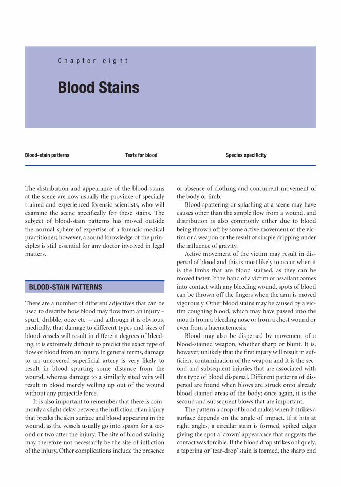
The distribution and appearance of the blood stains at the scene are now usually the province of speciallytrained and experienced forensic scientists, who willexamine the scene specifically for these stains. The subject of blood-stain patterns has moved outside the normal sphere of expertise of a forensic medicalpractitioner; however, a sound knowledge of the prin-ciples is still essential for any doctor involved in legalmatters.
There are a number of different adjectives that can beused to describe how blood may flow from an injury –spurt, dribble, ooze etc. – and although it is obvious,medically, that damage to different types and sizes ofblood vessels will result in different degrees of bleed-ing, it is extremely difficult to predict the exact type offlow of blood from an injury. In general terms, damageto an uncovered superficial artery is very likely toresult in blood spurting some distance from thewound, whereas damage to a similarly sited vein willresult in blood merely welling up out of the woundwithout any projectile force.
It is also important to remember that there is com-monly a slight delay between the infliction of an injurythat breaks the skin surface and blood appearing in thewound, as the vessels usually go into spasm for a sec-ond or two after the injury. The site of blood stainingmay therefore not necessarily be the site of inflictionof the injury. Other complications include the presence
or absence of clothing and concurrent movement ofthe body or limb.
Blood spattering or splashing at a scene may havecauses other than the simple flow from a wound, anddistribution is also commonly either due to bloodbeing thrown off by some active movement of the vic-tim or a weapon or the result of simple dripping underthe influence of gravity.
Active movement of the victim may result in dis-persal of blood and this is most likely to occur when itis the limbs that are blood stained, as they can bemoved faster. If the hand of a victim or assailant comesinto contact with any bleeding wound, spots of bloodcan be thrown off the fingers when the arm is movedvigorously. Other blood stains may be caused by a vic-tim coughing blood, which may have passed into themouth from a bleeding nose or from a chest wound oreven from a haematemesis.
Blood may also be dispersed by movement of ablood-stained weapon, whether sharp or blunt. It is,however, unlikely that the first injury will result in suf-ficient contamination of the weapon and it is the sec-ond and subsequent injuries that are associated withthis type of blood dispersal. Different patterns of dis-persal are found when blows are struck onto alreadyblood-stained areas of the body; once again, it is thesecond and subsequent blows that are important.
The pattern a drop of blood makes when it strikes asurface depends on the angle of impact. If it hits atright angles, a circular stain is formed, spiked edgesgiving the spot a ‘crown’ appearance that suggests thecontact was forcible. If the blood drop strikes obliquely,a tapering or ‘tear-drop’ stain is formed, the sharp end
BLOOD-STAIN PATTERNS
C h a p t e r e i g h t
Blood Stains
Blood-stain patterns Tests for blood Species specificity
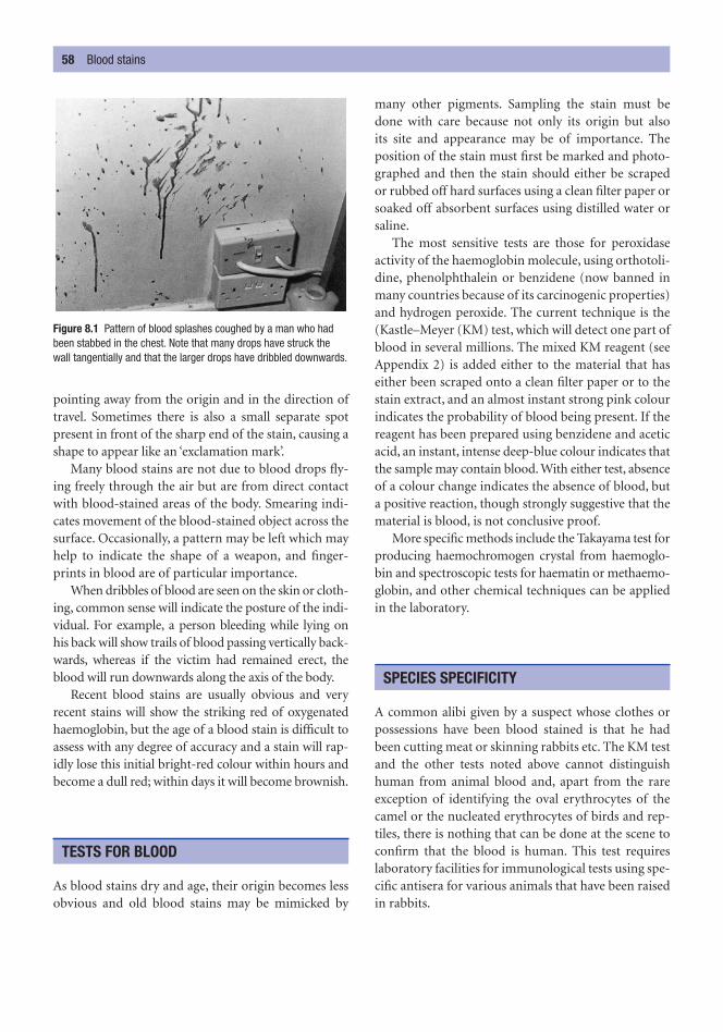
pointing away from the origin and in the direction oftravel. Sometimes there is also a small separate spotpresent in front of the sharp end of the stain, causing ashape to appear like an ‘exclamation mark’.
Many blood stains are not due to blood drops fly-ing freely through the air but are from direct contactwith blood-stained areas of the body. Smearing indi-cates movement of the blood-stained object across thesurface. Occasionally, a pattern may be left which mayhelp to indicate the shape of a weapon, and finger-prints in blood are of particular importance.
When dribbles of blood are seen on the skin or cloth-ing, common sense will indicate the posture of the indi-vidual. For example, a person bleeding while lying on his back will show trails of blood passing vertically back-wards, whereas if the victim had remained erect, theblood will run downwards along the axis of the body.
Recent blood stains are usually obvious and veryrecent stains will show the striking red of oxygenatedhaemoglobin, but the age of a blood stain is difficult toassess with any degree of accuracy and a stain will rap-idly lose this initial bright-red colour within hours andbecome a dull red; within days it will become brownish.
As blood stains dry and age, their origin becomes lessobvious and old blood stains may be mimicked by
many other pigments. Sampling the stain must bedone with care because not only its origin but also its site and appearance may be of importance. Theposition of the stain must first be marked and photo-graphed and then the stain should either be scraped or rubbed off hard surfaces using a clean filter paper orsoaked off absorbent surfaces using distilled water orsaline.
The most sensitive tests are those for peroxidaseactivity of the haemoglobin molecule, using orthotoli-dine, phenolphthalein or benzidene (now banned inmany countries because of its carcinogenic properties)and hydrogen peroxide. The current technique is the(Kastle–Meyer (KM) test, which will detect one part ofblood in several millions. The mixed KM reagent (seeAppendix 2) is added either to the material that haseither been scraped onto a clean filter paper or to thestain extract, and an almost instant strong pink colourindicates the probability of blood being present. If thereagent has been prepared using benzidene and aceticacid, an instant, intense deep-blue colour indicates thatthe sample may contain blood. With either test, absenceof a colour change indicates the absence of blood, buta positive reaction, though strongly suggestive that thematerial is blood, is not conclusive proof.
More specific methods include the Takayama test forproducing haemochromogen crystal from haemoglo-bin and spectroscopic tests for haematin or methaemo-globin, and other chemical techniques can be appliedin the laboratory.
A common alibi given by a suspect whose clothes orpossessions have been blood stained is that he hadbeen cutting meat or skinning rabbits etc. The KM testand the other tests noted above cannot distinguishhuman from animal blood and, apart from the rareexception of identifying the oval erythrocytes of thecamel or the nucleated erythrocytes of birds and rep-tiles, there is nothing that can be done at the scene toconfirm that the blood is human. This test requireslaboratory facilities for immunological tests using spe-cific antisera for various animals that have been raisedin rabbits.
SPECIES SPECIFICITY
TESTS FOR BLOOD
58 Blood stains
Figure 8.1 Pattern of blood splashes coughed by a man who hadbeen stabbed in the chest. Note that many drops have struck thewall tangentially and that the larger drops have dribbled downwards.

Any doctor may be called upon to examine an individ-ual who has sustained a wound as a result of an acci-dent, an act of self-harm or from an action by a thirdparty. It is essential that the doctor examines allwounds carefully and describes them completely andcorrectly. The examination and description of woundscan have far-reaching medico-legal implications,which may be civil or criminal and may not becomeapparent for many months or even years.
A ‘wound’ and an ‘injury’ are not distinguished in law,except in a charge of wounding where the skin must be‘breached’; such a definition would exclude rupture ofthe liver by a kick if the skin was not ‘breached’.
An ‘assault’ is simply the threat of an attack andrequires no physical contact; an actual attack involvingcontact is a ‘battery’. In general parlance, however, theword ‘assault’ is commonly used to describe incidentswith and without physical contact. Neither an assaultnor a battery will necessarily result in a wound.
The term homicide encompasses not only murderbut also several other types of non-accidental killing.The following broad categories are commonly found
in legal codes, but the exact definitions and termin-ology will vary from jurisdiction to jurisdiction.
• Murder: this offence occurs when death follows theactions of an assailant who either intends to kill orintends to cause severe injury or is not concernedwhether or not his actions will cause death orsevere injury.
• Manslaughter: this is a lesser offence than murderand generally occurs when there is no intent tocause death or severe injury but where the deathresulted from an unlawful act or gross negligence.Manslaughter may also be used when the assailanthas been provoked by the victim or when theassailant suffers from a psychiatric disease suchthat he is incapable of forming the intent to kill.In some countries, causing death by the recklessdriving of a motor vehicle is now a separate offence,distinguished from manslaughter.
• Justifiable homicide: this term applies to judicial exe-cutions in those countries where capital punishmentis still allowed by law, and it can also be applied whenan individual kills an assailant in order to defendhimself or others when their lives are directly undera real, direct and immediate threat. In some coun-tries, a police officer may also use lethal force to pre-vent the commission of a serious offence.
LAW ON WOUNDING
C h a p t e r n i n e
The Examination of Wounds
Law on woundingReportsTerminologyWoundsTypes of wound
Blunt force woundsAbrasionsContusionsLacerations
Sharp force wounds
Incised woundsStab wounds
Patterns of injurySurvivalSelf-inflicted injuries

• Infanticide: this is the killing of an infant who is less than 1 year of age by the mother. Death may bedue to an act of omission or commission when thebalance of the mother’s mind is disturbed by theeffects of childbirth or lactation.
In these increasingly litigious times, it is importantthat doctors complete all medical notes with care,especially those that are likely to be examined in acourt of law. In addition to recording the date, timeand place of the examination, the doctor should alsorecord, without comment, the history as related by thepatient. A full examination of the patient should thenbe performed unless it is clear that injury is located ina few areas only. A contemporaneous note of the type,size, shape and position of the injuries with referenceto fixed anatomical landmarks should be made.
The words used to describe injuries are important.The specialized medical meaning of words must onlybe used correctly and not in the ‘lay’ sense; for instance,the term ‘laceration’ must only be used to describe abreak in the continuity of the skin caused by blunttrauma. If in doubt, it is better to use the lay words ofcut, bruise, graze etc. when describing the nature ofthe wounds than to use medical terminology incor-rectly. The courts consider it reasonable to expect thatif doctors use specific medical terminology when writing a professional report, they will understand theterms they have used and they will have used thoseterms correctly.
The phase of recording information is a simple fac-tual step; it does not require any specialist knowledgeand it should be within the competence of any quali-fied medical practitioner. It may be that a doctor withno specialist forensic training will feel insufficientlyqualified to interpret the wounds and, if so, he shouldresist any request to do so and should suggest thatsomeone with greater experience in this field shouldprovide that opinion. If he does not accept his limita-tions at this early stage, they may be demonstrated tohim during cross-examination.
Types of wound
Wounds caused by the application of physical forcecan be divided into two main groups: blunt forcetrauma and sharp force trauma. A third group ofwounds is caused by the application of other forcesthat do not require movement or motion to producetheir effects (Table 9.1).
Blunt force trauma is caused when an object, usu-ally without a sharp or cutting edge, impacts the bodyor the body impacts the object. Three types of injurymay result from such an impact: abrasions, contusionsand lacerations.
Sharp force trauma occurs when an object with asharp or sharpened edge impacts the body. The type ofinjury caused in this case depends upon the depth ofthe wound: an incised wound is longer than it is deep,whereas a stab wound is deeper than it is long.
Non-motion trauma is the third type of wounding.This type of wounding occurs when tissues, usuallythe epidermis or the mucosal surfaces, are damaged bydirect application of a non-physical force, which canbe thermal, chemical, electrical or electromagnetic.
Blunt force wounds
ABRASIONS
An abrasion is the most superficial type of injury andaffects only the epidermis. As the epidermis does notcontain blood vessels, abrasions should not bleed, butthe folded nature of the junction between the dermisand the epidermis and the presence of loops of bloodvessels in the dermal folds will mean that deep abra-sions have a typical punctate or spotty appearance.
WOUNDS
TERMINOLOGY
REPORTS
60 The examination of wounds
Kinetic injuries Non-kinetic injuries
Blunt force injuries Thermal: heat or coldAbrasions ChemicalContusions Electrical: high or low voltageLacerations
ElectromagneticSharp force injuriesIncised woundsStab wounds
Table 9.1 Classification of Injuries
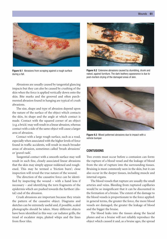
Abrasions are usually caused by tangential glancingimpacts but they can also be caused by crushing of theskin when the force is applied vertically down onto theskin. Bite marks and the grooved and often parch-mented abrasion found in hanging are typical of crushabrasions.
The size, shape and type of abrasion depend uponthe nature of the surface of the object which contactsthe skin, its shape and the angle at which contact ismade. Contact with the squared corner of an object(e.g. a brick) may well result in a linear abrasion, whereascontact with a side of the same object will cause a largerarea of abrasion.
Contact with a large rough surface, such as a road,especially when associated with the higher levels of forcefound in traffic accidents, will result in much broaderareas of abrasion, sometimes called ‘brush abrasions’or ‘gravel rash’.
Tangential contact with a smooth surface may wellresult in such fine, closely associated linear abrasionsthat the skin may simply appear reddened and rough-ened. This may be termed a ‘friction burn’; closeinspection will reveal the true nature of the wound.
The direction of the causative force can be identi-fied by inspecting the wound – with a hand lens ifnecessary – and identifying the torn fragments of theepidermis which are pushed towards the furthest (dis-tal) end of the abrasion.
Crush abrasions are important because they retainthe pattern of the causative object. Diagrams andsketches can be extremely useful and, if possible, scaledphotographs should be taken. Many different objectshave been identified in this way: car radiator grills, thetread of escalator steps, plaited whips and the linesfrom floor tiles.
CONTUSIONS
Two events must occur before a contusion can form:the rupture of a blood vessel and the leakage of bloodfrom the site of rupture into the surrounding tissues.Bruising is most commonly seen in the skin, but it canalso occur in the deeper tissues, including muscle andinternal organs.
The blood vessels that rupture are usually the smallarteries and veins. Bleeding from ruptured capillarieswould be so insignificant that it can be discounted inthe formation of a bruise. The extent of the damage tothe blood vessels is proportionate to the force applied:in general terms, the greater the force, the more bloodvessels are damaged, the greater the leakage of bloodand the bigger the bruise.
The blood leaks into the tissues along the fascialplanes and so a bruise will not reliably reproduce theobject which caused it and, as a bruise ages, the spread
Wounds 61
Figure 9.1 Abrasions from scraping against a rough surface during a fall.
Figure 9.2 Extensive abrasions caused by stumbling, drunk andnaked, against furniture. The dark leathery appearance is due topost-mortem drying of the damaged areas of skin.
Figure 9.3 Mixed patterned abrasions due to impact with a vehicle bumper.

of the blood and so the relationship between the sizeand shape of the bruise and the causative object reduce.
There is one exception, which is the very superficialbruise caused by a moderate force where the leakage of blood is confined to the epidermis and the upperstrata of the dermis, sometimes referred to as ‘intra-dermal’ bruises. In these bruises, the leakage of bloodis small and the blood is held where it has leaked. As a result, these intradermal bruises can reproduce thenature of the object that caused them. Intradermalbruises are often associated with diffuse compressionforces such as pressure from a car tyre or from a shoeduring a stamp or a kick.
Bruises are not static. Their major component,blood, is both fluid and biodegradable and, as a result,bruises will change with time and under the influenceof gravity. Indeed, some deep bruises (especially thoseto the deep muscles of the thigh) may ‘come out’ at the knee.
Once outside the confines of the blood vessel,blood is considered by the tissues to be foreign andthey immediately begin to degrade and remove it. Thisdegradation results in the colour change of a bruisethat is so well known. In general terms, a bruise willinitially be blue/dark blue/purple (depending on theamount of blood within the tissues) and it will then
change to brown to green to yellow as the haemo-globin passes through various stages of degradation.Eventually, all of the blood is removed and the skinreturns to its normal colour.
The speed of these changes is variable and it is notpossible to use the colour changes as a ‘clock’ or‘timetable’ of the bruise. Even in one individual, twobruises inflicted at the same time may differ in theirappearances during resolution. The size of the bruise,the age of the individual (in older people bruisesresolve more slowly), the presence of disease and manydrugs may affect both the formation and the resolu-tion of a bruise.
As a very broad rule of thumb, a small bruises in afit young adult will resolve in about 1 week. Researchin the 1980s showed that if yellow was identified, thebruise was over 18 hours old, whereas if no yellowcould be seen, the bruise could not be reliably aged. Itis to be hoped that further research will elucidate thismatter.
It is essential to remember that the skin fromAsians, Africans and blacks has a layer of melanin pigment of varying intensity. This will reduce the vis-ibility of a bruise and will mask the colour changesthat occur. In the examination of a living victim withdark skin, a bruise may only be identified by palpa-tion. Pathologically, it is possible to identify bruisingby dissection and inspection of the undersurface ofthe skin.
Bruising can occur after death: the blood vessels arejust as easily damaged by the application of force and,providing there is blood with some pressure withinthose vessels, bruising can occur. Such pressure mayexist at the lowermost vessels due to the static weightof the column of blood. Post-mortem bruises are generally small and lie on the dependent parts ofthe body. Bruising may also be found in the areas ofpost-mortem dissection and care must be taken ininterpreting new bruises when an autopsy has beenperformed.
Although bruises do not reflect with any accuracythe object causing them, there are some particular pat-terns that indicate the type of weapon used. One suchpattern is the ‘tramline’ or ‘railway line’ bruise causedby a blow from a linear object. This pattern occursbecause the major stretching and shearing of the skinoccur at the edges of contact and not directly beneaththe centre of the object, which is simply compressed.Impact by a hard ball produces concentric rings ofbruising for similar reasons.
62 The examination of wounds
Figure 9.4 Patterned intradermal bruise on the forehead due to afall onto ribbed ceramic tiles.

The patterns of bruises are extremely important: arow of oval or round bruises may be due to impact bythe knuckles in a punch; groups of small oval or roundbruises are also indicative of fingertip pressure as in
gripping and there is sometimes a single, larger, thumbbruise on the opposite side of the limb. Fingertip bruiseson the neck or along the jaw line are commonly seenin manual strangulation.
LACERATIONS
Lacerations are the result of a blunt force overstretch-ing the skin and the result is a split, which usuallypasses through the full thickness of the skin. A lacera-tion is deep and will bleed. Because the skin is com-posed of many different tissues, some of the moreresilient tissues will not be damaged by the forces thatsplit the weaker tissues. These most resilient tissues areoften nerves, the small fibrous bands of the fascialplanes and, sometimes, at the base of the laceration,the occasional medium-sized elastic blood vessels.These fibres arch across the defect in the skin and arereferred to as ‘bridging fibres’.
Lacerations are most common where the skin can becompressed between the applied force and underlyingbone (i.e. over the scalp, face, elbows, knees, shins etc).They are rare (unless severe force has been applied)
Wounds 63
Figure 9.5 Bruising of the upper arm. The pattern of these bruises is typical of forceful gripping. Small abrasions from fingernails arealso seen.
Figure 9.6 Recent bruising of the abdominal wall and scrotum dueto kicking.
Figure 9.7 Colour changes in a bruise. This injury is reported to be 10 days old and contains brown, blue and purple components.
Figure 9.8 Typical ‘railway-line’ bruises caused by a wooden rod.Note that the centre of the parallel contusions is unmarked.

over the soft, fleshy areas of the body such as the but-tocks, breasts and abdomen.
When the force applied to the skin is more tangen-tial, as in the rolling or grinding action of a vehiclewheel, the laceration may be horizontal and result in alarge area of separation of skin from the underlyingtissues. This can be termed ‘flaying’.
The margins of a laceration are always ragged;however, if a thin regular object inflicts an injury overa bony area of the body, the wound caused may look
very sharply defined and can be mistaken for anincised injury. Careful inspection of the margins willreveal some crushing and bruising, and examinationof the inner surfaces of the wound will reveal the pres-ence of bridging fibres.
The laceration may reflect the object causing it ifthat object is straight, but this wound is a poor repro-ducer in other circumstances.
Sharp force wounds
INCISED WOUNDS
Incised wounds are caused by objects with a sharp orcutting edge, most commonly a knife but an axe,shards of glass, broken glasses and bottles, an edge ofbroken pottery, a piece of metal etc. can also causethese wounds. An incised injury is distinguished froma stab wound by being longer than it is deep.
The edges of the wound will give some indicationas to the sharpness of the weapon causing it. A sharpedge will leave no bruising or abrasion of the woundmargins. Careful inspection of the internal structureof the wound will reveal that no bridging fibres arepresent because the cutting edge divides everything inits passage into the skin.
64 The examination of wounds
Figure 9.9 Laceration of eyebrow due to impact on the edge of abrick. The wound has clean edges but the abraded margins can beseen.
Figure 9.10 Laceration of an arm of a pedestrian struck by a car.The impact has been oblique, causing a flap of skin to tear away to the right.
Figure 9.11 Multiple lacerations from a blunt steel bar. These wereinitially mistaken by the police for axe wounds. The abraded orcrushed margins can be easily seen.

Incised wounds, by their nature, are rarely life threat-ening because they seldom penetrate deeply enough todamage a blood vessel of significant size. However,incised wounds over the wrist or neck, where majorarteries lie in the superficial tissues, can prove fatal.
STAB WOUNDS
A stab wound is deeper than it is long and it is thedepth of the injury that makes it so often fatal andhence of such forensic interest. Stabbing is the com-monest method of homicide in Britain and accountsfor over 50 per cent of all murders, many of which aredomestic in type.
Any weapon with a point or tip can cause a stabwound; the edge of the blade does not need to be sharp.For penetration to occur, the tip must be pressedagainst the skin with sufficient force to overcome thenatural elasticity of the skin. The factors that determinehow much force is needed for penetration to occur are:
• The sharpness of the tip of the weapon: the sharperthe tip, the easier it is to penetrate the skin.
• The speed of the contact: the faster the contact, thegreater the force applied and the easier it is to pen-etrate the skin.
• Whether clothing has been penetrated: clothingincreases the resistance to penetration.
• Whether bone or cartilage has been injured: skinoffers very little resistance to a stabbing action by asharp knife, but penetration of these denser tissueswill require greater force.
Experiments have shown that once a knife has pen-etrated the skin, the rest of the weapon follows with
Wounds 65
Figure 9.12 Incised wound to the flank; it is clearly longer than it is deep. Figure 9.13 Fatal stab wound of the chest and the knife that
caused it. The wound does not reproduce the size of the knife,due to contraction of the elastic fibres in the skin.
Figure 9.14 Multiple stab wounds from the same weapon as in Fig. 9.13, showing marked variation in size and appearance.
Figure 9.15 A complex stab wound where all three injuries arecaused by a single action. The first entry is in the right breast; thereis an exit wound in the middle and a re-entry wound over the centreof the chest.

almost no additional force being applied. In his text-book Forensic Pathology, Knight says that he believesthis is because the skin indents before penetration andacts as a reservoir of energy. The insertion of a sharpknife into the body, especially through the skin stretchedacross ribs, requires very little force, and pressure froma single finger is sufficient to push a sharp scalpelthrough the chest wall at this site. With these factors inmind, it is clear that it is possible, in some circum-stances, for a person to transfix himself by walking orfalling onto the tip of a sharp weapon. No forwardmovement of the weapon is necessary and, accordingto Knight, the hand or arm holding the knife does notneed to be braced or fixed for penetration to occur.
The commonest weapon used is a knife of somesort, but swords, shards of glass, broken bottles etc. canbe used and even screwdrivers, metal rods, railings etc.can all cause stab wounds if sufficient force is applied.
The appearances of a stab wound on the skin willreflect, but not necessarily mimic, the cross-sectionalshape of the weapon used. If a blade of some sort isused, the following general comments apply.
• A slit-like wound will distort after removal of theweapon by the action of the elastic fibres present inthe skin. If the fibres run at right angles to thewound, it will be pulled outwards and get shorterand wider; if they run parallel to the wound, it willbe pulled lengthways and the edges will tend toclose and the wound elongate slightly.
• Even if the edges of the wound are gently pushedtogether, the resulting defect is seldom the samesize as the knife, and personal experience suggeststhat the wounds and the weapon may differ by upto �1 cm.
• The size of the wound also depends on the shape ofthe blade and how deeply it was inserted.
• Movement of the knife in the wound, either by theassailant ‘rocking’ it or by the victim moving toescape, will cause the wound to be enlarged. If theknife is twisted or rotated, a triangular wound maybe caused.
• Many knives have only one cutting edge, the otherbeing blunt. This design may be reproduced in thewound where one ‘V’-shaped sharp point can beseen and one blunt point. The blunt point may splitand a tiny spike or triangle is sometimes formed,which is often referred to as a ‘fishtail’.
• If the knife is fully inserted, bruising or abrasion ofthe skin adjacent to the stab wound caused by a hilt
of the weapon or by the hand of the assailant can beseen provided that clothing has not intervened.
• The depth of a wound can be greater than thelength of the blade if a forceful stab is inflicted. Thisis because the abdomen and, to a lesser extent, thechest can be compressed by the force of the hilt orhand against the skin.
• A blunt object such as a screwdriver or spike willtend to indent, split and bruise the skin on insertion.Different types of screwdriver will cause differentpatterns of injury. Cross-head or ‘Phillips’ screw-drivers cause very distinctive cruciate skin wounds.
• Sharp but irregular instruments such as scissors orchisels will leave particular patterns of injury, witha ‘Z’ shape from scissors and a rectangular shapefrom a chisel. If the stab wounds are unusual, it isimportant to consider unusual instruments.
The three possible motives for infliction of a woundapply and there can be little to distinguish betweenaccidental and deliberate infliction by simply exam-ination of a single wound. However, examination ofthe site, orientation and pattern(s) of the wounds willoften reveal useful indications about the causation ofthe wound.
Particular actions result in particular and oftenidentifiable patterns of injury. Punches, kick, bites,holding or gripping injuries and defence injuries canbe identified by their patterns.
A punch is a blow delivered by the clenched fist.The blow can be directed anywhere, but usually leavesa wound only over those areas of the body where theskin is closely applied to bone, as in the face and skull.The wounds caused are bruises, abrasions and, wherethe skin is over an edge such as the eyebrow or cheeks,lacerations. The lips can also be forced back onto theteeth, resulting in lacerations inside the lips.
Bones may be broken, but punches will usuallyaffect only the thinner bones such as the nose or vul-nerable flat bones such as the mandible and the max-illa. Ribs may be fractured by blows of sufficient force.
Contusions of the face may result in extensivespreading of the blood, and periorbital haematomas(black eyes) are common, although it is important toremember that fractures of the nose can also result inhaematomas at these sites.
PATTERNS OF INJURY
66 The examination of wounds

In the very young and old, intra-abdominal injury,including mesenteric laceration, intestinal rupture andinjury to the major abdominal organs, may rarely occur.
Kicking and stamping injuries are caused by a footwhich is either swung or moved downwards with someforce. These actions can deliver blunt force trauma tothe body and, as a result, abrasions, contusions andlacerations may result. There are some minor differ-ences between the modes of application of force and,as stamping injuries have a more compressive compo-nent, they may result in more bruises and lacerationswith fewer abrasions. Stamping injuries, particularlythose caused by ‘trainers’, may leave areas of intra-dermal bruising that reflect the pattern of the sole ofthe shoe.
The victim is often on the ground when theseinjuries are inflicted, and blows from the foot are com-monly directed towards the head and face, the chestand the abdomen. The high levels of force that can beapplied by a foot mean that there are often underlyingskeletal injuries. These are most common in the facialskeleton and in the ribs. Stamping injuries to the frontof the abdomen may result in rupture of the internalorgans, particularly the liver and the spleen.
Bite marks are crush abrasions. They are com-monly associated with bruising and may be associatedwith lacerations if severe force is applied. Bite markscan be seen in sexual assaults, child abuse and occa-sionally on the sports field.
Bite marks are commonly composed of a pair ofcurved lines of bruises facing towards each other.Inspection may reveal distinctive linear crush abra-sions indicating the patterns of the teeth. Attempts atidentification of the assailant should be left to anodontologist.
Bite marks may be found on almost any surface ofthe body; specific sites are associated with specificforms of assault. The neck, breasts and shoulders areoften bitten in a sexually motivated attack, while inchild abuse bites of the arms and the buttocks arecommon. Adolescent self-inflicted bites can be seen onthe medial aspect of the arm.
Bites can also be inflicted by many common inhab-itants of a house, including family pets. Odontologicalexamination will reveal different sizes, different archshapes and different dentition in these cases.
If a suspected bite is found at examination, a swab ofthe area should be taken for DNA and the bite should bephotographed with a scale. Photography using ultra-violet light may be of assistance in older cases.
Defence injuries are caused as victims try to defendthemselves against an attack. The type of injury causeddepends upon the weapon used and the force applied:an attack by a blunt weapon may result in bruises on
Patterns of injury 67
Figure 9.16 Extensive bruising and abrasion of the face due tokicking.
Figure 9.17 Bite marks on the arm. The upper bite shows abrasionand bruising from the opposing dental arches. In the diffuse bruisingof the lower arm, a group of abrasions caused by the incisors can be seen.
Figure 9.18 Defence wounds on the palmar surface of the handcaused by attempts to grasp or deflect a blade.
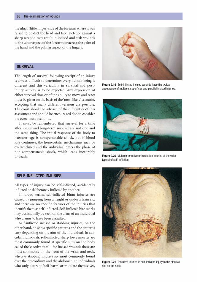
the ulnar (little finger) side of the forearm where it wasraised to protect the head and face. Defence against asharp weapon may result in incised and stab woundsto the ulnar aspect of the forearm or across the palm ofthe hand and the palmar aspect of the fingers.
The length of survival following receipt of an injury is always difficult to determine: every human being isdifferent and this variability in survival and post-injury activity is to be expected. Any expression ofeither survival time or of the ability to move and reactmust be given on the basis of the ‘most likely’ scenario,accepting that many different versions are possible.The court should be advised of the difficulties of thisassessment and should be encouraged also to considerthe eyewitness accounts.
It must be remembered that survival for a timeafter injury and long-term survival are not one and the same thing. The initial response of the body tohaemorrhage is compensatable shock, but if blood loss continues, the homeostatic mechanisms may beoverwhelmed and the individual enters the phase ofnon-compensatable shock, which leads inexorably to death.
All types of injury can be self-inflicted, accidentallyinflicted or deliberately inflicted by another.
In broad terms, self-inflicted blunt injuries arecaused by jumping from a height or under a train etc.and there are no specific features of the injuries thatidentify them as self-inflicted. Self-inflicted bite marksmay occasionally be seen on the arms of an individualwho claims to have been assaulted.
Self-inflicted incised or stabbing injuries, on theother hand, do show specific patterns and the patternsvary depending on the aim of the individual. In sui-cidal individuals, self-inflicted sharp force injuries aremost commonly found at specific sites on the bodycalled the ‘elective sites’ – for incised wounds these aremost commonly on the front of the wrists and neck,whereas stabbing injuries are most commonly foundover the precordium and the abdomen. In individualswho only desire to ‘self-harm’ or mutilate themselves,
SELF-INFLICTED INJURIES
SURVIVAL
68 The examination of wounds
Figure 9.19 Self-inflicted incised wounds have the typicalappearance of multiple, superficial and parallel incised injuries.
Figure 9.20 Multiple tentative or hesitation injuries of the wristtypical of self-infliction.
Figure 9.21 Tentative injuries in self-inflicted injury to the electivesite on the neck.

the site can be anywhere on the body that can bereached by the individual. Except in cases of severemental disease, the eyes, lips, nipples and genitaliatend to be spared.
The other features of self-inflicted injuries lie in themultiple, predominantly parallel nature of the woundsand, in suicidal acts, the more superficial injuries arereferred to as ‘hesitation’ or ‘tentative’ injuries.
Self-inflicted injuries 69

Many injuries, or rather their effects, will vary depend-ing on the exact area of the body that is injured. A stabwound to a limb is unlikely to have fatal consequences,whereas a similar injury to the chest may well be fatal.Similarly, the effects of a blow to the chest may be min-imal but the consequences of exactly the same blow tothe head may be catastrophic.
Injuries to the head are particularly important becauseof the brain’s vital role in sustaining the life of the indi-vidual. The brain is protected within the strong, bonyskull, but it is not well restrained within this compart-ment and injuries to the brain result from differencesbetween the motion of the skull and the brain. Thereare three main components of the head: the scalp, theskull and the brain.
Scalp
The scalp is vascular, hair-bearing skin; at its base is athick fibrous membrane called the galea aponeurotica.Lying between the galea and the skull is a very thinsheet of connective tissue that is penetrated by bloodvessels (emissary veins) emerging through the skull,
and beneath this connective tissue is the periosteum ofthe outer table of the skull.
The vascularity of the scalp, possibly the connec-tion with the intracranial blood vessels, results in pro-fuse bleeding if it is injured. Bleeding will also, verycommonly, continue after death if the head is in adependent position.
The scalp is easily injured by blunt trauma as it canbe crushed between the weapon and the underlyingskull, which acts like an anvil. Bruises of the scalp areassociated with prominent oedema because this nor-mal tissue response cannot spread and dissipate as eas-ily as in other areas of the body. The easiest way todetect scalp injuries is by palpation, but shaving isessential for proper documentation and photography.
Lacerations of the scalp will, if inspected carefully,reveal a band of abrasion adjacent to the laceration,and examination of the depths of the wound willreveal the bridging fibres that will confirm the trueorigin of these injuries. This is important because therelative thinness of the scalp and its immobility meanthat blunt trauma can result in sharply defined injuriesthat may, at first sight, appear to be incised injuries.
Tangential forces or glancing blows, either from aninstrument or from a rotating wheel as in a road traf-fic accident, may tear large flaps of tissue, exposing theunderlying skull. If hair becomes entangled in rotatingmachinery, portions of the scalp may be avulsed.
HEAD INJURIES
C h a p t e r t e n
Regional Injuries
Head injuriesScalpSkull fracturesIntracranial haemorrhage
EffectsTraumatic subarachnoid haemorrhageBrain injury
Coup and contra coup
Neck injuriesSpinal injuriesChest injuriesAbdomen
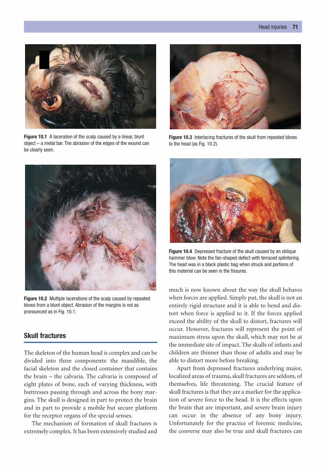
Head injuries 71
Skull fractures
The skeleton of the human head is complex and can bedivided into three components: the mandible, thefacial skeleton and the closed container that containsthe brain – the calvaria. The calvaria is composed ofeight plates of bone, each of varying thickness, withbuttresses passing through and across the bony mar-gins. The skull is designed in part to protect the brainand in part to provide a mobile but secure platformfor the receptor organs of the special senses.
The mechanism of formation of skull fractures isextremely complex. It has been extensively studied and
much is now known about the way the skull behaveswhen forces are applied. Simply put, the skull is not anentirely rigid structure and it is able to bend and dis-tort when force is applied to it. If the forces appliedexceed the ability of the skull to distort, fractures willoccur. However, fractures will represent the point ofmaximum stress upon the skull, which may not be atthe immediate site of impact. The skulls of infants andchildren are thinner than those of adults and may beable to distort more before breaking.
Apart from depressed fractures underlying major,localized areas of trauma, skull fractures are seldom, ofthemselves, life threatening. The crucial feature ofskull fractures is that they are a marker for the applica-tion of severe force to the head. It is the effects uponthe brain that are important, and severe brain injurycan occur in the absence of any bony injury.Unfortunately for the practice of forensic medicine,the converse may also be true and skull fractures can
Figure 10.1 A laceration of the scalp caused by a linear, blunt object – a metal bar. The abrasion of the edges of the wound can be clearly seen.
Figure 10.2 Multiple lacerations of the scalp caused by repeatedblows from a blunt object. Abrasion of the margins is not aspronounced as in Fig. 10.1.
Figure 10.3 Interlacing fractures of the skull from repeated blows to the head (as Fig. 10.2).
Figure 10.4 Depressed fracture of the skull caused by an obliquehammer blow. Note the fan-shaped defect with terraced splintering.The head was in a black plastic bag when struck and portions of this material can be seen in the fissures.
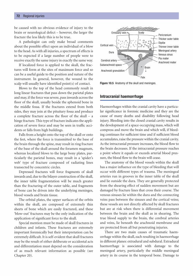
be caused with no obvious evidence of injury to thebrain or neurological defect – however, the larger thefracture the less likely this is to be true.
A pathologist can only make broad commentsabout the possible effect upon an individual of a blowto the head. As with all injuries, a spectrum of effects isto be expected if a large number of people were toreceive exactly the same injury in exactly the same way.
If localized force is applied to the skull, the frac-tures will form at the sites of maximum force and socan be a useful guide to the position and nature of theinstrument. In general, however, the wound to thescalp will usually have identified point(s) of contact.
Blows to the top of the head commonly result inlong linear fractures that pass down the parietal platesand may, if the force was severe, pass inwards across thefloor of the skull, usually beside the sphenoid bone inthe middle fossa. If the fractures extend from bothsides, they may join at the pituitary fossa and producea complete fracture across the floor of the skull – ahinge fracture. This type of fracture indicates the appli-cation of severe force and may be seen in traffic acci-dents or falls from high buildings.
Falls from a height onto the top of the skull or ontothe feet, where the force is transmitted to the base ofthe brain through the spine, may result in ring fractureof the base of the skull around the foramen magnum,whereas localized blows to the large bony plates, par-ticularly the parietal bones, may result in a ‘spider’sweb’ type of fracture composed of radiating lines transected by concentric circles.
Depressed fractures will force fragments of skullinwards and, due to the bilayer construction of the skull,the inner table fragmentation will be much greaterthan the fracturing of the outer table, and fragmentsof bone can be driven into the underlying meninges,blood vessels and brain tissue.
The orbital plates, the upper surfaces of the orbitswithin the skull, are composed of extremely thinsheets of bone which are easily fractured, and these‘blow-out’ fractures may be the only indication of theapplication of significant force to the skull.
Special mention must be made of skull fractures inchildren and infants. These fractures are extremelyimportant forensically but their interpretation can beextremely difficult. It is self-evident that skull fracturesmay be the result of either deliberate or accidental actsand differentiation must depend on the considerationof as much relevant information as possible (seeChapter 20).
Intracranial haemorrhage
Haemorrhages within the cranial cavity have a particu-lar significance in forensic medicine and they are thecause of many deaths and disability following headinjury. Bleeding into the closed cranial cavity results inthe development of a space-occupying mass, which willcompress and move the brain and which will, if bleed-ing continues for sufficient time and if sufficient bloodaccumulates, raise the pressure within the cranial cavity.As the intracranial pressure increases, the blood flow tothe brain decreases. If the intracranial pressure reachesa point where it equals or exceeds arterial blood pres-sure, the blood flow to the brain will cease.
The anatomy of the blood vessels within the skullhas a major influence on the type of bleeding that willoccur with different types of trauma. The meningealarteries run in grooves in the inner table of the skulland lie outside the dura. They are generally protectedfrom the shearing effect of sudden movement but aredamaged by fracture lines that cross their course. Thevenous sinuses lie within the dura and the connectingveins pass between the sinuses and the cortical veins;these vessels are not directly affected by skull fracturesbut are at risk when there is differential movementbetween the brain and the skull as in shearing. Thetrue blood supply to the brain, the cerebral arteriesand veins, lie beneath the arachnoid membrane andare protected from all but penetrating injuries.
There are two main causes of traumatic haem-orrhage within the skull, each resulting in haemorrhagein different planes: extradural and subdural. Extraduralhaemorrhage is associated with damage to themeningeal artery, particularly the middle meningealartery in its course in the temporal bone. Damage to
72 Regional injuries
Emissary vein ScalpPericraniumThicker outer table
Thinner inner tableMeningeal arteryVenous sinusPia materArachnoid mater
Diplöe
Brain
Falx
Arachnoid granulation
Subarachnoid space
Cerebral artery
Dura
Cortical vein
Figure 10.5 Anatomy of the skull and meninges.

this vessel results in arterial bleeding into the extraduralspace. As the blood accumulates, it separates the durafrom the overlying skull and forms a haematoma.Arterial bleeding is likely to be fast and the developmentof the haematoma will result in a rapid displacement ofthe brain and the rapid onset of symptoms. Rarely,extradural haemorrhage can develop as a result ofvenous bleeding from damaged perforating veins ordural sinuses, in which case the development of symp-toms will be slower.
The second cause of traumatic intracranial haem-orrhage is damage to the communicating veins as theycross the subdural space. This venous damage is notnecessarily associated with fractures of the skull. Inmany instances, particularly in the very young and thevery old, there may be no history or evidence of anytrauma to the head. These venous injuries are associ-ated with rotational or shearing forces that cause thebrain to move relative to the inner surface of the skull;this motion stretches the thin-walled veins, causingthem to rupture. The venous bleeding lies in the sub-dural space. Recent subdural haemorrhages are darkred in colour and shiny, but begin to turn brown after afew days; microscopically, haemosiderin can be identi-fied with Perl’s stain. Older subdural collections may beenclosed in gelatinous membranes, which can hardeninto a firm rubbery capsule in extreme cases. Such oldcollections of subdural blood are most commonly seenin the elderly, whose cerebral atrophy allows space forthe formation of the haematoma without significantclinical effects. Occasionally, these subdurals may bemany months or even years old. They may have beenthe cause of a change in neurological state that was vari-ously ascribed to a stroke or the onset of a psychiatriccondition.
Head injuries 73
Figure 10.6 A large extradural haemorrhage caused by damage tothe middle meningeal artery from a fracture of the temporal bone.
Figure 10.7 Fracture (arrowed) of the temporal bone resulting intearing of the middle meningeal artery and a large extraduralhaemorrhage. Collapse occurred 4 hours after receiving a ‘trivial’injury to the head.
Figure 10.8 A chronic subdural haemorrhage found incidentally at autopsy of an elderly person who had died from other causes.The brown, gelatinous material is seen on the under-surface of the meninges.

EFFECTS
The effects of both extradural and subdural haemor-rhages are essentially the same: they both act as space-occupying lesions compressing the brain and, at theirmost severe, causing herniation through the tentoriumcerebelli. The speed at which symptoms develop isvariable and both are associated with a period ofapparent normality – the lucid period – after the injuryand before the onset of neurological symptoms. Ingeneral terms, the arterial bleeding would be expectedto produce symptoms and signs of raised intracranialpressure more quickly than the venous bleeding of thesubdural haemorrhage.
Traumatic subarachnoid haemorrhage
Small areas of subarachnoid haemorrhage are com-mon where there has been direct trauma to the brain,either from an intrusive injury such as a depressedfracture or from movement of the brain against theinner surface of the skull as a result of acceleration ordeceleration injuries. These small injuries are usuallyassociated with areas of underlying cortical contusionand sometimes laceration.
Large basal subarachnoid haemorrhages can be oftraumatic origin and follow forceful blows or kicks tothe neck, particularly to the upper neck adjacent to theear, but any blow which results in rapid rotation or flex-ion of the neck can cause this damage. The vertebralarteries are confined within foraminae in the lateralmargins of the upper six cervical vertebrae and are sus-ceptible to trauma either with or without fracture of theforamina. It is thought that most of the damage to thearteries as a result of trauma occurs at the site at whichthey penetrate the spinal dura to enter the posteriorfossa of the skull.
It must be remembered that basal subarachnoidhaemorrhages are most commonly non-traumatic inorigin and arise from the spontaneous rupture of aberry aneurysm of one of the arteries in the circle ofWillis. Great care must be taken to exclude this naturalcause.
Brain injury
Injuries that have resulted in skull fractures or intracra-nial haemorrhage are clear macroscopic markers that
significant force has been applied to the head and there-fore to the brain. Sometimes, however, these markersare absent but, as a result of acceleration or deceler-ation, the brain has been significantly traumatized.Whatever the exact cause of the trauma, the effects onthe brain as a whole are the same and cerebral oedemadevelops. The mechanism of cerebral oedema is com-plex and not fully understood, but appears to be dueto transudation of fluid into the extracellular space.
As oedema develops, the brain swells and, as theskull cannot expand to allow for the swelling, theintracranial pressure rises and the brain is squeezedout of the spaces in the meninges and the skull. Thecerebrum is forced downwards through the tentoriumcerebelli and the cerebellum and brainstem are forceddownwards through the foramen magnum. Thesemovements produce secondary areas of injury in theunci of the cerebrum and the cerebellar tonsils.
The weight of the oedematous brain is increasedand the surface of the brain is markedly flattened,often with haemorrhage and necrosis of the unci andthe cerebellar tonsils; sectioning reveals compressionof the ventricles, sometimes into thin slits.
Direct injuries to the brain from depressed or comminuted skull fractures result in areas of bruisingand laceration to the cortex, often associated withlarger sites of haemorrhage. These injuries can occurat any site, above or below the tentorium. Penetratinginjuries from gunshots or from stab wounds will causeinjuries deep into the white matter, and the tissueadjacent to the wound tracks will often be associatedwith contusion.
There is also another group of injuries that areassociated with head injuries, particularly the injuriesthat do not result in either penetration or intracranialhaemorrhage. The brain is injured in these cases byrotation or by acceleration/deceleration, which causesshearing between the different layers or componentsof the brain as each moves in a different way or at adifferent speed. This shearing causes contusions andlacerations deep within the substance of the brain, butparticularly in the region of the corpus callosum.Shearing is also associated with the so-called ‘glidinglesions’ that occur beneath the cortex and in the cere-bral peduncles and brainstem.
The shearing effects are also identifiable at a micro-scopic level, where the damage to the axons can be iden-tified by the use of special staining techniques. Thesechanges have been termed diffuse axonal injury. Theytake a finite time to develop, or at least to become
74 Regional injuries
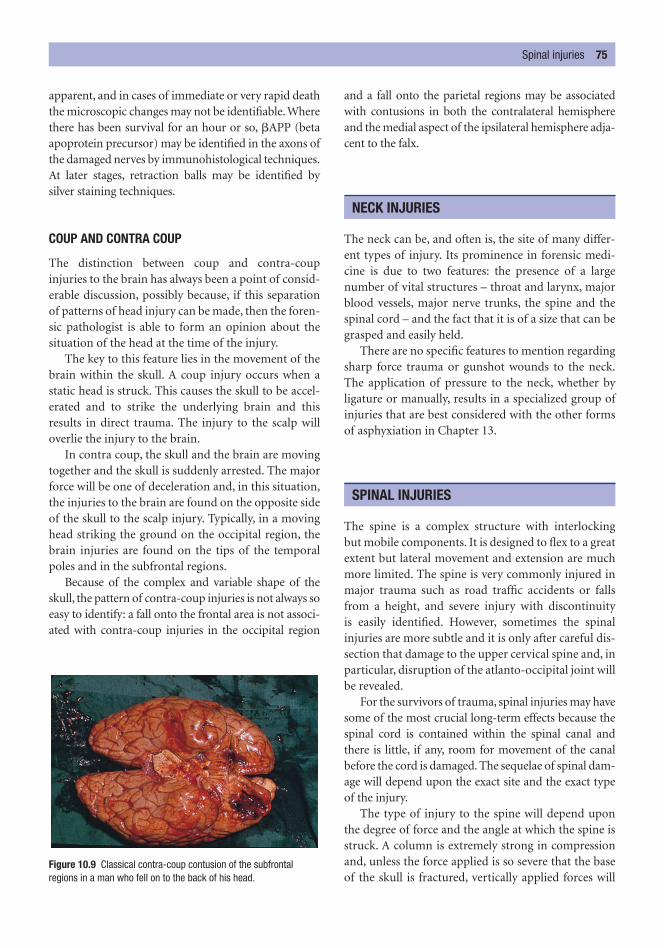
apparent, and in cases of immediate or very rapid deaththe microscopic changes may not be identifiable. Wherethere has been survival for an hour or so, �APP (betaapoprotein precursor) may be identified in the axons ofthe damaged nerves by immunohistological techniques.At later stages, retraction balls may be identified by silver staining techniques.
COUP AND CONTRA COUP
The distinction between coup and contra-coupinjuries to the brain has always been a point of consid-erable discussion, possibly because, if this separationof patterns of head injury can be made, then the foren-sic pathologist is able to form an opinion about the situation of the head at the time of the injury.
The key to this feature lies in the movement of thebrain within the skull. A coup injury occurs when astatic head is struck. This causes the skull to be accel-erated and to strike the underlying brain and thisresults in direct trauma. The injury to the scalp willoverlie the injury to the brain.
In contra coup, the skull and the brain are movingtogether and the skull is suddenly arrested. The majorforce will be one of deceleration and, in this situation,the injuries to the brain are found on the opposite sideof the skull to the scalp injury. Typically, in a movinghead striking the ground on the occipital region, thebrain injuries are found on the tips of the temporalpoles and in the subfrontal regions.
Because of the complex and variable shape of theskull, the pattern of contra-coup injuries is not always soeasy to identify: a fall onto the frontal area is not associ-ated with contra-coup injuries in the occipital region
and a fall onto the parietal regions may be associatedwith contusions in both the contralateral hemisphereand the medial aspect of the ipsilateral hemisphere adja-cent to the falx.
The neck can be, and often is, the site of many differ-ent types of injury. Its prominence in forensic medi-cine is due to two features: the presence of a largenumber of vital structures – throat and larynx, majorblood vessels, major nerve trunks, the spine and thespinal cord – and the fact that it is of a size that can begrasped and easily held.
There are no specific features to mention regardingsharp force trauma or gunshot wounds to the neck.The application of pressure to the neck, whether byligature or manually, results in a specialized group ofinjuries that are best considered with the other formsof asphyxiation in Chapter 13.
The spine is a complex structure with interlocking but mobile components. It is designed to flex to a greatextent but lateral movement and extension are muchmore limited. The spine is very commonly injured inmajor trauma such as road traffic accidents or fallsfrom a height, and severe injury with discontinuity is easily identified. However, sometimes the spinalinjuries are more subtle and it is only after careful dis-section that damage to the upper cervical spine and, inparticular, disruption of the atlanto-occipital joint willbe revealed.
For the survivors of trauma, spinal injuries may havesome of the most crucial long-term effects because thespinal cord is contained within the spinal canal andthere is little, if any, room for movement of the canalbefore the cord is damaged. The sequelae of spinal dam-age will depend upon the exact site and the exact type of the injury.
The type of injury to the spine will depend uponthe degree of force and the angle at which the spine isstruck. A column is extremely strong in compressionand, unless the force applied is so severe that the baseof the skull is fractured, vertically applied forces will
SPINAL INJURIES
NECK INJURIES
Spinal injuries 75
Figure 10.9 Classical contra-coup contusion of the subfrontalregions in a man who fell on to the back of his head.
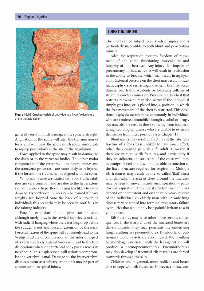
generally result in little damage if the spine is straight.Angulation of the spine will alter the transmission offorce and will make the spine much more susceptibleto injury, particularly at the site of the angulation.
Force applied to the spine may result in damage tothe discs or to the vertebral bodies. The other majorcomponents of the vertebrae – the neural arches andthe transverse processes – are more likely to be injuredif the force of the trauma is not aligned with the spine.
Whiplash injuries associated with road traffic fatal-ities are very common and are due to the hyperexten-sion of the neck, hyperflexion being less likely to causedamage. Hyperflexion injuries can be caused if heavyweights are dropped onto the back of a crouchingindividual; this scenario may be seen in roof falls inthe mining industry.
Forceful extension of the spine can be seen,although rarely now, in the cervical injuries associatedwith judicial hanging where there is a long drop beforethe sudden arrest and forceful extension of the neck.Forceful flexion of the spine will commonly lead to the‘wedge’ fracture or compression of the anterior aspectof a vertebral body. Lateral forces will lead to fracturedislocations where one vertebral body passes across itsneighbour – this displacement will seriously comprom-ise the vertebral canal. Damage to the intervertebraldiscs can occur as a solitary lesion or it may be part ofa more complex spinal injury.
The chest can be subject to all kinds of injury and isparticularly susceptible to both blunt and penetratinginjuries.
Adequate respiration requires freedom of move-ment of the chest, functioning musculature andintegrity of the chest wall. Any injury that impairs orprevents any of these activities will result in a reductionin the ability to breathe, which may result in asphyxi-ation. External pressure on the chest may result in trau-matic asphyxia by restricting movement; this may occurduring road traffic accidents or following collapse ofstructures such as mines etc. Pressure on the chest thatrestricts movement may also occur if the individualsimply gets into, or is placed into, a position in whichthe free movement of the chest is restricted. This posi-tional asphyxia occurs most commonly in individualswho are rendered immobile through alcohol or drugs,but may also be seen in those suffering from incapaci-tating neurological disease who are unable to extricatethemselves from these positions (see Chapter 13).
Blunt injury may result in fractures of the ribs. Thefracture of a few ribs is unlikely to have much effect,other than causing pain, in a fit adult. However, ifthere are numerous rib fractures, and particularly ifthey are adjacent, the structure of the chest wall maybe compromised and it will not be able to function asthe fixed structure required for respiration. Multiplerib fractures may result in the so-called ‘flail’ chestand, clinically, the area of chest around the fracturesmay be seen to move inwards on inspiration – para-doxical respiration. The clinical effects of such injuriesdepend on their extent and on the respiratory reserveof the individual: an elderly man with chronic lungdisease may be tipped into terminal respiratory failureby injuries that would only be a painful irritant to a fityoung man.
Rib fractures may have other more serious conse-quences. If the sharp ends of the fractured bones aredriven inwards, they may penetrate the underlyinglung, resulting in a pneumothorax. If subcostal or pul-monary blood vessels are also injured, the resultinghaemorrhage associated with the leakage of air willproduce a haemopneumothorax. Pneumothoracesmay also develop if fractured rib margins are forcedoutwards through the skin.
Children are, in general, more resilient and betterable to cope with rib fractures. However, rib fractures
CHEST INJURIES
76 Regional injuries
Figure 10.10 Crushed vertebral body due to a hyperflexion injury of the thoracic spine.

in children have a particular place in forensic medicine,as they are a marker for child abuse (see Chapter 20).
Rib fractures can also be artefactual and the use ofcardiopulmonary resuscitation (CPR) or cardiac mas-sage by both skilled and unskilled individuals hasresulted in an increase in the sternal and parasternalfractures that are seen at post mortem. They are usu-ally easily identified by their symmetrical, parasternalpattern and the relative lack of haemorrhage, whichindicates their peri-mortem or post-mortem origin.
Penetrating injuries of the chest, whether the resultof sharp force trauma (stab wounds) or gunshotwounds, may result in damage to any of the organs orvessels within the cavity. The effect of the penetrationwill depend mainly upon which organ(s) or vessel(s)are injured. The penetration of the chest wall will alsoresult in the possibility of a pneumothorax.
Injury to the lungs will also result in the develop-ment of pneumothoraces, and damage to the bloodvessels will result in haemorrhage, which may be con-fined to the soft tissues of the mediastinum or enterthe pleural cavities. Haemorrhage from penetratinginjuries to the chest will commonly remain concealedand it is not unusual to find several litres of bloodwithin the chest cavity at post mortem. Alternatively,the haemorrhage will become apparent when the vic-tim collapses, and the site of the injury then lies belowthe level of the blood in the chest cavity. This hasimportant consequences for scene examination becausethe site of obvious blood loss may be some distancefrom the site of infliction of the injury.
The anatomy of the abdominal cavity plays a majorrole in determining the type of injuries that are found.The vertebral column forms a strong midline verticalstructure posteriorly, and blunt trauma, especially inthe anterior/posterior direction, may result in com-pression of the organs lying in the midline against thevertebral column. This compressive injury may resultin bruising or even rupture of the mesentery, bruisingor even bisection of the duodenum or jejunum, rup-ture of the pancreas and laceration of the liver, espe-cially the left lobe.
The forces required to cause these injuries in anadult must be considered to be severe and they arecommonly found in road traffic accidents. It is unlikely
that a punch to the abdomen would cause more thansimple bruising, except in very unusual circumstances,but kicks and stamps are commonly the cause of majorintra-abdominal trauma. The kidneys and the spleenare attached only by their hila and are susceptible torotational as well as direct trauma, and severe rota-tional forces may result in avulsion from their pedicles.
Abdominal injuries in children may have the samecauses, but the forces required to cause them will be con-siderably reduced, and the slower compressive forcesassociated with squeezing of the abdomen during abusemay also result in the injuries described above.
Penetrating injuries to the abdomen can be theresult of either gunshots or sharp force trauma. Theeffects of these injuries will depend almost entirely on
ABDOMEN
Abdomen 77
Figure 10.12 Extensive rupture of the liver following severe bluntabdominal trauma in a road traffic accident.
Figure 10.11 Lacerations of the mesentery, which resulted in fatal intra-abdominal haemorrhage following a blow from a knee.

the organs and vessels involved in the track of the pro-jectiles. A penetrating injury to the aorta or inferiorvena cava will result in severe haemorrhage and mayproduce rapid death. Peritonitis from a rupturedbowel or stomach will be immediately painful but maynot, with adequate and prompt treatment, prove fatal.
Blunt trauma to the spleen is sometimes associatedwith delayed rupture leading to haemorrhage andpossibly death some hours or even days after receipt ofthe injury. Pancreatic trauma may lead to the develop-ment of a pseudocyst, with little or no short-term orlong-term sequelae.
78 Regional injuries
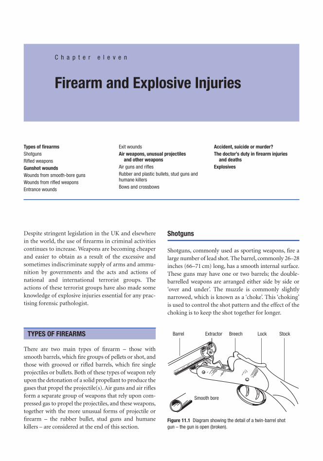
Despite stringent legislation in the UK and elsewherein the world, the use of firearms in criminal activitiescontinues to increase. Weapons are becoming cheaperand easier to obtain as a result of the excessive andsometimes indiscriminate supply of arms and ammu-nition by governments and the acts and actions ofnational and international terrorist groups. Theactions of these terrorist groups have also made someknowledge of explosive injuries essential for any prac-tising forensic pathologist.
There are two main types of firearm – those withsmooth barrels, which fire groups of pellets or shot, andthose with grooved or rifled barrels, which fire singleprojectiles or bullets. Both of these types of weapon relyupon the detonation of a solid propellant to produce thegases that propel the projectile(s). Air guns and air riflesform a separate group of weapons that rely upon com-pressed gas to propel the projectiles, and these weapons,together with the more unusual forms of projectile orfirearm – the rubber bullet, stud guns and humanekillers – are considered at the end of this section.
Shotguns
Shotguns, commonly used as sporting weapons, fire alarge number of lead shot. The barrel, commonly 26–28inches (66–71 cm) long, has a smooth internal surface.These guns may have one or two barrels; the double-barrelled weapons are arranged either side by side or‘over and under’. The muzzle is commonly slightlynarrowed, which is known as a ‘choke’. This ‘choking’is used to control the shot pattern and the effect of thechoking is to keep the shot together for longer.
TYPES OF FIREARMS
C h a p t e r e l e v e n
Firearm and Explosive Injuries
Types of firearmsShotgunsRifled weaponsGunshot woundsWounds from smooth-bore gunsWounds from rifled weaponsEntrance wounds
Exit woundsAir weapons, unusual projectiles
and other weaponsAir guns and riflesRubber and plastic bullets, stud guns andhumane killersBows and crossbows
Accident, suicide or murder?The doctor’s duty in firearm injuries
and deathsExplosives
Barrel Extractor Breech Lock Stock
Smooth bore
Figure 11.1 Diagram showing the detail of a twin-barrel shot gun – the gun is open (broken).

A shotgun is designed for use up to about 30–50 mand is unlikely to kill a man at this extreme range,although it may well cause serious and painful injuries.The length of the barrel makes handling and hidingdifficult and so it is not uncommon for the barrels tobe shortened – sawn off – for criminal activities. Thisshortening of the barrel has little effect on the proper-ties of the gun and there is no doubt that shotguns canbe devastating weapons at significant ranges.
The size of shotgun is usually given in relation to the ‘gauge’ or ‘bore’, which relates to the diameter of the barrel. The commonest size is the 12-bore or 12-gauge shotgun (which has a barrel diameter of18.2 mm). The other popular shotgun has a barrel thatis 0.410 inches (10.2 mm) in diameter and it is called,simply, a ‘410’.
The cartridges for shotguns consist of a metal basecontaining a central detonating cap, supporting a card-board or plastic tube containing the propellant chargeand a mass of lead shot, which rests on top of the pro-pellant. The open end of the cartridge is closed by a thin disc, which is crimped into the end of the tube.The shot may be contained within a plastic bucket orthere may be discs of felt, cork or cardboard acting as wads above and below the shot. The plastic contain-ers open into a star shape in flight and may themselvescause injury.
The shot in the cartridges falls into one of twomain groups – birdshot or buckshot – and the car-tridges are numbered according to the size of the shotthey contain. There are also cartridges that contain a single heavy projectile called a ‘slug’. Occasionally, car-tridges are tampered with and the loose shot is fusedinto a single mass. In some areas, especially developingcountries or amongst rebels, homemade guns, some-times called ‘country guns’, may be made from metaltubing with crude firing mechanisms. These weaponsmay be used to fire collections of debris such as nuts,bolts, wood-screws or metal shavings etc., and anywounds resulting from these weapons will obviouslynot comply with the usual descriptions.
Rifled weapons
These are weapons that fire one projectile at a timethrough a barrel that has a number of spiral grooves –usually between four and six and with a right-handtwist – cut into the interior of the barrel. The inter-vening projections, called ‘lands’, grip the bullet and
impart the gyroscopic spin that assists in maintainingan accurate trajectory. Rifled weapons fall into threemain groups – revolvers, ‘automatic’ pistols and rifles.
Revolvers and pistols are short-barrelled weaponsand the difference between them is the method of pre-senting a bullet to the breech. In the revolver, a cylin-der containing the bullets rotates under trigger pressureto line up a new cartridge with the trigger mechanismand the barrel. In a pistol, the forces generated by thefiring of a round are harnessed using a spring mecha-nism to eject the spent cartridge case, load a fresh car-tridge and reset the firing mechanism. The cartridgesare usually stored in a clip contained within the butt ofthe weapon.
Rifles are long-barrelled weapons whose muzzlevelocity and accuracy are much greater than those ofthe revolver or pistol. In older rifles, a bolt had to bemoved manually to eject the spent cartridge case andbring a new round into the breech from a magazine.The gases generated from the discharge of the weaponare now used to perform the same tasks.
The ammunition for rifled weapons is also called acartridge and comes in many sizes or calibres, usuallymeasured from land to land. A cartridge is essentially aclosed metal cylinder with the firing cap and propel-lant charge at one end and a single projectile – the bul-let – clamped into the other. The bullet may be madeof lead or lead alloy or it may have a lead core coveredwith a cupro-nickel, steel or other hard metal jacket.There may be a soft tip or an air space in the tip of thebullet, which is designed to distort and thus deceleratethe bullet on impact. Some modern bullets have asmall explosive charge in the tip for the same purpose.
80 Firearm and explosive injuries
Base of magazineclipped into stock
Ejector hole insliding breech cover
Revolving chamber ‘broken’(one round in place)
Spirally ‘rifled’bore
Figure 11.2 Diagrams of two types of handgun: a revolver (above)and a pistol (below). Note the rifling in the barrel.

On a historical note, the term ‘Dum-Dum bullets’relates to the .303 centrefire rifle cartridges with a hollow-point style bullet that were made at the Britisharsenal in Dum-Dum, India, in the late nineteenthcentury. The use of Dum-Dum and other expandingbullets was forbidden in wars between signatories ofThe Geneva Convention in 1864. This rule was reiter-ated by declarations of the Hague Conferences of 1899and 1907. It should be noted that the restrictions applyto war only and that no restriction applies to the use ofthis type of bullet if war has not been formally declared.
Modern propellants consist of nitro-cellulose orother synthetic compounds prepared as small colouredflakes, discs or balls. The process of firing a bullet orshotgun cartridge is as follows. The firing pin strikesthe primer cup and the primer compound explodes;small vents between the primer cup and the base of thecartridge case allow the flame of this detonation tospread to the propellant. The propellant burns rapidly,producing huge volumes of gas, which are furtherexpanded by the very high temperatures of the igni-tion, and it is the pressure of this gas that propels theshot or bullet from the barrel.
The speed with which the projectile leaves the endof the barrel – the muzzle velocity – varies from a fewhundred metres per second in a shotgun to a thousandor more in a high-velocity military weapon. The energyof the projectile is proportional to the speed at whichit travels and is calculated from the kinetic energy(1⁄2MV2) of the bullet, so higher muzzle velocities areconsiderably more effective at delivering energy to thetarget than larger bullets. The formation of wounds isrelated to the transfer of the energy of the bullet to thebody tissues.
When any weapon is fired, smoke, flame and the gasesof combustion will leave the barrel, together with por-tions of unburned, burning and burnt propellant.These contaminants will usually follow the projec-tile(s), but in some guns they may also precede them.The distance they will travel from the end of the muz-zle is extremely variable, depending mainly on thetype of weapon and the type of propellant. These con-taminants also escape from small gaps around thebreech and will soil hands or clothing that are close tothe breech at the time of discharge.
Wounds from smooth-bore guns
The pellets are forced along the barrel by the gases ofdetonation and will leave the muzzle in a compactmass, which spreads out as it travels away from thegun. The shot describes a long, shallow cone with itsapex close to the muzzle of the shotgun and the fur-ther along this cone the victim is situated, the largerthe wound pattern will be.
A contact wound occurs when the muzzle is touch-ing the skin and usually results in a circular entrancewound that is about the size of the muzzle. The woundedge will be regular and often has a clean-cut appear-ance with no individual pellet marks apparent. Therewill commonly be smoke soiling of at least some of themargin of the wound. There may be a narrow, circularrim of abrasion around some or all of the entrancewound, caused when the gases of the discharge enterthrough the wound and balloon the tissues upwards sothat the skin is pressed against the muzzle. If the dis-charge was over an area supported by bone, the gasescannot disperse as quickly as they would in soft areassuch as the abdomen, and the greater ballooning ofthe skin results in splits of the skin, which often have a radial pattern.
The wads or plastic cup will usually be recoveredalong the wound track. The tissues along the woundtrack will be blackened and the surrounding tissuesare said to be pinker than normal as a result of the car-bon monoxide contained within the discharge gases.
A close discharge, within a few centimetres of thesurface, will also produce a wound with a similarappearance, but as there is now room for gas escape,there will be no muzzle mark. More smoke soiling can
GUNSHOT WOUNDS
Gunshot wounds 81
Figure 11.3 Various types of ammunition: From left to right: cannonshell, automatic rifle, bolt action rifle, revolver, pistol, .22 rifle, two12-bore shotgun cartridges.
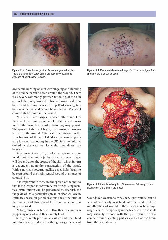
occur, and burning of skin with singeing and clubbingof melted hairs can be seen around the wound. Thereis also, very commonly, powder ‘tattooing’ of the skinaround the entry wound. This tattooing is due toburnt and burning flakes of propellant causing tinyburns on the skin and cannot be washed off. Wads willcommonly be found in the wound.
At intermediate ranges, between 20 cm and 1m,there will be diminishing smoke soiling and burn-ing of the skin, but powder tattooing may persist.The spread of shot will begin, first causing an irregu-lar rim to the wound. Often called a ‘rat-hole’ in theUSA because of the nibbled edges, the same appear-ance is called ‘scalloping’ in the UK. Separate injuriescaused by the wads or plastic shot containers may be seen.
At a range of over 1 m, smoke damage and tattoo-ing do not occur and injuries caused at longer rangeswill depend upon the spread of the shot, which in turnis dependent upon the construction of the barrel.With a normal shotgun, satellite pellet holes begin tobe seen around the main central wound at a range ofabout 2–3 m.
It is important to measure the spread of the shot sothat if the weapon is recovered, test firings using iden-tical ammunition can be performed to establish therange at which a particular spread of shot will occur.Estimates based on generalizations about the ratio ofthe diameter of this spread to the range should nolonger be used.
At long ranges, such as 20–50 m, there is a uniformpeppering of shot, and this is rarely fatal.
Shotguns rarely produce an exit wound when firedinto the chest or abdomen, although single pellet exit
wounds can occasionally be seen. Exit wounds can beseen when a shotgun is fired into the head, neck ormouth. The exit wound in these cases may be a hugeragged aperture, especially in the head, where the skullmay virtually explode with the gas pressure from acontact wound, ejecting part or even all of the brainfrom the cranial cavity.
82 Firearm and explosive injuries
Figure 11.4 Close discharge of a 12-bore shotgun to the chest.There is a large hole, partly due to disruption by gas, and noevidence of pellet scatter is seen.
Figure 11.5 Medium-distance discharge of a 12-bore shotgun. Thespread of the shot can be seen.
Figure 11.6 Complete disruption of the cranium following suicidaldischarge of a shotgun in the mouth.
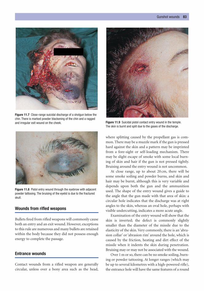
Wounds from rifled weapons
Bullets fired from rifled weapons will commonly causeboth an entry and an exit wound. However, exceptionsto this rule are numerous and many bullets are retainedwithin the body because they did not possess enoughenergy to complete the passage.
Entrance wounds
Contact wounds from a rifled weapon are generallycircular, unless over a bony area such as the head,
where splitting caused by the propellant gas is com-mon. There may be a muzzle mark if the gun is pressedhard against the skin and a pattern may be imprintedfrom a fore-sight or self-loading mechanism. Theremay be slight escape of smoke with some local burn-ing of skin and hair if the gun is not pressed tightly.Bruising around the entry wound is not uncommon.
At close range, up to about 20 cm, there will besome smoke soiling and powder burns, and skin andhair may be burnt, although this is very variable anddepends upon both the gun and the ammunitionused. The shape of the entry wound gives a guide tothe angle that the gun made with that area of skin: acircular hole indicates that the discharge was at rightangles to the skin, whereas an oval hole, perhaps withvisible undercutting, indicates a more acute angle.
Examination of the entry wound will show that theskin is inverted; the defect is commonly slightlysmaller than the diameter of the missile due to theelasticity of the skin. Very commonly, there is an ‘abra-sion collar’ or ‘abrasion rim’ around the hole, which iscaused by the friction, heating and dirt effect of themissile when it indents the skin during penetration.Bruising may or may not be associated with the wound.
Over 1 m or so, there can be no smoke soiling, burn-ing or powder tattooing. At longer ranges (which maybe up to several kilometres with a high-powered rifle),the entrance hole will have the same features of a round
Gunshot wounds 83
Figure 11.7 Close-range suicidal discharge of a shotgun below thechin. There is marked powder blackening of the chin and a raggedand irregular exit wound on the cheek.
Figure 11.8 Pistol entry wound through the eyebrow with adjacentpowder tattooing. The bruising of the eyelid is due to the fracturedskull.
Figure 11.9 Suicidal pistol contact entry wound in the temple.The skin is burnt and split due to the gases of the discharge.
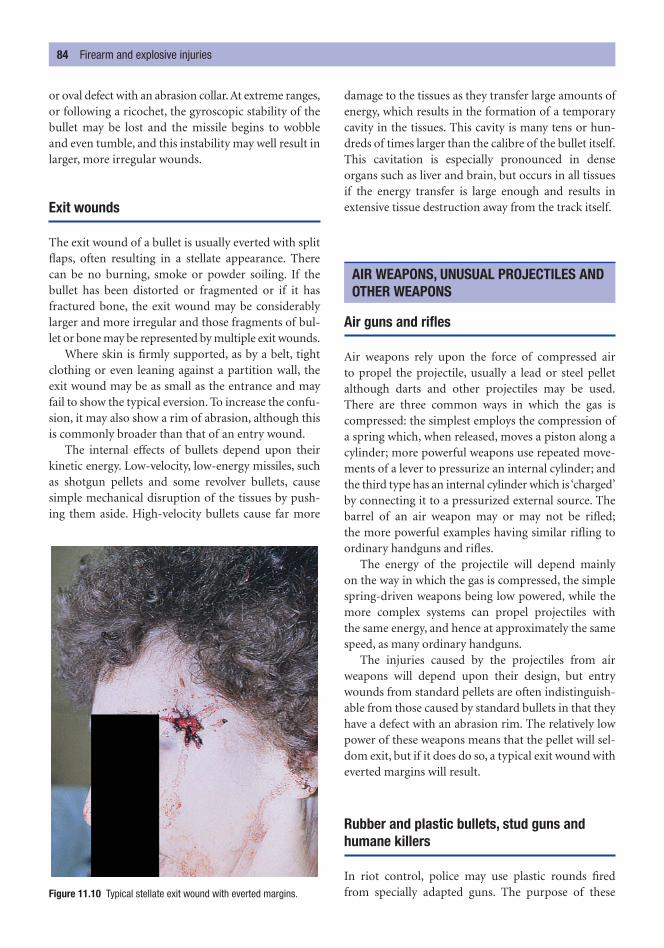
or oval defect with an abrasion collar. At extreme ranges,or following a ricochet, the gyroscopic stability of thebullet may be lost and the missile begins to wobbleand even tumble, and this instability may well result inlarger, more irregular wounds.
Exit wounds
The exit wound of a bullet is usually everted with splitflaps, often resulting in a stellate appearance. Therecan be no burning, smoke or powder soiling. If thebullet has been distorted or fragmented or if it hasfractured bone, the exit wound may be considerablylarger and more irregular and those fragments of bul-let or bone may be represented by multiple exit wounds.
Where skin is firmly supported, as by a belt, tightclothing or even leaning against a partition wall, theexit wound may be as small as the entrance and mayfail to show the typical eversion. To increase the confu-sion, it may also show a rim of abrasion, although thisis commonly broader than that of an entry wound.
The internal effects of bullets depend upon theirkinetic energy. Low-velocity, low-energy missiles, suchas shotgun pellets and some revolver bullets, causesimple mechanical disruption of the tissues by push-ing them aside. High-velocity bullets cause far more
damage to the tissues as they transfer large amounts ofenergy, which results in the formation of a temporarycavity in the tissues. This cavity is many tens or hun-dreds of times larger than the calibre of the bullet itself.This cavitation is especially pronounced in denseorgans such as liver and brain, but occurs in all tissuesif the energy transfer is large enough and results inextensive tissue destruction away from the track itself.
Air guns and rifles
Air weapons rely upon the force of compressed air to propel the projectile, usually a lead or steel pelletalthough darts and other projectiles may be used.There are three common ways in which the gas is compressed: the simplest employs the compression ofa spring which, when released, moves a piston along acylinder; more powerful weapons use repeated move-ments of a lever to pressurize an internal cylinder; andthe third type has an internal cylinder which is ‘charged’by connecting it to a pressurized external source. Thebarrel of an air weapon may or may not be rifled;the more powerful examples having similar rifling toordinary handguns and rifles.
The energy of the projectile will depend mainly on the way in which the gas is compressed, the simplespring-driven weapons being low powered, while themore complex systems can propel projectiles with the same energy, and hence at approximately the samespeed, as many ordinary handguns.
The injuries caused by the projectiles from airweapons will depend upon their design, but entrywounds from standard pellets are often indistinguish-able from those caused by standard bullets in that theyhave a defect with an abrasion rim. The relatively lowpower of these weapons means that the pellet will sel-dom exit, but if it does do so, a typical exit wound witheverted margins will result.
Rubber and plastic bullets, stud guns andhumane killers
In riot control, police may use plastic rounds firedfrom specially adapted guns. The purpose of these
AIR WEAPONS, UNUSUAL PROJECTILES ANDOTHER WEAPONS
84 Firearm and explosive injuries
Figure 11.10 Typical stellate exit wound with everted margins.

weapons is to disable and discourage rioters but not tokill or seriously injure them. The plastic rounds areabout 10 cm long, 3.5 cm in diameter and weigh about130 g. They should not be fired at ranges less thanabout 20 m and should only be fired at the lower part ofthe body. Some deaths and many serious injuries,including fractures of skull, ribs and limbs and also eyedamage and internal injuries, have occurred and theseare usually associated with improper use. The mark lefton the skin by a plastic round is usually distinctive.
Injuries and deaths from stud guns are well recorded.Stud guns are devices used in the building industry to fire steel pins into masonry or timber by means of a small explosive charge. They have been used forsuicide and even homicide, but accidental injuries aremore common. The skin injury often appears similarto many small-calibre entry wounds, although the find-ing of a nail will usually solve the diagnostic problem.
Humane killers are weapons used in abattoirs andby veterinary surgeons to kill animals. They may fireeither a small-calibre bullet or a ‘captive bolt’, where asliding steel pin is fired out for about 5 cm by an explo-sive charge. The skin injury will depend on the type ofweapon used. These weapons have been used for bothhomicide and suicide, but accidental discharges arealso recorded and may cause serious injury or death.
Bows and crossbows
These weapons fire arrows or bolts, which are shafts ofwood or metal with a set of flights at the rear to main-tain the trajectory of the projectile. The tips of theseprojectiles may have many shapes from the simple pointto complex, often triangular, forms. The energy pro-duced is extremely variable, depending on the con-struction of the weapon. The injuries caused dependon the energy of the projectile as well as on its con-struction. However, if the projectile has a simple pointedtip and if it has been removed from the body, the entrywounds can appear very similar to those caused bystandard bullets, with a central defect and surround-ing abrasion rim.
This is a major issue for the pathologist. Suicides musthave wounds whose site and range are within the reachof the deceased’s arm, unless some device has been
used to reach the trigger. The weapon must be presentat the scene, though it may be at a distance from thebody because it may have been thrown away from thebody by recoil or by movement of the individual ifdeath was not immediate. Suicidal gunshot injuriesare most commonly in the ‘sites of election’, whichvary with the length of the weapon used. Both long-barrelled and short-barrelled weapons can be used inthe mouth, below the chin, on the front of the neck,the centre of the forehead or, more rarely, the front of the chest over the heart. Discharges into the templesare almost unique to handguns and are usually on the side of the dominant hand, but this is not anabsolute rule. People almost never shoot themselves in the eye or abdomen or in inaccessible sites such asthe back. In Britain and many other countries, womenrarely commit suicide with guns and are rarely involvedin firearms accidents.
If suicide can be ruled out by the range of dis-charge, by absence of a weapon or by other features of the injury or the scene, a single gunshot injurycould be either accident or homicide. Multiple firearmwounds, on the other hand, strongly suggest homi-cide. However, there have been a number of publishedreports of suicidal individuals who have fired repeat-edly into themselves even when each wound is poten-tially fatal. The distinction between homicide, suicideand accident can sometimes be extremely difficult anda final conclusion can only be reached after a full policeinvestigation.
The immediacy of death following receipt ofinjuries is discussed later (see Chapter 16) and it is asunwise to state that a gunshot wound, as with anyother sort of injury, must have been immediately fatal.It is most likely that severe damage to the brain, heart,aorta and any number of other vital internal organswill lead to rapid collapse and death; however, thisauthor has seen a case of long-term survival followinga contact discharge of a shotgun into the head.
In the living, all efforts must be directed to saving lifebut, if at all possible, the treating doctor should makegood notes of the original appearances of the injuriesbefore any surgical cleaning or operative proceduresare performed. Any missiles, foreign bodies such as wads, bullets or shot and any skin removed from the
THE DOCTOR’S DUTY IN FIREARM INJURIESAND DEATHS
ACCIDENT, SUICIDE OR MURDER?
The doctor’s duty in firearm injuries and deaths 85

margin of a firearm wound during treatment shouldbe carefully preserved for the police.
The same general rules apply to the post-mortemrecovery of exhibits. The skin around the wounds maybe swabbed for powder residues if this is considered tobe necessary, but the retention of wounds themselves isno longer considered to be essential. Swabs of the handsof the victim should be taken. The pathologist mustensure that accurate drawings and measurements of thesite, size and appearance of the wound are obtained andthat distant and close-up photographs are taken of eachinjury with an appropriate scale in view.
In many countries, all firearm wounds, whether ornot they are fatal, must be reported to the police. Thisapplies in Northern Ireland (but not in mainlandEngland), where the anti-terrorism legislation makesit compulsory for doctors to inform the police of anyshooting or explosives injuries.
Terrorism and warfare lead to many deaths from explo-sive devices. In military bomb, shell and missile explo-sions, the release of energy may be so high that deathand disruption from blast effects may occur over a widearea. In contrast, terrorist devices, unless they containvery large amounts of explosive, rarely compare withmilitary effectiveness and thus the pure blast effects arefar more limited. According to the inverse square law,the energy developed by an explosion decreases rapidlyas the distance from the epicentre increases.
When an explosion occurs, the chemical interac-tion results in the generation of huge volumes of gas,which are further expanded by the great heat that isalso generated. This sudden generation of gas causes acompression wave to sweep outwards; at the origin,this is at many times the speed of sound.
The pure blast effects can cause either physical frag-mentation or disruption of the victim solely from theeffects of the wave of high pressure and hot gases strik-ing the body. A minimum pressure of about 700 kilo-pascals (100 lb/sq inch) is needed for tissue damage inhumans. There will also be pressure effects upon theviscera and these effects are far more damaging wherethere is an air/fluid interface, such as in the air passages,the lungs and the gut. Ruptures and haemorrhage ofthese areas represent the classical blast lesion.
Dangerous though the effect of blast is, many morecasualties, fatal and otherwise, are caused by second-ary effects of explosive devices, especially in the lower-powered terrorist bombs. These secondary effectsinclude:
• burns – directly from the near effects of the explo-sion and secondarily from fires started by the bomb;
• missile injuries from parts of the bomb casing orshrapnel or from adjacent objects;
• peppering by small fragments of debris and dustpropelled by the explosion;
• all types of injury due to collapse of structurescaused by the explosion;
• injuries and death from vehicular damage ordestruction, such as decompression, fire and groundimpact of bombed aircraft and crash damage to cars,trucks, buses etc.
Examination of either the living or the dead fol-lowing an explosion is essential, with careful docu-mentation of the sites and sizes of the abrasions,bruises, lacerations, burns and any other injuries.
If a near or massive explosion disrupts one or morebodies, identification of the fragments is essential,not least to determine how many victims were killed.This task can be extremely difficult and resembles theresponse to a mass disaster such as an air crash, in whichmany bodies may be severely disrupted and burnt. Thedifference is that, following a terrorist bombing, thepathologist has an additional task, which is to assist inthe identification and recovery of material that mighthave formed part of the bomb.
EXPLOSIVES
86 Firearm and explosive injuries
Figure 11.11 Multiple abrasions and lacerations due to flying debrisprojected by a terrorist bomb (courtesy of Professor T.K. Marshall,Belfast).

Every form of transport is associated with a risk and,as the speed of transport increases, particularly asmechanization is involved, the risks of an accident areincreased and the effects of an accident are magnified.
Those injured by accidents on the road can be dividedinto three broad groups: pedestrians, cyclists (pedal ormotor) and the drivers and passengers of vehicles. Ofthese three broad groups, it is the pedestrians that aremost often injured, although the proportion of pedes-trian victims in the overall statistics varies greatly accord-ing to the traffic patterns of different countries. It isperhaps self-evident that where there is greater minglingof motor transport and pedestrians there is also a greaterrisk of injury to the pedestrian. In 1998, 85 per cent of allroad deaths in the world occurred in developing coun-tries, where death rates of nearly 30 per 100 000 perannum are common; high-income countries generallyhave death rates of less than 5 per 100 000 per annum.
Pedestrians
Pedestrians struck by motor vehicles suffer injuriesfrom direct contact with the vehicle (primary injuries)
or from contact with other objects or the ground afterthe contact with the vehicle (secondary injuries).
The primary injuries can often form recognizablepatterns. When an adult is hit by the front of a car, thefront bumper (fender) usually strikes the victim atabout knee level. The exact point of contact, whetheron the front, side or back of the leg(s), will depend onthe orientation of the victim. There are often add-itional primary injury sites on the thigh, hip or pelvisdue to contact with the edge of the bonnet (hood). Atlow speeds (e.g. 20 kph/12 mph), the victim will com-monly be thrown off the bonnet either forwards or to one side. Between 20 and 60 kph (12–36 mph), thevictim will be tipped onto the bonnet (hood) and the head may strike the windscreen or the metal framethat surrounds it. At higher speeds (60–100 kph/36–60 mph), the victim may be projected (scooped)up into the air; sometimes they will pass completelyover the vehicle and will avoid hitting the windscreenand other points on the vehicle.
The secondary injuries are often more serious andpotentially lethal than the primary injuries. The constel-lation of these secondary injuries is wide; they varyfrom the ‘brush abrasions’ caused by skidding across thesurface of the road to fractures of the skull or axial skel-eton caused by direct contact with a surface, to fracturesof the spine, which can be caused by hyperflexion orextension. It is important to remember that the external
ROAD TRAFFIC INJURIES
C h a p t e r t w e l v e
Transportation Injuries
Road traffic injuriesPedestriansCar occupants
The function of seat belts
Air bagsMotor cycle and pedal cycle injuriesThe medical examination of victims of
road traffic accidents
Railway injuriesAircraft fatalitiesMass disasters and the doctor

injuries are seldom of themselves lethal; it is their asso-ciation with internal injuries that is important.
Even without fractures of the skull, brain damage isfrequent and is caused when the moving head of thevictim is suddenly stopped on impact; more diffusedamage to axons may be caused by the rotational orshearing forces acting upon the brain inside the rap-idly moving head. Fractures of the spine, especially inthe cervical and thoracic segments, may lead to corddamage. Fractures of the limbs are common but, apartfrom those of the legs that are associated with the pri-mary impact sites, they are somewhat unpredictablebecause of the random flailing of the limbs.
Where the vehicle is relatively larger than the victim – adults impacted by larger vehicles and chil-dren impacted by cars – the point of contact is higherup the victim and it is likely that the victim will makecontact with more of the front of the vehicle. This pat-tern of contact will be reflected in the resulting pri-mary injuries to the pelvis, abdomen, chest and head.It is more likely that the victim will be projected alongthe line of travel of the vehicle, which may increase therisk of ‘running-over’ injuries.
‘Running-over’ injuries are relatively unusual andthe effects are variable, depending on the area of thebody involved, the weight of the vehicle and the sur-face area of the contact. The skull may be disruptedand the brain externalized; internal organs may beruptured and there may be fractures of the spine.Compression of the chest may result in multiple ribfractures, causing a ‘flail chest’. The rotation of thewheel may strip off large areas of skin and subcuta-neous tissue; this is called a ‘flaying injury’.
Car occupants
Most impacts involve the front or the front corners ofthe vehicle and approximately 80 per cent of impactsare against either another vehicle or a stationaryobject. This type of impact rapidly decelerates thevehicle. Less commonly, the vehicle is hit from behind,causing an ‘acceleration’ impact. The least commonimpacts are side impacts and ‘roll-overs’.
In the common deceleration situation withoutrestraint, the front-seat occupants move forwards asthe vehicle decelerates around them and they strikethe vehicle structures that are in front of them. In gen-eral terms, the following may be expected in an unre-strained impact.
• The face and head hit the windscreen glass, frameor side-pillars, causing fractures to the skull andcervical spine and also brain injury.
• The chest and abdomen contact the fascia or thesteering wheel, causing rib, sternal, heart and liverdamage.
• The momentum of the heart within the thorax,perhaps aided by hyperflexion, may tear the aortaat the termination of the descending part of thearch, at the point where the vessel becomesattached to the vertebral column.
• The legs of the passenger are thrown forwards and the knees may strike the parcel shelf, causingfractures.
• The legs of the driver, which are commonly bracedon the brake and clutch pedals, may transmit the force of impact along the tibia and femur to the pelvis. All of these bones may be fractured ordislocated.
• On the rebound from these impacts, the heavyhead may swing violently backwards and causeinjury to the cervical or thoracic spine.
• The occupants of the car may be ejected out of thevehicle through the windscreen, increasing therisks of secondary injuries or running over.
The unrestrained rear-seat passenger is also liable toinjury through either deceleration or acceleration. Theinjuries, in general, may not be as severe as those causedto the front-seat occupants. In a deceleration impact,the rear-seat passengers will be thrown against thebacks of the front seats and may impact the front-seatoccupants. They may be projected over the front seat
88 Transportation injuries
Figure 12.1 Compound fracture of the right leg and laceration of the left knee in a pedestrian struck by a car. It is most likely that the vehicle struck the right leg.
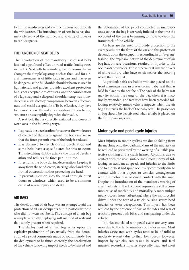
to hit the windscreen and even be thrown out throughthe windscreen. The introduction of seat belts has dra-matically reduced the number and severity of injuriesto car occupants.
THE FUNCTION OF SEAT BELTS
The introduction of the mandatory use of seat beltshas had a profound effect on road traffic fatality ratesin the UK. Seat belts have undergone numerous designchanges: the simple lap strap, such as that used for air-craft passengers, is of little value in cars and may evenbe dangerous; the full double shoulder harness used inlight aircraft and gliders provides excellent protectionbut is not acceptable to car users; and the combinationof a lap strap and a diagonal shoulder strap was intro-duced as a satisfactory compromise between effective-ness and social acceptability. To be effective, they haveto be worn correctly and any alteration in their fixing,structure or use rapidly degrades their value.
A seat belt that is correctly installed and correctlyworn acts in the following ways.
• It spreads the deceleration forces over the whole areaof contact of the straps against the body surface sothat the force per unit area of the body is reduced.
• It is designed to stretch during deceleration andsome belts have a specific area for this to occur.This stretching slightly extends the time of deceler-ation and reduces the force per unit time.
• It restrains the body during deceleration, keeping itaway from the windscreen, steering wheel and otherfrontal obstructions, thus protecting the head.
• It prevents ejection into the road through burstdoors or windows, which used to be a commoncause of severe injury and death.
AIR BAGS
The development of air bags was an attempt to aid theprotection of all car occupants but in particular thosewho did not wear seat belts. The concept of an air bagis simple: a rapidly deploying soft method of restraintthat is only present when required.
The deployment of an air bag relies upon theexplosive production of gas, usually from the deton-ation of a pellet commonly made of sodium azide. Forthe deployment to be timed correctly, the decelerationof the vehicle following impact needs to be sensed and
the detonation of the pellet completed in microsec-onds so that the bag is correctly inflated at the time theoccupant of the car is beginning to move towards theframework of the vehicle.
Air bags are designed to provide protection to theaverage adult in the front of the car and this protectiondepends upon the occupant responding in an ‘average’fashion; the explosive nature of the deployment of airbag has, on rare occasions, resulted in injuries to theoccupants of vehicles. Those especially at risk are driversof short stature who have to sit nearer the steeringwheel than normal.
At particular risk are babies who are placed on thefront passenger seat in a rear-facing baby seat that isheld in place by the seat belt. The back of the baby seatmay lie within the range of the bag when it is max-imally expanded, and fatalities have been recorded fol-lowing relatively minor vehicle impacts when the airbag has struck the back of the baby seat. The passengerairbag should be deactivated when a baby is placed onthe front passenger seat.
Motor cycle and pedal cycle injuries
Most injuries to motor cyclists are due to falling fromthe machine onto the roadway. Many of the injuries canbe reduced or prevented by the wearing of suitable pro-tective clothing and a crash helmet. Abrasions due tocontact with the road surface are almost universal fol-lowing an accident at speed, and injuries to the limbsand to the chest and spine occur very commonly due tocontact with other objects or vehicles, entanglementwith the motor bike or direct contact with the road.Despite the introduction of the mandatory wearing ofcrash helmets in the UK, head injuries are still a com-mon cause of morbidity and mortality. A more uniqueinjury occurs from ‘tail-gating’, where the motorcyclistdrives under the rear of a truck, causing severe headinjuries or even decapitation. This injury has beenreduced by the presence of bars at the sides and rear oftrucks to prevent both bikes and cars passing under thevehicle.
Injuries associated with pedal cycles are very com-mon due to the large numbers of cycles in use. Mostinjuries associated with cycles tend to be of mild ormoderate severity due to their low speeds. However,impact by vehicles can result in severe and fatalinjuries. Secondary injuries, especially head and chest
Road traffic injuries 89

damage, are common when cyclists fall from their rela-tively high riding position on such an inherentlyunstable machine.
A doctor may be called, either to treat living victims orto examine fatal casualties of road traffic accidents. Ifthe victim is still alive, the first duty of any doctor is toassist that individual to the best of his ability. Anyoneinvolved in the aftermath of an accident may be calledto an enquiry or court hearing, and civil actions fornegligence may arise and insurance claims may bepursued. Any doctor should make and keep a note oftreatment given, even if they have done so as a ‘GoodSamaritan’ act by the side of the road.
The victim should be carefully examined externallyand all injuries recorded (see Chapter 9). Injuriesshould be measured with a ruler and patterns shouldbe noted with great care. Tyre marks, often in the formof abrasions or intradermal bruising, may be presenton the skin of both living and dead victims and so aphotograph or accurate, measured drawing may be ofgreat value in helping to identify a ‘hit-and-run’ car ortruck. For pedestrian victims, the distance above theheel of the upper and lower margins of the majorinjuries should be measured because these heights canbe correlated with the height of the bumper (fender),the bonnet and other features on a suspect vehicle.
In countries where there is an established forensicscience service, examination of the clothing may rea-sonably be left to the scientists, but if no such service
exists or if it is not available to the doctor, the clothingshould be examined for tears, soiling by oil, grease orroad dirt etc. and any broken glass, paint flakes, rust orforeign material should be carefully documented andpreserved for the police.
The possibility that alcohol or drugs have con-tributed to the accident should always be consideredand samples retained for analysis at post mortem. Theliving victim must give consent to the taking of sam-ples for such a test or for the testing of samples takenfor other purposes (see Chapter 23).
THE MEDICAL EXAMINATION OF VICTIMS OFROAD TRAFFIC ACCIDENTS
90 Transportation injuries
Figure 12.2 Extensive ‘brush’ abrasion of the left flank in a motorcyclist thrown across a rough surface.
Figure 12.3 A ‘flaying’ injury due to a rotating wheel crossing theleft side of the face.
Figure 12.4 Pedestrian leg injury from a rotating wheel resulting in‘flaying’ of the skin.
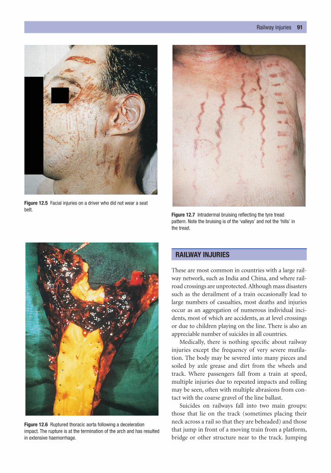
These are most common in countries with a large rail-way network, such as India and China, and where rail-road crossings are unprotected. Although mass disasterssuch as the derailment of a train occasionally lead tolarge numbers of casualties, most deaths and injuriesoccur as an aggregation of numerous individual inci-dents, most of which are accidents, as at level crossingsor due to children playing on the line. There is also anappreciable number of suicides in all countries.
Medically, there is nothing specific about railwayinjuries except the frequency of very severe mutila-tion. The body may be severed into many pieces andsoiled by axle grease and dirt from the wheels andtrack. Where passengers fall from a train at speed,multiple injuries due to repeated impacts and rollingmay be seen, often with multiple abrasions from con-tact with the coarse gravel of the line ballast.
Suicides on railways fall into two main groups:those that lie on the track (sometimes placing theirneck across a rail so that they are beheaded) and thosethat jump in front of a moving train from a platform,bridge or other structure near to the track. Jumping
RAILWAY INJURIES
Railway injuries 91
Figure 12.5 Facial injuries on a driver who did not wear a seat belt.
Figure 12.6 Ruptured thoracic aorta following a decelerationimpact. The rupture is at the termination of the arch and has resultedin extensive haemorrhage.
Figure 12.7 Intradermal bruising reflecting the tyre tread pattern. Note the bruising is of the ‘valleys’ and not the ‘hills’ in the tread.
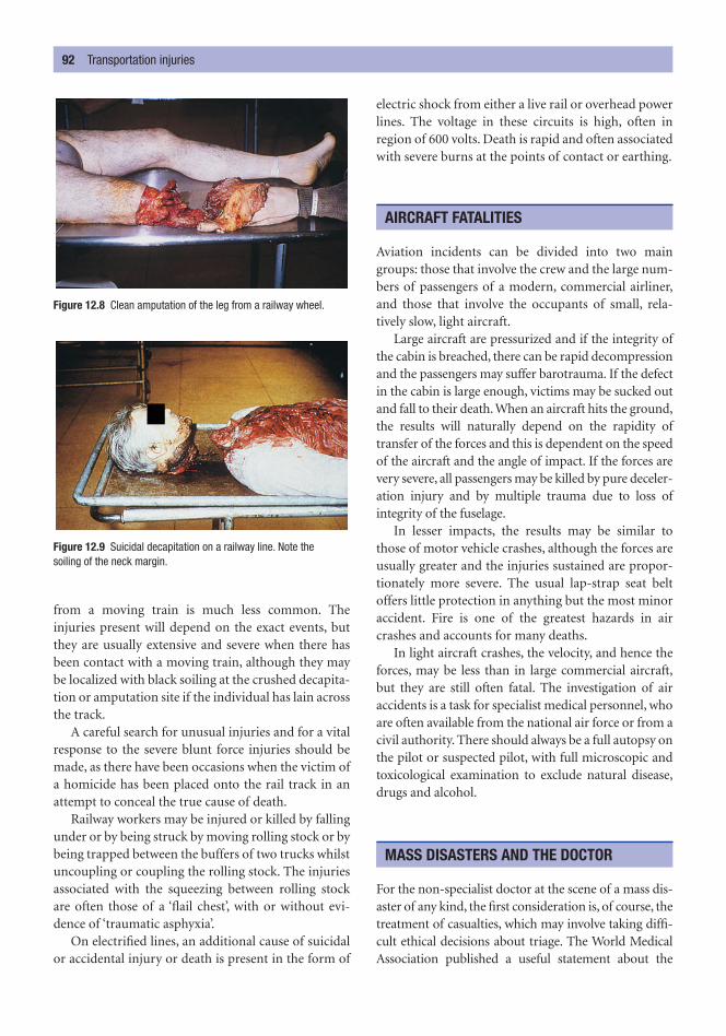
from a moving train is much less common. Theinjuries present will depend on the exact events, butthey are usually extensive and severe when there hasbeen contact with a moving train, although they maybe localized with black soiling at the crushed decapita-tion or amputation site if the individual has lain acrossthe track.
A careful search for unusual injuries and for a vitalresponse to the severe blunt force injuries should bemade, as there have been occasions when the victim ofa homicide has been placed onto the rail track in anattempt to conceal the true cause of death.
Railway workers may be injured or killed by fallingunder or by being struck by moving rolling stock or bybeing trapped between the buffers of two trucks whilstuncoupling or coupling the rolling stock. The injuriesassociated with the squeezing between rolling stockare often those of a ‘flail chest’, with or without evi-dence of ‘traumatic asphyxia’.
On electrified lines, an additional cause of suicidalor accidental injury or death is present in the form of
electric shock from either a live rail or overhead powerlines. The voltage in these circuits is high, often inregion of 600 volts. Death is rapid and often associatedwith severe burns at the points of contact or earthing.
Aviation incidents can be divided into two maingroups: those that involve the crew and the large num-bers of passengers of a modern, commercial airliner,and those that involve the occupants of small, rela-tively slow, light aircraft.
Large aircraft are pressurized and if the integrity ofthe cabin is breached, there can be rapid decompressionand the passengers may suffer barotrauma. If the defectin the cabin is large enough, victims may be sucked outand fall to their death. When an aircraft hits the ground,the results will naturally depend on the rapidity oftransfer of the forces and this is dependent on the speedof the aircraft and the angle of impact. If the forces arevery severe, all passengers may be killed by pure deceler-ation injury and by multiple trauma due to loss ofintegrity of the fuselage.
In lesser impacts, the results may be similar tothose of motor vehicle crashes, although the forces areusually greater and the injuries sustained are propor-tionately more severe. The usual lap-strap seat beltoffers little protection in anything but the most minoraccident. Fire is one of the greatest hazards in aircrashes and accounts for many deaths.
In light aircraft crashes, the velocity, and hence theforces, may be less than in large commercial aircraft,but they are still often fatal. The investigation of airaccidents is a task for specialist medical personnel, whoare often available from the national air force or from acivil authority. There should always be a full autopsy onthe pilot or suspected pilot, with full microscopic andtoxicological examination to exclude natural disease,drugs and alcohol.
For the non-specialist doctor at the scene of a mass dis-aster of any kind, the first consideration is, of course, thetreatment of casualties, which may involve taking diffi-cult ethical decisions about triage. The World MedicalAssociation published a useful statement about the
MASS DISASTERS AND THE DOCTOR
AIRCRAFT FATALITIES
92 Transportation injuries
Figure 12.8 Clean amputation of the leg from a railway wheel.
Figure 12.9 Suicidal decapitation on a railway line. Note the soiling of the neck margin.

medical ethics in disasters at its assembly in 1994.Among other things, the statement recommends thattriage should be entrusted to an authorized and experi-enced physician, who should be accompanied by com-petent staff. It also indicates that triage is not a ‘one-off ’review and that regular reassessments must be made ascases may evolve and change category.
Victims can be divided into five main groups:
1 those who can be saved and whose lives are inimmediate danger;
2 those whose lives are not in immediate danger butwho are in need or urgent but not immediate medical care;
3 those who have minor injuries only and who can betreated later or by unskilled personnel;
4 those who have been psychologically traumatizedand who need reassurance and/or sedation ifacutely disturbed;
5 those whose condition exceeds the available thera-peutic resources and who have suffered from suchextremely severe injuries of whatever type that theycannot be saved in the specific circumstances ofthat time and place: these victims can be termed‘beyond emergency care’.
The World Medical Association statement makes itclear that to abandon an injured person on account of
the priorities dictated by the disaster situation cannotbe considered to be a failure, but that it is justifiedbecause these decisions are intended to save the max-imum number of victims – the greatest good to thegreatest number.
The investigation of the causes of death and thecauses of the incident (such as a bomb) and the identi-fication of the dead are specialist operations involvingmortuary accommodation, pathology, dentistry, thepolice and the usual state agencies responsible for sud-den death; in England and Wales this is the coroner. Ateam of pathologists, assisted by police officers andmortuary staff and backed up by dental and radiologi-cal facilities, inspects every body and records all cloth-ing, jewellery and personal belongings still attached tothe bodies. The body or body part is then carefullyexamined for every aspect of identity, including sex,race, height, age and personal characteristics. All thesedetails are recorded on standard forms and charts andthe information is sent back to the identification teams,which can compare this information from the bodieswith the information obtained from relatives, friendsetc. An autopsy is performed to determine the cause ofdeath, to retrieve any foreign objects that, for example,may be related to an explosive device, and to seek any further identifying factors, such as operation sites,prostheses etc.
Mass disasters and the doctor 93

The literal translation of the word asphyxia from theGreek means ‘absence of pulse’ and how this word hascome to denote the effects of the lack of oxygen is notclear, but the word asphyxia is now commonly used todescribe a range of conditions for which the lack ofoxygen – whether it is partial (hypoxia) or complete(anoxia) – is considered to be the cause.
In the forensic context, asphyxia is usually obstruct-ive in nature, where some physical barrier preventsaccess of air to the lungs; this obstruction can occur atany point from the nose and mouth to the alveolarmembranes. Other conditions in which the body cannot gain sufficient oxygen may occur without anyobstruction to the air passages. Given the many pos-sible causes for this lack of sufficient oxygen to thecells of the body, it is not surprising that the clinicaland pathological features of the many different typesof ‘asphyxia’ are very varied.
The ‘classical’ features of asphyxia are found wherethe air passages are obstructed by pressure applied to theneck or to the chest and where there has been a struggleto breathe. The classical features of ‘asphyxia’ are:
1 congestion of the face;2 oedema of the face;3 cyanosis (blueness) of the skin of the face;4 petechial haemorrhages in the skin of the face and
the eyes.
A fifth feature – increased fluidity of the blood – isnow not accepted. The other four features, although notdiagnostic, are important and are considered below.
Congestion is the red appearance of the skin of theface and head. It is due to the filling of the venous sys-tem when compression of the neck or some otherobstruction prevents venous return to the heart.
Oedema is the swelling of the tissues due to transu-dation of fluid from the veins caused by the increasedvenous pressure as a result of obstruction of venousreturn to the heart.
Cyanosis is the blue colour imparted to the skin bythe presence of deoxygenated blood in the congestedvenous system and, possibly, in the arterial system.
Petechial haemorrhages (petechiae) are tiny, pin-point haemorrhages, most commonly seen in the skinof the head and face and especially in the lax tissues ofthe eyelids. They are also seen in the conjunctivae andsclera of the eye. They are due to leakage of blood fromsmall venules as a result of the raised pressure in thevenous system. The theory that they are caused, at leastin part, by hypoxia of the blood vessel walls is not sup-ported by research. They are not diagnostic of asphyxiabecause they can appear instantaneously in the faceand eyes following a violent episode of sneezing orcoughing.
Petechiae on the pleural surface of the lungs, theepicardium and the thymus in children, are the true
C h a p t e r t h i r t e e n
Asphyxia
SuffocationSmotheringGaggingChoking
Café coronaryPressure on the neck‘Vagal inhibition’ or reflex cardiac arrestManual strangulation
Ligature strangulationHangingThe sexual asphyxiasTraumatic asphyxia

Suffocation 95
Tardieu spots described in the nineteenth century byProfessor Ambroise Tardieu, who thought, incorrectly,that they were an infallible hallmark of asphyxia. A fewpetechial haemorrhages can be seen on the surface ofthe lungs, heart etc. in many other conditions, but ofparticular importance is the finding that approxi-mately 70 per cent of ‘cot deaths’ (SIDS) have themand they are often large and numerous in this situ-ation. It is important that these petechiae are not inter-preted as evidence of external respiratory obstruction.
A person with obstructed air entry will show vari-ous phases of distress and physical signs, the exact tim-ing and the sequence of events will be very variable,but they may progress as listed below.
1 Increased efforts to breathe, facial congestion andthe onset of cyanosis.
2 Deep, laboured respirations or attempts at respir-ation with a heaving chest; deepening congestionand cyanosis; the appearance of petechiae if venousreturn is impaired.
3 Loss of consciousness and possible convulsions;evacuation of bladder and vomiting.
4 Reduction in the depth and frequency of respir-ation; irreversible brain damage begins; the pupilsdilate and death ensues.
At any stage through this progression, death mayoccur suddenly from cardiac arrest.
The classification and nomenclature of the various‘asphyxial’ conditions are imprecise, adding to theconfusion over this group of deaths (Table 13.1).
Situations in which accumulations of irrespirablegases occur are commonly industrial or agricultural;they may be found in the deep tanks of ships whererust formation has removed oxygen, in farm silos con-taining grain, or in deep wells in chalk where carbondioxide has accumulated. The individual enteringthese situations without protective equipment will berendered unconscious extremely quickly and will dierapidly unless removed from the hypoxic environ-ment. In all these situations, asphyxial signs may beminimal as collapse and death may be so rapid.
Rapid deaths are also reported when a plastic bag isplaced over the head, either as a deliberate suicidal actor accidentally by children. Typically, no ‘classic signs’are seen and the face is usually pale when the bag isremoved. The bag need not be tied around the neck tobe effective.
SUFFOCATION
Figure 13.1 Petechial haemorrhages on the eyelid in a case ofmanual strangulation.
Figure 13.2 Conjunctival haemorrhages in manual strangulation.
Underlying cause of death Name
Lack of oxygen in the inspired air Suffocation
Blockage of the external orifices Suffocation/smothering
Blockage of the internal Gagging/chokingairways by obstruction
Blockage of the internal airways Strangulation/hangingby external pressure
Restriction of chest movement Traumatic asphyxia
Failure of oxygen transportation For example CO poisoning
Failure of oxygen utilization For example cyanide poisoning
Table 13.1 ‘Asphyxial’ Conditions

Smothering with a pillow or other object (including ahand) pressed over the nose and mouth will only veryrarely cause any petechiae, any significant cyanosis orcongestion unless the victim struggles and fights forbreath against the obstructed airways. Smothering maybe virtually impossible to diagnose if it is applied tothose who cannot or do not resist – the old, the infirmor the very young. If the victim does struggle, theremay be bruises and abrasions to the face, on the lips orinside the mouth (where lips are pressed against teeth).
The air passages may be obstructed when a cloth orsoft object is pushed into the mouth, or placed acrossthe mouth, often during a robbery when the victim istied up and the cloth is used to ensure their silence. Atfirst, breathing can take place via the nose, but as timegoes on, nasal mucus and oedema close the posteriornares and progressive asphyxia develops.
Manual strangulation is occasionally referred to as‘choking’, but this is incorrect. The term chokingshould be applied to the internal obstruction of theupper air passages by an object or substance impactedin the pharynx or larynx. Choking is, most commonly,
accidental and the causes include dentures in adultsand inhaled objects such as small toys, balls etc. inchildren. In medical practice there are risks associatedwith individuals who are sedated or anaesthetized,when objects such as extracted teeth or blood fromdental or ENT operations may occlude the airwaywithout provoking the normal reflex of coughing.Obstruction commonly leads to respiratory distresswith congestion and cyanosis of the head and face.
Café coronary
Perhaps the commonest cause of choking is the entryof food into the air passages. If food enters the larynxduring swallowing, it usually causes gross ‘chokingsymptoms’ of coughing, distress and cyanosis, whichcan be fatal unless the obstruction is cleared by coughing or some rapid treatment is offered. However,
CHOKING
GAGGING
SMOTHERING
96 Asphyxia
Figure 13.3 ‘Exit’ suicide with a plastic bag. The face is usually pale and devoid of asphyxial signs.
Figure 13.4 Impaction of food in the larynx – café coronary.
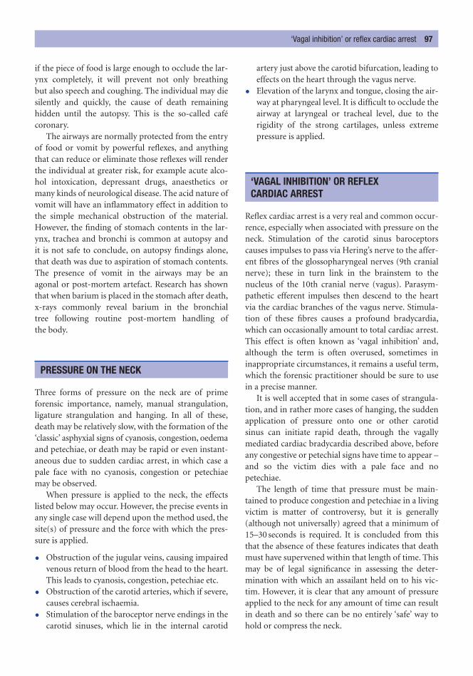
if the piece of food is large enough to occlude the lar-ynx completely, it will prevent not only breathing but also speech and coughing. The individual may diesilently and quickly, the cause of death remaining hidden until the autopsy. This is the so-called cafécoronary.
The airways are normally protected from the entryof food or vomit by powerful reflexes, and anythingthat can reduce or eliminate those reflexes will renderthe individual at greater risk, for example acute alco-hol intoxication, depressant drugs, anaesthetics ormany kinds of neurological disease. The acid nature ofvomit will have an inflammatory effect in addition tothe simple mechanical obstruction of the material.However, the finding of stomach contents in the lar-ynx, trachea and bronchi is common at autopsy and it is not safe to conclude, on autopsy findings alone,that death was due to aspiration of stomach contents.The presence of vomit in the airways may be an agonal or post-mortem artefact. Research has shownthat when barium is placed in the stomach after death,x-rays commonly reveal barium in the bronchial tree following routine post-mortem handling ofthe body.
Three forms of pressure on the neck are of primeforensic importance, namely, manual strangulation,ligature strangulation and hanging. In all of these,death may be relatively slow, with the formation of the‘classic’ asphyxial signs of cyanosis, congestion, oedemaand petechiae, or death may be rapid or even instant-aneous due to sudden cardiac arrest, in which case apale face with no cyanosis, congestion or petechiaemay be observed.
When pressure is applied to the neck, the effectslisted below may occur. However, the precise events inany single case will depend upon the method used, thesite(s) of pressure and the force with which the pres-sure is applied.
• Obstruction of the jugular veins, causing impairedvenous return of blood from the head to the heart.This leads to cyanosis, congestion, petechiae etc.
• Obstruction of the carotid arteries, which if severe,causes cerebral ischaemia.
• Stimulation of the baroceptor nerve endings in thecarotid sinuses, which lie in the internal carotid
artery just above the carotid bifurcation, leading toeffects on the heart through the vagus nerve.
• Elevation of the larynx and tongue, closing the air-way at pharyngeal level. It is difficult to occlude theairway at laryngeal or tracheal level, due to therigidity of the strong cartilages, unless extremepressure is applied.
Reflex cardiac arrest is a very real and common occur-rence, especially when associated with pressure on theneck. Stimulation of the carotid sinus baroceptorscauses impulses to pass via Hering’s nerve to the affer-ent fibres of the glossopharyngeal nerves (9th cranialnerve); these in turn link in the brainstem to thenucleus of the 10th cranial nerve (vagus). Parasym-pathetic efferent impulses then descend to the heartvia the cardiac branches of the vagus nerve. Stimula-tion of these fibres causes a profound bradycardia,which can occasionally amount to total cardiac arrest.This effect is often known as ‘vagal inhibition’ and,although the term is often overused, sometimes ininappropriate circumstances, it remains a useful term,which the forensic practitioner should be sure to usein a precise manner.
It is well accepted that in some cases of strangula-tion, and in rather more cases of hanging, the suddenapplication of pressure onto one or other carotid sinus can initiate rapid death, through the vagallymediated cardiac bradycardia described above, beforeany congestive or petechial signs have time to appear –and so the victim dies with a pale face and nopetechiae.
The length of time that pressure must be main-tained to produce congestion and petechiae in a livingvictim is matter of controversy, but it is generally(although not universally) agreed that a minimum of15–30 seconds is required. It is concluded from thisthat the absence of these features indicates that deathmust have supervened within that length of time. Thismay be of legal significance in assessing the deter-mination with which an assailant held on to his vic-tim. However, it is clear that any amount of pressureapplied to the neck for any amount of time can resultin death and so there can be no entirely ‘safe’ way tohold or compress the neck.
‘VAGAL INHIBITION’ OR REFLEX CARDIAC ARREST
PRESSURE ON THE NECK
‘Vagal inhibition’ or reflex cardiac arrest 97
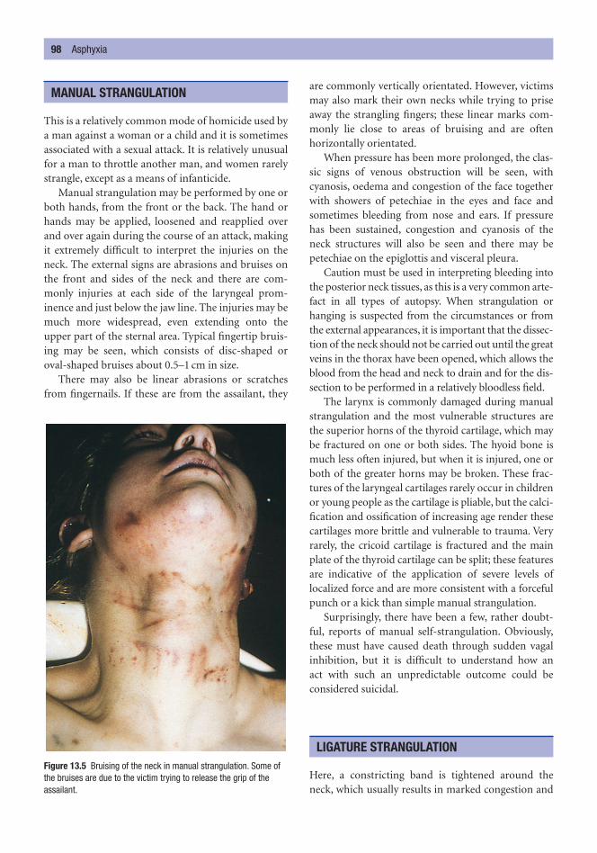
This is a relatively common mode of homicide used bya man against a woman or a child and it is sometimesassociated with a sexual attack. It is relatively unusualfor a man to throttle another man, and women rarelystrangle, except as a means of infanticide.
Manual strangulation may be performed by one orboth hands, from the front or the back. The hand orhands may be applied, loosened and reapplied overand over again during the course of an attack, makingit extremely difficult to interpret the injuries on theneck. The external signs are abrasions and bruises onthe front and sides of the neck and there are com-monly injuries at each side of the laryngeal prom-inence and just below the jaw line. The injuries may bemuch more widespread, even extending onto theupper part of the sternal area. Typical fingertip bruis-ing may be seen, which consists of disc-shaped oroval-shaped bruises about 0.5–1 cm in size.
There may also be linear abrasions or scratchesfrom fingernails. If these are from the assailant, they
are commonly vertically orientated. However, victimsmay also mark their own necks while trying to priseaway the strangling fingers; these linear marks com-monly lie close to areas of bruising and are often horizontally orientated.
When pressure has been more prolonged, the clas-sic signs of venous obstruction will be seen, withcyanosis, oedema and congestion of the face togetherwith showers of petechiae in the eyes and face andsometimes bleeding from nose and ears. If pressurehas been sustained, congestion and cyanosis of theneck structures will also be seen and there may bepetechiae on the epiglottis and visceral pleura.
Caution must be used in interpreting bleeding intothe posterior neck tissues, as this is a very common arte-fact in all types of autopsy. When strangulation orhanging is suspected from the circumstances or fromthe external appearances, it is important that the dissec-tion of the neck should not be carried out until the greatveins in the thorax have been opened, which allows theblood from the head and neck to drain and for the dis-section to be performed in a relatively bloodless field.
The larynx is commonly damaged during manualstrangulation and the most vulnerable structures arethe superior horns of the thyroid cartilage, which maybe fractured on one or both sides. The hyoid bone ismuch less often injured, but when it is injured, one orboth of the greater horns may be broken. These frac-tures of the laryngeal cartilages rarely occur in childrenor young people as the cartilage is pliable, but the calci-fication and ossification of increasing age render thesecartilages more brittle and vulnerable to trauma. Veryrarely, the cricoid cartilage is fractured and the mainplate of the thyroid cartilage can be split; these featuresare indicative of the application of severe levels oflocalized force and are more consistent with a forcefulpunch or a kick than simple manual strangulation.
Surprisingly, there have been a few, rather doubt-ful, reports of manual self-strangulation. Obviously,these must have caused death through sudden vagalinhibition, but it is difficult to understand how an act with such an unpredictable outcome could be considered suicidal.
Here, a constricting band is tightened around theneck, which usually results in marked congestion and
LIGATURE STRANGULATION
MANUAL STRANGULATION
98 Asphyxia
Figure 13.5 Bruising of the neck in manual strangulation. Some ofthe bruises are due to the victim trying to release the grip of theassailant.
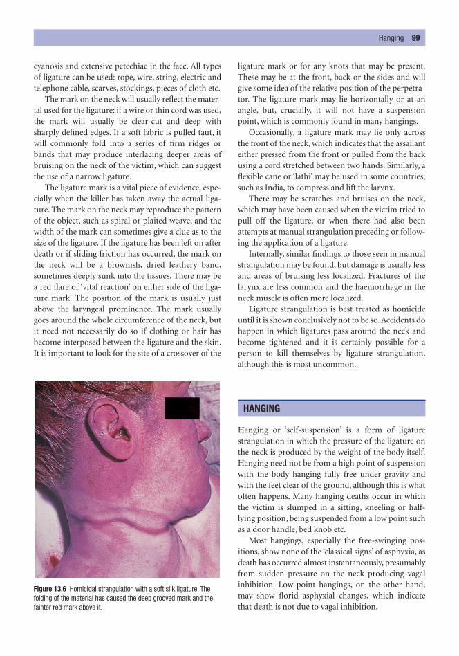
cyanosis and extensive petechiae in the face. All typesof ligature can be used: rope, wire, string, electric andtelephone cable, scarves, stockings, pieces of cloth etc.
The mark on the neck will usually reflect the mater-ial used for the ligature: if a wire or thin cord was used,the mark will usually be clear-cut and deep withsharply defined edges. If a soft fabric is pulled taut, itwill commonly fold into a series of firm ridges orbands that may produce interlacing deeper areas ofbruising on the neck of the victim, which can suggestthe use of a narrow ligature.
The ligature mark is a vital piece of evidence, espe-cially when the killer has taken away the actual liga-ture. The mark on the neck may reproduce the patternof the object, such as spiral or plaited weave, and thewidth of the mark can sometimes give a clue as to thesize of the ligature. If the ligature has been left on afterdeath or if sliding friction has occurred, the mark onthe neck will be a brownish, dried leathery band,sometimes deeply sunk into the tissues. There may bea red flare of ‘vital reaction’ on either side of the liga-ture mark. The position of the mark is usually justabove the laryngeal prominence. The mark usuallygoes around the whole circumference of the neck, butit need not necessarily do so if clothing or hair hasbecome interposed between the ligature and the skin.It is important to look for the site of a crossover of the
ligature mark or for any knots that may be present.These may be at the front, back or the sides and willgive some idea of the relative position of the perpetra-tor. The ligature mark may lie horizontally or at anangle, but, crucially, it will not have a suspensionpoint, which is commonly found in many hangings.
Occasionally, a ligature mark may lie only acrossthe front of the neck, which indicates that the assailanteither pressed from the front or pulled from the backusing a cord stretched between two hands. Similarly, aflexible cane or ‘lathi’ may be used in some countries,such as India, to compress and lift the larynx.
There may be scratches and bruises on the neck,which may have been caused when the victim tried topull off the ligature, or when there had also beenattempts at manual strangulation preceding or follow-ing the application of a ligature.
Internally, similar findings to those seen in manualstrangulation may be found, but damage is usually lessand areas of bruising less localized. Fractures of thelarynx are less common and the haemorrhage in theneck muscle is often more localized.
Ligature strangulation is best treated as homicideuntil it is shown conclusively not to be so. Accidents dohappen in which ligatures pass around the neck andbecome tightened and it is certainly possible for a person to kill themselves by ligature strangulation,although this is most uncommon.
Hanging or ‘self-suspension’ is a form of ligaturestrangulation in which the pressure of the ligature onthe neck is produced by the weight of the body itself.Hanging need not be from a high point of suspensionwith the body hanging fully free under gravity andwith the feet clear of the ground, although this is whatoften happens. Many hanging deaths occur in whichthe victim is slumped in a sitting, kneeling or half-lying position, being suspended from a low point suchas a door handle, bed knob etc.
Most hangings, especially the free-swinging pos-itions, show none of the ‘classical signs’ of asphyxia, asdeath has occurred almost instantaneously, presumablyfrom sudden pressure on the neck producing vagalinhibition. Low-point hangings, on the other hand,may show florid asphyxial changes, which indicatethat death is not due to vagal inhibition.
HANGING
Hanging 99
Figure 13.6 Homicidal strangulation with a soft silk ligature. Thefolding of the material has caused the deep grooved mark and thefainter red mark above it.

The cause of death in the hanging for judicial exe-cution is quite different, as the arrested drop of severalmetres completely disrupts the cervical spine,sometimes at the atlanto-occipital joint, but usually inthe mid-cervical portion. No asphyxial signs are seenin properly performed judicial hangings. It is veryunusual for the cervical spine to be broken in suicidalhanging unless there has been an unusually long drop.
The usual free-swinging suicidal hanging shows amark on the neck that is somewhat sloped and doesnot run around the full circumference of the neck. Thejunction of the noose and the vertical part of the ropeof the noose is pulled upwards and away from the skinand so no mark is left. The apex of the triangle formedin this way is called the suspension ‘peak’ or ‘point’ andindicates the position of the junction of the noose andvertical part of rope. This suspension peak or point isa distinguishing feature from ligature strangulation.
Excluding judicial execution, hanging is mostlycommonly a suicidal act of males. Some cases are accidental and entanglement with cords and ropes canoccur; amongst children, many tragedies have hap-pened due to leather or plastic restraint harnesses get-ting around the necks of unattended infants. The tying
100 Asphyxia
Figure 13.7 The spiral weave of the rope can be seen in thishorizontal ligature mark caused by hanging from a low suspensionpoint. The appearances may mimic homicidal ligature strangulation.
Figure 13.8 Suicidal hanging: the rope rises to a point, leaving agap in the ligature mark – the suspension point – on the neck.
Figure 13.9 Suicidal hanging with a dog lead, with the mark risingto a suspension point at the front of the neck.
Figure 13.10 Suicidal strangulation with a cable. The initialappearances were those of homicide, but a full examination of thecircumstances indicated suicide.
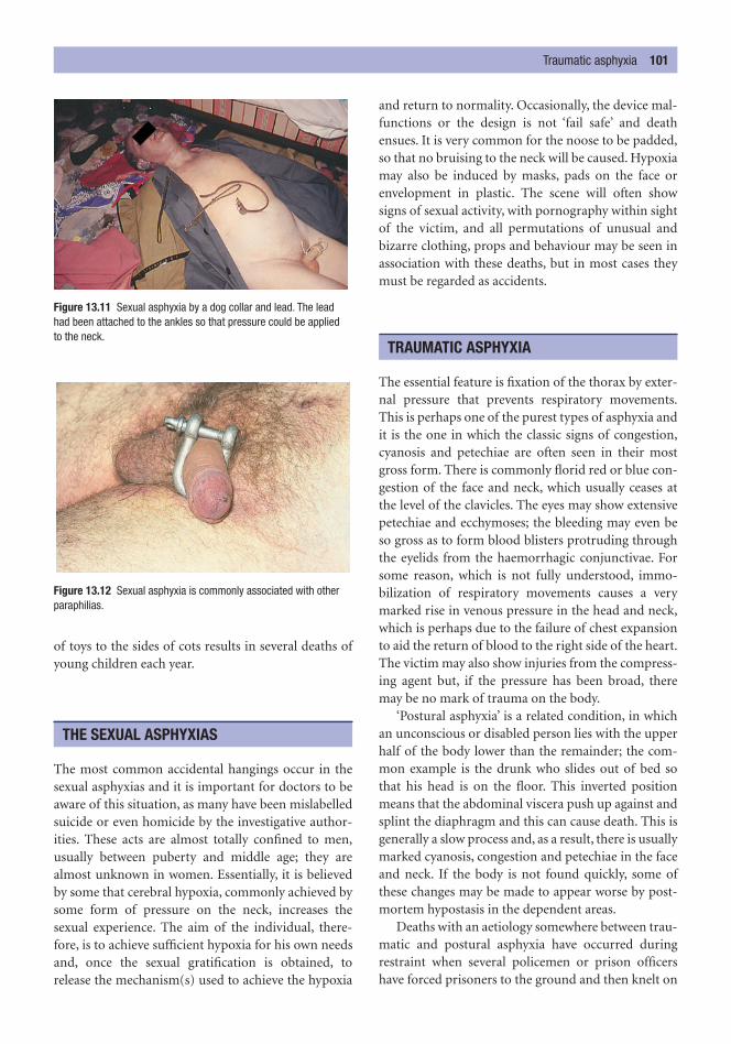
of toys to the sides of cots results in several deaths ofyoung children each year.
The most common accidental hangings occur in thesexual asphyxias and it is important for doctors to beaware of this situation, as many have been mislabelledsuicide or even homicide by the investigative author-ities. These acts are almost totally confined to men,usually between puberty and middle age; they arealmost unknown in women. Essentially, it is believedby some that cerebral hypoxia, commonly achieved bysome form of pressure on the neck, increases the sexual experience. The aim of the individual, there-fore, is to achieve sufficient hypoxia for his own needsand, once the sexual gratification is obtained, torelease the mechanism(s) used to achieve the hypoxia
and return to normality. Occasionally, the device mal-functions or the design is not ‘fail safe’ and deathensues. It is very common for the noose to be padded,so that no bruising to the neck will be caused. Hypoxiamay also be induced by masks, pads on the face orenvelopment in plastic. The scene will often showsigns of sexual activity, with pornography within sightof the victim, and all permutations of unusual andbizarre clothing, props and behaviour may be seen inassociation with these deaths, but in most cases theymust be regarded as accidents.
The essential feature is fixation of the thorax by exter-nal pressure that prevents respiratory movements.This is perhaps one of the purest types of asphyxia andit is the one in which the classic signs of congestion,cyanosis and petechiae are often seen in their mostgross form. There is commonly florid red or blue con-gestion of the face and neck, which usually ceases atthe level of the clavicles. The eyes may show extensivepetechiae and ecchymoses; the bleeding may even beso gross as to form blood blisters protruding throughthe eyelids from the haemorrhagic conjunctivae. Forsome reason, which is not fully understood, immo-bilization of respiratory movements causes a verymarked rise in venous pressure in the head and neck,which is perhaps due to the failure of chest expansionto aid the return of blood to the right side of the heart.The victim may also show injuries from the compress-ing agent but, if the pressure has been broad, theremay be no mark of trauma on the body.
‘Postural asphyxia’ is a related condition, in whichan unconscious or disabled person lies with the upperhalf of the body lower than the remainder; the com-mon example is the drunk who slides out of bed sothat his head is on the floor. This inverted positionmeans that the abdominal viscera push up against andsplint the diaphragm and this can cause death. This isgenerally a slow process and, as a result, there is usuallymarked cyanosis, congestion and petechiae in the faceand neck. If the body is not found quickly, some ofthese changes may be made to appear worse by post-mortem hypostasis in the dependent areas.
Deaths with an aetiology somewhere between trau-matic and postural asphyxia have occurred duringrestraint when several policemen or prison officershave forced prisoners to the ground and then knelt on
TRAUMATIC ASPHYXIA
THE SEXUAL ASPHYXIAS
Traumatic asphyxia 101
Figure 13.11 Sexual asphyxia by a dog collar and lead. The leadhad been attached to the ankles so that pressure could be applied to the neck.
Figure 13.12 Sexual asphyxia is commonly associated with otherparaphilias.
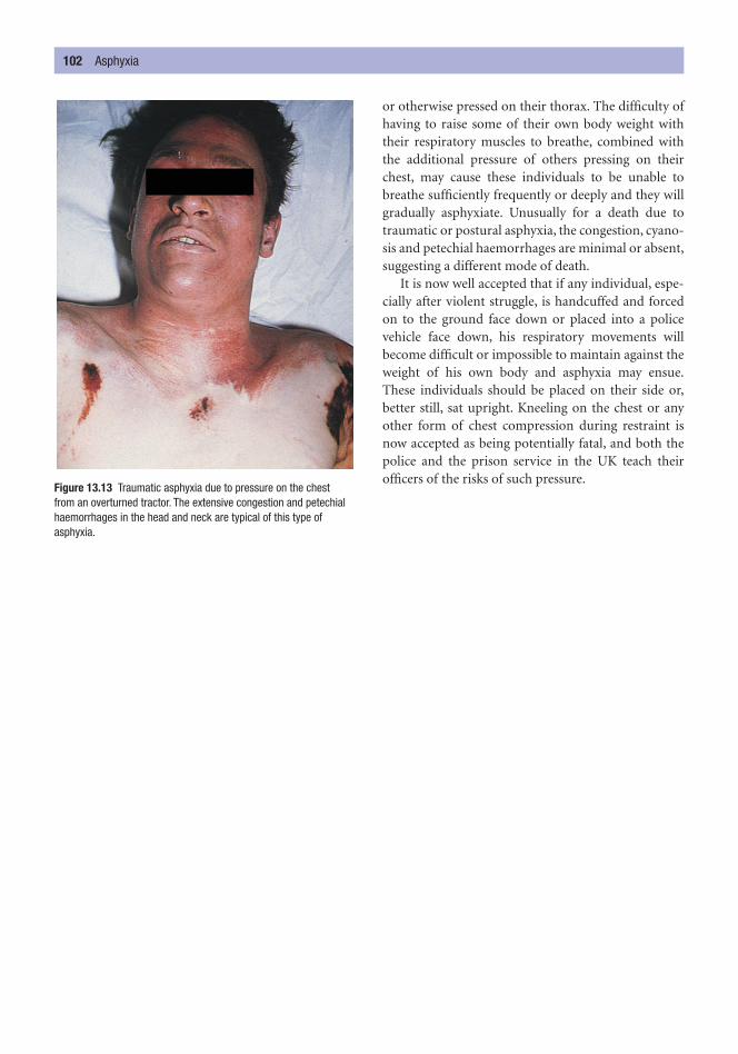
or otherwise pressed on their thorax. The difficulty ofhaving to raise some of their own body weight withtheir respiratory muscles to breathe, combined withthe additional pressure of others pressing on theirchest, may cause these individuals to be unable tobreathe sufficiently frequently or deeply and they willgradually asphyxiate. Unusually for a death due totraumatic or postural asphyxia, the congestion, cyano-sis and petechial haemorrhages are minimal or absent,suggesting a different mode of death.
It is now well accepted that if any individual, espe-cially after violent struggle, is handcuffed and forcedon to the ground face down or placed into a policevehicle face down, his respiratory movements willbecome difficult or impossible to maintain against theweight of his own body and asphyxia may ensue.These individuals should be placed on their side or,better still, sat upright. Kneeling on the chest or anyother form of chest compression during restraint isnow accepted as being potentially fatal, and both thepolice and the prison service in the UK teach theirofficers of the risks of such pressure.
102 Asphyxia
Figure 13.13 Traumatic asphyxia due to pressure on the chest from an overturned tractor. The extensive congestion and petechialhaemorrhages in the head and neck are typical of this type ofasphyxia.
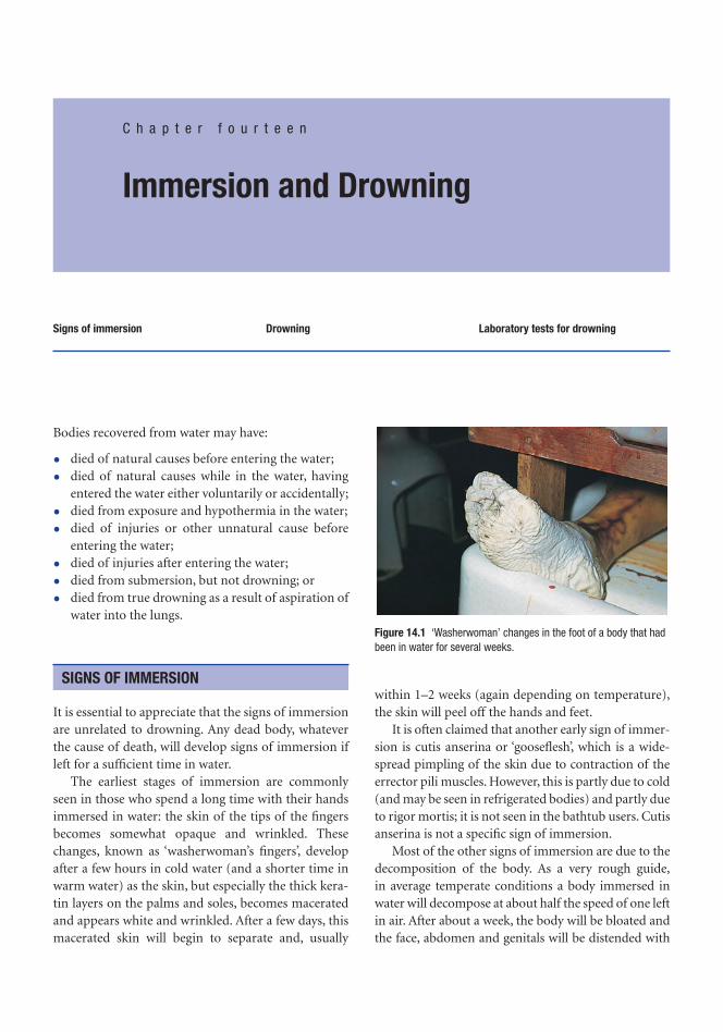
Bodies recovered from water may have:
• died of natural causes before entering the water;
• died of natural causes while in the water, havingentered the water either voluntarily or accidentally;
• died from exposure and hypothermia in the water;
• died of injuries or other unnatural cause beforeentering the water;
• died of injuries after entering the water;
• died from submersion, but not drowning; or
• died from true drowning as a result of aspiration ofwater into the lungs.
It is essential to appreciate that the signs of immersionare unrelated to drowning. Any dead body, whateverthe cause of death, will develop signs of immersion ifleft for a sufficient time in water.
The earliest stages of immersion are commonlyseen in those who spend a long time with their handsimmersed in water: the skin of the tips of the fingersbecomes somewhat opaque and wrinkled. Thesechanges, known as ‘washerwoman’s fingers’, developafter a few hours in cold water (and a shorter time inwarm water) as the skin, but especially the thick kera-tin layers on the palms and soles, becomes maceratedand appears white and wrinkled. After a few days, thismacerated skin will begin to separate and, usually
within 1–2 weeks (again depending on temperature),the skin will peel off the hands and feet.
It is often claimed that another early sign of immer-sion is cutis anserina or ‘gooseflesh’, which is a wide-spread pimpling of the skin due to contraction of theerrector pili muscles. However, this is partly due to cold(and may be seen in refrigerated bodies) and partly dueto rigor mortis; it is not seen in the bathtub users. Cutisanserina is not a specific sign of immersion.
Most of the other signs of immersion are due to thedecomposition of the body. As a very rough guide,in average temperate conditions a body immersed inwater will decompose at about half the speed of one leftin air. After about a week, the body will be bloated andthe face, abdomen and genitals will be distended with
SIGNS OF IMMERSION
C h a p t e r f o u r t e e n
Immersion and Drowning
Signs of immersion Drowning Laboratory tests for drowning
Figure 14.1 ‘Washerwoman’ changes in the foot of a body that hadbeen in water for several weeks.

gas. After about 2 weeks’ immersion in temperate con-ditions, the skin and hair become loose and will beginto detach. Bodies immersed in water are often not dis-covered until they float to the surface and the time atwhich this happens is very variable and is dependenton the extent of gas accumulation and also on the bodyweight. Obese people and some women may sink foronly a short period due to the effect of fat.
Post-mortem artefactual damage, including theeffects of predators – fish, crustaceans, molluscs andreptiles, rodents, dogs and, in some countries, largeranimals – will increase the rate of decomposition. Asthe body decomposes, so the tissues will becomeincreasingly fragile and will fragment and detach eas-ily until, eventually, skeletalization occurs.
As the body is moved by the flow of the water, con-tact with rocks, piers and many other underwaterobstructions can cause damage. These post-morteminjuries must be differentiated from ante-morteminjuries that may be evidence of criminal violence.Certain injuries are unique to bodies immersed inwater, for instance the parallel sliced lacerations charac-teristic of a rapidly revolving propeller of a motorboat.
The mechanism of death in true drowning is not oneof simple hypoxia from deprivation of air. The exacteffects on the body and the abnormalities that arecaused by the entry of water into the airways differaccording to the type of water in which drowningoccurs: hypotonic or hypertonic.
Fresh water is hypotonic compared to plasma, sothat when water enters the lungs, a rapid transfer of
fluid takes place from the alveoli into the vascular sys-tem through the alveolar membranes because of theosmotic difference between the water in the lungs andthe plasma. This transfer of fluids may increase theblood volume by as much as 50 per cent within aminute and this hypervolaemia will place a great strainon the heart. The rapid influx of water will render theplasma hypotonic, which in turn will result in haemoly-sis of red blood cells, and major shifts in the plasmaelectrolytes will occur extremely rapidly. It was oncethought that severe hyperkalaemia caused by the releaseof potassium from the haemolysed red blood cells con-tributed significantly to rapid myocardial failure, butmore recent work has shown this to be only a relativelyminor factor in death in fresh-water drowning.
Seawater is hypertonic, so that water is withdrawnby osmosis from the plasma into the fluid in the alveo-lar spaces in the lungs. There is also electrolyte trans-fer, with mainly sodium and chloride passing from thehypertonic inhaled fluid into the blood. The lack ofhypervolaemia, with the consequent absence of strainon the heart, probably accounts for the longer survivaltimes in salt-water submersion.
In all types of drowning, the deprivation of oxygencaused by obstruction of the alveolar spaces is, ofcourse, a factor, especially as the time of immersionlengthens. As in all kinds of hypoxia, irreversible cor-tical brain damage will occur if oxygenated blood failsto perfuse the brain for more than a number ofminutes. It was formerly thought that 3 minutes wasthe threshold of danger and this may still be so inmany cases, but oxygen deprivation times of over 10minutes with complete recovery of brain functionhave been recorded, although these are likely to be therare exception rather than the rule. Where hypother-mia also exists, particularly when it has a rapid onset,such as in immersion in very cold water or immersionof a child, the total immersion time compatible withfull brain recovery can be far longer and can even, onvery rare occasions, be in excess of 40 minutes.
The post-mortem findings in either type of drown-ing, whether on external observation or at autopsy,may be variable. Drowning is one of the most difficultmodes of death to prove at post mortem, especiallywhen the body is not examined in a fresh condition.Classically, there will be a plume of froth at the mouthand nostrils, which may sometimes be tinged withblood. This froth fills the air passages and exudes on tothe face. It is composed of water containing plasmaprotein and surfactant that has been whipped into a
DROWNING
104 Immersion and drowning
Figure 14.2 Hand from a body recovered from a shallow rivershowing the effects of post-mortem trauma against the river bed.

froth by the violent terminal respiratory actions. Theplume of froth is more common in drowning in hyper-tonic seawater. Externally this may be the only sign,but it is absent more often than it is present, especiallywhen the body has been dead for some time.
Internally, froth may still be found in the tracheaand bronchi for some time after death, but eventuallyeven this vanishes as decomposition begins. When thesternum is removed at autopsy, the lungs may be seento be over-distended, filling the thorax and coveringthe anterior surface of the heart, which is normallyexposed. The lungs may have a ‘doughy’ texture andremain inflated and spongy to the touch. There may beblurred areas of haemolysed blood under the pleura,but only rarely are there any true petechiae.
Dissection of the lung may show generalized pul-monary oedema, but where any water in the air spaceshas been absorbed into the pulmonary circulation, theoedema will be absent – this is the so-called ‘dry-lungdrowning’.
The mechanism of death in individuals who die asa result of submersion is not always classical drown-ing. In a significant proportion of cases, the persondies quickly, sometimes within seconds of entering thewater. The mode of death of these cases is considered tobe cardiac arrest, which is most likely to be due to a vagalinhibitory reflex triggered by a sudden stimulation of
sensitive areas. It is known that falling into cold watercan initiate this reflex, either by rapid cooling of thebody surface or by a sudden inrush of cold water intothe mouth and nasal passages impinging on the sensi-tive mucosa of the pharynx and larynx.
Sudden death on entering water is more likely indrunken people, in whom the skin is flushed fromalcohol-induced vasodilatation, and sailors falling intoa harbour on their way back from heavy drinking inthe local bars are a common example of this type ofdeath. This type of very rapid death on immersion isalso said to occur in emotionally tense individuals(such as intending suicides), in whom the nervousreflex arcs seem more active.
The diatom test depends on the presence of micro-scopic algae, called diatoms, which are often, but notalways, present in seawater and unpolluted naturalfresh water. There are about 25 000 species of diatomand all have an almost indestructible silicaceous cap-sule. The theory behind the test is that normal peopledo not have significant numbers of diatoms in the kid-neys, brain, marrow etc., but if a person drowns indiatom-containing water, the algae in that water willreach the lungs and some of them will penetrate thealveolar walls. If the heart is still beating, the diatomsthat have entered the bloodstream will be transportedaround the body in the circulation and may lodge indistant organs such as the kidney, brain and bone mar-row before death. An increase in the number ofdiatoms in the internal organs was thought to confirmthat the body had drowned and, if a sample of the
LABORATORY TESTS FOR DROWNING
Laboratory tests for drowning 105
Figure 14.3 Profuse blood-tinged froth around the mouth and nose in drowning. Figure 14.4 Diatoms from lake water.

water was also taken at the presumed site of drowning,the similarity of different species of diatom in thewater and the body could be compared.
On the other hand, if a dead body was dropped intowater, although diatoms could reach the lung by pas-sive percolation, no circulatory transfer could occurand so (theoretically) no diatoms would be present inthe distant organs. The advantages claimed for thistechnique include the fact that it could be used even inthe presence of putrefaction, if protected tissue such asbone marrow was examined.
Unfortunately, the diatom test is often negative,even in undoubted cases of drowning in waterways
full of diatoms, and there have been numerous false-positive results that are said to have occurred for avariety of technical reasons. All in all, this test is nowso unreliable that it should only be used, and the resultsinterpreted, with great circumspection.
Many attempts have been made to identify a chem-ical test or some other type of analysis of the bloodthat will assist in the diagnosis of either fresh-water orseawater drowning. Analyses of the concentrations ofsodium, chloride and magnesium have been used, butthe results are too variable (due to the rapid post-mortem diffusion of electrolytes throughout the bodyafter death) to be of any practical use.
106 Immersion and drowning
Heat, cold and electricity are some of the ‘physicalagents’ that can cause non-kinetic injuries to the body.
Exposure of living tissue to high temperatures willcause damage to the cells. The extent of the damagecaused is a function of the length of time of exposureas well as of the temperature to which the tissues areexposed. Damage to the skin can occur at tempera-tures as low as 44°C if exposed for several hours; attemperatures over 50°C or so, damage occurs morerapidly, and at 60°C tissue damage occurs in 3 seconds.
The heat source may be dry or wet; where the heatis dry, the resultant injury is called a ‘burn’, whereaswith moist heat from hot water, steam and other hotliquids it is known as ‘scalding’.
Burning
Dry burns are classified by both severity and extent.There are several systems of classification of the sever-ity of burns, the most useful of which is:
1 first degree – erythema and blistering (vesiculation);2 second degree – burning of the whole thickness of
the epidermis and exposure of the dermis;
3 third degree – destruction down to subdermal tis-sues, sometimes with carbonization and exposureof muscle and bone.
The size of the area of the burning may be moreimportant in the assessment of the dangers of theburn than the depth. The ‘Rule of Nines’ is used to cal-culate the approximate extent on the body surface.Where the burnt area exceeds 50 per cent, the progno-sis is poor, even in first-degree burns. The age of theindividual is also important: the elderly may die withonly 20 per cent burns, whereas children and youngadults are generally more resilient. There is, of course,considerable individual variation and the speed andextent of emergency treatment will play a significantpart in the morbidity and mortality of burns’ victims.In dry burns, any clothing will offer some protectionagainst heat unless it ignites.
Scalds
The general features of scalds are similar to those ofburns, with erythema and blistering, but charring ofthe skin is only found when the liquid is extremelyhot, such as with molten metal. The pattern of scald-ing will depend upon the way in which the body hasbeen exposed to the fluid: immersion into hot liquidresults in an upper ‘fluid level’, whereas splashed or
INJURY DUE TO HEAT
C h a p t e r f i f t e e n
Injury due to Heat, Cold and Electricity
Injury due to heatBurningScaldsThe examination of bodies recovered
from fires
Causes of death in burns and scaldsInjuries on burned bodiesCold injury (hypothermia)The signs of hypothermia
Electrical injuryThe electrical lesionDeath from lightning
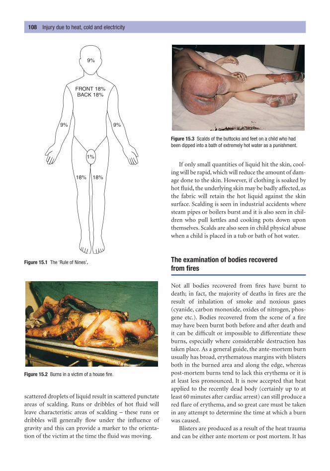
scattered droplets of liquid result in scattered punctateareas of scalding. Runs or dribbles of hot fluid willleave characteristic areas of scalding – these runs ordribbles will generally flow under the influence ofgravity and this can provide a marker to the orienta-tion of the victim at the time the fluid was moving.
If only small quantities of liquid hit the skin, cool-ing will be rapid, which will reduce the amount of dam-age done to the skin. However, if clothing is soaked byhot fluid, the underlying skin may be badly affected, asthe fabric will retain the hot liquid against the skinsurface. Scalding is seen in industrial accidents wheresteam pipes or boilers burst and it is also seen in chil-dren who pull kettles and cooking pots down uponthemselves. Scalds are also seen in child physical abusewhen a child is placed in a tub or bath of hot water.
The examination of bodies recovered from fires
Not all bodies recovered from fires have burnt todeath; in fact, the majority of deaths in fires are theresult of inhalation of smoke and noxious gases(cyanide, carbon monoxide, oxides of nitrogen, phos-gene etc.). Bodies recovered from the scene of a firemay have been burnt both before and after death andit can be difficult or impossible to differentiate theseburns, especially where considerable destruction hastaken place. As a general guide, the ante-mortem burnusually has broad, erythematous margins with blistersboth in the burned area and along the edge, whereaspost-mortem burns tend to lack this erythema or it isat least less pronounced. It is now accepted that heatapplied to the recently dead body (certainly up to atleast 60 minutes after cardiac arrest) can still produce ared flare of erythema, and so great care must be takenin any attempt to determine the time at which a burnwas caused.
Blisters are produced as a result of the heat traumaand can be either ante mortem or post mortem. It has
108 Injury due to heat, cold and electricity
1%
9%9%
9%
18% 18%
FRONT 18%BACK 18%
Figure 15.1 The ‘Rule of Nines’.
Figure 15.2 Burns in a victim of a house fire.
Figure 15.3 Scalds of the buttocks and feet on a child who hadbeen dipped into a bath of extremely hot water as a punishment.
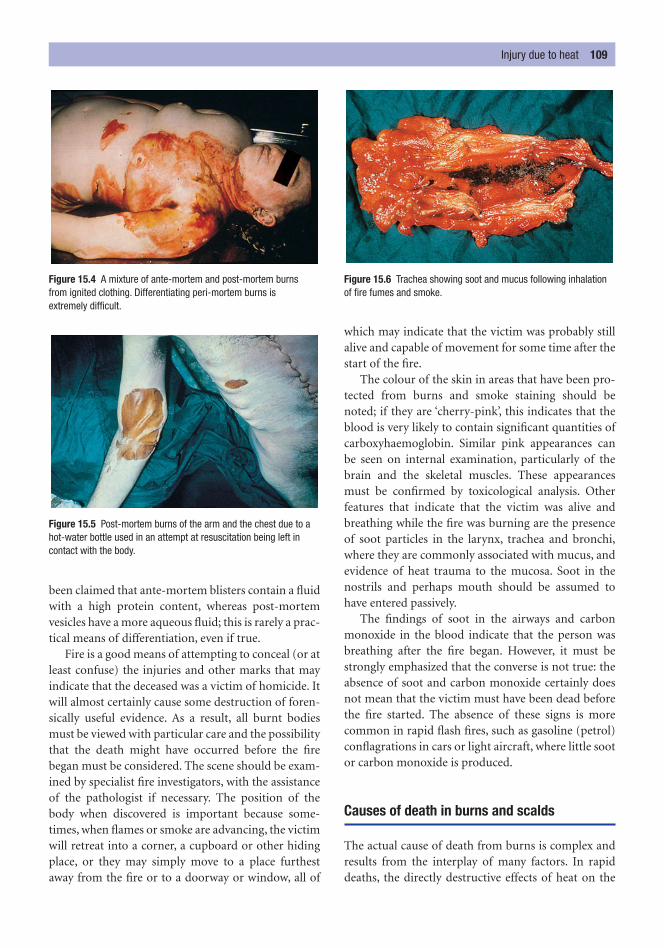
been claimed that ante-mortem blisters contain a fluidwith a high protein content, whereas post-mortemvesicles have a more aqueous fluid; this is rarely a prac-tical means of differentiation, even if true.
Fire is a good means of attempting to conceal (or atleast confuse) the injuries and other marks that mayindicate that the deceased was a victim of homicide. Itwill almost certainly cause some destruction of foren-sically useful evidence. As a result, all burnt bodiesmust be viewed with particular care and the possibilitythat the death might have occurred before the firebegan must be considered. The scene should be exam-ined by specialist fire investigators, with the assistanceof the pathologist if necessary. The position of thebody when discovered is important because some-times, when flames or smoke are advancing, the victimwill retreat into a corner, a cupboard or other hidingplace, or they may simply move to a place furthestaway from the fire or to a doorway or window, all of
which may indicate that the victim was probably stillalive and capable of movement for some time after thestart of the fire.
The colour of the skin in areas that have been pro-tected from burns and smoke staining should benoted; if they are ‘cherry-pink’, this indicates that theblood is very likely to contain significant quantities ofcarboxyhaemoglobin. Similar pink appearances canbe seen on internal examination, particularly of thebrain and the skeletal muscles. These appearancesmust be confirmed by toxicological analysis. Otherfeatures that indicate that the victim was alive andbreathing while the fire was burning are the presenceof soot particles in the larynx, trachea and bronchi,where they are commonly associated with mucus, andevidence of heat trauma to the mucosa. Soot in thenostrils and perhaps mouth should be assumed tohave entered passively.
The findings of soot in the airways and carbonmonoxide in the blood indicate that the person wasbreathing after the fire began. However, it must bestrongly emphasized that the converse is not true: theabsence of soot and carbon monoxide certainly doesnot mean that the victim must have been dead beforethe fire started. The absence of these signs is morecommon in rapid flash fires, such as gasoline (petrol)conflagrations in cars or light aircraft, where little sootor carbon monoxide is produced.
Causes of death in burns and scalds
The actual cause of death from burns is complex andresults from the interplay of many factors. In rapiddeaths, the directly destructive effects of heat on the
Injury due to heat 109
Figure 15.4 A mixture of ante-mortem and post-mortem burns from ignited clothing. Differentiating peri-mortem burns is extremely difficult.
Figure 15.5 Post-mortem burns of the arm and the chest due to ahot-water bottle used in an attempt at resuscitation being left incontact with the body.
Figure 15.6 Trachea showing soot and mucus following inhalationof fire fumes and smoke.

respiratory tract leading to asphyxia, the combinedtoxic effects of carbon monoxide, cyanide and themultitude of other noxious gases (oxides of nitrogen,phosgene etc.) that are inhaled, the release of toxicmaterial from the extensive tissue destruction, and‘shock’ due to pain all cause or contribute to death.Most of these factors do not apply to scalds, unless thevictim falls into the hot liquid.
Where death from burns or scalds is delayed, dehy-dration and electrolyte disturbances as a result ofplasma loss from the damaged skin surfaces are earlycauses of death. Later, renal failure, toxaemia from sub-stances absorbed from the burned area and infection ofwidespread burns may be responsible (see Chapter 16).
Injuries on burned bodies
A careful search must be made for any ante-morteminjuries that may have caused or contributed to death.These injuries can be of almost any type, but strangu-lation and shooting appear to be most common incases of homicide concealed by fire. It is always advis-able to x-ray a burned body, especially if the fire dam-age is so extensive as to make examination of the skinsurface impossible.
However, not all injuries on burned bodies are sin-ister, and where skin has been subjected to severeburning or charring, it will often split. These splits canalso be caused by post-mortem movement duringrecovery of the body when charred and brittle skin ismoved. These injuries sometimes worry the police orfire brigade, who suspect that they are due to previousinjury, but their appearance, position and lack of anyante-mortem tissue response are usually a clear indi-cation of their post-mortem nature.
Another misinterpreted injury is the ‘heathaematoma’ inside the skull. This spurious ‘extraduralhaematoma’ lies between the bone and dura and iscaused when severe heat has been applied to the scalp,resulting in expansion of the blood in the skull diplöeand the intracranial venous sinuses, which rupture,resulting in the formation of a collection of blood out-side the meninges. The blood is brown and spongy,unlike a true haematoma, but the simple presence ofthis collection of blood may mislead the unwary doc-tor performing an autopsy into thinking that this isthe result of a trauma.
There is seldom any doubt about the cause of thesecollections of blood. However, it has been said that if
there is carboxyhaemoglobin in the peripheral blood,then if the haematoma is due to heat, it will also con-tain carboxyhaemoglobin. On the other hand, a trueextradural haematoma, which had developed beforethe fire began, will not contain any appreciableamount of carboxyhaemoglobin. The test is not foolproof, but may be a useful adjunct to other methods ofinvestigation.
Hypothermia has both clinical and forensic aspects, asmany people suffer from and die of hypothermia evenin temperate climates in winter. In marine disasters,hypothermia may be as common a cause of death asdrowning. In the very cold waters of seas and lakes inhigh latitudes, death from immersion may occurwithin a couple of minutes from sheer heat loss andbefore true drowning can occur.
In practice, most deaths from hypothermia are seenin old people and in some children, both of whom areparticularly vulnerable to hypothermia. Old people maybecome severely hypothermic due to their restrictedeconomic circumstances or to senile mental changes,
COLD INJURY (HYPOTHERMIA)
110 Injury due to heat, cold and electricity
Figure 15.7 Skin splits in the victim of a house fire. These splitswere initially thought to be incised wounds.

and for a number of reasons they may go without adequate heating, food and clothing. An added factormay be myxoedema, which predisposes to low bodytemperature, or the use of phenothiazine drugs, whichalso predisposes towards hypothermia. It is importantto remember that the weather does not have to beunusually cold for hypothermia to develop and, evenin moderately cold winter weather, many elderly indi-viduals will become hypothermic.
Children have a high body surface-to-weight ratioand lose heat rapidly. In some cases of deliberate neglector careless family circumstances, infants may be left inunheated rooms in winter and suffer hypothermia.Another group includes drunken people, who may col-lapse or lie down to sleep after too much alcohol andbecome hypothermic, especially because alcohol causesvasodilatation of the skin, increasing heat loss.
The signs of hypothermia
Any fall in body core temperature of more than a fewdegrees is hypothermia, but clinical effects are slightproviding the core body temperature remains aboveabout 35°C. The body has a series of mechanisms toraise the body temperature, including diversion ofblood away from the skin and shivering. Below 32°C,shivering ceases and thus this extra muscular activitywill no longer generate heat, and so the situation worsens. People whose temperature falls to 28°C orless are almost certain to die, even with treatment.
When a dead person is found and hypothermia issuspected, it can be difficult or impossible to provebecause post-mortem cooling (including storage in amortuary refrigerator) can cause some similar changesto appear in the skin. However, if the body shows areaswith indistinct, blurred margins that are pink or brown-pink, particularly over and around joints such as theknees, elbows and hips and also in sites where they can-not be due to hypostasis due to their distribution, adiagnosis of hypothermia should be considered. Thepink colour is due to unreduced oxyhaemoglobin asthe cold tissues have little uptake of oxygen. The causeof the rather blurred, brownish tinge around the jointsis unclear, but suggests that there may be a degree ofred cell haemolysis. Very rarely, hypothermic victimswill have blisters in the affected areas of skin.
Internally, there may be no signs at all, but quitecommonly, the stomach lining is studded with numer-ous brown-black acute erosions, which are exactly the
same as those seen in many types of pre-death stress.Increased viscosity of the blood due to low tempera-tures may lead to sludging of blood in the small vesselsof many organs and this may lead to micro-infarcts.This mechanism is probably the cause of the haemor-rhagic pancreatitis and associated fat necrosis that arealso features of many cases of hypothermia.
Hypothermia may be linked with a strange condi-tion, particularly in the elderly, in which people take offsome or all of their clothing (reciprocal undressing).They may then hide themselves in corners, in cup-boards or under piles of furniture or household goods.This is called the ‘hide-and-die syndrome’ and is wellknown. An alternative, and perhaps more common,scenario is of a naked or partly clothed elderly individ-ual found amid a scene of utter confusion with furni-ture pulled over and drawers and cupboards emptiedout. Close examination of the scene will show that allof the disturbance is at low level and that the tops oftables etc. are not disturbed. When bodies are found ineither of these situations, the police may be suspiciousof some criminal action, but examination of the sceneand the post-mortem features of hypothermia willexplain these worrying circumstances.
If hypothermia of the extremities has been presentfor a long time, there may be ‘frost-bite’, which isinfarction of the peripheral digits with oedema, red-ness and later necrosis of the tissue beyond a line ofinflammatory demarcation. There need not be anygeneralized associated hypothermia.
Injury and death from the passage of an electric cur-rent through the body are common in both industrial
ELECTRICAL INJURY
Electrical injury 111
Figure 15.8 Discoloration of the skin in a case of fatal hypothermia.

and domestic circumstances. The essential factor incausing harm is the current (i.e. an electron flow)which is measured in milliamperes (mA). This in turnis determined by the resistance of the tissues in ohmsand the voltage of the power supply in volts (V),according to Ohm’s Law. To increase the current (andhence the damage), either the resistance must fall orthe voltage must increase, or both.
Almost all cases of electrocution, fatal or other-wise, originate from the public power supply, which isdelivered throughout the world at either 110 or 240 V.It is rare for death to occur at less than 100 V. The cur-rent needed to produce death varies according to thetime during which it passes and the part of the bodyacross which it flows. Usually, the entry point is a handthat touches an electrical appliance or live conductor,and the exit is to earth (or ‘ground’), often via theother hand or the feet. In either case, the current willcross the thorax, the most dangerous area for a shockbecause of the risks of cardiac arrest or respiratoryparalysis.
When a live metal conductor is gripped by thehand, pain and muscle twitching will occur if the cur-rent reaches about 10 mA. If the current in the armexceeds about 30 mA, the muscles will go into spasm,which cannot be voluntarily released because theflexor muscles are stronger than the extensors; theresult is for the hand to grip or to ‘hold on’. This ‘hold-on’ effect is very dangerous as it may allow the circuitto be maintained for long enough to cause cardiacarrhythmia, whereas the normal response would havebeen to let go so as to stop the pain.
If the current across the chest is 50 mA or more,even for only a few seconds, fatal ventricular fibrilla-tion is likely to occur, and alternating current (AC –very common in domestic supplies) is much moredangerous than direct current (DC) at precipitatingcardiac arrhythmias.
The tissue resistance is important. Thick dry skin,such as the palm of the hand or sole of the foot, mayhave a resistance of 1 million ohms (�), but when wet,this may fall to a few hundred ohms and the current,given a fixed supply voltage, will be markedly increased.This is relevant in wet conditions such as bathrooms,exterior building sites or when sweating.
The mode of death in most cases of electrocution isventricular fibrillation due to the direct effects of thecurrent on the myocardium and cardiac conductingsystem. These changes can be reversed when the cur-rent ceases, which may explain some of the remarkable
recoveries following prolonged cardiac massage afterreceipt of an electric shock. The victims of such anarrhythmia will be pale, whereas those who die as aresult of peripheral respiratory paralysis are usuallycyanosed. Even more rare are the instances in whichthe current has entered the head and caused primarybrainstem paralysis, which has resulted in failure of respiration. This may occur when workers on overhead power supply lines or electric railway wirestouch their heads against high-tension conductors,usually 660 V.
The electrical lesion
Unless the circumstances are accurately known, it canbe difficult to know whether a dead victim has been incontact with electricity. When high voltages or pro-longed contact have occurred, extensive and severeburns can be seen, but a few seconds’ contact with afaulty appliance may leave minimal signs. Where theskin is wet or where the body is immersed, as in a bath,there may be no signs at all, as the entrance and exit ofthe current may be spread over such a wide area thatno focal lesion exists.
Usually, however, there is a discrete focal point of entry and, as the electrical current is concen-trated at that point, enough energy can be released to cause a thermal lesion. The entry points may bemultiple and obvious, or they may be single and veryinconspicuous. As the most commonplace is on thehands, these should always be examined with particu-lar care.
The focal electrical lesion is usually a blister, whichoccurs when the conductor is in firm contact with theskin and which usually collapses soon after infliction,forming a raised rim with a concave centre. The skin ispale, often white, and an areola of pallor (due to localvasoconstriction) is a characteristic feature. The blistermay vary from a few millimetres to several centi-metres. The skin often peels off the large blisters leavinga red base. The other type of electrical mark is a ‘sparkburn’, where there is an air gap between metal andskin. Here, a central nodule of fused keratin, brown or yellow in colour, is surrounded by the typical areolaof pale skin. Both types of lesion often lie adjacent to each other. In high-voltage burns, multiple sparksmay crackle onto the victim and cause large areas ofdamage, sometimes called ‘crocodile skin’ due to itsappearance.
112 Injury due to heat, cold and electricity

Internally, there are no characteristic findings infatal electrocution. The skin lesions are mainly thermalin nature, but opinions vary as to whether histologicalappearances are specific to electricity. It has been said
that the cell nuclei line up in parallel rows due to theelectric field, but similar appearances can occur inpurely thermal burns. There are specific intracellularlesions on electron microscopy, but the gross patho-logical diagnosis relies upon the external appearances.
Hundreds of deaths occur each year from atmosphericlightning, especially in tropical countries. A lightningstrike from cloud to earth may involve property, ani-mals or humans. Huge electrical forces are involved,producing millions of amperes and phenomenal volt-ages. Some of the lesions caused to those who arestruck directly or simply caught close to the lightningstrike are electrical, but other will be from burns and
DEATH FROM LIGHTNING
Death from lightning 113
Figure 15.9 Multiple minute electrical marks on the hand caused bycontact with a faulty electrical drill.
Figure 15.10 Electrical mark from a mains wire wrapped aroundthe neck. There is marked hyperaemia and adjacent pallor, withevidence of blistering.
Figure 15.11 Hyperaemia from a defibrillator paddle, caused duringattempted resuscitation.
Figure 15.12 Extensive electrical burns with scorching andblistering.
Figure 15.13 The fern-like pattern of a lightning strike can be seenon the upper chest.

yet others are due to the compression wave of heatedair, which is like an explosion. All kinds of bizarreappearances may be found, especially the partial orcomplete stripping of clothing from the victim, whichmay arouse suspicions of foul play. Severe burns, frac-tures and gross lacerations can occur, along with the
well-known magnetization or even fusion of metallicobjects in the clothing. The usual textbook descriptionis of ‘fern or branch-like’ patterns on the skin, but infact most bodies do not show this. Red streaks follow-ing skin creases or sweat-damped tracks are morelikely, though many bodies are completely unmarked.
114 Injury due to heat, cold and electricity
Some injuries are so severe that they are immediatelyand obviously incompatible with life, for instance thegross disruption of a body caused by an explosion.Minimal injuries, for example abrasions, are extremelyunlikely to lead to any significant degree of incapacity,let alone disability or death. Between these two ends ofthe spectrum lie a wide range of types and severity ofinjuries that may or may not result in incapacity, dis-ability or death. The effects of trauma depend not onlyon the severity of the injury but also on the responseof the individual to that particular trauma.
The forensic practitioner must understand boththe immediate and the long-term effects of trauma inorder to be able to explain those effects to a court or tothe relatives.
External bleeding is usually obvious; internal bleedingis very commonly confined to the body cavities, tissuespaces or directly into large organs such as the lungs orthe liver. Bleeding within the skull is dangerous becauseof the space-occupying effect of the collection of bloodupon the brain, and the same may be said about bleed-ing into the pericardial sac and its effect on the heart.
The speed of blood loss is crucial. An adult humancan rapidly lose approximately 10 per cent of their total
blood volume without there being a significant effecton the stability of their cardiovascular system. Theeffects of blood loss are rapidly compensated by apowerful sympathetic response, which constricts boththe arterioles and the great veins and results in anincrease in the heart rate, which has the effect of main-taining blood pressure and circulating blood volume.Endocrine responses including vasopressin and angio-tensin also play their part in the longer term. This isdescribed as non-progressive or compensated shock.
If more blood is lost, the protective reflexes are over-whelmed: the blood pressure will begin to fall, whichwill have adverse effects upon the heart and, as themyocardium begins to deteriorate, there is a further fallof blood pressure and a worsening of the vicious cycle.Other effects of the falling blood pressure include fail-ure of the sympathetic centres themselves, thrombosis,increased capillary permeability and the release oftoxins from damaged or dead tissues. This is called progressive shock and will result in death if treatment is not given.
If bleeding continues, a third stage of shock isreached during which treatment may have a temporar-ily beneficial effect but will not affect the ultimatelyfatal outcome. This is called irreversible shock. Thetime scale for these various stages of shock is very vari-able and depends, among other things, upon the rate ofblood loss and the previous health of the individual.
HAEMORRHAGE
C h a p t e r s i x t e e n
Effects of Injuries
HaemorrhageInfectionEmbolismFat embolismAir embolism
Pulmonary thrombo-embolismForeign-body embolismAmniotic fluid embolismDisseminated intravascular
coagulation
Adult respiratory distress syndromeSuprarenal haemorrhageSubendocardial haemorrhages
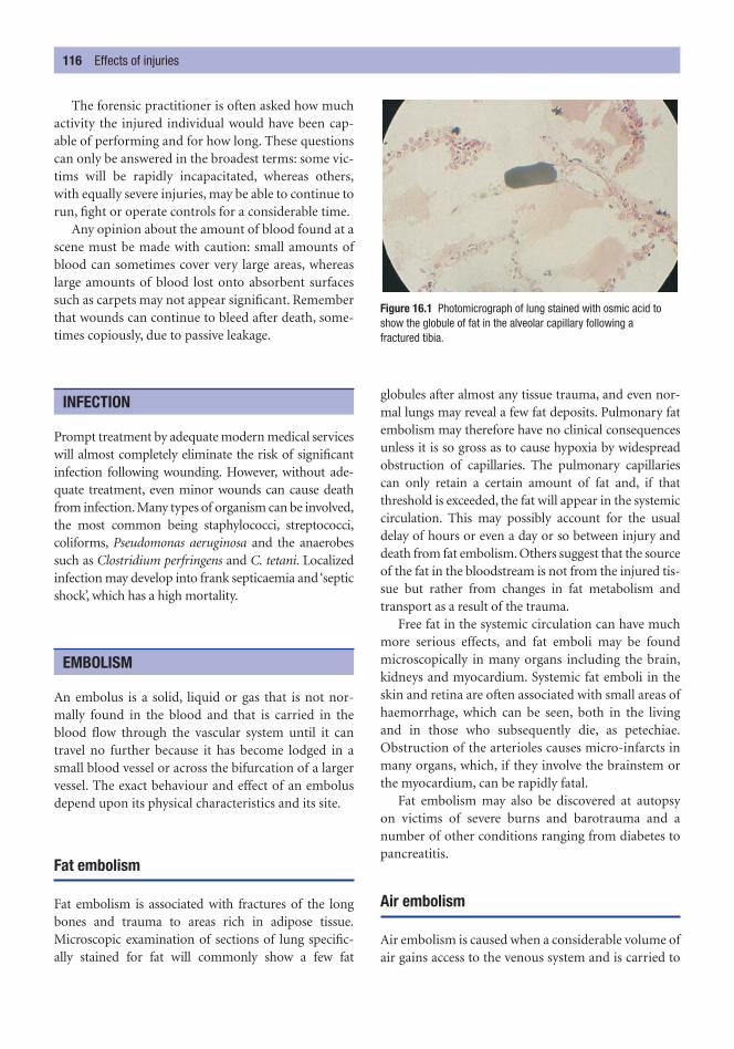
The forensic practitioner is often asked how muchactivity the injured individual would have been cap-able of performing and for how long. These questionscan only be answered in the broadest terms: some vic-tims will be rapidly incapacitated, whereas others,with equally severe injuries, may be able to continue torun, fight or operate controls for a considerable time.
Any opinion about the amount of blood found at ascene must be made with caution: small amounts ofblood can sometimes cover very large areas, whereaslarge amounts of blood lost onto absorbent surfacessuch as carpets may not appear significant. Rememberthat wounds can continue to bleed after death, some-times copiously, due to passive leakage.
Prompt treatment by adequate modern medical serviceswill almost completely eliminate the risk of significantinfection following wounding. However, without ade-quate treatment, even minor wounds can cause deathfrom infection. Many types of organism can be involved,the most common being staphylococci, streptococci,coliforms, Pseudomonas aeruginosa and the anaerobessuch as Clostridium perfringens and C. tetani. Localizedinfection may develop into frank septicaemia and ‘septicshock’, which has a high mortality.
An embolus is a solid, liquid or gas that is not nor-mally found in the blood and that is carried in theblood flow through the vascular system until it cantravel no further because it has become lodged in asmall blood vessel or across the bifurcation of a largervessel. The exact behaviour and effect of an embolusdepend upon its physical characteristics and its site.
Fat embolism
Fat embolism is associated with fractures of the longbones and trauma to areas rich in adipose tissue.Microscopic examination of sections of lung specific-ally stained for fat will commonly show a few fat
globules after almost any tissue trauma, and even nor-mal lungs may reveal a few fat deposits. Pulmonary fatembolism may therefore have no clinical consequencesunless it is so gross as to cause hypoxia by widespreadobstruction of capillaries. The pulmonary capillariescan only retain a certain amount of fat and, if thatthreshold is exceeded, the fat will appear in the systemiccirculation. This may possibly account for the usualdelay of hours or even a day or so between injury anddeath from fat embolism. Others suggest that the sourceof the fat in the bloodstream is not from the injured tis-sue but rather from changes in fat metabolism andtransport as a result of the trauma.
Free fat in the systemic circulation can have muchmore serious effects, and fat emboli may be foundmicroscopically in many organs including the brain,kidneys and myocardium. Systemic fat emboli in theskin and retina are often associated with small areas ofhaemorrhage, which can be seen, both in the livingand in those who subsequently die, as petechiae.Obstruction of the arterioles causes micro-infarcts inmany organs, which, if they involve the brainstem orthe myocardium, can be rapidly fatal.
Fat embolism may also be discovered at autopsy on victims of severe burns and barotrauma and anumber of other conditions ranging from diabetes topancreatitis.
Air embolism
Air embolism is caused when a considerable volume ofair gains access to the venous system and is carried to
EMBOLISM
INFECTION
116 Effects of injuries
Figure 16.1 Photomicrograph of lung stained with osmic acid toshow the globule of fat in the alveolar capillary following a fractured tibia.

the heart, where it fills the right atrium and ventriclewith frothy bubbles. These bubbles are compressibleand so cannot be pumped onward during systole and,as a result, they form an air lock, which causes circula-tory failure. Air can gain access to the veins by a num-ber of routes, including opening of the large veins inthe neck following cutting of the throat or duringoperations such as thyroidectomy. The head must beabove the level of the chest to cause a suction effectinto the superior vena cava.
Another cause of air emboli is the use of a Higginsonsyringe to procure an abortion: air or frothy fluid isforced into the placental bed and enters the blood-stream. A different mechanism operates in decompres-sion sickness in divers or high-altitude flyers, in whichbubbles of nitrogen gas are formed in the blood. Thismay happen in any blood vessel in the body and clearlymay result in direct effects on many organs, includingthe heart and brain.
The minimum volume of air needed to cause deathfrom cardiac embolism is debatable, but is probably ofthe order of at least 100 mL in humans. Any gas thatremains after absorption of oxygen by the erythro-cytes (which will be mainly nitrogen) cannot pene-trate the lung capillary bed to any extent, nor can itmove upwards in the jugular veins against the bloodflow. Therefore, ‘cerebral air embolism’ probably doesnot exist, and the descriptions and photographs ofbubbles of gas in the cerebral veins are probably arte-facts caused when the skull-cap is removed duringautopsy.
Pulmonary thrombo-embolism
This is mentioned in Chapter 17 as a common cause ofsudden death, but is also one of the most commonconsequences of injury of any type. After any signifi-cant tissue damage, there is an increase in the coagula-bility of the blood. If the injury results in immobility,this will have the effect of applying pressure on thecalves of the legs, which in turn reduces the blood flowin the legs. These factors encourage thrombosis in thedeep veins of the legs after any injury and many surgi-cal operations. However, leg vein thrombosis mayoccur after any period of stagnation or pressure on thelegs and it has been reported in long-distance air pas-sengers (now called the Economy Class Syndrome) andeven in those who sit in deck chairs for long periods.
Rarely, thromboses develop in other veins andform the source of pulmonary emboli, but the legveins are overwhelmingly the most common focus.Pelvic vein thrombosis, even in association with preg-nancy and childbirth, is less common than alleged.
Deep vein thromboses are extremely common, butmost remain in situ and many patients are symptomfree. The thromboses begin distally and spread proxim-ally in the blood flow; they are most commonly foundin the calves and may extend proximally as far as thesaphenous, femoral or even iliac veins. Many throm-boses spontaneously detach (in part or in whole) fromthe wall of the vein to form thrombo-emboli, which areswept off in the venous bloodstream to the right side ofthe heart. On leaving the heart, they encounter increas-ingly smaller vessels as the pulmonary arteries divideand, when the size of the embolus is equal to the size of the pulmonary artery, it can go no further and so itobstructs the vessel like a cork in a bottle. Pulmonaryemboli may cause clinical symptoms of chest pain andhaemoptysis or they may be silent. Larger emboli arecommonly preceded by one or more smaller emboli.The large emboli may impact in the main trunk or inthe large pulmonary arteries and cause rapid death.
The forensic significance of pulmonary emboli liesin their association with apparently non-fatal trauma.The victim, having survived the original injury, mayeven appear to be making an uncomplicated recovery,but suddenly collapses and dies some time later andpulmonary emboli are identified at the autopsy. About80 per cent of fatal pulmonary emboli have a predis-posing cause – trauma, surgical operation, immobility,childbirth etc. The remaining 20 per cent appear
Embolism 117
Figure 16.2 Fatal pulmonary emboli occluding the major pulmonaryarteries 2 weeks after repair of a fractured femur.
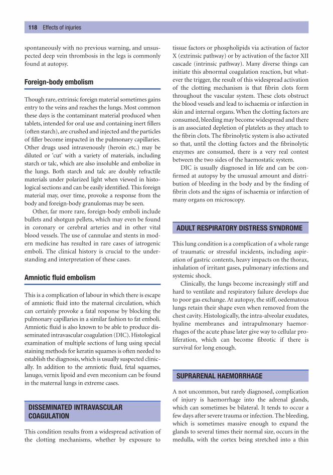
spontaneously with no previous warning, and unsus-pected deep vein thrombosis in the legs is commonlyfound at autopsy.
Foreign-body embolism
Though rare, extrinsic foreign material sometimes gainsentry to the veins and reaches the lungs. Most commonthese days is the contaminant material produced whentablets, intended for oral use and containing inert fillers(often starch), are crushed and injected and the particlesof filler become impacted in the pulmonary capillaries.Other drugs used intravenously (heroin etc.) may bediluted or ‘cut’ with a variety of materials, includingstarch or talc, which are also insoluble and embolize inthe lungs. Both starch and talc are doubly refractilematerials under polarized light when viewed in histo-logical sections and can be easily identified. This foreignmaterial may, over time, provoke a response from thebody and foreign-body granulomas may be seen.
Other, far more rare, foreign-body emboli includebullets and shotgun pellets, which may even be foundin coronary or cerebral arteries and in other vitalblood vessels. The use of cannulae and stents in mod-ern medicine has resulted in rare cases of iatrogenicemboli. The clinical history is crucial to the under-standing and interpretation of these cases.
Amniotic fluid embolism
This is a complication of labour in which there is escapeof amniotic fluid into the maternal circulation, whichcan certainly provoke a fatal response by blocking thepulmonary capillaries in a similar fashion to fat emboli.Amniotic fluid is also known to be able to produce dis-seminated intravascular coagulation (DIC). Histologicalexamination of multiple sections of lung using specialstaining methods for keratin squames is often needed toestablish the diagnosis, which is usually suspected clinic-ally. In addition to the amniotic fluid, fetal squames,lanugo, vernix lipoid and even meconium can be foundin the maternal lungs in extreme cases.
This condition results from a widespread activation ofthe clotting mechanisms, whether by exposure to
tissue factors or phospholipids via activation of factorX (extrinsic pathway) or by activation of the factor XIIcascade (intrinsic pathway). Many diverse things caninitiate this abnormal coagulation reaction, but what-ever the trigger, the result of this widespread activationof the clotting mechanism is that fibrin clots formthroughout the vascular system. These clots obstructthe blood vessels and lead to ischaemia or infarction inskin and internal organs. When the clotting factors areconsumed, bleeding may become widespread and thereis an associated depletion of platelets as they attach tothe fibrin clots. The fibrinolytic system is also activatedso that, until the clotting factors and the fibrinolyticenzymes are consumed, there is a very real contestbetween the two sides of the haemostatic system.
DIC is usually diagnosed in life and can be con-firmed at autopsy by the unusual amount and distri-bution of bleeding in the body and by the finding offibrin clots and the signs of ischaemia or infarction ofmany organs on microscopy.
This lung condition is a complication of a whole rangeof traumatic or stressful incidents, including aspir-ation of gastric contents, heavy impacts on the thorax,inhalation of irritant gases, pulmonary infections andsystemic shock.
Clinically, the lungs become increasingly stiff andhard to ventilate and respiratory failure develops dueto poor gas exchange. At autopsy, the stiff, oedematouslungs retain their shape even when removed from thechest cavity. Histologically, the intra-alveolar exudates,hyaline membranes and intrapulmonary haemor-rhages of the acute phase later give way to cellular pro-liferation, which can become fibrotic if there issurvival for long enough.
A not uncommon, but rarely diagnosed, complicationof injury is haemorrhage into the adrenal glands,which can sometimes be bilateral. It tends to occur afew days after severe trauma or infection. The bleeding,which is sometimes massive enough to expand theglands to several times their normal size, occurs in themedulla, with the cortex being stretched into a thin
SUPRARENAL HAEMORRHAGE
ADULT RESPIRATORY DISTRESS SYNDROME
DISSEMINATED INTRAVASCULAR COAGULATION
118 Effects of injuries

line around the haematoma. The bleeding is not due todirect mechanical damage, but appears to be neuro-logically mediated via the sympathetic nervous system,in much the same way as acute gastric erosions andsubendocardial haemorrhage are related to parasym-pathetic over-activity. When associated with meningo-coccal infection, these bilateral adrenal haemorrhagesform the typical Waterhouse–Friederichsen syndrome.
Subendocardial haemorrhages are a non-specific find-ing in many cases of death following general trauma,particularly when it was associated with hypotensionand following fatal head injuries. It is important thatsubendocardial haemorrhages are not confused withdirect cardiac trauma in these cases.
SUBENDOCARDIAL HAEMORRHAGES
Subendocardial haemorrhages 119
Figure 16.3 Adrenal haemorrhage several days after a head injury. The haemorrhage was bilateral and not related to abdominal trauma.

In countries where deaths have to be officially certified,the responsibility for certification falls either to thedoctor who attended the patient during life or one whocan reasonably be assumed to know sufficient of theclinical history to give a reasonable assessment of thecause of death. This is an ‘honest opinion, fairly given’,but many studies have shown that there is a large errorrate in death certificates and that in 25–60 per cent ofdeaths there are significant differences between theclinician’s presumption of the cause of death and thelesions or diseases actually displayed at the autopsy.
Unfortunately, it seems that there has been little or no improvement in the problems of certification ofdeath over the years and as a result the raw epidemi-ological data gathered by national statistical bureauxmust be treated with some caution. All doctors shouldtake the task of certifying the cause of death very ser-iously but, regrettably, it is a job usually delegated to the most junior, and least experienced, member ofthe team. A review of death certification is currentlybeing performed in the UK in the wake of the serialmurders of Harold Shipman, a general practitioner inManchester.
There is a different approach to sudden and unex-pected deaths, as these deaths are usually reportableto the authorities for medico-legal investigation. In
England and Wales, doctors should only issue a deathcertificate if they are satisfied that they know the causeof death and that it is definitely due to natural causes;additionally, they must have either examined thepatient (alive) within the previous 14 days or theymust have examined the body after death.
The World Health Organization (WHO) definitionof a sudden death is within 24 hours of the onset ofsymptoms, but in forensic practice most suddendeaths occur within minutes or even seconds of theonset of symptoms. Indeed, it is very likely that a deaththat is delayed by hours will not be referred to thecoroner or other medico-legal authority, as a diagnosismay well have been made, and a death certificate canbe completed by the attending doctors.
It is crucial to remember that a sudden death is notnecessarily unexpected and an unexpected death is not necessarily sudden, but these two facets are oftencombined.
When a natural death is very rapid, the cause is almostinevitably cardiovascular. Indeed, if a person collapses
CAUSES OF SUDDEN AND UNEXPECTEDDEATH
C h a p t e r s e v e n t e e n
Unexpected and Sudden Death from Natural Causes
Causes of sudden and unexpected deathCardiovascular systemDisease of the heart
Coronary artery diseaseHypertensive heart diseaseAortic stenosisSenile myocardial degeneration
Primary myocardial diseaseDisease of the arteries
Atheromatous aneurysm of the aortaDissecting aneurysm of the aortaSyphilitic aneurysms
Intracranial vascular lesionsRuptured berry aneurysm
Cerebral haemorrhageCerebral thrombosis and infarction
Respiratory systemGastrointestinal systemGynaecological conditionsDeaths from asthma and epilepsy

Causes of sudden and unexpected death 121
and is clinically dead before bystanders can assist, thiscan only be due to a cardiac event resulting in a cardiacarrest. Virtually no other mode of death operates soquickly. Extra-cardiac causes, even those elsewhere inthe cardiovascular system, are rarely so rapidly fatal.Of course, in all such discussions of this nature, ‘death’must be defined and, for our present purposes, irre-versible cardiac arrest is taken as the criterion of death.
Disease of the heart
Most sudden unexpected deaths (SUDs) are caused bydisease of the cardiovascular system. Although there isa huge geographical variation, due to the remarkabledifferences in the recorded incidence of atherosclero-sis, the prime causes of SUD usually lies in the heartitself. The following lesions are the most obvious.
CORONARY ARTERY DISEASE
• Coronary stenosis from narrowing of the lumen by atheroma may lead to chronic ischaemia of themuscle supplied by that coronary vessel. If themyocardium becomes ischaemic, it may also becomeelectrically unstable and liable to develop arrhyth-mias. The oxygen requirement of the myocardium isdependent upon the heart rate and so anything thatincreases the heart rate (exercise, a large meal or asudden adrenaline response to stress or to anger, fearor other emotion) will lead to an increase in theoxygen requirements. If these cannot be met due tothe restriction of the blood flow through the stenoticvessel, the myocardium distal to the stenosis willbecome ischaemic. There is no need for thisischaemia to produce a myocardial infarct; it just hasto be sufficiently severe to initiate fatal arrhythmiasand, if the region rendered ischaemic includes one ofthe pace-making nodes or a major branch of theconducting system, the risk that rhythm abnormali-ties will develop is greatly increased.
• Complications of atheromatous plaques may worsenthe coronary stenosis and subsequent myocardialischaemia. Bleeding may occur into a plaque and thiscan be seen as subintimal haemorrhage at autopsy.Sudden expansion of the plaque may lead to rupture,which may also occur if the plaque ulcerates. When a
plaque ruptures, the extruded cholesterol, fat andfibrous debris will be washed downstream in thecoronary artery and impact distally, often causingmultiple mini-infarcts. The endothelial cap of a rup-tured plaque may act as a flap valve within the vesseland cause a complete obstruction.
An atheromatous plaque is a site for the develop-ment of mural thrombus, which will further reducethe vessel lumen without necessarily fully blockingthe vessel.
• Coronary thrombosis is commonly over-diagnosedby clinicians as a cause of sudden death, and lessthan one-third of sudden cardiac deaths revealmacroscopic or microscopic evidence of coronarythrombus at autopsy. The simple stenosis and thecomplications of atheroma are both sufficient tocause death and much more common. However,coronary thrombosis is still a frequent finding atautopsy and it will be associated with an area ofmyocardial infarct, providing there has been a suffi-ciently long period of survival for the macroscopicchanges of infarction to develop.
• Myocardial infarction occurs when there is severestenosis or complete occlusion of a coronary arteryso that the blood supply is insufficient to maintainthe oxygenation of the myocardium. However, ifthere is adequate collateral circulation, blood canstill reach the myocardium by other routes. Thefatal effects of an infarct may appear at any timeafter the muscle has become ischaemic.
• The area of muscle damaged by a myocardialinfarction is further weakened by the process ofcellular death and the inflammatory response tothese necrotic cells. The area of the myocardialinfarct is weakest between 3 days and 1 week afterthe clinical onset of the infarct and it is at this timethat the weakened area of myocardium may rup-ture and cause sudden death from a haemoperi-cardium and cardiac tamponade. The ruptureoccasionally occurs through the interventricularseptum, resulting in a left–right shunt. If a papil-lary muscle is infarcted, it may rupture, which willallow part of the mitral valve to prolapse, whichmay be associated with sudden death or may pres-ent as a sudden onset of valve insufficiency.
• An infarct heals by fibrosis, and fibrotic plaques inthe wall of the ventricle or septum may interferewith physical or electrical cardiac function. Cardiacaneurysms may form at sites of infarction; theymay calcify and they may rupture.
CARDIOVASCULAR SYSTEM

122 Unexpected and sudden death from natural causes
• Physical lesions in the cardiac conducting systemhave been studied intensively in recent years, espe-cially in relation to sudden death. Many differentabnormalities have been found, varying from exten-sive fibrosis to haemorrhage, tumours and infectivelesions. It may be difficult to determine if suchlesions are the cause of the fatal arrhythmia or merelyan incidental finding, but in the absence of any otherabnormality, it is reasonable to conclude that theywere a significant factor in causing the death.
HYPERTENSIVE HEART DISEASE
This condition may lead to sudden cardiac death fromleft ventricular hypertrophy. The upper limit of normalheart weight is about 400 g (although this dependsgreatly on the body size and weight) and this mayincrease to 600 g or more, reflecting the increased thick-ness of the left ventricle. However, blood can only flowthrough the coronary arterioles during diastole becausethey are compressed during systole. At rest, when dias-tole is relatively long, the whole of the myocardium canbe adequately perfused, but if the heart rate increases,diastolic time is reduced and the perfusion of the sub-endocardial cells is reduced. These cells become unstableand irritable and may produce arrhythmias and fibrilla-tion. Atheroma is often associated with hypertension sothat the enlarged heart may also be deprived of a normalblood flow in the major coronary vessels by the presenceof atheromatous plaques and their complications.
AORTIC STENOSIS
Aortic stenosis is a disease that classically affects malesover the age of 60 years with tricuspid aortic valves,
Figure 17.1 The paler area at the lower edge is a regionalmyocardial infarction of several days’ duration.
Figure 17.2 Haemopericardium due to a ruptured anteriormyocardial infarct. Death was due to cardiac tamponade.
Figure 17.3 Ruptured atheromatous aortic aneurysm of theabdominal aorta causing massive retroperitoneal haemorrhage.

but which may also be seen in younger people whohave a congenital bicuspid aortic valve. The myocar-dial hypertrophy is similar to hypertension and it leadsto left ventricular hypertrophy, which may, in somecases, produce heart weights of over 700 g.
In aortic stenosis, the perfusion problem is wors-ened by the narrow valve, which results in a lower pres-sure at the coronary ostia and hence in the coronaryarteries. Sudden death is common in these patients.
SENILE MYOCARDIAL DEGENERATION
Senescence is a well-accepted concept in all animals, andfew humans survive beyond 90–100 years. The cause ofa sudden death in these elderly individuals can be verydifficult to determine. The senile heart is small, the sur-face vessels are tortuous and the myocardium is soft andbrown due to accumulated lipofuscins in the cells.
PRIMARY MYOCARDIAL DISEASE
These are much less common than the degenerativeconditions described above and they commonly affecta significantly younger age group. Myocarditis occursin many infective diseases, such as diphtheria and virusinfections, including influenza, but the clinical compli-cations and sudden death associated with the infectionmay occur some days or even weeks after the main clin-ical symptoms. Care must be taken in interpreting thehistological appearances of small foci of myocarditisbecause isolated collections of lymphocytes may beidentified in the myocardium of young adults whohave died suddenly from trauma, suggesting that thesefoci are simply incidental findings. These foci used tobe known as ‘isolated Fiedler’s myocarditis’. Otherinfective and inflammatory processes can also involvethe myocardium, including disseminated sarcoidosis.
A more definite group of intrinsic cardiac diseases isthe ‘cardiomyopathies’. The initial descriptions referredto cardiomegaly with huge hearts of over 1000 g andasymmetric thickening of the ventricular walls in thehypertrophic, obstructive type of cardiomyopathy(HOCM) or dilatation of the chambers in congestivecardiomyopathy. Both types of the disease are usuallyassociated with areas of disordered myocardial fibres.Extensive research has now shown that the cardio-myopathies are a much more complex group of primarymyocardial diseases, commonly with a genetic back-ground, which often do not show the typical macro-scopic appearances described above. Right ventricular
cardiomyopathy is now described and is associated withfatty infiltration of the wall of the right ventricle.
Other primary myocardial diseases or conductiondefects, many of which are inheritable, are nowdescribed and include long QT syndrome, Brugada syn-drome and catecholaminergic polymorphic ventriculartachycardia. The autopsy in these cases will be entirelynegative but, as DNA techniques improve, diagnosiswill be made in the laboratory rather than the mortuary.
Diseases of the arteries
The most common lesion of the arteries themselvesthat is associated with sudden death is the aneurysm.Several varieties must be considered as they are verycommonly found in autopsies on SUDs.
ATHEROMATOUS ANEURYSM OF THE AORTA
These aneurysms are most commonly found in elderlymales in the abdominal region of the aorta. They areformed when the elastic component of the aortic wallbelow an atheromatous plaque is damaged and theblood pressure is able to balloon the weakened wall. Theaneurysms may be saccular (expanding to one side) orfusiform (cylindrical). The wall of the aneurysm is com-monly calcified and the lumen is commonly lined byold laminated thrombus.
Many aneurysms remain intact and are found as an incidental finding at autopsy, but others eventu-ally rupture. The rupture may be repaired surgically if diagnosed in time, but many individuals die tooquickly for any help to be given. Because the aorta liesin the retroperitoneal space, that is where the bleeding isfound; it usually lies to one side and may envelope thekidney. Rarely, the aneurysm itself, or the retroperi-toneal haematoma, ruptures through the retroperito-neal tissues to cause a haemoperitoneum.
DISSECTING ANEURYSM OF THE AORTA
The damage caused by an atheromatous plaque canalso result in the weakening of the aortic media, and adefect in the intima, usually also associated with theplaque, allows blood from the lumen to dissect intothis weakened area of media. Once the dissection hasstarted, the pressure of the influx of blood extends thedissection along the aortic wall. The commonest site
Causes of sudden and unexpected death 123
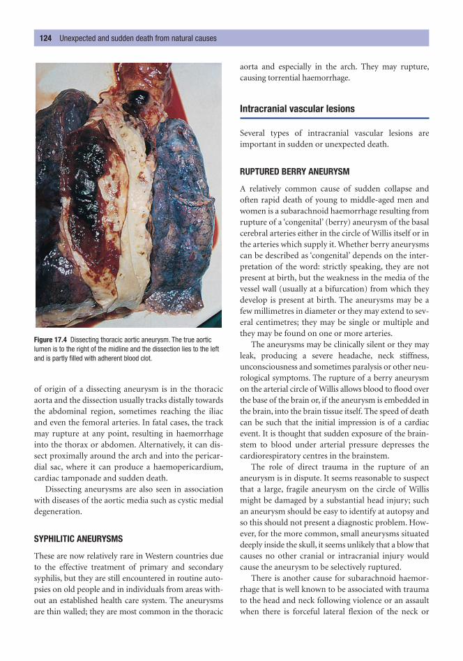
of origin of a dissecting aneurysm is in the thoracicaorta and the dissection usually tracks distally towardsthe abdominal region, sometimes reaching the iliacand even the femoral arteries. In fatal cases, the trackmay rupture at any point, resulting in haemorrhageinto the thorax or abdomen. Alternatively, it can dis-sect proximally around the arch and into the pericar-dial sac, where it can produce a haemopericardium,cardiac tamponade and sudden death.
Dissecting aneurysms are also seen in associationwith diseases of the aortic media such as cystic medialdegeneration.
SYPHILITIC ANEURYSMS
These are now relatively rare in Western countries dueto the effective treatment of primary and secondarysyphilis, but they are still encountered in routine auto-psies on old people and in individuals from areas with-out an established health care system. The aneurysmsare thin walled; they are most common in the thoracic
aorta and especially in the arch. They may rupture,causing torrential haemorrhage.
Intracranial vascular lesions
Several types of intracranial vascular lesions areimportant in sudden or unexpected death.
RUPTURED BERRY ANEURYSM
A relatively common cause of sudden collapse andoften rapid death of young to middle-aged men andwomen is a subarachnoid haemorrhage resulting fromrupture of a ‘congenital’ (berry) aneurysm of the basalcerebral arteries either in the circle of Willis itself or inthe arteries which supply it. Whether berry aneurysmscan be described as ‘congenital’ depends on the inter-pretation of the word: strictly speaking, they are notpresent at birth, but the weakness in the media of thevessel wall (usually at a bifurcation) from which theydevelop is present at birth. The aneurysms may be afew millimetres in diameter or they may extend to sev-eral centimetres; they may be single or multiple andthey may be found on one or more arteries.
The aneurysms may be clinically silent or they mayleak, producing a severe headache, neck stiffness,unconsciousness and sometimes paralysis or other neu-rological symptoms. The rupture of a berry aneurysmon the arterial circle of Willis allows blood to flood overthe base of the brain or, if the aneurysm is embedded inthe brain, into the brain tissue itself. The speed of deathcan be such that the initial impression is of a cardiacevent. It is thought that sudden exposure of the brain-stem to blood under arterial pressure depresses thecardiorespiratory centres in the brainstem.
The role of direct trauma in the rupture of ananeurysm is in dispute. It seems reasonable to suspectthat a large, fragile aneurysm on the circle of Willismight be damaged by a substantial head injury; suchan aneurysm should be easy to identify at autopsy andso this should not present a diagnostic problem. How-ever, for the more common, small aneurysms situateddeeply inside the skull, it seems unlikely that a blow thatcauses no other cranial or intracranial injury wouldcause the aneurysm to be selectively ruptured.
There is another cause for subarachnoid haemor-rhage that is well known to be associated with traumato the head and neck following violence or an assaultwhen there is forceful lateral flexion of the neck or
124 Unexpected and sudden death from natural causes
Figure 17.4 Dissecting thoracic aortic aneurysm. The true aorticlumen is to the right of the midline and the dissection lies to the leftand is partly filled with adherent blood clot.
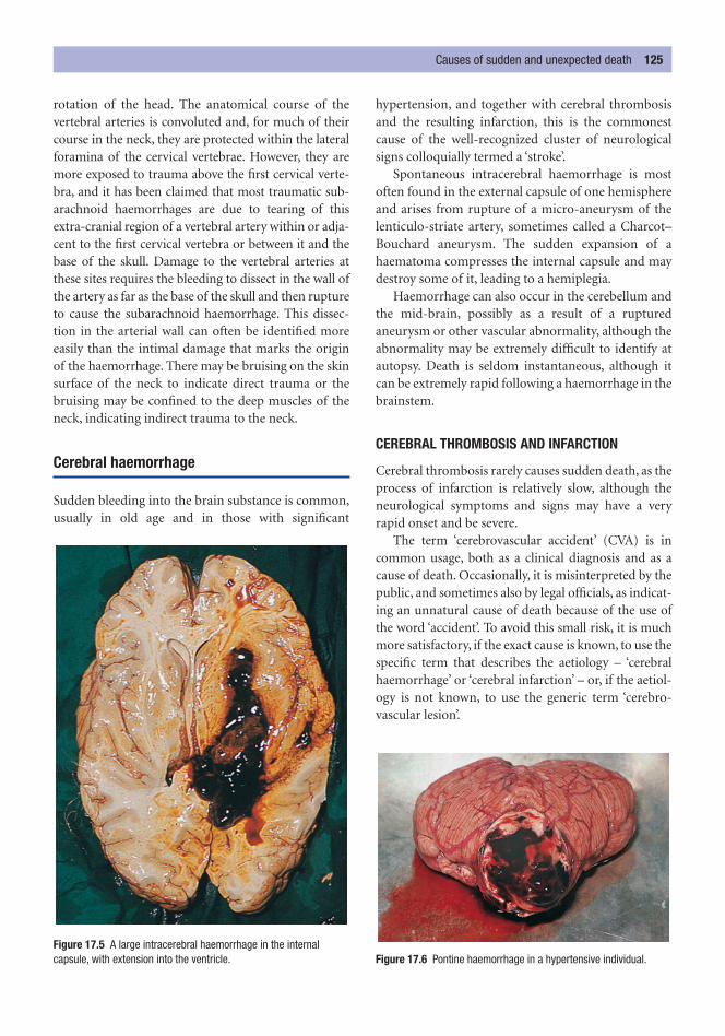
rotation of the head. The anatomical course of the vertebral arteries is convoluted and, for much of theircourse in the neck, they are protected within the lateralforamina of the cervical vertebrae. However, they aremore exposed to trauma above the first cervical verte-bra, and it has been claimed that most traumatic sub-arachnoid haemorrhages are due to tearing of thisextra-cranial region of a vertebral artery within or adja-cent to the first cervical vertebra or between it and thebase of the skull. Damage to the vertebral arteries atthese sites requires the bleeding to dissect in the wall ofthe artery as far as the base of the skull and then ruptureto cause the subarachnoid haemorrhage. This dissec-tion in the arterial wall can often be identified moreeasily than the intimal damage that marks the originof the haemorrhage. There may be bruising on the skinsurface of the neck to indicate direct trauma or thebruising may be confined to the deep muscles of theneck, indicating indirect trauma to the neck.
Cerebral haemorrhage
Sudden bleeding into the brain substance is common,usually in old age and in those with significant
hypertension, and together with cerebral thrombosisand the resulting infarction, this is the commonestcause of the well-recognized cluster of neurologicalsigns colloquially termed a ‘stroke’.
Spontaneous intracerebral haemorrhage is mostoften found in the external capsule of one hemisphereand arises from rupture of a micro-aneurysm of thelenticulo-striate artery, sometimes called a Charcot–Bouchard aneurysm. The sudden expansion of ahaematoma compresses the internal capsule and maydestroy some of it, leading to a hemiplegia.
Haemorrhage can also occur in the cerebellum andthe mid-brain, possibly as a result of a rupturedaneurysm or other vascular abnormality, although theabnormality may be extremely difficult to identify atautopsy. Death is seldom instantaneous, although itcan be extremely rapid following a haemorrhage in thebrainstem.
CEREBRAL THROMBOSIS AND INFARCTION
Cerebral thrombosis rarely causes sudden death, as theprocess of infarction is relatively slow, although theneurological symptoms and signs may have a veryrapid onset and be severe.
The term ‘cerebrovascular accident’ (CVA) is incommon usage, both as a clinical diagnosis and as acause of death. Occasionally, it is misinterpreted by thepublic, and sometimes also by legal officials, as indicat-ing an unnatural cause of death because of the use ofthe word ‘accident’. To avoid this small risk, it is muchmore satisfactory, if the exact cause is known, to use thespecific term that describes the aetiology – ‘cerebralhaemorrhage’ or ‘cerebral infarction’ – or, if the aetiol-ogy is not known, to use the generic term ‘cerebro-vascular lesion’.
Causes of sudden and unexpected death 125
Figure 17.5 A large intracerebral haemorrhage in the internalcapsule, with extension into the ventricle. Figure 17.6 Pontine haemorrhage in a hypertensive individual.

The major cause of sudden death within the respira-tory organs is again vascular. Pulmonary embolism isvery common and, in fact, is the most clinically under-diagnosed cause of death. In almost every case, thesource of the emboli is in the leg veins. Tissue trauma,especially where it is associated with immobility or bedrest, is a very common predisposing factor in the devel-opment of deep vein thrombosis. Most thrombosesremain silent and cause no problems, but a proportionembolize and block pulmonary arteries of varying size.Some produce no lung lesions at all, whereas othersproduce infarcts that may or may not lead to clinicalsigns, and a minority (though an appreciable number)block a major vessel and cause death.
About 80 per cent of pulmonary embolism deathshave a predisposing cause such as fractures, tissuetrauma, surgical operation, bed rest, forced immobil-ity etc., but the remainder occur unexpectedly in nor-mal, ambulant people who have reported no clinicalsymptoms. This makes establishing the relationship ofdeath to an injurious event difficult. For the purposesof civil law (where ‘the balance of probabilities’ isenough), the embolism can often be linked to thetrauma, but in a criminal trial in which the higherstandard of proof – ‘beyond reasonable doubt’ – isrequired, it is much harder to relate fatal pulmonaryemboli to the trauma.
Other rare causes of sudden death in the respira-tory system include a massive haemoptysis from cavi-tating pulmonary tuberculosis or from a malignanttumour. Rapid (but not sudden) deaths can also occurfrom fulminating chest infections, especially virulentforms of influenza.
Once again, the causes of sudden death have a vascularcomponent in that very severe bleeding from a gastricor duodenal peptic ulcer can be fatal in a short time,but, more commonly, the bleeding is less torrentialand is therefore amenable to medical or surgical treat-ment. Mesenteric thrombosis and embolism, usuallyrelated to aortic or more generalized atheroma, mayresult in infarction of the gut, but, once again, a rapidbut not sudden death is expected if the infarctionremains undiagnosed.
Perforation of a peptic ulcer can be fatal in hours ifnot treated and intestinal infarction due to a strangu-lated hernia or obstruction due to torsion of the bowelaround an area of peritoneal adhesions can also be ful-minating and fatal conditions, as can peritonitis arisingfrom diverticular disease or a perforated carcinoma.Many of these conditions present as sudden death inelderly people because they cannot or will not seekassistance at the onset of the symptoms and are thenunable to do so as their condition worsens.
When a woman of childbearing age is found unex-pectedly dead, a complication of pregnancy must beconsidered to be the most likely cause of her deathuntil all such causes are excluded. Abortion remainsone possibility anywhere in the world, but especially incountries where illegal abortion is still very common(see Chapter 19).
A ruptured ectopic pregnancy, which is usuallytubal in position, is another grave obstetric emergencythat can end in death from intraperitoneal bleedingunless rapidly treated by surgical intervention.
As a ‘rule of thumb’, the three commonest causes ofdeath in a woman of reproductive age are:
1 a natural complication of pregnancy such as rup-tured ectopic gestation or an induced complicationsuch as an abortion;
2 pulmonary embolism from leg vein thrombosis;3 ruptured cerebral aneurysm.
In both conditions, death may occur during an attackand the autopsy will reveal specific features to enable apositive diagnosis to be made. However, both of theseconditions are also associated with a few sudden andunexpected deaths each year where the specific fea-tures of an attack are absent and for which no obviouscause of death can be identified at autopsy.
Even well-controlled epileptic patients may dierapidly and inexplicably; it was once thought that theymust have been exhausted from status epilepticus,but this is now not thought to be so. There are somewho doubt that these patients are even having a fit
DEATHS FROM ASTHMA AND EPILEPSY
GYNAECOLOGICAL CONDITIONS
GASTROINTESTINAL SYSTEM
RESPIRATORY SYSTEM
126 Unexpected and sudden death from natural causes

Deaths from asthma and epilepsy 127
when they die because of the lack of pathological fea-tures, but many epileptics have fits that leave nopathological signs, rendering this theory unlikely.Epileptics are also at risk from the hazards of all typesof accidents during a pre-fit aura (if they have one),while having a fit or immediately afterwards; thesehazards include falls, drowning, suffocation and pos-tural asphyxia.
Bronchial asthma is also associated with rare unex-plained sudden and unexpected deaths, even where thereis no evidence of status asthmaticus or even of an unusu-ally severe or prolonged asthmatic episode. Severaldecades ago, an increase in sudden deaths in asthmaticswas traced to the over-use of adrenergic drugs – espe-cially the self-administered dose given by inhaler – butawareness amongst doctors soon reduced this hazard.

Though the legal definitions of particular sexualoffences will vary from country to country, the classi-fication of illegal sexual acts or actions is relativelystandard, even though the names of the variousoffences may differ. The following list is from Englishlaw but can be modified for other jurisdictions.
Rape
Rape is the most serious sexual offence. It can only becommitted by a biological male, but the victim can beof either sex. Rape was re-defined in the CriminalJustice and Public Order Act (1994) as ‘non-consen-sual penetration of the vagina or the anus by a penis’.Consent is crucial and the act is rape if the man eitherknows that the victim does not consent to the act ofintercourse or is reckless (unconcerned) as to whetherthey consent or not. This provision replaces the oldterminology of ‘fear, force or fraud’ because any con-sent given under any of those circumstances wouldnot be valid. The same will apply if alcohol or drugsare used to render the individual unconscious and alsoto intercourse with a sleeping individual.
The law in England and Wales does not distinguishbetween an act between strangers and one betweenthose with a relationship, legal or otherwise, and it is now possible for a man to be convicted of raping his wife. The law only applies to boys over the age of14 years, because English law presumes that a boyunder that age is incapable of rape. This is medicallyuntrue but cannot be challenged in England, althoughit may in Scotland.
Sexual intercourse is no longer part of the defin-ition of rape, having been replaced by the term ‘pene-tration’. The use of this term reflects the legal
TYPES OF SEXUAL OFFENCE
C h a p t e r e i g h t e e n
Sexual Offences
Types of sexual offenceRapeUnlawful sexual intercourseIndecent assaultIndecent exposureIndecency with children
IncestBestialityHomosexual offencesThe genuineness of allegations of sexual
assault
Forensic examination of victims of sexual offences
Examination of an alleged assailant
Figure 18.1 Victim of rape and strangulation. Removal of clothing,bleeding from the perineum and soiling of the skin can be seen.

Types of sexual offence 129
definition of sexual intercourse previously applied,which was any degree of penile insertion, even if this isonly just between the labia or just into the anal mar-gin. Neither full penetration nor ejaculation is neces-sary and, in terms of vaginal rape, the rupture of thehymen is irrelevant.
Individuals with substantial mental disease or defi-ciency that could be defined as ‘severe mental impair-ment’ under the Mental Health Act of 1983 cannot givevalid consent to sexual intercourse, whatever their age.
Unlawful sexual intercourse
In Britain, the age of consent to heterosexual inter-course is 16 years, but it is different in other countries,for instance in some states of the USA it is 18 years.Below that age a girl cannot give legal consent for sexualintercourse and hence any act of intercourse is always‘unlawful’. Sexual intercourse in these circumstances istermed ‘unlawful sexual intercourse’ and it is a chargethat is, in essence, divided into separate offencesdepending on the age of the girl and the man.
If the girl is under 13 years of age, this offence is thesame as rape and it is immaterial if consent was givenor not. The maximum penalty for this offence is lifeimprisonment.
If the girl is over 13 but under 16 years of age, aman can avoid conviction in England and Wales if:
a. (i) he is under 24 years of age and(ii) he has never have been charged with a similar
offence previously and(iii) he believed that the girl was over 16 and
(iv) he had reasonable cause for that belief due toher appearance, dress, make-up and behaviouretc.
or
b. he believed her to be his lawful wife.
Indecent assault
Indecent assault covers a very wide spectrum of acts oractions performed by members of either sex on victimsof either sex. These acts may range from a failed attemptat penile penetration of the vagina or anus, to the actualpenetration of the vagina or anus with a finger or anobject, to penetration of the mouth by the penis (forcedfellatio), to touching or fondling of the buttocks,breasts, thighs, perineum, penis or putting a hand up awoman’s skirt or inside a man’s trousers. Only the moreserious, penetrative forms of indecent assault are likelyto have physical medical aspects unless other injuriesare caused during the incident such as bruises, bitemarks and abrasions. All the acts are very likely to leavethe victims psychologically traumatized.
Indecent exposure
Indecent exposure, often known colloquially as ‘flash-ing’, occurs when a person, usually a man, displays hisgenitals in public to the annoyance and embarrass-ment of members of the public. This is physicallyharmless to victims but may cause them psychologicaldistress. Restraint and psychiatric treatment of theoffender are probably more important than a custo-dial sentence.
Indecency with children
More serious are the men and women who prey onsmall children, not to assault or touch them or theirgenitalia, but to encourage the child to handle or mas-turbate the offender’s sexual organs. In England andWales, a special law was passed in 1960 (Indecencywith Children Act) to make this behaviour illegal witha child below 14 years of age; this closed a loop-hole inthe Sexual Offences Act of 1956.
Figure 18.2 Anal penetration of a young child with dilatation,abrasion, marginal tearing and skin tags. Dilatation alone isinsufficient for a firm diagnosis of buggery.
Incest
Incest is not universally considered to be a criminaloffence; indeed, it was (and is) a socially acceptablepart of normal behaviour in some cultures. The mostcommon situation occurs when a father has sexualintercourse with his own teenage daughter when hiswife was in the later stages of pregnancy or in the puer-perium. In some countries and religions, even fromancient times, it was recognized that there were geneticrisks attached to births arising from the mating of closerelations, and from the Biblical period onward lists of‘prohibited degrees’ of relationships were declared.
The extent of these lists varies from country tocountry and even between religious and secular codesin the same country. In Britain the law forbids mar-riage or penile/vaginal sexual intercourse between aman and a woman he knows to be his mother, daugh-ter, grand-daughter, sister or half-sister; a similar listof exclusions exists for the female side. Both the manand the woman are liable to prosecution for such anact. Other sexual acts between relations are covered byother laws and, if truly consensual, they do not consti-tute an offence.
Doctors, especially family practitioners, may be thefirst to learn of incest in a family and if one participantis a child, they must act immediately to prevent anyfurther acts of incest and to treat the child physicallyand psychologically. If pregnancy or sexually trans-mitted diseases are diagnosed, the child must bereferred to specialists for treatment.
There is a range of further options open to doctorsin socially advanced countries and they must decidehow best to respond, but there is no doubt that the doc-tor’s over-riding duty is to the welfare of the child. Thedoctor may decide to involve the police immediately ormay decide to take professional advice from a coun-selling organization such as the National Society for thePrevention of Cruelty to Children (or the equivalentin other countries), the Social Services Department ofthe city or county authority or possibly from a seniorpaediatrician at a local hospital. Whatever the decision,the doctor should keep full notes of all telephone calls,conversations or other communications with profes-sional organizations and the police as well as with thechild and his or her parents.
When incest occurs between adult relatives, thesituation is far more difficult, as legally the woman (orman) is a free agent and it might be thought that she
could remove herself from the relationship. However,in practice, this is seldom an easy option for her and,in fact, the relationship is often more abusive thansimply incestuous and requests for assistance must betreated very seriously, irrespective of the wishes ofother members of the family.
Bestiality
Bestiality is sexual intercourse with animals, eithervaginal or anal. This includes all animals, includingbirds, the usual victims being pets and farm animals,which are sometimes tortured and killed in the process.Though doctors are rarely involved in the investigationof this offence, they may sometimes be asked to exam-ine genital injuries or infections in a man or womanthat have been sustained in one of these acts.Veterinarysurgeons and forensic scientists are more commonlyinvolved in relation to injuries to animals and in thedetection and identification of animal secretions andhairs found on a suspect and human semen in the ani-mal. Though bestiality is more common than usuallythought, especially in the countryside, few prosecu-tions are ever brought.
Homosexual offences
In many countries, all sexual acts between men areillegal and the penalties may be severe. In England andWales, the Sexual Offences Act (1967) allowed forhomosexual acts in private between consenting malesover the age of 21 years, but with the specific exclusionof members of the armed forces or merchant seamen.The age of consent has now been reduced to 18 yearsand there is considerable pressure to reduce it furtherto 16 to match the heterosexual age of consent forfemales. In Scotland and Ireland, no such act wasintroduced, but prosecutions are now rarely if everbrought. Homosexual acts between females (‘lesbian-ism’) have never been a criminal offence unless theyoffend against public decency.
Under the terms of the 1967 act, anal intercoursebetween consenting males is not an offence, but analintercourse with a woman or a boy over the age of 16but below the minimum age stipulated in the act isalways an offence whether they consent or not. Oral or
130 Sexual offences
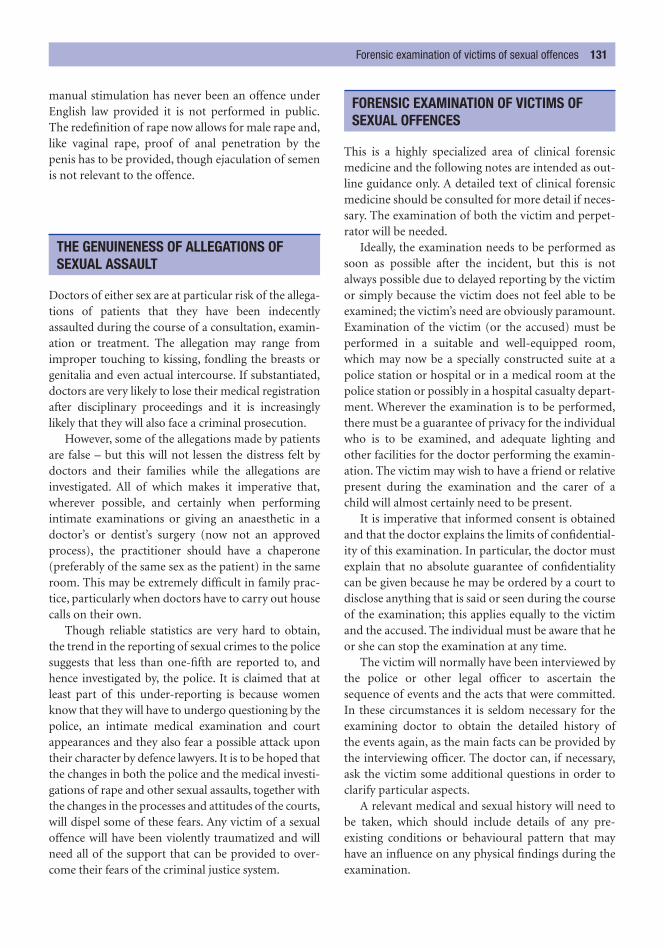
manual stimulation has never been an offence underEnglish law provided it is not performed in public.The redefinition of rape now allows for male rape and,like vaginal rape, proof of anal penetration by thepenis has to be provided, though ejaculation of semenis not relevant to the offence.
Doctors of either sex are at particular risk of the allega-tions of patients that they have been indecentlyassaulted during the course of a consultation, examin-ation or treatment. The allegation may range fromimproper touching to kissing, fondling the breasts orgenitalia and even actual intercourse. If substantiated,doctors are very likely to lose their medical registrationafter disciplinary proceedings and it is increasinglylikely that they will also face a criminal prosecution.
However, some of the allegations made by patientsare false – but this will not lessen the distress felt bydoctors and their families while the allegations areinvestigated. All of which makes it imperative that,wherever possible, and certainly when performingintimate examinations or giving an anaesthetic in adoctor’s or dentist’s surgery (now not an approvedprocess), the practitioner should have a chaperone(preferably of the same sex as the patient) in the sameroom. This may be extremely difficult in family prac-tice, particularly when doctors have to carry out housecalls on their own.
Though reliable statistics are very hard to obtain,the trend in the reporting of sexual crimes to the policesuggests that less than one-fifth are reported to, andhence investigated by, the police. It is claimed that atleast part of this under-reporting is because womenknow that they will have to undergo questioning by thepolice, an intimate medical examination and courtappearances and they also fear a possible attack upontheir character by defence lawyers. It is to be hoped thatthe changes in both the police and the medical investi-gations of rape and other sexual assaults, together withthe changes in the processes and attitudes of the courts,will dispel some of these fears. Any victim of a sexualoffence will have been violently traumatized and willneed all of the support that can be provided to over-come their fears of the criminal justice system.
This is a highly specialized area of clinical forensicmedicine and the following notes are intended as out-line guidance only. A detailed text of clinical forensicmedicine should be consulted for more detail if neces-sary. The examination of both the victim and perpet-rator will be needed.
Ideally, the examination needs to be performed assoon as possible after the incident, but this is notalways possible due to delayed reporting by the victimor simply because the victim does not feel able to beexamined; the victim’s need are obviously paramount.Examination of the victim (or the accused) must beperformed in a suitable and well-equipped room,which may now be a specially constructed suite at apolice station or hospital or in a medical room at thepolice station or possibly in a hospital casualty depart-ment. Wherever the examination is to be performed,there must be a guarantee of privacy for the individualwho is to be examined, and adequate lighting andother facilities for the doctor performing the examin-ation. The victim may wish to have a friend or relativepresent during the examination and the carer of achild will almost certainly need to be present.
It is imperative that informed consent is obtainedand that the doctor explains the limits of confidential-ity of this examination. In particular, the doctor mustexplain that no absolute guarantee of confidentialitycan be given because he may be ordered by a court todisclose anything that is said or seen during the courseof the examination; this applies equally to the victimand the accused. The individual must be aware that heor she can stop the examination at any time.
The victim will normally have been interviewed bythe police or other legal officer to ascertain thesequence of events and the acts that were committed.In these circumstances it is seldom necessary for theexamining doctor to obtain the detailed history ofthe events again, as the main facts can be provided bythe interviewing officer. The doctor can, if necessary,ask the victim some additional questions in order toclarify particular aspects.
A relevant medical and sexual history will need tobe taken, which should include details of any pre-existing conditions or behavioural pattern that mayhave an influence on any physical findings during theexamination.
FORENSIC EXAMINATION OF VICTIMS OF SEXUAL OFFENCES
THE GENUINENESS OF ALLEGATIONS OF SEXUAL ASSAULT
Forensic examination of victims of sexual offences 131
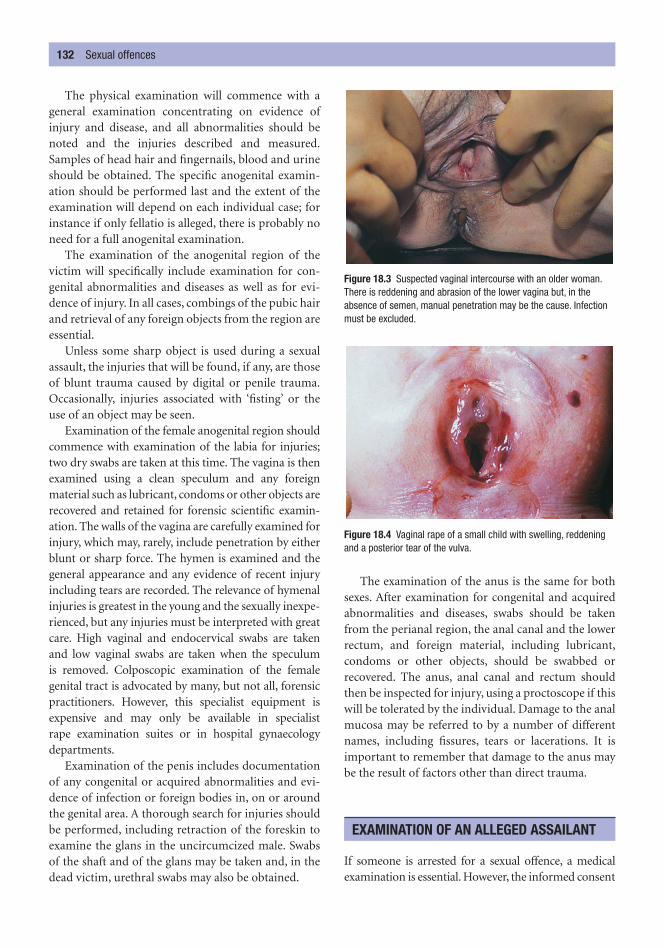
The physical examination will commence with ageneral examination concentrating on evidence ofinjury and disease, and all abnormalities should benoted and the injuries described and measured.Samples of head hair and fingernails, blood and urineshould be obtained. The specific anogenital examin-ation should be performed last and the extent of theexamination will depend on each individual case; forinstance if only fellatio is alleged, there is probably noneed for a full anogenital examination.
The examination of the anogenital region of thevictim will specifically include examination for con-genital abnormalities and diseases as well as for evi-dence of injury. In all cases, combings of the pubic hairand retrieval of any foreign objects from the region areessential.
Unless some sharp object is used during a sexualassault, the injuries that will be found, if any, are thoseof blunt trauma caused by digital or penile trauma.Occasionally, injuries associated with ‘fisting’ or theuse of an object may be seen.
Examination of the female anogenital region shouldcommence with examination of the labia for injuries;two dry swabs are taken at this time. The vagina is thenexamined using a clean speculum and any foreignmaterial such as lubricant, condoms or other objects arerecovered and retained for forensic scientific examin-ation. The walls of the vagina are carefully examined forinjury, which may, rarely, include penetration by eitherblunt or sharp force. The hymen is examined and thegeneral appearance and any evidence of recent injuryincluding tears are recorded. The relevance of hymenalinjuries is greatest in the young and the sexually inexpe-rienced, but any injuries must be interpreted with greatcare. High vaginal and endocervical swabs are takenand low vaginal swabs are taken when the speculum is removed. Colposcopic examination of the femalegenital tract is advocated by many, but not all, forensicpractitioners. However, this specialist equipment isexpensive and may only be available in specialist rape examination suites or in hospital gynaecologydepartments.
Examination of the penis includes documentationof any congenital or acquired abnormalities and evi-dence of infection or foreign bodies in, on or aroundthe genital area. A thorough search for injuries shouldbe performed, including retraction of the foreskin toexamine the glans in the uncircumcized male. Swabsof the shaft and of the glans may be taken and, in thedead victim, urethral swabs may also be obtained.
The examination of the anus is the same for bothsexes. After examination for congenital and acquiredabnormalities and diseases, swabs should be takenfrom the perianal region, the anal canal and the lowerrectum, and foreign material, including lubricant,condoms or other objects, should be swabbed orrecovered. The anus, anal canal and rectum shouldthen be inspected for injury, using a proctoscope if thiswill be tolerated by the individual. Damage to the analmucosa may be referred to by a number of differentnames, including fissures, tears or lacerations. It isimportant to remember that damage to the anus maybe the result of factors other than direct trauma.
If someone is arrested for a sexual offence, a medicalexamination is essential. However, the informed consent
EXAMINATION OF AN ALLEGED ASSAILANT
132 Sexual offences
Figure 18.3 Suspected vaginal intercourse with an older woman.There is reddening and abrasion of the lower vagina but, in theabsence of semen, manual penetration may be the cause. Infectionmust be excluded.
Figure 18.4 Vaginal rape of a small child with swelling, reddeningand a posterior tear of the vulva.

of the individual is still needed in most countries. Theaccused may wish to have a legal representative presentand these wishes should be accommodated. The doctorwill need to take, and to make a note of, a general his-tory of the events as given by the perpetrator and alsotheir general medical and social history, and the limita-tions of confidentiality must be explained clearly, aswith the victims of a sexual assault.
A general examination of the body for signs of anyinjuries is essential. If there has been a struggle, the per-petrator may have received injuries, and groups of linearparallel scratches on the face, neck and chest from thevictim’s fingernails are not uncommon. Examination ofthe anogenital region rarely reveals any significant signs,although there may be some non-specific tendernessand reddening. Exceptionally, the glans or prepuce maybe bruised and the frenum of the penis damaged, andfaecal material may be recovered if anal penetration hasbeen achieved. The use of Lugol’s iodine test is nowredundant: the idea was that iodine solution paintedon to the glans would reveal the presence of vaginal
squames by turning them brown due to the glycogencontained in the vaginal cells.
The recovery of trace evidence is often more import-ant than the genital inspection. Samples of pubic hairshould be taken, along with head and even moustacheor beard hair if appropriate. The pubic hair should becombed out to recover foreign hairs or fibres. Bloodsamples for grouping, alcohol and DNA are taken andany evidence of venereal infection noted.
In small boys and children, damage to the anal ringis more likely than in older victims, though caution isneeded in assessing anal size, especially in the dead,in whom post-mortem dilatation can be marked. Inextreme cases, tearing of the mucosa may be seen. Inolder habitual homosexuals, the anus may be perman-ently dilated, with loss of sphincter tone and exposureof the anal mucosa. The older description of a ‘funnel-shaped anus’ with keratinization and thickening of theexternal skin is a rare phenomenon, but some silveryscaliness of the skin can occur adjacent to the anusfrom repeated penetration.
Examination of an alleged assailant 133
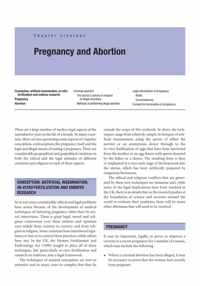
There are a large number of medico-legal aspects of thereproductive years in the life of a female. In many coun-tries, there are laws governing some aspects of virginity,conception, contraception, the pregnancy itself and thelegal and illegal means of ending a pregnancy. There areconsiderable geographical and geopolitical variations inboth the ethical and the legal attitudes of differentcountries and religions to each of these aspects.
In recent years considerable ethical and legal problemshave arisen because of the development of medicaltechniques of initiating pregnancy other than by sex-ual intercourse. There is great legal, moral and reli-gious controversy over these matters and opinionsvary widely from country to country and from reli-gion to religion. Some countries have introduced legis-lation to ban or to control these practices, while othershave not. In the UK, the Human Fertilization andEmbryology Act (1990) sought to place all of thesetechniques, but particularly in-vitro fertilization andresearch on embryos, into a legal framework.
The techniques of assisted conception are now soextensive and in many cases so complex that they lie
outside the scope of this textbook. In short, the tech-niques range from relatively simple techniques of arti-ficial insemination using the sperm of either thepartner or an anonymous donor through to the in-vitro fertilization of eggs that have been harvestedfrom the mother or an egg donor with sperm donatedby the father or a donor. The resulting fetus is then re-implanted at a very early stage of development intothe uterus, which has been artificially prepared byexogenous hormones.
The ethical and religious conflicts that are gener-ated by these new techniques are immense and, whilesome of the legal implications have been resolved inthe UK, there is no doubt that as the research pushes atthe boundaries of science and societies around theworld re-evaluate their positions, there will be manyother dilemmas that will need to be resolved.
It may be important, legally, to prove or disprove acurrent or a recent pregnancy for a number of reasons,which may include the following.
• Where a criminal abortion has been alleged, it maybe necessary to prove that the woman had recentlybeen pregnant.
PREGNANCY
CONCEPTION: ARTIFICIAL INSEMINATION,IN-VITRO FERTILIZATION AND EMBRYORESEARCH
C h a p t e r n i n e t e e n
Pregnancy and Abortion
Conception: artificial insemination, in-vitrofertilization and embryo research
PregnancyAbortion
Criminal abortionThe doctor’s actions in respect
of illegal abortionsMethods of performing illegal abortion
Legal termination of pregnancyRulesCircumstances
Consent to termination of pregnancy

Abortion 135
• In some civil disputes about the estate of a deadhusband, the pregnancy of the wife may affect thedisposition of property.
• Women in advanced pregnancy may be excusedattendance as a witness in a court: in those coun-tries that still have capital punishment, the preg-nancy of a convicted woman may postpone orcancel her execution.
• In charges of infanticide or concealment of birth,it may be relevant to show that the woman hadrecently been pregnant.
• For a variety of reasons, pregnancy may be a pre-tence and medical evidence may be required to dis-prove the condition.
Many of the reasons previously advanced for a clin-ical medical examination to determine the fact of, or theduration of, a pregnancy in an attempt to establishpaternity have now been superseded by DNA analysis ofthe fetus or the baby and of the putative father(s). Morerecently, DNA has also been used to establish maternityand a woman has been charged with infanticide.
Evidence of pregnancy consists of both clinical signsand biochemical testing. Biochemical tests on urine forpregnancy-related hormones are now so sophisticatedand accurate that they are sensitive enough to be reli-ably positive as early as a week or so after conception.
Clinical signs in the first trimester include breastswelling and tenderness, changes in the nipples, amen-orrhoea, frequency of micturition, (inconstant) nausea,leucorrhoea, softening of the cervix and ballotment of the uterus on vaginal examination (Hegar’s sign).Ultrasound scanning can identify pregnancy as early as4 weeks.
Later in pregnancy, the uterine enlargementextends over the pelvic brim; palpation of the fetus ispossible at about 16 weeks and palpation of fetal partsand the detection of fetal heart sounds are possible atabout 24 weeks. Fetal movements may be detected andradiological and ultrasonic evidence of the fetus canbe easily obtained.
Evidence of a recent or past pregnancy and of par-turition may be needed for medico-legal purposes, inthe living or the dead. No single feature is diagnostic,but the presence of groups of features may allow for anaccurate assessment. Following a recent vaginal deliv-ery there will be dilatation and laxity of the vaginalintroitus, sometimes associated with perineal damageor evidence of an episiotomy. The uterus remains palp-able above the symphysis pubis for up to 2 weeks.
A vaginal discharge (lochia) may be bloodstained for a few days and then brown for a week or more.The breast changes will depend on whether feeding isestablished, discontinued or suppressed by hormones.Whatever the ultimate appearance, immediately afterdelivery the breasts will be swollen and tense and will often have prominent surface veins. Milk will beexpressible after the second or third day. The nippleand areolae will be darkened, a change that is non-specific but that may persist in some degree almostpermanently.
On the abdomen, the stretch marks (striae gravi-darum), which are pink in late pregnancy and aroundthe time of delivery, become paler with time and, aftera year or so, eventually appear as white scars (lineaealbicantes). The cervix remains dilated and soft forsome time and may show a tear, which eventuallyforms a scar. The breast changes subside with the ces-sation of breast-feeding but the nipple darkening maypersist, especially in dark-complexioned women. Theremains of the hymen will either vanish completely orremain only as small remnants (carunculae mytri-formes) and there is inconstant loss of the rugosity ofthe vaginal walls seen in virgins.
The normal duration of pregnancy is about 40weeks or 280 days, though there is naturally no min-imum period. The maximum duration of pregnancy isdisputed, but medically it is unlikely to extend formore than 42–43 weeks. Legal decisions that preg-nancy may extend to 350 days or more are medicallyuntenable and were probably introduced to associate apregnancy with the last known intercourse with thehusband. In Nigeria, a claim that a pregnancy had ‘laindormant’ for a number of years was accepted by a reli-gious court; it is doubtful that such a claim would havebeen accepted elsewhere. The problems of paternityare now easily resolved by the use of DNA profiles,although these tests are expensive and may not beavailable throughout the world.
A considerable proportion of pregnancies fail to pro-ceed to full term. About 20 per cent of known preg-nancies fail and it is estimated that many others havefailed so early that pregnancy is not even recognized.After a pregnancy is apparent, the loss of the fetus upto 24 weeks is usually called a ‘spontaneous abortion’
ABORTION
or ‘miscarriage’; from 24 weeks to full term, it may becalled a ‘premature birth’. However, these temporaldefinitions are not universally accepted.
In addition to the spontaneous (natural) abor-tions, abortions may also be induced and theseinduced abortions may be therapeutic or criminal,depending on the circumstances and the prevailinglaw of the country in which the abortion occurs.
Criminal abortion
A criminal abortion is the deliberate ending of a preg-nancy outside any legal provisions that have beenmade by the state for this act. In most countries, per-forming a criminal abortion is a serious criminaloffence and, in addition to any criminal conviction, itwould form grounds for the strict disciplining of anydoctor who was shown to be involved.
Before the introduction of legal abortion in Britainin 1967, it was thought that between 40 and 80 per centof all reported abortions were deliberately induced,either by the woman herself or by another person. Theintroduction of easily available and reliable contracep-tion has reduced the number of unwanted pregnan-cies, and the introduction of legal abortion in manyparts of the world has greatly reduced the morbidityand mortality from criminal abortion, as it has allowedfor state control of the conditions under which abor-tions are performed and for control of the training andskills of the operators performing them.
There is continual pressure throughout the worldfor the repeal of liberal abortion laws, and counter-movements motivated by politics or religion mayagain restrict or even abolish legal termination ofpregnancy. There is no doubt that the inevitable resultof the abolition of legal abortion will be increasedwork for the gynaecologist and pathologist in dealingwith a revival of criminal abortion and with the dis-eases, disability and deaths that will travel in its wake.
Where no provisions exist for legal abortions,women are still at the mercy of ‘back-street abortion-ists’, who are sometimes doctors, nurses or midwivesbut whose methods may be as unsatisfactory as theirpremises and instruments. Alternatively, an abortionmay be induced by the woman herself – in some placesthis is said to be a common practice amounting almostto regular birth control.
Nearly all criminal abortions used to take place inthe second or third month of pregnancy, when the
woman could be certain that the missed periods andmorning sickness had confirmed her pregnancy. Theincreasing use of sophisticated ‘do-it-yourself ’ preg-nancy tests in many countries will allow for earlierdiagnosis of pregnancy and may result in earlier attend-ance for abortion.
THE DOCTOR’S ACTIONS IN RESPECT OF ILLEGAL ABORTIONS
Unless the law of the country allows termination ofpregnancy, albeit sometimes only in specific and verylimited circumstances, no doctor may abort a womanfor any reason; as noted above, a criminal convictionand loss of medical registration may follow such anact. Where the law of a country does allow for thera-peutic abortion, doctors with strong religious ormoral objections to abortion cannot be forced by theirterms of employment or contractual obligations toperform or assist in any termination of pregnancy. Evenwhere abortion is permitted by law, doctors cannot beforced to take part in this act, but neither should theyobstruct a patient wishing to obtain an abortion, butshould instead refer her to a doctor who will be willingto assist her.
When a doctor learns that a criminal abortion hastaken place, his obligations depend on the circum-stances. If the act was performed by the woman her-self, the doctor should confine himself to giving allnecessary medical care, but it is not part of his duty toreport the fact to the police or anyone else unless deathoccurs, in which case he is naturally obliged to notifythe coroner or whatever other legal authority investi-gates unnatural deaths.
Unless doctors feel that their duty to the publicover-rides their duty of confidentiality to their patient,they should not divulge the matter unless the womangives her consent for them to do so. The law has some-times thought otherwise, as in Britain in 1914, when ajudge said that, ‘The desire for a doctor to preserveconfidentiality must be subordinated to the duty whichis cast upon every good citizen to assist in the investi-gation of a serious crime’. The British Medical Associ-ation and the Royal College of Physicians took legaladvice and resolved that, ‘A medical practitioner shouldnot under any circumstances disclose voluntarily, with-out the patient’s consent, information obtained fromthat patient in the course of professional duties’. Thisis to be supported, but it must be remembered that thedoctor has a duty of confidentiality to the patient and
136 Pregnancy and abortion

not to the abortionist, whose name, if known, could bedisclosed to the police or other authority without anybreach of confidentiality.
METHODS OF PERFORMING ILLEGAL ABORTION
The common methods of abortion vary greatly fromcountry to country and range from semi-magical folkremedies to the sophisticated clinical methods identi-cal to those used during a legal abortion. The follow-ing are the most common means of procuring abortion;however, few of them will be seen in countries withlegal abortion laws.
Drugs and toxins have been used for millennia totry to remove unwanted pregnancies, but most ofthem are quite ineffective. In Western countries, thelong list of substances formerly documented in foren-sic textbooks is of historical interest only. However, indeveloping countries, many folk medicines and moredrastic poisons may be encountered, including a hostof vegetable compounds such as juniper, pennyroyal,savin, rue, colocynth and aloes. In South-East Asia,unripe pineapple is alleged to produce miscarriage.However, none of these substances is known to haveany predictable or significant effect on the uterus andthey are often sold to exploit distressed women.
Other substances that have a contractile effect onsmooth muscle have a better theoretical chance ofsuccess and these include ergot, pituitary extracts andquinine. Other substances normally used in obstetrics,particularly prostaglandins, are also used to induceabortions and are potentially more potent. However, inearly pregnancy the uterus is not primed to respond tothese drugs. Another group of chemicals is the strongpurgatives such as colocynth, croton oil or jalap, whichsimply cause general debility to the woman, but theyare highly unlikely to cause an abortion.
Instrumentation is commonly practised, involvinga wide variety of devices from surgical probes andbougies to the crudest implements made of metal orwood, as well as bicycle spokes, metal coat-hangers oreven twigs from trees. The dangers of instrumentationare perforation and infection. Medical or paramedicalabortionists may have an adequate knowledge of theanatomy of the female genital tract and of the need for the instruments to be clean and preferably sterile,and so the dangers are less than with the same actsperformed by many lay people who have no idea ofthe structure of the genital tract or of the need forcleanliness.
Instruments may be forced through the vaginalwalls, especially the posterior fornix, or through thewalls of the cervix or uterus. The fundus may be perfo-rated and the instrument pushed up into the intestinesor even the liver. All of these injuries can cause rapiddeath from haemorrhage or shock or delayed deathfrom haemorrhage, shock and infection. The Higginsonsyringe (widely sold as a method of giving an enema) isstill used as a means of inducing an abortion, despite itscontinued association with fatal air embolism.
Local interference other than instrumentation maytake the form of chemicals applied to the cervix orinterior of the uterus. Potassium permanganate crys-tals applied to the upper vagina and cervix have causedchemical burns and death from absorption of per-manganate. Chemical mixtures (‘Utus Paste’ or‘Interruptin’) were once used to induce therapeuticabortions by acting as endometrial irritants.
General violence in an attempt to induce a ‘sponta-neous’ abortion may still be encountered on rare occa-sions. This may involve widespread injury from theinfliction of multiple injuries or from a fall downstairs or from a height, or there may be more localizeddirect blunt injury (often punching or kicking) to theabdominal wall. These injures may be self-inflicted orthey may be inflicted by the husband, the father orsome other ‘helper’. The more diffuse injuries will bevery difficult to distinguish from an assault that maypossibly have been occasioned by the announcementof the pregnancy.
Other methods that are thought to be capable ofinducing a ‘spontaneous’ abortion include the taking ofvery hot baths, ingestion of large quantities of alcohol,vaginal douches with many substances, and violentexercise such as skipping, jumping and even ridinghorses or bicycles over rough ground. However, fetusesare generally very persistent and these methods very sel-dom work. Indeed, pregnant women who die as a resultof trauma (for instance road traffic accidents) usuallyhave no evidence of dislodgement of the pregnancy.
Syringe aspiration is a method used by both legal andcriminal abortionists. A large syringe with an attachedplastic cannula is pushed through the cervix, suction isapplied, which ruptures the early gestational sac andleads to aspiration or later expulsion of the contents.
The dangerous or fatal effects of illegal abortionconsist of:
• haemorrhage from local genital trauma duringclumsy instrumental interference;
Abortion 137
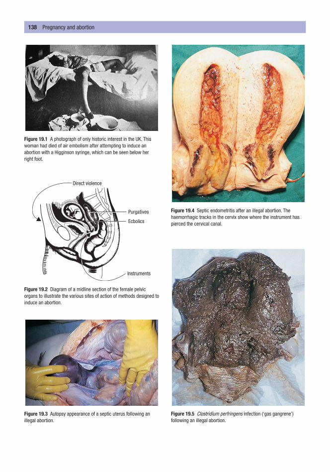
138 Pregnancy and abortion
Figure 19.1 A photograph of only historic interest in the UK. Thiswoman had died of air embolism after attempting to induce anabortion with a Higginson syringe, which can be seen below herright foot.
Figure 19.3 Autopsy appearance of a septic uterus following anillegal abortion.
Figure 19.4 Septic endometritis after an illegal abortion. Thehaemorrhagic tracks in the cervix show where the instrument haspierced the cervical canal.
Figure 19.5 Clostridium perfringens infection (‘gas gangrene’)following an illegal abortion.
Direct violence
Purgatives
Ecbolics
Instruments
Figure 19.2 Diagram of a midline section of the female pelvicorgans to illustrate the various sites of action of methods designed toinduce an abortion.

• sepsis due to the use of non-sterile instruments ormethods and the lack of antibiotic prophylaxis;
• ‘shock’, which may be associated with haemorrhageor perforation of the vagina, uterus or adjacentorgans; it may also be an immediate and some-times fatal response to the dilatation of the cervixof an unanaesthetized woman;
• air embolism, which may occur during any form of instrumentation within the uterus, althoughinjection of fluid into the uterus (with a Higginsonsyringe) is a common cause;
• later systemic complications, which include: throm-bosis of the leg and pelvic veins with the subse-quent risk of fatal pulmonary embolism; infection,which brings the risk of septicaemia and the associ-ated complications of disseminated intravascularcoagulation; hepatic and renal failure etc; and
• sterility, which is a very common complication ofthese highly dangerous procedures.
Legal termination of pregnancy
Many countries now have legal provisions for themedical termination of pregnancy. This is often called‘therapeutic abortion’, which is not exactly synonym-ous with ‘medical termination’ because in some coun-tries a woman has the right to have an ‘abortion ondemand’ without having to satisfy any medical pre-conditions; such an operation can hardly be called‘therapeutic’.
Most countries, including Britain, require thatsome medical condition be proved before the abortionis sanctioned, though the scope and strictness of suchproof vary widely.
The Hippocratic Oath totally banned the involve-ment of doctors in abortions, a view that was con-firmed by the World Medical Association Declarationof Geneva in 1948. This view was modified by the sameassociation 22 years later in 1970, when the Declarationof Oslo was approved, which accepted that doctorscould participate in abortions but only where this wassanctioned by the laws of their own country.
The Roman Catholic and Islamic religions are fun-damentally opposed to any termination of pregnancy,although most religious codes would not prohibit thetruly therapeutic termination of a pregnancy in awoman who was likely to die if the pregnancy pro-ceeded to full term. The Infant Life Preservation Act of1929 gave limited protection to a doctor who per-
formed an abortion ‘for the purpose only of preserv-ing the life of the mother’, provided that the operationwas performed in good faith.
The wide variation in the laws and regulations thatrelate to abortions from country to country meansthat it is impossible to give an account here of the dif-ferent legal provisions for termination of pregnancyaround the world. These laws and regulations arechanging all the time, some countries becoming moreliberal while others become more restrictive. One ofthe best-known pieces of legislation that allows fortherapeutic abortion on medical grounds was theAbortion Act of 1967, which relates to England, Walesand Scotland but not to Northern Ireland. ThisAbortion Act was modified in 1990 by the HumanFertilization and Embryology Act. The fundamentalrules governing the provision of staff and premises forabortions and the four circumstances that allow for adecision to be taken to perform a therapeutic abortionare set out below.
RULES
• The termination must be performed by a registeredmedical practitioner.
• It must be carried out in a Health Service hospitalor a place specifically registered for the purpose.
• Two registered medical practitioners must examinethe woman (not necessarily together) and certifythat grounds for termination exist. Neither of thesedoctors needs to be the doctor who actually per-forms the operation.
• The termination must be notified to medical offi-cers of the appropriate government department.
CIRCUMSTANCES
• The pregnancy must not exceed its twenty-fourthweek and that the continuance of pregnancy wouldinvolve risk, greater than if the pregnancy were ter-minated, of injury to the physical or mental healthof the pregnant woman or any existing children ofher family; or
• The termination is necessary to prevent grave per-manent injury to the physical or mental health ofthe pregnant woman; or
• The continuance of the pregnancy would involverisk to the life of the pregnant woman, greater thanif the pregnancy were terminated; or
Abortion 139

140 Pregnancy and abortion
• There is a substantial risk that if the child wereborn, it would suffer from such physical or mentalabnormalities as to be seriously handicapped.
In an emergency, to save the life of or to prevent‘grave permanent injury to the physical or mentalhealth of the pregnant woman’, a termination may becarried out anywhere on the certification of only onemedical practitioner.
Consent to termination of pregnancy
The rules for consent to a termination of pregnancyare no different from those applicable to any othermedical or surgical procedure. In most Western coun-tries (where therapeutic abortion is lawful), the onlyconsent that is required for termination of pregnancyis from the woman herself, provided she is competentto make that decision. The consent of a husband orpermanent partner (if he accompanies the woman)may be obtained as good clinical practice; however, thewoman’s wishes are paramount and a husband’s orpartner’s objections are not relevant in English law. Itis possible that when the grounds for the abortion arethe health of the children of the existing family, theviews of the husband may be relevant and may influ-ence the decision of the two certifying doctors.
The consent or otherwise of a more casual‘boyfriend’ or even the putative father of the child isnot relevant and although several attempts have beenmade through the courts to establish the right of theputative father to object to an abortion of ‘his’ child,none has yet succeeded.
Any competent young girl may seek medial adviceand may consent on her own to an abortion, althoughfor girls under 16 the doctor should seek to encouragethem to discuss the matter with their parents or carers.This principle was established in 1985 by a ruling fromthe House of Lords in the Gillick case. The key ques-tion is the competence of the girl and if, in the opinionof the doctor who will perform the abortion, she iscapable of understanding the details, implications andpossible consequences of the abortion, she can legallyconsent. There are two exceptions, one absolute andone partial. The absolute exception is a girl who is award of court, in which case only the court can giveconsent; the partial exception is a girl who is legally inthe care of the local authority, in which case the BritishMedical Association advises that it would be prudentfor the consent of her carers to be obtained.
Conversely, a girl of any fertile age, even under 16,certainly cannot be made to have an abortion on thewishes of her parents or anyone else if she herself doesnot agree.

The results and effects of disease and trauma are, ingeneral terms, similar in all victims, but there are somespecial features of injuries to infants and children thatrequire consideration. It is important to rememberthat babies and children are not just small adults andthey have their own unique problems, such as still-birth and sudden infant death syndrome, which haveimportant medico-legal aspects. They are also particu-larly dependent and hence vulnerable to abuse, bothphysical and sexual.
A significant number of pregnancies that continueinto the third trimester fail to deliver a live child andresult in a stillbirth. The definitions of a stillbirth varyfrom country to country, but the British definition,which dates from 1990 and takes into account theadvances in resuscitation and care of premature infants,is perhaps typical: the child must be of more than 24 weeks’ gestational age and, after being completelyexpelled from the mother, did not breathe or show anysigns of life. These ‘signs of life’, in addition to respira-tion and heart beat, are taken to mean movement,crying or pulsation of the umbilical cord.
If death has occurred more than a couple of daysbefore birth, the fetus is commonly macerated due to
the effects of early decomposition combined withexposure to fluid. The infant is discoloured, usually apinkish-brown or red, with extensive desquamation ofthe skin; the tissues have a soft, slimy translucence andthere may be partial collapse of the head with over-riding of the skull plates. The appearance of the childis quite different from that seen in an infant who hasdied following a live birth and then begun to putrefy.Because many, if not most, of these deaths occur afterthe onset of labour and during the process of birthitself, no evidence of decomposition will be present.
In English law, a child does not have a ‘separateexistence’ (and thus does not become a legal person,
STILLBIRTHS
C h a p t e r t w e n t y
Deaths and Injury in Infancy
StillbirthsInfanticideThe estimation of maturity of a newborn
baby or fetus
Sudden infant death syndromeChild abuse
Child physical abuseEpidemiology
General featuresInjuries
Child sexual abuse
Figure 20.1 The body of a newborn baby with the placenta stillattached was left in a blanket by the side of a country road.At autopsy, no evidence of a separate existence could be proved.

capable of acquiring legal rights of inheritance, titleetc.) until he or she is completely free from the mother’sbody, though the cord and placenta may still be withinthe mother. The Infant Life Preservation Act (1929)made it an offence to destroy a baby during birth; how-ever, an exception was made in the Act for doctors act-ing ‘in good faith’ who, to save the life of the mother,have to destroy a baby that becomes impacted in thepelvis during birth.
As babies who are stillborn have never ‘lived’ in thelegal sense, they cannot ‘die’ and so a death certificatecannot be issued. In Britain, a special ‘stillbirth certifi-cate’ may be completed either by a doctor or a midwifeif either was present at the birth or by either one of themif they have examined the body of the child after birth.
The causes of stillbirth are varied and may be unde-terminable, even after a full autopsy. Indeed, it may beimpossible to determine on the pathological featuresalone if the death occurred before, during or after birth.Recognized conditions leading to intrauterine or peri-mortem death include prematurity, fetal hypoxia, pla-cental insufficiency, intra-uterine infections (viral,bacterial or fungal), congenital defects (especially in thecardiovascular or nervous system) and birth trauma.
The term infanticide has a very specific meaning in the many countries that have introduced legislationdesigned to circumvent the criminal charge of murderwhen a mother kills her child during its first year of life.The Infanticide Act of England and Wales states that:
Where a woman by a wilful act of omission or com-mission causes the death of her child being underthe age of twelve months but at the time the balanceof her mind was disturbed by the effects of child-birth or lactation she may be dealt with as if she hadcommitted manslaughter.
Because manslaughter is a less serious charge thanmurder and does not result in the mandatory penaltyof life imprisonment that is attached to murder, a ver-dict of infanticide allows the court to make an appro-priate sentence for the mother, which is more likely tobe probation and psychiatric supervision than cus-tody. The Infanticide Act recognizes the special natureof infanticide and the particular effects pregnancy andlactation may have upon the woman.
In English law, there is a presumption that all deadbabies are stillborn and so the onus is on the prosecu-tion, and hence the pathologist, to prove that the childwas born alive and had a separate existence. In order todo this, it must be shown that the infant breathed orshowed other signs of life, such as movement or pulsa-tion of the umbilical cord, after having been com-pletely expelled from the mother.
INFANTICIDE
142 Death and injury in infancy
Figure 20.2 A newborn infant showing marked maceration due toan intrauterine death. The cord around the neck is significant, but a full autopsy is required to establish the cause of death.
Figure 20.3 Examination of the brain of a newborn infant showshaemorrhage over the temporal lobe due to an unattended andprecipitate birth.

In the absence of eyewitness accounts, pathologistscan make no comment about the viability or other-wise of a baby at the moment of complete expulsionfrom the mother, and one way that they may be able to comment on the possibility of separate existence is if they can establish that the child had breathed.However, establishing that breathing had taken placeis still not absolute evidence of a ‘separate existence’, asa baby in a vertex delivery can breathe after the headand thorax have been delivered but before delivery ofthe lower body.
The ‘flotation test’, which used to be the definitivetest for breathing, and hence separate existence, andwhich was depended upon for many centuries, is nowconsidered to be unreliable – although it still appearsin some textbooks. All that can be said with regard toflotation of the lungs is that if a lung or piece of lungsinks in water, the baby had not breathed sufficientlyto expand that lung and so the child may have beenstillborn. The converse is definitely not true, as thelungs of babies who are proven to have been stillbornsometimes float. This test is useless in differentiatingbetween live-born and stillborn infants and should nolonger be used.
Some textbooks recommend sets of highly compli-cated procedures at autopsy in an attempt to resolvethis problem, but all of these tests are far too unreliableand unpredictable and although they might allow for medical speculation and discussion, they do notallow the pathologist to say, in court, under oath andbeyond all reasonable doubt, that a child has or hasnot breathed.
To complicate matters further, many dead new-born babies are hidden and may not be discovered
until decomposition has begun, which precludes anyreliable assessment of the state of expansion of thelungs. Even in fresh bodies, the problems are immenseand any attempt at mouth-to-mouth resuscitation or even chest compression will prevent any reliableopinion being given on the possibility of spontaneousbreathing.
On the other hand, if there is milk in the stomachor if the stump of the umbilical cord is shrivelled orshows an inflammatory ring of impending separation,the child must have lived for some time after birth.
Establishing the identity of the infant and the iden-tity of the mother is often a matter of great difficulty,as these babies are often found hidden or abandoned.When the baby is found in the home, there is seldomany dispute about who the mother is. Commonly, it isa teenage girl, usually unmarried and sometimes evenunaware that she was pregnant until the birth occurred.
In those cases where the mother is traced, furtherlegal action depends on whether the pathologist candefinitely decide if the baby was born alive or was still-born. If live-born, no charge of infanticide can bebrought in English law unless a wilful act of omissionor commission can be proved to have caused thedeath. Omission means the deliberate failure to pro-vide the normal care at birth such as tying and cuttingthe cord, clearing the air passages of mucus and keep-ing the baby warm and fed. The wilful or deliberatewithholding of these acts, as opposed to the ignoranceand inexperience of a frightened teenage girl, is hardto establish. Acts of commission are more straightfor-ward for the doctor to demonstrate as they includehead injuries, stabbing, drowning, strangulation etc.
The maturity of the infant is rarely an issue as mostinfants found dead after birth are at or near full termof 38–40 weeks. The legal age of maturity in Britain isnow 24 weeks, though medical advances have allowedfetuses of only 20 weeks’ or less gestation to survive inspecialist neonatal units. In infanticide, the maturity isnot legally material as it is the deliberate killing of anybaby that has attained a separate existence, and thisdoes not depend directly upon the gestational age.
It may be necessary to estimate the length of gesta-tion from the body of a baby or fetus in relation to
THE ESTIMATION OF MATURITY OF A NEWBORN BABY OR FETUS
The estimation of maturity of a newborn baby or fetus 143
Figure 20.4 Abrasion of the neck of a baby. These marks were said to have been caused by traction of the neck of the baby by the mother during self-delivery. Similar marks could be caused by strangulation.

an abortion, stillbirth or alleged infanticide. Two veryrough ‘rule of thumb’ formulae for estimating matu-rity are:
1 up to the twentieth week, the length of the fetus incentimetres is the square of the age in months(Haase’s rule);
2 after the twentieth week, the length of the fetus incentimetres equals five times the age in months.
There is considerable variation in any of the meas-ured parameters due to sex, race, nutrition and indi-vidual variation, but it is possible to form a reasonableestimate of the maturity of a fetus by using the briefnotes that follow.
4 weeks 1.25 cm, showing limb buds, envelopedin villous chorion.
12 weeks 9 cm long, nails formed on digits, pla-centa well formed, lanugo all over body.
20 weeks 18–25 cm, weight 350–450 g, hair on head.24 weeks 30 cm crown–heel, vernix on skin.28 weeks 35 cm crown–heel, 25 cm crown–rump,
weight 900–1400 g.32 weeks 40 cm crown–heel, weight 1500–2000 g.36 weeks 45 cm crown–heel, weight 2200 g.40 weeks 48–52 cm crown–heel, 28–32 cm crown–(full term) rump, 33–38 cm head circumference,
lanugo now absent or present only overshoulders, head hair up to 2–3 cm long,testes palpable in scrotum/vulval labiaclose vaginal opening, dark meconium inlarge intestine.
Development can also be assessed using the ossifi-cation centres and the histological appearances of the
major organs. Specialist anthropological and paediatrictextbooks should be consulted for further details ofany of these methods.
Sudden infant death syndrome (SIDS) is also known as‘cot death’ or ‘crib death’ and in westernized countriesnow has an incidence of approximately 0.6 per 1000live births, which is a considerable reduction from theearly 1990s when the incidence was between 1.5 and 2 per 1000 live births. The decline in SIDS deaths beganin 1991 and has accelerated since the introduction in1994 of the worldwide ‘Back to Sleep’ campaign, whichencouraged mothers to place babies on their back tosleep rather than face down or on their side. Publicitycampaigns have also advised mothers to refrain fromsmoking during pregnancy or near to their babies afterbirth and to avoid overheating babies by wrappingthem up too closely. However, despite this significantdecline, SIDS still forms the most common cause ofdeath in the post-perinatal period in countries with arelatively low infant mortality rate.
A definition of SIDS may be stated as ‘the suddendeath of an infant which is unexpected by history andby examination of the scene and in whom a thoroughautopsy fails to reveal an adequate cause of death’. Thefollowing are the main features of the syndrome.
• The accepted age range is between 2 weeks and 2 years, but most deaths takes place between 1 and7 months, with a peak at 2–3 months.
• There is little sex difference, although there is aslight preponderance of males similar to that seenin many types of death.
• The incidence is markedly greater in multiplebirths, whether identical or not. This can be partlyexplained by the greater incidence of prematureand low birth-weight infants in multiple births.
• There is a marked seasonal variation in temperatezones: SIDS is far more common in the colder andwetter months, in both the Northern and Southernhemispheres.
• There are apparent social, racial and ethnic differ-ences, but these are explained by fundamentalunderlying socio-economic factors, which show thatthere is a higher incidence in any disadvantagedfamilies such as those with poor housing, lower
SUDDEN INFANT DEATH SYNDROME
144 Death and injury in infancy
Figure 20.5 Cross-section of the lower end of the femur showingthe ossification centre.

occupational status, one-parent families etc. How-ever, no class, race or creed is exempt from thesedevastating deaths.
The essence of SIDS is that the deaths are unex-pected and autopsy reveals no adequate cause ofdeath. The history is usually typical: a perfectly wellchild – or one with only trivial symptoms – is put inthe sleeping place at night only to be found dead in themorning. At autopsy, nothing specific is found in thetrue SIDS death, although in about 70 per cent of casesthe autopsy reveals intrathoracic petechiae on thepleura, epicardium and thymus, which formerly gaverise to the misapprehension that SIDS was due tomechanical suffocation. These are now believed to beagonal phenomena and not a cause of death.
The true aetiology of SIDS is unknown, but it islikely that there are many different causes, often multi-factorial, which act via a final common pathway ofcardiorespiratory failure. Theories about possible‘causes’ of SIDS abound and include allergy to cows’milk or house-mites, botulism, prolonged sleep apnoea,spinal haemorrhages, deficiencies of liver enzymes,selenium or vitamin E, various metabolic defects,vaccinations, hyperthermia, hypothermia, carbonmonoxide or dioxide poisoning, viral bronchiolitis,muscle hypotonia, and many others. As yet, none ofthese has been substantiated.
One persistent claim is for a possible role of deliber-ate mechanical suffocation by the wilful action of a par-ent. Speculation that up to 10 per cent of SIDS arecaused in this way is difficult to refute but has neverbeen substantiated by confessions, statistics or research.
The doctor must certainly be vigilant in any infantdeath, but the dangers of reinforcing the inevitableguilt in the great majority of innocent parents must beweighed against the chance of detecting a deliberatesuffocation. The doctor’s main concern in the man-agement of SIDS is to support the family by explana-tions and to make sure that the counselling servicesestablished in many Western countries are made avail-able to the parents.
Child abuse is a major social problem, which involves many medical specialties including paediatri-cians, general practitioners, psychiatrists and forensic
practitioners. Child abuse is a generic term that coversboth the physical and sexual abuse of children.
Child physical abuse
The syndrome of physical abuse was noticed in themiddle of the twentieth century by doctors such asCaffey, who was a radiologist and who described limbfractures and subdural haemorrhages in infants thathe first thought were due to a new disease. Subsequently,this combination of injuries was shown to be due toill-treatment by the carers. In fact, Caffey and his co-workers had only rediscovered a syndrome, as similarfeatures had been described by Tardieu in Paris duringthe middle of the nineteenth century.
Following the description by Caffey and others ofthis syndrome – variously called ‘baby battering’ or‘non-accidental injury’ – there was a very significantincrease in the frequency with which it was diagnosedand it is not entirely clear if this increase was due sim-ply to increased awareness of the syndrome leading toincreased reporting or whether it was due to a trueincrease in the incidence of abuse. It is most likely thatthere is truth in both of those statements. All types ofchild abuse are more frequently reported in the moreadvanced, industrialized countries, but there is nodoubt that it is a global problem.
The threshold between actions that are accepted aslegitimate physical punishment of a child by a parent,carer or other authority figure (school teacher etc.)varies with time, geography and culture. What wasapproved in the Victorian era as ‘beneficial chastise-ment’ would now almost certainly be condemned ascruelty, and some countries have gone so far as to pro-hibit all forms of physical chastisement of children byanyone, including their parents.
EPIDEMIOLOGY
The physically abused child can be of either sex and ofany age. Fatalities are more common in children underthe age of 2 years. Sexually abused children tend to beslightly older, although this may reflect the problemsin perceiving and reporting sexual abuse in very youngchildren. The upper age limit is very difficult to define:physical abuse is more likely to stop at a younger age,whereas sexual abuse commonly continues into theteens and even older. In developed societies, there is
CHILD ABUSE
Child abuse 145

no clear distinction in incidence between the differentsocial strata, but there may be a slight preponderancein the families of skilled manual workers.
GENERAL FEATURES
There are four general features that should make doc-tors suspicious that they are dealing with injuries thatare the result of physical child abuse and not from achildhood accident:
1 if the child has suffered from repeated injuries;2 if there is a delay in seeking medical assistance by
the carers;3 if the child has been taken to different hospitals or
doctors for treatment of the different injuries; or4 if the explanation(s) given by the carer(s) does not
correlate with the type or the extent of the injuriesthat are present on the child.
Any child suspected of being a victim of physicalchild abuse should be fully examined by an experi-enced practitioner and every injury, old or recent,should be carefully recorded on a diagram, in the writ-ten notes and, if possible, by photography. All of theskin of the child should be examined for injuries,although this does not mean that the child has to beexamined naked because this may cause great distressto some children.
INJURIES
Bruising
All children will sustain injuries that result in bruisingon many occasions while they are growing up. Theseinjuries are particularly common when children beginto mobilize and later when they begin to play and toexplore their environment. The type and extent ofinjury that may reasonably be found on a child willvary with their age and the environment in which theyare living. One would not expect a toddler to havemultiple bruises of varying ages on his lower legs,whereas that pattern of injury would be acceptable(indeed, it might be almost expected) in an enthusias-tic 10-year-old footballer.
The features of bruises that are important can besummed up as site, age and multiplicity. Bruising ofthe arms and legs, especially around the upper arms,forearms, wrists, ankles and knees, may be evidence of
gripping by an adult. Bruises on the face, ears, lips,neck, lateral thorax, anterior abdomen, buttocks andthighs are very suspicious, as these sites are less likelyto be injured in childhood falls.
146 Death and injury in infancy
Figure 20.6 Fingertip bruises on the chest and abdomen of a childwho died in the care of its mother’s boyfriend. Autopsy revealed a ruptured liver.
Figure 20.7 Bruising of the ear of a small child. X-rays revealedmultiple rib fractures suggestive of child abuse.

The age of an injury cannot be reliably determined(see Chapter 9) from the colour of the bruise alone.However, areas of bruising on the same part of thebody that show different colours are a strong indica-tion that the injuries are of different ages, and multipleinjuries inflicted over a period of time are highly sus-picious of physical abuse.
The explanation given by the carers of how all ofthe injuries present on the child were caused must benoted with great care for two reasons: firstly, so thatthe doctor understands exactly what happened, allow-ing for a good interpretation of the injuries; and sec-ondly, so that any changes in the explanation given bythe carers over time or to different individuals can becompared.
Skeletal injury
Fractures, displaced epiphyses and other evidence ofskeletal trauma are highly suggestive of physical childabuse. X-rays of the living child should be used spar-ingly, but suspected physical child abuse is one condi-tion for which a full skeletal survey is justified. Theseconsiderations of radiation risk do not affect thepathologist, who should always examine the x-rays ofa full skeletal survey before commencing an examina-tion in suspected physical child abuse. It is also impor-tant to consider physical abuse when examining anyx-ray of a child, even when that diagnosis is not underimmediate consideration. If there is any doubt aboutfeatures seen on an x-ray of a child, the films must bereviewed by a paediatric radiologist.
Recent fractures, especially of the skull, ribs andlimbs, are of particular significance, but evidence ofprevious fractures is also vital in confirming long-term abuse. Old fractures and trauma can be indicatedby callus formation, subperiosteal calcification in oldhaematomas, slipped epiphyses, metaphyseal damageetc. The classical radiological picture of a child whohas been physically abused shows a combination ofrecent and old fractures with areas of callus formation.
Rib fractures are rarely accidental in children. Theymay occasionally be associated with birth trauma, butin general they are a feature of the application of sub-stantial force. One particular pattern that may be seenon x-ray or at autopsy comprises areas of callus on the posterior ribs, often lying in a line adjacent to thevertebrae, and giving a ‘string-of-beads’ appearance.This pattern is interpreted as indicating an episode orepisodes of forceful squeezing of the chest by adult
hands. The possibility that even amateur or inexperi-enced and over-enthusiastic cardiopulmonary resusci-tation can cause multiple rib fractures is remote, giventhe tremendously flexible nature of a baby’s or younginfant’s ribs.
A skull fracture is always a marker of significanttrauma to the head, and skull fractures are common infatal cases of physical child abuse, but they are not nec-essarily accompanied by brain damage. They oftencomprise a linear split across the temporal or parietalbones, which may sometimes cross the sagittal suture.Less often, fractures of the occipital or frontal bonesare present and basal fractures are uncommon.
Research in Germany has demonstrated that skullfractures in children can be caused by passive fallsfrom only 80 cm onto carpeted floors. For compari-son, the seat of a ‘standard’ settee or chair is about40 cm from the floor, the mattress of a ‘standard’ bedabout 60 cm from the floor, and a ‘standard’ tabletop isabout 70 cm from the floor. These figures would sug-gest that, unless the furniture was unusually high, orsome other factors have to be considered, falls fromfurniture onto the floor are unlikely to be the cause ofa skull fracture in a child.
Head injuries
Head injuries are the most frequent cause of death inchild abuse, and even when they are non-fatal, theymay result in severe and permanent neurological dis-ability. Head injuries may be caused in one of twoways: direct trauma to the head and shaking (shakenbaby syndrome). Subdural haemorrhages, cerebraltrauma and axonal damage can be caused by both ofthese mechanisms, whereas skull fractures can only becaused by direct trauma. Discussion continues aboutthe more likely cause of the intracranial injuries inchildren who have no evidence of direct trauma to thehead. Many paediatricians favour shaking as a mecha-nism and, although many pathologists would acceptthis, they are also aware that significant direct traumato the head – especially from a broad object such as ahand – may not leave any mark of that impact.
Perhaps the important factors here are not so muchthe exact mechanism of delivery of the energy to thehead of the child, but how much energy is deliveredand how quickly. Some research suggests that the forcesdelivered to the head from a ‘substantial’ blow can bemany times greater than those delivered by shaking, butif the lesser forces of shaking are more than sufficient
Child abuse 147
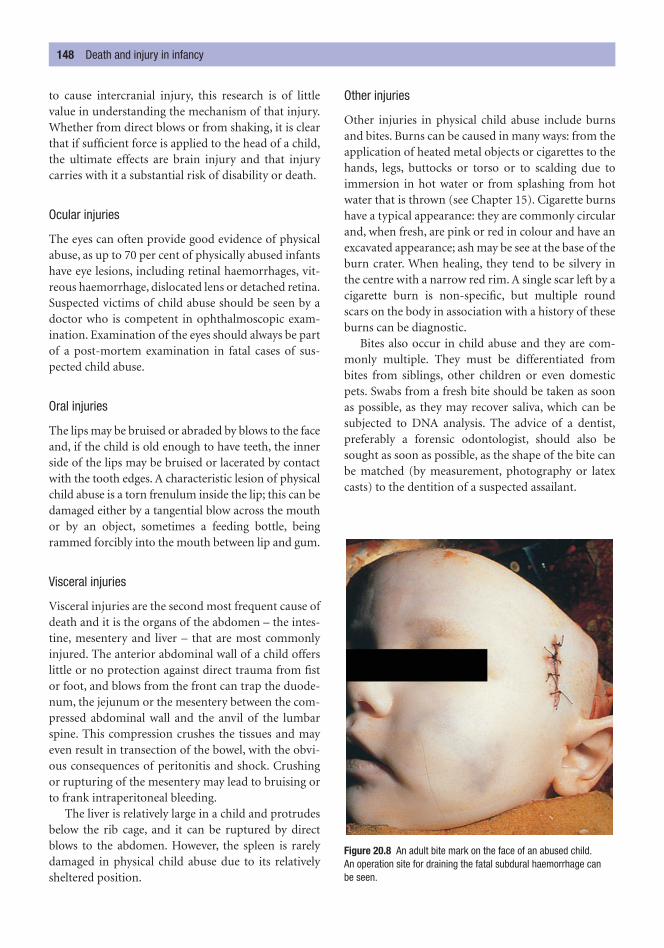
to cause intercranial injury, this research is of littlevalue in understanding the mechanism of that injury.Whether from direct blows or from shaking, it is clearthat if sufficient force is applied to the head of a child,the ultimate effects are brain injury and that injurycarries with it a substantial risk of disability or death.
Ocular injuries
The eyes can often provide good evidence of physicalabuse, as up to 70 per cent of physically abused infantshave eye lesions, including retinal haemorrhages, vit-reous haemorrhage, dislocated lens or detached retina.Suspected victims of child abuse should be seen by adoctor who is competent in ophthalmoscopic exam-ination. Examination of the eyes should always be partof a post-mortem examination in fatal cases of sus-pected child abuse.
Oral injuries
The lips may be bruised or abraded by blows to the faceand, if the child is old enough to have teeth, the innerside of the lips may be bruised or lacerated by contactwith the tooth edges. A characteristic lesion of physicalchild abuse is a torn frenulum inside the lip; this can bedamaged either by a tangential blow across the mouthor by an object, sometimes a feeding bottle, beingrammed forcibly into the mouth between lip and gum.
Visceral injuries
Visceral injuries are the second most frequent cause ofdeath and it is the organs of the abdomen – the intes-tine, mesentery and liver – that are most commonlyinjured. The anterior abdominal wall of a child offerslittle or no protection against direct trauma from fistor foot, and blows from the front can trap the duode-num, the jejunum or the mesentery between the com-pressed abdominal wall and the anvil of the lumbarspine. This compression crushes the tissues and mayeven result in transection of the bowel, with the obvi-ous consequences of peritonitis and shock. Crushingor rupturing of the mesentery may lead to bruising orto frank intraperitoneal bleeding.
The liver is relatively large in a child and protrudesbelow the rib cage, and it can be ruptured by directblows to the abdomen. However, the spleen is rarelydamaged in physical child abuse due to its relativelysheltered position.
Other injuries
Other injuries in physical child abuse include burnsand bites. Burns can be caused in many ways: from theapplication of heated metal objects or cigarettes to thehands, legs, buttocks or torso or to scalding due toimmersion in hot water or from splashing from hotwater that is thrown (see Chapter 15). Cigarette burnshave a typical appearance: they are commonly circularand, when fresh, are pink or red in colour and have anexcavated appearance; ash may be see at the base of theburn crater. When healing, they tend to be silvery inthe centre with a narrow red rim. A single scar left by acigarette burn is non-specific, but multiple roundscars on the body in association with a history of theseburns can be diagnostic.
Bites also occur in child abuse and they are com-monly multiple. They must be differentiated frombites from siblings, other children or even domesticpets. Swabs from a fresh bite should be taken as soonas possible, as they may recover saliva, which can besubjected to DNA analysis. The advice of a dentist,preferably a forensic odontologist, should also besought as soon as possible, as the shape of the bite canbe matched (by measurement, photography or latexcasts) to the dentition of a suspected assailant.
148 Death and injury in infancy
Figure 20.8 An adult bite mark on the face of an abused child.An operation site for draining the fatal subdural haemorrhage can be seen.

Genuine accidents to children are far more fre-quent than acts of child physical abuse. However, itcan be extremely difficult to distinguish between them.Any doctor who examines children must always bearin mind the possibility of physical child abuse – evenin the well-spoken, middle-class family – and althoughit is vital that innocent parents are not unjustly accusedof ill-treating their children, it is equally importantthat abuse is recognized, as the next time the child, ora sibling, presents for treatment may be when he isfatally injured or dead.
Child sexual abuse
The problem of the sexual abuse and exploitation ofchildren is global and thousands of children are sexu-ally abused every day around the world. Sexual abusehas come to prominence more recently than physicalabuse, although sexual contact between adults andchildren has been recorded in many situations andmany countries for centuries. The modern definitionof child sexual abuse may be expressed as ‘the involve-ment of dependent, developmentally immature chil-dren and adolescents in sexual activities they do nottruly comprehend, to which they are unable to giveinformed consent or which violate social taboos orfamily roles’.
The diagnosis and management of child sexualabuse are multidisciplinary, involving not only doctorsbut also social agencies and sometimes law enforce-ment agencies. On the medical side, there needs to bea partnership between paediatricians and forensicmedical examiners (police surgeons), although in gen-eral terms the physical findings on medical examina-tion may be minimal or absent, especially if the abuseis not reported for weeks, months or even years afterthe actual events, and may form only a very small partof the diagnosis.
Great care needs to be exercised in the examinationof the suspected victims of sexual abuse, and specialistdoctors are now available throughout the UK. If nospecialist doctors are available and the opinions of
both a paediatrician and a forensic practitioner areneeded, arrangements must be made for a joint exam-ination so that the search for physical signs and theretention of samples for forensic tests can be donetogether. When more than 72 hours have passed sinceintercourse, the chances of recovering sperm from thevagina are too low to make the taking of high vaginalswabs justified, but swabs of the introitus and exami-nation of underwear should be considered.
In general terms, much of the examination routineis similar to that recommended in sexual offencesagainst older age groups: history-taking, consent, con-fidentiality, good examination facilities and the needfor a general examination before any perineal exami-nation are all essential, but uppermost in the mind ofthe doctor must be the concern that any further psy-chological trauma to the child is avoided.
The possibility of sexually transmitted diseasesbeing present must always be borne in mind. This hasboth forensic and therapeutic aspects for the examin-ing doctor, because they may provide a positive link toa perpetrator while also needing urgent treatment.
The difficulties of getting accurate evidence or awitness statement from a very immature witness andplacing it before a court have led to the use of video-recorded interviews and examinations including theuse of dolls to indicate parts of the body. These inter-views must be performed by specially trained individ-uals – usually police officers or social workers.
No doctor should voluntarily commence an exam-ination of a child who might have been sexuallyabused without having had paediatric and forensictraining, because it is the appreciation of the widerange of normal appearances of the genitalia and anusthat is the key-stone of a reliable opinion, and there isgreat concern that unless the doctor has had this spe-cialist training, the value of his or her opinion to acourt may be reduced. Extensive, clear notes must, ofcourse, be taken, with diagrams to support the text.
Digital examination of the vagina in a pre-pubertalchild is rarely indicated and may need to be performedunder general anaesthesia if thought to be absolutelyessential.
Child abuse 149

In an increasingly violent and divided world, abuses ofhuman rights, both physical and psychological, aremore common. The term ‘ethnic cleansing’ has beenadded to the vocabulary, and concentration camps,mass rape and murder and the torture of men, womenand children have again been seen in Europe in the lastdecade. Regrettably, about one-third of the memberstates of the United Nations have been involved inthese abhorrent activities during the past few decades,even though they are all signatories to declarationsoutlawing such practices.
The 1975 Tokyo Declaration of the World MedicalAssociation on ‘Torture and other cruel and degradingtreatment or punishment’ specifically forbids theinvolvement of medical practitioners in the adminis-tration of torture, and this view is strongly reinforcedby the British Medical Association in its report on ‘TheMedical Profession and Human Rights: handbook fora changing agenda’.
Forensic practitioners have a crucial primary roleto play in the impartial examination of individualswho reach asylum and claim refugee status. In this situation, the authorities of the host country oftenrequire definite forensic evidence that will support aclaim of torture in order to verify the request for asy-lum based on physical abuse.
If the injuries are fresh, their documentation andinterpretation will generally follow the standard routeand practices of the examination of a victim of a vio-lent physical assault. However, one of the major prob-lems faced by the doctor is the long interval, often ofmonths or even years, that will commonly haveelapsed between the infliction of the injuries and thedate of examination. This delay in examination isoften inevitable due to periods of imprisonment anddelays while the victim manages to escape to a countryof sanctuary. During this time, the normal healingprocesses will occur and many acute injuries will dis-appear completely, while others will remain as scars.It requires very careful examination combined withexperience of either similar cases or of forensic medi-cine to detect and interpret such old lesions.
Features of physical abuse
The ways of deliberately harming victims are limitedonly by the ingenuity of the torturers, and the forensicevidence left upon the body of the victim will reflectthe methods used. Blunt trauma, sharp trauma, appli-cation of heat or electricity, asphyxia and suspensionare all commonly encountered.
Beating with a variety of objects is the most com-mon form of abuse and will result in the usual triad ofsigns associated with that type of trauma: bruising,
PHYSICAL ABUSE OF HUMAN RIGHTS: TORTURE
C h a p t e r t w e n t y - o n e
Neglect, Starvation and Abuse ofHuman Rights
Physical abuse of human rights: torture Features of physical abuse Neglect and starvation

Physical abuse of human rights: torture 151
abrasion and laceration. Particular weapons or objectsmay leave particular patterns on the skin, for examplea blow from a rod or thong will commonly result inparallel tramline bruises (see Chapter 9). Healing ofthese bruises and abrasions will generally be completeand leave no residual signs, although in some dark-skinned victims there is sometimes a linear increase inpigmentation that may provide permanent evidence.Where the skin is lacerated, the scar left may provide
clear evidence of the site of the injury. Susceptibleindividuals may develop keloid scarring.
The sites of injury are very variable; blows to thehead, back, buttocks, perineum and soles of the feet(‘falanga’) are favourite places for beating. Falangaconsists of the soles of the feet being beaten with a thinrod or cane and, because of the thick skin and densefascia at this site, permanent signs of injury are slighteven though deep tenderness may be long-lasting afterthe initial pain and swelling have subsided.
Cutting and piercing injuries are common featuresof torture; knives, bayonets, glass fragments, needles orany object with a cutting edge or a point may be usedon any part of the body. The injuries may be incisedwounds, stab wounds or a combination of the two. Theresulting scar may reflect the type of injury caused.
Burns may be inflicted from anything that will eitherburn or can be heated: cigarette lighters, hot irons, elec-tric heating rings, branding irons, cigarettes, ignitedkerosene-soaked rags or molten rubber. Acutely, theburns show the usual features, but if healing hasoccurred, the residual lesions will depend upon the siteand severity of the burns.
Burns can also be caused by both acid and causticmaterials, the favourite being sulphuric acid in theform of ‘battery fluid’. These chemicals, if strongenough, will damage skin and eyes. Their fluid naturecan often be confirmed by the presence of the typicaltrickle-marks of a corrosive fluid. Scarring will dependon the depth of the injury.
Electrical torture is excruciatingly painful and isoften used because it leaves little permanent signs. Theshocks can be of any voltage, but are most commonlytaken direct from the mains supply of 110 or 240 V.Higher voltages derived from a magneto or a motorignition coil have also been used. The electrodes maybe clipped to, or bare wires applied to, the skin, tongue,breasts, nipples, penis or vulva. With mains voltage, therisk of fatal electrocution is present but seems rare,perhaps because of the short duration of each shock.The points of contact may leave small scars but, ingeneral, the residual effects of electricity are almostundetectable in the living. If death occurs during orsoon after electrical torture, special histological exami-nation of the sites of electrocution can be used to con-firm the presence of recent electrical injuries.
Damage to the ears is frequent, especially from the‘telefono’ technique in which hands are clapped vio-lently against the side of the head. This action mayrupture the eardrum and damage the inner ear.
Figure 21.1 This woman was tied to a chair beneath a burning cartyre. The dripping burning rubber caused these injuries.
Figure 21.2 Extreme keloid scarring following burns inflicted by anelectric hot plate and burning rags.
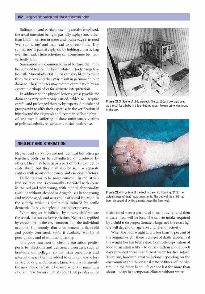
Suffocation and partial drowning are also employed,the usual intention being to partially asphyxiate, ratherthan kill. Immersion in water and foul sewage is termed‘wet submarino’ and may lead to pneumonias. ‘Drysubmarino’ is partial asphyxia by holding a plastic bagover the head. These activities can sometimes be inad-vertently fatal.
Suspension is a common form of torture, the limbsbeing roped to a ceiling beam while the body hangs freebeneath. Musculoskeletal injuries are very likely to resultfrom these acts and they may result in permanent jointdamage. These injuries may require examination by anexpert in orthopaedics for accurate interpretation.
In addition to the physical lesions, gross psychiatricdamage is very commonly caused, which will requirecareful and prolonged therapy by experts. A number ofgroups exist to offer their expertise in the verification ofinjuries and the diagnosis and treatment of both physi-cal and mental suffering in these unfortunate victimsof political, ethnic, religious and racial intolerance.
Neglect and starvation are not identical but often gotogether; both can be self-inflicted or produced byothers. They may be seen as a part of torture or delib-erate abuse, but they may also be seen as separate entities with many other causes and associated factors.
Neglect seems to be more common in industrial-ized societies and is commonly associated with abusein the old and very young, with mental abnormality(with or without alcohol or drug abuse) in the youngand middle aged, and as a result of social isolation inthe elderly, which is sometimes induced by seniledementia. Rarely is neglect due to sheer poverty.
When neglect is inflicted by others, children are the usual, but not exclusive, victims. Neglect is typifiedby excess dirt in the environment that the individualoccupies. Commonly, that environment is also coldand poorly ventilated. Food, if available, will be ofpoor quality and of minimal quantity.
The poor nutrition of chronic starvation predis-poses to infections and deficiency disorders, such asberi-beri and pellagra, so that skin conditions andinternal disease become added to catabolic tissue losscaused by calorie deficiency. Emaciation is commonlythe most obvious feature because, when the minimumcalorie intake for an adult of about 1500 per day is not
maintained over a period of time, body fat and thenmuscle mass will be lost. The calorie intake requiredby a child is disproportionately large and the exact fig-ure will depend on age, size and level of activity.
When the body weight falls to less than 40 per cent ofthe original weight, there is danger of death, especially ifthe weight loss has been rapid. Complete deprivation offood in an adult is likely to cause death in about 50–60days provided there is sufficient water for free intake.There are, however, great variations depending on theenvironment and the original state of fitness of the vic-tim. On the other hand, life cannot last for more thanabout 10 days in a temperate climate without water.
NEGLECT AND STARVATION
152 Neglect, starvation and abuse of human rights
Figure 21.3 Scene of child neglect. The cardboard box was used as the cot for a baby in this unheated room. Frozen urine was foundin the box.
Figure 21.4 Frostbite of the foot in the child from Fig. 21.3. Theactual cause of death was pneumonia. The body of the child hadbeen disposed of by the parents down the farm well.

Two main types of starvation are described: the‘wet’ and the ‘dry’. The former shows marked oedemaof most of the body, with ascites and pleural effusionsdue to hypoproteinaemia caused by the muscle massbeing metabolized to produce energy. The dry formhas little or no oedema, but there is hypotension andincipient cardiac failure.
The appearance in both the living and the dead isusually of a sunken, cachectic face, with skin stretchedover the cheekbones and prominent jaw, zygoma andorbital margins. The eyes are sunken due to loss oforbital fat, though accompanying dehydration may
also contribute to these appearances. The ribs, spine,iliac crests and all bony landmarks are prominent; theabdomen may be ‘scaphoid’ (i.e. concave between thecostal margin and iliac crests) or it may be protuberantif ascites is a major factor. The diameter of the limbs ismarkedly reduced and the head may appear undulylarge due to the narrowing of the neck. The skin maypresent various abnormalities. In children it is oftenpale and translucent, sometimes with a bluish tint due to the transparency of the tissues over veins. In the chronically starved, various skin diseases due toavitaminoses and infections may present as scaling,thickening or inflammatory lesions.
There may be pressure sores on the buttocks, sacrumand heels, due to loss of the padding effect of subcuta-neous fat coupled with a reduction of skin viability andreduced mobility. There may be gum swelling, loosen-ing of the teeth, sores on the lips and other signs of vita-min and protein deficiency, as well as infective lesions.
Internally, most organs except the brain may bereduced in weight compared to the normal range forage, sex and size. The stomach and intestines will com-monly be distended with gas and appear thin walledand translucent. Occasionally, remnants of the ingestedforeign material may be found in the stomach orbowel – in one case of a starved child, the remains of apaper nappy liner were found in all parts of the bowel.The gall bladder may be tense with bile due to lack ofstimulus for emptying. There may hard faecoliths inthe rectum, but sometimes there is an enteritis withloose stools. There may be a smell of ketones on thebreath and in the urine.
One of the medico-legal problems, especially whena child is alleged to have been denied proper feeding,is the possibility that some natural disease has causedthe emaciated state. Full clinical investigations mustthen be made of either the living or the dead child,including an extensive radiological survey. An autopsyand histological survey are essential to confirm or refutethe claim that conditions such as malignancy, Addison’sdisease, coeliac disease or a congenital metabolic defectare present.
Neglect and starvation 153
Figure 21.5 Extreme emaciation due to deliberate starvation,showing the scaphoid abdomen, shrunken limbs and ‘Hippocraticfacies’ of dehydration and wasting.
Figure 21.6 The abdominal contents of the child in Fig. 21.5.The bowel is empty apart from gas.
In Britain, self-inflicted poisoning still accounts for a huge number of acute hospital consultations andadmissions each day. Fortunately, only a minority ofcases are fatal, but the cost to the health service is considerable.
The definition of poisoning can be difficult and itcannot be defined simply as an unwanted effect of inges-tion of a chemical, because death is often the desiredeffect of ingesting the chemical in self-poisoning. It isperhaps better to discuss ‘toxicity’, which is simply theadverse effect of a chemical on cell, organ or organismfrom an objective viewpoint. Using this definition,virtually anything can be toxic if it is ingested in suffi-cient quantity and, in a chemical deliberately adminis-tered for treatment, it is the margin between anydesired therapeutic effect and the adverse effects that isimportant.
Poisoning is very commonly accidental but, as notedabove, a large number of cases are self-inflicted, oftenwith the intention of committing suicide. Homicide bypoison is now relatively rarely detected in advancedcountries, despite the availability of well-equipped toxi-cology laboratories. The increasing cost of analysis anddecreasing awareness of the problem of poisoning mayhave led to a false sense of complacency, which may inturn have led to the under-detection of poisoning fromall causes. This has been clearly demonstrated recentlyin the UK by the murder of over 200 of his elderlypatients by a general practitioner.
Accidental poisoning can vary from the individualcase of a child eating medicinal tablets in mistake forsweets, to families dying from carbon monoxide dueto a faulty gas appliance, to mass industrial disasterssuch as Bhopal in India, the leakage of dioxin gas inItaly and contaminated cooking oil in Spain. In war-fare, the deliberate use of chemical weapons on a civil-ian population in Iraq and natural disasters such as theescape of carbon dioxide gas from a lake bed in Africahave claimed thousands of victims.
Agriculture is commonly associated with accidentalpoisoning because of the availability, often in a rela-tively uncontrolled environment, of toxic substancessuch as paraquat, organophosphorous insecticides etc.The history of toxicology is full of tragedies involvingsacks of grain being left in rail-trucks contaminatedwith dieldrin, or of soft-drink bottles being re-filledwith ethylene glycol anti-freeze or organophosphateweed killers. Other industrial processes are also associ-ated with the use of potentially lethal chemicals: forexample, the extraction of gold from ore and electro-plating both use cyanide.
Suicidal poisoning is now the most common methodof self-destruction for females and the second mostcommon for males in England and Wales. The drugsand chemicals used depend upon their availability andupon some national and local practices. In Britain,there was a shift in the latter half of the twentieth cen-tury from barbiturates and aspirin to paracetamol,
C h a p t e r t w e n t y - t w o
General Aspects of Poisoning
The toxic and fatal doseTolerance and idiosyncrasyThe doctor’s duty in a case of suspected
poisoning
Samples required for toxicological analysisBloodUrineVomit and stomach contents
FaecesLiver and other organsHair and nail clippings
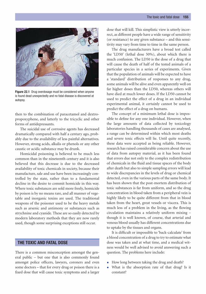
The toxic and fatal dose 155
then to the combination of paracetamol and dextro-propoxyphene, and latterly to the tricyclic and otherforms of antidepressants.
The suicidal use of corrosive agents has decreaseddramatically compared with half a century ago, prob-ably due to the availability of less painful alternatives.However, strong acids, alkalis or phenols or any othercaustic or acidic substance may be drunk.
Homicidal poisoning is believed to be much lesscommon than in the nineteenth century and it is alsobelieved that this decrease is due to the decreasedavailability of toxic chemicals in society, because theirmanufacture, sale and use have been increasingly con-trolled by the state, rather than to a fundamentaldecline in the desire to commit homicide in this way.Where toxic substances are sold more freely, homicideby poison is by no means rare, and all manner of vege-table and inorganic toxins are used. The traditionalweapons of the poisoner used to be the heavy metalssuch as arsenic and antimony or substances such asstrychnine and cyanide. These are so easily detected bymodern laboratory methods that they are now rarelyused, though some surprising exceptions still occur.
There is a common misconception amongst the gen-eral public – but one that is also commonly foundamongst police officers, lawyers, coroners and evensome doctors – that for every drug or poison there is afixed dose that will cause toxic symptoms and a larger
dose that will kill. This simplistic view is utterly incor-rect, as different people have a wide range of sensitivity(or resistance) to any given substance – and this sensi-tivity may vary from time to time in the same person.
The drug manufacturers have a broad test calledthe ‘LD50’ (lethal dose 50%), about which there ismuch confusion. The LD50 is the dose of a drug thatwill cause the death of half of the tested animals of aparticular species in a series of experiments. Giventhat the population of animals will be expected to havea ‘standard’ distribution of responses to any drug,some animals will be alive and even apparently well onfar higher doses than the LD50, whereas others willhave died at much lower doses. If the LD50 cannot beused to predict the effect of a drug in an individualexperimental animal, it certainly cannot be used topredict the effect of a drug on humans.
The concept of a minimum lethal dose is impos-sible to define for any one individual. However, whenthe large amounts of data collected by toxicology laboratories handling thousands of cases are analysed,a range can be determined within which most deathsand severe toxic effects will lie. Until quite recently,these data were accepted as being reliable. However,research has raised considerable concern about the useof data from autopsy material, as it has been foundthat errors due not only to the complex redistributionof chemicals in the fluid and tissue spaces of the bodyafter death but also to simple sampling errors will leadto wide discrepancies in the levels of drug or chemicaldetected, even in the various parts of the same body. Ithas been shown that the post-mortem distribution oftoxic substances is far from uniform, and so the drugconcentration in blood taken from a peripheral vein ishighly likely to be quite different from that in bloodtaken from the heart, great vessels or viscera. This ismuch less of a problem in the living, as the flowing circulation maintains a relatively uniform mixing –though it is well known, of course, that arterial andvenous blood usually has different concentrations dueto uptake by the tissues and organs.
It is difficult or impossible to ‘back-calculate’ froma blood concentration of a drug to try to estimate whatdose was taken and at what time, and a medical wit-ness would be well advised to avoid answering such aquestion. The problems here include:
• How long between taking the drug and death?
• What is the absorption rate of that drug? Is it constant?
THE TOXIC AND FATAL DOSE
Figure 22.1 Drug overdosage must be considered when anyone is found dead unexpectedly and no fatal disease is discovered atautopsy.

• Is the drug completely absorbed from the alimen-tary canal?
• What is the rate of metabolism? Is it constant?
• What is the rate of excretion? Is it constant?
• Does the drug diffuse uniformly throughout allbody tissues or does it vary between the aqueous,lipoid and other body compartments?
• Was the sample taken from an appropriate site?
• Has it been stored and analysed correctly?
One of the problems of determining toxic or fataldoses of a drug or other chemical, and the equivalentblood levels, is that not only do different people havewidely varying innate susceptibility, but tolerance canbuild up to a substance so that large doses no longerhave the effect that they originally had. In analgesia,this tolerance, especially of the opiates, can cause thera-peutic problems, as increasing doses need to be givento obtain the same beneficial effect. This can result inthe administration of quantities of drugs that wouldnormally be expected to cause death and the subse-quent detection of greatly raised blood levels in indi-viduals who are not only alive but conscious. Thistolerance can develop rapidly, being established withsome drugs within a couple of days or weeks, or it maytake considerably longer. In evaluating toxicologyresults, the doctor has to take tolerance into account,as what may appear to be a toxic or fatal level in theblood or tissues may have had far less clinical effectthan might be expected if tolerance has developed.
Tolerance is lost when exposure to the drug orchemical ceases, a fact that can have fatal results in therelapsing intravenous opiate drug abuser who, after aperiod of abstinence (either voluntary or enforced),returns to the same amount of drug and dies as aresult. Generally speaking, the more rapidly tolerancedevelops, the more rapidly it will be lost. Many abuseddrugs – amphetamines, barbiturates, benzodiazepinesand the morphine–heroin–methadone group – exhibitthis phenomenon.
The opposite situation is that of idiosyncrasy, inwhich abnormally small amounts of substances –including penicillin, aspirin, cocaine and heroin – maycause dramatic or even fatal effects, some of whichmay be mediated by anaphylaxis and require previousexposure, whereas others induce a true idiosyncraticresponse on first exposure.
Whatever the cause or circumstances of poisoning, themedical attendant has a responsibility to manage thesituation in the most effective way, by:
1 recognizing the fact or possibility of poisoning atthe earliest possible stage;
2 instituting primary treatment and arranging admis-sion to hospital when necessary;
3 identifying and retaining any drugs or possible poi-sons in the bedroom or household, workplace etc.and ensuring that these substances are taken to thetreatment centre or hospital along with the patientso that they can be identified; and
4 where death has occurred, co-operating fully with the police, coroner, magistrate etc. in theinvestigation.
Commonly, an individual will present to a doctorin hospital with a history of accidental or deliberatepoisoning, in which case the type(s) of drugs takenand when they were taken is crucial information.Diagnosis in an unconscious patient may depend asmuch on information from the scene as on physicalexamination of the body – empty tablet containers or‘blister packs’ in the room or waste bins, notes describ-ing intentions etc. are all important.
The recognition of concealed poisoning (whetheraccidental or homicidal) can be considerably moredifficult, but the following symptoms and signs shouldat least cause the doctor to include poisoning in hisfirst differential diagnosis:
• sudden vomiting and diarrhoea;
• unexplained coma, especially in children;
• coma in an adult known to have a depressive illness;
• rapid onset of a peripheral neuropathy, such aswrist-drop; and
• rapid onset of a neurological or gastrointestinal illness in someone known to be occupationallyexposed to chemicals.
It is not all that uncommon for patients to allegethat they are being poisoned (usually by a relative) orfor a family member to allege that a recent death wasdue to poisoning. The majority of these claims arewithout any truth, and are due to misunderstandings,family feuds, malice, delusion or mental abnormality.In the living patient, the medical practitioner has firstto decide whether the allegations may be true, based
THE DOCTOR’S DUTY IN A CASE OF SUSPECTED POISONING
TOLERANCE AND IDIOSYNCRASY
156 General aspects of poisoning
on his knowledge of the family situation and the per-sonalities involved. If the signs and symptoms of thealleged victim are genuine and not inconsistent withsome form of toxic effect, the doctor must take somesteps to investigate and, in most instances, if the doc-tor has reason to believe the victim, he should advisehim or her to inform the police so that a full investiga-tion can be performed.
If there is real suspicion and cause for concernabout the health of the victim, it would also makesense to remove the victim from the potential sourceof the poison, and this has been achieved in the past byadmitting people to hospital or moving them to someother environment. It is then possible to monitor theirsymptoms and, if they clear up, this might suggest thatsome toxin was indeed being given. If an oral poison issuspected, friends and relatives should not be allowedto bring food or drink to the patient.
The possibility of deliberate self-administration of a poison for psychiatric reasons (Munchausen’ssyndrome) should always be borne in mind and,in children, the related problem of Munchausen’s syndrome-by-proxy (in which one of the methodsused by the carer to cause the child’s illness is theadministration of drugs or salt) has to be considered.
Samples for toxicological analysis, whether fromclinical cases under investigation or under treatmentor from autopsies, must be collected and stored withparticular care. It is best practice that all samples mustbe meticulously labelled and identified at the time ofremoval or sampling, but this is an absolute necessityif there is any possibility of criminal action.
The doctor who takes a sample of blood from avenepuncture must place the blood into the appropri-ate tube and then immediately label it with the patient’sname, address or hospital number, the date and prefer-ably the time of sampling. The doctor’s signature (or atleast initials) should also be placed on the label. Thetube will then usually be sealed into another receptaclesuch as a plastic bag, which may also be labelled withthe details of the individual, the specimen and the per-son who took it. If the police are present (for instance atan autopsy), this sample will commonly be given aunique exhibit number. The doctor is the first person inwhat may be a long chain of custody of any specimen,and it is important for doctors to record in the medicalnotes what specimen(s) they took and at what time andto whom they gave the specimen and at what time.
This process is also called ‘continuity of evidence’and every step in the chain from vein to laboratorymust be capable of later proof in court if the opposing
lawyers challenge the authenticity of the analysis. Onno account should a specimen taken for these reasonsbe allowed to lie in a surgery, on a ward or elsewhereoutside a sealed bag where it may be tampered with.
As to the nature of the preservation of samples, muchdepends upon the type of toxic substance suspected.The advice of the laboratory that is to carry out theanalysis should be obtained, where there is any doubt,but the following is a generally accepted routine.
Blood
It is very important to obtain blood samples from thecorrect site. During life, any venous sample drawn froma limb vein is usually satisfactory, except in unusual cir-cumstances when arterial blood is required. However, atautopsy, the site of sampling is of great importancebecause the interpretation of the results of analysis canbe markedly distorted by an incorrect sample. The bestplace to obtain blood is from the femoral or iliac veinsor, if no blood is available from those sites, from theaxillary veins. The jugular venous system is not suitableas it may be contaminated by reflux from the upperthorax. Blood should never be taken from the generalbody cavity as it is highly likely to be grossly contami-nated by intestinal contents, effusions, urine, faeces etc.Neither should blood be taken from the heart or greatvessels in the chest as post-mortem diffusion of drugsand alcohol from the stomach or from aspirated vomitin the air passages can easily and rapidly contaminatethe blood in these vessels. It has been shown, for exam-ple, that if fluid containing alcohol and paracetamol isinstilled into the trachea after death to simulate aspira-tion of stomach contents, high blood concentrations of these substances can be detected in the thoracic ves-sels. Also, alcohol swallowed immediately before death,when it has had no time to be absorbed or to cause anyphysiological effects, can diffuse post mortem throughthe gastric wall into the blood in the heart and great ves-sel and so lead to a false-positive result on samples takenfrom these sites.
For most analyses, about 15 mL of blood from theiliac or femoral veins is placed in a plain tube, so thatclotting can occur, together with 5–10 mL in a tube
SAMPLES REQUIRED FOR TOXICOLOGICALANALYSIS
Samples required for toxicological analysis 157

containing an anticoagulant, such as EDTA, potas-sium oxalate or heparin. In addition, an alcohol esti-mation is almost invariably required and 5 mL ofblood for this should be placed in a sodium fluoridetube to inhibit production or destruction of alcohol by micro-organisms.
In autopsies upon victims of solvent abuse (‘gluesniffers’), the chemical(s) responsible can often berecovered from the blood. Blood samples in cases ofsuspected solvent abuse should fill the bottle to the topso that there is no ‘head space’ into which the solventscan diffuse, only to be lost immediately on openingthe bottle. It is far better to have a full small bottle inthese cases than a half-empty large bottle.
Urine
Up to 20–30 mL of urine should be placed in a ‘universalcontainer’. No preservative is required unless there is tobe a delay in reaching the laboratory. If this is so, then asmall amount of sodium azide or fluoride can be added.
Vomit and stomach contents
If produced by a living patient or recovered from the scene of death, vomit may usefully be sent to thelaboratory placed either in a clean glass jar or a plastictub with a tight-fitting lid. At autopsy, the stomach con-tents may be saved into a similar container, by openingthe greater curvature with scissors and allowing thecontents to gravitate into the receptacle. Some labora-tories also request the stomach wall itself, as powderand tablet debris may remain adherent to the mucosalfolds in higher concentration.
Some well-organized toxicology laboratories pro-vide kits consisting of a large and a small plastic tub,several universal containers, a fluoride tube, togetherwith a syringe and needles and an instruction sheet,for use by doctors and pathologists.
Faeces
The contents of the rectum are not often required foranalysis, except in suspected heavy-metal poisoning,such as arsenic, mercury or lead. A sample of 20–30 gshould be taken into a plain screw-topped jar or aplastic container with a snap-on lid.
Liver and other organs
At autopsy, the most common organ to be saved foranalysis is the liver, as higher concentrations of manydrugs may be found, making them identifiable whenthe blood and urine concentrations have declined tovery low or undetectable levels. The pathologist mustexamine the organ macroscopically for anatomical orpathological abnormalities and must sample it formicroscopic examination before sending it for toxico-logical examination. Some laboratories request thewhole organ, whereas others require only part of theliver. Whatever sample is required, it should be placedin a clean container. Some laboratories require thetotal weight of the organ, but this should routinely benoted as part of the internal examination and will beavailable if requested.
Bile is sometimes required for analysis and it is particularly useful for seeking evidence of chlorpro-mazine or morphine use.
Some laboratories analyse for solvents direct froman intact lung. In such a case, a lung should be takenstraight from the body and placed in an impervious bag,which should be securely tied to avoid escape of vapour.Nylon bags, as used by arson investigators, are prefer-able to the usual polyvinyl chloride bags, as the latter arenot completely impervious to organic solvents.
Hair and nail clippings
Heavy-metal poisonings by elements such as antim-ony, arsenic or thallium are often chronic events andsome hair cut or pulled at the roots, together with nailclippings, should be submitted for analysis. Thesemetals are laid down in keratin in a sequence depend-ing on the time of administration, and it may even bepossible, by neutron-activation analysis of a few hairs,to determine when in relation to the growth span ofthe hair the poison was administered, as the mostrecent doses will be nearer the root.
Recent advances in microchemistry have alsoallowed for the detection and analysis of opiate drugsin hair, and this can be used to confirm or refute prolonged drug use and even to identify approxi-mately when (in terms of weeks or months) the drugswere used. Clearly, this is only of use if hair is presentand is of sufficient length. The samples for this analy-sis should include the root to the tip of the hair, andthe hairs should all be orientated in one direction.
158 General aspects of poisoning

Alcohol (ethyl alcohol, ethanol) is by far the mostwidely used drug in the world, even in those countrieswhere religious beliefs theoretically prevent its con-sumption. Ethanol is a small, water-soluble moleculethat becomes distributed evenly throughout the bodywater; it passes easily across the blood–brain barrierand has a profound depressant effect upon the cere-bral function. It is a drug with huge morbidity andmortality, with direct toxic effects on the body tissues,but alcohol also has significant indirect effects and it isa very common catalyst in the majority of assaults andhomicides. The use, or rather abuse, of alcohol per-vades all aspects of legal medicine from the precipita-tion of many road traffic accidents and accidents atwork through to professional misconduct of doctors,for whom alcohol abuse provides one of the mostcommon reasons for disciplinary proceedings. Everymedical practitioner needs to know something of themetabolism and effects of this ubiquitous substance.
Ethanol is extremely easy to produce and almost allethanol is produced by the action of yeast enzymes onvegetable substrates containing sugars. Direct (pri-mary) fermentation cannot raise the concentration tomore than 12–15 per cent, as the yeast is killed by therising levels of alcohol. However, distillation of the
primary fermentation can concentrate the alcohol to40–60 per cent strength.
The usual forms of alcohol that are drunk have thefollowing approximate strengths, excluding ‘special’varieties, which are becoming more prevalent.
Beer, stout, cider 3–6%Table wine 9–12%Sherry, port (fortified with brandy) 17–21%Spirits (whisky, brandy, gin,
rum, vodka etc.) 35–45%
The concentration of alcohol must be written onthe labels of most bottles containing it. It is usuallywritten in the form of weight of alcohol per volume ofliquid (w/v), rather than as volume of alcohol per vol-ume of liquid, because alcohol has an appreciablylower specific gravity than water; for example 40% v/vwhisky only has about 32 g per 100 mL. In order tosimplify the understanding of the amount of alcoholconsumed, the concept of a ‘unit’ of alcohol was introduced – a unit being about 8 g of alcohol – and inBritain this information is also on the labels of bottles.The following drinks all contain approximately 1 unitof alcohol.
Beer half a pint (300 mL)Wine a 100-mL glass of table wineSpirits a small measure of spirits (25 mL)
It is generally stated that liver damage is unlikely if a man consumes fewer than 21 units per week,
SOURCES OF ALCOHOL
C h a p t e r t w e n t y - t h r e e
Alcohol
Sources of alcoholAbsorption of alcoholElimination of alcohol
The measurement of alcoholThe effects of alcoholDangers of drunkenness
Drinking and drivingScheme of examination for impairment of
ability to drive

whereas a woman should consume fewer than14 unitsper week to avoid liver damage.
Most absorption of alcohol takes place in the stomach,duodenum and first part of the jejunum; the latter twoare the most effective areas of absorption, partly becausethe gastric mucosa contains an alcohol dehydrogenase.Individuals with rapid stomach emptying or a gastro-jejunostomy may absorb alcohol unusually rapidly andthis will produce a higher peak blood-alcohol concentra-tion (BAC). A single bolus of alcohol taken into anempty stomach will be completely absorbed into theportal bloodstream within 30–90 minutes. As soon asalcohol enters the bloodstream, the liver and kidneys willbegin to eliminate it, so the BAC achieved will be gov-erned by the relative rates of absorption and elimination.
Absorption of alcohol is affected by:
• The presence of food in the stomach: food,especially fatty food, delays emptying through thepylorus, which will in turn delay contact with themucosa of the more rapidly absorbing duodenal–jejunal segment of the intestines. Fatty food alsophysically obstructs the contact of alcohol mole-cules with the gastric mucosa.
• The concentration of alcohol: the optimum con-centration for rapid absorption is about 20 percent. The alcohol in weak drinks, such as beer,passes only slowly through the mucosa and into thebloodstream, whereas strong spirits irritate the gas-tric mucosa, resulting in the production of excessmucus, which will tend to coat the lining and pro-duce a physical barrier to absorption. The pylorusmay also undergo spasm, thus delaying gastricemptying and absorption.
Well over 90 per cent of alcohol is metabolized by theliver, which accounts for the damaging effect that thelong-term high intake of alcohol has upon that organ.Only 5–8 per cent of the alcohol is excreted unchangedby the kidneys, sweat and breath.
The rate of elimination from the bloodstream variesin different people: chronic alcoholics, particularly,
who have induced liver cytochrome P450 enzymes(until their livers fail) can metabolize alcohol at anabnormally high rate. The ‘normal’ rate of eliminationfrom the blood for non-alcoholics lies between about11 and 25 mg/100 mL/h, with an average of18 mg/100 mL/h; this can eliminate approximately 1unit of alcohol (8 g) per hour.
The concentration of alcohol in both blood and urineis usually expressed in milligrams per hundred milli-litres (mg/100 mL), sometimes written as mg/dL(decilitre). In some countries, as in the USA, the con-centration is expressed as a percentage. The BAC is themost useful measure, because the rapid equilibrationof alcohol across the blood–brain barrier means thatthe BAC reflects the concentration of alcohol cur-rently affecting the brain.
The urine alcohol is more concentrated than theblood level, due to tubular resorption of water in theratio of 1.3 : 1. However, it is less useful than blood-alcohol measurements, as the ureteric urine concen-tration varies with the rising or falling BAC and, as allthe urine from the kidneys is pooled in the bladder,any sample of urine can only represent the mean valueover the period of collection in the bladder.
Breath alcohol is in equilibrium with the alcohol in the blood, even though in a very small concentra-tion of about 1 : 2300. At 37°C, a level of 1 mg/100 mLin blood will be equivalent to about 0.43 �g/100 mL inbreath (and thus 1 �g/100 mL in breath is equivalentto about 2.28 mg/100 mL in blood). The exact ratio ofblood alcohol/breath alcohol is temperature depend-ent and varies slightly with other factors such as thedepth of respiration and the concentration of alcohol.Because the ratio is not constant, legal systems that usebreath alcohol for detecting drinking drivers do notattempt to relate the breath level to the blood, butmake the offence solely that of having exceeded a cer-tain level in the breath itself.
Alcohol depresses the nervous system and any apparentinitial excitant effect is due to suppression of inhibitionby the cerebral cortex. The drug begins to act at the
THE EFFECTS OF ALCOHOL
THE MEASUREMENT OF ALCOHOL
ELIMINATION OF ALCOHOL
ABSORPTION OF ALCOHOL
160 Alcohol

lowest concentrations upon the higher centres and itaffects the lower centres of the central nervous systemonly when the BAC becomes higher. The effects of highlevels of alcohol on the lower centres may jeopardize thefunction of the cardiorespiratory centres in the brain-stem, with a consequent danger of death.
The earliest signs of alcohol intoxication can befound on objective testing with BAC as low as30 mg/100 mL, when driving skills begin to deterio-rate. Behaviour begins to change at a very variablelevel, but this is also influenced by emotional and envir-onmental factors. A person is likely to appear moreuninhibited in a student party than in a very formalfunction given the same level of alcohol.
The guide in Table 23.1 is a reasonable summation,given the great variations, in relation to the effects ofalcohol in blood.
To illustrate the uncertainty of this type of table, inAustralia, numerous drunken drivers have been foundto have BACs of over 500 mg/100 mL, when they shouldtechnically be dead, and people have survived alcoholconcentrations of over 1500 mg/100 mL (albeit withmedical support in intensive care) – these individualsare almost always chronic alcoholics.
Apart from the danger of respiratory paralysis at highconcentrations of alcohol, there are many other hazards
(some of which can be fatal), which can beset thedrunken person. Accidents of all types are more com-mon: individuals under the influence of drink falldown and frequently suffer injuries, especially to theirheads; falls down stairs or steps and into the roadwayare common and people who are under the influenceof alcohol are more likely to be pedestrian victims ofroad traffic accidents.
Drinking and personal violence are a commoncombination and altercations, fights and assaults are afrequent sequel to intoxication, with all the possibleinjurious and fatal consequences.
Burns are another possibility and typically occurwhen a drunken person falls asleep in bed while smok-ing a cigarette, which may be dropped into the bed-ding, death ensuing from burns or carbon monoxideinhalation. Drowning is another common sequel, par-ticularly in seamen and others who work or livearound ships or docks, as they fall from quays, gang-ways and decks when unsteady due to drink.
Vomiting is not uncommon in those intoxicated byalcohol, due to the irritant effect of alcohol upon thegastric mucosa. Vomiting combined with decreasedlaryngeal reflexes allows the irritant gastric contents to enter the airways, where they may cause death bystimulation of the vagus nerve, with resultant cardiacdepression, or by simple mechanical obstruction.
The police are most commonly left to deal withthose members of the community who cause a disturb-ance or otherwise offend the public after consumingtoo much alcohol. The police have few places to putthese people, other than in a cell in a police station.Although most police forces have a strict regime forplacing inebriated individuals in a semi-prone pos-ition and inspecting them at frequent intervals, deathsof these people while in custody are not uncommonand represent a disaster – for the family, for the policeand for society. It is interesting to note that we do notplace an individual with any other form of drug abuseinto custody simply because they are under the influ-ence of that drug, and many deaths would be pre-vented each year if these cases were treated in the sameway as other types of acute poisoning.
Alcohol-induced disease is extremely common andan appreciable proportion of national medical care isconsumed by drink-related conditions. Liver disease isdue to progressive damage to the hepatic cells causedby high concentrations of alcohol and its breakdownproducts. Fatty change is common following theingestion of alcohol, although at low levels it may be
DANGERS OF DRUNKENNESS
Dangers of drunkenness 161
Blood-alcohol Effectsconcentration(mg/dL)
30–50 Impairment of driving and similar skills
50–100 Reduced inhibitions, talkativeness,laughter, slight sensory disturbance
100–150 Incoordination, unsteadiness, slurred speech
150–200 Obvious drunkenness, nausea and ataxia
200–300 Vomiting, stupor, possibly coma
300–350 Danger of aspirating vomit, stupor or coma
350–450� Progressive danger of death from respiratory paralysis
Table 23.1 Guide to the Likely Effects of Alcohol on a Normal Adult
apparent only microscopically. Continued ingestion ofalcohol will be followed by fibrosis, which, if itbecomes sufficiently extensive, is then termed cirrho-sis. Once cirrhosis is established, a series of clinicalevents will follow, including splenomegaly, portalhypertension with the development of varices andpossibly gastrointestinal bleeding.
Liver function tests will assist in monitoring thecondition of the liver and a specific enzyme marker isgamma-glutamyl transpeptidase (GGT), which is ele-vated well above 36 units in alcoholic liver damage.Alcohol also affects the brain in long-term alcoholics,even when full-blown Wernicke’s encephalopathy isnot present. The myocardium also suffers andalthough there are no definite histological criteria foralcoholic cardiomyopathy, there is no doubt of itsexistence as a clinical entity. Alcohol is also the causeof considerable fetal abnormality and the features ofthe fetal alcohol syndrome are well defined.
This is a major problem all over the world, even inIslamic countries where alcohol is officially banned.Even small quantities of alcohol impair driving effi-ciency and objective testing, demonstrating deterior-ation of skills at a BAC of only 30 mg/100 mL. Somecountries, such as in Eastern Europe, have a zero limitfor driving, but elsewhere the threshold of blood orbreath concentration varies considerably.
For example, the countries of the British Isles havea BAC limit of 80 mg/100 mL in blood, 35 mg/100 mLin breath or 107 mg/100 mL in urine. This is thoughtto be too high by the British Medical Association,which wishes the government to bring the limit downto 50 mg/100 mL in blood. However, it must be admit-ted that over half the drivers charged with excess alco-hol have more than twice the legal limit in their breathor blood, so reducing the lower limit would be irrele-vant in their case.
The role of the doctor in the investigation of drink-ing and driving varies according to the method ofenforcement used in a particular country. Where thereis no statutory limit by blood or breath, a doctor mayhave to carry out clinical testing of the driver to decideif his ability to control the vehicle is impaired. This can be a very subjective decision in marginal cases,although the evidence of blood and breath testing
shows that many drinking drivers are so far ‘over thelimit’ that a decision as to their condition is not diffi-cult. Where a statutory limit is part of the law, somecountries require blood samples to be taken by a doc-tor by venepuncture, whereas others, including Britain,accept evidential breath testing performed by trainedpolice officers. If urine is used in place of blood orbreath, it can also be collected by police officers.
In addition to testing for driving ability, a doctormay be required to examine a suspected drunkendriver to exclude or confirm natural disease or injurythat may mimic alcoholic intoxication. Diabetic pre-coma, insulin overdose with hypoglycaemia, carbonmonoxide poisoning from a leaking exhaust system, ahead injury and many other disease processes have tobe considered in this examination. Another problem isthe impairment of driving ability from drugs otherthan alcohol – either medicinal substances or drugs ofdependence – and this has been the subject of recentresearch in the UK.
Scheme of examination for impairment ofability to drive
This is an examination not to discover if the driver is ina drunken state but to decide whether his ability tocontrol a vehicle safely is impaired for whatever reason.Unless the examination also includes the taking ofsamples for blood, breath or urine for analysis, its valuein confirming alcoholic intoxication is limited, exceptwhen the subject is obviously inebriated. Informedconsent to the examination is essential and the drivermust be aware that the facts may be used in criminalproceedings. If the person refuses permission, noattempt should be made forcibly to examine him, asthis would be an assault. However, the person can beobserved and a report offered on that basis. If the sub-ject is too drunk to understand or give consent, onlythe results of observation can be provided to the police.In these circumstances, it may also be necessary, for theperson’s own safety, to perform some examination toexclude serious injury and to protect him from thepotential dangers of severe drunkenness. Even whenconsent has been obtained, the driver may wish a doc-tor of his own choice to be present on his behalf; this isquite acceptable but it must not so delay the examina-tion that the intoxication passes off or is significantlyreduced. The British Medical Association has recom-mended that no more than half an hour should elapse.
DRINKING AND DRIVING
162 Alcohol
The examination begins with history taking, espe-cially about past medical history and the taking of anymedicines or drugs, especially insulin, oral hypogly-caemic agents, sedatives, anti-epileptic drugs, anti-histamines (which might produce drowsiness) andother hypnotics or tranquillizers. This is not only rele-vant to the present examination, but also gives thedoctor an opportunity to assess coherence of speech,confusion, memory and general mental ability. Thedemeanour, whether excited, depressed, drowsy oraggressive etc., is recorded. The amount, type and tim-ing of the person’s recent drinking should be deter-mined as well as some idea of his long-term drinkinghabits. The quantities admitted by the person shouldbe noted – but not necessarily believed!
General tests of memory and mental alertness maybe incorporated into the history taking, unless thedriver is obviously drunk, when simple direct ques-tions about the date and time may have to be asked.
The subject should then be asked to write some-thing either from dictation or copied from a book orpaper, but in making this assessment the doctorshould allow for the apparent social and educativestandard of the person: many may be unable to writewell or at all, even when sober, in which case drawingsimple patterns, such as triangles and diamonds, maybe preferable. Simple tests of dexterity such as pickingup small objects from the floor are preferable to walk-ing a chalk-line on the floor, which even sober peopleoften cannot perform well.
When undressing for examination, the ability toundo buttons, any fumbling with zips etc. should benoted. The physical examination should follow a normalmedical routine, seeking physical evidence of naturaldisease or injury. The scalp should be inspected and pal-pated for evidence of any head injury. The eyes shouldbe carefully examined, seeking evidence of visual acuity,such as reading the time on a clock across a room. Thefull range of eye movements, nystagmus, strabismus andpupil reactions must be tested. Lateral nystagmus mustbe distinguished from nystagmoid jerks due to theexaminer’s finger moving beyond the field of vision.
The condition of the skin should be noted – dry,cold, sweaty or flushed. The pulse rate and volume andthe blood pressure are to be taken. In intoxication, theskin may be warm and flushed, as are the conjuncti-vae. The pulse is full and bounding.
The smell of the breath may confirm that alcoholhas been taken and, on occasion, the type of drink, butis no guide whatsoever to the amount. If blood is to betaken, the antecubital fossa of the elbow is the chosensite. It is vital when cleaning the skin not to use anyantiseptic that contains alcohol. Some proprietarypre-packed swabs have ethanol as a solvent and eventhe slightest hint of vapour from these swabs on theskin may totally distort the analytical result.
About 5 mL of blood are taken into a tube contain-ing sodium fluoride, with or without potassiumoxalate as an anticoagulant. The tube is thoroughlymixed by inversion and carefully labelled with the dri-ver’s details and countersigned by the doctor. If it can-not immediately be sent for analysis, it should be keptat ordinary refrigerator temperature (5 °C, not deep-frozen) until despatch. In many jurisdictions, twosamples are taken for the laboratory and a third isoffered to the driver in case he wishes to have his ownsample analysed by an independent laboratory.
If urine is to be taken, the driver should empty hisbladder completely and then, after 30 or 60 minutes,they should be asked to provide a specimen direct intoa clean and sterile container.
Breath samples are usually taken by trained policeofficers and are rarely the concern of the doctor.
The report on the examination should be written atthe time and, at the end of the examination, the policeshould be informed of the doctor’s opinion, as they willhave to make decisions about whether to charge thedriver and also whether he is fit to be detained in thepolice station, whether he can be allowed to return tohis vehicle or whether he must be admitted to hospital.
If a driver is admitted to hospital after an accident,the decision to allow a doctor employed by the policeto examine him rests with the hospital clinician. Theclinician must decide if the clinical state of the driveris satisfactory enough for this to be done, but the doc-tor must not use clinical freedom to shelter someonewho might be a continuing danger to the public. If it isdesired to take blood for alcohol analysis from a driverin hospital, he must give permission: it is not ethical(unless the law of that country allows it) to use a bloodsample taken for clinical diagnostic purposes for alco-hol analysis without the consent of the patient. If he isunconscious, the sample must be kept until he is ableto decide on consent.
Drinking and driving 163

Drug abuse is a worldwide problem with numerousmanifestations: from the ‘drug cartel’ assassinations inSouth America to the prostitutes in the cities ofEurope, to the pathetic death of a solitary drug user ina public toilet of any city, town or village. The deadsolitary drug abuser represents the commonest face ofdrug abuse, but it is a problem that affects all strata ofsociety and is not confined to the dirty and the desti-tute. A doctor may self-prescribe analgesics and middle-class, middle-aged women are not uncommonlyaddicted to benzodiazepines. It is essential for a foren-sic practitioner to know about the major drugs ofdependence and abuse and to be able to recognize thesigns and symptoms of drug abuse on physical exam-ination of either the living or dead.
Drugs may be taken by injection (intravenous, sub-cutaneous or, rarely, intramuscular), by sniffing(snorting) into the nose, by inhalation or orally. Thedifferent routes of ingestion may produce differentphysical lesions. It is very rare for drugs to be abusedby the vaginal or rectal route, but large amounts ofdrugs may be transported, wrapped, in these orifices.In general, most drug users will appear perfectly nor-mal; the popular image of an emaciated wreck coveredin septic sores is true only in a very small minority.
In those who do exhibit some physical abnormality,it is usually not due to the drugs themselves, but ratherto the associated complications of the prolonged use
of the drugs: general ill-health, loss of weight due toanorexia or simple lack of money for food. The lack ofa suitable diet may lead to avitaminosis and intercur-rent infections in addition to the more specific infec-tions directly related to drug usage.
Most of the commonly abused drugs, such as heroin,cocaine, methadone, amphetamines and barbiturates,invoke ‘tolerance’, which is not the same, or evenlinked to, dependence. Tolerance means that a dose ofa drug does not produce the ‘normal’ or ‘standard’physical or psychological effects. Natural tolerance isgenetically determined (fewer receptor sites, increasedenzyme activity etc.) and it is not due to previous useof the drug. Acquired tolerance depends upon previ-ous exposure to the drug and may be due to a numberof factors, including reduced sensitivity of receptor sitesor induction of metabolic enzymes. Rapidly developingtolerance is known as tachyphylaxis. Cross-tolerancefor other chemically similar drugs is common and, sur-prisingly, tolerance can also be demonstrated betweenchemically dissimilar compounds (e.g. alcohol andbarbiturates). Increasing dosages mean that any dose-related toxic side-effects also increase and, because
TOLERANCE AND SYNERGY
C h a p t e r t w e n t y - f o u r
Drugs of Dependence and Abuse
Tolerance and synergyDependence and withdrawal symptomsThe dangers of drug dependenceComplications of injectionsShared syringes
Solid drugsOverdosage and hypersensitivityHeroin, morphine and other opiatesBarbiturates and other hypnoticsAmphetamines
CocaineCannabisLysergic acid diethylamide (LSD)Solvent abuse
more drug needs to be taken, the social consequencesare also magnified and so more money is needed tobuy the drugs.
The opposite of cross-tolerance is synergy, whenone drug increases the effects of another, either bysummation (alcohol and ether) or by potentiation(amphetamine and monoamine oxidase inhibitors).
Dependence, on the other hand, means the inability ofthe user to give up the habit, and dependence goeshand in hand with tolerance to worsen the vicious cir-cle that is soon established. Dependence to some drugscan develop very quickly and some authorities wouldgo so far as to claim that even a single exposure to somepotent drugs is sufficient to ‘hook’ some recipients.
When the drugs are withdrawn, due either toattempts at treatment or to the inability to sustain thehabit, severe symptoms can sometimes develop. Thesesymptoms are sometimes called ‘cold turkey’, but thisterminology is most commonly used in relation to themost common variety – opiate withdrawal. Placeboscan reduce the symptoms of withdrawal for a whileand the withdrawal ‘syndrome’ is, for the most part,psychological, but it also has some genuinely physio-logical aspects. The symptoms of acute withdrawalinclude fear and anxiety, muscle twitching, yawning,sweating, lacrimation, tremors, ‘gooseflesh’ of the skinand cramps, which may be followed by diarrhoea andincontinence. Restlessness and insomnia accompanytachycardia and hypertension.
Complications of injections
The repeated injection of drugs damages the periph-eral veins wherever they are used, most commonly inthe arms, hands and legs but sometimes in the groin or neck, and repeated use of the same vein or group ofveins leads to phlebitis and thrombosis, especially ifthe injected substance is either irritant or non-sterile.On examination, it is possible to see that the veinseventually become dark in colour under the skin and
may feel hard and cord-like due to thrombosis andfibrosis; the overlying skin may even ulcerate. Skinabscesses, which may extend deep into the subcuta-neous tissues and depressed areas of fat atrophy, maycomplicate sites of intravenous drug abuse and there is often fat necrosis and, with deep injection sites,chronic myositis. When healed, the superficial multi-ple puncture sites along a vessel may result in linear,white or silvery scars lying along the axis of the limb.There are numerous other complications, includingsepticaemia and subacute bacterial endocarditis, whichmay occur where pyogenic organisms are injected orenter the bloodstream from abcesses and other sites ofinfection.
Other complications of drug dependence are pul-monary tuberculosis and pneumonias, which developfrom a combination of increased exposure to theinfective organisms due to poor living conditions andreduced resistance and poor nutrition.
The use of shared syringes and needles betweengroups of addicts can transmit hepatitis B and C, HIVvirus and even malaria. In many countries, needle andsyringe swap schemes have been introduced to reducethis risk.
Tablets are crushed or capsules are opened and thesolid contents are then dissolved in water, commonlywith a little citric acid to acidify the water and usingsome heat from a lighter or candle. The water may beclean but is very seldom sterile and, more commonly,it is from a highly contaminated source such as a toilet.The solution obtained is drawn up into a syringe andthen injected. Any undissolved fragments of drug ortablet filler will become impacted in the capillary bedsand commonly lead to micro-emboli in the lungs andliver. Birefringent material (commonly the starch fillerfrom the tablets) and granulomas can often be seen onmicroscopic examination of the lungs and there mayalso be a wide spectrum of pulmonary changes, fromacute abscesses to chronic fibrosis, associated withthese debris emboli.
SOLID DRUGS
SHARED SYRINGES
THE DANGERS OF DRUG DEPENDENCE
DEPENDENCE AND WITHDRAWAL SYMPTOMS
Solid drugs 165

Deaths may occur very rapidly, especially with theintravenous use of heroin; indeed, death may be sorapid that the needle and syringe may still be in thevein when the body is found. A few of the deaths of‘first-time users’ may be due to some personal hyper-sensitivity to the drug. However, the relative lack ofhypersensitivity when using heroin or other opiatesclinically suggests that if hypersensitivity does occur,it is more likely that it is to some contaminant in thematerial that was injected rather than to the drugitself. Habituated users are not immune to suddendeath. This may be due to hypersensitivity on the basisof previous exposure, but it may also be due to takingan excessive amount of drug, either deliberately oraccidentally, because a different supply has less diluentcut into it and so the same amount of powder containsa greater amount of active drug.
The post-mortem features are commonly those ofacute left ventricular failure with gross pulmonaryoedema, which may rarely be apparent on externalexamination as a plume of froth exuding from themouth and nose.
The need to obtain money to sustain the drug habitmay lead to squalor, theft and prostitution. Drugabusers are much more likely to be involved in acci-dents of all kinds, including traffic accidents, falls andfires, because of their impaired awareness and behav-iour and also because of the environment in whichmany live. Personal violence and murder are alsomuch more common. Death can also occur from poi-sonous contaminants (strychnine etc.) that have beenused, accidentally or deliberately, to dilute the drugs.
Opiates, both naturally occurring and synthetic, arevery common drugs of abuse, heroin being preferredby addicts due to its more intense action. Pure mor-phine is rarely used in urban drug dependence; it ispoorly absorbed from the gut and so is usuallyinjected or smoked as opium, especially in Asia. Otheropioid drugs include codeine (di-methyl morphine),dihydrocodeine (DF 118), papaverine (Omnopon),pethidine, dipipanone, methadone, dextromoramide,pentazocine, cyclizine, diphenoxylate, propoxypheneand dextropropoxyphene. Most of these are legitimate
pharmacological substances but find their way ontothe illicit market.
Solid heroin (di-acetyl morphine) can be eitherdissolved in a liquid (see above) that is then injected orit can be heated (usually on a piece of silver foil) andthe smoke or vapour inhaled (‘chasing the dragon’).Opiates are central nervous system depressants andheroin causes excitation by lifting cortical inhibitionsin a similar manner to alcohol. Opiates soon inducetolerance, so that increasing doses must be used for thesame effect.
The signs of opiate toxicity commonly include pin-point pupils, sweating, warm skin, low blood pressureand slow breathing, which may progress to respiratoryarrest if the dosage is high enough. Heroin can alsocause sudden death, but the exact mechanism involvedin any individual case will probably remain obscureunless admission to hospital and clinical monitoringhave occurred. Even people who have been abusingthe drug for some time may be found dead, and some-times it is clear that they have died extremely rapidly.Severe pulmonary oedema is the common autopsyfinding and this has probably developed from a sud-den ventricular dysrhythmia.
HEROIN, MORPHINE AND OTHER OPIATES
OVERDOSAGE AND HYPERSENSITIVITY
166 Drugs of dependence and abuse
Figure 24.1 Death of a heroin abuser in a public toilet. The needle is still in the vein.

Pethidine is more excitant than hypnotic and appearsto have an atropine-like effect, with dilated pupils, tachy-cardia and dry mouth. Monoamine oxidase inhibitorsand phenothiazines can produce severe reactions andeven death when taken in conjunction with pethidine.
Methadone was introduced as a less dangerous alter-native to heroin and was used in substitution treatmentfor heroin addiction in an effort to reduce dependence.However, it has become a drug of addiction in its ownright and in some places it is claimed that more deathsoccur from methadone than from the drugs it wasintended to replace.
Dipipanone is an analgesic drug, often combinedwith cyclizine under the trade name Diconal, whichhas also become a widely used drug of abuse.
In terms of sheer quantity, barbiturates used to be byfar the most common addictive drugs, but stringentcontrols on dispensing and prescribing together withchanging fashions have seen a marked reduction intheir use. A wide range of compounds is available, fromthe short-acting anaesthetic drugs such as thiopentonesodium through to the medium-acting amylobar-bitone. The long-acting barbiturates such as phenobar-bitone are now only used in the treatment of epilepsy inhumans, but they are still widely available as veterinaryproducts. Tolerance is rapidly induced and withdrawalsymptoms can be severe. Barbiturates (‘downers’) maybe combined with stimulant amphetamines (‘uppers’)in the same tablet, once known as a ‘purple heart’.Alcohol and barbiturates have a powerful additiveaction and have been responsible for many deaths.
Amphetamine (benzedrine) and dextroamphetamine(dexedrine) were originally prescribed to delay fatigueand for appetite suppression; they have a strong stimu-lant effect, which in chronic use may lead to hyperexcite-ment, hallucinations and psychoses. Commonly, there ishyperpyrexia and hypertension, which can occasionallyprecipitate a cerebral or subarachnoid haemorrhage,and there is also a risk of cardiac arrhythmias.
MDMA (methylene-dioxy-methamphetamine) –also known also as ‘Ecstasy’, ‘XTC’ or ‘ADAM’ – which
appeared a number of years ago as a ‘designer drug’,has been responsible for a number of deaths. Its use inWestern countries is now widespread, especiallyamongst teenagers as an aid to ‘rave’ activities such as all-night dancing. It is said that, in 1996, probably a third of young people in Britain had used ‘Ecstasy’,with appreciable morbidity and some mortality.MDMA use may result in neurological, renal, hepaticand pulmonary complications and, in addition, rhab-domyolysis and disseminated intravascular coagula-tion may be present. Some users have been known todrink large quantities of water, which may result inwater intoxication and death from cerebral oedema.
This substance, known for centuries through the effectsof consumption of the coca leaf in South America, is acompetitor for heroin (with which it may be combined)as the largest-volume drug of abuse. Cocaine is eitherinhaled into the nostrils or injected, though some issmoked. Ulceration of the nasal septum is alwaysrecorded as a complication of long-term nasal abuse ofcocaine but this is, in fact, a rare phenomenon.
When cocaine usage is suspected during a medicalexamination or an autopsy, cotton-wool swabs fromeach nostril should be sent for laboratory analysis.Tolerance is rapidly developed and the user requireslarger and larger doses for the same excitant effect.Abuse of cocaine is said to be followed by profounddepression and may lead to severe mental dysfunction.Unlike the opioids, there are few physical withdrawalsymptoms.
Cocaine produces hypertension, which (like amphet-amines) may lead to cerebral bleeding. Dilated pupils,hyperpyrexia, marked sweating and confusion may leadto coma and death from either respiratory depression orcardiac arrhythmia. In the last few years, an allegedlymore potent form of cocaine called ‘crack’ has appeared;it is made by heating cocaine together with an alkali suchas bicarbonate. The name arises from the noise madewhen it is being prepared as well as the fissured appear-ance of the heated cocaine. Increased rapidity of actionand heightened effect are claimed for ‘crack’, and this issaid to be associated with an increase in the speed ofdevelopment of dependence.
Cocaine is also known to have chronic effects upona number of organs, particularly the heart, which
COCAINE
AMPHETAMINES
BARBITURATES AND OTHER HYPNOTICS
Cocaine 167

may show foci of scarring that may themselves be the source of fatal dysrhythmias, even in the absenceof circulating cocaine. Ingestion of cocaine is alsoassociated with the development of an unusual butpotentially fatal state of over-excitement called exciteddelirium. The bizarre behaviour exhibited in this statemay result in attempts at restraint by the police andmany sudden deaths have been recorded in these circumstances.
Under a wide variety of names, such as hashish, bhang,hemp, marihuana, ganja etc., cannabis is used through-out much of the world, either smoked or by mouth. Itnever causes death and its dangers are denied by many,though long-term users may develop psychotic statesand there is some evidence of genetic defects in the off-spring of users.
This hallucinogenic substance, which is not addictive,was commonly used in the 1960s. It was associatedwith a number of deaths because of accidents duringthe so-called ‘trip’, as distortion of perception per-suaded the users that they could fly from high win-dows or stop passing buses with their bare hands. Thehallucinations and visual disturbances associated withthis drug can be terrifying (‘bad trip’) and some usershave developed frankly psychotic states resemblingschizophrenia. The tiny (microgram) doses neededmeant that LSD could be circulated with relativeimpunity, carried in a single drop soaked into a lumpof sugar or a scrap of blotting paper.
Other hallucinogenic drugs include mescaline,originally extracted from a Mexican cactus, and psilo-cybin, a drug obtained from the ‘magic mushrooms’that are collected and dried on a large scale in Europe.
More dangerous is phencyclidine, nick-named‘angel dust’ or PCP (an acronym of its chemical name,phenyl-cyclohexyl-piperidine). This can be injected,inhaled or smoked, often in conjunction with otherdrugs. It produces hyperexcitability, often aggressive,and has led to many deaths, some of them suicidal orhomicidal.
During the 1980s and 1990s, a new form of addictiveor dependent behaviour emerged, which was originallycalled ‘glue sniffing’ because the most widely used sub-stance that contained the active compound – toluene –was an adhesive. The practice is still common, althoughmuch less publicized in the UK. It is said to be mostfrequent in older children and teenagers, boys out-number girls and 90 per cent of the deaths are in malesunder the age of 30. It has been claimed that between3.5 and 10 per cent of British teenagers have experi-mented with the habit.
Toluene is not the only substance abused andnowadays a wide variety of volatile substances is used,most of which are organic solvents, giving the name tothe syndrome.
The usual way in which these substances are used isby placing some of the solvent-containing material ina plastic bag and holding the open end over the noseand mouth. Alternatively, the pure solvent is soakedonto a handkerchief or rag and the vapour is inhaled.Both methods give the desired effect of intoxicationand hallucination.
Volatiles such as paint stripper, typewriting erasingfluid, brush cleaner, household aerosols, fabric cleaner,gasoline as well as glue solvents are misused in thisway, as are many common materials that contain sub-stances such as ethylene chloride, carbon tetrachlor-ide, benzene, halon, trichloethane, toluene, xylene andtrichlorethylene.
Another variation is the use of gases, such asbutane, propane and bromo-fluoro-carbons. Thesecan be obtained from cylinders for cigarette-lighterrefills and cooking stoves and from fire extinguishers.The gas, under high pressure, may be sprayed directlyinto the mouth for inhalation, sometimes causingsudden death, perhaps from vagal stimulation by thefreezing gas.
Death in any form of solvent abuse may be suddenand unexpected and from a variety of causes. Thedirect use of gas has been mentioned, but many of thesolvents seem to sensitize the myocardium to cat-echolamines (adrenaline and noradrenaline) so that,even after the sniffing has ceased, any sudden frightthat releases catecholamines can precipitate ventricu-lar fibrillation and cardiac arrest. Vomit entering theair passages, sheer hypoxia (especially if the plastic bagis placed over the head) or a direct toxic effect of the
SOLVENT ABUSE
LYSERGIC ACID DIETHYLAMIDE (LSD)
CANNABIS
168 Drugs of dependence and abuse

substance on the tissues, especially the brain andmyocardium, are alternative mechanisms.
Little is found on examination of these victims,although if glue has been used frequently, there maybe sores and excoriations on the lips, and microscopicdamage to the cells of the liver, cerebral and cerebellum
may occur. If solvent abuse is suspected, blood sam-ples taken for toxicology must fill the container so thatthe solvent is not lost from the ‘head space’ when thelid is removed. Organ samples must be placed in nylonbags to prevent loss of the solvent during transporta-tion to the laboratory.
Solvent abuse 169

Almost any pharmaceutical substance is potentiallypoisonous if taken in sufficient dosage or for a longperiod. However, toxic and fatal consequences aremost commonly accidental or suicidal, although homi-cide should always be borne in mind, even though it is rare.
It is impossible in a book of this size to mentionmore than a few drugs of particular forensic interest;however, there is great variation in the drugs pre-scribed, used and, most importantly, available to thepublic in different countries. In many countries, thereare strict controls on the supply of drugs but manypotentially lethal compounds are still available to thepublic without prescription in the shops. In othercountries, most drugs can be easily bought fromchemists without the need for a prescription. In less-developed countries, the availability and cost of drugsare the main limiting factors and so few drugs areavailable, either with or without a prescription. Wheredrugs are easily available, they are by far the mostcommon method of suicide and suicidal gestures. Thedrugs used and the other ‘fashions’ in self-poisoningvary with place and time; for example aspirin overdoseused to be common in Britain and rare in continentalEurope, and barbiturates were once the most frequentcause of fatal overdose in Britain, yet both havedeclined markedly and paracetamol (acetaminophen)is now one of the most common agents used in sui-cide. In countries where pharmaceutical drugs arerare, it follows that overdoses using these drugs will
also be rare and other methods of suicide will be morecommon.
The autopsy appearances in most medicinal poi-sonings are usually unhelpful unless corrosive or irri-tative substances are ingested (which is now rare) oridentifiable tablet residues are present in the mouth orstomach. The diagnosis depends on the history, thecircumstances and the results of the analysis of bodyfluids and tissues.
The following groups of drugs and individual com-pounds are representative of the dangerous usage ofpharmaceuticals, but the subject has expanded sogreatly that it properly belongs in the field of clinicaltoxicology rather than legal medicine, and a textbookshould be consulted if more detailed information isrequired.
C h a p t e r t w e n t y - f i v e
Medicinal Poisons
Analgesics Barbiturates Phenacetin InsulinAntidepressant and sedative drugs Chloral Lithium
Figure 25.1 An overdose of drugs may, rarely, still be in the mouth.
Analgesics 171
These include codeine, paracetamol (acetaminophen),aspirin, dichlorophenazone, dextropropoxyphene,acetanilide, phenazone, phenylbutazone, amidopyrine,ibuprofen etc. Many combined preparations such asdistalgesic or coproxamol, both of which contain bothparacetamol and dextropropoxyphene, are also avail-able and many are sold without prescription.
Aspirin is acetyl-salicylic acid, often modified intoless irritant forms such as soluble aspirin or calciumaspirin. It was once a favoured drug for suicide andparticularly for suicidal ‘gestures’. Its toxicity variesgreatly in different people, some having virtually nosymptoms after taking a hundred 300-mg tablets,whereas others with some rare idiosyncratic sensitivityto the substance will die after ingesting only a fewtablets. These latter victims usually suffer a hypersen-sitivity reaction with urticaria, angio-neurotic oedema,hypotension, vasomotor disturbances and laryngealand glottal oedema. Deaths in adults can occur aftertaking between 25 and 50 tablets if no treatment isgiven. In children, a quarter of that dose can be verydangerous and an association of aspirin with Reye’ssyndrome has led to a ban on the administration ofthis drug to children under 12 years, except in veryspecific circumstances.
In many people, large doses of aspirin lead to vom-iting, which beneficially removes much of the drugfrom the stomach, but even if they retain a largeamount of the drug, they suffer no effects at all or theysuffer only from a range of symptoms related to themetabolic effects of the acidic aspirin, including ring-ing in the ears, dizziness, faintness, air hunger, pallorand sweating. Occasionally, hyperventilation and comamight ensue. A particular danger, even where symp-toms are mild or even absent, is the onset of cardiacarrhythmias and sudden death, and this risk persistsfor a day or so after the overdose. Aspirin is a potentanticoagulant and some bleeding disorders can followoverdose; gastric haemorrhage is not uncommon,especially as the solid compound can erode the gastricmucosa when in close contact. At autopsy on fatalcases, multiple haemorrhagic foci may be seen in thegut and on serosal surfaces in the thorax.
Patients who are on long-term salicylate therapyfor rheumatic disorders may have blood levels inexcess of those seen in some fatal overdoses, whichmakes interpretation of toxicological results difficult.
For example, patients on 3 g aspirin per day may haveblood concentrations as high as 300 mg/L – fatal levelsvary between about 60 and 750 mg/L, with a meanlevel at around 500 mg/L.
As already mentioned, paracetamol is now themost common cause of self-administered overdosagein Britain. The drug is often taken in one of the combination preparations also containing dextro-propoxyphene, which will cause death before the toxiceffects of the paracetamol are apparent. Paracetamol isused therapeutically in doses of up to 1 g per day; over-doses of 20 g or more are potentially fatal. Paracetamolis a potent hepatic toxin, a small part being convertedby the mixed function oxidase system (P450) into N-acetyl-p-benzoquinoneimine (NAPQI); normallyglutathione and other sulphydryl compounds detoxifythis substance. In overdose, the detoxification mecha-nism is overloaded and the NAPQI accumulates suffi-ciently to cause a severe centrilobular liver necrosis.This is a relatively slow but inexorable process if suffi-cient drug has been taken and most paracetamoldeaths are delayed for several days until liver failureoccurs.
Typical blood levels in overdoses of about 10–15 gare in the region of 100–400 mg/L and the toxicologicalanalysis of blood levels provides a good guide to thevalue of treatment and the ultimate prognosis. A graphof paracetamol concentration against time since inges-tion can be used to determine if treatment with intra-venous acetylcysteine or oral methionine is required.As a rough guide, 4 hours after ingestion a paracetamollevel of more than 200 mg/L (1.3 mmol/L) requirestreatment; this falls to 100 mg/L at 8 hours and 50 mg/Lat 12 hours. It has now been noted that certain individ-uals are considered to be at higher risk of developinghepatic necrosis with paracetamol. This group includesthose with anorexia and alcoholism and those who areHIV positive as well as those taking enzyme-inducingdrugs, including phenytoin, phenobarbital, carba-mazepine and rifampicin.
Rarely, a massive overdose results in a quite rapiddeath from direct effects on the brainstem. At autopsy,liver damage and sometimes an associated renal tubu-lar necrosis may be the only findings.
Ibuprofen is one of a large group (naproxen,fenbrufen etc.) of the newer non-steroidal anti-inflammatory drugs (NSAIDs) that are derived frompropionic acid; other members of the NSAID group(diclofenac, phenylbutazone, mefenamic acid etc.) arederived from other sources. The effects of overdoses
ANALGESICS
are as variable as the members of the NSAID group. Ingeneral, there may be some local gastrointestinalsymptoms and some members of the group are associ-ated with some aspirin-like symptoms, but these drugsare better therapeutically and safer when taken inexcess than aspirin.
Though relatively innocuous, the antidepressants (pre-scribed in huge quantities in many parts of the world)can cause toxic symptoms and even death in overdose.The tricyclic group, amitriptyline, dothiepin, trim-ipramine (which also have a sedative action) andimipramine, nortriptyline, desipramine etc., and thetetracyclic antidepressants, including maprotiline andmianserin, commonly have anticholinergic actionsand, though relatively safe in therapeutic dosage, at the high levels in overdose they can cause death frombrainstem depression with respiratory failure, convul-sions and sudden cardiac arrhythmias.
Selective serotonin re-uptake inhibitor (SSRI)drugs form the latest group of antidepressant drugsand include fluoxetine, paroxetine and sertraline.Hypersensitivity reactions and side-effects can occurand, although the drugs are relatively safe, deaths havebeen reported with ingestion of 1500 mg or more.
Benzodiazepines are widely used for their sedativeand tranquillizing properties. A wide variety is manu-factured, varying mainly in terms of their length oftherapeutic action. Each has a chemical and a trade orproprietary name, which is often different in differentparts of the world. Short-acting benzodiazepinesinclude triazolam and midazolam; intermediate-actingdrugs comprise oxazepam, temezepam, flunitrazepam,lorezepam etc.; and the longer-acting group includescompounds such as flurazepam, nitrazepam, diazepam,chlordiazepoxide etc.
Blood levels in fatal cases vary greatly, but the fol-lowing is a guide to severe and possibly lethal toxicity:nitrazepam 5–10 mg/L, diazepam 5–18 mg/L, chlor-diazepoxide more than 20 mg/L.
The phenothiazines are a group of major tranquil-lizing psychoactive drugs (often termed neurolepticdrugs), which includes chlormethiazole, chlorpro-mazine (Largactil), haloperidol (butyrophenone),fluphenazine, promazine, trifluoroperazine andprochlorperazine. Autopsy appearances in deaths
from these substances are essentially negative, andanalysis of body fluids is the only practical way – inconjunction with the circumstantial evidence – ofproving the cause of death. Most deaths are thought tobe due to abrupt cardiac dysrhythmias, convulsions or respiratory depression. A rare side-effect of many of these drugs is the neuroleptic malignant syndrome,which comprises hyperthermia, sweating, decreasedconsciousness and cardiovascular symptoms, whichmay lead to death. Combination of these drugs withalcohol and other hypnotics or sedatives markedlyenhances their dangers.
The monoamine oxidase inhibitors (MAOIs) arewell known to have specific dangers and should not be taken with a wide range of drugs (morphine, anti-histamines, adrenaline, amphetamines etc.) or somefoods containing tyramine (cheeses, yeast extracts, redwine, beans etc.), as the ensuing sympathomimeticeffects may significantly raise the blood pressure andcause a cerebral haemorrhage. Drugs of this typeinclude tranylcypromine, isocarboxazid and phenelzine.A reversible MAOI, moclobemide, is said to have fewerinteractions with either tyramine or the drugs notedabove.
Although it has already been mentioned that action bydoctors and the introduction of pharmaceutical con-trols in many countries have led to the virtual outlawingof these drugs as hypnotics, they are still prescribed inmany countries around the world, where they are con-sumed in large quantities and, as a result, they are acommon form of drug dependence. These drugs areeasily obtained on the illicit market. They are descrip-tively divided into long-acting, intermediate-actingand short-acting compounds. This division has aneffect on the dosage that may be toxic and the bloodlevels that may be expected in fatal cases.
The short-acting barbiturates include the anaes-thetics thiopentone, hexobarbitone, cyclobarbitoneand secobarbital. Large doses of these drugs can causedeath within about half an hour. The blood levelfound in fatal poisoning is about 17 mg/L (range5–50 mg/L). The intermediate-acting drugs includeamylobarbitone, pentobarbitone, allobarbitone andbutobarbitone. The mean fatal level of this type ofbarbiturate is around 25 mg/L (range 10–70 mg/L. The
BARBITURATES
ANTIDEPRESSANT AND SEDATIVE DRUGS
172 Medicinal poisons
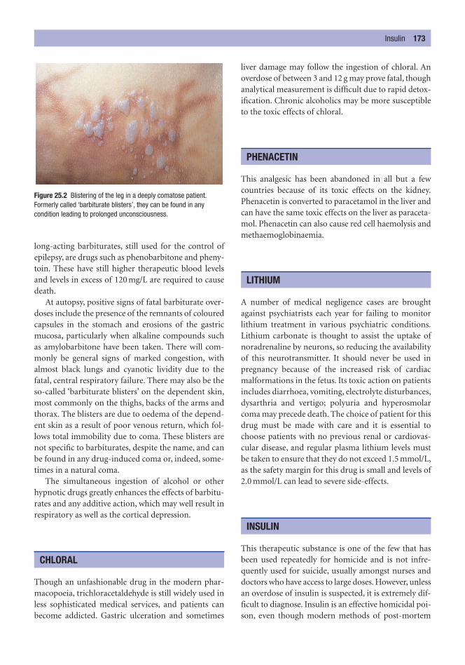
long-acting barbiturates, still used for the control ofepilepsy, are drugs such as phenobarbitone and pheny-toin. These have still higher therapeutic blood levelsand levels in excess of 120 mg/L are required to causedeath.
At autopsy, positive signs of fatal barbiturate over-doses include the presence of the remnants of colouredcapsules in the stomach and erosions of the gastricmucosa, particularly when alkaline compounds suchas amylobarbitone have been taken. There will com-monly be general signs of marked congestion, withalmost black lungs and cyanotic lividity due to thefatal, central respiratory failure. There may also be theso-called ‘barbiturate blisters’ on the dependent skin,most commonly on the thighs, backs of the arms andthorax. The blisters are due to oedema of the depend-ent skin as a result of poor venous return, which fol-lows total immobility due to coma. These blisters arenot specific to barbiturates, despite the name, and canbe found in any drug-induced coma or, indeed, some-times in a natural coma.
The simultaneous ingestion of alcohol or otherhypnotic drugs greatly enhances the effects of barbitu-rates and any additive action, which may well result inrespiratory as well as the cortical depression.
Though an unfashionable drug in the modern phar-macopoeia, trichloracetaldehyde is still widely used inless sophisticated medical services, and patients canbecome addicted. Gastric ulceration and sometimes
liver damage may follow the ingestion of chloral. Anoverdose of between 3 and 12 g may prove fatal, thoughanalytical measurement is difficult due to rapid detox-ification. Chronic alcoholics may be more susceptibleto the toxic effects of chloral.
This analgesic has been abandoned in all but a fewcountries because of its toxic effects on the kidney.Phenacetin is converted to paracetamol in the liver andcan have the same toxic effects on the liver as paraceta-mol. Phenacetin can also cause red cell haemolysis andmethaemoglobinaemia.
A number of medical negligence cases are broughtagainst psychiatrists each year for failing to monitorlithium treatment in various psychiatric conditions.Lithium carbonate is thought to assist the uptake ofnoradrenaline by neurons, so reducing the availabilityof this neurotransmitter. It should never be used inpregnancy because of the increased risk of cardiacmalformations in the fetus. Its toxic action on patientsincludes diarrhoea, vomiting, electrolyte disturbances,dysarthria and vertigo; polyuria and hyperosmolarcoma may precede death. The choice of patient for thisdrug must be made with care and it is essential tochoose patients with no previous renal or cardiovas-cular disease, and regular plasma lithium levels mustbe taken to ensure that they do not exceed 1.5 mmol/L,as the safety margin for this drug is small and levels of2.0 mmol/L can lead to severe side-effects.
This therapeutic substance is one of the few that hasbeen used repeatedly for homicide and is not infre-quently used for suicide, usually amongst nurses anddoctors who have access to large doses. However, unlessan overdose of insulin is suspected, it is extremely dif-ficult to diagnose. Insulin is an effective homicidal poi-son, even though modern methods of post-mortem
INSULIN
LITHIUM
PHENACETIN
CHLORAL
Insulin 173
Figure 25.2 Blistering of the leg in a deeply comatose patient.Formerly called ‘barbiturate blisters’, they can be found in anycondition leading to prolonged unconsciousness.
assay now exist, as the complex investigations that arenecessary to establish the diagnosis are unlikely to belaunched unless there is some suspicion attached towhat appears to be a natural death.
Insulin is, of course, a potent hypoglycaemic agentand, if severe lowering of the blood sugar persists formany hours, brain damage and death will occur. Inmassive doses, especially intravenously, death can takeplace within a very few hours. If death from insulin issuspected, whether suicide, homicide or accident, asearch of the body must be made for recent needlemarks, and the surrounding skin, subcutaneous tissueand underlying muscle must be excised and sent,unfixed, for assay. Blood samples should also be taken,
as modern analytical methods can now distinguishbetween human, bovine and porcine insulin and candetect adjuvants such as zinc, which may assist in trac-ing the origin of any extrinsic insulin.
Post-mortem samples should be taken as soon aspossible after death and the plasma immediately sepa-rated from the cells and then kept deep-frozen untilanalysis. Post-mortem blood glucose levels are gener-ally unhelpful in confirming hypoglycaemia, but vitre-ous humour glucose levels may be more useful.
Oral hypoglycaemic agents, such as the sulphonyl-ureas and biguanides, may be taken in overdose, eithersuicidally or accidentally, and produce hypoglycaemia,hyperkalaemia and acidosis.
174 Medicinal poisons
Corrosive and metallic poisons used to be quite com-mon in Westernized countries as both suicidal andhomicidal agents. Severe restrictions on their availabil-ity and the increasing ease with which these poisonsare detected have markedly limited their use. However,in other parts of the world they are still used sufficientlyfrequently to ensure their continued inclusion in aforensic textbook. It is also important to rememberthese poisons whenever a death occurs, as lack of recentexperience of them may lead the death investigator or the pathologist to assume that this type of poisoningcould not happen in their country and so incorrectlydismiss it as a possibility.
Corrosive substances are used as suicidal poisons andfor homicide and assaults in the form of ‘acid bombs’;they are also used as agents of torture (see Chapter 21).
Corrosives irritate, necrose or destroy any surfacewith which they come into contact. These actions are afunction of both the concentration of the substance andthe length of time for which it acts, so rapid dilutionand washing will greatly reduce the damage, whetherinternally or externally. If used in a very concentratedform, the damage is almost instantaneous and is likelyto be very severe.
The action of any corrosive substance is a surfacephenomenon, whether on the skin or the respiratoryor gastrointestinal mucosa, but continued contact willenable the corrosion to penetrate deeply, so perfor-ation of internal structures such as the oesophagus orstomach may occur with prolonged exposure.
There is often spillage of the corrosive agent on theexterior of the body, and the patterns of skin injury canhelp to reconstruct the way in which the substance wastaken or applied. For example, a person swallowing acidor alkali from a cup whilst standing or sitting usually hasoverflow marks around the corners of the mouth anddribbles that run down the chin, neck and perhaps chest.If they were lying down or had immediately fallen back,the trickle marks are likely to be down the side of theface, cheeks and on to the side of the neck. Coughingand spluttering may project the poison on to the hands,clothing or nearby objects – the fingers are often heldto the mouth and also become corroded. Deliberatesplashing with acid will result in areas or spots of injuryand there may be vertical dribbles from the points ofcontact if the victim was standing. The patterns pro-duced by accidental splashing will depend upon theposition of the individual when splashed, and thisinformation should be specifically sought from workcolleagues and other witnesses.
Internally, oesophageal and gastric damage is to be expected if the substance is swallowed, and theremay also be overflow into the glottis and larynx, with
CORROSIVE POISONS
C h a p t e r t w e n t y - s i x
Corrosive and Metallic Poisons
Corrosive poisonsOxalic acidHeavy-metal poisoning
ArsenicAntimonyMercury
LeadThalliumIron
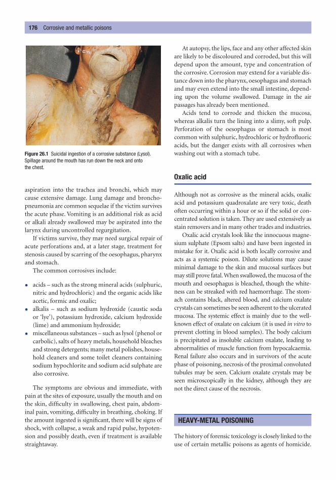
aspiration into the trachea and bronchi, which maycause extensive damage. Lung damage and broncho-pneumonia are common sequelae if the victim survivesthe acute phase. Vomiting is an additional risk as acidor alkali already swallowed may be aspirated into thelarynx during uncontrolled regurgitation.
If victims survive, they may need surgical repair ofacute perforations and, at a later stage, treatment forstenosis caused by scarring of the oesophagus, pharynxand stomach.
The common corrosives include:
• acids – such as the strong mineral acids (sulphuric,nitric and hydrochloric) and the organic acids likeacetic, formic and oxalic;
• alkalis – such as sodium hydroxide (caustic soda or ‘lye’), potassium hydroxide, calcium hydroxide(lime) and ammonium hydroxide;
• miscellaneous substances – such as lysol (phenol orcarbolic), salts of heavy metals, household bleachesand strong detergents; many metal polishes, house-hold cleaners and some toilet cleaners containingsodium hypochlorite and sodium acid sulphate arealso corrosive.
The symptoms are obvious and immediate, withpain at the sites of exposure, usually the mouth and onthe skin, difficulty in swallowing, chest pain, abdom-inal pain, vomiting, difficulty in breathing, choking. Ifthe amount ingested is significant, there will be signs ofshock, with collapse, a weak and rapid pulse, hypoten-sion and possibly death, even if treatment is availablestraightaway.
At autopsy, the lips, face and any other affected skinare likely to be discoloured and corroded, but this willdepend upon the amount, type and concentration ofthe corrosive. Corrosion may extend for a variable dis-tance down into the pharynx, oesophagus and stomachand may even extend into the small intestine, depend-ing upon the volume swallowed. Damage in the air passages has already been mentioned.
Acids tend to corrode and thicken the mucosa,whereas alkalis turn the lining into a slimy, soft pulp.Perforation of the oesophagus or stomach is mostcommon with sulphuric, hydrochloric or hydrofluoricacids, but the danger exists with all corrosives whenwashing out with a stomach tube.
Oxalic acid
Although not as corrosive as the mineral acids, oxalicacid and potassium quadroxalate are very toxic, deathoften occurring within a hour or so if the solid or con-centrated solution is taken. They are used extensively asstain removers and in many other trades and industries.
Oxalic acid crystals look like the innocuous magne-sium sulphate (Epsom salts) and have been ingested inmistake for it. Oxalic acid is both locally corrosive andacts as a systemic poison. Dilute solutions may causeminimal damage to the skin and mucosal surfaces butmay still prove fatal.When swallowed, the mucosa of themouth and oesophagus is bleached, though the white-ness can be streaked with red haemorrhage. The stom-ach contains black, altered blood, and calcium oxalatecrystals can sometimes be seen adherent to the ulceratedmucosa. The systemic effect is mainly due to the well-known effect of oxalate on calcium (it is used in vitro toprevent clotting in blood samples). The body calciumis precipitated as insoluble calcium oxalate, leading toabnormalities of muscle function from hypocalcaemia.Renal failure also occurs and in survivors of the acutephase of poisoning, necrosis of the proximal convolutedtubules may be seen. Calcium oxalate crystals may beseen microscopically in the kidney, although they arenot the direct cause of the necrosis.
The history of forensic toxicology is closely linked to theuse of certain metallic poisons as agents of homicide.
HEAVY-METAL POISONING
176 Corrosive and metallic poisons
Figure 26.1 Suicidal ingestion of a corrosive substance (Lysol).Spillage around the mouth has run down the neck and onto the chest.
The literature of nineteenth-century European forensicmedicine is full of notorious cases of murder by arsenic,antimony and mercury. Occasional cases still occur soall doctors should retain some index of suspicion,especially where atypical gastrointestinal symptomsexist and do not respond to treatment. There are veryfew instances of metallic poisoning from industrialexposure in the UK because of the awareness of the dan-gers and the supervision of the health and safety inspec-tors. However, toxic effects and deaths from metallicpoisons are still common in regions where such occupa-tional and environmental safeguards are less effective.
Arsenic
Arsenic is the twelfth most common element and is itselfnot poisonous; it is the compounds of arsenic, partic-ularly arsenious oxide and the various arsenites of cop-per, sodium and potassium, that are toxic to humans.These compounds exert their effects by combiningwith the sulphydryl groups of mitochondrial enzymesand so prevent cellular respiration. Vascular endothe-lium appears to be particularly susceptible to the toxiceffects of arsenic. Arsenical substances are widely usedin industry and agriculture but are now seldom, if ever,used in conventional medicine. Of historical interest isthe fact that many former editions of this book neededto list a number of sources of pharmacological arsenic.Once ingested, arsenic is stored for long periods in cer-tain tissues and remains permanently in hair, skin andnails (sulphur-containing keratin) until they are shed.
Poisoning is rare and may be suicidal or homicidal,but accidental and environmental causes are morelikely. The signs and symptoms of arsenical poisoninghave so often been mistaken for a natural intestinaldisease that some account must be offered. Acute andchronic poisoning must be considered separately, asthere are marked differences between a single inges-tion of a large dose, as in most suicides, and accidentalor environmental poisoning, where there is morelikely to be the insidious onset of chronic toxicity fromrepeated small doses.
In acute poisoning, if sufficient chemical is ingested,there may be initial burning in the mouth and then,after an interval, signs of intense gastroenteritis develop:abdominal pain, nausea, burning regurgitation andvomiting. Diarrhoea ensues and this, together with the sickness, leads to prostration, mainly from dehydra-tion and electrolyte imbalance. The faeces soon become
watery and often blood streaked. Thirst, oliguria,cramps, collapse and perhaps convulsions lead to car-diovascular failure and death, sometimes within hoursif the dose is large, though survival can last for severaldays. In massive dosage, death may occur before thesesigns have time to appear.
In chronic poisoning, a classic state of ill-healthdevelops, which has often been attributed, incorrectly,to some systemic natural illness. Loss of appetite, mildnausea, some vomiting, jaundice, loss of weight andanaemia are relatively non-specific symptoms, which –especially in the days when ancillary investigationswere minimal – have been accepted as being incapableof further diagnosis or treatment. More specific signscan be seen in the skin, where chronic arsenical poi-soning causes hyperkeratosis of the palms of the handsand soles of the feet, a speckled pigmentation of thegeneral skin surface (sometimes described as a ‘rain-drop’ appearance with patches of pale leucoplakia),loss of hair, brittle nails and a peripheral neuritis withitching, tingling and paraesthesia of the extremities,sometimes with painful swelling (erythromelalgia).
The fatal dose is variable, but 200–300 mg of arseni-ous oxide are likely to be needed for acute fatal toxicity.At autopsy, little may be found in acute deaths apartfrom a haemorrhagic gastritis. The stomach mucosahas been likened to red velvet, due to the bleeding alongthe top ridges of the mucosal folds, which are alsooedematous. Subendocardial bleeding may be seen inthe left ventricle, though this is not specific to arsenicand can be seen in any death when there has been a precipitous drop in blood pressure due to ‘shock’.
In chronic poisoning, the autopsy will reveal a seriesof non-specific signs – wasting, atrophy and dehydrationtogether with skin thickening, hair loss and puffiness ofthe face – may be found and degenerative changes can beidentified in liver, myocardium and kidneys. There is nogood evidence for the long-standing claim that peopledying of chronic arsenic poisoning resist post-mortemdecomposition far longer than normal, apart from thepossibility that dehydration sometimes inhibits therapid onset of moist putrefaction.
Internally, the stomach may show a chronic gastri-tis with excess mucus and patchy mucosal erosion. Thegut may reveal a non-specific mixed oedema andenteritis or it may appear quite normal, apart from acopious ‘rice-water’ content. The liver may suffer fattydegeneration or even necrosis, usually at the peripheryof the lobules. Renal tubular damage and sometimesmyocardial necrosis can occur.
Heavy-metal poisoning 177

In any suspected arsenical poisoning, samples ofhair, nails, blood, urine, faeces and any vomit should becollected from live subjects, supplemented by stomachand contents, intestinal contents, liver, kidneys and bilefrom autopsies. The samples should be submitted to anexpert toxicologist for analysis, as the days of do-it-yourself meddling with the Marsh, Reinsch and Gutzeittests are now strictly part of medical history.
The ubiquitous nature of arsenic means that if abody is being exhumed because of possible arsenicalpoisoning, control samples of soil from the grave and ofthe grave water should be taken so that the role of nat-ural exposure to arsenic after burial can be considered.
It was formerly thought that it took a week or twofor ingested arsenic to appear in the keratinized tissuessuch as hair and nails, but the more sensitive analyticaltechniques now used have shown that the metal canappear there within hours. The test for determiningwhen the arsenic was administered, by measuring theconcentration in different parts of the hair shaft atvarying distances from the growing root, is still a use-ful tool for establishing chronicity of exposure. Themetal can remain in keratinized tissues for severalyears after administration of arsenic has ceased.
Antimony
Antimony is quite similar to arsenic, both in symp-tomatology and fatal dosage. The usual compoundencountered in suicide, accident and the very rarehomicide is antimony potassium tartrate, often called‘tartar emetic’. Poisoning of numbers of people in thesame incident has taken place following the absorptionof antimony from the enamel lining of cooking utensils.Although 150 mg has proved fatal, it is more commonfor there to be a full recovery from vastly greater doses;indeed, 150 mg used to be the intravenous dosage whenantimony was used in the treatment of bilharzia.
Mercury
Mercury is usually an industrial or environmental poi-son. Although historically some deaths occurred fromits use in medicine, mercurial diuretics and antisepticsare now almost a thing of the past.
Acute poisoning resembles that of arsenic and antimony: gastrointestinal symptoms are common, butthere are also excessive salivation and signs of renalfailure. Chronic mercury poisoning is almost wholly
industrial in nature and the clinical hallmarks are gingivitis, loose teeth, salivation, black gums, man-dibular necrosis, peripheral neuritis, encephalopathyand renal failure.
Lead
Lead poisoning is also now an industrial and environ-mental problem, especially since the last forensic aspectsof lead-induced criminal abortions have passed intohistory.
Thallium
Thallium has been used homicidally several times inrecent years, most notably by Graham Young inBritain in 1971, when two victims died and others nar-rowly avoided death. This case was forensically uniquein that it was probably the first murder in which thepoison was detected posthumously in the ashes of acremated victim. Thallium is also unusual in beingdetectable radiologically in the intestine and liver.
Thallium is used as a rat poison and is widelyemployed in industry, especially for optical glass. It isno longer used medically to remove hair in skin dis-eases because of its toxicity. Hair loss is a major sign ofpoisoning and begins 1 or 2 weeks after oral adminis-tration. The other common symptoms of abdominalpain and vomiting characterize acute poisoning. Insucceeding days and weeks, muscle pains, peripheralneuritis, confusion, sleep disturbances and psychiatricsymptoms appear. At autopsy, widespread, but non-specific, degeneration in the heart, liver, kidneys, brainand other organs is discovered.
Iron
Iron is best known in toxicology as an acute poison inchildren who have eaten the attractive-looking tabletsof ferrous sulphate that had been prescribed for theirmothers to treat anaemia. Death has occurred in smallinfants with as few as three to five tablets, gastro-intestinal symptoms occurring soon after ingestion.There may be a temporary respite in symptoms, then arelapse, which is sometimes fatal due to liver damageand acidosis from the release of free iron into the cir-culation as a result of overloading the transferrin sys-tem, which normally binds excess iron to protein.
178 Corrosive and metallic poisons

Agrochemicals are important because they are used insuch vast quantities in most parts of the world. Thesechemicals include pesticides and herbicides, which havegreatly increased food yields but have brought theirown dangers. They are particularly common and there-fore particularly dangerous in rural and agriculturalcommunities. Worldwide, there are millions of admis-sions to hospital and more than 250 000 deaths eachyear as a result of the ingestion of agrochemicals. Someare suicidal, a few homicidal, but most are the result ofeither accidental or environmental exposure. In the UK,over 40 per cent of suspected childhood poisonings arerelated to the ingestion of rodenticides.
First amongst this group are the organophosphate com-pounds, which kill insect pests by inhibiting cholineesterase enzymes, thus preventing the breakdown ofacetylcholine. One of the most toxic is parathion,though others such as malathion, dithion, demeton,octamethylpyrophosphamide (OMPA), hexaethylte-traphosphate (HETP) and tetraethylpyrophosphate(TEPP) are also poisonous. These and similar com-pounds have caused thousands of deaths, especially inSouth-East Asia, either by direct absorption or by con-tamination of foodstuffs with insecticides during stor-age or transit.
The organophosphates are sprayed in liquid form in a kerosene base or used as dusting powders. They canbe absorbed through the intact skin, by inhalation ororally. Toxic effects begin when the choline esterase leveldrops below about 30 per cent of normal. With largedoses, death may be very rapid; otherwise, nausea,vomiting, blurred vision, sweating, colic, contractedpupils and fits may precede death.
The organo-chlorine compounds include lindane,dieldrin and dicophane (DDT). They are soluble inbody fat and are not nearly as toxic as the organophos-phate compounds; indeed, fatalities are rare, thoughcontaminated grain has caused extensive harm. Theirdisadvantage is that they get into the food chain andcause widespread harmful effects on wildlife; this iswhy the parathion-type substances were developed, asthey are far less stable than DDT-like compounds.
The dipyridyl herbicides are also used in great quan-tities in many countries. The best known is paraquat,marketed to the trade as a liquid concentrate underthe trade name Gramoxone and as a much weakergranular form for garden and horticultural use, asWeedol. Gramoxone is extremely toxic and one swal-lowed mouthful is commonly fatal unless immediateenergetic treatment is undertaken. Most fatalities are
HERBICIDES (WEED KILLERS)
PESTICIDES AND INSECTICIDES
C h a p t e r t w e n t y - s e v e n
Agrochemical Poisons
Pesticides and insecticidesHerbicides (weed killers)

suicidal, but quite a number of accidental poisoningsoccur, usually because the liquid is mistaken for a drink.In some countries, including the Republic of Ireland,it is a criminal offence to remove Gramoxone from itsoriginal marked container into any other receptacle,because of this risk of accidental poisoning.
Paraquat is corrosive, causing a burning sensationin the mouth and pharynx, with ulceration of themucosa, but the grave effects are upon the liver, kidneys
and lungs. Hepato-renal failure may occur within the first few days, with a centrilobular necrosis andtubular damage. If the victim survives for longer than aweek, lung damage is common, with what is essentiallythe adult respiratory distress syndrome developing.A hyaline membrane forms and the alveoli fill withpneumocytes, which lead to pulmonary fibrosis ifthe patient lives for a few weeks. Diquat is a similar herbicide.
180 Agrochemical poisons

A number of unrelated poisons are in gaseous form.Though there are many gaseous poisons in existence,especially in an industrial environment, only a few areseen frequently in forensic practice.
In many urbanized countries, ‘coal gas’ or ‘town gas’used to be easily available in almost every home and,as it contained between 7 per cent and 20 per cent car-bon monoxide (CO), it was a potent poison and wascommonly used as a method of suicide, it also causedmany accidental deaths. When natural gas becamegenerally available, especially throughout Europe andNorth America, the number of deaths due to poison-ing by CO was drastically reduced.
The change in house gas supply has not eliminatedCO poisoning, and other sources of the gas have sim-ply become more prominent. The exhaust gas frompetrol engines (unless fitted with a modern catalyticconverter) contains between 4 and 8 per cent CO;diesel engines produce less. In a common suicidalsequence, the victim either sits in the car in a closedgarage with the engine running or leads a pipe fromthe exhaust through the window of the car.
The other common causes of CO poisoning, almostalways accidental, are faulty gas appliances and heat-ing systems. When any hydrocarbon fuel is burned inoxygen (O2 – air), it will be completely converted into
carbon dioxide (CO2) and water only if there is anadequate O2 (air) supply. If air is restricted, CO will beformed instead of CO2. This may occur in gas waterheaters, boilers, fires, cookers etc. and in central heat-ing units fuelled by coal, coke, butane, propane or oil.Faulty adjustment, insufficient ventilation and blockedchimneys and flues may lead to this danger. Such faultyappliances can insidiously kill whole families as theysleep in a house or caravan.
The other major source of CO is a fire in a house orbuilding, where burning timber, furniture and fabricscan produce large volumes of the gas, together withother toxic gases, which kill far more people than actualburns. Other sources of CO are in industry, where hugevolumes are produced and used in steel-works etc.
CO is a heavy, colourless gas, odourless when pure,but often contaminated by impurities, which have asmell. Its poisonous qualities are due to its great affin-ity for haemoglobin, as it has a combining power some250 times greater than O2. This means that even smallconcentrations of CO can displace O2 from the redblood cells and progressively diminish the ability of theblood to transport O2 to the tissues. Strong concentra-tions of CO, such as a leak from a laboratory cylinder,can kill within a few moments. Exercise (causing fasterbreathing) and the quicker respirations of children canhasten the absorption of a fatal level of CO.
There is great variation in personal susceptibilityand in the speed with which saturation takes place.Levels of carboxyhaemoglobin saturation above 50–60per cent are likely to be fatal in healthy adults, whereas
CARBON MONOXIDE
C h a p t e r t w e n t y - e i g h t
Gaseous Poisons
Carbon monoxide Carbon dioxide Ammonia Cyanogen gas and cyanides

old people and those with pulmonary or cardiac dis-ease may well die at much lower levels, even down to25 per cent saturation. The individual sensitivity ofdifferent people is well illustrated by multiple deathsin the same room, where bodies lying side by side mayhave markedly different saturation levels.
The clinical symptoms of CO poisoning are stealthyand progressive so that victims may not notice any-thing, except a headache, until they lapse into comaand die. Many cigarette smokers tolerate levels of up to10–15 per cent without symptoms. At up to about 30per cent saturation of the haemoglobin in fit adults,there may simply be headache and slight nausea; this isvery likely to be associated with lack of concentrationand even a slight ‘drunkenness’, which may be mistakenfor alcohol, especially if driving skills are impaired.
At from 30 to 40 per cent saturation, nausea, pos-sibly vomiting, faintness, loss of visual acuity, weak-ness and a slide towards stupor and coma begin. Over40–50 per cent, sickness, weakness, incoordination,convulsions and coma will progress towards cardiores-piratory failure and death. Some fit young adults mayreach 70 per cent saturation or more before dying. Itmust be emphasized that these figures are very variable,depending on age, fitness and personal susceptibility.
The stability of carboxyhaemoglobin makes it acumulative poison, and the blood will continue toabsorb the gas from the lungs if it is present in theinspired air, so that remarkably small concentrationsof inspired CO may eventually prove fatal. Within 2–3hours, even 0.1 per cent CO in the atmosphere mayresult in 55–60 per cent saturation. Within 20 minutes,1 per cent CO can cause unconsciousness, and the airin a single garage with a 2-litre car engine running canreach a lethal level within 5 minutes.
The external signs of CO poisoning, apart from thesymptoms, are a pink coloration of the skin, usuallydescribed as ‘cherry-pink’. The nail beds and lips mayalso show this characteristic colour, but this may not beobvious in the living until high saturations are reached.In the hypostatic areas of the dead body, the pink col-oration is usually obvious, but exceptions may be foundin the old or anaemic, in whom reduction in haemo-globin content reduces the intensity of the coloration.
Internally, all the organs are pink in colour due to carboxyhaemoglobin and carboxymyoglobin. Pul-monary oedema is common but there are no specificorgan changes, except in the brain of individuals whohave survived for a time following an episode of COpoisoning, in which case there may be bilateral cystic
degeneration of the basal ganglia. Individuals withlong-term survival following significant exposure mayhave a parkinsonian syndrome or may develop evenworse neurological states. Long-term psychologicaldamage may also be caused and this, too, is associatedwith the profound cerebral hypoxia of CO poisoning.
Confirmation of CO poisoning in both the livingand dead depends upon analysis of blood.
CO2 is a relatively inert gas and, unlike the monoxide, isneither directly poisonous nor is it a cumulative poison.However, it can still kill, because it is an irrespirable gas.Three per cent in the atmosphere will cause headache,drowsiness, giddiness and loss of muscle power.
Sources of CO2 are often industrial as it is used verywidely in the manufacture of many products. In anaes-thesia, deaths have occurred when an O2 cylinder hasbecome empty whilst connected to a functioning CO2
cylinder, so that the patient has received only CO2.Vagrants ‘sleeping rough’ near lime kilns are said to
have died due to this heavier-than-air gas creeping overthem during the night. Most modern fatalities are asso-ciated with wells, deep shafts and farming exposure. Inchalk rock, CO2 can accumulate in deep holes and thishas caused deaths of workmen and, in one instance, ofa doctor who descended a well to give help. On farms,the tall grain silos can fill with CO2 produced by therespiration of the seed; the CO2 sinks to the bottom andwhen workers enter through a hatch to clear blockagesin the delivery chutes, they may be rapidly overcome.
An unexplained feature of CO2 is the speed of itstoxicity on many occasions. Pure asphyxia should take
CARBON DIOXIDE
182 Gaseous poisons
Figure 28.1 Cherry-pink hypostasis of carbon monoxide poisoning.

several minutes and be associated with cyanosis andcongestion, but CO2 deaths can be very rapid and thereare seldom any ‘asphyxial signs’. It is presumed that thisis due to some vasovagal reflex causing cardiac arrest,triggered by a chemoreceptor stimulus.
The minimum fatal concentration of CO2 is said to be 25–30 per cent, the high concentrations causingsudden death being 60–80 per cent.
Ammonia gas is an uncommon poison outside indus-try. It was formerly used extensively in refrigerationbut has been largely replaced by more inert fluids.Ammonia is occasionally used criminally for aidingassaults and robberies by being thrown in the face, andthis is sometimes also done in attempts to blind or scar.In gaseous form, escapes in factories may cause severelung symptoms, choking and coughing, and pulmonaryoedema may be followed by bronchopneumonia.
Hydrogen cyanide (HCN) is a gas; hydrocyanic acid(prussic acid) is a solution of HCN in water, either 2per cent or 4 per cent, the latter being called Scheele’sacid. Salts of cyanide, usually sodium and potassium,are white solids. Cyanides are extremely poisonous,their action depending upon the inhibition of the respiratory enzyme cytochrome oxidase, thus prevent-ing the utilization of O2 by tissue cells.
Cyanides are used in industry for a wide variety ofprocesses, as well as in photography, electroplating andlaboratory techniques. They are also used as verminkillers and, regrettably, are still in use as weapons of war.Fumigation of trees, fruit and ships is common andaccounts for a number of accidental deaths. Chemistsand laboratory workers are sometimes overcome byHCN generated by cyanides being poured into sinksand drains that already contain acid. Suicide by cyanide
is not uncommon, especially amongst laboratory staffwho have easy access to the substances. In one surveyof cyanide deaths, 70 per cent were found to be self-administered. The use of cyanide in homicides is notcommon, but it has been deliberately substituted fordrugs of dependence and it has been used to contam-inate medicinal drugs for sale in pharmacies and, inthe notorious Jonesville tragedy in Guyana, 900 peopledied of cyanide poisoning instigated by the ‘Reverend’Jim Jones.
Cyanide is only poisonous as free cyanide and there-fore swallowed salts need to encounter either water orgastric acid before liberating HCN, a process whichtakes only a few seconds. The fatal dose of cyanide, inthe form of a salt, is only 150–300 mg, which allows it tobe concealed as ‘suicide pills’ in rings or hollow teeth.However, recovery has been recorded with far largerdoses of up to 2.4 g of potassium cyanide.
Symptoms and death are often very rapid but notnecessarily so – treatment has saved many victims ofcyanide ingestion. Though some literally ‘drop dead’within seconds, many linger for 15–20 minutes, evenafter substantial doses, depending upon the speed ofabsorption.
The blood remains pink, due to failure of uptake ofO2 by the tissues. The skin, especially after death, maybe a brick-red colour, particularly in the dependenthypostasis. This may be confused with CO poisoning,though cyanide usually gives a darker, sometimes purp-lish hue, whereas CO has a pinker colour.
Cyanides have a characteristic smell, but up to 80per cent of the general population have a congenitalinability to detect the odour of bitter almonds. Thosewho cannot detect the smell of cyanide may insteadreport a sudden onset of a severe headache due to thecerebral vasodilatation caused by this chemical. Atautopsy, great care must be taken to reduce the expos-ure of individuals in the mortuary to a minimum, andbreathing hoods should be worn. The organs will becongested and bright or dark red. If cyanide has beenswallowed, the oesophagus and stomach lining will bedeep red or black, especially along the rugal folds, dueto erosion and haemorrhage, and the smell of cyanidemay be most obvious when the stomach is opened.
CYANOGEN GAS AND CYANIDES
AMMONIA
Cyanogen gas and cyanides 183

Strychnine is an alkaloid prepared from the seeds ofthe Nux vomica tree. In pure form it is a colourlesscrystal and has a notable extremely bitter taste. It is oneof the cruellest poisons in existence as it causes excru-ciating agony to any form of animal life whilst notaffecting consciousness. Used to kill ‘pests’, from earth-worms to dogs, there is no justification for its continueduse or even manufacture, as it no longer has any placein medical treatment and its use in the barbaric killingof animals is quite unjustifiable.
A fatal dose may be as little as 30–60 mg, though,as usual, there is great individual variation. Its majoreffect is upon the muscles, as twitching begins soonafter absorption, deepening into muscle spasms, thendeveloping into convulsions. Opisthotonus and risussardonicus – the grin induced by the contracted facialmuscles – indicate the widespread muscle stimulation.
Through stimulation of the central nervous sys-tem, especially the spinal reflexes, muscles go intotetanic spasms and tetanus may be the differentialdiagnosis where no history is available. The spasmsmay tear muscles from their ligaments and tendons bythe gross contractions; all the while, the maintenanceof consciousness allows the appreciation of the excru-ciating pain. Episodes of convulsive muscle spasm aretriggered by minimal stimulation, and exhaustionaided by respiratory failure due to spasm and loss of
function in the intercostal and diaphragmatic musclesleads to death.
There are no specific features at autopsy, apartfrom haemorrhage into traumatized muscle, and thetendon insertions, routine retention of samples ofgastric contents, blood and urine will reveal strych-nine on toxicological analysis.
This group comprises a number of compounds inwhich part of the aliphatic chain is replaced by one ormore halogen atoms. The resulting substances are inwidespread used in industry, refrigeration, dry cleaningand medicine, one of the latter being Trilene anaesthetic(tri-chlorethylene). All of these chemicals are active fatsolvents and have deleterious effects on the liver, caus-ing hepatic cell damage that may proceed to necrosis.
Though most exposure is accidental, self-administration as a form of addiction may also be seenand this may lead to anaesthetists inhaling their ownagents or to abuse of solvents or other halogenatedcompounds on the streets. Absorption may be fromingestion, inhalation or even skin contact, where thebody surface may suffer from the strong fat-solventaction of these substances.
Methylene chloride is widely used in refrigerationand many deaths and toxic states have occurred from
HALOGENATED HYDROCARBONS
STRYCHNINE
C h a p t e r t w e n t y - n i n e
Miscellaneous Poisons
StrychnineHalogenated hydrocarbons
Gasoline and keroseneThe glycols
Nicotine

Nicotine 185
leakage from refrigeration plants. Chronic exposure maylead to cerebellar signs, with staggering gait, dizziness,vertigo and visual disturbances. Methyl bromide is usedin fire extinguishers and as an insecticide. It can blisterskin and mucous membranes. Exposure may lead tomuscular incoordination, ataxia and muscle twitch-ing. Renal tubular necrosis may also develop.
Carbon tetrachloride used to be available as adomestic dry-cleaning agent (Thawpit) and a fire extin-guisher (Pyrene) as well as commercial solvent. It is a liver poison, but not as dangerous in this respect astetrachlorethane. Excessive domestic use of the stainremover could lead to anaesthesia and it could alsobecome addictive. Death can occur, especially if drunkby children. When sprayed on a burning surface as afire extinguisher, carbon tetrachloride could be con-verted to the even more toxic gas phosgene.
Tetrachlorethane is nine times as toxic as carbontetrachloride. It is also a solvent and plasticizer used forfilms, artificial pearls etc. Its original use as a solvent forcellulose in aircraft ‘dope’ was abandoned early in thetwentieth century when jaundice and deaths occurred inthe factory workers. Liver necrosis may be accompaniedby a polyneuritis affecting the extremities of the limbs.
Petrol and paraffin, alternative names for these fuels,can cause coma and death within a few minutes ifswallowed or inhaled in high concentration. If drunk,they may also be coughed or vomited into the air passages, where they spread widely and rapidly due totheir low surface tension, damaging surfactant andprecipitating bronchopneumonia if the victim sur-vives the initial direct toxic effects.
Used as industrial solvents and anti-freeze agents formotor engines, the glycols are sometimes misused,either for suicide or, more often, as a cheap substitutefor alcoholic drinks. Accidental ingestion usually occurswhen they have been put into an unlabelled soft-drinkbottle. Of the several glycols – dioxan (diethylene diox-ide), ethylene chlorhydrin etc. – ethylene glycol is mosttoxic and most widely available. If drunk in excess ofabout 100–200 mL, it is likely to be fatal unless energetictreatment such as renal dialysis is instituted.
Though symptoms resembling mild drunkennessappear at first, death may supervene within a day or sobecause part of the metabolic pathway converts glycolthrough formic acid to oxalic acid. Renal failureoccurs, with fan-shaped crystals of calcium oxalate inthe kidney tubules and interstitial tissues. Liver dam-age may also occur.
Though homicides have been committed with nicotine,most fatal poisonings are accidental, usually from insec-ticides, some of which may be almost pure nicotine.The poison can be absorbed by mouth, by inhalation orthrough the skin and death has resulted from the inges-tion or application of as little as 40 mg. Even tobaccoleaves wrapped around sweaty skin during smugglinghave caused serious toxic effects, as have the tar andremnants of pipe and cigarette smoking to children.
Nicotine paralyses muscles and usually kills bydepressing the respiratory musculature, as well as pro-ducing sweating, dizziness, fainting and convulsions.
NICOTINE
THE GLYCOLS
GASOLINE AND KEROSENE
1 Where the death is definitely due to crime or if thereis a possibility of crime (a suspicious death), thedoctor should attend the scene (locus) before thebody is moved in order to gain an understanding ofthe surroundings, blood distribution in relation tothe body etc. Notes of attendance, of people presentand of the observations should be made. Photo-graphs should be taken of the scene in general, ofthe body in particular and of any other significantfeatures; these are usually taken by the police.
2 The identity of the body should be confirmed tothe doctor by a relative or by a police officer orother legal officer who either knows the deceasedpersonally or who has had the body positively iden-tified to them by a relative or by some other means(e.g. fingerprints).
3 If the remains are mummified, skeletalized, decom-posed, burnt or otherwise disfigured to a point atwhich visual identification is impossible or uncer-tain, or if the identity is unknown, other methods ofestablishing the identity of the remains must be used,but the autopsy cannot be delayed while this is done.
4 In a suspicious death, if there can be no direct iden-tification of the body, a police officer must confirmdirectly to the doctor that the body or the remainspresented for autopsy are those that are the focus ofthe police inquiry.
5 In a suspicious death, the body should be examinedwith the clothing in place so that defects caused by
trauma that may have damaged the body (stabswounds, gunshot injuries etc.) can be identified.When removed, the clothing must be retained innew, clean bags that are sealed and carefully labelledfor later forensic science examination.
6 In suspicious deaths or if there are any unusual fea-tures, the body should be photographed clothed andthen unclothed and then any injuries or other abnor-malities should be photographed in closer detail.
7 X-rays are advisable in victims of gunshot woundsand explosions and where there is a possibility ofretained metal fragments, and are mandatory in allsuspicious deaths in children.
8 The surface of the body should be examined for thepresence of trace evidence: fibres, hair, blood, saliva,semen etc. This examination may be performed bypolice officers or by forensic scientists, often withthe assistance of the pathologist. Where samplesare to be removed from the body itself as opposedto the surface of the body – fingernail clippings,head and pubic hair, anal and genital swabs – theseshould be taken by the pathologist.
9 Forensic scientists may also wish to examine thebody using specialist techniques, and the patholo-gist must be aware of their needs and allow themaccess at appropriate times.
10 Careful documentation of the external features ofinjuries or abnormalities, their position, size, shapeand type, is often the most important aspect of aforensic examination and often has much greatervalue in understanding and in reconstructing the
GUIDELINES FOR A MEDICO-LEGAL AUTOPSY
A p p e n d i x o n e
Guidelines for an Autopsy andExhumation
Guidelines for a medico-legal autopsyThe autopsy
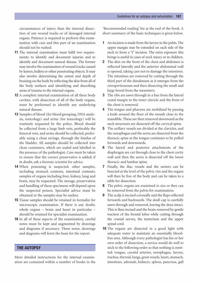
Guidelines for an autopsy and exhumation 187
circumstances of injury than the internal dissec-tion of any wound tracks or of damaged internalorgans. Patience is required to perform this exam-ination with care and this part of an examinationshould not be rushed.
11 The internal examination must fulfil two require-ments: to identify and document injuries and toidentify and document natural disease. The formermay involve the examination of wound tracks causedby knives, bullets or other penetrating objects. It mayalso involve determining the extent and depth ofbruising on the body by reflecting the skin from all ofthe body surfaces and identifying and describingareas of trauma to the internal organs.
12 A complete internal examination of all three bodycavities, with dissection of all of the body organs,must be performed to identify any underlying natural disease.
13 Samples of blood (for blood grouping, DNA analy-sis, toxicology) and urine (for toxicology) will beroutinely requested by the police. Blood should be collected from a large limb vein, preferably thefemoral vein, and urine should be collected, prefer-ably using a clean syringe, through the fundus ofthe bladder. All samples should be collected intoclean containers, which are sealed and labelled inthe presence of the pathologist. Care must be takento ensure that the correct preservative is added; ifin doubt, ask a forensic scientist for advice.
14 When poisoning is suspected, other samples,including stomach contents, intestinal contents,samples of organs including liver, kidney, lung andbrain, may be requested. The storage, preservationand handling of these specimens will depend uponthe suspected poison. Specialist advice must beobtained or the samples may be useless.
15 Tissue samples should be retained in formalin formicroscopic examination. If there is any doubt,whole organs – brain and heart in particular –should be retained for specialist examination.
16 In all of these aspects of the examination, carefulnotes must be kept and augmented by drawingsand diagrams if necessary. These notes, drawingsand diagrams will form the basis for the report.
More detailed instructions for the internal examin-ation are contained within a number of books in the
‘Recommended reading’ list at the end of the book. Ashort summary of the basic techniques is given below.
1 An incision is made from the larynx to the pubis. Theupper margin may be extended on each side of theneck to form a ‘Y’ incision. The extra exposure thisbrings is useful in cases of neck injury or in children.
2 The skin on the front of the chest and abdomen isreflected laterally and the anterior abdominal wallis opened, taking care not to damage the intestines.The intestines are removed by cutting through thethird part of the duodenum as it emerges from theretroperitoneum and then dissecting the small andlarge bowel from the mesentery.
3 The ribs are sawn through in a line from the lateralcostal margin to the inner clavicle and the front ofthe chest is removed.
4 The tongue and pharynx are mobilized by passing a knife around the floor of the mouth close to themandible. These are then removed downward as theneck structures are dissected off the cervical spine.
5 The axillary vessels are divided at the clavicles, andthe oesophagus and the aorta are dissected from thethoracic spine as the tongue continues to be pulledforwards and downwards.
6 The lateral and posterior attachments of thediaphragm are cut through close to the chest cavitywall and then the aorta is dissected off the lowerthoracic and lumbar spine.
7 Finally, the iliac vessels and the ureters can bebisected at the level of the pelvic rim and the organswill then be free of the body and can be taken to atable for dissection.
8 The pelvic organs are examined in situ or they canbe removed from the pelvis for examination.
9 The scalp is incised coronally and the flaps reflectedforwards and backwards. The skull-cap is carefullysawn through and removed, leaving the dura intact.This is then incised and the brain removed by gentletraction of the frontal lobes while cutting throughthe cranial nerves, the tentorium and the upperspinal cord.
10 The organs are dissected in a good light with adequate water to maintain an essentially blood-free area. Although every pathologist has his or herown order of dissection, a novice would do well tostick to the following order so that nothing is omit-ted: tongue, carotid arteries, oesophagus, larynx,trachea, thyroid, lungs, great vessels, heart, stomach,intestines, adrenals, kidneys, spleen, pancreas, gall
THE AUTOPSY

188 Appendix one
bladder and bile ducts, liver, bladder, uterus andovaries or testes and finally the brain.
11 Samples should be taken for toxicology and hist-ology as necessary.
12 Make detailed notes at the time of your examin-ation and write your report as soon as possible, evenif you cannot complete it because further tests arebeing performed.
13 All reports should include all of the positive findingsand all of the relevant negative findings, because incourt the absence of a comment may be taken to
mean that it was not examined or specifically lookedfor and, if a hearing or trial is delayed for manymonths or years, it would not be credible to statethat specific details of this examination can beremembered with clarity.
14 The conclusions should be concise and address allof the relevant issues concerning the death of theindividual. A conclusion about the cause of deathwill be reached in most cases, but in some it isacceptable to give a differential list of causes fromwhich the court may choose.
The following reagent is prepared:
130 mg phenolphthalein1.3 g potassium hydroxide100 mL distilled water
This is boiled until clear, then 20 g of powdered zincare added. A few drops of this reagent are mixed withan equal volume of 20-vol. hydrogen peroxide. Nocolour should result, indicating that the reagents and
glassware are not contaminated. The mixed reagent isthen added to the filter paper or stain extract and analmost instant strong pink colour indicates the prob-ability of blood being present. If benzidene is still usedin your laboratory, a fresh 20 per cent solution of thepowder in glacial acetic acid is added to an equal vol-ume of peroxide before adding to the test material. Aninstant, intense deep-blue colour indicates positivity.
A p p e n d i x t w o
Preparation of the Reagent for the Kastle–Meyer Test
Burton, J. and Rutty, G. (eds) 2001. The Hospital Autopsy,2nd edition. London: Arnold.
Haglund, W.D. and Sorg, M.H. 1996. ForensicTaphonomy. Boca Raton, IL: CRC Press.
Krogman, W. and Iscan, M.Y. 1986. The Skeleton inForensic Medicine, 2nd edition. Springfield, IL:Charles C Thomas.
Ludwig, J. 1979. Current Methods in Autopsy Practice,2nd edition. Philadelphia: W.B. Saunders.
Trotter, M. and Gleser, G.C. 1958. A Re-evaluation ofEstimation of Stature on Measurements of StatureTaken During Life and of Long Bones After Death.American Journal of Physical Anthropology 16: 79–123.
Trotter, M. and Gleser, G.C. 1977. Corrigenda to‘Estimation of Stature from Long Limb Bones ofAmerican Whites and Negroes’. American Journal ofPhysical Anthropology 77: 355–56.
Brogdon, B.G. 1998. Forensic Radiology. Boca Raton,IL: CRC Press.
Byard, R.W. and Cohle, S.D. 1994. Sudden Death inInfancy, Childhood and Adolescence. Cambridge,Cambridge University Press.
Di Maio, V.J.M. 1999. Gunshot Wounds, 2nd edition.Boca Raton, IL: CRC Press.
Mason, J.K. 1989. Paediatric Forensic Medicine andPathology. London: Chapman & Hall Medical.
Mason, J.K. and Perdue, B.N. 1999. The Pathology ofTrauma, 3rd edition. London: Arnold.
McClay, W. 1996. Clinical Forensic Medicine. London:Greenwich Medical Media.
Robinson, S. 1996. Principles of Forensic Medicine.London: Greenwich Medical Media.
Saukko, P. and Knight, B. 2003. Knight’s ForensicPathology, 3rd edition. London: Arnold.
Stark, M.M. (ed.) 2000. A Physicians’ Guide to ClinicalForensic Medicine. Totowa, NJ: Humana Press.
Valdés-Dapena, M. 1993. Histopatholgy Atlas for SuddenInfant Death Syndrome. Washington, DC: AFIP.
Whittaker, D. and MacDonald, D. 1989. A Colour Atlas of Forensic Dentistry. London: Wolfe MedicalPublications.
BMA 2001. The Medical Profession and Human Rights:A Handbook for a Changing Agenda. London: BMA.
Mason, J. and McCall, R. 1999. Law and Medical Ethics.London: Butterworth Tolley.
Palls, C. and Harley, D.H. 1996. ABC of BrainstemDeath, 2nd edition. London: BMJ Books.
MEDICAL ETHICSFORENSIC MEDICINE AND PATHOLOGY
AUTOPSY PROCEDURES AND ANTHROPOLOGY
Recommended Reading
Autopsy procedures and anthropologyForensic medicine and pathology
Medical ethicsToxicology
Websites

Recommended reading 191
Ellenhorn, M.J. 1997. Ellenhorn’s Medical Toxicology,2nd edition. Baltimore, MD: Williams and Wilkins.
Amnesty International www.amnesty.org.ukBritish Medical Association www.bma.org.ukInternational Red Cross www.ifr.orgPhysicians for Human Rights www.phsusa.orgUnited Nations www.un.orgWorld Medical Association www.wma.net
WEBSITESTOXICOLOGY

This page intentionally left blank
Index
Note: Page numbers in italic indicate figures; bold type refers to tables
abdominal injury 77–8, 77in children 77penetrating 77
abnormal behaviour 22abortion 135–40
illegal 136–9, 138methods of performing 137–9spontaneous (miscarriage) 135–6
Abortion Act 139abrasions 60–61, 61, 67accident 85accident surgery 18acid bombs 175adipocere 46, 46adrenal haemorrhage 119adult respiratory destress
syndrome 118Adults with Incapacity Act 14affective psychoses 23age determination 50, 55agrochemical poisons 179–80air bags 89air embolism 116–17air guns and rifles 84air weapons 84aircraft fatalities 92alcohol 159–63
absorption 160concentration 159effects 25, 160–61elimination 160evaluation for impairment of
ability to drive 162–3measurement 160sources of 159–60
ammonia 183ammunition 80–81, 81amnesia 25amniotic fluid embolism 118amphetamines 167amputation 92anaesthesiology 18–19analgesics 171–2aneurysm 123
aortic 123
berry 124–5syphilitic 124
animal tissue 30anoxia see asphyxiaanthropology, recommended
reading 190antidepressants 172antimony 178aorta
atheromatous aneurysm of 122, 123dissecting aneurysm of 123–4penetrating injury 78
aortic stenosis 122–3arsenic poisoning 36, 177–8arteries, diseases of 123–5artificial insemination 134asphyxia 94–102
classical features 94conditions 95use of term 94
aspirin 171assault 59asthma 126–7atheroma 122atheromatous aortic aneurysm 122,
123autopsy 30, 33–5, 92, 143, 158, 173
basic techniques 187–8guidelines 186–7observation in 35recommended reading 190reports 7
barbiturate blisters 173, 173barbiturates 167, 172–3battery 59benzodiazepines 164, 172bestiality 130�APP (beta apoprotein precursor) 75bile analysis 158bite marks 67bites in child abuse 148, 148bleeding see haemorrhageblood-alcohol concentration (BAC)
160, 161
blood cells, individuality 51–2, 52blood group systems 51–2, 52blood samples 157, 163blood stains 57–8, 58blood tests 58
species specificity 58blood transfusion 29blood vessels, rupture 61body chart 6body piercing 54body temperature
as indicator of time of death 41–8measurement of 43
bones, stature determination from 56bows 85brain damage 18, 23, 88brain death, Code of Practice in
diagnosing 28brain injury 74–5British Medical Association (BMA) 11,
20, 162bruises 61–2, 62–3
colour changes 63in child abuse 146–7resolution 62tramline (railway) line 62, 63tyre tread pattern 91
brush abrasion 61, 90burial 47burns 107, 108, 109
causes of death 109–10in child abuse 148injuries due to 110
cadaveric donation 29–30cadaveric rigidity 38–9, 39café coronary 96–7, 96cannabis 168capital punishment 59car occupant injuries 88–9carbon dioxide (CO2) poisoning
182–3carbon monoxide (CO) poisoning
181–2carbon tetrachloride 185
cardiac arrest 28, 37cardiopulmonary resuscitation
(CPR) 77catatonia 23cellular death 27cerebral haemorrhage 125, 125cerebral infarction 125cerebral thrombosis 125cerebral tumours 23cerebrovascular accident (CVA) 125cerebrovascular lesion 125chest injury 76–7
blunt injury 76penetrating 77
child abuse 145–6child physical abuse 145
epidemiology 145–6general features 146injuries due to 146–9, 146, 148
child sexual abuse 149children
criminal responsibility 24indecency with 129
chloral 173choking 96civil courts 2
role of the expert 3civil rights 6cloning of animals 30Clostridium perfringens 116, 138Clostridium tetani 116cocaine 167–8Code of Practice in diagnosing brain
death 28cold injury 110–11‘cold turkey’ 165compensation 17conception 134confidentiality 6, 11–13, 136–7
absolute duty of 12congestion of the face 94conjuctival haemorrhages 95consent
disclosure 12express 14implied 14inferred 14, 131–3, 140treatment 13–14
contamination of genetic material 52Continuing Professional Development
(CPD) 15continuity of evidence 157contusions 61–3, 66coronary artery disease 121coronary stenosis 121coronary thrombosis 121coroner 32–4corrosive poisons 175–8, 176
common agents 176symptoms 176
cot deaths see sudden infant deathsyndrome (SIDS)
coup and contra-coup injuries 75, 75
courtdisclosure of information 12professional demeanour in 5
court attendance 4court behaviour 5court structure 3crash helmets 89criminal abortion see abortionCriminal Justice and Public Order
Act 128criminal justice system 23–4criminal law 2criminal responsibility, age and mental
capacity 24–5crossbows 85cross-examination 5crush abrasions 61, 67cyanides 183cyanogen 183cyanosis 94
damages 17death 27–36
changes after 37–48cooling of body after 40–41, 41definition 27early changes 37–8estimation of time 41–8medico-legal investigation 32–4, 33,
34methods of estimating time of 44
death certificates 30–32, 31, 120Declarations of the World Medical
Association 10decomposition 37, 44–5deep vein thrombosis 117, 126defence wounds 67, 68delusions 23dementias 23dental X-rays 51diatom test 105–6, 105diminished responsibility 25dipipanone 167Diquat 180disclosure of information 12–13dissecting aneurysm of the aorta see
aneurysmdisseminated intravascular coagulation
(DIC) 118DNA analysis 135
samples required for 53DNA profiling 52documentary evidence 2drinking and driving see alcoholdrowning 104–6, 152
laboratory tests 105–6, 105drug abuse 164–9drug addiction 164drug dependence 164–9drug effects 25drug hypersensitivity 166drug ingestion 164drug injections 165
drug overdose 155, 166, 170, 171drug synergy 165drug tolerance 164–5drug withdrawal 165drunkenness, dangers 161–2Dum-Dum bullets 81duty of care 15, 16
Economy Class Syndrome 117ectopic pregnancy 126electric shock 92electrical injury 111–13, 113electrical lesion 112–13embolism 116
amniotic fluid 118air 116fat 116foreign body 118pulmonary 117–18, 117
embryo research 134entrance wounds 82–3epilepsy 23, 126–7epiphyseal union 50ethanol 159ethical responsibilities 1ethics of medical practice see medical
ethicsexhumation 35–6exit wounds 84, 84expert witness 3explosive injuries 79, 86, 86
facial reconstruction from skull 55–6faeces, analysis 158falls 72fat embolism 116fatal dose 155–6fingerprints 50firearms 79–86
doctor’s duty in 85–6types 79–85
fires, examination of bodies 108–9, 110flaying injury 90flotation test 143foreign-body embolism 118Forensic Medical Examiner (FME) 24forensic odontology (forensic
dentistry) 50fractures see specific regional injuriesfunctional psychoses 23
gagging 96gamma-glutamyl transpeptidase
(GGT) 162gaseous poisons 181–3gasoline 185gastrointestinal system 126General Medical Council (GMC) 11,
20–21general medical practice 18giving evidence 4, 5glycols 185Gramoxone 179
194 Index

gravel rash 61gunshot wounds 81–4, 81–4
Haase’s rule 144haemopericardium 122haemopneumothorax 76haemorrhage 72–4, 73, 115–16hair samples 158hallocinogenic drugs 168hallucinations 23halogenated hydrocarbons 184–5hanging 97, 99–101, 100head injuries 70–75, 89
in child abuse 147–8heat, injury due to 107–10heavy-metal poisoning 158, 176–8Henssge’s Normogram 42–4herbicides 179–80heroin 166Hippocratic Oath 8–9homicide 59, 85, 154Homicide Act 25homosexual offences 130–31Human Fertilization and Embryology
Act 134, 139human rights 6, 150–57Human Tissue Act 30humane killers 84–5hydrogen cyanide (HCN) 183hyperflexion injuries 76hypertensive heart disease 122hypertrophic obstructive
cardiomyopathy (HOCM) 123hypnotic drugs 167hypoglycaemic agents 174hypothermia 110–11hypoxia see asphyxia
ibuprofen 171–2identification of the living and the
dead 49–56illusions 23immersion 46–7, 103incest 130indecency with children 129indecent assault 129indecent exposure 129infancy, deaths and injury in 141–9Infant Life Preservation Act (1929) 142infanticide 60, 135, 142–3, 142–3Infanticide Act 142infection 116inferior vena cava, penetrating injury 78injuries
classification 60effects 115–19patterns 66–8regional 70–78self-inflicted 68–9, 68survival following 68terminology 60see also specific injuries
inquest and verdicts 34
insecticides 179, 185insulin 173–4intracranial haemorrhage 72–4, 73in-vitro fertilization 134iron 178
justifiable homicide 59juveniles, criminal responsibility 24
Kastle–Meyer test 58, 189kerosene 185kicking 67kidney transplantation 29
lacerations 63–4, 64lead 178left ventricular failure 166left ventricular hypertrophy 122leg injury, pedestrian 90legal system 1, 2ligature strangulation 98–9, 99lightning, death from 113–14, 113lithium 173live donation 29liver analysis 158liver damage 159–60liver function tests 162liver rupture 77lung injury 77lysergic acid diethylamide (LSD) 168
McNaghten Rules 25magnetic resonance imaging (MRI) 50manic–depressive psychosis 23manslaughter 59manual strangulation 98, 98marrow transplantation 29mass disasters, medical ethics in 92–3MDMA 167medical abbreviations 7medical ethics 8–14
in mass disasters 92–3in practice 10–11recommended reading 190
medical examination, road trafficaccidents 90
medical information disclosure 6medical legislation 1–7medical malpractice 15–21medical negligence 15–17, 18
allegations of 16types 17–19
Medical Register 20medical reports 4–5
alterations 7preparation 5–6structure 6–7
medicinal poisons 170–74medico-legal autopsies 34mental abnormality
acquired 22congenital 22criminal responsibility 24
mental disease, medico-legal aspects22–6
mental health legislation 23–4mental subnormality 22mercury 178mercy killing 23mesentery, lacerations 77methadone 167methylene chloride 184–5miscarriage see abortion, spontaneousmonoamine oxidase inhibitors
(MAOIs) 172morphine 166motor cycle injuries 89–90mummification 45–6murder 59myocardial hypoxia 28myocardial infarction 121, 122
nail clippings 158neck injuries 75necropsy 34neglect 152–3, 152negligence see medical negligencenicotine poisoning 185non-steroidal anti-inflammatory drugs
(NSAIDs) 171–2
oath or affirmation 5ocular injuries in child abuse 148opiates 166
withdrawal 165oral injuries in child abuse 148organ donation and transplantation
29–30organic psychoses 23ossification centres 50oxalic acid 176
pancreatic trauma 78paracetamol 171paraffin 185paranoid schizophrenia 23paraquat 179–80paternity 135paternity/maternity testing 52–3pathology 34
recommended reading 190patients’ rights 6pedal cycle injuries 89–90pedestrians 87–9, 88, 90Perl’s stain 73pesticides 179petechial haemorrhages 94, 95pethidine 167petrol 185pharmaceuticals, dangerous usage
170phenacetin 173phencyclidine 168phenobarbitone 167phenothiazines 172physical abuse 150–52, 151
Index 195
physiciansduties to each other 10duties to the sick 9–10
pistols 80–81, 80plastic bullets 84–5pneumothoraces 76, 77poisoning 36
accidental 154, 180concealed 156definition 154diagnosis 156differential diagnosis 156general aspects 154–8heavy-metal 158homicidal 155management 156self-administration 157self-inflicted 154suspected 156–7see also agrochemical poisoning;
corrosive poisons; heavy-metallicpoisons; medicinal poisons
police, disclosure of information 12–13police surgeon see Forensic Medical
Examinerpolymerase chain reaction (PCR) 53Pontine haemorrhage 125post-mortem examination 34post-mortem hypostasis 39–40post-mortem injuries 47–8, 47post-mortem recovery of exhibits 86pregnancy 126, 134–5
consent to termination 140duration 135illegal abortion 136–9, 138legal termination 139–40proving/disproving 134–5
presistent vegetative state (PVS) 29professional demeanour in court 5professional misconduct 19–21professional witness 2–3pseudocyst 78Pseudomonas aeruginosa 116psychoses 23pulmonary embolism 117–18, 117pulmonary oedema 166punches 66putrefaction 45, 45
radiology 35railway injuries 91–2rape 128–9, 128reflex cardiac arrest 97regional injuries 70–78Registrar of Deaths 33regurgitation 38respiratory system 126resuscitation 28, 38retroperitoneal haemorrhage 122revolvers 80–81, 80rib fractures 76–7rifled weapons 80–81
wounds from 82–3rifles 80–81
rigor mortis 38road traffic injuries 87–90
facial injuries in 91medical examination 90
rubber bullets 84–5Rule of Nines 107, 108
scalds 107–10, 108scalp injuries 70–71, 71schizophrenia 22, 23seat belts 89sedative drugs 172selective serotonin re-uptake inhibitor
(SSRI) drugs 172self-inflicted injuries 68–9, 68senile myocardial degeneration 123sexual asphyxias 101, 101sexual assault, allegations of 131sexual intercourse, unlawful 129sexual matters, disclosure of 12sexual offences 128–33
examination of alleged assailant132–3
forensic examination of victims131–2, 132
Sexual Offences Act (1967) 130sexually transmitted diseases 149shotguns 79–80, 79skeletal injury, in child abuse 147skeletal remains, identity of 54–5,
54–5skeletalization 47skull
anatomy 72facial reconstruction from 55–6
skull fractures 71–2, 71in children and infants 72
smothering 96solvent abuse 158, 168–9somatic death 27–8spinal injuries 75–6, 76spleen, blunt trauma 78stab wounds 65–6, 65stamping injuries 67starvation 152–3, 153stature determination from bones 56stillbirths 141–2, 141–2stomach contents 158strangulation 95, 97–9, 99strychnine 184stud guns 84–5subarachnoid haemorrhage 74subdural haemorrhage 73subendocardial haemorrhages 119sudden and unexpected deaths (SUDs)
120–27causes 120–25definition 120
sudden infant death syndrome (SIDS)95, 144–5
suffocation 95, 152suicide 83, 85, 91, 96, 154, 176, 180suprarenal haemorrhage 118–19surgery 18
survival following injury 68, 85suspension 152
Takayama blood test 58tattoos 53, 53teeth, identity from 50–51, 51temporal bone, fracture 73testamentary capacity 25–6tetrachlorethane 185thallium 178thoracic aorta aneurysm 124thoracic aorta rupture 91throttling 98tissue donation and transplantation
29–30toluene 168toxic dose 155–6toxicity 154toxicology, analysis 157–8transplantation 29–30transportation injuries 87–93traumatic asphyxia 101–2, 102traumatic subarachnoid haemorrhage
74triage 92Trilene anaesthetic 184
unethical conduct 11unlawful sexual intercourse 129urine samples 158, 163
vagal inhibition 97ventilation techniques 28view and grant 33
websites 191wedge fracture 76weed killers 179–80Weedol 179whiplash injuries 76wills 25–6World Health Organization (WHO)
30, 120World Medical Association 10,
92, 139wounding, law 59–60wounds
blunt force 60–64defence 67, 68definition 59examination 59–69explosives 86, 86gunshot 81–4, 81–4incised 64–5, 65recording information 60reports 60rifled weapons 82–3sharp force 64–6smooth-bore guns 81–2stab 65–6, 65terminology 60types 60
xenografts 30
196 Index