x-ray crystal structure and cation binding properties of a ...journalarticle.ukm.my/12061/1/ukm...
TRANSCRIPT
Sains Malaysiana 47(4)(2018): 755-762http://dx.doi.org/10.17576/jsm-2018-4704-14
X-Ray Crystal Structure and Cation Binding Properties of a New Phthalide-fused Indoline Derivative
(Struktur Hablur Sinar-X dan Sifat Pengikat Kation bagi Terbitan Ftalida-Indolin)
Wong Sheryn, Jalifah latip, nurul izzaty haSSan & Siti aiShah haSbullah*
abStract
An efficient and a green route to synthesize phthalide-fused indoline, 3-[(5-chloro-1,3,3-trimethylindolin-2-ylidene)methyl]isobenzofuran-1(3H)-one (3) has been developed by the coupling reaction of 5-chloro-2-methylene-1,3,3-trimethylindoline, 1 and 2-formylbenzoic acid, 2 under solvent-free domestic microwave irradiation. The compound was produced in 85% yield. Compound 3 was characterized by analytical and spectral methods and its structure was confirmed by chemical crystallography. Compound 3 was successfully crystallized in triclinic system with space group Pī. The molecular structure consists of fused 1 and 2 groups connected by the enamine carbon. Binding study of 3 towards different types of metal cations was done by colorimetric detection and UV-vis titrations. Compound 3 exhibited good selectivity and sensitivity for Sn2+ compared to other metal cations tested. The stoichiometric binding ratio of 3 toward Sn2+ is found to be 1:1 and the binding constant (Ka) is 1.07×10⁴ M–1 on the basis of Job’s plot and Benesi-Hildebrand analysis.
Keywords: Binding study; chemical sensor; green synthesis; Phthalide-indoline; Sn(II) ion
abStrak
Satu kaedah sintesis hijau dan cekap bagi sintesis ftalida-indolin, 3-[(5-kloro-1,3,3-trimetilindolin-2-ylidin)metil]isobenzofuran-1(3H)-on (3), telah dibangunkan melalui tindak balas pengkupelan antara 5-kloro-2-metilin-1,3,3-trimetilindolin, 1 dan asid 2-formilbenzoik, 2 menggunakan iradiasi gelombang mikro tanpa pelarut dengan peratusan 85%. Sebatian 3 telah dicirikan melalui kaedah analisis dan spektrum, serta strukturnya disahkan melalui kristalografi kimia. Hablur tunggal sebatian 3 telah berjaya dihasilkan dan mempunyai sistem hablur triklinik dengan kumpulan ruang Pī. Struktur molekul 3 adalah merupakan gabungan kumpulan 1 dan 2 yang berikat pada karbon enamin. Kajian pengikatan antara sebatian 3 dengan pelbagai kation logam telah dijalankan menggunakan kaedah kolorimetri dan kaedah pentitratan ultralembayung boleh nampak (UV-vis). Sebatian 3 menunjukkan kepilihan dan kepekaan yang baik terhadap ion Sn2+ berbanding dengan kation logam lain yang diuji. Nisbah stoikiometri pengikatan antara 3 dengan Sn2+ dan pemalar pengikatan (Ka) masing-masing adalah 1:1 dan 1.07×10⁴ M–1 berdasarkan plot Job dan analisis Benesi-Hildebrand.
Kata kunci: Ftalida-indolin; ion Sn(II); kajian pengikatan; penderia kimia; sintesis hijau
introduction
Study on selective and sensitive chemosensors are widely attractive because of their instantaneous response toward a certain analyte, high sensitivity with low limit of detection, accuracy and uncomplicated design as compared to the costly conventional method of detecting metal ions (Li & Xiao 2016; Qu et al. 2017). Heterocyclic molecule with ring opening ability is one of the important aspects in designing a highly selective and sensitive chemosensor (Chatterjee et al. 2016). Colorimetric chemosensor has also attracted much attention whereby a target analyte can be identified by ‘naked eye’ (Zhu et al. 2015). In a recent study on tin compounds in landfill leachates, Sn2+ ionic species exhibited bacterial methylation forming toxic methyltin which may be released into the environment (Peeters et al. 2014). Sn2+ and inorganic tin compounds have several unfavourable biological effects on human and moderate
toxicity to aquatic organisms and the environment (Howe & Watts 2005). Meanwhile in another study by Mathews et al. (2015), Sn(II) chloride was successfully used as a treatment system to remove mercury from wastewater, however, possible impacts of tin bioaccumulation toward human or ecological risk may be of concern. Thus, an in situ method of detecting and quantifying tin metal ions is essential in monitoring the environmental and biological systems.
Phthalides, also known as isobenzofuranones, are classified under lactones with its five-membered O-heterocyclic ring and is fused with benzene. 3-substituted phthalides are usually found in natural products and are well known for their wide range of biological application (Beck & Chou 2007; Karmakar et al. 2014). It is also one of the main classes in the production of commercial dyes (Bamfield 2010). Indoline, an aromatic N-heterocycle,
756
possess biological activities like antibacterial (Michael Barbour et al. 2014) and cytotoxicity against cancer cells (Azizian et al. 2012). Besides, it is also a building block of highly conjugated cyanine with good optical properties, great biocompatibility and low toxicity toward biosamples (Sun et al. 2016). These are the desirable properties of a chemosensor.
Much effort is being expended in synthesizing natural products containing the phthalide and N-heterocyclic moiety. Green route of synthesizing 3-substituted phthalides using the microwave irradiation was reported with better yield and efficiency (Landge et al. 2008; Srivastava et al. 2013). The use of microwave irradiation in reaction provides many advantages which include performing solventless reactions, reduction of time taken for a reaction, instantaneous and efficient heating and less waste production (Gangrade et al. 2015). This work will follow the green route of synthesis whereby reaction will be done in a solventless medium using the microwave irradiation method.
MaterialS and MethodS
Reaction was performed under microwave irradiation method using a domestic microwave oven (Electrolux, model EMM2017X, PCR). Chemicals and solvents were purchased from Sigma Aldrich and Systerm and used directly without further purification. All UV-vis spectra were recorded on Shimadzu UV-1800 spectrophotometer using a quartz cell with a path length of 1 cm in 95% ETOH; FTIR spectrum was recorded on Perkin-Elmer Spectrum GX spectrophotometer in the range 400-4000 cm-1 using KBr pellet method; 1H and 13C NMR (in CDCl3) spectra recorded on a Bruker Avance 400 MHz spectrometer at 400.2 MHz and at 100.6 MHz, respectively using TMS as internal standard; mass spectrum recorded on a Bruker micrOTOF-Q spectrometer by ESI-MS in the positive ion mode. Single crystal X-ray experiments were performed on Bruker D8 QUEST diffractometer with MoKα radiation.
MicroWaVe irradiation Method
A mixture of 5-chloro-2-methylene-1,3,3-trimethylindoline, 1 (1 mmol) and 2-formylbenzoic acid, 2 (1 mmol) was added into a dry reaction vial. The mixture was then irradiated using a domestic microwave at 100 W for 1 min. The reaction was monitored by TLC (petroleum ether/ethyl acetate, 2:1). After reaction was completed, cold ethanol (2 mL) was added into the reaction mixture. Precipitate was formed upon sonication for 10 s. The reaction mixture was then evaporated to approximately half its original volume and the precipitate was filtered off. The crude product was crystallized by slow evaporation from acetone to obtain pure crystal plates.
3-[(5-Chloro-1,3,3-trimethylindolin-2-ylidene)methyl]isobenzofuran-1(3H)-one(3)
The compound was obtained as greenish-yellow crystals in 85% yield (0.29 g). UV (EtOH): lmax = 226, 290 nm. IR
υmax (KBr)/cm–1: 3053 (C-H sp2), 2928, 2960 (C-H sp3), 1750 (C=O), 1649 (C=C), 1466, 1493, 1602 (aromatic C=C), 1139, 1287 (C-O), 1062, 1094, 1265 (C-N), 900. 1H NMR (400 MHz; CDCl3) dH 1.70 (6H, s, CH3), 3.00 (3H, s, N-CH3), 4.21 (1H, d, J = 10.8 Hz, CH-O), 6.51 (1H, d, J = 7.6 Hz, HAr), 6.60 (1H, d, J = 10.4 Hz, C=CH), 7.12 (2H, m, J = 8.0 Hz, HAr), 7.45 (1H, d, J = 6.8 Hz, HAr), 7.56 (1H, t, J = 6.8 Hz, 7.2 Hz, HAr), 7.70 (1H, t, J = 6.4 Hz, 7.2 Hz, HAr), 7.91 (1H, d, J = 7.2 Hz, HAr). 13C NMR (100 MHz, CDCl3) 29.2, 29.9 (CH3-C-CH3), 45.2 (N-CH3), 79.1 (C=CH), 88.7 (CH-O), 106.4 (CHAr), 122.1 (CHAr), 122.7 (CHAr), 124.3 (CAr-Cl), 125.4 (CHAr), 126.4 (CAr), 127.7 (CHAr), 129.1 (CHAr), 134.1 (CHAr), 139.7 (CAr), 143.8 (CAr), 150.9 (CAr), 160.9 (N-C=CH), 170.7 (C=O). ESI-MS m/z calcd for C20H18ClNO2Na [M + Na]+: 362.0924. Found: 362.0921.
SAMPLE PREPARATION FOR BINDING STUDY WITH METAL CATIONS
For colorimetric detection of metal ions, stock solutions (1 × 10⁻³ M) of 3 and metal ions (Ag+, Co2+, Cu2+, Fe2+, Hg2+, Ni2+, Sn2+ and Zn2+) were prepared in 95% EtOH, respectively. The color change of 1 mL of 3 (5 × 10⁻⁴ M) was observed immediately after mixing with 1 mL of stock solution of respective metal ions. Metal ion screening was further analyzed by UV-vis absorption by diluting 50 mL of stock solution of 3 and 100 mL of stock solution of respective metal ions in a 5 mL volumetric flask with EtOH. The final concentrations of 3 and metal ions were 1 × 10⁻⁵ M and 2 × 10⁻⁵ M, respectively.
For UV-vis titration, stock solutions of 3 (1 × 10⁻⁵ M) and SnCl2 (1 × 10⁻³ M) were prepared. 3 mL of 3 was taken directly into a quartz cuvette. The spectral measurements were recorded after each aliquot addition (10 mL) of the metal cation solution using a micropipette. All titration experiments were recorded at room temperature.
The stoichiometric ratio between 3 and Sn2+ ions was studied using Job’s plot. Equimolar (1 × 10⁻³ M) stock solutions of 3 and Sn2+ ions were prepared in 95% EtOH. Both the compound 3 and Sn2+ ions were mixed at different volumes (from 0 to 200 mL) in 10 mL volumetric flasks and was diluted with EtOH to make a total volume of 10 mL. Each volumetric flask had a total concentration of 3 and Sn2+ of 2 × 10⁻⁵ M. The UV-vis spectra were taken at room temperature.
reSultS and diScuSSion
SYNTHESIS AND CHARACTERIZATION
Compound 3 was synthesized from the reaction of a Fischer base and phthalaldehydic acid. The reaction scheme is shown in Scheme 1. Compound 3 was synthesized in a quantitative yield of 85% by microwave irradiation.
The UV spectrum of 3 in ethanol displayed absorption maximum at around 226 nm and 290 nm indicative of aromatic ring compounds due to π → π* of benzene chromophores and n → π* transition of carbonyl groups.
757
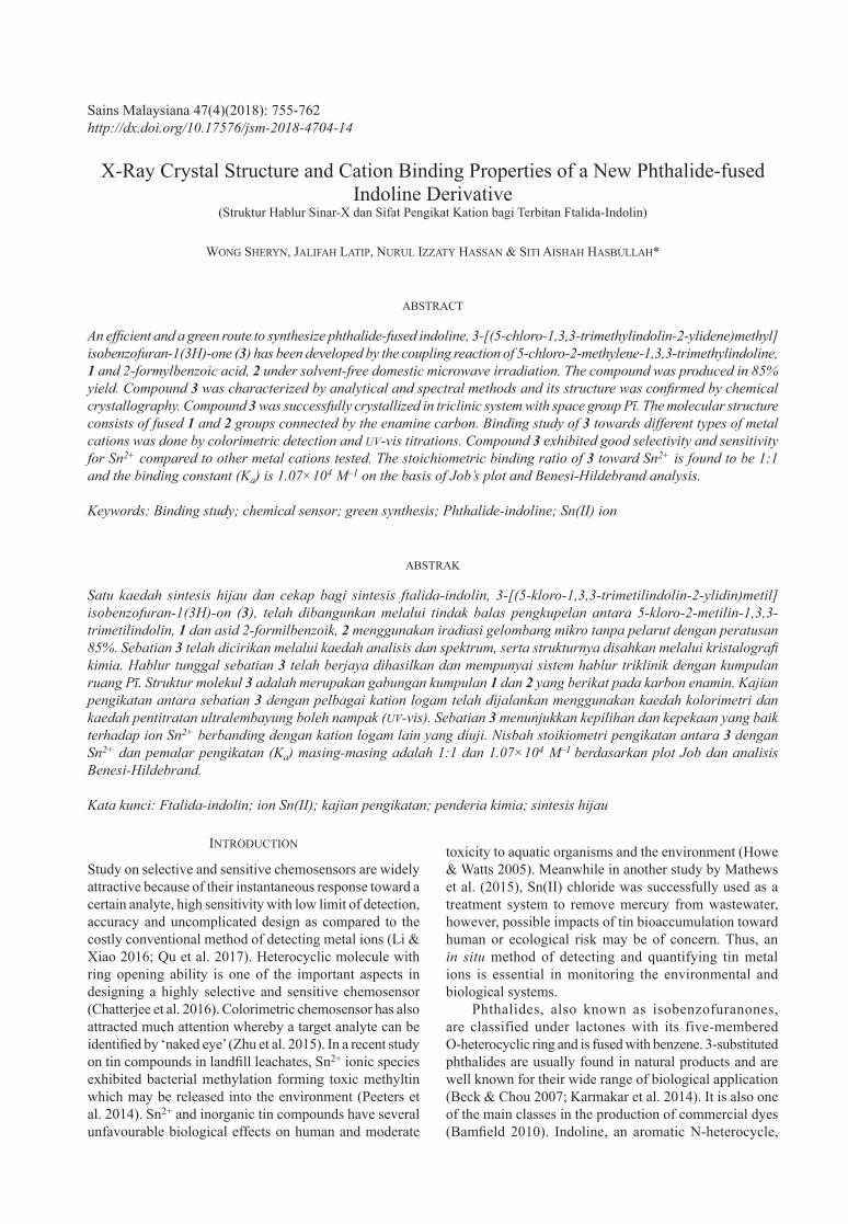
The FTIR spectrum showed characteristic C=O stretch of lactone ring at 1750 cm–1 (Figure 1). Bands between 1062 and 1287 cm–1 were assigned as C-O and C-N stretching.
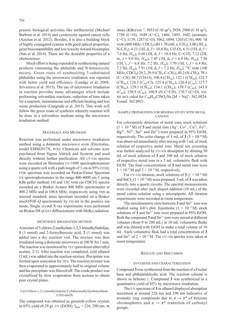
1H NMR spectrum (Figure 2) of compound 3 displayed 18 hydrogen integrals. According to report by Keum et al.(2011), the indoline group showed regular 1H NMR resonance pattern for the protons on adjacent methyl
figure 2. 1H NMR of 3
ScheMe 1. Preparation of a phthalide-fused indoline derivative under microwave irradiation
figure 1. FTIR spectrum of 3
100
90
80
70
60
50
40
30
20
10
0
%T
cm–1
4000 3600
2928
400800120016002000240028003200
2960
3053C-O and
C-N stretch
1287113914931602
1062 900
1750
Lactone C=O stretch
1649 C=C bend C=C bend
sp3 C-H stretch
sp2 C-H stretch
3 (Yield = 85%)21
758
groups. The gem-dimethyl protons appeared as a singlet at 1.70 ppm, while N-methyl protons resonated at 3.00 ppm. A doublet at 4.20 ppm belongs to the chiral CH-O proton. The olefi nic proton was detected as a multiplet at 6.60 ppm, with a J value of 10.4 Hz. Peaks between 6.5 and 8.0 ppm were resonated by aromatic protons. The 13C NMR spectrum had 19 discrete carbon resonance signals and each peak was assigned by comparing to the spectrum of a phthalide-fused indoline derivative reported by Chunaev et al. (1982).
The molecular weight of C20H20NO2 is 339.8190 g/mol. The ESI-MS spectrum showed an intense pseudomolecular ion peak at m/z 362.0921, [M + Na]+.
X-RAY CRYSTALLOGRAPHY
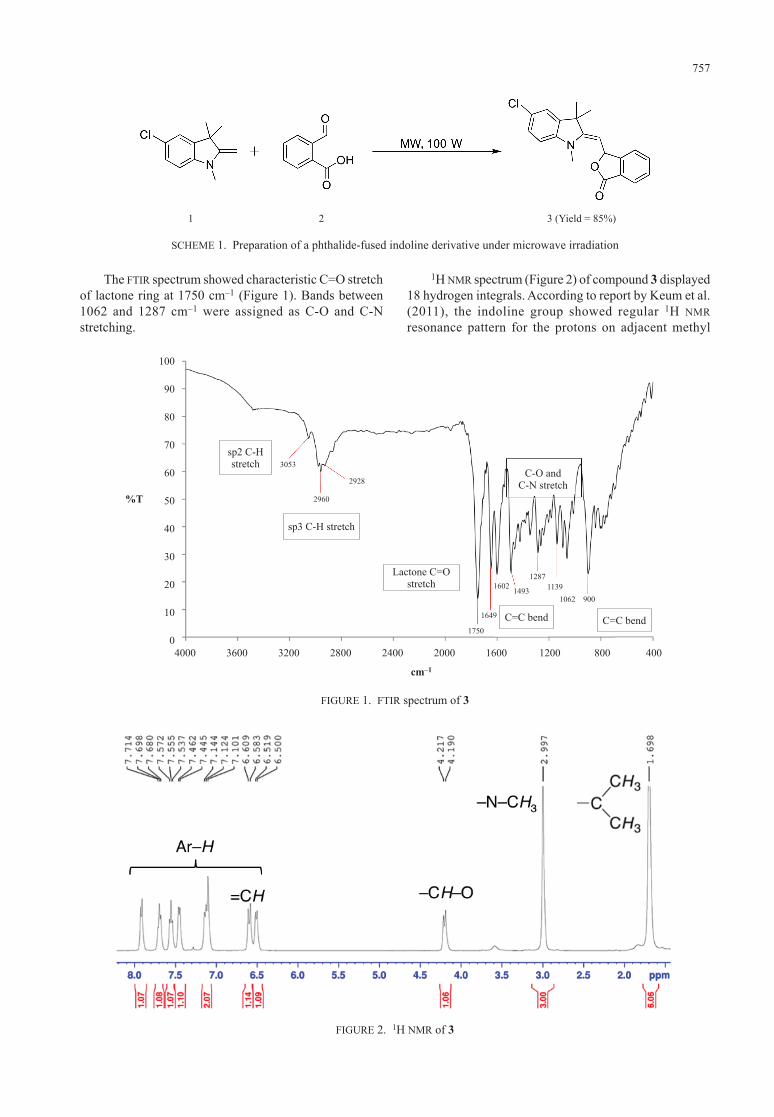
Single crystal of 3 was subjected to X-ray crystallographic analyses to further confi rm its chemical structure. The crystal was grown by slow evaporation of acetone solution at room temperature. Compound 3 crystallized in monoclinic system with space group Pī. The crystal system and refinement parameters are shown in Table 1.
The molecule 3 consists of two fused 5-chloro-2-methylene-1,3,3-trimethylindoline and isobenzofuran-1(3H)-one groups connected by the enamine carbon C9 (Figure 3) with C8–C9 and C9–C10 of bond lengths 1.475 (3) and 1.335 (3) Å, respectively.
BINDING STUDY WITH METAL IONS
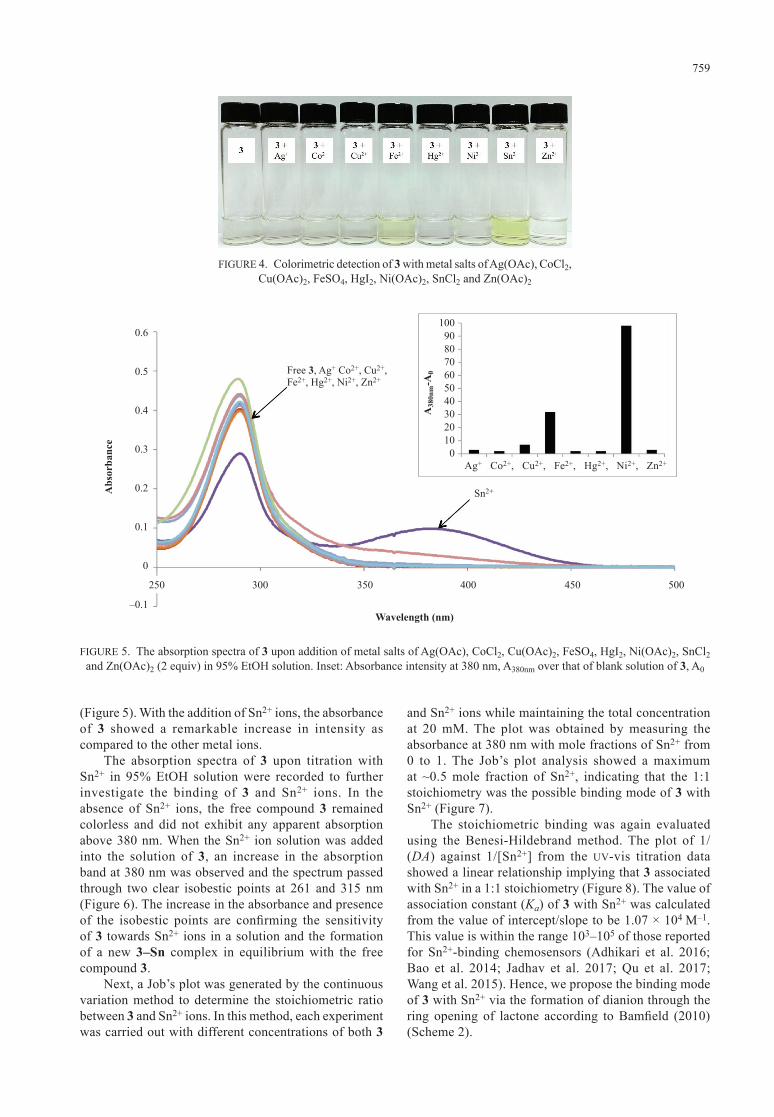
The colorimetric detection of mixture of compound 3 and metal salts were done qualitatively and observed by naked eyes. Color change was observed when colorless solution of compound 3 (5 × 10⁻⁴ M) was mixed with 2 equivalent of Fe2+ and Sn2+, respectively (Figure 4). No color change was detected for all the other metal cations tested. The color of the mixture changed from colorless to greenish-yellow showed that compound 3 possessed ionochromic behavior with Fe2+ and Sn2+.
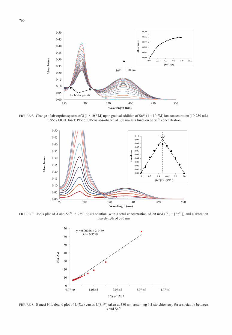
The interaction between compound 3 and various metal ions was studied by UV-vis spectroscopy in EtOH solution. Figure 5 shows the results of metal ion screening with 3. Solution of 3 was prepared at a concentration of 1 × 10⁻⁵ M. No absorption band was found in the visible region from the absorption spectrum of EtOH solution of 3 and is, therefore, colorless. The UV-vis spectra of 3 (1 × 10⁻⁵ M) was recorded upon addition of Ag(OAc), CoCl2, Cu(OAc)2, FeSO4, HgI2, Ni(OAc)2, SnCl2 and Zn(OAc)2 (2 equiv) in 95% EtOH solution, respectively. New absorption band was seen with maximum absorbance at around 380 nm upon addition of Sn2+. The absorption spectrum of 3 upon addition of Sn2+ also showed a drop in the absorbance intensity at around 290 nm, which may be an evidence of a host-guest complex interaction or binding. While upon the addition of other metal ions, no obvious change on the maximum absorbance or new band was detected. The absorbance intensity at 380 nm (A380nm) over the absorbance intensity of blank solution of 3 with various metal ions is represented in a chart
table 1. Crystallographic data collection parameters for 3
Compound 3Empirical formula C20 H18 Cl N O2
Formula weight 339.80Temperature (K) 304 (2)Wavelength (Å) 0.71073Crystal system TriclinicSpace groupUnit cell dimensions (Å) a = 8.178 (7)
b = 10.163 (8)c = 11.995 (8)
α, β, γ (°) α = 83.46 (4)β = 89.30 (5)γ = 69.53 (5)
Volume (Å3) 850.9 (12)Z 2Density (calculated) (Mg/m3) 1.326Absorption coeffi cient (mm–1) 0.236F (000) 356Crystal size (mm) 0.60 × 0.43 × 0.25θ range (º) 3.009 – 28.288Index ranges –10 ≤ h ≤ 10
–13 ≤ k ≤ 13–14 ≤ l ≤ 14
Refl ections collected 39140Independent refl ections 4200 [R(int) = 0.072]Completeness to theta 99.3%Refi nement method Full-matrix least-squares
on F2
Data/restraints/parameters 4200 / 0 / 221Goodness-of-fi t on F2 1.046Final R indices [I>2sigma(I)] R1 = 0.0559, wR2 =
0.1320R indices (all data) R1 = 0.0925, wR2 =
0.1577Largest diff. peak and hole 0.225 and –0.314 e.Å–3
CCDC No. 1574258
figure 3. ORTEP diagram of 3 drawn at 50% probability ellipsoids
759
figure 4. Colorimetric detection of 3 with metal salts of Ag(OAc), CoCl2, Cu(OAc)2, FeSO4, HgI2, Ni(OAc)2, SnCl2 and Zn(OAc)2
figure 5. The absorption spectra of 3 upon addition of metal salts of Ag(OAc), CoCl2, Cu(OAc)2, FeSO4, HgI2, Ni(OAc)2, SnCl2and Zn(OAc)2 (2 equiv) in 95% EtOH solution. Inset: Absorbance intensity at 380 nm, A380nm over that of blank solution of 3, A0
Wavelength (nm)
250 300 350 400 450 500
Abs
orba
nce
0.6
0.5
0.4
0.3
0.2
0.1
0
–0.1
1009080706050403020100
A38
0nm
-A0
Sn2+
Free 3, Ag+ Co2+, Cu2+, Fe2+, Hg2+, Ni2+, Zn2+
Ag+ Co2+, Cu2+, Fe2+, Hg2+, Ni2+, Zn2+
(Figure 5). With the addition of Sn2+ ions, the absorbance of 3 showed a remarkable increase in intensity as compared to the other metal ions.
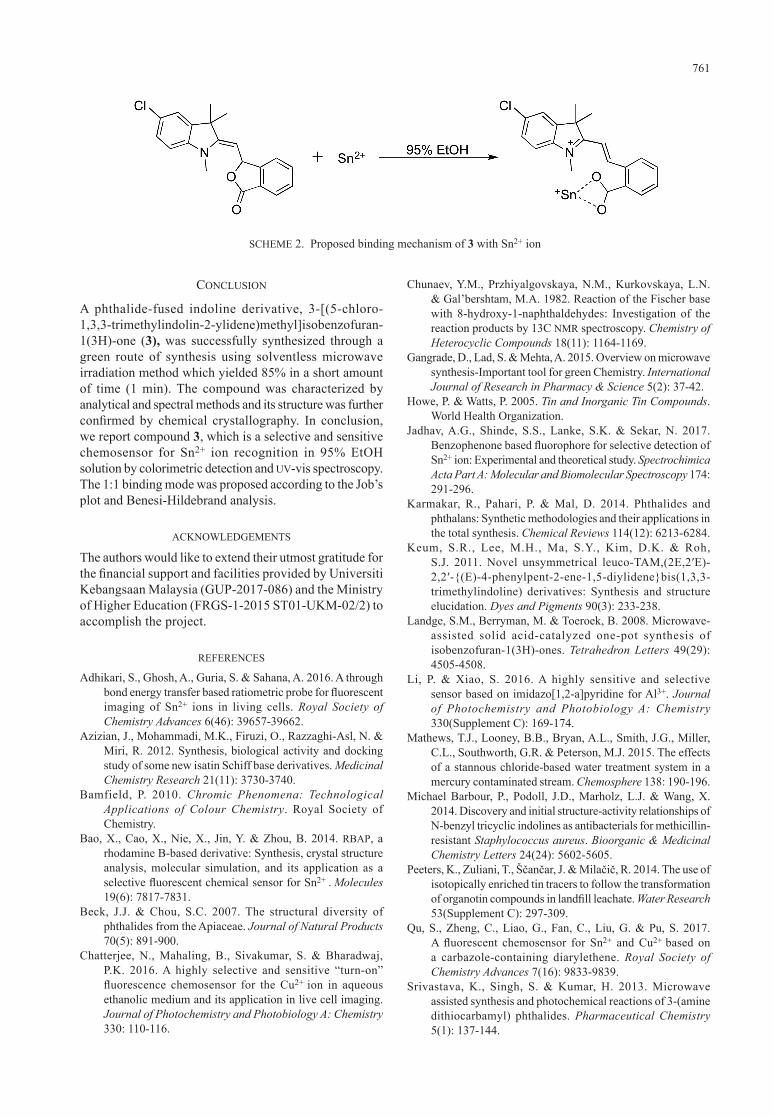
The absorption spectra of 3 upon titration with Sn2+ in 95% EtOH solution were recorded to further investigate the binding of 3 and Sn2+ ions. In the absence of Sn2+ ions, the free compound 3 remained colorless and did not exhibit any apparent absorption above 380 nm. When the Sn2+ ion solution was added into the solution of 3, an increase in the absorption band at 380 nm was observed and the spectrum passed through two clear isobestic points at 261 and 315 nm (Figure 6). The increase in the absorbance and presence of the isobestic points are confi rming the sensitivity of 3 towards Sn2+ ions in a solution and the formation of a new 3–Sn complex in equilibrium with the free compound 3.
Next, a Job’s plot was generated by the continuous variation method to determine the stoichiometric ratio between 3 and Sn2+ ions. In this method, each experiment was carried out with different concentrations of both 3
and Sn2+ ions while maintaining the total concentration at 20 mM. The plot was obtained by measuring the absorbance at 380 nm with mole fractions of Sn2+ from 0 to 1. The Job’s plot analysis showed a maximum at ~0.5 mole fraction of Sn2+, indicating that the 1:1 stoichiometry was the possible binding mode of 3 with Sn2+ (Figure 7).
The stoichiometric binding was again evaluated using the Benesi-Hildebrand method. The plot of 1/(DA) against 1/[Sn2+] from the UV-vis titration data showed a linear relationship implying that 3 associated with Sn2+ in a 1:1 stoichiometry (Figure 8). The value of association constant (Ka) of 3 with Sn2+ was calculated from the value of intercept/slope to be 1.07 × 10⁴ M–1. This value is within the range 103–105 of those reported for Sn2+-binding chemosensors (Adhikari et al. 2016; Bao et al. 2014; Jadhav et al. 2017; Qu et al. 2017; Wang et al. 2015). Hence, we propose the binding mode of 3 with Sn2+ via the formation of dianion through the ring opening of lactone according to Bamfi eld (2010) (Scheme 2).
760
figure 7. Job’s plot of 3 and Sn2+ in 95% EtOH solution, with a total concentration of 20 mM ([3] + [Sn2+]) and a detection wavelength of 380 nm
figure 8. Benesi-Hildebrand plot of 1/(DA) versus 1/[Sn2+] taken at 380 nm, assuming 1:1 stoichiometry for association between 3 and Sn2+
figure 6. Change of absorption spectra of 3 (1 × 10⁻⁵ M) upon gradual addition of Sn2+ (1 × 10⁻³M) ion concentration (10-250 mL) in 95% EtOH. Inset: Plot of UV-vis absorbance at 380 nm as a function of Sn2+ concentration
Wavelength (nm)250 300 350 400 450 500
Sn2+ 380 nm
Abs
orba
nce
0.50
0.45
0.40
0.35
0.30
0.35
0.20
0.15
0.10
0.05
0.00Isobestic points
10.0
0.20
0.16
0.12
0.08
0.04
0.008.06.04.02.00.0
[Sn2+]/[3]
Abs
orba
nce
Wavelength (nm)250 300 350 400 450 500
Abs
orba
nce
0.50
0.45
0.40
0.35
0.30
0.35
0.20
0.15
0.10
0.05
0.00
10
0.100.090.080.070.060.050.040.030.020.010.00
0.80.60.40.20
[Sn2+]/(3]+{SN2+])
Abs
orba
nce
0.0E+0
70
60
50
40
30
20
10
0
1/[Sn2+]M–1
1/(A
-A0)
1.0E+5 2.0E+5 3.0E+5 4.0E+5
y = 0.0002x + 2.1469R2 = 0.9799
761
concluSion
A phthalide-fused indoline derivative, 3-[(5-chloro-1,3,3-trimethylindolin-2-ylidene)methyl]isobenzofuran-1(3H)-one (3), was successfully synthesized through a green route of synthesis using solventless microwave irradiation method which yielded 85% in a short amount of time (1 min). The compound was characterized by analytical and spectral methods and its structure was further confi rmed by chemical crystallography. In conclusion, we report compound 3, which is a selective and sensitive chemosensor for Sn2+ ion recognition in 95% EtOH solution by colorimetric detection and UV-vis spectroscopy. The 1:1 binding mode was proposed according to the Job’s plot and Benesi-Hildebrand analysis.
acknoWledgeMentS
The authors would like to extend their utmost gratitude for the fi nancial support and facilities provided by Universiti Kebangsaan Malaysia (GUP-2017-086) and the Ministry of Higher Education (FRGS-1-2015 ST01-UKM-02/2) to accomplish the project.
referenceS
Adhikari, S., Ghosh, A., Guria, S. & Sahana, A. 2016. A through bond energy transfer based ratiometric probe for fl uorescent imaging of Sn2+ ions in living cells. Royal Society of Chemistry Advances 6(46): 39657-39662.
Azizian, J., Mohammadi, M.K., Firuzi, O., Razzaghi-Asl, N. & Miri, R. 2012. Synthesis, biological activity and docking study of some new isatin Schiff base derivatives. Medicinal Chemistry Research 21(11): 3730-3740.
Bamfield, P. 2010. Chromic Phenomena: Technological Applications of Colour Chemistry. Royal Society of Chemistry.
Bao, X., Cao, X., Nie, X., Jin, Y. & Zhou, B. 2014. RBAP, a rhodamine B-based derivative: Synthesis, crystal structure analysis, molecular simulation, and its application as a selective fl uorescent chemical sensor for Sn2+ . Molecules19(6): 7817-7831.
Beck, J.J. & Chou, S.C. 2007. The structural diversity of phthalides from the Apiaceae. Journal of Natural Products70(5): 891-900.
Chatterjee, N., Mahaling, B., Sivakumar, S. & Bharadwaj, P.K. 2016. A highly selective and sensitive “turn-on” fl uorescence chemosensor for the Cu2+ ion in aqueous ethanolic medium and its application in live cell imaging. Journal of Photochemistry and Photobiology A: Chemistry330: 110-116.
Chunaev, Y.M., Przhiyalgovskaya, N.M., Kurkovskaya, L.N. & Gal’bershtam, M.A. 1982. Reaction of the Fischer base with 8-hydroxy-1-naphthaldehydes: Investigation of the reaction products by 13C NMR spectroscopy. Chemistry of Heterocyclic Compounds 18(11): 1164-1169.
Gangrade, D., Lad, S. & Mehta, A. 2015. Overview on microwave synthesis-Important tool for green Chemistry. International Journal of Research in Pharmacy & Science 5(2): 37-42.
Howe, P. & Watts, P. 2005. Tin and Inorganic Tin Compounds. World Health Organization.
Jadhav, A.G., Shinde, S.S., Lanke, S.K. & Sekar, N. 2017. Benzophenone based fl uorophore for selective detection of Sn2+ ion: Experimental and theoretical study. Spectrochimica Acta Part A: Molecular and Biomolecular Spectroscopy 174: 291-296.
Karmakar, R., Pahari, P. & Mal, D. 2014. Phthalides and phthalans: Synthetic methodologies and their applications in the total synthesis. Chemical Reviews 114(12): 6213-6284.
Keum, S.R., Lee, M.H., Ma, S.Y., Kim, D.K. & Roh, S.J. 2011. Novel unsymmetrical leuco-TAM,(2E,2′E)-2,2′-{(E)-4-phenylpent-2-ene-1,5-diylidene}bis(1,3,3-trimethylindoline) derivatives: Synthesis and structure elucidation. Dyes and Pigments 90(3): 233-238.
Landge, S.M., Berryman, M. & Toeroek, B. 2008. Microwave-assisted solid acid-catalyzed one-pot synthesis of isobenzofuran-1(3H)-ones. Tetrahedron Letters 49(29): 4505-4508.
Li, P. & Xiao, S. 2016. A highly sensitive and selective sensor based on imidazo[1,2-a]pyridine for Al3+. Journal of Photochemistry and Photobiology A: Chemistry 330(Supplement C): 169-174.
Mathews, T.J., Looney, B.B., Bryan, A.L., Smith, J.G., Miller, C.L., Southworth, G.R. & Peterson, M.J. 2015. The effects of a stannous chloride-based water treatment system in a mercury contaminated stream. Chemosphere 138: 190-196.
Michael Barbour, P., Podoll, J.D., Marholz, L.J. & Wang, X. 2014. Discovery and initial structure-activity relationships of N-benzyl tricyclic indolines as antibacterials for methicillin-resistant Staphylococcus aureus. Bioorganic & Medicinal Chemistry Letters 24(24): 5602-5605.
Peeters, K., Zuliani, T., Ščančar, J. & Milačič, R. 2014. The use of isotopically enriched tin tracers to follow the transformation of organotin compounds in landfi ll leachate. Water Research 53(Supplement C): 297-309.
Qu, S., Zheng, C., Liao, G., Fan, C., Liu, G. & Pu, S. 2017. A fl uorescent chemosensor for Sn2+ and Cu2+ based on a carbazole-containing diarylethene. Royal Society of Chemistry Advances 7(16): 9833-9839.
Srivastava, K., Singh, S. & Kumar, H. 2013. Microwave assisted synthesis and photochemical reactions of 3-(amine dithiocarbamyl) phthalides. Pharmaceutical Chemistry 5(1): 137-144.
ScheMe 2. Proposed binding mechanism of 3 with Sn2+ ion

762
Sun, W., Guo, S., Hu, C., Fan, J. & Peng, X. 2016. Recent development of chemosensors based on cyanine platforms. Chemical Reviews 116(14): 7768-7817.
Wang, J., Lv, M., Wang, Z., Zhou, M., Gu, C. & Guo, C. 2015. Highly sensitive and selective fluorescent detection of rare earth metal Sn(II) ion by organic fluorine Schiff base functionalized periodic mesoporous material in aqueous solution. Journal of Photochemistry and Photobiology A: Chemistry 309(Supplement C): 37-46.
Zhu, H., Fan, J., Wang, B. & Peng, X. 2015. Fluorescent, MRI, and colorimetric chemical sensors for the first-row d-block metal ions. Chemical Society Reviews 44(13): 4337-4366.
Siti Aishah Hasbullah*Pusat Pengajian Sains dan Teknologi MakananFakulti Sains dan TeknologiUniversiti Kebangsaan Malaysia43600 UKM Bangi, Selangor Darul EhsanMalaysia
*Corresponding author; email: [email protected]
Received: 15 September 2017Accepted: 2 November 2017