x-ray irradiation of blood components - transfusion · pdf filex-ray irradiation of blood...
TRANSCRIPT

Page 1 of 19
JJPPAACC 0088--7755
X-ray irradiation of blood components
1 Background The risk of development of Transfusion-Associated Graft versus Host Disease (TA-GVHD) following transfusion of blood components containing viable lymphocytes to susceptible individuals has been well documented (1). It is routinely prevented by irradiation of components, and British Committee for Standards in Haematology (BCSH) Guidelines outline the use of gamma irradiation for the prevention of TA-GVHD (1). Gamma irradiation, although effective in preventing TA-GVHD, has some drawbacks. It involves the use of a radioactive source, which is subject to stringent health and safety regulations. The machines are also very expensive to buy and decommission when no longer required. In addition, due to decay of the source, regular recalibration is required and irradiation time is increased. X-ray irradiation is a possible alternative to gamma irradiation. This has been delivered in some areas using linear accelerators, though dose mapping these machines for this purpose may be difficult. However, a shielded cabinet X-ray radiation source is now available - the Raycell, manufactured by Nordion. SACBC have reviewed data on the use of this machine.
2 Raycell x-ray irradiator In 1998, this machine (then the ‘RS 3000 shielded cabinet X-ray source’, manufactured by Rad-Source and later sold to Nordion) was granted a marketing license by the FDA to market the device as ‘substantially equivalent’ to a gamma irradiator. It is CE marked (for conformance to electrical specifications - CE mark does not deal with the effects of ionising radiation on blood components). It is in use in several European and US sites, including Puget Sound in Seattle, Sweden, Italy, France and Germany. In France, EFS and AFSAPS approval has been granted based on published data (2). It is dose-mapped prior to release from the factory and at installation, and the manufacturers recommend routine dosimetry at 6-monthly intervals. They also manufacture a radiation sensitive label specifically for use with X-radiation.
3 Blood component data
3.1 Efficacy for prevention of TA-GVHD Several publications state that X-ray irradiation is equivalent to gamma irradiation (2-4). Moroff and Luban state that “Two types of ionising radiation, ? rays and X-rays, inactivate T lymphocytes. Both can be used to irradiate blood and blood components. At a given absorbed dose, both ? and X-rays are equivalent in their ability to inactivate T lymphocytes” (5). Janatpour et al (2) compared X-ray with gamma irradiation using the Raycell irradiator. They performed a small study on lymphocyte function as part of this work. They reported as follows: “Lymphocytes isolated from both gamma- and X-ray-irradiated (25 Gy) portions of one unit showed an identical lack of proliferation when stimulated with mitogen or with allogeneic leucocytes. One cell division was observed in the cultures with PHA, no cell division was observed in the MLC cultures. Lymphocytes from the control portion showed expected proliferation in both assays”.

Page 2 of 19
JJPPAACC 0088--7755
Herva and Kiviniity in an earlier study found no difference between the effects of X-ray irradiation, cobalt and 45 MeV electron irradiation on lymphocyte response when equivalent doses were given (6). During Process Qualification prior to implementation, Dinwiddie et al from the Puget Sound Blood Centre in Seattle compared lymphocyte viability following irradiation of red cells in the x-ray irradiator with gamma irradiation (7). Compared to control cells, those irradiated in the X-ray irradiator showed a 6 – 30 fold decrease in viability and were not significantly different from those irradiated in the gamma irradiator. On personal communication with a senior radiation physicist, we are advised that in general, X-rays should have very similar effects to gamma rays on blood components as the energy disposition mechanisms are similar, provided the doses delivered are equivalent. X-rays however are attenuated more rapidly therefore dose distributions can be variable – dosimetry therefore needs to be checked carefully. Use of X-irradiation is already established in Centres in Europe and the US. Communication from a large transplant unit in Seattle confirmed that X-irradiated components were being transfused to stem cell transplant patients without problems. In addition, it is licensed as equivalent to gamma irradiation with the FDA.
3.2 Component quality Irradiation can be applied to all cellular components: red cells, platelets and granulocytes. Below are summaries of 1) published data; 2) data provided by the Karolinska Institute; and 3) data generated by the NHSBT Component Development Laboratory.
3.2.1 Published data
3.2.1.a Red cells Janatpour and co-workers compared red cell quality after irradiation with gamma and X-rays at 2 doses (25 Gy and 35 Gy). The recommended dose in the UK is a minimum of 25 Gy and maximum of 50 Gy. They looked specifically at free plasma haemoglobin (Hb), and extracellular potassium levels, as these have previously been shown to be parameters affected by irradiation. They found that at 25 Gy, X-ray irradiated units had slightly higher levels of free plasma Hb, whereas at 35 Gy, gamma irradiated units showed higher levels of extracellular potassium. They concluded that small differences in red cell membrane permeability were found between ? irradiated and X-irradiated units, but that these differences were not likely to be clinically important. This study however was conducted using non-leucodepleted red cells in CPDA-1. Leucodepletion may increase free Hb levels further. Most red cell units in the UK are resuspended in SAG-M additive solution, and one report has suggested that the presence of mannitol after irradiation may increase extracellular potassium (4).
3.2.1.b Granulocytes
One study has shown that neutrophil chemotaxis and chemiluminescence is unaltered by exposing buffy coats to 1500 rads (15 Gy) of x-irradiation (8). A further study has shown that neutrophil function is only marginally affected by exposure to 10,000-20,000 rads (100-200 Gy) of x-irradiation (9). Therefore exposure to 25-50 Gy is unlikely to have an impact on neutrophil function in granulocyte concentrates.
It is unlikely that red cell haemolysis or potassium leakage would be an issue in granulocyte components due to the lower red cell content and shorter shelf life than red cell concentrates. Puget Sound Blood Center is a centre of excellence for granulocyte studies, and routinely uses X-irradition for its components.
Page 3 of 19
JJPPAACC 0088--7755
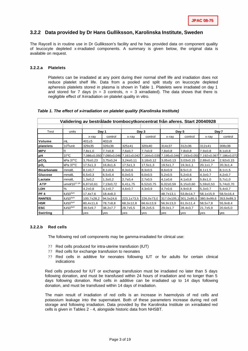
3.2.2 Data provided by Dr Hans Gulliksson, Karolinska Institute, Sweden The Raycell is in routine use in Dr Gulliksson’s facility and he has provided data on component quality of leucocyte depleted x-irradiated components. A summary is given below, the original data is available on request.
3.2.2.a Platelets
Platelets can be irradiated at any point during their normal shelf life and irradiation does not reduce platelet shelf life. Data from a pooled and split study on leucocyte depleted apheresis platelets stored in plasma is shown in Table 1. Platelets were irradiated on day 1 and stored for 7 days (n = 3 controls, n = 3 x-irradiated). The data shows that there is negligible effect of X-irradiation on platelet quality in vitro.
Table 1. The effect of x-irradiation on platelet quality (Karolinska Institute)
Test units
x-ray control x-ray control x-ray control x-ray control
Volume mL 401±5 402±9
platelets 109/unit 329±35 326±36 325±41 320±40 314±37 312±36 312±41 308±38
MPV f l 7,8±1,0 7,7±0,8 7,6±0,7 7,7±0,9 7,8±0,8 7,8±0,8 7,9±0,8 8,1±0,8
pH 37oC 7,086±0,050 7,090±0,046 7,161±0,042 7,164±0,039 7,195±0,046 7,193±0,050 7,192±0,067 7,186±0,072
pCO2 kPa 37oC 3,76±0,23 3,75±0,24 3,24±0,11 3,18±0,12 3,05±0,13 3,03±0,15 2,89±0,14 2,92±0,15
pO2 kPa 37oC 17,5±1,3 16,8±1,6 17,5±1,3 17,5±1,3 19,5±1,7 19,3±1,1 20,1±1,7 20,3±1,4
Bicarbonate mmol/L 8,1±0,7 8,1±0,6 8,3±0,6 8,3±0,5 8,6±0,9 8,5±1,0 8,1±1,5 8,1±1,5
Glucose mmol/L 6,5±0,3 6,5±0,4 6,0±0,5 6,0±0,5 5,2±0,5 5,2±0,6 4,3±0,7 4,2±0,7
Lactate mmol/L 1,3±0,2 1,3±0,2 2,7±0,4 2,7±0,5 4,1±0,6 4,1±0,8 5,8±1,0 5,7±1,0
ATP umol/1011 plt 6,87±0,61 7,13±0,72 6,41±,75 6,52±0,75 6,02±0,59 6,15±0,60 5,59±0,53 5,74±0,70
LDH % 4,2±0,8 4,1±0,7 4,6±0,7 4,3±0,9 4,7±0,6 4,9±0,8 5,3±0,7 5,4±0,7
PF 4 IU/106plt 17,4±7,6 18,4±8,3 48,7±13,1 53,8±14,7 58,1±15,9 58,5±16,4
RANTES IU/106plt 100,7±28,2 94,5±24,6 223,1±73,5 226,0±73,0 317,0±105,1 301,2±85,5 380,9±99,6 353,9±89,3
HSR IU/106plt 80,4±11,6 78,7±8,8 68,3±12,8 66,6±13,9 56,9±13,0 61,0±11,4 58,5±7,8 55,9±8,4
ESC IU/106plt 39,5±9,7 38,2±7,7 28,7±5,5 28,2±5,5 29,0±1,7 28,4±3,7 21,7±5,9 20,6±5,0
Swirling yes yes yes yes yes yes yes yes
Validering av bestrålade trombocytkoncentrat från aferes. Start 20040928
Day 1 Day 3 Day 5 Day 7
3.2.2.b Red cells
The following red cell components may be gamma-irradiated for clinical use:
?? Red cells produced for intra-uterine transfusion (IUT) ?? Red cells for exchange transfusion to neonates ?? Red cells in additive for neonates following IUT or for adults for certain clinical
indications
Red cells produced for IUT or exchange transfusion must be irradiated no later than 5 days following donation, and must be transfused within 24 hours of irradiation and no longer than 5 days following donation. Red cells in additive can be irradiated up to 14 days following donation, and must be transfused within 14 days of irradiation. The main result of irradiation of red cells is an increase in haemolysis of red cells and potassium leakage into the supernatant. Both of these parameters increase during red cell storage and following irradiation. Data provided by the Karolinska Institute on x-irradiated red cells is given in Tables 2 - 4, alongside historic data from NHSBT.

Page 4 of 19
JJPPAACC 0088--7755
Table 2. X-irradiation of LD red cells in SAG-M irradiated day 1 (mean ± SD, n=6)
Day 1 Day 7 Day 14 Day 21 Day 28 Haemolysis (%) 0.06 ± 0.00 0.14 ± 0.05 0.24 ± 0.08 0.37 ± 0.15 0.44 ± 0.07 ATP (?g/gHb) 5.00 ± 0.57 5.30 ± 0.88 5.18 ± 0.54 4.47 ± 0.48 4.04 ± 0.60 Supernatant Potassium (mmol/l)
6 ± 1 39 ± 4 48 ± 3 53 ± 3 56 ± 3
Table 3. X-irradiation and gamma-irradiation of LD red cells in SAG-M irradiated day 14
Day 14/15 Day 21 Day 28 Haemolysis (%) – x-irradiated 0.20 ± 0.25 0.24 ± 0.13 0.40 ± 0.20 Haemolysis (%) – gamma-irradiated 0.07 (0.05-0.10) 0.16 (0.13-0.24) 0.31 (0.24-0.40) ATP (?g/gHb) –x-irradiated 5.27 ± 0.46 5.09 ± 0.41 4.25 ± 0.42 Supernatant Potassium (mmol/l) – x-irradiated
23 ± 5 44 ± 5 50 ± 5
Supernatant Potassium (mmol/l) – gamma-irradiated
42 (38-45) 60 (52-64) 66 (63-69)
x-irradiated unit data from Karolinska (mean ± SD, n=6), gamma-irradiated data from NHSBT (mean with range, n=10) Table 4. X-irradiation and gamma-irradiation of LD red cells in plasma irradiated day 4/5 (for neonatal exchange transfusion) Day 4/5 Day 6/7 Day 7/8 Haemolysis (%) – x-irradiated 0.21 ± 0.13 0.29 ± 0.12 0.28 ± 0.10 Haemolysis (%) – gamma-irradiated 0.04 (0.02-0.12) 0.05 (0.03-0.19) ATP (?g/gHb) –x-irradiated 4.90 ± 0.22 5.18 ± 0.04 5.63 ± 0.03 Supernatant Potassium (mmol/l) – x-irradiated
6 ± 1 10 ± 1 12 ± 1
Supernatant Potassium (mmol/l) – gamma-irradiated
13 (11-19) 21 (19-25)
x-irradiated unit data from Karolinska (mean ± SD, n=3), gamma-irradiated units from NHSBT (mean with range, n=10)
The data in Tables 2 - 4 suggest that the quality of red cells in SAG-M are suitable up to 14 days following x-irradiation and for red cells for exchange up to 24 hours after irradiation. However, the levels of haemolysis appear to be higher than in historical data from the NHSBT on gamma-irradiated units and no data are available on IUT units. Therefore it was decided that haemolysis and potassium levels should be further assessed during validation studies as follows:
Component Day of irradiation
Time points following irradiation to assess
Red cells for IUT 4 0, 24 hours
Red cells for exchange
4 0, 24 hours
Red cells in additive 14 0, 7 days, 14 days
This work was undertaken by the NHSBT Component Development Laboratory (CDL), in collaboration with NHSBT Oxford (X-irradiated units), and NHSBT Brentwood (gamma-irradiated units).

Page 5 of 19
JJPPAACC 0088--7755
3.2.3 Data from NHSBT Component Development Laboratory (EVAL/CD/2007/57)
3.2.3.a Study design
Study 1: Effect of X-ray irradiation on red cell units in SAG-M
10 units of whole blood were processed into RCC-SAGM. 6 units were prepared by TAT processing and other 4 units were prepared by BAT processing. All the units were X-irradiated (central dose 35.9 Gy) using the Raycell X-ray irradiator on day 14. Samples were taken pre-irradiation, post-irradiation (0 hrs), post-irradiation (7 days) and post-irradiation (14 days). Supernatant samples were achieved by double centrifugation at 2000 rpm for 30 and 15 minutes. All samples were stored at -40°C. All the frozen samples were couriered to CDL on dry ice for analysis of supernatant potassium and haemoglobin.
Study 2: Effect of X-ray irradiation on red cell units for Exchange
10 units of whole blood units were prepared by TAT processing into red cell units for Exchange. All the units were X-irradiated (35.9 Gy) using the Raycell X-ray irradiator on day 4. Samples were taken pre-irradiation, post-irradiation (0 hrs) and post-irradiation (24 hrs). Samples were processed as for study 1.
Study 3: Effect of X-ray irradiation on red cell units for IUT- Intrauterine Transfusions
10 units of whole blood units were prepared by TAT processing into red cell units for IUT. All the units were X-irradiated (35.9 Gy) using the Raycell X-ray irradiator on day 4. Samples were taken pre-irradiation, post-irradiation (0 hrs) and post-irradiation (24 hrs). Samples were processed as for study 1
3.2.3.b Results
Please note that for clarity the figures show only the results of statistical tests for difference (p value) between the irradiation treatment (X-ray v gamma) at the end of shelf life.

Page 6 of 19
JJPPAACC 0088--7755
Study 1 Effect of X-ray irradiation on RCC in SAG-M
a) Supernatant Potassium
No difference was seen between pre and post-X-ray irradiation (0 hrs). Although supernatant potassium levels increased by Days 7 and 14 post X-ray irradiation.
There was no significant difference in Supernatant Potassium levels in X-ray and Gamma irradiated RCC in SAG-M at the end of shelf life, as shown in Figure 1.
Figure 1A Figure 1B
pre- X
irr
post-
X irr
0 hrs
post
-X irr
7 day
s
post-
X irr 1
4 day
s
Post
Gamma-ir
r d7
Post
Gamma-ir
r d14
0
20
40
60
80
RCC in SAG-M NS
Su
per
nat
ant
Po
tass
ium
(mm
ol/l
)
RCC in SAG-M
pre- X
irr
post-
X irr 0
hrs
post -
X irr 7
days
post-
X irr 1
4 day
s
Post
Gamma-ir
r d7
Post
Gamma-ir
r d14
0
2
4
6
8
NS
Su
per
nat
ant
Po
tass
ium
(m
mo
l/un
it#)
Figure 1 Potassium levels in X-ray irradiated (at Day 14) and CDL data from gamma irradiated RCC in SAG-M. K+ expressed as mmol/l in figure 1A and mmol/unit in figure 1B.
NS – p value is Non Significant # Potassium levels calculated using unit volume corrected to account for sampling. Data plotted as median with range n=10 for X-ray irradiated units n=20 for gamma reference data

Page 7 of 19
JJPPAACC 0088--7755
b) Haemolysis
No difference was seen between pre and post-X-irradiation (0 hrs). Although haemolysis increased by Days 7 and 14 post X-irradiation, all were all below the UK limit of 0.8%.
There was no significant difference in haemolysis in X-ray and Gamma irradiated RCC in SAG-M at the end of shelf life, as shown in Figure 1c
RCC in SAG-M
pre- X
irr
post-
X irr 0 h
rs
post
-X irr 7
days
post-
X irr 14
days
Post G
amma-i
rr d7
Post G
amma-ir
r d14
0.0
0.2
0.4
0.6 NS
Hae
mol
ysis
(%)
Figure 1c Haemolysis in X-ray irradiated at Day 14 and CDL data from gamma irradiated RCC in SAG-M.
NS – p value is NonSignificant Data plotted as median with range n=10 for X-ray irradiated units n=20 for gamma reference data

Page 8 of 19
JJPPAACC 0088--7755
Study 2 Effect of X-ray irradiation on red cell units for exchange transfusion
a) Supernatant Potassium No difference was seen between pre and post-X-irradiation (0 hrs). Units were X-irradiated on day 4, and tested at 24 hours post X-irradiation (end of shelf life). Units showed significantly higher levels of supernatant potassium, and free potassium per ml of RCC than CDL reference data for Gamma irradiated Red cell units (Figure 2).
Figure 2A Figure 2B
pre- X
irr
post-
X irr 0
hrs
post-
X irr 24
hrs
post-
Gamma ir
r 24 hr
s
0
10
20
30***
RCC for Exchange Transfusion
Sup
erna
tant
Pot
assi
um (m
mo
l/l)
RCC for Exchange transfusion
pre- X
irr
post-
X irr 0
hrs
post-
X irr 24
hrs
post-
Gamma ir
r 24 h
rs
0.000
0.003
0.006
0.009
0.012 ***
Pota
ssiu
m
mm
ol/m
l RC
C#
Figure 2A & 2B Potassium levels in X-irradiated (at Day 4) and CDL data from gamma irradiated RCC for Exchange. K+ expressed as mmol/l of supernatant in figure 2A and mmol/ml of RCC in figure 2B.
*** p < 0.001 # Potassium levels calculated as mmol/ml of RCC. Data plotted as median with range n=10 for X-irradiated units n=20 for gamma-irradiated reference data

Page 9 of 19
JJPPAACC 0088--7755
b) Haemolysis There was no difference in haemolysis pre and post-X-irradiation (0 hrs) of RCC for exchange transfusion. There was no difference in haemolysis between X-ray and Gamma irradiated RCC for exchange at the end of shelf life (24 hours post-irradiation), and all were below the European limit of 0.8% (Figure 4).
pre- X
irr
post-
X irr 0
hrs
post-
X irr 24
hrs
post-
Gamma ir
r 24 h
rs
0.00
0.05
0.10
0.15
0.20 RCC for Exchange Transfusion
Hae
mol
ysis
(%)
Figure 2C Haemolysis in X-ray irradiated (at Day 4) and historic data from gamma irradiated Red cell units for exchange.
Data plotted as median with range n=10 for X-irradiated units n=20 for gamma-irradiated reference data

Page 10 of 19
JJPPAACC 0088--7755
Study 3 Effect of X-ray irradiation on red cell units for IntrauterineTransfusion
There was no difference in supernatant potassium pre and post-X-irradiation (0 hrs) of RCC-IUT. Although supernatant potassium levels increased in RCC-IUT units 24 hrs post X-irradiation, there was no significant difference in supernatant potassium levels in X-ray and Gamma irradiated RCC for IUT with 90% Hct at the end of shelf life (24 hours post-irradiation; Figure 3A). This was also the case for the total amount of potassium per ml of RCC (Figure 3B). At 24 hours (end of shelf life) post X-ray irradiation RCC for IUT with 90 % Hct had significantly lower levels of supernatant potassium than Gamma irradiated RCC for IUT with 80% Hct, as shown in Figure 3.
Figure 3A Figure 3B
RCC for IUT
pre- X
irr
post-
X irr 0
hrs
post-
X irr 24
hrs 9
0% Hc
t
post-
Gamma ir
r 24hrs
80% Hc
t
post-
Gamma ir
r 24 hr
s-90%
Hct
0.0
0.5
1.0
1.5
2.0
2.5
3.0
NS
**
Sup
erna
tant
Pot
assi
umm
mol
/uni
t#
RCC for IUT
pre- X
irr
post-
X irr 0
hrs
post- X
irr 24 h
rs 90%
Hct
post- G
amma irr
24hrs
80% Hc
t
post- G
amma irr
24 hrs
-90% Hc
t
0.000
0.003
0.006
0.009
0.012
0.015
0.018
NS
* P
otas
sium
mm
ol/m
l RC
C#
Figure 3A & 3B Supernatant Potassium levels in X-ray irradiated (on Day 4) Red Cell units for IUT. K+ expressed as mmol/L of supernatant in figure 3A and mmol/mL of RCC in figure 3B.
# Potassium levels calculated using corrected unit volume to calculate K+ in mmol/ml of RCC. NS –p value is not significant ** p < 0.01 Data plotted as median with range. n = 10 for X-ray irradiated units with 90% Hct (Eval/CD/2007/57) n = 10 for gamma irradiated units with 80% Hct (Eval/CD/2007/57b) n = 9 for gamma irradiated units with 90% Hct (Eval/CD/2007/57b)

Page 11 of 19
JJPPAACC 0088--7755
b) Haemolysis
There were no significant differences in haemolysis pre and post-X-ray irradiation (0 hrs) of RCC for IUT. There was no increase in haemolysis in X-irradiated RCC for IUT at 24 hrs (end of shelf life), as shown in figure 3C. All gamma irradiated RCC-IUT with 80 % Hct were below the European limit of 0.8% haemolysis at 24 hrs (end of shelf life), but one of ten gamma irradiated RCC-IUT with 90 % Hct showed high haemolysis. However, there was no statistically significant difference between X-irradiated or gamma-irradiated units.
RCC for IUT
pre- X irr
post-
X irr 0 h
rs
post- X
irr 24
hrs 90
% Hct
post- G
amma irr
24hrs
80% Hct
post- G
amma ir
r 24 hrs
-90% Hct
0.0
0.2
0.4
0.6
0.8
1.0
1.2
1.4
1.6
1.8NS
Hae
mol
ysis
%
Figure 3C Haemolysis in X-ray irradiated (on Day 4) Red cell units for IUT.
NS – p value is Not Significant Data plotted as median with range n = 10 for X-ray irradiated units with 90% Hct (Eval/CD/2007/57) n = 10 for gamma irradiated units with 80% Hct (Eval/CD/2007/57b) n = 9 for gamma irradiated units with 90% Hct (Eval/CD/2007/57b)

Page 12 of 19
JJPPAACC 0088--7755
3.2.3.c Discussion Irradiation causes damage to the RBC membrane, increasing the membrane permeability(1,2) and leading to potassium leakage from red cells. Supernatant potassium levels post–gamma irradiation are reported to be elevated to clinically significant levels (3,4,5) and have been associated with foetal and neonatal arrhythmia and hyperkalemic cardiac arrest. Several publications have shown that X-irradiation is equivalent to gamma–irradiation (6,7,8,9) for inactivation of leucocytes. This study investigated the effect of X-irradiation on red cell concentrates. Components Development Laboratory Gamma-irradiated RCC reference data from EVAL/CD/2006/43 and EVAL/CD/2006/35 were used for comparison to determine whether X-irradiation has a significant impact on two markers of red cell quality - haemolysis and potassium leakage. X-irradiated red cells in SAG-M or for exchange or IUT did not exceed the current European limit of 0.8% haemolysis at the end of shelf life in any units studied. When X-irradiation of RCC in SAGM or RCC for exchange transfusion were compared with reference data from gamma-irradiated units, no significant differences were seen in haemolysis levels at the end of shelf life. Furthermore, the haemolysis data was also comparable to the Karolinska Institute’s data. The supernatant potassium levels of RCC in SAG-M doubled 7 days after X-ray irradiation, but no significant difference was seen between X-ray and gamma irradation up to day 14 of storage post-irradiation (end of shelf life). Supernatant potassium levels seen after X-irradiation in this study are similar to those reported in gamma–irradiated red cell units by other workers (10), but slightly higher than in X-irradiated RCC in SAG-M in the data from the Karolinska Institute. However, irradiation did lead to higher supernatant potassium concentrations in RCC for Exchange transfusion. At 24 hours post X-irradiation RCC for Exchange transfusion (haematocrit 53 - 58%) had significantly higher supernatant potassium than gamma irradiated RCC. Although these levels were markedly higher than the Karolinska data, similar values have been reported in other studies using gamma radiation (11;12), and higher levels than those seen in NHSBT studies have been reported in non-leucodepleted X-irradiated RCC in CPDA-1 with haematocrit of 70 - 80% (2). The data generated from NHSBT studies was discussed with Dr Helen New, Consultant Haematologist at St Mary’s who has specialist expertise in transfusion to neonates. Her conclusion, following discussion with other colleagues, was that the magnitude of the differences seen in potassium levels in red cells for exchange transfusion was not likely to be clinically significant. X-ray and gamma-irradiation of RCC for IUT with 90% Hct led to comparable potassium leakage, which was lower than that seen following gamma-irradiation of RCC for IUT with haematocrit of 80 %. In addition, when the potassium concentration was calculated per mL of RCC the results were very similar (around 0.01 mmol/mL RCC) to gamma-irradiated RCC for IUT safely used for IUT by Win et al (13).

Page 13 of 19
JJPPAACC 0088--7755
4 Overall conclusions X-irradiation has considerable logistical advantages over the use of gamma irradiation. There is now a CE marked, FDA approved, machine available to deliver X-irradiation, which has already been implemented in some Centres outside the UK. Published data on the efficacy of X-irradiation for leucocyte inactivation are sufficient. Internal data on red cells that were X-irradiated do not show any clinically significant differences to those irradiated with gamma irradiation. Data from the Karolinska Institute indicate that X-irradiated platelet components are of acceptable quality for clinical use. Published data on granulocyte components indicate adequate neutrophil function after X-irradiation.
5 Recommendation The use of X-irradiation using the Raycell device is approved for irradiation of red cells, platelets and granulocytes. If approved, the Red Book Guidelines will require limited review, as in several entries irradiation is specified as gamma-irradiation. Stephen Thomas & Rebecca Cardigan for SACBC 29 October 2008

Page 14 of 19
JJPPAACC 0088--7755
References
1. Love EM, Williamson LM, Cohen H. Serious Hazards of Transfusion Annual Report 1998-99.
Manchester: 2000.
2. Janatpour K, Denning L, Nelson K, Betlach B, Mackenzie M, Holland P. Comparison of X-ray vs. gamma irradiation of CPDA-1 red cells. Vox Sang. 2005;215-9.
3. Klein HG. Transfusion-associated graft-versus-host disease: less fresh blood and more gray (Gy) for an aging population. Transfusion 2006;878-80.
4. Hirayama J, Abe H, Azuma H, Ikeda H. Leakage of potassium from red blood cells following gamma ray irradiation in the presence of dipyridamole, trolox, human plasma or mannitol. Biol.Pharm.Bull. 2005;1318-20.
5. Moroff G, Luban NL. The irradiation of blood and blood components to prevent graft-versus-host disease: technical issues and guidelines. Transfus.Med.Rev. 1997;15-26.
6. Herva E, Kiviniity K. The effect of in vitro irradiation on the responses of human lymphocytes to PHA, PPD and allogeneic cells. Strahlentherapie. 1975;504-12.
7. Dinwiddie SC, Yadock W, Johnson DO. X-ray radiation as an alternative to gamma radiation for irradiation of blood components. Transfusion 2000.
8. Wheeler JG, Abramson JS, Ekstrand K. Function of irradiated polymorphonuclear leukocytes obtained by buffy-coat centrifugation. Transfusion 1984;238-9.
9. Valerius NH, Johansen KS, Nielsen OS, Platz P, Rosenkvist J, Sorensen H. Effect of in vitro X-irradiation on lymphocyte and granulocyte function. Scand.J.Haematol. 1981;9-18.
10. Al Amiri AH, Raouf MY. Safer Storage. MedLab Magazine 2005;22-4.
11. Ramirez AM, Woodfield DG, Scott R, McLachlan J. High potassium levels in stored irradiated blood. Transfusion 1987;444-5.
12. Rivet C, Baxter A, Rock G. Potassium levels in irradiated blood. Transfusion 1989;185.
13. Win N, Logan RW, Cameron A. Post-irradiation plasma potassium and intra-uterine transfusion. Vox Sang. 1997;56-7.

Page 15 of 19
JJPPAACC 0088--7755
APPENDIX 1

Page 16 of 19
JJPPAACC 0088--7755

Page 17 of 19
JJPPAACC 0088--7755

Page 18 of 19
JJPPAACC 0088--7755

Page 19 of 19
JJPPAACC 0088--7755