y nikon corporation - mikroskop · imagine the perfect digital-imaging platform f or all today’s...
TRANSCRIPT

En
Advanced Research Microscope ECLIPSE 80i
This brochure is printed on recycled paper made from 40% used material.
WARNINGTO ENSURE CORRECT USAGE, READ THE CORRESPONDING MANUALS
CAREFULLY BEFORE USING YOUR EQUIPMENT.
Specifications and equipment are subject to change without any notice or obligationon the part of the manufacturer. December 2003. ©2003 NIKON CORPORATION
NIKON CORPORATIONYokohama Plant
NIKON CORPORATIONInstruments Company
ISO 9001Accredited by theDutch Council for
Accreditation ISO 14001
Printed in Japan (0312-15)T Code No. 2CE-MSGH-1
www.nikon.com/
Fuji Bldg., 2-3, Marunouchi 3-chome, Chiyoda-ku, Tokyo 100-8331, Japan
NIKON CORPORATION
NIKON INSTECH CO., LTD.Parale Mitsui Bldg., 8, Higashida-cho, Kawasaki-ku,Kawasaki, Kanagawa 210-0005, Japanphone: +81-44-223-2167 fax: +81-44-223-2182 www.nikon-instruments.jp/eng/www.i-series.com
NIKON INSTRUMENTS (SHANGHAI) CO., LTD.CHINA phone: +86-21-5058-5055 fax: +86-21-5058-5060
NIKON SINGAPORE PTE LTDSINGAPORE phone: +65-6559-3618 fax: +65-6559-3668
NIKON MALAYSIA SDN. BHD.MALAYSIA phone: +60-3-78763887 fax: +60-3-78763387
NIKON INSTRUMENTS EUROPE B.V.P.O. Box 222, 1170 AE Badhoevedorp, The Netherlandsphone: +31-20-44-96-222 fax: +31-20-44-96-298www.nikon-instruments.com/
NIKON FRANCE S.A.FRANCE phone: +33-1-45-16-45-16 fax: +33-1-45-16-00-33NIKON GMBHGERMANY phone: +49-211-9414-0 fax: +49-211-9414-322NIKON INSTRUMENTS S.p.A.ITALY phone: + 39-55-3009601 fax: + 39-55-300993NIKON AGSWITZERLAND phone: +41-43-277-2860 fax: +41-43-277-2861NIKON UK LTD. UNITED KINGDOM phone: +44-20-8541-4440 fax: +44-20-8541-4584
NIKON INSTRUMENTS INC.1300 Walt Whitman Road, Melville, N.Y. 11747-3064, U.S.A.phone: +1-631-547-8500; +1-800-52-NIKON (within the U.S.A.only) fax: +1-631-547-0306/www.nikonusa.com/
NIKON CANADA INC.CANADA phone: +1-905-625-9910 fax: +1-905-625-0103
* Monitor images are simulated.Company names and product names appearing in this brochure are their registered trademarks or trademarks.
. . . magine perfection in digital microscopy
Nikon has reduced the amount of chromium, cadmium and lead used in the Eclipse-i Seriesto an absolute minimum to diminish its environmental impact.
Please contact Nikon for a handy pamphlet listingcompatible accessories, including objectives andepi-fluorescence filters.
ECLIPSE i-Series Website
www.nikon-i.com
Microscopy images courtesy of: 1 Momoki Hirai, Professor, Department of Integrated Biosciences, Graduate School of Frontier Sciences, The University of Tokyo.2 Naoyuki Miyokawa, M.D., Ph.D., Associate Professor, Dept. of Surgical Pathology, Asahikawa Medical College Hospital.3 Dr. Torsten Wittmann, The Scripps Research Institute.

. . . breaking new ground in microscopy for the digital age.
Imagine a high-performance digital-imaging microscope systemthat can visualize weakly fluorescing molecules with the highestcontrast and brightness you have experienced. Well, imagine nolonger. Such a system has arrived.
The Eclipse 80i is the culmination of Nikon’s breakthroughs inoptical technologies and precision engineering. After listeningcarefully to our customers, we have incorporated an array ofoptimal digital-imaging optics that ensure uniform brightnessover the whole view field and offer superb resolution to theperipheries, taking digital imaging to new heights.In configuration with the DS-5M-L1 digital camera and a newlydeveloped digital-imaging head, microscopy data of the capturedimage are automatically detected and can be saved togetherwith the image file.Epi-fluorescence microscopy featuring unparalleled S/N ratiosand DIC imaging with an exceptional balance betweenresolution and contrast has become a reality.An operating platform that is ergonomic and easy to manipulatehas been incorporated into the Eclipse i-series, including a stay-in-position stage handle and tilting/telescoping ergonomiceyepiece tube whose length and inclination can be adjusted tosuit each operator. A model with a rotatable stage that isespecially useful for pathology documentation procedures andresearch-level DIC observations is also available.The feature-packed 80i is the perfect platform for digitalimaging in any laboratory or research situation.
— Built-in “fly-eye” optics ensure ultra uniform illumination—perfect for digital imaging. (See page 6)
— New ultra-high definition objectives, the Plan Apo VC series,deliver crisp high-resolution images right to the edge of the fieldof view. (See page 6)
— “Hi S/N Fluorescence System” incorporates a unique “NoiseTerminator” that delivers an S/N ratio five times that of ourprevious fluorescence system. (See page 4)
— Digital-imaging head includes integrated reflected lightillumination, binocular tube, dual imaging ports with an opticalzoom lens optimized for digital imaging of Hi S/N fluorescenceimages. (See page 6)
— Imaging data, such as the magnification and fluorescencefilter in use, are automatically detected and can be savedtogether with the image file when the image is captured withthe DS-5M-L1 digital camera, which attaches to the digital-imaging head. (See page 6)
— High-contrast DIC images with superb resolution arepossible, even at low magnifications. (See page 4)
— Solid construction ensures high-precision, stable focusing.(See page 5)
— Ergonomic eyepiece tube and platform design ensurecomfortable operation for all users. (See page 7)
1

Imagine the clearest, highest-contrast images possible . . .
CFI 60 infinity opticsThe objectives of Nikon’s acclaimed CFI60 infinityoptics have a 60mm parfocal distance, resulting inlonger working distances and high N.A.’s, whileproducing crisp, clear images with high contrast andminimal flare. A flexible upgrade path is available toaccommodate various intermediate modules.
Solid construction enables high-precision focusing Utilizing computer-aided engineering (CAE), Nikonhas significantly increased the stability of both thestage ‘Z’ movement and the arm section comparedwith previous Eclipse models. The increased stabilityminimizes the chance of the unwanted blur or imageshifts that can occur during high-magnificationobservations.
Superhard stage surface The superhard, smooth “Alumite” treatment appliedto the stage surface ensures years of use.
Hi S/N Fluorescence Systememploys a Noise TerminatorThe digital imaging head and the universal epi-fluorescence illuminator incorporate Nikon’sunique Hi S/N Fluorescence System. TheNoise Terminator directs stray light out ofthe objective light-collection path, delivering asignal-to-noise (S/N) ratio five times that ofour previous fluorescence system, increasingthe image contrast during fluorescencemicroscopy and further extending thedetection-level limit.
“Excitation Balancer”continuously adjustsexcitation wavelengthDuring observations or imaging of multi-stained specimens, the operator can easilyemphasize the specific wavelength of theexcitation light without changing the filtercubes. The spectral intensity of eachexcitation wavelength of the multi-bandfilter can be changed continuously byadjusting the sliding distance of theExcitation Balancer (option) into the opticalpath.
Six-filter turretThe filter turret can accommodate six, easilyexchangeable filter cubes. The filters ormirror in the filter cubes can be easilyreplaced to create the desired combination.Phosphorescent filter labels are used on theturret cover, making it easy to see thenames and positions of filter cubes indarkened rooms.
Uniformly crisp images withhigh contrast and resolutionThe composition of the material used in theDIC prism has been changed to make itpossible to obtain high-contrast DIC imageswith excellent resolution and uniformcoloration at any magnification.
— Two types of new DIC modules (dry)cover observations at 10X-100Xmagnifications.
— Three types of DIC prisms are available:standard, high-contrast and high-resolution.
— The shade (3D effect) of the image canbe adjusted on the rotatable-stage model.
54
Unparalleled S/N ratio and contrast during fluorescence imaging
Upgraded DIC performance
Adjustmentscan be made bysimply slidingthe excitationbalancer backand forth.
B excitation isemphasized.
Standardexcitation.
UV excitationis emphasized.
Stray light is thoroughly eliminated fromthe optical path in the filter turret.
Filter cube labels glow in the dark for easy identification.
Specimen
Noise
Lightsource
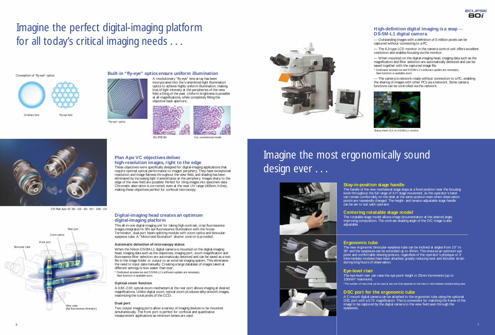
Imagine the perfect digital-imaging platformfor all today’s critical imaging needs . . .
Imagine the most ergonomically sounddesign ever . . .
Plan Apo VC objectives deliver high-resolution images, right to the edgeThese objectives were specifically designed for digital-imaging applications thatrequire optimal optical performance to images’ periphery. They have exceptionalresolution and image flatness throughout the view field, and shading has beeneliminated by increasing light transmittance at the periphery. Images sharp to theedge of the view field are possible: Perfect for tiling images into specimen data.Chromatic aberration is corrected, even at the near UV range (405nm; h-line),making these objectives perfect for confocal microscopy.
Digital-imaging head creates an optimum digital-imaging platformThis all-in-one digital-imaging unit for taking high-contrast, crisp fluorescenceimages integrates Hi S/N epi-fluorescence illumination with the NoiseTerminator, dual-port beam-splitting module with zoom optics and binocular-eyepiece tube. A “Motorized Excitation” shutter control is provided.
Automatic detection of microscopy statusWhen the Nikon DS-5M-L1 digital camera is mounted on the digital-imaginghead, imaging data such as the objectives, imaging port, zoom magnification andfluoresence filter selection are automaticcaly detected and can be saved as a textfile in the image folder or output to an external imaging system. This eliminatesthe need to input data manually. Creating a large database of images taken atdifferent settings is now easier than ever.* Dedicated accessories and DS-5M-L1’s software update are necessary.
Save function is available soon.
Optical-zoom functionA 0.8X-2.0X optical-zoom mechanism at the rear port allows imaging at desiredmagnifications. Unlike digital zoom, optical zoom produces silky-smooth images,maximizing the total pixels of the CCD.
Dual portTwo output imaging ports allow a variety of imaging devices to be mountedsimultaneously. The front port is perfect for confocal and quantitativemeasurement applications as minimum lenses are used.
76
High-definition digital imaging is a snap — DS-5M-L1 digital camera— Outstanding images with a definition of 5 million pixels can becaptured without connecting to a PC.
— The 6.3-type LCD monitor in the camera control unit offers excellentresolution and enables focusing via the monitor.
— When mounted on the digital-imaging head, imaging data such as themagnification and filter selection are automatically detected and can besaved together with the captured image file.* Dedicated accessories and DS-5M-L1’s software update are necessary.
Save function is available soon.
— The camera is network-ready without connection to a PC, enablingthe sharing of images with other PCs via a network. Some camerafunctions can be controlled via the network.
Stay-in-position stage handleThe handle of the new mechanical stage stays at a fixed position near the focusingknob throughout the full range of X/Y stage movement, so the operator’s handcan remain comfortably on the desk at the same position even when observationpoints are repeatedly changed. The height- and tension-adjustable stage handlecan be set to suit each operator.
Centering rotatable stage modelThe rotatable-stage model allows image documentation at the desired angle,improving composition. The contrast-shading angle of the DIC image is alsoadjustable.
Ergonomic tubeThe new ergonomic binocular-eyepiece tube can be inclined at angles from 10° to30° and the eyepieces can be extended up to 40mm. This ensures an optimum eyepoint and comfortable viewing posture, regardless of the operator’s physique or ifintermediate modules have been attached, greatly reducing neck and shoulder strainduring long hours of observation.
Eye-level riserThe eye-level riser can raise the eye point height in 25mm increments (up to100mm* maximum).*The number of risers that can be used at any one time depends on the tube or intermediate modules being used.
DSC port for the ergonomic tubeA C-mount digital camera can be attached to the ergonomic tube using the optionalDSC port with a 0.7X magnification. This is convenient for matching the frame of theimage to be captured by the digital camera to the view field seen through theeyepieces.
Built-in “fly-eye” optics ensure uniform illumination A revolutionary “fly-eye” lens array has beenincorporated into the transmitted-light illuminationoptics to achieve highly uniform illumination, makingloss of light intensity at the peripheries of the viewfield a thing of the past. Uniform brightness is possibleat all magnifications, while completely filling theobjective back aperture.
Ordinary lens
Conception of “fly-eye” optics
Fly-eye lens
Front port
Filter cube(Epi-fluorescence illminator)
Zoom optics
Rear port
Binocular tube
ECLIPSE 80i Our conventional model
Status-check GUI on DS-5M-L1 monitor
“Fly-eye” optics
CFI Plan Apo VC 60×Oil、60×WI、100×Oil

For high-end bioscience researchConfocal microscopy and digtal-imaging techniques complement each other withthis configuration.— New Plan Apo VC objectives provide razor-sharp high-contrast images in
confocal microscopy.— Status-check function, which automatically detects imaging data, can be used
to create an image database.— Hi S/N Fluorescence System produces
images with extraordinary brightnessand a high S/N ratio.
— Clear and crisp DIC images arepossible at any magnification.
— Optical-zoom function ensures high-definition images during magnification.
— The front port of the dual port isperfect for confocal and quantitativemeasurement applications.
Imagine the widest range of system expansionto meet all applications . . .
“Hi S/N” epi-fluorescence microscopyThe new epi-fluorescence illuminators come standard with the NoiseTerminator, which eliminates stray light leaking from the filter cube,producing high-contrast images with greater S/N ratios when observingweakly fluorescing specimens. The desired wavelength of a multi-stained specimen can also be emphasized with the unique ExcitationBalancer. Up to six filter cubes can be mounted in the filter turret,while the mirror and all filters can be easily changed to suit individualapplications.
Brightfield microscopyThe new “fly-eye” lens array in the illumination optics providesuniform brightness to the edge of the view field. The CFI60 infinityoptics are acclaimed for their excellent sharpness and superior colorfidelity, while the Plan Apo VC objectives provide extraordinarilyhigh resolution over the entire image. By using the 1X-100Xcondenser, users can view images at all magnification ranges, fromultralow to high, without having to change the condenser. The 1Xobjective is ideal for pathology work requiring a larger image field.
Darkfield microscopyOur dedicated condensers for darkfield microscopy allow clearobservation of blood or the minute structure of flagella. Dry- andoil-type condensers are available.
Simple polarizing microscopyPolarizing microscopy is as simple as inserting a polarizer over thefield lens and an analyzer in the arm slot. It is ideal for observingbirefringent samples such as collagen, amyloids and crystals.
Nomarski DIC microscopyThe new DIC method results in well-balanced images with outstandingcontrast and resolution. Crisp and clear images with perfectly even colorare obtainable, even at low magnifications. Three types of DIC prismsare available: standard, high-contrast and high-resolution. The shadedirection of DIC images can also be adjusted on the rotatable-stagemodel.
Hi S/N epi-fluorescence/DIC microscopyBy using the high-performance DIC method in combination with HiS/N epi-fluorescence illumination, researchers can accurately locatefluorescent-tagged structures or proteins and visualize the cellularmorphology of a specimen. Used in conjunction with the new PlanApo VC objectives, digital imaging with the highest resolution andaberration correction throughout the view field is possible.
Phase-contrast microscopyNikon has specially developed its unique Apodized Phase Contrastobjectives for phase-contrast microscopy. These objectives enableresearchers to detect minute structures—previously difficult todetect due to annoying halos—with excellent contrast and a muchwider tonal range. This is ideal for specimens with varied refractiveindices.
For pathology inspection or documentationThis configuration is ideal for pathology and related applications. High-definitiondigital images can be easily documented in a relaxed posture.
— “Fly-eye” optics ensure evenillumination intensity.
— Ergonomic tilting tube reduces strainduring long hours of observation.
— Specimen holder for one slidefacilitates quick, one-handed changes ofspecimens.
— 5-megapixel images can be easilycaptured and shared among PCs usingthe DS-5M-L1 standalone digitalcamera.
98
For various experiments or general researchThis configuration is suitable for all types of image documentation and research,from pathology and cell biology to material science investigations.— Noise Terminator ensures digital fluorescence images with exceptional S/N
ratios.— Excitation Balancer allows specific
wavelengths in multi-stained specimensto be emphasized.
— Universal epi-illuminator enablesvarious episcopic observations formaterial science research.
— Optimal camera settings for eachobservation method can be selectedfrom the menu of the DS-5M-L1 digitalcamera.
Epi-fluorescence
Nomarski DIC
Epi-fluorescence/DIC
Phase contrast
Brightfield
. . . from basic laboratory research to top-level, cutting-edge research
Configured with confocal system, digitalcamera and digital imaging head
Configured with universal epi-fluorescenceilluminator and digital camera
Configured with digital camera
. . . applying various methodologies
2
3

DS-5M-L1 “Digital Sight” all-in-one digital camera systemThe standalone design of the DS-5M-L1 allows independent operation forhigh-definition digital imaging at a resolution of 5 megapixels (2560 x 1920effective pixels) without connecting to a PC or external monitor.
— A large 6.3-inch high-definition LCD monitor built into the cameracontroller allows the operator to focus the image on the monitor withoutneeding to use the eyepieces.
— Image-processing capabilities include shading correction, tone settings andsimple measurements.
— An exclusive scene function that utilizes a preprogrammed mode providesoptimum imaging for each observation method.
C1 confocal microscope systemThe C1 is a compact, personal type of confocal laser microscope system thatprovides the highest quality images of its class. Resolution, contrast andfluorescent-image brightness are all state of the art. Image sizes of up to 2Kby 2K at 12-bit image depth can be easily scanned.
— Filters are interchangeable to match fluorescent dyes, enablingresearchers to use the latest probes or dyes available.
— 3-channel simultaneous detection is possible, including simultaneous 3-channel fluorescence, 3-channel plus DIC, time-lapse recording and spatialanalysis.
DXM1200F ultrahigh-definition digital cameraThe DXM1200F is a top-level digital camera with 12 million output pixels. Itfeatures a CCD more than twice as sensitive as conventional models,providing stunning fluorescence capabilities. Frame selection, autocategorizing, auto print and other features facilitate imaging of a largenumber of images.
— Sparkling digital images that are better than those taken with film-basedcameras, thanks to Nikon’s proprietary IPS (Inter Pixel Stepping) high-density imaging technology.
— Low-noise design that incorporates high-S/N digital-circuit technology anda new CCD with outstanding sensitivity.
Universal epi-fluorescence illuminatorThe filter turret can accommodate up to six filter cubes. The built-in Noise Terminator dramatically improves the S/N ratio and imagecontrast. By sliding the Excitation Balancer through the optical path,it is possible to continuously adjust the intensity of each excitationwavelength of a multi-stained specimen. The epi-fluorescence illuminatorcan be used for variousapplications that requireepiscopic illumination, suchas material scienceinvestigations.
Ergonomic tubeThe tube can be inclined at angles from 10° to 30° and theeyepieces can be extended up to 40mm. With the optional DSCport, featuring a 0.7X magnification, a C-mount digital camera canbe easily attached to the ergonomic tube.
Quadrocular adapter Two CCTV cameras or one CCTVcamera and a digital camera can beattached to the trinocular eyepiece tubewhile maintaining the same eye level.
Double-lamphouse adapterThe double-lamphouse adapter allowstwo different light sources to besimultaneously attached to themicroscope, eliminating the need tochange the lamphouse and carry outtime-consuming centering procedures.
Eye-level riserThe eye-level riser can raise the eye-point height in 25mm increments, upto a maximum of 100mm*, to suitindividual requirements.
* The number of risers that can be used at any onetime depends on the tube or intermediate modulesbeing used.
Teaching headsThis option, which includes abuilt-in pointer, allowssimultaneous viewing of a singlespecimen withoutcompromising the brightness.Styles suitable for two people(side by side and face-to-face)to 10 people are available.
Double portMounted between the main body and theeyepiece tube, the double port enablesthe simultaneous use of two CCTVcamera systems or one CCTV cameraand one digital camera.
Drawing tubeThe drawing tube allows both theimage of a specimen and thedrawing to be seen through theeyepieces. When needed, 100% ofthe light can be sent to theobservation port.
Magnification moduleThe turret system allows the intermediatemagnification to be changed to 1X, 1.25X,1.5X or 2X, enabling the operator toframe the image to be captured with adigital camera so it matches the view fieldseen through the eyepieces.
FX-III series photomicrographic equipmentThe FX-III series utilizes a direct-projection system with a swing-out prism for fast exposure setting and accurate metering.
U-III: 0.1% and 1% spot exposure, and 35% integrated-averagemeasurement modes
H-III: 1% spot and 35% integrated-average measurement modes
P-III: Manual exposure model
Digital-imaging headThe digital-imaging head can be integrated with the Hi S/N epi-fluorescence illuminator, dual port with 0.8X–2.0X optical zoomlenses and a binocular eyepiece tube. Thanks to Nikon’s uniqueNoise Terminator, digital imaging with a significantly improved S/Nratio is also possible. And when used with the DS-5M-L1 digitalcamera, imaging data such asthe magnification and filter inuse are automatically detectedand can be stored togetherwith the captured image file. *: Accessories such as dedicated nosepieceand DS-5M-L1’s software update arenecessary.
1110
. . . a wide range of accessories too.
Tube inclination is adjustable 10°–30° Eyepiece can be extended up 40mm

System Diagram
1312
0.20.40.60.81.01.21.4
Achr-Apl N.A=1.4
ENG-mount Camera C-mount Camera
C-mount Camera
F E
S
S
P
P
O
O
O
O
RR
EF
O
P K Q R S
F
G
H
K
K
L
B L
W
D
W
C
D
CB
J JJ
I I
N
I
D
A
A
K
F
G
G
M
H
ENG-mount TV Adapter 0.45x*1
0.6x
ENG-mount TV Adapter Adapter
Relay Lens 1x
ENG-mount Zooming
Adapter Zooming
C-mount
TV Zoom Lens
Relay Lens 1x
C-mountTV Adapter
C-mount TVAdapter 0.35x*2
0.38x 0.45x 0.6x
C-mountTV Adapter A
C-mountTV Adapter VM2.5x
C-mountTV Adapter
VM4x
V-TPhotoAdapter
CFI12.5x
CFI10x
CFI10x M
CFI15x
CFI UW10x
CFI UW10x M
Y-TVTV Tube
Y-TBBinocularTube B
CFI60Objective Lens
Y-TFTrinocularTube FUW
Y-TTTrinocularTube TUW
C-TEErgonomic Binocular Tube
C-TEP
DSC Port
C-HL2 Specimen Holder Specimen Holder Specimen Holder (2 slides: Left)
C-HR2
(2 slides: Right)
C-HC1
(1 slide)
C-SRMechanicalStage
Darkfieid DarkfieidCondenser(oil)
Condenser(dry)
C-C
AplanatCondenser
C-CAchromatAchromat /Condenser
C-CAbbeCondenser
LWDAchromatCondenser
C-CAchromatSwing-outCondenser1-100X
C-CPhase ContrastCondenser*3
C-FL Epi-flFilter Cube
C-ER Eye-level Riser
C-ISA Intermediate Tubew/Simple Analyzer
C-IA Intermediate Tubew/Analyzer
Y-IDP Double Port
Y-IM Magnification Module
Y-IDT Drawing Tube
Y-THF Teaching Unit Face to Face
Y-THS Teaching Unit Side by Side B
Y-THPS Support for Side by Side
Y-THM Main Teaching Unit
C-FCEpi-FlCollector Lens
Quartz Epi-FI Collector Lens
C-FCEpi-FlCollector Lens
C-FCEpi-FlCollector Lens
Mercury LampSocket S 100W
Xe LampSocket 75W
Halogen LampSocket 100W/HMX
Hg Lamphouse HMX-3B
Hg Lamphouse HMX-4B
Xe Lamphouse HMX-4
HMX Lamphouse
C-SPSimplePolarizer
C-TPPolarizer withFirst-order RedTint Plate
C-NSextupleNosepiece
C-mount Camera
F.STOP A.STOP
EX.ADJ.
D-FLDDF Filter Cube
D-FLEBF Filter Cube
D-FB
λ
ERG
POWER
SHUTTER
DC12V3A
D-LH 12V100WPrecentered LamphouseD-CB C-BOX
C-SRRRotatableMechanicalStage
M
TE C1adapter
D-ES ND Slider Set
D-FAFL/DIC Analyzer
YM-POPolarizer
D-FBExcitation Balancer
D-DH Digital-Imaging Head
D-DP DICRotatablePolarizer
D-CUDUniversalCondenserDry
D-C DIC Module Dry
D-C PH Module
D-C Darkfield Ring
D-C 2-4X Auxiliary Lens
D-DADIC Analyzer
D-LPLambda Plate
D-CDIC Slider
JAPAN
C-HSHand Switch
MIN. MAX.
POW
I 0
SUPER HIGH MERCURY POWER
RUN
IGNITIO
POWLAREA
REPLACE BEFORE SPECIFIED
O I
MADE IN
LAMP
PUSH
POWLIGH
XENON POWER SUPPLY
C-SHG1Power Supply for HG100W
Xenon Power Supply 75W
UN2 Transformer 100W
D-FLUniversal Epi-Fluorescence Attachment
D-NID6IntelligentSextupleDIC Nosepiece
D-ND6SextupleDIC Nosepiece
(for Rotatable Mechanical Stage) (for Mechanical Stage)
LU NosepieceAdapter
CFI LU BD Objective Lens
L2-DIC DIC Prism
CFI L/LU EPI Objective Lens
L-NU5UniversalQuintupleNosepieceESD
L-NBD5 BDQuintupleNosepeiceESD
DIH FPAdapter
Y-THR Teaching Unit Side by Side A
Y-THP Pointer Unit
Y-THA AC Adapter
C-CTCenteringTelescope
D-C DIC Module Oil
D-CUODICCondenserOil
2
3
4
D-NI7IntelligentSeptupleNosepiece
EF
EF
V
C
A
UT
VU
T
T
U
V
Camera Mounts
Eyepieces
Eyepiece Tubes
Epi-fl Attachment Accessories Intermediate Modules
Polarizers
Condensers
Nosepieces
Stages / Specimen Holders
Digital-imaging Head
Lamphouses
FX-III Series Photomicrographic System
Projection LensPLI 2x
PLI 2.5xPLI 4xPLI 5x
C-0.7xDXM Relay Lens
Confocal Scanning Head C1
C-CLow PowerCondenser
C-CEL Expander Lens
N
N
N
TE-ATDouble Lamphouse
G
G
Y-QTQuadrocularAdaper
N
N
N
TE-AT Double Lamphouse Adapter
H
(45-55 / 0-100, 0-100)
*3: Cannot be used with 80i model for rotatable mechanical stage.*1: Use the dedicated 0.45X adapter for the double port sub-port. The 0.6X adapter cannot be used with the double port sub-port.
*2: Use the dedicated 0.35X adapter for the double port sub-port.

Magnification 10–1500X
Optical system CFI60 Infinity Optical System
Coarse/fine focusing Fine: 0.1mm per rotationCoarse: 14mm per rotationMinimum reading: 1 micron on left side knobCoarse motion torque adjustableRefocusing function (focus clamp)
Illumination 12V-100W halogen lamp100-240V (worldwide voltage)Built-in "Fly-eye" optics for optimal digital-imaging iIllumination
Built-in filter NCB11, ND8, ND32
Eyepiece tube Digital Imaging Head Binocular tube B (for F.O.V. 22mm)Trinocular tube "F" UW (for F.O.V. 22mm/25mm, observation/photo: 100/0, 0/100)Trinocular tube "T" UW (for F.O.V. 22mm/25mm, observation/photo: 100/0, 20/80, 0/100)Ergonomic binocular tube (for F.O.V. 22mm, inclination:10-30°, extension: 40mm) DSC port: 50/50, 100/0 (optional)
Eyepiece lens 10X (F.O.V.: 22mm), 10X M photo mask (F.O.V.: 25mm), 12.5X (F.O.V.: 16mm), 15X (F.O.V.: 14.5mm),UW 10X (F.O.V.: 25mm), UW 10X M photo mask (F.O.V.: 25mm)
Nosepiece Sextuple nosepiece, DIC sextuple nosepiece, Intelligent DIC sextuple nosepiece, Intelligent septuple nosepiece
Stage Super-hard Alumite coated surfaceStay-in-position stage handleStage handle height and tension adjustableRectangular 159mm x 243mm surface stage, 78mm x 54mm cross travel (x-y movement)1-slide or 2-slide specimen holder available (option)
Stage rotation Centerable, 220 degrees (80i Rotatable Stage model)
Condenser focusing stroke 37mm
Intermediate accessories Universal epi-fluorescence Illuminator (6 filter positions, material application capability)Teaching heads, Double port, Magnification module, Drawing tube, Eye-level riser
Observation method Brightfield, Epi-fluorescence, DIC, Phase contrast, Darkfield, Simple polarizing, Epi/brightfield, Epi/Darkfield, Epi/DIC, Epi/Simple polarizing
Digital Imaging Head Universal Epi-illuminator
Application Epi-fluorescence, Materials, Confocal, Quantative analysis
Dual camera (Depends on eyepiece tube or double port)
Light distribution 3-way (Eye/Front/Rear) 100% to each port (Depends on eyepiece tube)
Optical output ports Front port: 1X, Diameter 52mm (Depends on eyepiece tube or double port)Rear port: Optical zoom 0.8X - 2.0X(continuous), Zoom ratio 2.5 :1, C-mount
Inclination angle 25 degrees (Depends on eyepiece tube)
F.O.V. Max. 25mm
Filter turret 6 positions
HiS/N Noise Terminator fluorescence Available
Aperture diaphragm Centerable, detachable; diameter 1-9mm
Field diaphragm Centerable, detachable; diameter 1-9mm
ND filter ND4, ND8, ND16
Light shield shutter Motorized (controlled by external switch) Manual
Analyzer slot Available
Polarizer slot Available
Usable light sources Mercury, Xenon, Halogen (centering)
External connection USB, C1 interlock, C-box connector —
Status-check function Available with digital camera DS-5M-L1 combination —
Digital-Imaging Head, Universal Epi-illuminator Specifications
Specifications Dimensional Diagrams
144300
144300
435
435
203
60
338
561
6020
3
33841
313
1.6
203
338
478
60
435
144300
1514
Configured with digitalimaging head
Configured with universalepi-fluorescence illuminator
and ergo tube
Configured withtrinocular tube TUW
Unit: mm

En
Advanced Research Microscope ECLIPSE 80i
This brochure is printed on recycled paper made from 40% used material.
WARNINGTO ENSURE CORRECT USAGE, READ THE CORRESPONDING MANUALS
CAREFULLY BEFORE USING YOUR EQUIPMENT.
Specifications and equipment are subject to change without any notice or obligationon the part of the manufacturer. December 2003. ©2003 NIKON CORPORATION
NIKON CORPORATIONYokohama Plant
NIKON CORPORATIONInstruments Company
ISO 9001Accredited by theDutch Council for
Accreditation ISO 14001
Printed in Japan (0312-15)T Code No. 2CE-MSGH-1
www.nikon.com/
Fuji Bldg., 2-3, Marunouchi 3-chome, Chiyoda-ku, Tokyo 100-8331, Japan
NIKON CORPORATION
NIKON INSTECH CO., LTD.Parale Mitsui Bldg., 8, Higashida-cho, Kawasaki-ku,Kawasaki, Kanagawa 210-0005, Japanphone: +81-44-223-2167 fax: +81-44-223-2182 www.nikon-instruments.jp/eng/www.i-series.com
NIKON INSTRUMENTS (SHANGHAI) CO., LTD.CHINA phone: +86-21-5058-5055 fax: +86-21-5058-5060
NIKON SINGAPORE PTE LTDSINGAPORE phone: +65-6559-3618 fax: +65-6559-3668
NIKON MALAYSIA SDN. BHD.MALAYSIA phone: +60-3-78763887 fax: +60-3-78763387
NIKON INSTRUMENTS EUROPE B.V.P.O. Box 222, 1170 AE Badhoevedorp, The Netherlandsphone: +31-20-44-96-222 fax: +31-20-44-96-298www.nikon-instruments.com/
NIKON FRANCE S.A.FRANCE phone: +33-1-45-16-45-16 fax: +33-1-45-16-00-33NIKON GMBHGERMANY phone: +49-211-9414-0 fax: +49-211-9414-322NIKON INSTRUMENTS S.p.A.ITALY phone: + 39-55-3009601 fax: + 39-55-300993NIKON AGSWITZERLAND phone: +41-43-277-2860 fax: +41-43-277-2861NIKON UK LTD. UNITED KINGDOM phone: +44-20-8541-4440 fax: +44-20-8541-4584
NIKON INSTRUMENTS INC.1300 Walt Whitman Road, Melville, N.Y. 11747-3064, U.S.A.phone: +1-631-547-8500; +1-800-52-NIKON (within the U.S.A.only) fax: +1-631-547-0306/www.nikonusa.com/
NIKON CANADA INC.CANADA phone: +1-905-625-9910 fax: +1-905-625-0103
* Monitor images are simulated.Company names and product names appearing in this brochure are their registered trademarks or trademarks.
. . . magine perfection in digital microscopy
Nikon has reduced the amount of chromium, cadmium and lead used in the Eclipse-i Seriesto an absolute minimum to diminish its environmental impact.
Please contact Nikon for a handy pamphlet listingcompatible accessories, including objectives andepi-fluorescence filters.
ECLIPSE i-Series Website
www.nikon-i.com
Microscopy images courtesy of: 1 Momoki Hirai, Professor, Department of Integrated Biosciences, Graduate School of Frontier Sciences, The University of Tokyo.2 Naoyuki Miyokawa, M.D., Ph.D., Associate Professor, Dept. of Surgical Pathology, Asahikawa Medical College Hospital.3 Dr. Torsten Wittmann, The Scripps Research Institute.