y90 radioembolization of colorectal hepatic metastases using...
TRANSCRIPT

1
Y90 Radioembolization of Colorectal Hepatic
Metastases using Glass Microspheres: Safety and
Survival Outcomes from a 531-Patient Multicenter
Study
Ryan Hickey MD1, Robert J. Lewandowski MD1, Totianna Prudhomme CNP2,
Eduardo Ehrenwald MD2, Brian Baigorri MD3, Jeffrey Critchfield MD3, Joseph
Kallini MD1, Ahmed Gabr MD1, Boris Gorodetski4, Jean-Francois Geschwind
MD4, Andrea Abbott MD5, Ravi Shridhar MD6, Sarah B. White MD7, William S.
Rilling MD7, Brendan Boyer PA-C8, Shannon Kauffman MD8, Sharon Kwan MD9,
Siddarth A. Padia MD9, Vanessa L. Gates MS1, Mary Mulcahy MD10, Sheetal
Kircher MD10, Halla Nimeiri MD10, Al B. Benson MD10, Riad Salem MD MBA1,10
1Department of Radiology, Section of Interventional Radiology, Northwestern
Memorial Hospital, Robert H. Lurie Comprehensive Cancer Center, Chicago IL 2Department of Interventional Radiology, Abbott Northwestern Hospital,
Minneapolis, MN 3Department of Radiology, Wayne State University, Detroit Medical Center,
Detroit, MI 4Interventional Radiology Center, Johns Hopkins Hospital, Baltimore, MD 5Department of Surgery, Moffitt Cancer Center, Tampa, FL 6Department of Radiation Oncology, Moffitt Cancer Center, Tampa, FL 7Department of Radiology, Division of Vascular/Interventional Radiology, Medical
College of Wisconsin, Milwaukee, WI 8Department of Radiology, Miami Valley Hospital, Dayton, OH 9Department of Radiology, Section of Interventional Radiology, University of
Washington, Seattle, WA 10Department of Medicine, Division of Hematology and Oncology, Robert H. Lurie
Comprehensive Cancer Center, Northwestern University, Chicago, IL
Journal of Nuclear Medicine, published on December 3, 2015 as doi:10.2967/jnumed.115.166082

2
Corresponding Author: Riad Salem, MD, MBA
Chief, Interventional Radiology
Section of Interventional Radiology
Department of Radiology
676 N. St. Clair, Suite 800
Chicago, Illinois 60611 USA
email: [email protected]
Role of Funding: There was no funding provided for this study.
Conflict of Interest: Ryan Hickey, Robert Lewandowski, Jean-Francois
Geschwind, Siddarth Padia, Mary Mulcahy and Riad Salem are advisors to BTG.
None of the other authors have any conflict of interest.
Word Count: 2850
Key words: radioembolization, colorectal metastases, survival, yttrium-90
Running title: Glass Y90 and Colorectal Metastases

3
ABSTRACT
Background: Hepatic metastases of colorectal carcinoma are a leading cause of
cancer-related mortality. The vast majority of colorectal liver metastases become
refractory to chemotherapy and biologic agents, at which point the median overall
survival declines to 4-5 months. Radioembolization with yttrium-90 has been
used in the salvage setting with favorable outcomes. This study reports the
survival and safety outcomes of 531 patients treated with glass-based 90Y
microspheres at eight institutions, making it the largest 90Y study for patients with
colorectal liver metastases.
Patients and methods: Data were retrospectively compiled from eight institutions
for all 90Y glass microsphere treatments for colorectal liver metastases. Exposure
to chemotherapeutic/biologic agents, prior liver therapies, biochemical
parameters prior to and following treatment, radiation dosimetry and
complications were recorded. Uni- and multivariate analyses for predictors of
survival were performed. Survival outcomes, clinical/biochemical adverse events
are reported.
Results: 531 patients received 90Y radioembolization for colorectal liver
metastases. The most common clinical adverse events were fatigue (55%),
abdominal pain (34%) and nausea (19%). Grade 3 or 4 hyperbilirubinemia
occurred in 13% of patients at anytime. Median overall survival from first Y90
treatment was 10.6 months (95% CI 8.8-12.4). Performance status, tumor burden
≤25%, no extra-hepatic metastases, albumin >3 g/dL and having received ≤2
chemotherapeutic agents independently predicted better survival outcomes.
Conclusion: Multi-institutional review of a large cohort of patients with colorectal
liver metastases treated with 90Y radioembolization using glass microspheres
demonstrates promising survival outcomes with low toxicities and side-effects
that are reproducible and consistent with prior reports of radioembolization.

4
INTRODUCTION
Metastases of colorectal carcinoma are the third most common cause of
cancer-related mortality worldwide. Advances in chemotherapy regimens,
biologic agents, and liver resection in select cases have lead to a prolongation in
overall survival from the diagnosis of hepatic metastases. Nonetheless, most
colorectal liver metastases become refractory or resistant to these regimens, at
which point survival estimates range from 4-5 months. Yttrium-90 (Y90)
radioembolization of hepatic metastases has been increasingly used in this
salvage setting with favorable survival outcomes after Y90 radioembolization
exceeding 10 months (1-3). The majority of published outcomes report results of
Y90 radioembolization using resin microspheres. However, a substantial number
of patients with colorectal liver metastases undergo Y90 radioembolization with
glass microspheres. This study reports the survival and safety outcomes of 531
patients with hepatic metastases of colorectal cancer treated with glass Y90
microspheres at several institutions.
METHODS
Patient cohort
Between 2001 and 2014, 531 consecutive patients underwent
radioembolization of hepatic metastases of colorectal carcinoma at eight
institutions. The study was approved by Institutional Review Boards and was
Health Insurance Portability and Accountability Act compliant. Data were
retrospectively compiled into a common database and analyzed at one
institution. The clinical trials registration number was NCT00532740.
Clinical side effects and biochemical toxicities according to National
Cancer Institute common terminology criteria (CTCAE) version 4.0 were

5
recorded at follow up. Clinical adverse events and biochemical toxicities
occurring at any time after treatment are reported (not limited to 30 days). This
report also complies with the research reporting standards for radioembolization.
(4)
This study represents retrospectively collected data at cancer centers with
expertise in locoregional therapies. Y90 radioembolization treatment indications
included unresectable metastases from colorectal cancer; progressive disease
confirmed on imaging refractory to previous systemic/locoregional therapy;
Eastern Cooperative Oncology Group (ECOG) status ≤2; ability to undergo
angiography and selective visceral catheterization; and adequate hematology
(granulocyte count ≥1.5 × 10⁹ /L, platelets ≥50 × 109 /L), renal function
(creatinine ≤2.0 mg/dL) and liver function (bilirubin ≤2.0 mg/dL). Exclusion criteria
included significant extrahepatic disease (life expectancy <3 months); evidence
of uncorrectable gastrointestinal flow observed on angiography or 99mTc-
macroaggregated albumin (MAA) scans; estimated lung dose >30 Gy in a single
session.
Patient Evaluation and Workup
All patients underwent baseline laboratory tests and radiological imaging
within one month of treatment. Imaging information was used to determine
baseline tumor burden, uni/multifocality and presence/absence of extrahepatic
metastases. Pretreatment angiography was performed to determine proper
catheter position and identify any collateral flow to the gastrointestinal tract (5-9).
99mTc-MAA scan was performed to detect GI flow and lung shunt fraction (6).
Prophylactic embolization of aberrant vessels was performed when appropriate.
Treatment Plan
All radioembolization procedures were performed with a glass-based
Yttrium-90 device (TheraSphere®, BTG, United Kingdom). This device is
currently approved for hepatocellular carcinoma in the United States and liver

6
neoplasia (worldwide) (10). The method for determining injected activity required
to deliver 120 Gy has been published previously (4,6,11,12).
Overall survival
Median overall survival was calculated from the dates of diagnosis of the
primary cancer, hepatic metastases and first Y90 treatment, censored to the date
of last follow-up. Survival analyses were substratified based on exposure to
cytotoxic chemotherapeutics (5-fluorouracil/capecitabine, oxaliplatin, irinotecan)
and biologic agents (bevacizumab, cetuximab/panitumumab, regorafenib).
Uni/Multivariate Analysis and Statistical Plan
Uni/multivariate analyses were performed using Cox proportional hazards
model. Variables entered into the univariate analyses included patient
demographics (gender, age), performance status (ECOG), prior therapy
(cytotoxic/biologic agents), presence of metastatic disease at diagnosis, tumor
burden, presence of extra-hepatic metastases and liver function. Variables with
p≤0.25 by univariate model were included in the multivariate model. Analysis
were performed using IBM SPSS Statistics v22.0 (Armonk, NY); p<0.05 was
considered significant.
RESULTS
Baseline characteristics
Most patients (63%) were <65 years old at the time of treatment, of whom
59% were male. The majority of patients had an ECOG status of 0 or 1 (96%).
Most patients (70%) had ≤25% of the liver volume involved by tumor. The
majority of patients (62%) had liver-only disease, while 38% had limited extra-
hepatic disease. Eighteen percent of patients had prior hepatic resection, 14%
had prior liver ablation and 4% had prior transarterial chemoembolization (TACE)
(Table 1).

7
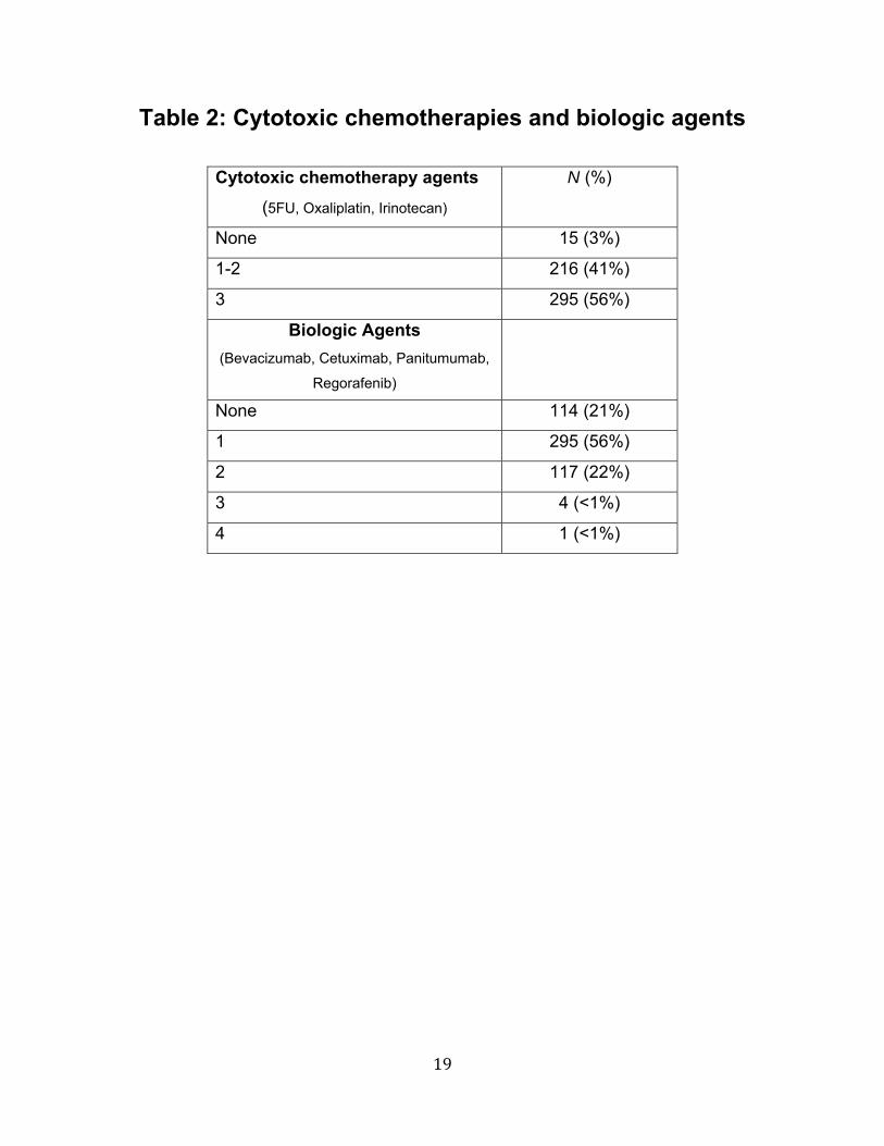
Prior to Y90 radioembolization, 56% of patients received three cytotoxic
chemotherapeutics (5-fluorouracil (5FU), oxaliplatin and irinotecan), while 41% of
patients received only one or two of these agents. Fifteen patients (3%) received
no cytotoxic chemotherapy prior to Y90. Twenty-two percent of patients received
no biologic agents, 56% received one biologic agent and 22% received three
biologic agents (Table 2).
Nearly all patients underwent lobar or selective radioembolization at the
first treatment. Only 2% of patients received whole-liver treatment in a single
setting.
Y90 Dosimetry & Delivery
The median radiation dose delivered to the liver was 120.2 Gy (range 35-391
Gy). Ninety percent or greater of the dose was delivered in all treatments. Extra-
hepatic arterial coil embolization was performed in 25% of patients.
Side Effects & Biochemical Toxicities
The most frequent clinical side effects included fatigue (55%), abdominal
pain and/or discomfort (34%) and nausea (19%). Vomiting, anorexia and fever
occurred in less than 10% of patients (Table 3). No gastrointestinal ulcers were
reported.
Biochemical toxicities of grade 3 or 4, recorded at any time following
treatment, included bilirubin (13%), alkaline phosphatase (9%), albumin (8%),
aspartate transaminase (AST) (3%) and alanine transaminase (ALT) (<1%)
(Table 4).
Overall Survival
Two hundred eighty-four patients had died at the time of data compilation.
Survival analysis is provided in Table 5. Median overall survival (OS) for the
patient cohort from the date of diagnosis of the primary tumor, censored to last

8
follow-up, was 48.7 months (95% CI 44.2-53.2), 37.7 months (95% CI 33.7-41.7)
from diagnosis of hepatic metastases, and 10.6 months (95% CI 8.8-12.4) from
first Y90 treatment. Median time from the diagnosis of hepatic metastases to first
Y90 was 17.5 months (95% CI 15.2-19.7). Median OS from first Y90 was longer
in patients without extra-hepatic disease compared to those with extra-hepatic
disease (14.4 vs. 6.6 months, P<0.001).
Time from diagnosis of hepatic metastases to first Y90 was longer in
patients who received three cytotoxic chemotherapeutics compared to those who
received ≤2 (22.6 vs. 10.9 months, P<0.001). Median OS from first Y90 was
shorter for patients who received three cytotoxic chemotherapeutics compared to
those who received ≤2 (9.2 vs. 14.7 months, P<0.001). For patients without
extra-hepatic disease at the time of first Y90, median OS was longer for patients
receiving ≤2 compared to three cytotoxic chemotherapeutics (16.5 vs. 13.1
months, P<0.05). For patients with extra-hepatic disease at first Y90, median OS
was longer for patients receiving ≤2 compared to three cytotoxic
chemotherapeutics (9.6 vs. 5.4 months, P=0.003).
Median OS from first Y90 was not significantly different for patients who
received no biologic agents compared to those who received one (11.5 vs. 12.9
months). Median OS from first Y90 was longer for patients who received ≤1
biologic agents compared to those who received ≥2 (12.9 vs. 7.0 months,
P<0.001).
Median OS from first Y90 was longer for patients treated after 2004
compared to patients treated prior to 2004 (10.9 vs. 7.0 months, P<0.05).
Uni/Multivariate Analyses
Better survival outcomes were predicted by ECOG performance status 0
(P<0.001), American Joint Committee on Cancer (AJCC) stage ≤3 at time of
diagnosis (p=0.113), lack of extra-hepatic metastases (p<0.001), hepatic tumor

9
burden ≤25% (p<0.001), ≤2 cytotoxic chemotherapeutics (P<0.001), no biologic
therapy (P<0.001), bilirubin <1.3 mg/dL (P<0.001) and albumin >3 g/dL
(P<0.001).
On multivariate analysis, ECOG performance status 0 (HR 0.61; 95%CI
0.46-0.79), hepatic tumor burden ≤25% (HR 0.37; 95%CI 0.28-0.49), no extra-
hepatic metastatic disease (HR 0.50; 95%CI 0.38-0.64), albumin >3 g/dL (HR
0.47; 95%CI 0.35-0.63) and ≤2 cytotoxic chemotherapeutics (HR 0.61; 95%CI
0.46-0.79) independently predicted better survival outcomes (Table 6).
DISCUSSION
Colorectal carcinoma remains one of the most common cancers
worldwide with approximately half of affected individuals eventually developing
liver metastases. Only a minority of these patients are candidates for potentially
curative therapies such as surgical resection or ablation. Modern chemotherapy
regimens and the advent of biologic agents have lead to a significant
prolongation of median overall survival for patients with hepatic metastases to
approximately 29 to 32 months (13,14). Nonetheless, once hepatic metastases
become chemorefractory, survival estimates are poor, typically between four and
five months (15-17).
Transarterial Y90 radioembolization has been increasingly used as a
locoregional therapy in the setting of chemorefractory hepatic metastases and
was recently included in the European Society for Medical Oncology clinical
practice guidelines for the treatment of liver-limited colorectal metastases failing
chemotherapeutic options (18). Radioembolization relies on the differential blood
supply of hepatic tumors compared to normal liver parenchyma. Primary and
metastatic hepatic tumors receive the majority of their blood supply from the
hepatic arteries, as opposed to the predominantly portal venous blood supply to

10
liver parenchyma. Hepatic arterial injection of the radioactive microspheres leads
to preferential deposition within the tumor capillaries, providing the anti-tumoral
effects of radiation that are not otherwise utilized in the standard treatment
paradigms of colorectal liver metastases.
Several studies have demonstrated the benefits of Y90 radioembolization
in the treatment of metastatic colorectal cancer. A randomized phase II analysis
of the addition of a single treatment of Y90 resin microspheres to the standard
regimen of 5FU/leucovorin chemotherapy for patients with colorectal liver
metastases demonstrated a significant prolongation in median overall survival
compared to chemotherapy alone (29.4 vs. 12.8 months, P=0.02) (19). The
addition of Y90 radioembolization to systemic chemotherapy has been shown to
prolong time to liver tumor progression for patients with chemorefractory
colorectal liver metastases (5.5 vs. 2.1 months, P=0.003) (20), and matched-pair
analysis of patients with chemorefractory colorectal liver metastases
demonstrated a survival benefit for patients receiving Y90 radioembolization
compared to best supportive care (8.3 vs. 3.5 months, P<0.001) (21). A
prospective, multi-center, phase II analysis of Y90 radioembolization for
chemorefractory liver metastases reported a hepatic progression-free survival of
3.0 months for colorectal metastases. Median overall survival for all patients with
metastatic colorectal cancer was 8.8 months and increased to 10.5 months for
patients with liver-only colorectal metastases (22).
However, most of the published literature on Y90 radioembolization for
colorectal liver metastases use resin microspheres, which is in part due to
different approved indications for glass and resin microspheres (20,21,23,24).
Nonetheless, a significant number of patients undergo Y90 radioembolization of
colorectal liver metastases with glass microspheres. The results of this multi-
institutional review of 531 patients with colorectal liver metastases treated with
Y90 radioembolization using glass microspheres underscore the consistent,
reproducible and favorable survival outcomes of the therapy in the salvage

11
setting. The data show that radioembolization using glass microspheres provides
reliable dose delivery, and that the treatment is safe and well tolerated.
Once colorectal liver metastases become chemorefractory, patients’
median overall survival approaches four to five months (15-17). In addition to the
studies cited previously, a number of recent large cohort studies have reported
the survival of patients undergoing Y90 radioembolization in the salvage setting.
The survival data from these studies are remarkably consistent and nearly
identical to our own findings. Median overall survival from first Y90 treatment in
our cohort was 10.6 months. In the next largest published study of 302 patients
with chemorefractory colorectal liver metastases using resin microspheres,
Saxena et al. report a median overall survival of 10.5 months from first Y90 (1),
which mirrors the 10.6-month median overall survival of 214 patients treated with
glass microspheres reported by Lewandowski et al (2). Kennedy et al. also
reported a median overall survival of 10.5 months among responders in a multi-
institutional review of 208 patients treated with resin Y90 microspheres (3). While
the 214 patients reported by Lewandowski et al. were included in our multi-
institutional cohort, removal of these 214 patients from the survival analysis still
indicates a median overall survival of 10.5 months for the remaining 317 patients.
The significance of these survival outcomes achieved in the salvage
setting should not be overlooked. While data obtained from retrospective review
certainly cannot be equated with data ascertained from prospective, randomized
trials, these results resonate favorably considering the median overall survival
outcomes achieved in the salvage setting with other agents. Cetuximab
demonstrated a median overall survival of 9.5 months in the salvage setting for
patients with the wild type K-ras gene compared to 4.8 months achieved with
best supportive care (16). Subsequent trials investigating its use earlier in the
treatment of metastatic colorectal cancer have led to its acceptance as a
cornerstone of colorectal cancer treatment (18,25). More recently, regorafenib
has become standard of care treatment of chemorefractory colorectal liver

12
metastases for demonstrating a prolongation in median overall survival from 5
months with best supportive care and placebo compared to 6.4 months with best
supportive care and regorafenib (15). A number of prospective, randomized trials
are currently evaluating the role of Y90 radioembolization at different points in the
treatment of colorectal liver metastases, including the EPOCH trial and combined
1100-patient SIRFLOX, FOXFIRE and FOXFIRE global studies.
The precise and reliable dose delivery achieved with glass Y90
microspheres is a critical observation. Glass microspheres have a low embolic
load, which does not limit delivery of the prescribed activity of Y90 (26). In our
cohort, delivery of ≥90% of the dose was achieved in all treatments. The heavy
embolic load of resin microspheres, on the other hand, can result in arterial
stasis, limiting the actual Y90 dose delivered. In a recent phase I trial of resin
microspheres for treatment of colorectal metastases, arterial stasis limiting the
total administered activity occurred in 37.5% of treatment sessions (27). Such
reliability of dose delivery with glass microspheres is not only a vital component
of consistent and reproducible brachytherapeutic treatments, but it is also central
to the principles of oncologic clinical trials, as the safety and effectiveness of a
therapy cannot be evaluated if there is variability in the actual dose delivered.
This was confirmed in a recent phase I study of the radiosensitizing
chemotherapy capecitabine used with escalating, whole-liver doses of Y90 glass
microspheres in which controlled and predictable escalation of Y90 dose was
performed (28). During the same time period, a phase I study of resin Y90
radioembolization in the setting of capecitabine therapy was published, but
escalation of Y90 dose could not be performed due to the unpredictability of dose
delivery with resin microspheres, particularly in the setting salvage colorectal liver
metastases, where vessels can be adversely affected by chemotherapy
exposure. In addition, only 41.7% of patients received whole-liver
radioembolization, limiting assessment of the true liver tolerance to
radioembolization performed during concomitant administration of
radiosensitizing chemotherapy.

13
Given the changes in hepatic vasculature and flow dynamics seen in
patients who have been heavily pre-treated with chemotherapy and biologic
agents, the high activity and low embolic load of glass microspheres offer
tremendous value. A frequent criticism of Y90 radioembolization is the necessity
of complex coil embolization of extra-hepatic vessels in order to prevent non-
target Y90 radioembolization, or coil embolization of hepatic vasculature for intra-
hepatic flow redistribution in order to achieve dose delivery, both of which not
only prolong procedures and render them more complex, but also introduce the
inherent risk of promoting collateral arterial networks perfusing tumors that may
complicate future treatments. In our cohort, however, only 25% of patients
underwent arterial coil embolization, which is in line with the increasingly low
rates of extra-hepatic arterial embolization during radioembolization with glass
microspheres previously reported by our group (29). In our group’s more
contemporary patient population, prophylactic coil embolization approaches 10%.
The safety of this practice is underscored by the fact that no gastrointestinal
ulcers were reported.
The low rate of clinical side-effects and biochemical toxicities in this large
patient cohort confirm the safety of Y90 performed across institutions. The most
common clinical side-effects of fatigue, abdominal pain/discomfort and nausea
are comparable to the known and expected side effects of radioembolization.
Grade 3 and 4 biochemical toxicities following radioembolization affected a
minority of patients. These may in fact be overestimated in this series, as hepatic
toxicities at any time following radioembolization were recorded, inevitably
capturing the effects of hepatic decompensation related to tumor progression.
This study has limitations. The retrospective nature of data collection limits
the accurate comparison of patient subgroups. For this reason, our study focused
on outcomes least prone to misinterpretation and bias, namely the survival and
safety of a treatment when performed at diverse institutions. The study is

14
confounded by the variability in the number of chemotherapy and biologic agent
regimens patients received, and a high percentage of patients did not receive all
available systemic options for a variety of reasons, including poor tolerance and
KRAS mutation status. Nonetheless, despite local oncology practices and
variability in the nuances of the radioembolization procedure, Y90
radioembolization with glass microspheres is a safe therapy that offers consistent
survival outcomes for chemorefractory colorectal liver metastases.
These results add to growing evidence, now comprising >1000 patients, in
support of Y90 use for treatment of colorectal liver metastases. Collaboration
among oncologists and interventional radiologists is necessary in order to
conduct the large-scale, prospective trials necessary to more precisely define the
role of Y90 radioembolization in the treatment of colorectal liver metastases.
CONCLUSION
Multi-institutional review of the largest cohort of patients with colorectal
liver metastases treated with glass Y90 radioembolization demonstrates very
promising survival outcomes that are reproducible and consistent with prior
reports. Glass microspheres provide reliable and precise radiation dose delivery
that is safe and well-tolerated. Results of the large-scale randomized studies are
awaited.

15
REFERENCES
1. Saxena A, Meteling B, Kapoor J, Golani S, Morris DL, Bester L. Is yttrium-90 radioembolization a viable treatment option for unresectable, chemorefractory colorectal cancer liver metastases? A large single-center experience of 302 patients. Ann Surg Oncol. 2015;22:794-802. 2. Lewandowski RJ, Memon K, Mulcahy MF, et al. Twelve-year experience of radioembolization for colorectal hepatic metastases in 214 patients: survival by era and chemotherapy. Eur J Nucl Med Mol Imaging. 2014;41:1861-1869. 3. Kennedy AS, Coldwell D, Nutting C, et al. Resin 90Y-microsphere brachytherapy for unresectable colorectal liver metastases: modern USA experience. Int J Radiat Oncol Biol Phys. 2006;65:412-425. 4. Salem R, Lewandowski RJ, Gates VL, et al. Research reporting standards for radioembolization of hepatic malignancies. J Vasc Interv Radiol. 2011;22:265-278. 5. Lewandowski RJ, Sato KT, Atassi B, et al. Radioembolization with (90)y microspheres: angiographic and technical considerations. Cardiovasc Intervent Radiol. 2007;30:571-592. 6. Salem R, Thurston KG. Radioembolization with 90Yttrium Microspheres: A State-of-the-Art Brachytherapy Treatment for Primary and Secondary Liver Malignancies: Part 1: Technical and Methodologic Considerations. J Vasc Interv Radiol. 2006;17:1251-1278. 7. Salem R, Thurston KG. Radioembolization with 90Yttrium Microspheres: A State-of-the-Art Brachytherapy Treatment for Primary and Secondary Liver Malignancies: Part 2: Special Topics. J Vasc Interv Radiol. 2006;17:1425-1439. 8. Salem R, Thurston KG. Radioembolization with yttrium-90 microspheres: a state-of-the-art brachytherapy treatment for primary and secondary liver malignancies: part 3: comprehensive literature review and future direction. J Vasc Interv Radiol. 2006;17:1571-1593. 9. Salem R, Lewandowski RJ, Sato KT, et al. Technical aspects of radioembolization with 90Y microspheres. Tech Vasc Interv Radiol. 2007;10:12-29. 10. Salem R, Thurston KG, Carr BI, Goin JE, Geschwind JF. Yttrium-90 microspheres: radiation therapy for unresectable liver cancer. J Vasc Interv Radiol. 2002;13:S223-229.

16
11. Ho S, Lau WY, Leung TW, Chan M, Johnson PJ, Li AK. Clinical evaluation of the partition model for estimating radiation doses from yttrium-90 microspheres in the treatment of hepatic cancer. Eur J Nucl Med. 1997;24:293-298. 12. Lau WY, Ho S, Leung TW, et al. Selective internal radiation therapy for nonresectable hepatocellular carcinoma with intraarterial infusion of 90yttrium microspheres. Int J Radiat Oncol Biol Phys. 1998;40:583-592. 13. Venook AP ND, Lenz H, Innocenti F, Mahoney MR, O'Neil BH, Shaw JE, Polite BN, Hochster HS, Atkins JN, Goldberg RM, Mayer RJ, Schilsky RL, Bertagnolli MM, Blanke CD. CALGB/SWOG 80405: Phase III trial of irinotecan/5-FU/leucovorin (FOLFIRI) or oxaliplatin/5-FU/leucovorin (mFOLFOX6) with bevacizumab (BV) or cetuximab (CET) for patients (pts) with KRAS wild-type (wt) untreated metastatic adenocarcinoma of the colon or rectum (MCRC). 2014 ASCO Annual Meeting. Chicago, IL; 2014. 14. Lenz H ND, Innocenti F, Blanke C, Mahony MR, O'Neil BH, Shaw JE, Polite B, Hochster H, Atkins J, Goldberg R, Mayer R, Schilsky RL, Bertagnolli M, Venook A. CALGB/SWOG 80405: PHASE III trial of irinotecan/5-FU/leucovorin (FOLFIRI) or oxaliplatin/5-FU/leucovorin (mFOLFOX6) with bevacizumab (BV) or cetuximab (CET) for patients (pts) with expanded ras analyses untreated metastatic adenocarcinoma of the colon or rectum (MCRC). ESMO 2014. Madrid, Spain; 2014. 15. Grothey A, Van Cutsem E, Sobrero A, et al. Regorafenib monotherapy for previously treated metastatic colorectal cancer (CORRECT): an international, multicentre, randomised, placebo-controlled, phase 3 trial. Lancet. 2013;381:303-312. 16. Karapetis CS, Khambata-Ford S, Jonker DJ, et al. K-ras mutations and benefit from cetuximab in advanced colorectal cancer. N Engl J Med. 2008;359:1757-1765. 17. Jonker DJ, O'Callaghan CJ, Karapetis CS, et al. Cetuximab for the treatment of colorectal cancer. N Engl J Med. 2007;357:2040-2048. 18. Van Cutsem E, Cervantes A, Nordlinger B, Arnold D, Group EGW. Metastatic colorectal cancer: ESMO Clinical Practice Guidelines for diagnosis, treatment and follow-up. Ann Oncol. 2014;25 Suppl 3:iii1-9. 19. Van Hazel G, Blackwell A, Anderson J, et al. Randomised phase 2 trial of SIR-Spheres plus fluorouracil/leucovorin chemotherapy versus fluorouracil/leucovorin chemotherapy alone in advanced colorectal cancer. J Surg Oncol. 2004;88:78-85. 20. Hendlisz A, Van den Eynde M, Peeters M, et al. Phase III trial comparing protracted intravenous fluorouracil infusion alone or with yttrium-90 resin microspheres radioembolization for liver-limited metastatic colorectal cancer refractory to standard chemotherapy. J Clin Oncol. 2010;28:3687-3694.

17
21. Seidensticker R, Denecke T, Kraus P, et al. Matched-pair comparison of radioembolization plus best supportive care versus best supportive care alone for chemotherapy refractory liver-dominant colorectal metastases. Cardiovasc Intervent Radiol. 2012;35:1066-1073. 22. Benson AB, 3rd, Geschwind JF, Mulcahy MF, et al. Radioembolisation for liver metastases: results from a prospective 151 patient multi-institutional phase II study. Eur J Cancer. 2013;49:3122-3130. 23. SIRTeX. http://www.sirtex.com/, 2015. 24. TheraSphere. http://www.therasphere.com. Accessed 2015. 25. NCCN Clinical Practice Guidelines in Oncology: Colon Cancer v 2.2015. http://www.nccn.org/professionals/physician_gls/pdf/colon.pdf. 26. Sato K, Lewandowski RJ, Bui JT, et al. Treatment of unresectable primary and metastatic liver cancer with yttrium-90 microspheres (TheraSphere): assessment of hepatic arterial embolization. Cardiovasc Intervent Radiol. 2006;29:522-529. 27. Sofocleous CT, Garcia AR, Pandit-Taskar N, et al. Phase I trial of selective internal radiation therapy for chemorefractory colorectal cancer liver metastases progressing after hepatic arterial pump and systemic chemotherapy. Clin Colorectal Cancer. 2014;13:27-36. 28. Hickey R, Mulcahy MF, Lewandowski RJ, et al. Chemoradiation of hepatic malignancies: prospective, phase 1 study of full-dose capecitabine with escalating doses of yttrium-90 radioembolization. Int J Radiat Oncol Biol Phys. 2014;88:1025-1031. 29. Hamoui N, Minocha J, Memon K, et al. Prophylactic embolization of the gastroduodenal and right gastric arteries is not routinely necessary before radioembolization with glass microspheres. J Vasc Interv Radiol. 2013;24:1743-1745.

18
Table 1: Baseline characteristics
N (%)
Age (years) <65 334 (63%)
≥65 197 (37%)
Gender Male 314 (59%)
Female 217 (41%)
Tumor burden
≤25% 370 (70%)
26-50% 103 (19%)
>50% 58 (11%)
AJCC Stage IV at
diagnosis
242 (46%)
Extrahepatic disease Absent 329 (62%)
Present 202 (38%)
ECOG 0 or 1 509 (96%)
Albumin ≤3 g/dL 106 (20%)
Bilirubin >1.3 mg/dL 39 (7%)
Prior liver therapy
None 275 (71%)
Chemoembolization 22 (4%)
Ablation 73 (14%)
Resection 98 (18%)
19
Table 2: Cytotoxic chemotherapies and biologic agents
Cytotoxic chemotherapy agents
(5FU, Oxaliplatin, Irinotecan)
N (%)
None 15 (3%)
1-2 216 (41%)
3 295 (56%)
Biologic Agents
(Bevacizumab, Cetuximab, Panitumumab,
Regorafenib)
None 114 (21%)
1 295 (56%)
2 117 (22%)
3 4 (<1%)
4 1 (<1%)

20
Table 3: Clinical side-effects
N (%)
Fatigue 290 (55%)
Abdominal pain/discomfort 182 (34%)
Nausea 98 (19%)
Anorexia 36 (7%)
Fever/chills 36 (7%)
Vomiting 32 (6%)
Diarrhea 10 (2%)

21
Table 4: Biochemical toxicities grade 3-4*
N (%)
Bilirubin 69 (13%)
Alkaline phosphatase 46 (9%)
Albumin 40 (8%)
AST 18 (3%)
ALT 3 (<1%)
*CTCAE version 4.0

22
Table 5: Survival analysis Overall survival (months) Median (CI) P-Value
From diagnosis of primary 48.7 (44.2-53.2)
From diagnosis of hepatic metastases 37.7 (33.7-41.7)
From 1st Y90 Treatment 10.6 (8.8-12.4)
Time from Hepatic Metastases to Y90 17.5 (15.3-19.7)
From Y90 (No extra-hepatic metastases) (n=329) 14.4 (12.7-16.1) P<0.001
From Y90 (with extra-hepatic metastases) (n=202) 6.6 (5.2-8.1)
Survival in Months By Exposure to Cytotoxic Agents (5FU, Oxaliplatin, Irinotecan)
≤2 Drugs All 3 drugs P-Value
From diagnosis of primary 49.4 (40.1-58.7) (n=222) 47.5 (42.2-52.8) (n=293) P=0.20
From diagnosis of hepatic metastases 37.2 (31.4-43.0) (n=222) 39.8 (35.5-44.1) (n=294) P=0.36
From 1st Y90 Treatment 14.7 (12.9-16.5) (n= 231) 9.2 (7.8-10.6) (n=295) P<0.001
Time from Hepatic Metastases to Y90 10.9 (9.9-11.9) (n= 222) 22.6 (20.5-24.7) (n= 294) P<0.001
From Y90 (No extra-hepatic metastases) 16.5 (11.92-21.1) (n=160) 13.1 (10.0-16.2) (n=164) P=0.007
From Y90 (with extra-hepatic metastases) 9.6 (4.7-14.5) (n=71) 5.4 (3.9-7.0) (n=131) P≤0.003
Survival in Months by Exposure to Biologic Agents (Bevacizumab, Cetuximab, Panitumumab, Regorafenib)
None Received 1 Received 2 P-Value
From diagnosis of primary 48.9 (35.4-62.4) (n=110)
49.4 (43.1-55.7)
(n=287)
47.5 (39.2-55.8)
(n=117) P=0.98
From diagnosis of hepatic metastases 33.2 (27.5-38.9) (n=111)
37.6 (33.3-41.9)
(n=288)
42.0 (34.8-49.2)
(n=117) P=0.30
From 1st Y90 Treatment 11.5 (6.3-16.7) (n=114)
12.9 (11.0-14.9)
(n=295)
7.0 (4.9-9.1)
(n=117) P=0.001
Time from Hepatic Metastases to Y90 11.1 (8.0-14.2) (n=111)
15.4 (13.4-17.4)
(n=288)
27.6 (24.7-30.5)
(n=117) P<0.001
From Y90 (No metastases) 14.7 (8.7-20.8) (n=90)
15.4 (11.0-19.8)
(n=178)
9.6 (5.8-13.4)
(n=57) P=0.113
From Y90 (with metastases)
7.6 (1.1-14.1)
(n=24) 7.8 (6.2-9.4) (n=117) 4.5 (2.7-6.3) (n=60) P=0.113
Survival By Era (months)
Before 2004 After 2004 P-Value
From diagnosis of primary 33.3 (31.8-34.8) (n=14) 49.4 (44.8-54.0) (n=505) P≤0.003
From diagnosis of hepatic metastases 33.2 (17.6-48.8) (n=14) 38.7 (35.0-42.4) (n=507) P=0.041
From 1st Y90 Treatment 7.0 (4.1-9.9) (n=14) 10.9 (9.0-12.8) (n=517) P=0.06
Time from Hepatic Metastases to Y90 12.7 (10.0-15.5) (n=14) 17.6 (15.5-19.7) (n=507) P=0.37
From Y90 (No metastases) 7.0 (0.0-15.5) (n=9) 14.7 (13.0-16.4) (n=320) P=0.12
From Y90 (with metastases) 4.3 (1.9-6.8) (n=5) 6.8 (5.2-8.4) (n=197) P=0.13

23
Table 6: Multivariate analysis for survival
Category Hazard Ratio (95% CI) P-value
Bilirubin <1.3 mg/dL 1.23 (0.80-1.87) 0.349
Albumin >3 g/dL 0.47 (0.35-0.63) P<0.001
ECOG 0 0.60 (0.46-0.79) P<0.001
≤2 cytotoxic agents 0.61 (0.46-0.79) P<0.001
No biologics 0.93 (0.68-1.28) 0.663
Tumor burden ≤25% 0.37 (0.28-0.49) P<0.001
Extra-hepatic disease
absent 0.50 (0.38-0.64) P<0.001
AJCC Stage IV at
Diagnosis 0.88 (0.69-1.13) 0.33