yoga teacher training anatomy of movement for yoga...
TRANSCRIPT

Yoga Teacher Training
Anatomy of Movement for Yoga Teachers
By Nancy Wile
© Yoga Education Institute, 2012 All rights reserved. Any unauthorized use, sharing, reproduction or distribution of these materials by any means is strictly prohibited.

1
Table of Contents Introduction……………………………………………………………….. 2 Anatomical Terminology……………………………………………….. 2 Anatomical Location Terms…………………………………………… 3 Movement Terms…………………………….…………………………... 5 Bones, Joints and Ligaments………………………………………….. 6 Muscle Forms……………………………………………………………… 9 The Spine and Pelvic Girdle…………………………………………….. 10 Biomechanics of Movement……………………………………………. 11 Neck and Spine…………………………………………………………… 13 Deep Spinal Muscles……………………………………………………… 14 Erector Spinae Muscles………………………………………………… 15 Torso and Shoulder Muscles…………………………………………….. 16 Lower Arm Muscles………………………………………………………. 22 Hip and Thigh Muscles…………………………………………………… 24 Lower Leg/Ankle Muscles……………………………………………….. 27 Major Muscles of the Body………………………………………………. 30 Developing a Well-Rounded Yoga Practice…………………………. 31 Further Resources………………………………………………………… 34

2
Introduction As a yoga teacher, it’s important to have an understanding of how yoga asanas affect specific muscles, so you can plan a well-rounded class that aims to strengthen and stretch the muscles within each major muscle group. And, because the physical practice of yoga often focuses on the spine, we will pay special attention to that area of the body. Although this guide was written with adult students in mind, the same anatomy of movement principles can be applied to kids. The following guide gives the names and illustrates the location of specific muscles, as well as describing their actions and ways to strengthen and stretch the muscle. Specific yoga poses that strengthen or stretch the muscle can be found in parentheses in the “to strengthen” and “to stretch” columns of the charts. Note: This section of the yoga teacher’s guide does not provide a complete cataloging of the relation of anatomy of movement to yoga. That would have taken an entire book. But, this section will provide you with general information about the actions of major skeletal muscles and the relationship between those muscles and specific yoga poses. The more you understand how yoga movements affect muscles, the easier it becomes to plan a well-rounded and safe class. To learn more about the anatomy of movement in hatha yoga, please consider reading the reference materials listed at the end. Anatomical Terminology There are a common set of terms used to describe the spatial positions and relationships in the human body when speaking of anatomy or movement. They are all related to anatomical position, which is standing erect with the palms of the hands forward, as seen in most anatomy charts. Terms Used to Describe Muscle Location and Movement In this section, anatomical terms are used to describe location and movement. If you are unfamiliar with these terms, please see the charts below. Planes In order to describe where anatomical structures are located three-dimensionally, the body is divided into three planes:
1) Saggital Plane: The vertical plane dividing the body into left and right halves 2) Frontal Plane (Coronal Plane): The vertical plane dividing the body into front
and back halves. 3) Transverse Plane: The horizontal plane dividing the body into upper and lower
parts.

3
Location Terms
Term Description of Location
Anterior Towards the front of the body (abdomen/chest are anterior to the back)
Posterior Towards the back of the body (back is posterior to the abdomen/chest)
Ventral Towards the front of the torso (on the front of the body)
Dorsal Towards the back of the torso (on the back of the body)
Medial Towards the center or midline of the body (the sternum is medial to the shoulder).
Lateral Away from the midline of the body – to the side (the shoulder is lateral to the sternum).
Inferior (Caudal) Below – in relation to another structure (feet are inferior to knees)
Superior (Cranial) Above – in relation to another structure (knees are superior to feet)
Proximal Nearest the trunk or point of origin of the limb (shoulders are proximal to elbows)
Distal Situated away from the center or midline of the body or away from the point of origin, closer to the end of the limbs (the hand is distal).
Contralateral Pertaining or relating to the opposite side.
Ipsilateral On the same side
Transverse Horizontally across the body

4
5
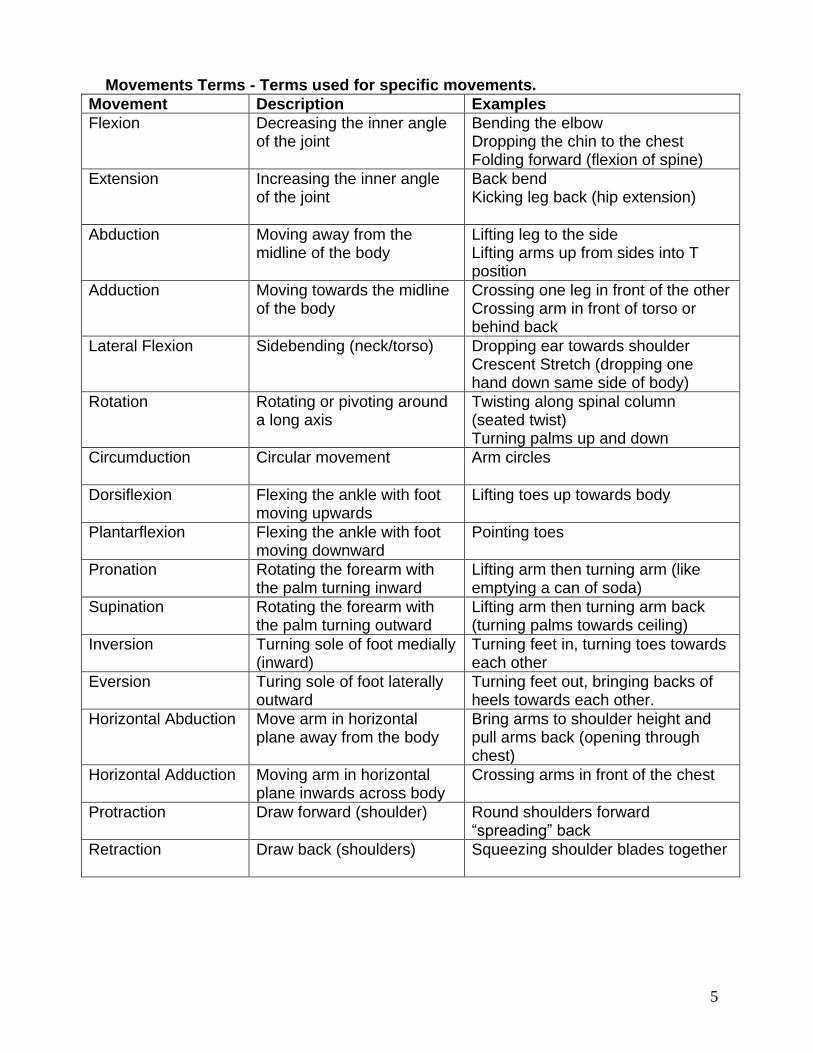
Movements Terms - Terms used for specific movements.
Movement Description Examples
Flexion Decreasing the inner angle of the joint
Bending the elbow Dropping the chin to the chest Folding forward (flexion of spine)
Extension Increasing the inner angle of the joint
Back bend Kicking leg back (hip extension)
Abduction Moving away from the midline of the body
Lifting leg to the side Lifting arms up from sides into T position
Adduction Moving towards the midline of the body
Crossing one leg in front of the other Crossing arm in front of torso or behind back
Lateral Flexion Sidebending (neck/torso) Dropping ear towards shoulder Crescent Stretch (dropping one hand down same side of body)
Rotation Rotating or pivoting around a long axis
Twisting along spinal column (seated twist) Turning palms up and down
Circumduction Circular movement Arm circles
Dorsiflexion Flexing the ankle with foot moving upwards
Lifting toes up towards body
Plantarflexion Flexing the ankle with foot moving downward
Pointing toes
Pronation Rotating the forearm with the palm turning inward
Lifting arm then turning arm (like emptying a can of soda)
Supination Rotating the forearm with the palm turning outward
Lifting arm then turning arm back (turning palms towards ceiling)
Inversion Turning sole of foot medially (inward)
Turning feet in, turning toes towards each other
Eversion Turing sole of foot laterally outward
Turning feet out, bringing backs of heels towards each other.
Horizontal Abduction Move arm in horizontal plane away from the body
Bring arms to shoulder height and pull arms back (opening through chest)
Horizontal Adduction Moving arm in horizontal plane inwards across body
Crossing arms in front of the chest
Protraction Draw forward (shoulder) Round shoulders forward “spreading” back
Retraction Draw back (shoulders) Squeezing shoulder blades together

6
Bones, Joints and Ligaments Bones form the structural framework for the body. They are comprised of calcium salts, connective tissue, cells and blood vessels. Bones also serve as levers that are acted upon by muscles, and come in varied shapes and sizes. The shape of a bone reflects its function. Long bones are found in the limbs, where they act as levers for support and locomotion. Short bones function for strength and compactness. Flat bones have a protective function (skull) or provide broad surfaces for muscular attachment (shoulder blades). Regular yoga practice strengthens your bones because it provides weight bearing exercise in a variety of directions. Major Bones in the Body
7
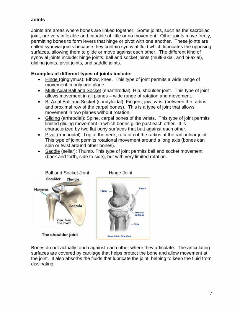
Joints Joints are areas where bones are linked together. Some joints, such as the sacroiliac joint, are very inflexible and capable of little or no movement. Other joints move freely, permitting bones to form levers that hinge or pivot with one another. These joints are called synovial joints because they contain synovial fluid which lubricates the opposing surfaces, allowing them to glide or move against each other. The different kind of synovial joints include: hinge joints, ball and socket joints (multi-axial, and bi-axial), gliding joints, pivot joints, and saddle joints. Examples of different types of joints include:
Hinge (ginglymus): Elbow, knee. This type of joint permits a wide range of movement in only one plane.
Multi-Axial Ball and Socket (enarthrodial): Hip, shoulder joint. This type of joint allows movement in all planes – wide range of rotation and movement.
Bi-Axial Ball and Socket (condyloidal): Fingers, jaw, wrist (between the radius and proximal row of the carpal bones). This is a type of joint that allows movement in two planes without rotation.
Gliding (arthrodial): Spine, carpal bones of the wrists. This type of joint permits limited gliding movement in which bones glide past each other. It is characterized by two flat bony surfaces that butt against each other.
Pivot (trochoidal): Top of the neck, rotation of the radius at the radioulnar joint. This type of joint permits rotational movement around a long axis (bones can spin or twist around other bones).
Saddle (sellar): Thumb. This type of joint permits ball and socket movement (back and forth, side to side), but with very limited rotation. Ball and Socket Joint Hinge Joint
Bones do not actually touch against each other where they articulate. The articulating surfaces are covered by cartilage that helps protect the bone and allow movement at the joint. It also absorbs the fluids that lubricate the joint, helping to keep the fluid from dissipating.

8
Joints are bound together by ligaments. All primary joints are firmly bound together by ligaments that connect bone to bone. Torn ligaments result from undue stress on joints, with knee and ankle injuries being the most common. Muscles are attached to bones and cartilage by tendons. By contracting, muscles produce movement. So bones function as levers, and muscles as motors that move the levers. Fascia are tendinous fibers that connect the skin and underlying structures to the muscles. The words “origin” and “insertion” indicate where muscles are attached to bones in relation to the most common movement at a joint. The origin of a muscle is on the bone that is usually relatively stationary, and the insertion of the muscle is on the bone that is most often moved. For example, in flexion of the elbow, it is the forearm (not the upper arm) that is usually most moved. So, the biceps brachii and the triceps brachii take origin from the upper arm and shoulder, and insert on the forearm.
9
Muscle Forms Muscles have different forms and fiber arrangements, depending on their function. Muscles in the limbs tend to be long. Because of this, they can contract more and are capable of producing greater movement. Muscles in the trunk tend to be broader and to form sheets that wrap around the body. Muscles that stabilize parts of the body tend to be short and squat, like those found in the hip. Muscles are also defined by the number of joints they cross; from their origin to their insertion. Monoarticular muscles cross only one joint, while polyarticular cross more than one joint (for example hamstrings). Types of Muscle Contractions Muscles are composed of bundles of fibers held together by very thin membranes. Within these fibers are thousands of tiny filaments, which slide along each other when the muscle is stimulated by a nerve. This causes the muscle to shorten or contract. Muscles that produce a specific movement are called agonists, while the muscles that produce the opposite movement are called antagonists. When we think of a muscle contracting, we tend to think of the muscle shortening as it generates force. While this is one way that muscles contract, there are also other forms of muscle contraction. Concentric Contractions In this form of contraction, a muscle shortens in length while contacting. An example is when the biceps brachii muscle in the forearm contracts to lift a book off a table and bring it in close to you to read, or when you perform a bicep curl with a free weight. Eccentric Contractions When you slowly extend your elbow to put a book you were reading back on a table, you are lengthening the muscle (biceps brachii) while keeping some of its muscle fibers in a state of contraction. Whenever this happens, we call this movement eccentric lengthening; increasing muscle length against resistance or gravity. Isometric Contraction In isometric contraction, muscles are active while held at a fixed length. The muscle is neither lengthened or shortened, but is held at a constant length. An example of isometric contraction would be carrying an object in front of you. The weight of the object would be pulling down, but your hands and arms would be opposing the motion with equal force going upwards. Since your arms are neither raising or lowering, your biceps will be isometrically contracting.
10
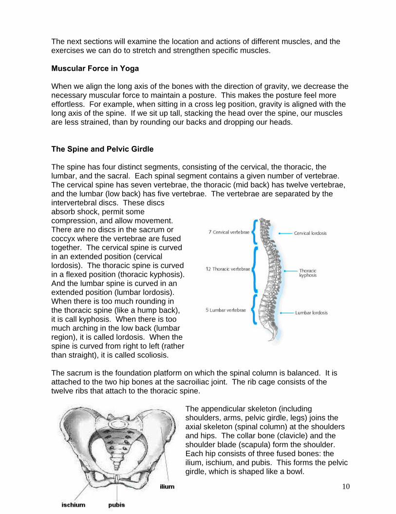
The next sections will examine the location and actions of different muscles, and the exercises we can do to stretch and strengthen specific muscles. Muscular Force in Yoga When we align the long axis of the bones with the direction of gravity, we decrease the necessary muscular force to maintain a posture. This makes the posture feel more effortless. For example, when sitting in a cross leg position, gravity is aligned with the long axis of the spine. If we sit up tall, stacking the head over the spine, our muscles are less strained, than by rounding our backs and dropping our heads. The Spine and Pelvic Girdle The spine has four distinct segments, consisting of the cervical, the thoracic, the lumbar, and the sacral. Each spinal segment contains a given number of vertebrae. The cervical spine has seven vertebrae, the thoracic (mid back) has twelve vertebrae, and the lumbar (low back) has five vertebrae. The vertebrae are separated by the intervertebral discs. These discs absorb shock, permit some compression, and allow movement. There are no discs in the sacrum or coccyx where the vertebrae are fused together. The cervical spine is curved in an extended position (cervical lordosis). The thoracic spine is curved in a flexed position (thoracic kyphosis). And the lumbar spine is curved in an extended position (lumbar lordosis). When there is too much rounding in the thoracic spine (like a hump back), it is call kyphosis. When there is too much arching in the low back (lumbar region), it is called lordosis. When the spine is curved from right to left (rather than straight), it is called scoliosis. The sacrum is the foundation platform on which the spinal column is balanced. It is attached to the two hip bones at the sacroiliac joint. The rib cage consists of the twelve ribs that attach to the thoracic spine.
The appendicular skeleton (including shoulders, arms, pelvic girdle, legs) joins the axial skeleton (spinal column) at the shoulders and hips. The collar bone (clavicle) and the shoulder blade (scapula) form the shoulder. Each hip consists of three fused bones: the ilium, ischium, and pubis. This forms the pelvic girdle, which is shaped like a bowl.

11
Biomechanics of Movement Forward bends Forward bends stretch and strengthen the back portion of the spine, pelvic girdle, shoulders and legs. They also strengthen the abdominal muscles, which contract as we bend forward, and gently compress abdominal organs, which stimulates their function. Proper Technique in Bending Forward When folding forward, it’s best to maintain a “flat” back, neither arched nor rounded, with the neck in line with the rest of the spine. So, as you bend forward you maintain the normal curvature of the spine. It’s important to press the hips back and hinge from the hips, instead of rounding the back, when folding forward. If hamstrings are tight, it is best to bend the knees, so the back can remain flat. Rounding the back due to tight hamstrings can lead to a backwards rotation of the pelvis and a collapsing of the chest over the belly. This can result in intervertebral disc compression in the anterior (front) of the spine. Low back pain is often a result of poor mechanical relationship between the lumbar spine and the pelvis. Although many chronic back conditions can occur due to improper forward bending, forward bending done properly can also help us strengthen and stabilize our back and body. Obstacles to forward bends result from tightness in the hamstrings, spinal muscles and gluteals. Back Bends Backward bending helps to stretch the front portion of the torso, shoulders, pelvic girdle and legs. In addition, they stretch the abdominal organs, relieving compression. Backbends also help develop more strength in the muscles in the back, which must contract during back bends. Proper Technique in Bending Backwards It’s important in backbends to control the proportional relation between lengthening the thoracic curve and deepening the lumbar curve. You don’t want too much arch in the lumbar spine without any movement in the thoracic spine. Bending that way can cause compression and strain in the lumbar region. Like the lumbar region, the cervical region should not arch excessively in relation to the movement of the thoracic region. One way to maintain a balance between the movement of the thoracic region and the lumbar region is to first expand and lift the chest on inhale (which lengthens the thoracic spine), and then keep the abdominal muscles slightly contracted as you exhale and bend backwards. Keeping the abdominal muscles slightly contracted helps prevent excessive arch in the lumbar region and helps to bring more of the arch into the thoracic region, which keeps the backbend in balance. Students should also be encouraged to keep the legs straight (or slightly internally rotated) to keep the sacroiliac joints stable.

12
Twists Twisting creates a rotation between the vertebrae, which builds strength and flexibility in the deep and superficial muscles of the spine and abdomen. Twisting alternating stretches and strengthens each side of the torso, including the intestines, which may help improve digestion. Proper Technique in Twisting: It’s important in twisting to control the spinal rotation, rather than simply force it through the use of leverage. The key to spinal rotation is to start the twist as you exhale and contract the abdominal muscles. As with forward bends, there can be a tendency to slump forward in the thoracic region of the spine. This can be avoided by lengthening between the chest and belly on inhalation. Lengthening the spine helps create more space between each vertebrae in which to twist. In standing twisting postures (revolved triangle), the pelvis is stabilized to emphasize the rotation of the spine and shoulders. Because the shoulder girdle has more range of motion than the spine, it is best to start a twist without using the arms as leverage, and add the arms at the end. Lateral Bends Lateral bends alternately stretch and compress the deep spinal muscles, intervertebral discs, and intercostal muscles of the ribs. They stretch and strengthen the muscles of the spine, rib cage, shoulders, and pelvis. They also help restore balance to asymmetries of the spine. The capacity for lateral flexion of the spine is limited, so it is often not done during daily activities. Because of this, it is an important movement to add to a yoga practice. Proper Technique in lateral bends When practicing a lateral bend, people often turn their hips and rotate their chest towards the side they are bending. For example, in Triangle posture, as students slide their hand down their right leg, their chest often turns towards the floor and their hip moves to the right. This causes them to lose the lateral stretch, as it moves towards a forward bending position. In Triangle, as with other postures requiring lateral flexion, the shoulders should stack one on top of the other and the chest should remain open facing forward. One way to make sure that the shoulders remain stacked is to practice Triangle posture with your back next to the wall, keeping both shoulder blades pressed into the wall. To help with lateral bending, again lengthen the spine on the inhale, and then move into the lateral bend on the exhale, keeping the abdominal muscles contracted. Obstacles to lateral bends include tightness in the shoulder joints or latissimus dorsi.
13
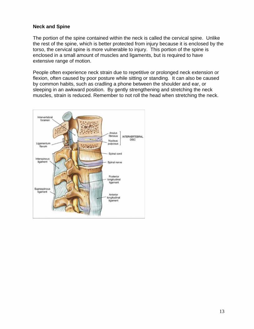
Neck and Spine The portion of the spine contained within the neck is called the cervical spine. Unlike the rest of the spine, which is better protected from injury because it is enclosed by the torso, the cervical spine is more vulnerable to injury. This portion of the spine is enclosed in a small amount of muscles and ligaments, but is required to have extensive range of motion. People often experience neck strain due to repetitive or prolonged neck extension or flexion, often caused by poor posture while sitting or standing. It can also be caused by common habits, such as cradling a phone between the shoulder and ear, or sleeping in an awkward position. By gently strengthening and stretching the neck muscles, strain is reduced. Remember to not roll the head when stretching the neck.
14
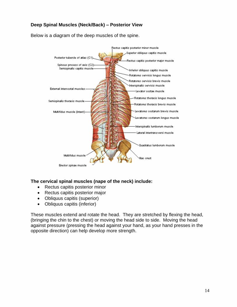
Deep Spinal Muscles (Neck/Back) – Posterior View Below is a diagram of the deep muscles of the spine.
The cervical spinal muscles (nape of the neck) include:
Rectus capitis posterior minor
Rectus capitis posterior major
Obliquus capitis (superior)
Obliquus capitis (inferior) These muscles extend and rotate the head. They are stretched by flexing the head, (bringing the chin to the chest) or moving the head side to side. Moving the head against pressure (pressing the head against your hand, as your hand presses in the opposite direction) can help develop more strength.
15
Erector Spinae Muscles The erector spinae are the long muscles that run parallel to the spine. The erector spinae are composed of the following muscles:
Muscles Action To Strengthen To Stretch
Iliocostalis (lumbar, thoracic, cervicis)
Extension and lateral flexion (side-bend) of spine
Back extension in prone position (locust, bow, cobra), moving against gravity.
Flexion of the spine (forward fold), side bend (crescent stretch opposite side, gate), neck stretches
Longissimus (thoracis, cervicis, capitis)
Extension and lateral flexion of spine and rotate head
Back extension in prone position (locust, bow), moving against gravity.
Flexion of the spine (seated forward fold), side bend (crescent stretch opposite side), neck stretches.
Splenius (cervicis, capitis)
Extend and rotate head
Head extension – head back (sun worshipper, neck stretches-turn head)
Head flexion (drop chin to chest) and turn head side to side
These muscles are responsible for intevertebral movements, including extension, sidebending and rotation Yoga movements to strengthen these muscles:
Standing Backbend
Camel
Sun Worshipper
Crescent Stretch
Locust
Bow
Cobra
Seated Twist Yoga movements to stretch these muscles:
Neck Stretches (chin to chest)
Seated Forward Fold
Child’s Pose
Seated Twist (stretches opposite side of twist)
Plow
Rabbit
Crescent Stretch (opposite side) Note – In standing forward fold, if you bend from the waist, you will contract only abdominal muscles as you fold forward. If you bend from the hips, you will contract (use/strengthen) both your adominal muscles and your erector spinae muscles.

16
Muscles of Torso/Shoulder – Posterior View (Back)
Chart of Muscles of Shoulder Girdle/ Scapular (Shoulder Blade) Stabilization (Posterior) – How to Strengthen/Stretch
Muscle Action To Strengthen To Stretch
Shoulder Blade
Levator Scapula Elevates (raises) scapula, rotates and side bends head
Rotate neck, keep head raised against gravity (dropping ear to shoulder, triangle look to raised hand), Elevating scapula against gravity (standing backbend – with arms overhead)
Rotate head to and flex cervical spine (neck stretch – bringing chin towards armpit or drop ear to shoulder, child’s pose, rabbit)
Rhomboid (major and minor)
Adduction of scapula, draw scapula down
Abduct shoulder, squeeze shoulder blades (camel, locust, chest expander)
Protract scapula while keeping shoulders down (child’s pose, rabbit, plow, thread the needle)
Trapezeus (upper, lower, middle)
Elevation and adduction of scapula. Upper fibers extend head
Abduct arm and shoulder, squeeze shoulder blades together (camel,locust chest expander)
Flex neck, protract scapula (thread the needle, rabbit, shoulder stretch)
Serratus Anterior
Protraction and upward rotation of scapula
Push ups (yoga push ups, eagle)
Retract scapula (serpent stretch, fish, frog)

17
Movers of the Shoulder Joint (Posterior)
Shoulder Mover Muscles
Action To Strengthen To Stretch
Deltoid (anterior, lateral, posterior)
Abduct arm, anterior also draws arm forward, posterior also draws arm back
Abducting arms to shoulder height – arms in T position (warrior 2, warrior 3, chair)
Adduction – crossing arm across torso (thread the needle, shoulder stretches) or extension (chest expander)
Teres Major Extension, internal rotation and adduction of shoulder joint
Internal rotation against resistance (eagle, swaying palm tree)
External rotation of shoulder in 90 degree abducted position (tree pose with arms out to sides and palms turned up)
Latissimus Dorsi Adduction, extension, internal rotation and horizontal abduction of shoulder joint
Exercises in which arms are pulled down (swimming dolphin)
External rotation of shoulder in 90 degree abducted position (tree pose with arms out to sides and palms turned up)
18
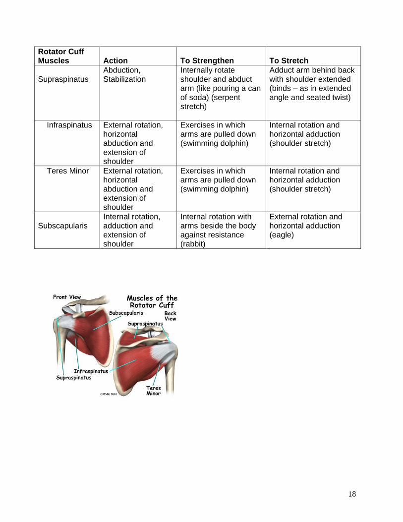
Rotator Cuff Muscles
Action
To Strengthen
To Stretch
Supraspinatus
Abduction, Stabilization
Internally rotate shoulder and abduct arm (like pouring a can of soda) (serpent stretch)
Adduct arm behind back with shoulder extended (binds – as in extended angle and seated twist)
Infraspinatus External rotation, horizontal abduction and extension of shoulder
Exercises in which arms are pulled down (swimming dolphin)
Internal rotation and horizontal adduction (shoulder stretch)
Teres Minor External rotation, horizontal abduction and extension of shoulder
Exercises in which arms are pulled down (swimming dolphin)
Internal rotation and horizontal adduction (shoulder stretch)
Subscapularis
Internal rotation, adduction and extension of shoulder
Internal rotation with arms beside the body against resistance (rabbit)
External rotation and horizontal adduction (eagle)
19
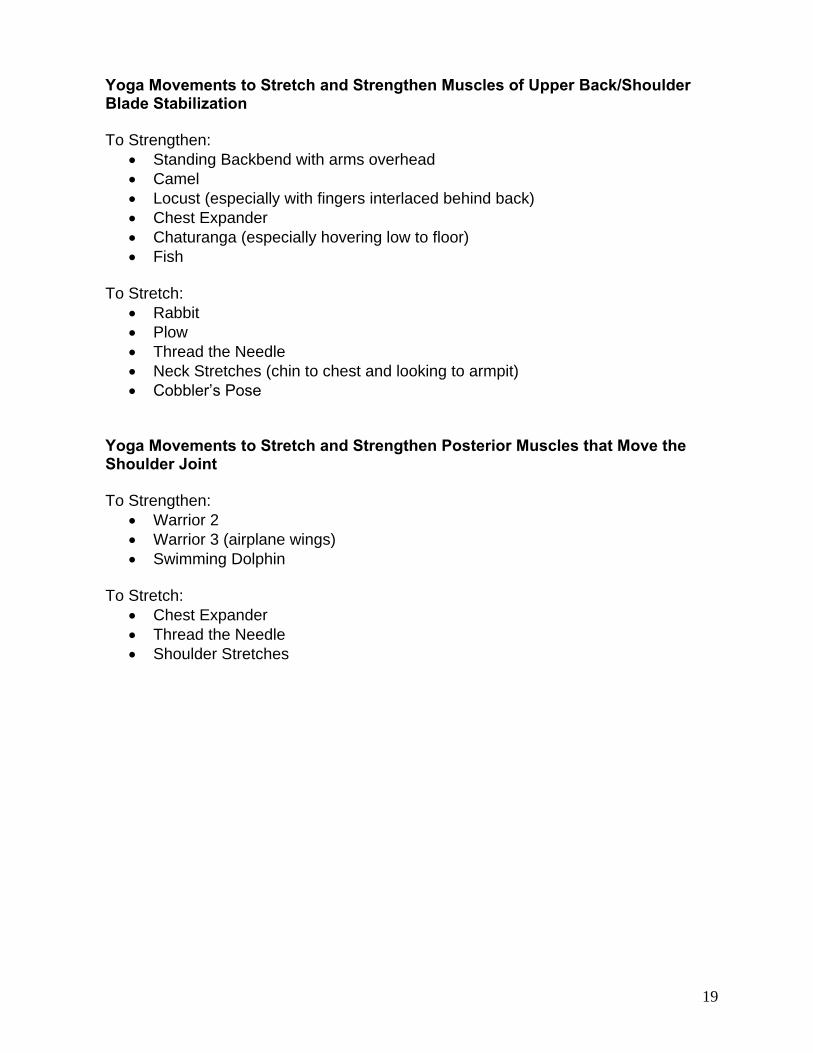
Yoga Movements to Stretch and Strengthen Muscles of Upper Back/Shoulder Blade Stabilization To Strengthen:
Standing Backbend with arms overhead
Camel
Locust (especially with fingers interlaced behind back)
Chest Expander
Chaturanga (especially hovering low to floor)
Fish To Stretch:
Rabbit
Plow
Thread the Needle
Neck Stretches (chin to chest and looking to armpit)
Cobbler’s Pose Yoga Movements to Stretch and Strengthen Posterior Muscles that Move the Shoulder Joint To Strengthen:
Warrior 2
Warrior 3 (airplane wings)
Swimming Dolphin To Stretch:
Chest Expander
Thread the Needle
Shoulder Stretches

20
Muscles of the Torso/Chest/Shoulder – Anterior View
Chart of Muscles of Torso/ Chest (Anterior) – How to Strengthen/Stretch
Muscles Action To Strengthen To Stretch
Sternocleido-mastoid
Flex and rotate head, raise ribs
Flex head (bring chin to chest), rotate head
Extend head (drop head back), rotate head
Pectoralis Major Internal rotation of arm, horizontal adduction, and adduction
Push ups (chaturanga, yoga push up)
Externally rotating shoulder with arm adducted behind back (chest expander), horizontal abduction of shoulder (prone twist)
Obliques (external, internal, transverse)
Rotate, flex and side bend trunk
Rotate trunk while flexing hips, knees flexed (leg pumps - (bring elbow to opposite leg, lunge with twist, chair with twist)
Laterally flex the opposite side while rotating lumbar region (triangle, gate, supine twist)
Rectus Abdominus Flex trunk (forward bend)
Flex hip with knees flexed (leg pumps, plank - with knee to chest, knee to chest standing balance, boat, marichyasana)
Extend lumbar and thoracic spine, and extend hips to accentuate the rotation of the pelvis (bow, upward bow, standing backbend, camel)

21
Yoga Movements to Stretch and Strengthen Muscles of the Chest and Front Torso To Strengthen:
Rotate head, bring chin to chest
Lunge with twist
Chair with twist
Bringing opposite elbow to opposite knee
Boat
Plank with knee to chest To Stretch:
Triangle
Supine (lying on back) twist
Bow
Upward Bow/Wheel
Camel
Chest Expander
Prone Twist
22
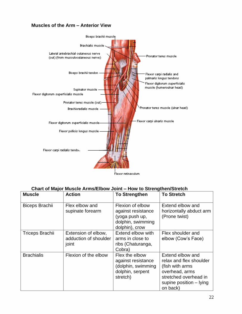
Muscles of the Arm – Anterior View
Chart of Major Muscle Arms/Elbow Joint – How to Strengthen/Stretch
Muscle Action To Strengthen To Stretch
Biceps Brachii Flex elbow and supinate forearm
Flexion of elbow against resistance (yoga push up, dolphin, swimming dolphin), crow
Extend elbow and horizontally abduct arm (Prone twist)
Triceps Brachii Extension of elbow, adduction of shoulder joint
Extend elbow with arms in close to ribs (Chaturanga, Cobra)
Flex shoulder and elbow (Cow’s Face)
Brachialis Flexion of the elbow Flex the elbow against resistance (dolphin, swimming dolphin, serpent stretch)
Extend elbow and relax and flex shoulder (fish with arms overhead, arms stretched overhead in supine position – lying on back)
23
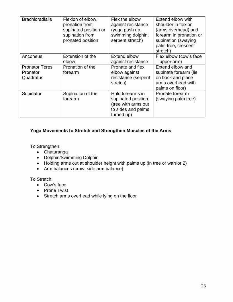
Brachioradialis Flexion of elbow, pronation from supinated position or supination from pronated position
Flex the elbow against resistance (yoga push up, swimming dolphin, serpent stretch)
Extend elbow with shoulder in flexion (arms overhead) and forearm in pronation or supination (swaying palm tree, crescent stretch)
Anconeus Extension of the elbow
Extend elbow against resistance
Flex elbow (cow’s face – upper arm)
Pronator Teres Pronator Quadratus
Pronation of the forearm
Pronate and flex elbow against resistance (serpent stretch)
Extend elbow and supinate forearm (lie on back and place arms overhead with palms on floor)
Supinator Supination of the forearm
Hold forearms in supinated position (tree with arms out to sides and palms turned up)
Pronate forearm (swaying palm tree)
Yoga Movements to Stretch and Strengthen Muscles of the Arms To Strengthen:
Chaturanga
Dolphin/Swimming Dolphin
Holding arms out at shoulder height with palms up (in tree or warrior 2)
Arm balances (crow, side arm balance) To Stretch:
Cow’s face
Prone Twist
Stretch arms overhead while lying on the floor
24
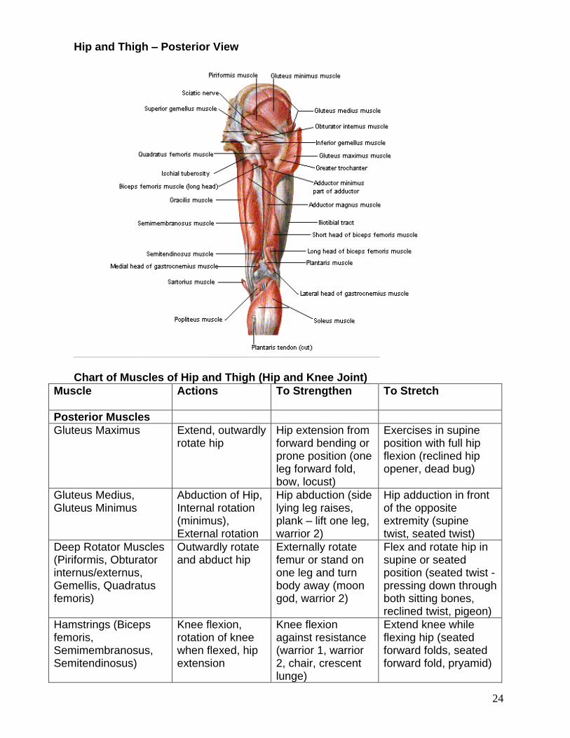
Hip and Thigh – Posterior View
Chart of Muscles of Hip and Thigh (Hip and Knee Joint)
Muscle Actions To Strengthen To Stretch
Posterior Muscles
Gluteus Maximus Extend, outwardly rotate hip
Hip extension from forward bending or prone position (one leg forward fold, bow, locust)
Exercises in supine position with full hip flexion (reclined hip opener, dead bug)
Gluteus Medius, Gluteus Minimus
Abduction of Hip, Internal rotation (minimus), External rotation
Hip abduction (side lying leg raises, plank – lift one leg, warrior 2)
Hip adduction in front of the opposite extremity (supine twist, seated twist)
Deep Rotator Muscles (Piriformis, Obturator internus/externus, Gemellis, Quadratus femoris)
Outwardly rotate and abduct hip
Externally rotate femur or stand on one leg and turn body away (moon god, warrior 2)
Flex and rotate hip in supine or seated position (seated twist - pressing down through both sitting bones, reclined twist, pigeon)
Hamstrings (Biceps femoris, Semimembranosus, Semitendinosus)
Knee flexion, rotation of knee when flexed, hip extension
Knee flexion against resistance (warrior 1, warrior 2, chair, crescent lunge)
Extend knee while flexing hip (seated forward folds, seated forward fold, pryamid)
25
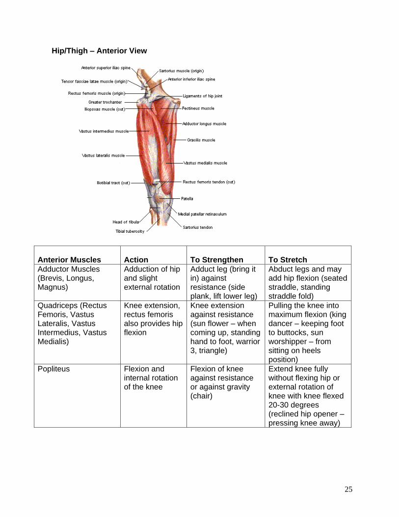
Hip/Thigh – Anterior View
Anterior Muscles
Action
To Strengthen
To Stretch
Adductor Muscles (Brevis, Longus, Magnus)
Adduction of hip and slight external rotation
Adduct leg (bring it in) against resistance (side plank, lift lower leg)
Abduct legs and may add hip flexion (seated straddle, standing straddle fold)
Quadriceps (Rectus Femoris, Vastus Lateralis, Vastus Intermedius, Vastus Medialis)
Knee extension, rectus femoris also provides hip flexion
Knee extension against resistance (sun flower – when coming up, standing hand to foot, warrior 3, triangle)
Pulling the knee into maximum flexion (king dancer – keeping foot to buttocks, sun worshipper – from sitting on heels position)
Popliteus Flexion and internal rotation of the knee
Flexion of knee against resistance or against gravity (chair)
Extend knee fully without flexing hip or external rotation of knee with knee flexed 20-30 degrees (reclined hip opener – pressing knee away)
26
Yoga Movements to Stretch and Strengthen the Hip and Back of Thigh To Strengthen:
One leg standing forward fold (back of lifted leg)
Half Moon
Locust
Bow
Crescent Lunge (lunge with back knee bent, but off the floor) To Stretch:
Pigeon
Reclined Twist
Seated forward fold
Pyramid
Reclined hip opener
Happy baby Yoga Movements to Stretch and Strengthen the Front of Thigh To Strengthen:
Chair
Warrior 3
Triangle (front leg)
Warrior 1 (front leg)
Boat To Stretch:
Reclined Hero
Windshield wipers
Camel
Pigeon (sitting up, bending back knee, holding back foot)
Bow
27
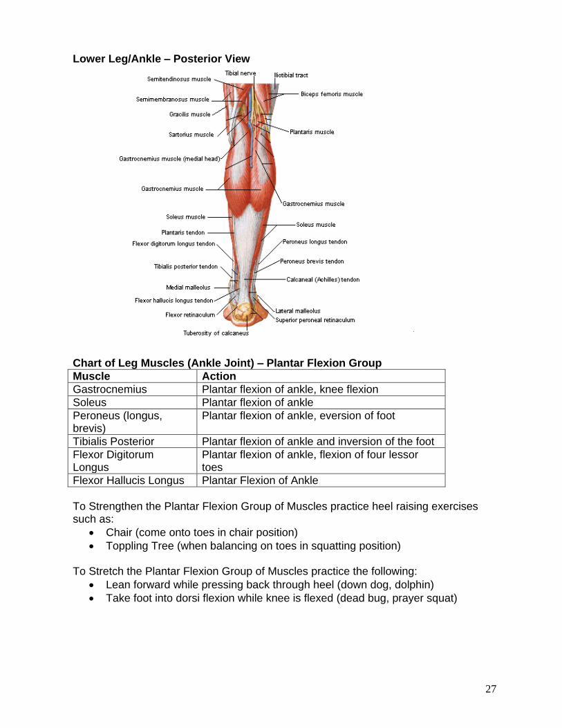
Lower Leg/Ankle – Posterior View
Chart of Leg Muscles (Ankle Joint) – Plantar Flexion Group
Muscle Action
Gastrocnemius Plantar flexion of ankle, knee flexion
Soleus Plantar flexion of ankle
Peroneus (longus, brevis)
Plantar flexion of ankle, eversion of foot
Tibialis Posterior Plantar flexion of ankle and inversion of the foot
Flexor Digitorum Longus
Plantar flexion of ankle, flexion of four lessor toes
Flexor Hallucis Longus Plantar Flexion of Ankle
To Strengthen the Plantar Flexion Group of Muscles practice heel raising exercises such as:
Chair (come onto toes in chair position)
Toppling Tree (when balancing on toes in squatting position) To Stretch the Plantar Flexion Group of Muscles practice the following:
Lean forward while pressing back through heel (down dog, dolphin)
Take foot into dorsi flexion while knee is flexed (dead bug, prayer squat)
28
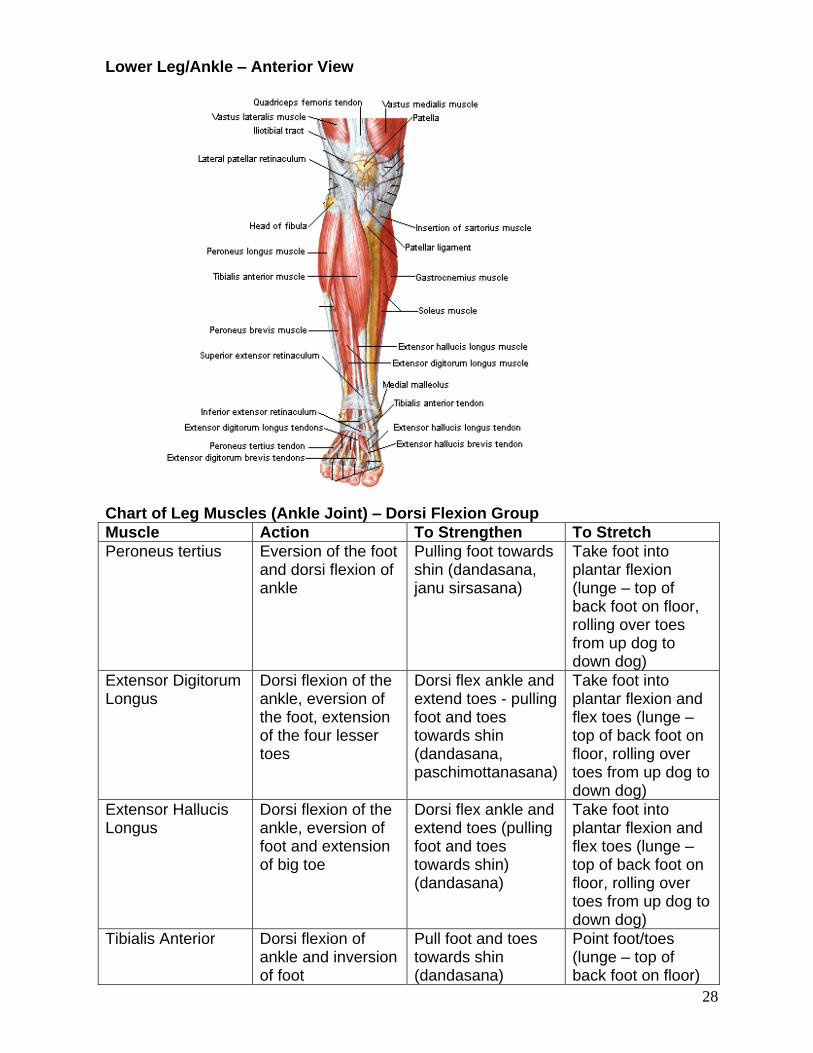
Lower Leg/Ankle – Anterior View
Chart of Leg Muscles (Ankle Joint) – Dorsi Flexion Group
Muscle Action To Strengthen To Stretch
Peroneus tertius Eversion of the foot and dorsi flexion of ankle
Pulling foot towards shin (dandasana, janu sirsasana)
Take foot into plantar flexion (lunge – top of back foot on floor, rolling over toes from up dog to down dog)
Extensor Digitorum Longus
Dorsi flexion of the ankle, eversion of the foot, extension of the four lesser toes
Dorsi flex ankle and extend toes - pulling foot and toes towards shin (dandasana, paschimottanasana)
Take foot into plantar flexion and flex toes (lunge – top of back foot on floor, rolling over toes from up dog to down dog)
Extensor Hallucis Longus
Dorsi flexion of the ankle, eversion of foot and extension of big toe
Dorsi flex ankle and extend toes (pulling foot and toes towards shin) (dandasana)
Take foot into plantar flexion and flex toes (lunge – top of back foot on floor, rolling over toes from up dog to down dog)
Tibialis Anterior Dorsi flexion of ankle and inversion of foot
Pull foot and toes towards shin (dandasana)
Point foot/toes (lunge – top of back foot on floor)
29
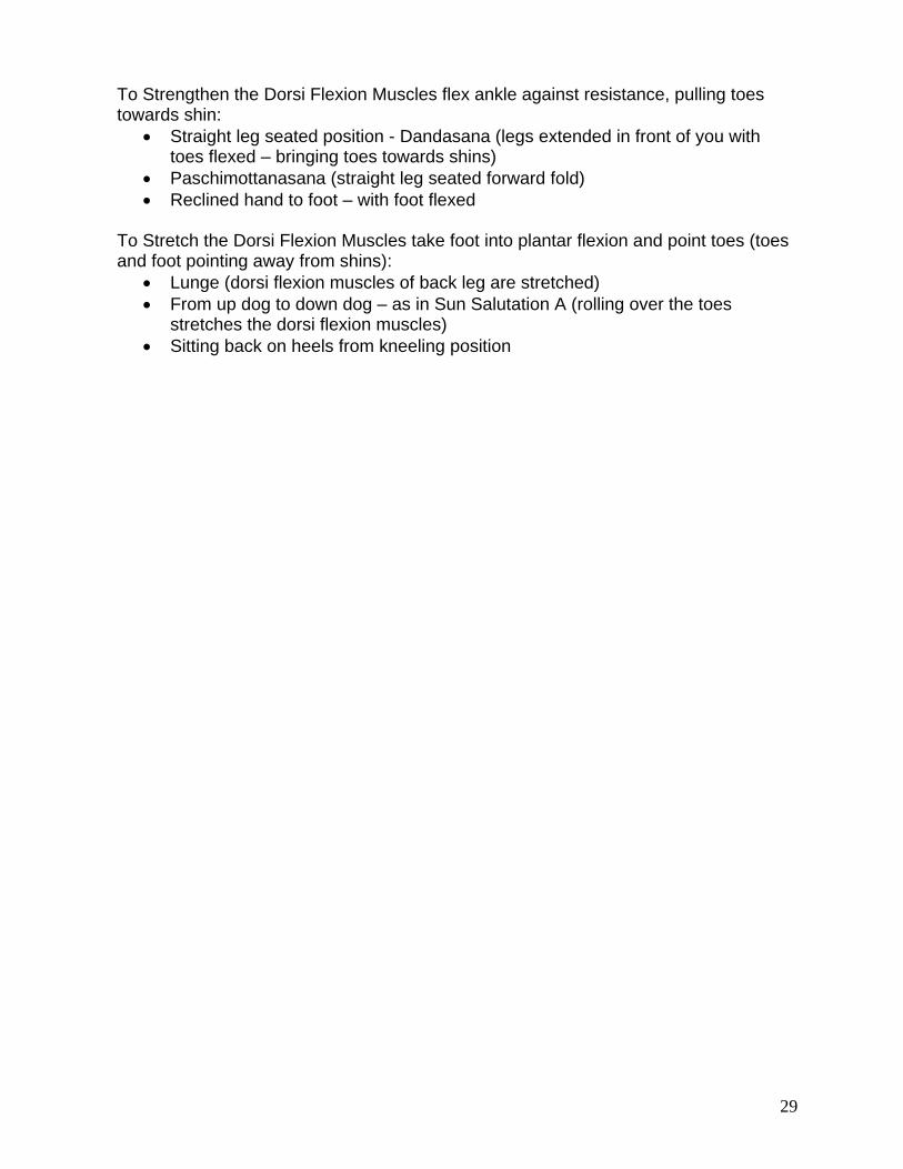
To Strengthen the Dorsi Flexion Muscles flex ankle against resistance, pulling toes towards shin:
Straight leg seated position - Dandasana (legs extended in front of you with toes flexed – bringing toes towards shins)
Paschimottanasana (straight leg seated forward fold)
Reclined hand to foot – with foot flexed To Stretch the Dorsi Flexion Muscles take foot into plantar flexion and point toes (toes and foot pointing away from shins):
Lunge (dorsi flexion muscles of back leg are stretched)
From up dog to down dog – as in Sun Salutation A (rolling over the toes stretches the dorsi flexion muscles)
Sitting back on heels from kneeling position

30
Major Muscles of the Body

31
Developing a Well-Rounded Yoga Practice When developing a yoga practice, it’s important to include exercises and postures that strengthen and stretch muscles within all the major body parts, including:
Neck
Upper Back/ Shoulder Girdle (Scapular Stabilizing Muscles)
Chest/Shoulder Joints (Anterior)
Spine/Erector Spinae (including flexion, extension, lateral flexion and rotation)
Arms/Wrists
Hips/Back of Thighs
Front of Thighs
Lower Legs/Feet/Ankles Design a warm-up routine that would warm up the major muscles of the body parts listed above.
32
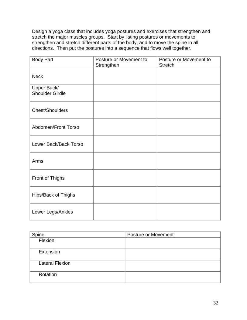
Design a yoga class that includes yoga postures and exercises that strengthen and stretch the major muscles groups. Start by listing postures or movements to strengthen and stretch different parts of the body, and to move the spine in all directions. Then put the postures into a sequence that flows well together.
Body Part Posture or Movement to Strengthen
Posture or Movement to Stretch
Neck
Upper Back/ Shoulder Girdle
Chest/Shoulders
Abdomen/Front Torso
Lower Back/Back Torso
Arms
Front of Thighs
Hips/Back of Thighs
Lower Legs/Ankles
Spine Posture or Movement
Flexion
Extension
Lateral Flexion
Rotation

33
Re-order the postures you listed on the last page to create a practice sequence from those postures:

34
For Further Reading Calais-Germain, Blandine (1993). Anatomy of Movement. Eastland Press. Seattle, Washington. Calais-Germain, B. & Lamotte, A. (1996). Anatomy of Movement Exercises. Eastland Press. Seattle, Washington. Coulter, David (2001). Anatomy of Hatha Yoga. Breath and Body, Inc. Honesdale, PA. Kapit, Wynn & Elson, L. (2002). The Anatomy Coloring Book. Pearson Education. Glenview, IL. Powers, Scott & Howley, E. (2004). Exercise Physiology: Theory and Application to Fitness and Performance, Fifth Edition. McGraw Hill. New York, NY. Robin, Mel (2002). A Physiological Handbook for Teachers of Yogasana. Fenestra Books. Tucson, AZ. Stiles, Mukunda (2003). Structural Yoga Therapy. Samuel Weiser, Inc. York Beach, ME.

35
Review Complete the following chart
Yoga Posture What muscles are strengthened?
What muscles are stretched?
Bow (dhanurasana)
One leg seated forward fold (janu sirsasana)
Tree (vrksasana)
Triangle (trikonasana)
Rabbit