yolk-shell structure of polyaniline coated sulfur for
TRANSCRIPT

S1
Supporting Information
Yolk-Shell Structure of Polyaniline Coated Sulfur for Lithium-
Sulfur Batteries
Weidong Zhou,*,† Yingchao Yu,
† Hao Chen,
† Francis J. DiSalvo
† & Héctor D. Abruña
†,*
† Department of Chemistry and Chemical Biology, Cornell University, Ithaca, New York 14853,
United States
* Corresponding author: [email protected], [email protected]
Methods
Materials Synthesis. Na2S2O3 (2.37 g) in 50 ml water was slowly added into a dilute sulfuric
acid solution (500ml, 3mM) containing 1% (weight ratio) of polyvinylpyrrolidone (PVP,
Mw~40,000). After stirring for 2 hours at room temperature, the sulfur particles were collected
by centrifugation and re-dispersed into 300ml aqueous solution of PVP (1%). 200mg aniline and
10ml sulfuric acid (1M) were added into the above emulsion. 0.5g ammonium persulphate in
30ml water was then added dropwise under a nitrogen flow at 00C. After stirring at 0
0C for
24hours, the polyaniline coated sulfur particles were collected by centrifugation and dried under
vacuum overnight. To prepare the sulfur-polyaniline yolk-shell structures, the powder of the
core-shell particles was sealed into a glass tube filled with argon and heated to 1800C for 12hours.
Electrochemical Measurements. To prepare the cathodes, sulfur based materials were first
mixed with carbon black and water soluble binder sodium alginate (80:15:5 by weight) through
ground in a mortar. The mixture was then spread evenly on the aluminum foil and roll-pressed to
produce electrode films with an average sulfur loading of 2mg cm-2
, which were heated at 500C
S2
for 12 hours under vacuum before using to fabricate the coin cells. 2032 type coin cells were
fabricated in an argon filled glove box using lithium foil as the anode and TFSI (1M in
DOL/DME) containing LiNO3 (1 wt%) as the electrolyte. The sulfur contents of S-Pani core-
shell and yolk-shell in the cathode films were calculated to be 65.6% and 46.4%, respectively.
Material Characterization. Electron microscopy imaging was carried out using a Schottky-
field-emission-gun Tecnai F20 scanning transmission electron microscope (STEM) operated at
200 keV. The energy dispersive x-ray (EDX) analysis was per-formed in the F20 using an
Oxford detector, at a beam current of about 1 nA. An EDX resolution of 1-5 nm is routinely
achieved on this setup. Sulfur was not found to sublime into vacuum within the electron
microscope under the testing conditions, likely due to the core-shell or yolk-shell structure,
which protects sulfur against sublimation.
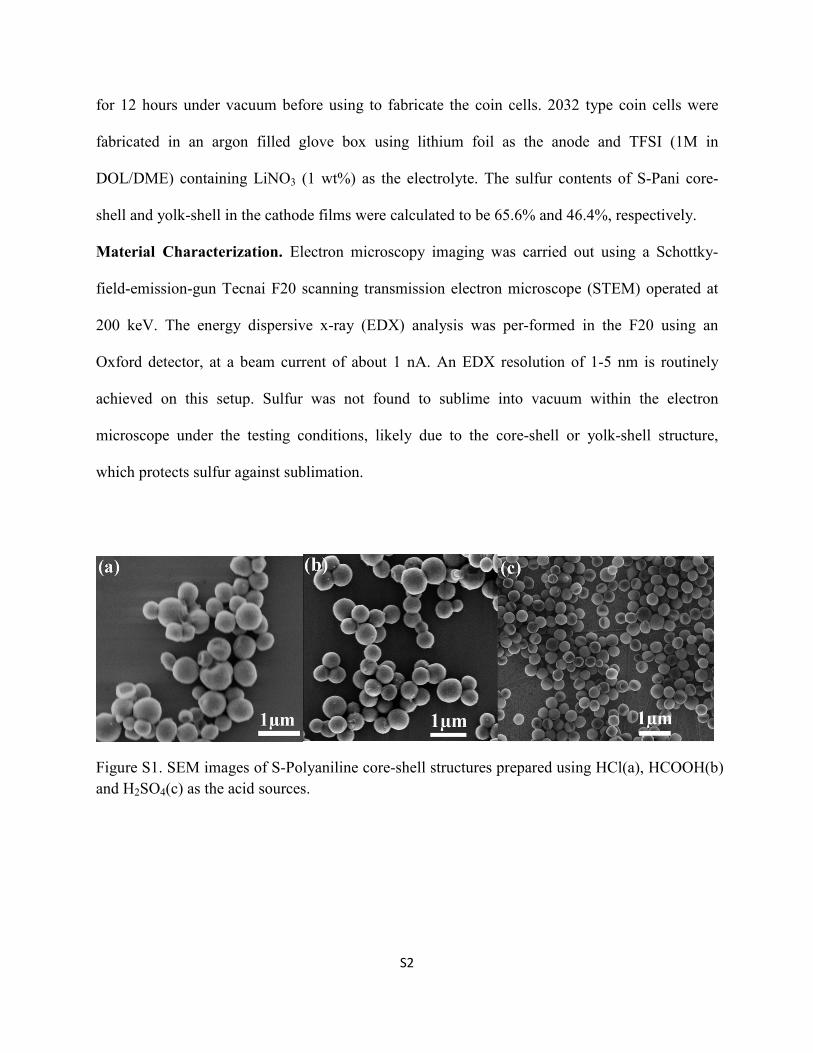
Figure S1. SEM images of S-Polyaniline core-shell structures prepared using HCl(a), HCOOH(b)
and H2SO4(c) as the acid sources.

S3
Figure S2 The magnified SEM image of the S-Pani core-shell structure.
Figure S3 X-ray diffraction (XRD) of S-Pani composite shows it matches PDF card of pure
sulfur(orthorhombic) since polyaniline (PAN) is amorphous. X-Ray Diffraction (XRD) data
were collected using the D8-Advance DaVinci system with the Bragg-Brentano geometry and
Cu Kα radiation (40kV and 40mA). The samples were prepared conservatively on quartz discs.
Scans range from 10°-90° with fixed sample illumination and a 0.04°/s step size and 1s/step rate
with variable rotation at 15rpm.

S4
Figure S4 The thermal gravimetric analysis (TGA) of the S-Pani core-shell composite in argon
flow.
Figure S5 The image of S-Pani core-shell(left) and heat treated yolk-shell(right) composite.
S5
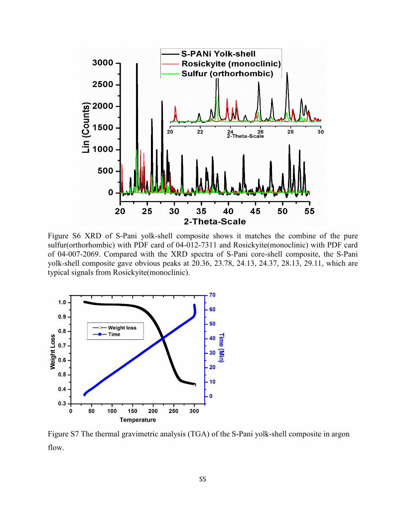
Figure S6 XRD of S-Pani yolk-shell composite shows it matches the combine of the pure
sulfur(orthorhombic) with PDF card of 04-012-7311 and Rosickyite(monoclinic) with PDF card
of 04-007-2069. Compared with the XRD spectra of S-Pani core-shell composite, the S-Pani
yolk-shell composite gave obvious peaks at 20.36, 23.78, 24.13, 24.37, 28.13, 29.11, which are
typical signals from Rosickyite(monoclinic).
Figure S7 The thermal gravimetric analysis (TGA) of the S-Pani yolk-shell composite in argon
flow.