zeiss primo star iled - mikroskop-center · the international journal of tuberculosis and lung...
TRANSCRIPT
Application Note
ZEISS Primo Star iLEDSelected Fluorescence Applications in Laboratories and Education
Application Note
2
ZEISS Primo Star iLEDSelected Fluorescence Applications in Laboratories and Education
Author: Hung Pham Carl Zeiss Microscopy GmbH, Germany
Date: January 2015
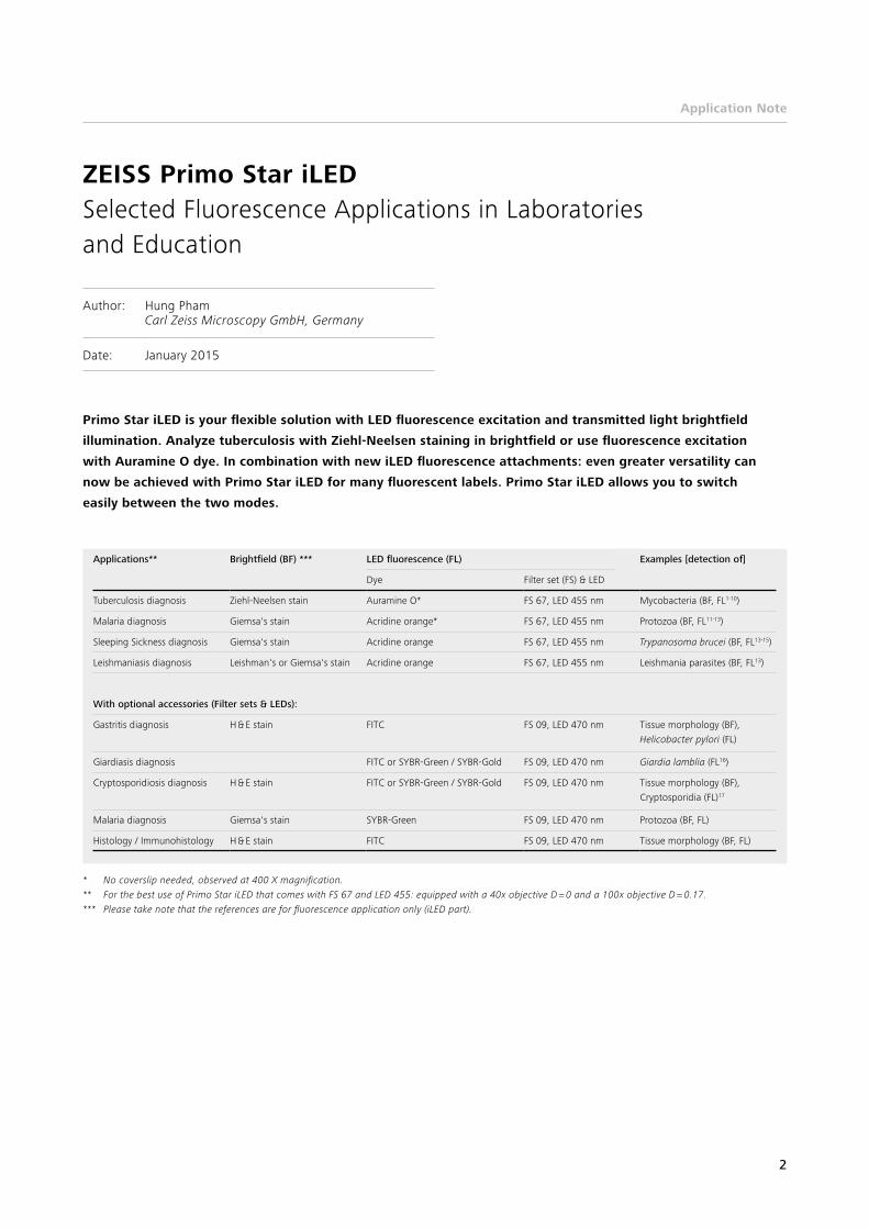
Primo Star iLED is your flexible solution with LED fluorescence excitation and transmitted light brightfield
illumination. Analyze tuberculosis with Ziehl-Neelsen staining in brightfield or use fluorescence excitation
with Auramine O dye. In combination with new iLED fluorescence attachments: even greater versatility can
now be achieved with Primo Star iLED for many fluorescent labels. Primo Star iLED allows you to switch
easily between the two modes.
* No coverslip needed, observed at 400 X magnification.** For the best use of Primo Star iLED that comes with FS 67 and LED 455: equipped with a 40x objective D = 0 and a 100x objective D = 0.17.*** Please take note that the references are for fluorescence application only (iLED part).
Applications** Brightfield (BF) *** LED fluorescence (FL) Examples [detection of]
Dye Filter set (FS) & LED
Tuberculosis diagnosis Ziehl-Neelsen stain Auramine O* FS 67, LED 455 nm Mycobacteria (BF, FL1-10)
Malaria diagnosis Giemsa's stain Acridine orange* FS 67, LED 455 nm Protozoa (BF, FL11-13)
Sleeping Sickness diagnosis Giemsa's stain Acridine orange FS 67, LED 455 nm Trypanosoma brucei (BF, FL13-15)
Leishmaniasis diagnosis Leishman's or Giemsa's stain Acridine orange FS 67, LED 455 nm Leishmania parasites (BF, FL13)
With optional accessories (Filter sets & LEDs):
Gastritis diagnosis H & E stain FITC FS 09, LED 470 nm Tissue morphology (BF), Helicobacter pylori (FL)
Giardiasis diagnosis FITC or SYBR-Green / SYBR-Gold FS 09, LED 470 nm Giardia lamblia (FL16)
Cryptosporidiosis diagnosis H & E stain FITC or SYBR-Green / SYBR-Gold FS 09, LED 470 nm Tissue morphology (BF), Cryptosporidia (FL)17
Malaria diagnosis Giemsa's stain SYBR-Green FS 09, LED 470 nm Protozoa (BF, FL)
Histology / Immunohistology H & E stain FITC FS 09, LED 470 nm Tissue morphology (BF, FL)

Application Note
3
CA B
FD E
IG H
Short notes: A. Tubercle bacilliB. Plasmodium knowlesi C. TrypanosomesD. Leishmania donovani promastigotesE. Helicobacter pyloriF. GiardiaG. CryptosporidiaH. Plasmodium falciparumI. Mouse capillary
Legend:
(A) Tubercle bacilli stained with auramine O (courtesy of CDC). (B) Malaria parasites (small orange structures) inside red blood
cells (green) of a baboon experimentally infected with Plasmodium knowlesi and stained with acridine orange.
White blood cells also stain orange (blood smear courtesy of Dr. Maina Ngotho, Institute of Primate Research, Nairobi).
(C) A thin blood smear stained with acridine orange showing trypanosomes (orange) alongside red blood cells (green).
(D) Cultured Leishmania donovani promastigotes (orange with flagella) stained with acridine orange (slide courtesy of
Dr. Maina Ngotho).13 (E) Helicobacter pylori immuno-labeled with FITC. (F) Giardia from contaminated water immuno-labeled
with FITC (courtesy of Dr. H.P. Fuechslin, Bachema AG). (G) Cryptosporidia from contaminated drinking water immuno-labeled
with FITC (courtesy of Mr. Brian Oram, Wilkes University).17 (H) Plasmodium falciparum stained with SYBR Green I
(courtesy of Dr. West Suhanic11). (I) Mouse capillary immuno-labeled with FITC.
Application examples:

Application Note
4
References:
[1] Turnbull, E.R. et al. An evaluation of the performance and acceptability of three LED fluorescent microscopes in Zambia:
lessons learnt for scale-up. PLoS One 6, e27125 (2011).
[2] Minion, J., Pai, M., Ramsay, A., Menzies, D. & Greenaway, C. Comparison of LED and Conventional Fluorescence Microscopy for
Detection of Acid Fast Bacilli in a Low-Incidence Setting. PLoS ONE 6, e22495 (2011).
[3] Drobniewski, F., Nikolayevskyy, V., Balabanova, Y., Bang, D. & Papaventsis, D. Diagnosis of tuberculosis and drug resistance: what can
new tools bring us? The international journal of tuberculosis and lung disease: the official journal of the International Union against
Tuberculosis and Lung Disease 16, 860-870 (2012).
[4] Albert, H. et al. Feasibility of magnetic bead technology for concentration of mycobacteria in sputum prior to fluorescence microscopy.
BMC infectious diseases 11, 125 (2011).
[5] Parsons, L.M. et al. Laboratory diagnosis of tuberculosis in resource-poor countries: challenges and opportunities. Clinical microbiology
reviews 24, 314-350 (2011).
[6] Minion, J., Sohn, H. & Pai, M. Light-emitting diode technologies for TB diagnosis: what is on the market? Expert Review of Medical
Devices 6, 341-345 (2009).
[7] Alfred, N. et al. Optimising Mycobacterium tuberculosis detection in resource limited settings. BMJ open 4, e004093 (2014).
[8] Albert, H. et al. Performance of three LED-based fluorescence microscopy systems for detection of tuberculosis in Uganda. PLoS One 5,
e15206 (2010).
[9] Henostroza, G. et al. The high burden of tuberculosis (TB) and human immunodeficiency virus (HIV) in a large Zambian prison:
a public health alert. PLoS One 8, e67338 (2013).
[10] Ramsay, A., Steingart, K.R., Cunningham, J. & Pai, M. Translating tuberculosis research into global policies: the example of an
international collaboration on diagnostics. The international journal of tuberculosis and lung disease : the official journal of the
International Union against Tuberculosis and Lung Disease 15, 1283-1293 (2011).
[11] Suhanic, W., Crandall, I. & Pennefather, P. An informatics model for guiding assembly of telemicrobiology workstations for malaria
collaborative diagnostics using commodity products and open-source software. Malaria journal 8, 164 (2009).
[12] Lenz, D. et al. Assessment of LED fluorescence microscopy for the diagnosis of Plasmodium falciparum infections in Gabon.
Malaria journal 10, 194 (2011).
[13] Ndung’u, J.M., Bieler, S. & Roscigno, G. “Piggy-backing” on diagnostic platforms brings hope to neglected diseases: the case of sleeping
sickness. PLoS neglected tropical diseases 4, e715 (2010).
[14] Diagnostics, F.f.I.N. Practical fluorescence microscopy for detection of trypanosomes. (2010).
[15] Matovu, E., Kazibwe, A.J., Mugasa, C.M., Ndungu, J.M. & Njiru, Z.K. Towards Point-of-Care Diagnostic and Staging Tools for Human
African Trypanosomiaisis. Journal of tropical medicine 2012, 340538 (2012).
[16] Keserue, H.A. et al. Comparison of rapid methods for detection of Giardia spp. and Cryptosporidium spp. (oo)cysts using transportable
instrumentation in a field deployment. Environmental science & technology 46, 8952-8959 (2012).
[17] http://www.water-research.net/cryptosporidium.htm
[18] http://www.mycology.adelaide.edu.au/Laboratory_Methods/Microscopy_Techniques_and_Stains/calcofluor.html
Highlights
Use ZEISS Primo Star in combination with iLED fluorescence
attachments for many fluorescent labels and profit from.
• fast and efficient testing
• reflected-light fluorescence (FL) together with transmitted-
light brightfield (BF)
• easy switching between FL excitation and BF illumination
• economical LED concept: long-lasting, retrofittable with
any Primo Star
• versatility with options for fluorescence attachments
• battery pack for operation without a main power supply
• special eyecups eliminate the need for a dark room
during a test for tuberculosis, malaria, sleeping sickness,
or leishmaniasis
• easy to operate
• durable and robust
• tried-and-tested ZEISS optics made from high-quality glass
• high-quality materials
• worldwide support from ZEISS

Carl Zeiss Microscopy GmbH 07745 Jena, Germany [email protected] www.zeiss.com/microscopy
EN_4
1_01
3_04
3 | C
Z 01
-201
5 | D
esig
n, s
cope
of
deliv
ery
and
tech
nica
l pro
gres
s su
bjec
t to
cha
nge
with
out
notic
e. |
© C
arl Z
eiss
Mic
rosc
opy
Gm
bH