zinc based antibacterial formulations for cosmetic ... · zinc based antibacterial formulations for...
TRANSCRIPT
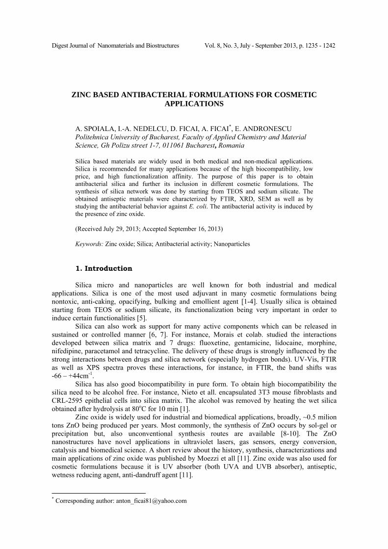
Digest Journal of Nanomaterials and Biostructures Vol. 8, No. 3, July - September 2013, p. 1235 - 1242
ZINC BASED ANTIBACTERIAL FORMULATIONS FOR COSMETIC APPLICATIONS
A. SPOIALA, I.-A. NEDELCU, D. FICAI, A. FICAI*, E. ANDRONESCU
Politehnica University of Bucharest, Faculty of Applied Chemistry and Material Science, Gh Polizu street 1-7, 011061 Bucharest, Romania Silica based materials are widely used in both medical and non-medical applications. Silica is recommended for many applications because of the high biocompatibility, low price, and high functionalization affinity. The purpose of this paper is to obtain antibacterial silica and further its inclusion in different cosmetic formulations. The synthesis of silica network was done by starting from TEOS and sodium silicate. The obtained antiseptic materials were characterized by FTIR, XRD, SEM as well as by studying the antibacterial behavior against E. coli. The antibacterial activity is induced by the presence of zinc oxide. (Received July 29, 2013; Accepted September 16, 2013) Keywords: Zinc oxide; Silica; Antibacterial activity; Nanoparticles
1. Introduction Silica micro and nanoparticles are well known for both industrial and medical
applications. Silica is one of the most used adjuvant in many cosmetic formulations being nontoxic, anti-caking, opacifying, bulking and emollient agent [1-4]. Usually silica is obtained starting from TEOS or sodium silicate, its functionalization being very important in order to induce certain functionalities [5].
Silica can also work as support for many active components which can be released in sustained or controlled manner [6, 7]. For instance, Morais et colab. studied the interactions developed between silica matrix and 7 drugs: fluoxetine, gentamicine, lidocaine, morphine, nifedipine, paracetamol and tetracycline. The delivery of these drugs is strongly influenced by the strong interactions between drugs and silica network (especially hydrogen bonds). UV-Vis, FTIR as well as XPS spectra proves these interactions, for instance, in FTIR, the band shifts was -66 – +44cm-1.
Silica has also good biocompatibility in pure form. To obtain high biocompatibility the silica need to be alcohol free. For instance, Nieto et all. encapsulated 3T3 mouse fibroblasts and CRL-2595 epithelial cells into silica matrix. The alcohol was removed by heating the wet silica obtained after hydrolysis at 80oC for 10 min [1].
Zinc oxide is widely used for industrial and biomedical applications, broadly, ~0.5 milion tons ZnO being produced per years. Most commonly, the synthesis of ZnO occurs by sol-gel or precipitation but, also unconventional synthesis routes are available [8-10]. The ZnO nanostructures have novel applications in ultraviolet lasers, gas sensors, energy conversion, catalysis and biomedical science. A short review about the history, synthesis, characterizations and main applications of zinc oxide was published by Moezzi et all [11]. Zinc oxide was also used for cosmetic formulations because it is UV absorber (both UVA and UVB absorber), antiseptic, wetness reducing agent, anti-dandruff agent [11].
* Corresponding author: [email protected]

1236
SiO2/ZnO core-shell nanostructures or composite materials were already synthesized for different applications such as catalysis, adsorbent or photo-luminescence, for instance [12-14].
Recently, Li et colab. presents their results in the field of preparing and characterization of porous silica spheres with self-dispersing properties with potential application in drug delivery [15]. Silica was obtained starting from TEOS by using a water/oil emulsion procedure. The specific surface area was determined and reached 772.3m2/g while the total pore volume reached 0.663cm3/g. The self-dispersing properties is induced by a post-synthesis treatment with NH4Cl. The main purpose of this paper is to obtain antiseptic silica based micro and nanopowder. In perspective, the micro- and nanopowders will be used for obtaining cosmetic formulations, ZnO assuring antiseptic, faster healing or even radiation protective role.
2. Materials and methods Silicon dioxide was obtained by two independent route starting from sodium silicate
(Sigma Aldrich, reagent grade) and TEOS (Fluka, puriss; >99%). Zinc oxide was synthesized by precipitation starting from zinc acetate dehydrate (Sigma-Aldrich, ACS reagent). All other chemicals were reagent grade and were used without further purification.
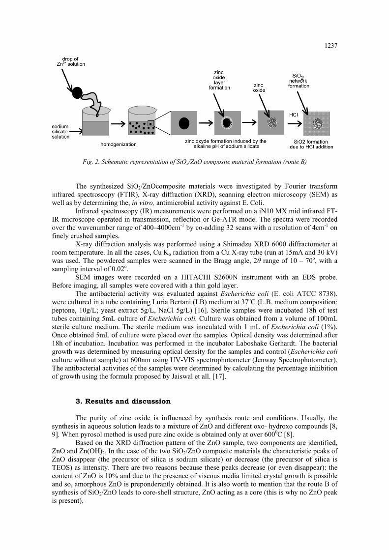
The SiO2/ZnO composite materials were obtained according to Figure 1. Two synthesis routes can be identified, the first one (route A) can be assimilated with a sol-gel followed by alkaline precipitation while the second route (route B) consists into an acidic precipitation of induced by the addition of HCl 1M into the Zn2+ and sodium silicate precursors.
Fig. 1. Synthesis of SiO2/ZnO composite materials - flow chart
Water, pH=2
Sodium silicate
Hydrolysis
Homogenization
Zn2+, saturated aq. solution
TEOS
Homogenization
Zn2+, saturated aq. solution
Precipitation
Purification
Freeze-drying
Precipitation
Purification
Freeze-drying
HCl, 1M
NaOH, 1M
A B
1237
Fig. 2. Schematic representation of SiO2/ZnO composite material formation (route B)
The synthesized SiO2/ZnOcomposite materials were investigated by Fourier transform infrared spectroscopy (FTIR), X-ray diffraction (XRD), scanning electron microscopy (SEM) as well as by determining the, in vitro, antimicrobial activity against E. Coli.
Infrared spectroscopy (IR) measurements were performed on a iN10 MX mid infrared FT-IR microscope operated in transmission, reflection or Ge-ATR mode. The spectra were recorded over the wavenumber range of 400–4000cm-1 by co-adding 32 scans with a resolution of 4cm-1 on finely crushed samples.
X-ray diffraction analysis was performed using a Shimadzu XRD 6000 diffractometer at room temperature. In all the cases, Cu Kα radiation from a Cu X-ray tube (run at 15mA and 30 kV) was used. The powdered samples were scanned in the Bragg angle, 2θ range of 10 – 70o, with a sampling interval of 0.02o.
SEM images were recorded on a HITACHI S2600N instrument with an EDS probe. Before imaging, all samples were covered with a thin gold layer.
The antibacterial activity was evaluated against Escherichia coli (E. coli ATCC 8738). were cultured in a tube containing Luria Bertani (LB) medium at 37oC (L.B. medium composition: peptone, 10g/L; yeast extract 5g/L, NaCl 5g/L) [16]. Sterile samples were incubated 18h of test tubes containing 5mL culture of Escherichia coli. Culture was obtained from a volume of 100mL sterile culture medium. The sterile medium was inoculated with 1 mL of Escherichia coli (1%). Once obtained 5mL of culture were placed over the samples. Optical density was determined after 18h of incubation. Incubation was performed in the incubator Laboshake Gerhardt. The bacterial growth was determined by measuring optical density for the samples and control (Escherichia coli culture without sample) at 600nm using UV-VIS spectrophotometer (Jenway Spectrophotometer). The antibacterial activities of the samples were determined by calculating the percentage inhibition of growth using the formula proposed by Jaiswal et all. [17].
3. Results and discussion The purity of zinc oxide is influenced by synthesis route and conditions. Usually, the
synthesis in aqueous solution leads to a mixture of ZnO and different oxo- hydroxo compounds [8, 9]. When pyrosol method is used pure zinc oxide is obtained only at over 6000C [8].
Based on the XRD diffraction pattern of the ZnO sample, two components are identified, ZnO and Zn(OH)2. In the case of the two SiO2/ZnO composite materials the characteristic peaks of ZnO disappear (the precursor of silica is sodium silicate) or decrease (the precursor of silica is TEOS) as intensity. There are two reasons because these peaks decrease (or even disappear): the content of ZnO is 10% and due to the presence of viscous media limited crystal growth is possible and so, amorphous ZnO is preponderantly obtained. It is also worth to mention that the route B of synthesis of SiO2/ZnO leads to core-shell structure, ZnO acting as a core (this is why no ZnO peak is present).

1238
10 20 30 40 50 60 70 800
100
200
300
400
500
600
700
800
900
1000
*
* * *
***
I, C
PS
2Theta
SiO2_ZnO_TEOS SiO2_ZnO_silicate ZnO
Zn(OH)2
*
ZnO
Fig. 3. XRD pattern of the ZnO based samples
Based on Sherrer’s relation the mean crystallite sizes, determined as an average of the size of the most important peaks of ZnO ad Zn(OH)2 are presented in Table 1.
Table 1. Crystallite size of the components of ZnO
Sample Component 2Theta d, A I/Io D, nm
nm ,D
ZnO
Zn(OH)2 13.29 6.66 55 9.49
10.48 19.87 4.46 21 11.47
ZnO 28.51 3.13 22 5.96
6.24 33.44 2.68 100 6.08 59.28 1.56 45 6.69
All samples were analyzed by SEM. As a general rule, in all cases agglomeration occurs. Even at 5000x magnification smooth surfaces can be visualized which means that these agglomerates are formed from smaller particles. In both cases, the agglomerated can reach up to several tens of micrometers

Fig. 4. SEM images of ZnO
1239

1240
Figg. 5. SEM imaages of SiO2/ZnZnO compositee materials ob
tained by routte A

1241
The bacteriological experiments performed in vitro demonstrated the effectiveness of ZnO as well as SiO2/ZnO composite materials in inhibiting the growth of Escherichia coli bacteria (Table 2). Analyzing the antibacterial activity of the samples it can conclude that pure ZnO have the maximal antibacterial activity against E. coli. The SiO2/ZnO composite materials exhibit 75 – 87% effectiveness against E. coli. The antibacterial activity of SiO2/ZnO obtained starting from TEOS (route A) is 83 and respectively 87% function of the used base. The higher antibacterial activity of the samples obtained by using NH3 can be explained based on the oxo- hydroxy compounds of Zn formed at higher pH (NaOH). The lowest effectiveness against E. coli is manifested by the composite material obtained from sodium silicate perhaps, because during the synthesis route, a higher content of ZnO is encapsulated into the SiO2 network as schematically present in Fig. 2.
Table 2. Antimicrobial activity against E. Coli
No. Sample I, %
1 ZnO ~100
2 SiO2/ZnO (NH3) 87
3 SiO2/ZnO (NaOH) 83
4 SiO2/ZnO (sodium silicate) 75
4. Conclusion SiO2/ZnO materials with antibacterial activity were obtained by two routes starting from
TEOS and sodium silicate as silica precursors. Based on the XRD diffraction patterns it can be conclude that ZnO crystallization is strongly influenced by the synthesis route. When starting from sodium silicate, once the ZnO drops fall into the sodium silicate solution a thin film of ZnO is formed. Under the experimental conditions, the antibacterial activity of the ZnO is almost 100%. The better antibacterial activity is specific for the samples obtained from TEOS perhaps because of the higher level of ZnO on the surface.
Acknowledgements This paper is supported by the Sectorial Operational Programme Human Resources
Development, financed from the European Social Fund, and by the Romanian Government under the contract number POSDRU/86/1.2/S/58146 (MASTERMAT)" and ‘Novel nanostructured prosthetic tubular devices with antibacterial and antibiofilm properties induced by physicochemical and morphological changes’ PN-II-PT-PCCA-2011-3.2-0284, funded by the National University Research Council in Romania.
References
[1] A Nieto, S Areva, T Wilson, R Viitala, M. Vallet-Acta Biomaterialia Nov; 5(9), 3478 (2009). [2] KR. Martin, Journal of Nutrition, Health and Aging;11(2):94 (2007). [3] C Fu, T Liu, L Li, H Liu, D Chen, F.Tang Biomaterials; 34(10), 2565 (2013). [4] C. Gumu, OM Ozkendir, H Kavak, Y.Ufuktepe, J. Optoelectron. Adv. Mater. 8(1), 299 (2006). [5] D Ficai, A Ficai, M Alexie, M Maganu, C Guran, E.Andronescu REV CHIM (Bucharest) 62(6), 622 (2011)

1242
[6] EC Morais, GG Correa, R Brambilla, C Radtke, IM Baibich, JHZD Santos. Colloids and Surfaces B: Biointerfaces 103, 422 (2013). [7] MG Florea, A Ficai, O Oprea, C Guran, D Ficai, L Pall, et al. Romanian Journal of Materials; 42(3), 313 (2012). [8] OR Vasile, E Andronescu, C Ghitulica, BS Vasile, O Oprea, E Vasile, et al. Journal of Nanoparticle Research 14(12), 2012. [9] O Oprea, E Andronescu, BS Vasile, G Voicu, C. Covaliu Digest Journal of Nanomaterials and Biostructures; 6(3): 1393 (2011). [10] F Vaja, C Comanescu, O Oprea, D Ficai, C.Guran, Revista De Chimie; 63(7),722 (2012). [11] A Moezzi, AM McDonagh, MB Cortie, Chemical Engineering Journal 185-186, 1-22 (2012). [12] L Shastri, MS Qureshi, MM.Malik, Journal of Physics and Chemistry of Solids 74(4),595 (2013). [13] HW Kim, HS Kim, HG Na, JC. Yang, Silica-coated ZnO nanowires. Vacuum; 86(6), 78993 (2012) [14] M Hussain, N Abbas, D Fino, N.Russo Chemical Engineering Journal; 188, 222 (2012). [15] Y Li, B Zou, X Wang, Z.Wang Preparation of porous silica spheres with self-dispersing properties. Particuology 2013;in press. [16] MA Ansari, HM Khan, AA Khan, A Malik, A Sultan, M Shahid, et al. Biology and Medicine 3(2), 141 (2011). [17] S. Jaiswal, B Duffy, AK Jaiswal, N Stobie, P. McHale Int J Antimicrob Ag; 36(3), 280 (2010).