ª 2018 by the american collegeof cardiology … · peer reviewer information.....2828 appendix c...
TRANSCRIPT
J O U R N A L O F T H E A M E R I C A N C O L L E G E O F C A R D I O L O G Y V O L . 7 1 , N O . 2 4 , 2 0 1 8
ª 2 0 1 8 B Y T H E AM E R I C A N C O L L E G E O F C A R D I O L O G Y F O U N D A T I O N
P U B L I S H E D B Y E L S E V I E R
EXPERT CONSENSUS DOCUMENT
ISSN 0735-1097/$36.0
2018 ACC/HRS/NASCI/SCAI/SCCTExpert Consensus Documenton Optimal Use of Ionizing Radiation
in Cardiovascular Imaging—Best Practices for Safety and Effectiveness,Part 1: Radiation Physics andRadiation Biology A Report of the American College of Cardiology Task Force onExpert Consensus Decision Pathways
Developed in Collaboration With Mended Hearts
Writing John W. Hirshfeld, JR, MD, FACC, FSCAI, Chair
CommitteeMembersVictor A. Ferrari, MD, FACC, Co-Chair
Frank M. Bengel, MD*Lisa Bergersen, MD, MPH, FACCCharles E. Chambers, MD, FACC, MSCAIyAndrew J. Einstein, MD, PHD, FACCMark J. Eisenberg, MD, MPH, FACCMark A. Fogel, MD, FACCThomas C. Gerber, MD, FACCDavid E. Haines, MD, FACCzWarren K. Laskey, MD, MPH, FACC, FSCAIMarian C. Limacher, MD, FACCKenneth J. Nichols, PHDxDaniel A. Pryma, MD
0
This document was approved by the American College of Cardiology Clinic
the Heart Rhythm Society, North American Society for Cardiovascular Imagi
of Cardiovascular Computed Tomography in January 2018.
The American College of Cardiology requests that this document be cited
CE, Einstein AJ, Eisenberg MJ, Fogel MA, Gerber TC, Haines DE, Laskey WK,
AE, Thomas SA, Tsai TT, Wagner LK, Wann LS. 2018 ACC/HRS/NASCI/SCAI/
cardiovascular imaging—best practices for safety and effectiveness, part 1: ra
Cardiology Task Force on Clinical Expert Consensus Documents. J Am Coll
Copies: This document is available on the World Wide Web site of the Am
please contact Elsevier Reprint Department via fax (212) 633-3820 or e-mail
Permissions: Multiple copies, modification, alteration, enhancement, and
permission of the American College of Cardiology. Requests may be com
business/policies/copyright/permissions).
Gilbert L. Raff, MD, FACCkGeoffrey D. Rubin, MD, MBA, FNASCI{Donnette Smith#Arthur E. Stillman, MD, PHD, FNASCISuma A. Thomas, MD, MBA, FACCThomas T. Tsai, MD, MSC, FACCLouis K. Wagner, PHDL. Samuel Wann, MD, MACC
*Society of Nuclear Medicine and Molecular Imaging Representative.
ySociety for Cardiovascular Angiography and Interventions
Representative. zHeart Rhythm Society Representative. xAmerican Society
of Nuclear Cardiology Representative. kSociety for Cardiovascular
Computed Tomography Representative. {North American Society for
Cardiovascular Imaging Representative. #Mended Hearts Representative.
https://doi.org/10.1016/j.jacc.2018.02.017
al Policy Approval Committee in November 2017 and the approval bodies of
ng, Society for Cardiovascular Angiography and Interventions, and Society
as follows: Hirshfeld JW Jr, Ferrari VA, Bengel FM, Bergersen L, Chambers
Limacher MC, Nichols KJ, Pryma DA, Raff GL, Rubin GD, Smith D, Stillman
SCCT expert consensus document on optimal use of ionizing radiation in
diation physics and radiation biology: a report of the American College of
Cardiol 2018;71:2811–28.
erican College of Cardiology (www.acc.org). For copies of this document,
/or distribution of this document are not permitted without the express
pleted online via the Elsevier site (http://www.elsevier.com/about/our-

Hirshfeld Jr. et al. J A C C V O L . 7 1 , N O . 2 4 , 2 0 1 8
Radiation Safety ECD Part 1: Physics and Biology J U N E 1 9 , 2 0 1 8 : 2 8 1 1 – 2 8
2812
ACC Task Forceon ExpertConsensusDecisionPathways**
Adrian F. Hernandez, MD, MHS, FACCWilliam Hucker, MD, PHD
James L. Januzzi, JR, MD, FACCLuis C. Afonso, MBBS, FACCBrendan Everett, MD, FACC
Hani Jneid, MD, FACCDharam Kumbhani, MD, SM, FACCJoseph Edward Marine, MD, FACC
Pamela Bowe Morris, MD, FACCRobert N. Piana, MD, FACCKarol E. Watson, MD, FACCBarbara S. Wiggins, PHARMD, AACC
**Formerly named ACC Task Force on Clinical Expert Consensus
Documents.
TABLE OF CONTENTS
ABSTRACT . . . . . . . . . . . . . . . . . . . . . . . . . . . . . . . . . . . . 2812
PREAMBLE . . . . . . . . . . . . . . . . . . . . . . . . . . . . . . . . . . . . 2813
1. INTRODUCTION . . . . . . . . . . . . . . . . . . . . . . . . . . . . . 2813
2. PURPOSE . . . . . . . . . . . . . . . . . . . . . . . . . . . . . . . . . . . 2814
2.1. Document Purpose . . . . . . . . . . . . . . . . . . . . . . . . 2814
2.2. The Radiation Safety Issue . . . . . . . . . . . . . . . . . 2814
2.3. The Need for Physician RadiationSafety Education . . . . . . . . . . . . . . . . . . . . . . . . . . 2814
2.4. Appropriateness of Medical Radiation . . . . . . . . 2814
3. CURRENT TRENDS IN AND CONSEQUENCES OF
PATIENT AND MEDICAL PERSONNEL RADIATION
EXPOSURE FROM CARDIOVASCULAR
PROCEDURES . . . . . . . . . . . . . . . . . . . . . . . . . . . . . . . 2815
4. THE MANY MEASURES OF RADIATION . . . . . . . . . 2816
4.1. Radiation Exposure and Dose Metrics . . . . . . . . 2816
4.2. Challenges in Relating Radiation Exposure andDose to Risk of Detrimental Effects . . . . . . . . . . 2817
4.3. Types of Ionizing Radiation Used inMedical Imaging . . . . . . . . . . . . . . . . . . . . . . . . . . 2817
4.3.1. X-Rays and Gamma Rays . . . . . . . . . . . . . 2817
4.3.2. Positrons . . . . . . . . . . . . . . . . . . . . . . . . . . . 2817
4.4. Relationships Between Exposure andAbsorbed Dose . . . . . . . . . . . . . . . . . . . . . . . . . . . 2817
4.4.1. Measures of Exposure FromExternal Beams . . . . . . . . . . . . . . . . . . . . . 2817
4.4.2. Exposure From Radionuclides . . . . . . . . . 2818
4.5. Estimating Effective Dose . . . . . . . . . . . . . . . . . . 2818
4.6. Synopsis of Measures of Radiation Exposureand Dose . . . . . . . . . . . . . . . . . . . . . . . . . . . . . . . . 2818
5. HOW RADIATION CAN HARM PEOPLE . . . . . . . . . 2818
5.1. Mechanism of Radiation-InducedBiological Effects . . . . . . . . . . . . . . . . . . . . . . . . . 2818
5.2. Types of Radiation-Induced Health Effects . . . . 2819
5.2.1. Tissue Reactions (Formerly CalledDeterministic Effects) . . . . . . . . . . . . . . . . . 2819
5.2.2. Stochastic Effects: Cancer . . . . . . . . . . . . . 2820
5.2.3. Stochastic Effects: Heritable Effects inOffspring . . . . . . . . . . . . . . . . . . . . . . . . . . . 2820
5.3. Tissue Reactions: Dose-Effect Relationships . . . 2820
5.3.1. Skin Injury . . . . . . . . . . . . . . . . . . . . . . . . . 2820
5.3.2. Bone Injury . . . . . . . . . . . . . . . . . . . . . . . . . 2821
5.3.3. Cataracts . . . . . . . . . . . . . . . . . . . . . . . . . . . 2821
5.3.4. Tissue Reactions: Managing Skin Injuries . . 2821
5.4. Stochastic Effects: Radiation-Induced Cancer . . 2821
5.4.1. Stochastic Effects: Attribution Challenges . . 2821
5.4.2. Stochastic Effects: Risk Metrics . . . . . . . . 2821
5.4.3. Stochastic Risk: Dose-Risk Relationships . . 2822
5.4.4. Incremental Cancer Risk Attributable toRadiation Exposure for OccupationallyExposed Healthcare Workers . . . . . . . . . . 2823
REFERENCES . . . . . . . . . . . . . . . . . . . . . . . . . . . . . . . . . 2825
APPENDIX A
Author Relationships With Industry andOther Entities (Relevant) . . . . . . . . . . . . . . . . . . . . . . . 2826
APPENDIX B
Peer Reviewer Information . . . . . . . . . . . . . . . . . . . . . 2828
APPENDIX C
Abbreviations . . . . . . . . . . . . . . . . . . . . . . . . . . . . . . . . 2828
ABSTRACT
The stimulus to create this document was the recognitionthat ionizing radiation-guided cardiovascular procedures

J A C C V O L . 7 1 , N O . 2 4 , 2 0 1 8 Hirshfeld Jr. et al.J U N E 1 9 , 2 0 1 8 : 2 8 1 1 – 2 8 Radiation Safety ECD Part 1: Physics and Biology
2813
are being performed with increasing frequency, leading togreater patient radiation exposure and, potentially, togreater exposure for clinical personnel. Although theclinical benefit of these procedures is substantial, there isconcern about the implications of medical radiationexposure. The American College of Cardiology leadershipconcluded that it is important to provide practitionerswith an educational resource that assembles and in-terprets the current radiation knowledge base relevant tocardiovascular procedures. By applying this knowledgebase, cardiovascular practitioners will be able to selectprocedures optimally, and minimize radiation exposureto patients and to clinical personnel.
Optimal Use of Ionizing Radiation in CardiovascularImaging: Best Practices for Safety and Effectiveness is acomprehensive overview of ionizing radiation use incardiovascular procedures and is published online. Toprovide the most value to our members, we divided theprint version of this document into 2 focused parts. PartI: Radiation Physics and Radiation Biology addresses theissue of medical radiation exposure, the basics of radi-ation physics and dosimetry, and the basics of radiationbiology and radiation-induced adverse effects. Part II:Radiological Equipment Operation, Dose-Sparing Meth-odologies, Patient and Medical Personnel Protectioncovers the basics of operation and radiation delivery forthe 3 cardiovascular imaging modalities (x-ray fluoros-copy, x-ray computed tomography, and nuclearscintigraphy).
PREAMBLE
This document has been developed as an ExpertConsensus Document by the American College of Cardi-ology (ACC) in collaboration with the American Society ofNuclear Cardiology, Heart Rhythm Society, MendedHearts, North American Society for Cardiovascular Imag-ing, Society for Cardiovascular Angiography and In-terventions, Society for Cardiovascular ComputedTomography, and Society of Nuclear Medicine and Mo-lecular Imaging. Expert Consensus Documents are inten-ded to inform practitioners, payers, and other interestedparties of the opinion of ACC and document cosponsorsconcerning evolving areas of clinical practice and/ortechnologies that are widely available or new to thepractice community. Topics chosen for coverage by expertconsensus documents are so designed because the evi-dence base, the experience with technology, and/or clin-ical practice are not considered sufficiently welldeveloped to be evaluated by the formal ACC/AmericanHeart Association practice guidelines process. Often thetopic is the subject of considerable ongoing investigation.
Thus, the reader should view the Expert ConsensusDocument as the best attempt of the ACC and documentcosponsors to inform and guide clinical practice in areaswhere rigorous evidence may not yet be available or evi-dence to date is not widely applied to clinical practice.
To avoid actual, potential, or perceived conflicts ofinterest that may arise as a result of industry relationshipsor personal interests among the writing committee, allmembers of the writing committee, as well as peerreviewers of the document, are asked to disclose allcurrent healthcare-related relationships, including thoseexisting 12 months before initiation of the writing effort.The ACC Task Force on Expert Consensus Decision Path-ways (formerly the ACC Task Force on Clinical ExpertConsensus Documents) reviews these disclosures todetermine which companies make products (on the mar-ket or in development) that pertain to the document un-der development. Based on this information, a writingcommittee is formed to include a majority of memberswith no relevant relationships with industry (RWI), led bya chair with no relevant RWI. Authors with relevant RWIare not permitted to draft or vote on text or recommen-dations pertaining to their RWI. RWI is reviewed on allconference calls and updated as changes occur. Authorand peer reviewer RWI pertinent to this documentare disclosed in Appendixes A and B, respectively.Additionally, to ensure complete transparency, authors’comprehensive disclosure information—including RWInot pertinent to this document—is available online.Disclosure information for the ACC Task Force onClinical Expert Consensus Documents is also availableonline, as is the ACC disclosure policy for documentdevelopment.
James L. Januzzi, MD, FACCChair, ACC Task Force on Expert Consensus Decision Pathways
1. INTRODUCTION
1.1. Document Development Process and Methodology
1.1.1. Writing Committee Organization
The work of the writing committee was supportedexclusively by the ACC without commercial support.Writing committee members volunteered their time tothis effort. Conference calls of the writing committeewere confidential and attended only by committeemembers and ACC staff.
The writing committee consisted of a broad rangeof members representing 9 societies and the followingareas of expertise: interventional cardiology, generalcardiology, pediatric cardiology, nuclear cardiology,nuclear medicine, electrophysiology, cardiac computed
Hirshfeld Jr. et al. J A C C V O L . 7 1 , N O . 2 4 , 2 0 1 8
Radiation Safety ECD Part 1: Physics and Biology J U N E 1 9 , 2 0 1 8 : 2 8 1 1 – 2 8
2814
tomography, cardiovascular imaging, and the con-sumer patient perspective. Both a radiation safetybiologist and physicist were included on the writingcommittee.
This writing committee met the College’s disclosurerequirements for RWI as described in the Preamble.
1.1.2. Document Development and Approval
The Writing Committee convened by conference call ande-mail to finalize the document outline, develop theinitial draft, revise the draft per committee feedback, andultimately sign off on the document for external peerreview. All participating organizations participated inpeer review, resulting in 21 reviewers representing 299comments. Comments were reviewed and addressed bythe writing committee. A member of the ACC Task Forceon Expert Consensus Decision Pathways served as leadreviewer to ensure that all comments were addressedadequately. Both the writing committee and the task forceapproved the final document to be sent to the ACC Clin-ical Policy Approval Committee. This committee reviewedthe document, including all peer review comments andwriting committee responses, and approved the docu-ment in November 2017. The Heart Rhythm Society, NorthAmerican Society for Cardiovascular Imaging, Society forCardiovascular Angiography and Interventions, and So-ciety of Cardiovascular Computed Tomography endorsedthe document in January 2018. This document is consid-ered current until the Task Force on Expert ConsensusDecision Pathways revises or withdraws it frompublication.
2. PURPOSE
2.1. Document Purpose
This print-published document is part 1 of an abbreviatedversion of a larger, more comprehensive document that ispublished concurrently online. The online version con-tains additional technical detail for readers who wish tounderstand a topic in greater depth. The online publisheddocument, in addition to covering the topics in the 2print-published documents in greater depth, also coversadditional topics not covered in the print-published doc-uments including: 1) dose reduction strategies; 2) oper-ator education and certification; 3) quality assurance; and4) patient radiation tracking.
This document covers radiation physics, radiationdosimetry and its determinants, and radiation harm. Thedocument’s purpose is to provide a comprehensive in-formation source about ionizing radiation use in cardio-vascular procedures. The writing group has assembledthis information to assist cardiovascular practitioners toprovide optimal cardiovascular care when employing
ionizing radiation-based procedures. The goal is toenhance cardiovascular practitioners’ ability to select theoptimal imaging technique for a given clinical circum-stance while balancing a technique’s risk and benefits,and to apply that technique optimally to generate high-quality diagnostic images of greatest clinical value andminimal radiation exposure.
2.2. The Radiation Safety Issue
Cardiovascular procedures that employ ionizing radiationhave great value for diagnosis and treatment of properlyselected patients with known or suspected cardiovasculardisease. However, ionizing radiation has molecular-leveldetrimental effects on tissue, with potential for injuryboth to patients and to exposed medical personnel. It isdesirable to minimize radiation exposure both to patientsand to medical personnel while achieving optimal bene-fits to health. This principle requires that cliniciansemploy judicious use and conduct of radiation-employingprocedures.
Currently, cardiovascular diagnostic and therapeuticprocedures are a major source of patient exposure tomedical ionizing radiation, accounting for approximately40% of total medical radiation exposure (exclusive of ra-diation oncology) (1,2). Among occupationally exposedhealthcare workers, interventional cardiologists andclinical electrophysiologists are among the most highlyexposed, and there is potential for exposure to supportpersonnel as well (3,4).
2.3. The Need for Physician Radiation Safety Education
Cardiovascular specialists have a responsibility to:
1. Apply knowledge of the radiation safety knowledgebase to make appropriate case selection choices.
2. Conduct radiation-assisted procedures optimally,minimizing exposure to patients and personnel.
There is evidence that many cardiovascular special-ists who order and conduct radiation-employing pro-cedures are not fully informed about the radiationdoses that accompany the procedure or the associatedhealth implications for their patients and for them-selves (5,6).
2.4. Appropriateness of Medical Radiation
The balance between a procedure’s risk and benefit de-termines its appropriateness. The hazard associated withionizing radiation is a potentially important determinantof a procedure’s risk-benefit relationship. Physicians whoeither order or conduct such procedures need to:
1. Know the magnitude of a patient’s risk associated witha procedure’s radiation exposure.

TABLE 1 Typical Effective Doses for Cardiac Procedures
Modality ProtocolTypical Effective
Dose (mSv)
MDCT Coronary CT angiography:helical, no tube current modulation
8–30
MDCT Coronary CT angiography:helical, tube current modulation
6–20
MDCT Coronary CT angiography:prospectively triggered axial
0.5–7
MDCT Coronary CT angiography:high-pitch helical
<0.5–3
MDCT CT angiography, pre-TAVR:coronary (multiphase) andchest/abdomen/pelvis
5–50
MDCT Calcium score 1–5
MDCT Attenuation correction <0.5–2.0
EBCT Calcium Score 1
SPECT 10 mCi 99mTc sestamibi rest/30 mCi 99mTc sestamibi stress
11
SPECT 15 mCi 99mTc sestamibi rest/45 mCi 99mTc sestamibi stress
17
SPECT 30 mCi 99mTc sestamibi rest/30 mCi 99mTc sestamibi stress
18
SPECT 10 mCi 99mTc sestamibi stress only 2.7
SPECT 30 mCi 99mTc sestamibi stress only 8
SPECT 10 mCi 99mTc tetrofosmin rest/30 mCi 99mTc tetrofosmin stress
9
SPECT 15 mCi 99mTc tetrofosmin rest/45 mCi 99mTc tetrofosmin stress
14
SPECT 30 mCi 99mTc tetrofosmin rest/30 mCi 99mTc tetrofosmin stress
14
SPECT 10 mCi 99mTc tetrofosmin stress only 2.3
SPECT 30 mCi 99mTc tetrofosmin stress only 7
SPECT 3.5mCi 201Tl 15
SPECT Dual isotope: 3.5 mCi 201Tl rest/30 mCi 99mTc sestamibi stress
23
SPECT Dual isotope: 3.5 mCi 201Tl rest/30 mCi 99mTc tetrofosmin stress
22
Continued in the next column
TABLE 1 Continued
Modality ProtocolTypical Effective
Dose (mSv)
PET 50 mCi 82Rb rest/50 mCi 82Rb stress
4
PET 15 mCi 13N ammonia rest/15 mCi 13N ammonia stress
2
PET 10 mCi 18F FDG 7
Planar 30 mCi 99mTc-labeled erythrocytes 8
Fluoroscopy Diagnostic invasive coronary angiography 2–20
Fluoroscopy Percutaneous coronary intervention 5–57
Fluoroscopy TAVR, transapical approach 12–23
Fluoroscopy TAVR, transfemoral approach 33–100
Fluoroscopy Diagnostic electrophysiological study 0.1–3.2
Fluoroscopy Radiofrequency ablation of arrhythmia 1–25
Fluoroscopy Permanent pacemaker implantation 0.2–8
Note: Current and ongoing engineering physical design and image processing softwarerefinements enable dose reductions for all 3 modalities since the data in Table 1 werecompiled. These lower doses can be achieved only if radiological equipment is currentgeneration and if operators consciously take advantage of their improved capabilities.As the majority of the currently installed base of equipment is earlier generation, thedata in Table 1 reflect most current exposure levels. Reproduced with permission fromEinstein et al. (7).
CT ¼ computed tomography; EBCT ¼ electron-beam computed tomography; FDG ¼fluorodeoxyglucose; MDCT ¼ multidetector-row computed tomography; PET ¼ posi-tron emission tomography; Rb ¼ rubidium; SPECT ¼ single-photon emission computedtomography; TAVR ¼ transcatheter aortic valve replacement; Tc ¼ technetium;Tl ¼ thallium.
J A C C V O L . 7 1 , N O . 2 4 , 2 0 1 8 Hirshfeld Jr. et al.J U N E 1 9 , 2 0 1 8 : 2 8 1 1 – 2 8 Radiation Safety ECD Part 1: Physics and Biology
2815
2. Apply that understanding to determining the appro-priate procedure and selecting the approach that pro-vides the best balance of benefit and risk.
3. CURRENT TRENDS IN AND CONSEQUENCES OF
PATIENT AND MEDICAL PERSONNEL
RADIATION EXPOSURE FROM
CARDIOVASCULAR PROCEDURES
The past 2 decades have seen substantial develop-ment and refinement of x-ray fluoroscopy, x-raycomputed tomography, and radionuclide scintigraphy.Engineering advances have improved image qualitywhile in many cases reducing the radiation dosesemployed.
Despite these engineering refinements, the patient ra-diation doses that accompany these procedures remainsubstantial and, for the most part, are at the upper rangeof radiation-based diagnostic studies. Medical pro-fessionals should be aware of the radiation dose thatthese studies deliver to patients. In addition, within aparticular type of study, the radiation dose can varysubstantially depending on image acquisition protocoland patient characteristics. For reference, the commonlyperformed cardiovascular diagnostic studies and theirradiation dose ranges are listed in Table 1. Note that thedoses delivered by x-ray computed tomography (CT) andnuclear cardiology can vary substantially depending onparticulars of image acquisition protocols.
Patient radiation dose ranges (in millisieverts) are lis-ted for the 3 principal radiation-based cardiovascularimaging studies: x-ray fluoroscopy, x-ray computed to-mography, and nuclear cardiology. Individual procedurecategories are further subdivided according to types ofimage acquisition protocols. Note that for a particularprocedure category, the dose can vary considerablydepending on image acquisition protocol and, within agiven image acquisition protocol, procedure conduct andpatient characteristics.
However, augmented capabilities have led to increasedutilization levels, necessarily accompanied by greater ra-diation exposure both at the individual and population
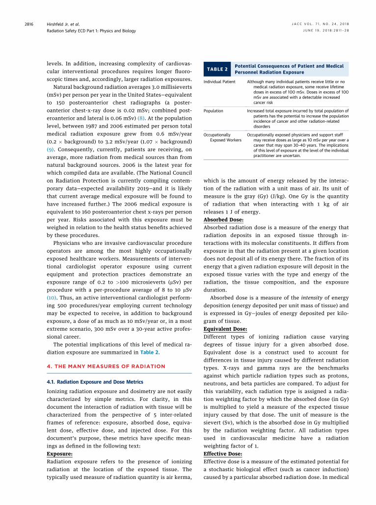
TABLE 2Potential Consequences of Patient and MedicalPersonnel Radiation Exposure
Individual Patient Although many individual patients receive little or nomedical radiation exposure, some receive lifetimedoses in excess of 100 mSv. Doses in excess of 100mSv are associated with a detectable increasedcancer risk
Population Increased total exposure incurred by total population ofpatients has the potential to increase the populationincidence of cancer and other radiation-relateddisorders
OccupationallyExposed Workers
Occupationally exposed physicians and support staffmay receive doses as large as 10 mSv per year over acareer that may span 30–40 years. The implicationsof this level of exposure at the level of the individualpractitioner are uncertain.
Hirshfeld Jr. et al. J A C C V O L . 7 1 , N O . 2 4 , 2 0 1 8
Radiation Safety ECD Part 1: Physics and Biology J U N E 1 9 , 2 0 1 8 : 2 8 1 1 – 2 8
2816
levels. In addition, increasing complexity of cardiovas-cular interventional procedures requires longer fluoro-scopic times and, accordingly, larger radiation exposures.
Natural background radiation averages 3.0 millisieverts(mSv) per person per year in the United States—equivalentto 150 posteroanterior chest radiographs (a poster-oanterior chest-x-ray dose is 0.02 mSv; combined post-eroanterior and lateral is 0.06 mSv) (8). At the populationlevel, between 1987 and 2006 estimated per person totalmedical radiation exposure grew from 0.6 mSv/year(0.2 � background) to 3.2 mSv/year (1.07 � background)(9). Consequently, currently, patients are receiving, onaverage, more radiation from medical sources than fromnatural background sources. 2006 is the latest year forwhich compiled data are available. (The National Councilon Radiation Protection is currently compiling contem-porary data—expected availability 2019—and it is likelythat current average medical exposure will be found tohave increased further.) The 2006 medical exposure isequivalent to 160 posteroanterior chest x-rays per personper year. Risks associated with this exposure must beweighed in relation to the health status benefits achievedby these procedures.
Physicians who are invasive cardiovascular procedureoperators are among the most highly occupationallyexposed healthcare workers. Measurements of interven-tional cardiologist operator exposure using currentequipment and protection practices demonstrate anexposure range of 0.2 to >100 microsieverts (mSv) perprocedure with a per-procedure average of 8 to 10 mSv(10). Thus, an active interventional cardiologist perform-ing 500 procedures/year employing current technologymay be expected to receive, in addition to backgroundexposure, a dose of as much as 10 mSv/year or, in a mostextreme scenario, 300 mSv over a 30-year active profes-sional career.
The potential implications of this level of medical ra-diation exposure are summarized in Table 2.
4. THE MANY MEASURES OF RADIATION
4.1. Radiation Exposure and Dose Metrics
Ionizing radiation exposure and dosimetry are not easilycharacterized by simple metrics. For clarity, in thisdocument the interaction of radiation with tissue will becharacterized from the perspective of 5 inter-relatedframes of reference: exposure, absorbed dose, equiva-lent dose, effective dose, and injected dose. For thisdocument’s purpose, these metrics have specific mean-ings as defined in the following text:Exposure:
Radiation exposure refers to the presence of ionizingradiation at the location of the exposed tissue. Thetypically used measure of radiation quantity is air kerma,
which is the amount of energy released by the interac-tion of the radiation with a unit mass of air. Its unit ofmeasure is the gray (Gy) (J/kg). One Gy is the quantityof radiation that when interacting with 1 kg of airreleases 1 J of energy.Absorbed Dose:
Absorbed radiation dose is a measure of the energy thatradiation deposits in an exposed tissue through in-teractions with its molecular constituents. It differs fromexposure in that the radiation present at a given locationdoes not deposit all of its energy there. The fraction of itsenergy that a given radiation exposure will deposit in theexposed tissue varies with the type and energy of theradiation, the tissue composition, and the exposureduration.
Absorbed dose is a measure of the intensity of energydeposition (energy deposited per unit mass of tissue) andis expressed in Gy—joules of energy deposited per kilo-gram of tissue.Equivalent Dose:
Different types of ionizing radiation cause varyingdegrees of tissue injury for a given absorbed dose.Equivalent dose is a construct used to account fordifferences in tissue injury caused by different radiationtypes. X-rays and gamma rays are the benchmarksagainst which particle radiation types such as protons,neutrons, and beta particles are compared. To adjust forthis variability, each radiation type is assigned a radia-tion weighting factor by which the absorbed dose (in Gy)is multiplied to yield a measure of the expected tissueinjury caused by that dose. The unit of measure is thesievert (Sv), which is the absorbed dose in Gy multipliedby the radiation weighting factor. All radiation typesused in cardiovascular medicine have a radiationweighting factor of 1.Effective Dose:
Effective dose is a measure of the estimated potential fora stochastic biological effect (such as cancer induction)caused by a particular absorbed radiation dose. In medical
J A C C V O L . 7 1 , N O . 2 4 , 2 0 1 8 Hirshfeld Jr. et al.J U N E 1 9 , 2 0 1 8 : 2 8 1 1 – 2 8 Radiation Safety ECD Part 1: Physics and Biology
2817
radiation exposures, absorbed dose is typically not uni-form throughout all tissues. For x-ray imaging, dose isconcentrated in the body region being examined andvaries with depth from the beam entrance port. For nu-clear imaging, dose is concentrated in the tissues thatmost avidly take up the tracer or are involved in itselimination.
Effective dose is the sum of the equivalent dosesreceived by each organ, with each organ equivalent dosemultiplied by a coefficient that reflects that organ’ssensitivity to a stochastic effect. The unit of effective doseis also the Sv. The Sv has the same unit as the Gy (J/kg).The connection between effective dose and absorbeddose is that an effective dose of 1 Sv (which may beconcentrated in only a few organs) confers the sameestimated stochastic risk that would be caused by a uni-form total absorbed body dose of 1 Gy of radiation that hasa radiation weighting factor of 1.
Different tissues have different sensitivities toradiation-induced effects. In the effective dose construct,each tissue is assigned a tissue-weighting factor thatspecifies its sensitivity to radiation effects. To calculatethe effective dose in Sv, each exposed tissue’s equivalentdose is multiplied by its tissue-weighting factor yieldingthat tissue’s contribution to the overall risk. The contri-butions to risk from all exposed tissues are summedyielding total risk, which is expressed as the effectivedose in Sv.Injected Dose:
Injected dose describes the quantity of radioactivityinjected into a patient for a nuclear scintigraphy study(expressed in millicuries). The relationship between aninjected dose and the previously described dose param-eters is complex and is discussed in Part II: RadiologicalEquipment Operation, Dose-Sparing Methodologies andProtection.
4.2. Challenges in Relating Radiation Exposure and Dose toRisk of Detrimental Effects
Detrimental effects of radiation exposure typically pre-sent weeks to years following exposure. In addition,many detrimental effects, principally cancer, have a largebackground frequency. This complicates the attributionof an effect in a particular subject to prior radiationexposure.
4.3. Types of Ionizing Radiation Used in Medical Imaging
Radiation in cardiovascular imaging consists of photonswith energy >10 kiloelectron volts (keV) (x-rays andgamma rays) and positrons. The physical effect of suchradiation is to eject electrons from atoms forming ionsand free radicals. This is the basis for the term “ionizingradiation.” The resulting ions and free radicals react withtissue molecules, damaging them.
4.3.1. X-Rays and Gamma Rays
X-ray and gamma ray photons travel at the speed of lightand have no mass and no charge. Their electromagneticenergy ranges from a few electron volts (eV) to millions ofelectron volts (MeV). X-rays used in x-ray fluoroscopy andx-ray CT have a photon energy spectrum between 30 and140 keV. Thallium-201 releases photons primarily in the68-80 keV range, similar to diagnostic x-rays.Technetium-99m releases photons primarily in the 140keV range.
4.3.2. Positrons
Positrons are positively charged electrons. They havemass and charge. When they travel through a medium,their electrostatic charge causes them to interact readilywith electrons in the medium, leaving a trail of ionization.Consequently, they have a very short mean free path intissue (6 to 7 mm, with a maximum of 15.2 mm). Positronsare annihilated by colliding with an electron of a con-stituent atom releasing two 511 keV gamma ray photonsthat travel in opposite directions. These high-energyphotons are minimally attenuated in tissue, and the ma-jority reach the imaging detector. Rubidium-82 is themost commonly used positron emitter for myocardialperfusion imaging. Nitrogen-13 ammonia is used lessfrequently for this purpose. Fluorine-18 deoxyglucose isused in cardiology for metabolic imaging and to detectmyocardial sarcoid and other inflammatory conditions.
4.4. Relationships Between Exposure and Absorbed Dose
Medical radiation exposures occur in 2 ways:
1. Exposure from an external radiation beam (x-ray fluo-roscopy and x-ray CT)
2. Exposure from radioactive decay within the subject(nuclear scintigraphy)
4.4.1. Measures of Exposure From External Beams
For external radiation beams, the absorbed dose isdetermined by the total incident exposure, the propertiesof the incident radiation, and the volume of tissueexposed.
Air kerma (“kinetic energy released in material”) is thestandard unit of measure for x-ray beam exposure. It is anenergy intensity measured in Gy. 1 Gy ¼ 1 J of energyreleased per kilogram of absorbing material. The metric“air kerma” is used because the measurement is madeusing air as the absorbing material.Absorbed Dose From an External Beam
Radiation absorbed dose, as distinguished from exposure,is an energy intensity, the concentration of radiation en-ergy actually deposited in the exposed tissue. Not all ra-diation energy that impinges on a tissue is absorbed.Some radiation (a variable quantity depending on both

TABLE 3Tissue Weighting Factors Used to CalculateEffective Dose in Sieverts
OrgansTissue Weighting Factors
(ICRP103–2007)
Red bone marrow 0.12
Colon 0.12
Lung 0.12
Stomach 0.12
Breasts 0.12
Gonads 0.08
Bladder 0.04
Liver 0.04
Esophagus 0.04
Thyroid 0.04
Skin 0.01
Bone surface 0.01
Salivary glands 0.01
Brain 0.01
Remainder of body 0.12
Total 1.00
Adapted from the International Commission on Radiological Protection (ICRP) (12).
Hirshfeld Jr. et al. J A C C V O L . 7 1 , N O . 2 4 , 2 0 1 8
Radiation Safety ECD Part 1: Physics and Biology J U N E 1 9 , 2 0 1 8 : 2 8 1 1 – 2 8
2818
radiation and tissue characteristics) passes through thetissue without interacting with it, depositing no energy (itis this radiation that contributes to image formation).Radiation absorbed dose is also measured in Gy. 1 Gy ¼ 1 Jof energy deposited per kilogram of irradiated tissue.
External beam energy deposition in tissue is not uni-form. X-ray radiation is attenuated exponentially as itpasses through tissue, decreasing by approximately afactor of 2 for each 5 cm of tissue. The incident beam airkerma is a good measure of dose at the body surface, butstructures deep to the body surface receive smaller doses.X-Ray Fluoroscopy Kerma-Area Product: Incorporating
the Exposed Tissue Volume
The risk of radiation harm is related both to the intensityof the radiation dose, and also to the quantity of tissuethat receives the dose. Kerma-area product (KAP) is theproduct of the beam’s kerma and its cross-sectional areaincorporating the volume of tissue irradiated. Thisconcept is particularly important in x-ray fluoroscopy, asimaging field sizes vary leading to very different KAPs.X-Ray Computed Tomography Kerma-Length Product:
Incorporating the Exposed Tissue Volume
CT delivers radiation to a patient in a manner quitedifferent from that of projectional imaging or fluoroscopy.The dose is distributed more uniformly around thepatient.
The total dose delivered by a CT examination is themeasured kerma multiplied by the axial length of thescan. A variety of dose metrics for x-ray CT are derivedfrom this model.
4.4.2. Exposure From Radionuclides
Unlike external beam exposures, radionuclide exposurescome from radioactive decay within the subject. Exposureis determined by the activity administered, the tracerdistribution, the tracer elimination rate, and the tracer’stime-activity relationships.
4.5. Estimating Effective Dose
The effective dose construct assigns each organ/tissue aweighting factor that reflects the tissue’s sensitivity toradiation-induced stochastic risk. The calculation ofeffective dose involves estimating each organ’s actualequivalent dose (in Gy). Each organ dose is adjusted bymultiplying it by the organ’s tissue-weighting factor. Theorgan sensitivity-adjusted individual organ doses aresummed to yield a total effective dose (in Sv) (11).
For a chest exposure, absorbed dose is concentrated inthe skin, mediastinal structures, lungs, breast, andthoracic bone marrow. Doses to these organs contributethe largest components to the effective dose calculation.Smaller quantities of scattered radiation expose the
abdominal viscera and upper neck. As these organs wouldreceive smaller exposures, their contribution to theeffective dose calculation would be smaller.
The International Commission on Radiation Protection(ICRP) published the most recent organ sensitivity esti-mates in 2007 in ICRP Publication 103 (12). These esti-mates are listed in Table 3.
4.6. Synopsis of Measures of Radiation Exposure and Dose
The existence of the many different measures of radia-tion exposure and dose has the potential to causeconfusion leading to misapplication of units of measure.Table 4 contains a synopsis of the principal metricsdescribed in this section. It should be noted that, becauseeffective dose in the table is based on gender- and age-averaged tissue weighting factors, (not accounting forthe fact that children and females are more sensitive), itspractical value is in comparing the effects of differentexposures rather than in estimating an individual’s sto-chastic risk.
5. HOW RADIATION CAN HARM PEOPLE
5.1. Mechanism of Radiation-Induced Biological Effects
Radiation-induced tissue injury is due to molecularalteration caused by particles or photons that have suffi-cient energy to induce ionization. Atoms ionized by ra-diation are frequently chemically unstable and transform

TABLE 4 Synopsis of Radiation Exposure and Dose Metrics
Metric Unit Utility
Absorbed Dose-Related Parameters:Characterize Dose to Organ/Tissue or Whole Body
Absorbed dose Gy Amount of ionizing radiation energy deposited per unit mass of tissue. 1 Gy ¼ 1 Joule of energy deposited perkg of tissue. This metric is a concentration of energy deposition—not the total quantity of energydeposited.
Equivalent dose Sv Absorbed dose adjusted by a radiation weighting factor that adjusts for the specific tissue-injuring potentialof the particular radiation type. Photons (x-rays and gamma rays) have a weighting factor of 1. Electronsalso have a weighting factor of 1. Neutrons have larger weighting factors that vary with their energylevel. For medical imaging, because only photons and positrons are used, absorbed dose and equivalentdose take the same value.
Effective dose mSv Calculated whole-body quantity used to roughly compare potential stochastic risks from different partial-body exposures. It is expressed as the uniform whole-body dose that would confer the stochastic riskequivalent to that caused by a regional exposure.
Modality-Specific Parameters
X-ray fluoroscopic air kerma (free-in-air) Gy Used to assess level of radiation present at a location. In x-ray fluoroscopy, cumulative air kerma at theinterventional reference point can be used to approximate beam entrance port skin dose. (For isocentricC-arms, the reference point is located 15 cm from isocenter in the direction toward the x-ray source. Thispoint in space approximates the location of beam entry into the patient, but due to variation in tableheight and tube angulation, is only an estimate of beam entrance port skin dose).
X-ray fluoroscopic Air-KAP, also referred toas dose-area product (DAP)
Gy,cm2 Used to assess the total quantity of radiation delivered by an external beam. It is the product of thecumulated amount of air kerma and the area of a radiographic or fluoroscopic field. KAP is often used asthe basis for estimating effective dose from a fluoroscopic procedure.
Computed tomographic dose index(CTDIFDA, CTDI100, CTDIw, and CTDIvol)
mGy Used to assess relative level of radiation applied during a CT imaging sequence. This metric is a concentrationof energy deposition in the exposed volume. It is not a total deposited energy quantity, as it does notincorporate the actual exposed volume (See DLP below). Different versions are used for varied purposes.
Computed tomographic dose-lengthproduct (DLP)
mGy,cm Used to assess integrated amount of radiation applied along an axial length of a patient during a CTexamination. Can be used to estimate effective dose from the procedure.
Radionuclide injected dose mCi A measure of the quantity of radioactivity injected for a nuclear scintigraphy study. The relationship ofinjected dose to other dose parameters is complex and includes the nature of the nuclide’s radiation, thenuclide’s half-life, the distribution in the body, and the elimination kinetics.
CT ¼ computed tomography; CTDI ¼ computed tomographic dose index; KAP ¼ Kerma-Area Product.
J A C C V O L . 7 1 , N O . 2 4 , 2 0 1 8 Hirshfeld Jr. et al.J U N E 1 9 , 2 0 1 8 : 2 8 1 1 – 2 8 Radiation Safety ECD Part 1: Physics and Biology
2819
into free radicals. A common example is ionization ofwater, which, upon interacting with an x-ray photon,decomposes into a free electron, a proton, and a hydroxylradical. The hydroxyl radical, because of its unpairedelectron, is highly reactive and interacts avidly with bio-molecules (proteins or nucleic acids). Similarly, an x-rayphoton can ionize an atom that is a constituent of abiomolecule. Thus, a biomolecule can be altered by eitherreacting with a radiation-generated free radical or bydirect ionization from radiation. The resulting structuralchange can alter or degrade its function.
5.2. Types of Radiation-Induced Health Effects
Radiation-induced health effects are divided into 2groups that differ in mechanism, the nature of effects,relationship to absorbed dose, and time between expo-sure and manifestation.
5.2.1. Tissue Reactions (Formerly Called Deterministic Effects)
Tissue reactions are caused by radiation-induced injury tostructural and functional molecules in cells. Cell necrosiswill occur if the amount of molecular damage exceeds the
cell’s ability to repair itself and maintain function. Tissuereactions only become macroscopically evident if athreshold radiation dose is exceeded, causing a sufficientfraction of an exposed tissue’s cells to malfunction ornecrose. A dose below the threshold dose may cause un-apparent cellular injury but will not cause a detectablereaction (13).
Tissue reactions typically exhibit dose-related severityand occur with a time delay (typically 4 to 8 weeks) be-tween exposure and the appearance of tissue injury.Above the threshold dose, a greater dose causes moreextensive injury to a greater fraction of cells in proportionto the dose.
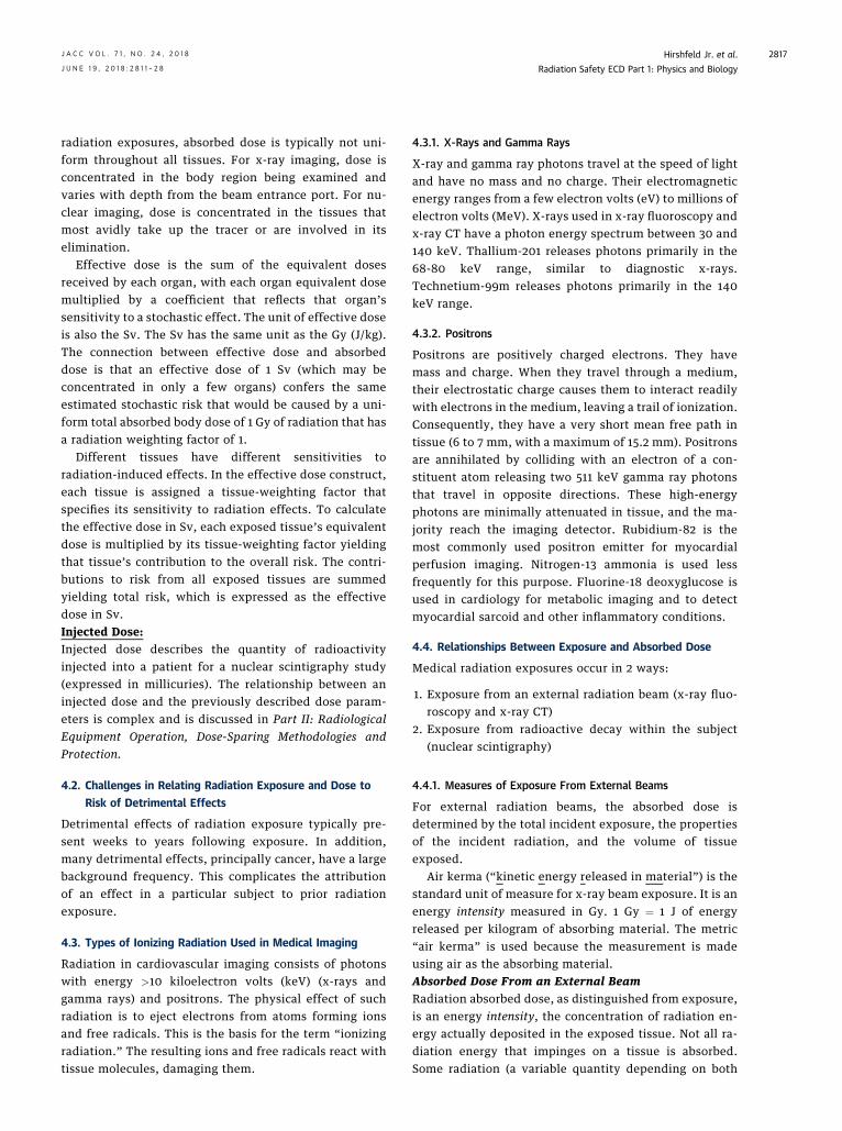
Skin injury is the most common tissue reactionobserved in cardiovascular imaging. It occurs almostexclusively from x-ray fluoroscopic exposures. Othertissue reactions include cataract formation, bonenecrosis and, in the heart, damage to myocardium,cardiac valves, and coronary arteries. In addition, ifa fetus incurs sufficient cellular injury at criticalstages of organogenesis, development will beimpaired (14).

FIGURE 1 Full Thickness Skin Necrosis Caused By a Large-Dose
X-Ray Fluoroscopic Procedure
An example of full thickness skin necrosis (underlying muscle and fat
are exposed) caused by a large-dose x-ray fluoroscopic procedure (90
minutes of fluoroscopy time). Note the rectangular area of skin
discoloration surrounding the area of skin necrosis. The injury is on the
left side of the subject’s back indicating that the exposure was con-
ducted in the right anterior oblique projection (17). (This image is
available on the U.S. Food and Drug Administration Web site and is in
the public domain.)
Hirshfeld Jr. et al. J A C C V O L . 7 1 , N O . 2 4 , 2 0 1 8
Radiation Safety ECD Part 1: Physics and Biology J U N E 1 9 , 2 0 1 8 : 2 8 1 1 – 2 8
2820
5.2.2. Stochastic Effects: Cancer
Stochastic effects are caused by radiation-induced dam-age to a cell’s genetic material that reprograms thedamaged cell’s deoxyribonucleic acid (DNA) intodysfunctional operation. The principal stochastic event ofclinical importance is radiation-induced cancer.
TABLE 5 Radiation-Induced Skin Injuries—Relationship of Sever
Single ExposureDose Range (Gy) 0–2 Weeks 2–8 Weeks
0–2 N
2–5 Transient erythema Possible epilation R
5–10 Transient erythema Erythema epilation R
10–15 Transient erythema Epilation, possibledesquamation
P
>15 Transient erythema, after veryhigh doses ulceration
Epilation, moistdesquamation
D
Adapted from Balter et al. (13).
Stochastic effects differ from tissue reactions in theirdose relationship. Whereas tissue reactions exhibit dose-related severity and have a definite dose threshold, sto-chastic events, in contrast, have a probabilistic relation-ship to dose. They are not known to have a dose thresholdand do not have a quantitative dose-related severity.Radiation-induced cancer either does or does not occur(or may not present within the subject’s lifetime). A singlecritically located DNA damage event can create an onco-gene (15). This is the theoretical basis for the concept thatthere is no threshold dose below which stochastic risk iszero (16).
5.2.3. Stochastic Effects: Heritable Effects in Offspring
Theoretically, radiation injury to DNA in germ cells couldcause a clinically important mutation that would notaffect the exposed individual, but would be transmittedto that individual’s offspring. Such effects have beendemonstrated in animal models but have not beenobserved in humans with statistical significance (17).
5.3. Tissue Reactions: Dose-Effect Relationships
5.3.1. Skin Injury
The most common radiation-induced tissue reaction isskin injury at the beam entrance port (typically on thepatient’s back) following an x-ray fluoroscopic exami-nation. Skin entrance port injuries are rectangular,reflecting the beam shape. These injuries vary in severityfrom erythema to desquamation to ulceration andnecrosis.
Skin injury typically appears 4 to 8 weeks following theexposure. In extreme cases, the ulceration can becomeconfluent and full thickness necrosis of skin may develop,exposing underlying fat, muscle, and even bone (Figure 1).
The skin injury threshold dose is variable, as is therelationship between dose and injury severity. A proced-ure’s cumulative air kerma can be used to estimate a pa-tient’s skin injury risk.
ity to Dose
Skin Reaction
8–40 Weeks Long-Term (>40 weeks)
o observable effects
ecovery of hair loss Complete healing
ecovery or permanent hair loss At higher doses dermal atrophy orinduration
rolonged erythema, permanenthair loss
Dermal atrophy or induration
ermal atrophy, secondaryulceration, necrosis
Dermal atrophy, possible late skinbreakdown, ulceration, and necrosis ofsubcutaneous tissues

J A C C V O L . 7 1 , N O . 2 4 , 2 0 1 8 Hirshfeld Jr. et al.J U N E 1 9 , 2 0 1 8 : 2 8 1 1 – 2 8 Radiation Safety ECD Part 1: Physics and Biology
2821
General guideline values for the ranges of thresholdvalues for a single first-time exposure for absorbed dosesassociated with degrees of skin injury severity are tabu-lated in Table 5. Injury thresholds for a subsequentexposure are lower.
Fluoroscopic entrance skin doses vary greatly becauseof variations in procedure complexity and duration andvariations in patient radiological characteristics. Skindose is strongly affected by the patient’s characteristicsand procedural techniques. Body habitus is the mostimportant patient characteristic. Larger patients require agreater skin entrance port dose. Dose is also determinedby equipment calibration and imaging protocol settings.
The prototypical patient at risk for a skin injury is anobese diabetic who has undergone 1 or more long-duration procedures within the past several months.
5.3.2. Bone Injury
In addition to skin injury, on occasion, incident radia-tion can cause necrosis of superficial bones such as ribs.Although the dose to bone needed to cause osteonec-rosis is greater than the dose that causes skin necrosis,bone’s high calcium content imparts a greater capacityto absorb x-ray photons, causing a greater absorbeddose to bone.
5.3.3. Cataracts
The single dose threshold that will cause vision-impairingcataracts in humans is not well characterized but isbelieved to be on the order of 500 mGy with a minimumlatency of approximately 1 year (18). Cataracts are alsoincreasingly being observed in physician operators withlong career experience. This area is currently a subject ofongoing study.
5.3.4. Tissue Reactions: Managing Skin Injuries
Less-severe degrees of skin injury have the potential toheal if managed with good supportive dermatologicalcare.
The cornerstone of optimizing the outcome of a skininjury is mechanical protection of the affected skin. X-rayinjured skin is fragile. Mechanical trauma to the skin canaggravate the injury. Dressings and other mechanismsthat help the patient avoid applying pressure or friction tothe affected area is important.
Early recognition of a radiation-induced skin injury isessential to initiate protection and early treatment. Theinherent delay of weeks between exposure and theinitial signs of skin injury may interfere with recognitionof the cause, delaying appropriate treatment. The beststrategy to facilitate prompt recognition is to warn thepatient, family, and primary care physician of the skin
injury potential. The 2011 ACCF/AHA/SCAI Guideline forPercutaneous Coronary Intervention state that it is aClass I recommendation for all patients who receive anair kerma at the interventional reference point >5 Gy tobe counseled about the possibility of a skin injury andinstructed how to react to the earliest signs should theyoccur (19).
5.4. Stochastic Effects: Radiation-Induced Cancer
Note: Considerable additional detail for this section is
provided in the online version of this document.
Radiation-induced cancer is potentially the mostimportant consequence of medical radiation exposure. Itis an important determinant of a cardiovascular proced-ure’s risk-benefit relationship and an occupational hazardto healthcare workers who work in a radiationenvironment.
5.4.1. Stochastic Effects: Attribution Challenges
It is difficult to attribute a particular cancer to medicalradiation exposure. The large background cancer preva-lence (the lifetime risk of developing cancer is roughly46% (16) and the risk of developing fatal cancer is about23%) and the latent period (2 years to decades) betweenexposure and presentation present challenges to effortsto construct evidence-based models that relate dose torisk.
Population-based studies have demonstrated a statis-tical association between leukemia and other childhoodcancers in children exposed to large medical radiationdoses (20,21). Pearce et al. (21) found a 3.18-fold increasein incidence of leukemia in a large cohort of childrenexposed to a mean dose of 51 mGy from CT scanning. In acohort of 674 children who underwent cardiac catheteri-zation with a mean follow-up of 28.6 years (12,978patient-years), Modan et al. (20) found a 4.75 timesincreased risk of malignancies, with a 6.3 times increasein lymphomas, and a 4.9 times increased risk ofmelanoma.
5.4.2. Stochastic Effects: Risk Metrics
At the population level, stochastic risk can be quantifiedas an increased cancer incidence in an exposed popula-tion compared with the background incidence in a com-parable unexposed population. This risk is measuredusing by 2 related but different metrics:
1. Excess relative risk. The rate of disease in an exposedpopulation divided by the rate of disease in an unex-posed population minus 1.0.a. Excess relative risk is a ratio derived from the disease
incidence in exposed and unexposed populations.

Hirshfeld Jr. et al. J A C C V O L . 7 1 , N O . 2 4 , 2 0 1 8
Radiation Safety ECD Part 1: Physics and Biology J U N E 1 9 , 2 0 1 8 : 2 8 1 1 – 2 8
2822
2. Excess absolute risk. The rate of disease in an exposedpopulation minus the rate of disease in an unexposedpopulation.
Excess absolute risk is an incidence.
5.4.3. Stochastic Risk: Dose-Risk Relationships
Understanding of dose-risk relationships in humans isderived from epidemiological studies of exposed humanpopulations. These studies have clearly identified adose-related risk for cancers including both leukemiasand solid tumors. The LSS (Life Span Study), conductedby the Radiation Effects Research Foundation in resi-dents of Hiroshima and Nagasaki, provides some of thebest quantitative data relating dose to future cancerrisk (22).Stochastic Risk: Qualitative Dose-Risk Relationships
Most models derived from epidemiological data find alinear relationship between dose and increased futurecancer risk, with no dose threshold below which there isno risk. This is the basis of the “linear-no threshold”theory, which is the basis for the concept that radiationexposure should always be minimized (ALARA: “As LowAs Reasonably Achievable”) (16).
Children and young adults are more sensitive to ra-diation and, accordingly, for a given exposure have agreater risk of radiation-induced cancer than theelderly. Children born with congenital heart disease areat greater risk, compared with other children, forincreased radiation exposure given their ongoing needfor cardiac catheterization and other radiation-basedprocedures. In addition, because radiation-inducedcancer has a latent period for induction, young peopleare more likely to live long enough for a stochasticevent to present.
The Committee to Assess Health Risks from Exposureto Low Levels of Ionizing Radiation of the NationalResearch Council has examined a number of statisticalmodels that relate incremental cancer risk to absorbedradiation dose for individual solid organ cancers andleukemia. These models also incorporate important pa-tient characteristics including age and gender. Themodels were published in the 2006 report, Biological Ef-fects of Ionizing Radiation (BEIR) VII (16).
The models have several common features that are ofpragmatic importance.
1. Risk has a graded relationship to total dose.2. Excess cancer incidence is statistically detectable in
population studies at a dose of 100 mSv in adults and insmaller doses in children (23–25).
3. Dose-response risk for solid organ cancer correlatesloosely with the organ’s intrinsic mitotic activity. Themost radiation-sensitive solid organs are: lung, femalebreast, colon, bladder, and thyroid.
4. Hematopoietic tissues have a higher dose sensitivityand a shorter latent period for leukemia induction thansolid organs have for primary cancers.
5. Women have greater risk and a steeper dose-risk rela-tionship than men. Some, but not all, of this differenceis attributable to breast sensitivity.
6. Risk and dose-risk relationships have a strong rela-tionship with age, with subjects younger than 30 yearsof age having greater dose sensitivity (20). Beyondage 30 years, dose sensitivity is less strongly age-related (26).
7. The length of the latent period for clinical presentationof an induced cancer decreases the importance ofradiation-related risk for elderly patients who havelimited natural life expectancies.
Stochastic Risk: Quantitative Dose-Risk Relationships
The quantitative relationship between radiation expo-sure and increased cancer risk has implications both fora patient undergoing a medical procedure and foroccupationally-exposed healthcare workers.
Background Cancer Risk in the Overall Population
An unexposed subject’s lifetime risk of developing solidcancer or leukemia is approximately 46% and lifetime riskof cancer mortality is approximately 23% (16).
Incremental Cancer Risk Attributable to Patient Medical
Radiation Exposure
The BEIR VII models calculate coefficients that estimatethe excess relative risk and excess absolute risk per Sv ofexposure. Because subject age and gender are importantrisk determinants, different age ranges and genders havedifferent coefficients (larger coefficients for youngersubjects and females).
The lifetime attributable risk for cancer incidence andmortality is the percent of exposed patients who areprojected to develop a cancer attributable to an expo-sure. Figures 2 and 3 display the model-predicted inci-dence and mortality estimates for a whole-body100 mGy(100 mSv) exposure (a moderately large, but plausible,medical exposure dose). The impact of gender and age atexposure is highly evident. Children age 15 years andyounger are projected to have incremental incidencerates in the range of 2% for males and 4% for females(27). In older patient groups, the predicted incrementalrates are substantially smaller, but not negligible, with
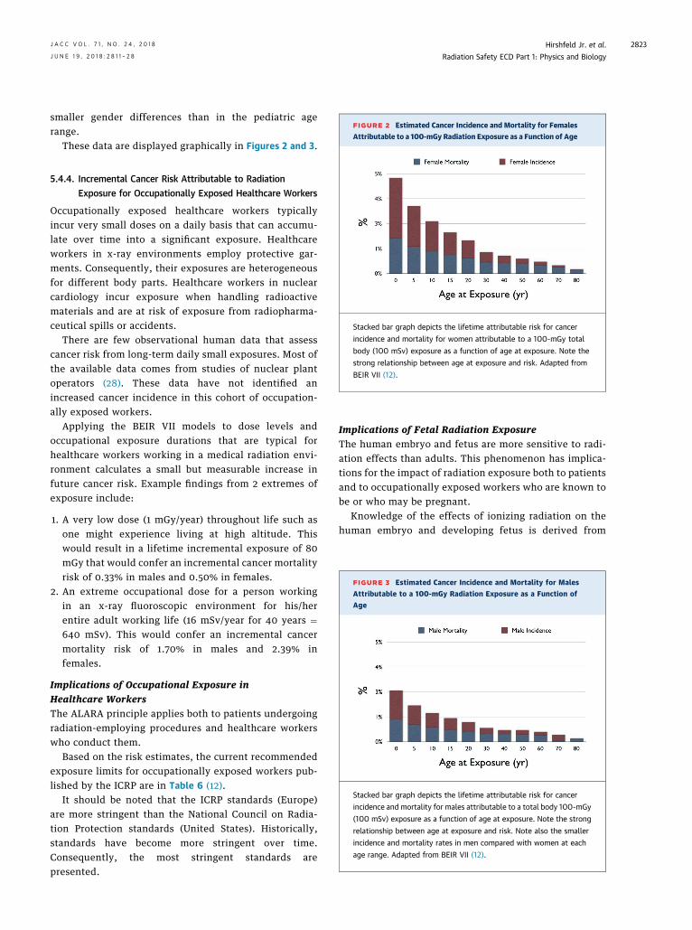
FIGURE 2 Estimated Cancer Incidence and Mortality for Females
Attributable to a 100-mGy Radiation Exposure as a Function of Age
Stacked bar graph depicts the lifetime attributable risk for cancer
incidence and mortality for women attributable to a 100-mGy total
body (100 mSv) exposure as a function of age at exposure. Note the
strong relationship between age at exposure and risk. Adapted from
BEIR VII (12).
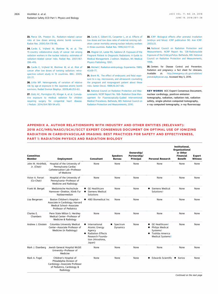
FIGURE 3 Estimated Cancer Incidence and Mortality for Males
Attributable to a 100-mGy Radiation Exposure as a Function of
Age
Stacked bar graph depicts the lifetime attributable risk for cancer
incidence and mortality for males attributable to a total body 100-mGy
(100 mSv) exposure as a function of age at exposure. Note the strong
relationship between age at exposure and risk. Note also the smaller
incidence and mortality rates in men compared with women at each
age range. Adapted from BEIR VII (12).
J A C C V O L . 7 1 , N O . 2 4 , 2 0 1 8 Hirshfeld Jr. et al.J U N E 1 9 , 2 0 1 8 : 2 8 1 1 – 2 8 Radiation Safety ECD Part 1: Physics and Biology
2823
smaller gender differences than in the pediatric agerange.
These data are displayed graphically in Figures 2 and 3.
5.4.4. Incremental Cancer Risk Attributable to Radiation
Exposure for Occupationally Exposed Healthcare Workers
Occupationally exposed healthcare workers typicallyincur very small doses on a daily basis that can accumu-late over time into a significant exposure. Healthcareworkers in x-ray environments employ protective gar-ments. Consequently, their exposures are heterogeneousfor different body parts. Healthcare workers in nuclearcardiology incur exposure when handling radioactivematerials and are at risk of exposure from radiopharma-ceutical spills or accidents.
There are few observational human data that assesscancer risk from long-term daily small exposures. Most ofthe available data comes from studies of nuclear plantoperators (28). These data have not identified anincreased cancer incidence in this cohort of occupation-ally exposed workers.
Applying the BEIR VII models to dose levels andoccupational exposure durations that are typical forhealthcare workers working in a medical radiation envi-ronment calculates a small but measurable increase infuture cancer risk. Example findings from 2 extremes ofexposure include:
1. A very low dose (1 mGy/year) throughout life such asone might experience living at high altitude. Thiswould result in a lifetime incremental exposure of 80mGy that would confer an incremental cancer mortalityrisk of 0.33% in males and 0.50% in females.
2. An extreme occupational dose for a person workingin an x-ray fluoroscopic environment for his/herentire adult working life (16 mSv/year for 40 years ¼640 mSv). This would confer an incremental cancermortality risk of 1.70% in males and 2.39% infemales.
Implications of Occupational Exposure in
Healthcare Workers
The ALARA principle applies both to patients undergoingradiation-employing procedures and healthcare workerswho conduct them.
Based on the risk estimates, the current recommendedexposure limits for occupationally exposed workers pub-lished by the ICRP are in Table 6 (12).
It should be noted that the ICRP standards (Europe)are more stringent than the National Council on Radia-tion Protection standards (United States). Historically,standards have become more stringent over time.Consequently, the most stringent standards arepresented.
Implications of Fetal Radiation Exposure
The human embryo and fetus are more sensitive to radi-ation effects than adults. This phenomenon has implica-tions for the impact of radiation exposure both to patientsand to occupationally exposed workers who are known tobe or who may be pregnant.
Knowledge of the effects of ionizing radiation on thehuman embryo and developing fetus is derived from

TABLE 6Recommended Exposure Limits forOccupationally Exposed Workers
Total body 20 mSv/yr averaged over defined periods of 5 yrs withno individual annual exposure to exceed 50 mSv.
Lens of the eye 100 mSv/5 yrs (20 mSv/yr)
Skin 500 mSv/yr
Hands and feet 500 mSv/yr
Adapted from the International Commission on Radiological Protection (12).
Hirshfeld Jr. et al. J A C C V O L . 7 1 , N O . 2 4 , 2 0 1 8
Radiation Safety ECD Part 1: Physics and Biology J U N E 1 9 , 2 0 1 8 : 2 8 1 1 – 2 8
2824
multiple sources, including the Hiroshima, Nagasaki, andChernobyl experiences as well as radiation of pregnantexperimental animals (29). Detrimental radiation effectsinclude embryonic death, fetal malformations, impairedfetal development (particularly neurological), andincreased risk of future cancer (16,30,31). The type ofevent and the dose-risk relationship for them is variablethroughout the stages of pregnancy and is summarized inTables 7 and 8 (32–34).
The principal risk of radiation exposure to the earlyembryo during the blastogenesis phase of development isintrauterine death, which would be experienced asfailure to establish a pregnancy. Exposure during the
TABLE 7 Estimates of Adverse Embryonic and Fetal Events as a
Acute Radiation Dose*to the Embryo/Fetus
T
Blastogenesis(up to 2 wks)
Organogenesis(2–7 wks)
<0.05 Gy (5 rads)† Noncancer health effects NOT detectable
0.05-0.50 Gy (5–50 rads) Incidence of failure to implantmay increase slightly, butsurviving embryos willprobably have nosignificant (noncancer)health effects
n Incidence of majormalformations mayincrease slightly
n Growth retardationpossible
>0.50 Gy (50 rads)The expectant mother
may be experiencingacute radiationsyndrome in thisrange, depending onher whole body dose.
Incidence of failure to implantwill likely be large.‡depending on dose, butsurviving embryos willprobably have nosignificant (noncancer)health effects
n Incidence ofmiscarriage mayincrease, dependingon dose
n Substantial risk ofmajor malforma-tions such asneurological andmotor deficiencies
n Growth retardationlikely
Note: This table is intended only as a guide. The indicated doses and times post conceptioFractionated or chronic doses: doses delivered over time. For fractionated or chronic doses thand the rad are units of absorbed dose and reflect the amount of energy deposited into a massthe entire fetus (whole-body fetal dose). The referenced absorbed dose levels in this documproduces many of the health effects described herein at lower absorbed dose levels. ‡A feta100% of human embryos or fetuses before 18 weeks’ gestation is about 5 Gy (500 rads). §Fodays) is about 3 to 5 Gy (300 to 500 rads) and the LD100 (the dose necessary to kill 100% ofthe Centers for Disease Control and Prevention (35).
organogenesis phase has the potential to cause fetalmalformations. Later exposure during the fetogenesisphase can cause growth retardation and impaired neuro-logical development, and can potentially increase thefetus’ future cancer risk.
In considering these risks, it is important to link therisk to threshold radiation doses. This knowledge base hasbeen summarized by the Centers for Disease Control andPrevention (35). In this document, dose ranges areexpressed in Gy rather than in Sv, as the Sv construct isnot applicable to embryos and fetuses.
The increased childhood cancer risk caused by fetalradiation exposure is less well characterized, andwhether fetal radiation exposure might confer a life-long increased cancer risk is not known. Estimates ofchildhood cancer risk are summarized in Table 8. Theavailable data indicate minimal detectable childhoodrisk at fetal doses <50 mGy but increased risk atdoses >50 mGy.
A general synthesis of the fetal radiation dose dataindicates that fetal doses <50 mGy (as distinguishedfrom maternal exposures to other body regions) are notassociated with a detectable increase in frequency of
Function of Fetal Radiation Dose
ime Post Conception
Fetogenesis
(8–15 wks) (16–25 wks) (26–38 wks)
n Growth retardationpossible
n Reduction in IQ possible(up to 15 points,depending on dose)
n Incidence of severemental retardation up to20%. depending ondose
Noncancer health effects unlikely
n Incidence of miscarriageprobably will increase,depending on dose
n Growth retardationlikely
n Reduction in IQ possible(>15 points, dependingon dose)
n Incidence of severemental retardation>20%, depending ondose
n Incidence of major mal-formations will probablyincrease
n Incidence of miscar-riage may increase,depending on dose
n Growth retardationpossible, dependingon dose
n Reduction in IQpossible, dependingon dose
n Severe mental retar-dation possible,depending on dose
n Incidence of majormalformations mayincrease
Incidence ofmiscarriage andneonatal deathwill probablyincreasedepending ondose§
n are approximations. *Acute dose: dose delivered in a short time (usually minutes).e health effects to the fetus may differ from what is depicted here. †Both the gray (Gy)of tissue (1 Gy ¼ 100 rads). In this document, the absorbed dose is that dose received byent are assumed to be from beta, gamma, or x radiation. Neutron or proton radiationl dose of 1 Gy (100 rads) will likely kill 50% of the embryos. The dose necessary to killr adults, the LD50/60 (the dose necessary to kill 50% of the exposed population in 60the exposed population) is around 10 Gy (1,000 rads). Reproduced with permission from

TABLE 8Estimated Risk for Cancer from Prenatal Radia-tion Exposure
Radiation DoseEstimated ChildhoodCancer Incidence*†
No radiation exposure above background 0.3% 38%
0.00–0.05 Gy (0–5 rads) 0.3%–1% 38%–40%
0.05–0.50 Gy (5–50 rads) 1%–6% 40%–55%
>0.50 Gy (50 rads) >6% >55%
Estimated lifetime‡ cancer incidence§ (exposure at age 10 years). The right columntabulates the estimated lifetime incidence of cancer for the same exposure incurred atage 10 for comparison to the estimated childhood incidence from fetal exposure.*Data published by the International Commission on Radiation Protection.†Childhood cancer mortality is roughly half of childhood cancer incidence.‡The lifetime cancer risks from prenatal radiation exposure are not yet known. Thelifetime risk estimates given are for Japanese males exposed at age 10 years frommodels published by the United Nations Scientific Committee on the Effects of AtomicRadiation.§Lifetime cancer mortality is roughly one third of lifetime cancer incidence. Reproducedwith permission from the Centers for Disease Control (35).
J A C C V O L . 7 1 , N O . 2 4 , 2 0 1 8 Hirshfeld Jr. et al.J U N E 1 9 , 2 0 1 8 : 2 8 1 1 – 2 8 Radiation Safety ECD Part 1: Physics and Biology
2825
any adverse fetal outcomes. For external beammaternal exposures (x-ray fluoroscopy and x-ray CT),fetal exposures are substantially less than the exposureto the imaged or unshielded body region unless theuterus is directly in the imaged field. For
occupationally exposed pregnant health care workers inx-ray fluoroscopy environments, proper shielding andpractices should keep uterine exposures substantiallybelow 50 mGy.
Specific recommendations for management of preg-nant, possibly pregnant, or lactating patients are dis-cussed in depth in the online version of this document.
PRESIDENT AND STAFF LIST
American College of Cardiology
Mary Norine Walsh MD, FACC, PresidentShalom Jacobovitz, Chief Executive OfficerWilliam J. Oetgen, MD, MBA, FACC, Executive Vice Pres-
ident, Science, Education, and QualityJoseph M. Allen, MA, Senior Director, Clinical Policy and
PathwaysLea Binder, Team Leader, Clinical Policy and PathwaysAmanda Ladden-Stirling, Associate, Clinical Policy and
PathwaysShira Klapper, Publications Manager (Acting), Clinical
Policy and Pathways
RE F E RENCE S
1. Mettler FA Jr., Bhargavan M, Faulkner K, et al.Radiologic and nuclear medicine studies in the UnitedStates and worldwide: frequency, radiation dose, andcomparison with other radiation sources—1950-2007.Radiology. 2009;253:520–31.
2. Picano E, Vano E, Rehani MM, et al. The appropriateand justified use of medical radiation in cardiovascularimaging: a position document of the ESC Associationsof Cardiovascular Imaging, Percutaneous Cardiovascu-lar Interventions and Electrophysiology. Eur Heart J.2014;35:665–72.
3. Vano E, Gonzalez L, Guibelalde E, et al. Radiationexposure to medical staff in interventional and cardiacradiology. Br J Radiol. 1998;71:954–60.
4. Linet MS, Kim KP, Miller DL, et al. Historical reviewof occupational exposures and cancer risks in medicalradiation workers. Radiat Res. 2010;174:793–808.
5. Einstein AJ, Tilkemeier P, Fazel R, et al. Radiationsafety in nuclear cardiology-current knowledge andpractice: results from the 2011 American Society ofNuclear Cardiology member survey. JAMA Intern Med.2013;173:1021–3.
6. Correia MJ, Hellies A, Andreassi MG, et al. Lack ofradiological awareness among physicians working in atertiary-care cardiological centre. Int J Cardiol. 2005;103:307–11.
7. Einstein AJ, Berman DS, Min JK, et al. Patient-centered imaging: shared decision making for cardiacimaging procedures with exposure to ionizing radia-tion. J Am Coll Cardiol. 2014;63:1480–9.
8. Wall BF, Hart D. Revised radiation doses for typicalX-ray examinations. Report on a recent review of dosesto patients from medical X-ray examinations in the UK
by NRPB. National Radiological Protection Board. Br JRadiol. 1997;70:437–9.
9. National Council on Radiation Protection and Mea-surements. NCRP Report No. 160– Ionizing RadiationExposure of the Population of the United States.Bethesda, MD: National Council on Radiation Protec-tion and Measurements, 2009.
10. Kim KP, Miller DL, Berrington de Gonzalez A, et al.Occupational radiation doses to operators performingfluoroscopically-guided procedures. Health Phys.2012;103:80–99.
11. Martin CJ. Effective dose: how should it be appliedto medical exposures? Br J Radiol. 2007;80:639–47.
12. ICRP. The 2007 recommendations of the Interna-tional Commission on Radiological Protection. ICRPpublication 103. Ann ICRP. 2007;37:1–332.
13. Balter S, Hopewell JW, Miller DL, et al. Fluoro-scopically guided interventional procedures: a reviewof radiation effects on patients’ skin and hair. Radi-ology. 2010;254:326–41.
14. National Council on Radiation Protection andMeasurements. NCRP Report No. 174– Preconceptionand Prenatal Radiation Exposure: Health Effects andProtective Guidance In: National Council on RadiationProtection and Measurements. Bethesda, MD: NationalCouncil on Radiation Protection and Measurements,2013.
15. National Council on Radiation Protection andMeasurements. NCRP Report No. 136– Evaluation ofthe Linear-Nonthreshold Dose-Response Model forIonizing Radiation. Bethesda, MD: National Council onRadiation Protection and Measurements, 2001.
16. National Research Council. Health Risks fromExposure to Low Levels of Ionizing Radiation: BEIR VIIPhase 2. Washington, DC: The National AcademiesPress, 2006.
17. Neel JV, Schull WJ, Awa AA, et al. The children ofparents exposed to atomic bombs: estimates of thegenetic doubling dose of radiation for humans. Am JHum Genet. 1990;46:1053–72.
18. Bouffler S, Ainsbury E, Gilvin P, et al. Radiation-induced cataracts: the Health Protection Agency’sresponse to the ICRP statement on tissue reactions andrecommendation on the dose limit for the eye lens.J Radiol Prot. 2012;32:479–88.
19. Levine GN, Bates ER, Blankenship JC, et al. 2011ACCF/AHA/SCAI guideline for percutaneous coronaryintervention: a report of the American College of Car-diology Foundation/American Heart Association TaskForce on Practice Guidelines and the Society for Car-diovascular Angiography and Interventions. J Am CollCardiol. 2011;58:e44–122.
20. Modan B, Keinan L, Blumstein T, et al. Cancerfollowing cardiac catheterization in childhood. Int JEpidemiol. 2000;29:424–8.
21. Pearce MS, Salotti JA, Little MP, et al. Radia-tion exposure from CT scans in childhood andsubsequent risk of leukaemia and brain tumours:a retrospective cohort study. Lancet. 2012;380:499–505.
22. Preston DL, Shimizu Y, Pierce DA, et al. Studies ofmortality of atomic bomb survivors. Report 13: Solidcancer and noncancer disease mortality: 1950-1997.Radiat Res. 2003;160:381–407.
Hirshfeld Jr. et al. J A C C V O L . 7 1 , N O . 2 4 , 2 0 1 8
Radiation Safety ECD Part 1: Physics and Biology J U N E 1 9 , 2 0 1 8 : 2 8 1 1 – 2 8
2826
23. Pierce DA, Preston DL. Radiation-related cancerrisks at low doses among atomic bomb survivors.Radiat Res. 2000;154:178–86.
24. Cardis E, Vrijheid M, Blettner M, et al. The15-country collaborative study of cancer risk amongradiation workers in the nuclear industry: estimates ofradiation-related cancer risks. Radiat Res. 2007;167:396–416.
25. Cardis E, Vrijheid M, Blettner M, et al. Risk ofcancer after low doses of ionising radiation: retro-spective cohort study in 15 countries. BMJ. 2005;331:77.
26. Little MP. Heterogeneity of variation of relativerisk by age at exposure in the Japanese atomic bombsurvivors. Radiat Environ Biophys. 2009;48:253–62.
27. Glatz AC, Purrington KS, Klinger A, et al. Cumula-tive exposure to medical radiation for childrenrequiring surgery for congenital heart disease.J Pediatr. 2014;164:789–94.e10.
CommitteeMember Employment
John W. Hirshfeld,Jr. (Chair)
Hospital of the University ofPennsylvania Cardiac
Catheterization Lab—Professorof Medicine
Victor A. Ferrari(Co-Chair)
Hospital of the University ofPennsylvania—Professor ofMedicine and Radiology
Frank M. Bengel Medizinische HochschuleHannover—Direktor, Klinik Fur
Nuklearmedizin
n
n
Lisa Bergersen Boston Children’s Hospital—Associate in Cardiology; Harvard
Medical School—AssociateProfessor of Pediatrics
n
Charles E.Chambers
Penn State Milton S. HersheyMedical Center—Professor of
Medicine & Radiology
Andrew J. Einstein Columbia University MedicalCenter—Associate Professor of
Medicine (in Radiology)
n
n
Mark J. Eisenberg Jewish General Hospital McGillUniversity—Professor of
Medicine
Mark A. Fogel Children’s Hospital ofPhiladelphia Division of
Cardiology—Associate Professorof Pediatrics, Cardiology &
Radiology
28. Cardis E, Gilbert ES, Carpenter L, et al. Effects oflow doses and low dose rates of external ionizing radi-ation: cancer mortality among nuclear industry workersin three countries. Radiat Res. 1995;142:117–32.
29. Wagner LK, Lester RG, Saldana LR. Exposure of thePregnant Patient to Diagnostic Radiations: A Guide toMedical Management. 2 edition. Madison, WI: MedicalPhysics Publishing, 1997.
30. Michel C. Radiation embryology. Experientia. 1989;45:69–77.
31. Brent RL. The effect of embryonic and fetal expo-sure to x-ray, microwaves, and ultrasound: counselingthe pregnant and nonpregnant patient about theserisks. Semin Oncol. 1989;16:347–68.
32. National Council on Radiation Protection and Mea-surements. NCRP Report No. 168– Radiation Dose Man-agement for Fluoroscopically-Guided InterventionalMedical Procedures. Bethesda, MD: National Council onRadiation Protection and Measurements, 2010.
ConsultantSpeakersBureau
Ownership/Partnership/Principal
None None None
None None None
GE HealthcareSiemens MedicalSolutions
None None
480 Biomedical Inc None None
None None None
InternationalAtomic EnergyAgencyRadiation EffectsResearch Founda-tion (Hiroshima,Japan)
n SpectrumDynamics
None
None None None
None None None
33. ICRP. Biological effects after prenatal irradiation(embryo and fetus). ICRP publication 90. Ann ICRP.2003;33.
34. National Council on Radiation Protection andMeasurements. NCRP Report No. 128-RadionuclideExposure of the Embryo/Fetus. Bethesda, MD: NationalCouncil on Radiation Protection and Measurements,1998.
35. Centers for Disease Control and Prevention.Radiation and pregnancy: a fact sheet for clinicians.Available at: http://emergency.cdc.gov/radiation/prenatalphysician.asp. Accessed May 5, 2016.
KEY WORDS ACC Expert Consensus Document,nuclear cardiology, positron emissiontomography, radiation, radiation risk, radiationsafety, single-photon computed tomography,x-ray computed tomography, x-ray fluoroscopy
APPENDIX A. AUTHOR RELATIONSHIPS WITH INDUSTRY AND OTHER ENTITIES (RELEVANT):
2018 ACC/HRS/NASCI/SCAI/SCCT EXPERT CONSENSUS DOCUMENT ON OPTIMAL USE OF IONIZING
RADIATION IN CARDIOVASCULAR IMAGING: BEST PRACTICES FOR SAFETY AND EFFECTIVENESS,
PART 1: RADIATION PHYSICS AND RADIATION BIOLOGY
Personal Research
Institutional,Organizational
or OtherFinancialBenefit
ExpertWitness
None None None
None None None
n Siemens MedicalSolutions†
None None
None None None
None None None
n GE Healthcare†n Philips Medical
Systems†n Toshiba America
Medical Systems†
None None
None None None
n Edwards Scientific n Kerios None
Continued on the next page

CommitteeMember Employment Consultant
SpeakersBureau
Ownership/Partnership/Principal Personal Research
Institutional,Organizational
or OtherFinancialBenefit
ExpertWitness
Thomas C. Gerber Mayo Clinic, Rochester—Professor of Medicine and
Radiology
None None None None None None
David E. Haines William Beaumont Hospital—Director, Heart Rhythm Center
None None None n Boston Scientificn Medtronic
None None
Warren K. Laskey University of New Mexico Schoolof Medicine—Professor ofMedicine; Chief, Division of
Cardiology
None None None None None None
Marian C.Limacher
University of Florida—Professorof Medicine, Division ofCardiovascular Medicine
None None None None None None
Kenneth J. Nichols Northwell Health, Division ofNuclear Medicine and Molecular
Imaging—Professor ofRadiology, Hofstra University
n Syntermed Inc.* None None None None None
Daniel A. Pryma Perelman School of Medicine atthe University of Pennsylvania—
Associate Professor ofRadiology & Radiation Oncology
Chief, Nuclear Medicine &Clinical Molecular Imaging
None None None n AdvancedAcceleratorApplications†
n Bayer HealthcarePharmaceutical†
n ProgenicsPharmaceuticals†
n Siemens†
None None
Gilbert L. Raff William Beaumont Hospital—Medical Director, AdvancedCardiovascular Imaging
None None None None None None
Geoffrey D. Rubin Duke University MedicalCenter—Professor of Radiology
and Bioengineering
n Fovian GE Healthcare
None n Terareconn HeartFlow
None None None
Donnette Smith Mended Hearts—Executive VicePresident
None None None None None None
Arthur E. Stillman Emory University Hospital—Professor of Radiology &
Imaging Sciences; Professor ofMedicine
None None None None None None
Suma A. Thomas Cleveland Clinic Heart andVascular Institute—Vice-
Chairman Strategic Operations
None None None None None None
Thomas T. Tsai Kaiser Permanente—Interventional Cardiology;
Kaiser Permanente ColoradoInstitute for Health Research;
University of Colorado Denver—Associate Clinical Professor.
None None None None None None
Louis K. Wagner University of Texas/HoustonMedical School Department of
Radiology—Professor
None None None None None None
L. Samuel Wann University of Wisconsin,Madison and Medical College ofWisconsin, Milwaukee—Clinical
Professor of Medicine
n United Healthcaren Astellas
Pharmaceutical
None None None None None
This table represents the relationships of committee members with industry and other entities that were determined to be relevant to this document. These relationships werereviewed and updated in conjunction with all meetings and/or conference calls of the writing committee during the document development process. The table does not necessarilyreflect relationships with industry at the time of publication. A person is deemed to have a significant interest in a business if the interest represents ownership of $5% of the votingstock or share of the business entity, or ownership of $$5,000 of the fair market value of the business entity; or if funds received by the person from the business entity exceed 5% ofthe person’s gross income for the previous year. Relationships that exist with no financial benefit are also included for the purpose of transparency. Relationships in this table aremodest unless otherwise noted. According to the ACC, a person has a relevant relationship IF: a) the relationship or interest relates to the same or similar subject matter, intellectualproperty or asset, topic, or issue addressed in the document; or b) the company/entity (with whom the relationship exists) makes a drug, drug class, or device addressed in thedocument, or makes a competing drug or device addressed in the document; or c) the person or a member of the person’s household, has a reasonable potential for financial, professionalor other personal gain or loss as a result of the issues/content addressed in the document.*Significant relationship.†No financial benefit.
APPENDIX A. CONTINUED
J A C C V O L . 7 1 , N O . 2 4 , 2 0 1 8 Hirshfeld Jr. et al.J U N E 1 9 , 2 0 1 8 : 2 8 1 1 – 2 8 Radiation Safety ECD Part 1: Physics and Biology
2827

Hirshfeld Jr. et al. J A C C V O L . 7 1 , N O . 2 4 , 2 0 1 8
Radiation Safety ECD Part 1: Physics and Biology J U N E 1 9 , 2 0 1 8 : 2 8 1 1 – 2 8
2828
APPENDIX B. PEER REVIEWER INFORMATION: 2018 ACC/HRS/NASCI/SCAI/SCCT EXPERT CONSENSUS
DOCUMENT ON OPTIMAL USE OF IONIZING RADIATION IN CARDIOVASCULAR IMAGING: BEST
PRACTICES FOR SAFETY AND EFFECTIVENESS, PART 1: RADIATION PHYSICS AND RADIATION BIOLOGY
This table represents the individuals, organizations, andgroups that peer reviewed this document. A list of
Reviewer Representation
Lyndon Box Official Reviewer—SCAI
James Case Official Reviewer—ASNC
Manuel D. Cerqueira Official Reviewer—ASNC Cl
Panithaya Chareonthaitawee Official Reviewer—SNMMI
Mina K. Chung Official Reviewer—HRS Cle
Mehmet Cilingiroglu Official Reviewer—SCAI
B. Kelly Han Official Reviewer—SCCT Chi
Jacobo Kirsch Official Reviewer—NASCI C
Dharam Kumbhani Official Reviewer—ACC Task Force onClinical Expert Consensus Documents
Thomas H. Schindler Official Reviewer—SNMMI Jo
Todd C. Villines Official Reviewer—ACC Board of Governors
Paul C. Zei Official Reviewer—HRS
James A. Arrighi Content Reviewer— CompetencyManagement Committee
Noel G. Boyle Content Reviewer—EP Council UCL
Rami Doukky Content Reviewer—Imaging Council C
Jonathan Halperin Content Reviewer— CompetencyManagement Committee
Mahadevappa Mahesh Content Reviewer—SCCT John
Joseph E. Marine Content Reviewer—ACC Task Force onClinical Expert Consensus Documents
J
Pamela B. Morris Content Reviewer—ACC Task Force onClinical Expert Consensus Documents
Me
William A. Van Decker Content Reviewer—Health AffairsCommittee
Robert Vincent Content Reviewer—ACPC Council
ABIM ¼ American Board of Internal Medicine; ACC ¼ American College of Cardiology; ACPCAssociation; ASNC ¼ American Society of Nuclear Cardiology; HRS ¼ Heart Rhythm Societydiovascular Angiography and Interventions; SCCT ¼ Society of Cardiovascular Computed TomoTexas.
corresponding comprehensive healthcare-related disclo-sures for each reviewer is available as an Online Appendix.
Employment
West Valley Cardiology—Interventional Cardiologist; Cardiac CatheterizationLaboratory Director
Cardiovascular Imaging Technologies—Chief Scientific Officer
eveland Clinic Foundation—Chairman, Department of Molecular and FunctionalImaging
Mayo Clinic—Consultant and Associate Professor of Medicine, Department ofCardiovascular Medicine
veland Clinic Lerner College of Medicine of Case Western Reserve University—Associate Professor of Medicine
Arkansas Heart Hospital—Professor of Medicine
ldren’s Hospitals and Clinics of Minnesota and the Minneapolis Heart Institute—Director of Advanced Congenital Cardiac Imaging
leveland Clinic Florida—Professor; Harper University Hospital, Detroit MedicalCenter—Director, Center for Hospital Specialties
UT Southwestern Medical Center—Assistant Professor of Medicine
hns Hopkins University—Director of Cardiovascular Nuclear Medicine; AssociateProfessor of Medicine and Radiology
Walter Reed National Military Medical Center—Director, Cardiovascular CT
Stanford University School of Medicine—Clinical Professor of Medicine,Electrophysiology, and Cardiovascular Medicine
Rhode Island Hospital—Director, Graduate Medical Education
A Medical Center, Cardiac Arrhythmia Center—Professor of Medicine; Director ofCardiac EP Labs & Fellowship Program
ook County Health and Hospitals System—Professor of Medicine, PreventiveMedicine, and Radiology; Chairman, Division of Cardiology
Mount Sinai Medical Center—Professor of Medicine
s Hopkins Hospital—Professor of Radiology and Radiological Science; Professor ofMedicine, Cardiology, Chief Physicist
ohns Hopkins University School of Medicine—Associate Professor of Medicine
dical University of South Carolina—Director, Seinsheimer Cardiovascular HealthProgram; Women’s Heart Care Co-Director
Temple University Hospital—Assistant Professor of Medicine
Children’s Healthcare of Atlanta, Sibley Heart Center—Pediatric Cardiologist
¼ Adults with Congenital Heart Disease and Pediatric Cardiology; AHA ¼ American Heart; NASCI ¼ North American Society for Cardiovascular Imaging; SCAI ¼ Society for Car-graphy; SNMMI ¼ Society of Nuclear Medicine and Molecular Imagine, UT ¼ University of
APPENDIX C. ABBREVIATIONS
Gy ¼ gray
Kerma ¼ kinetic energy released in material
keV ¼ kiloelectron volts
mSv ¼ millisieverts
RWI ¼ relationship with industry
Sv ¼ sievert