all human body cells belong to one of these tissues: ◦ epithelial tissue ◦ connective tissue ◦...
TRANSCRIPT

Tissues
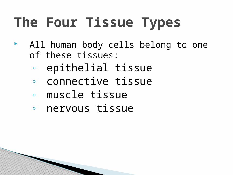
All human body cells belong to one of these tissues:◦ epithelial tissue◦ connective tissue◦ muscle tissue◦ nervous tissue
The Four Tissue Types

Epithelial Tissue
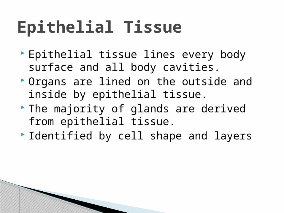
Epithelial tissue lines every body surface and all body cavities.
Organs are lined on the outside and inside by epithelial tissue.
The majority of glands are derived from epithelial tissue.
Identified by cell shape and layers
Epithelial Tissue
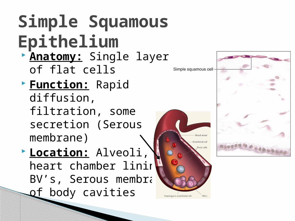
Anatomy: Single layer of flat cells
Function: Rapid diffusion, filtration, some secretion (Serous membrane)
Location: Alveoli, heart chamber lining, BV’s, Serous membrane of body cavities
Simple Squamous Epithelium
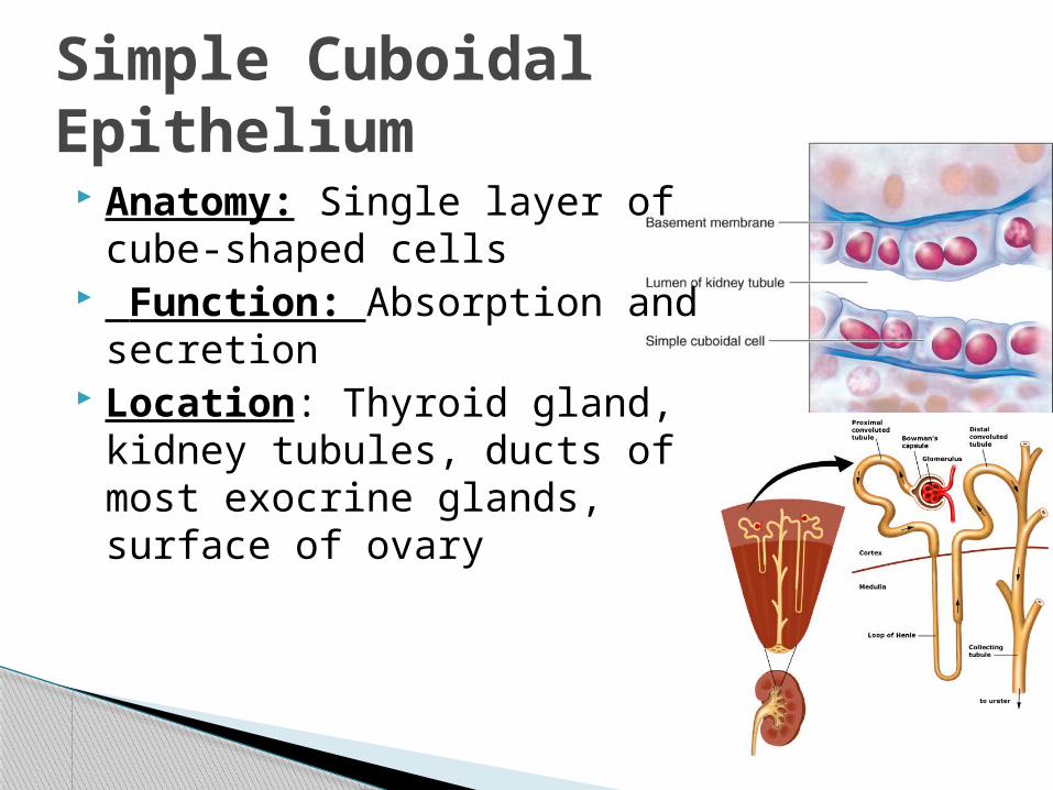
Anatomy: Single layer of cube-shaped cells
Function: Absorption and secretion
Location: Thyroid gland, kidney tubules, ducts of most exocrine glands, surface of ovary
Simple Cuboidal Epithelium

Anatomy: Single layer of cells that are taller than they are wide. Microvilli & Goblet cells.
Function: Absorption, secrete mucin
Location: Digestive tract lining. (No goblets in stomach)
Non Ciliated: Simple Columnar Epithelium
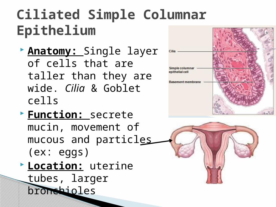
Anatomy: Single layer of cells that are taller than they are wide. Cilia & Goblet cells
Function: secrete mucin, movement of mucous and particles (ex: eggs)
Location: uterine tubes, larger bronchioles
Ciliated Simple Columnar Epithelium
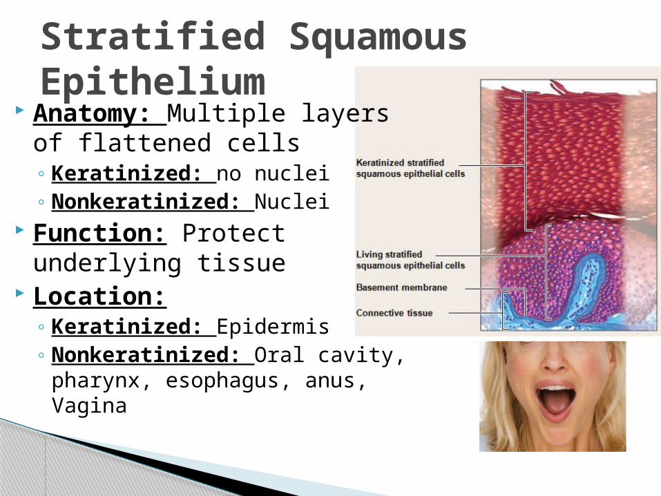
Anatomy: Multiple layers of flattened cells◦ Keratinized: no nuclei ◦ Nonkeratinized: Nuclei
Function: Protect underlying tissue
Location:◦ Keratinized: Epidermis◦ Nonkeratinized: Oral cavity,
pharynx, esophagus, anus, Vagina
Stratified Squamous Epithelium
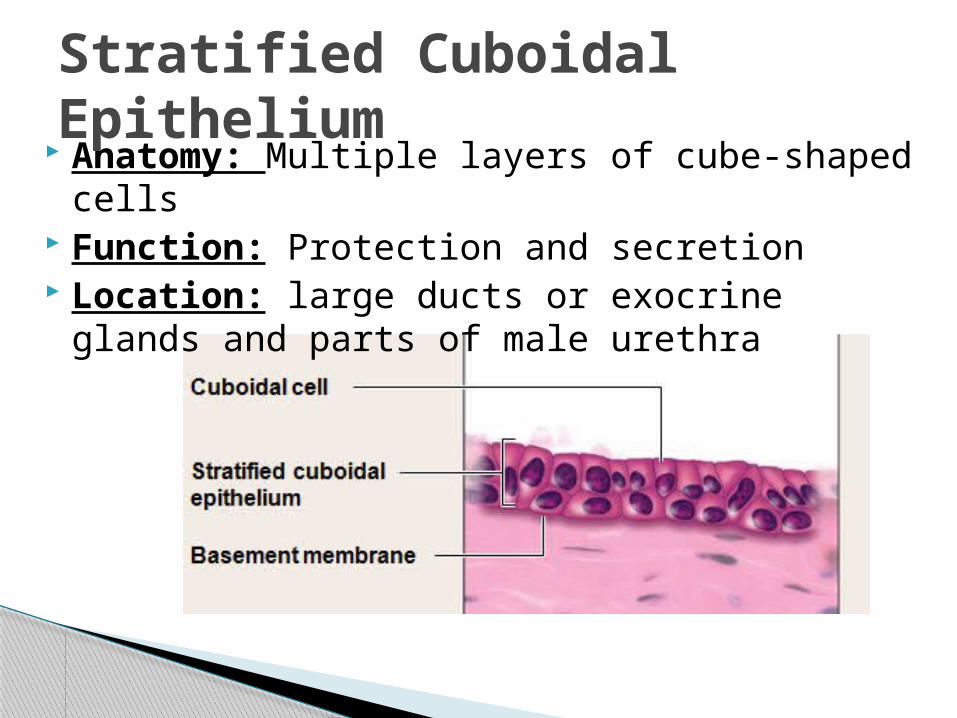
Anatomy: Multiple layers of cube-shaped cells Function: Protection and secretion Location: large ducts or exocrine glands and
parts of male urethra
Stratified Cuboidal Epithelium
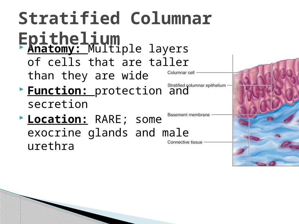
Anatomy: Multiple layers of cells that are taller than they are wide
Function: protection and secretion
Location: RARE; some exocrine glands and male urethra
Stratified Columnar Epithelium
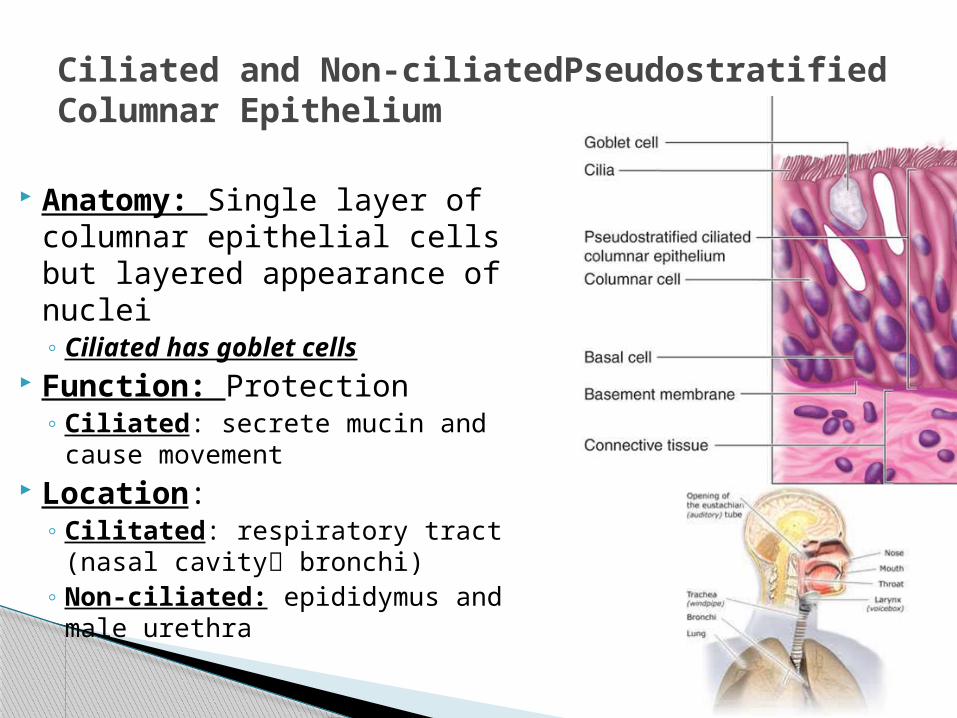
Anatomy: Single layer of columnar epithelial cells but layered appearance of nuclei◦ Ciliated has goblet cells
Function: Protection◦ Ciliated: secrete mucin and
cause movement Location:
◦ Cilitated: respiratory tract (nasal cavity bronchi)
◦ Non-ciliated: epididymus and male urethra
Ciliated and Non-ciliatedPseudostratified Columnar Epithelium
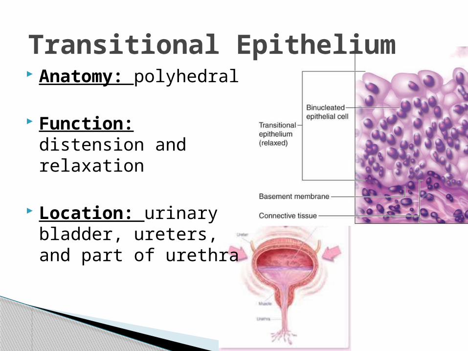
Anatomy: polyhedral
Function: distension and relaxation
Location: urinary bladder, ureters, and part of urethra
Transitional Epithelium
Muscle TissueSupportive
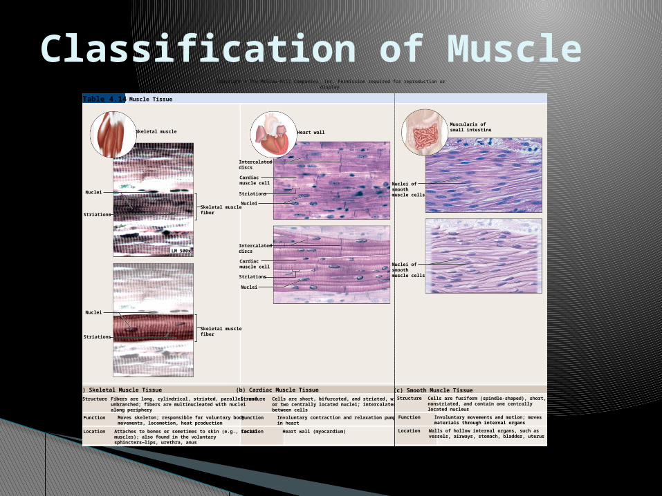
Classification of MuscleCopyright © The McGraw-Hill Companies, Inc. Permission required for reproduction or display.
Structure
Function
Location Location
Function
Structure
Skeletal muscle Heart wall
Nuclei
Striations
LM 500x
Nuclei
Striations
Skeletal musclefiber
Intercalateddiscs
Cardiacmuscle cell
Striations
NucleiSkeletal musclefiber
Intercalateddiscs
Nuclei
Striations
Cardiacmuscle cell
Table 4.14 Muscle Tissue
(a) Skeletal Muscle Tissue (b) Cardiac Muscle Tissue
Fibers are long, cylindrical, striated, parallel, andunbranched; fibers are multinucleated with nucleialong periphery
Moves skeleton; responsible for voluntary bodymovements, locomotion, heat production
Attaches to bones or sometimes to skin (e.g., facialmuscles); also found in the voluntarysphincters—lips, urethra, anus
Cells are short, bifurcated, and striated, with oneor two centrally located nuclei; intercalated discsbetween cells
Involuntary contraction and relaxation pump bloodin heart
Heart wall (myocardium)
a:© The McGraw-Hill Companies, Inc./Photo by Dr. Alvin Telser; b,c: © Victor Eroschenko
Muscularis ofsmall intestine
Nuclei ofsmoothmuscle cells
Nuclei ofsmoothmuscle cells
Structure
Function
Location Walls of hollow internal organs, such asvessels, airways, stomach, bladder, uterus
Cells are fusiform (spindle-shaped), short,nonstriated, and contain one centrallylocated nucleus
Involuntary movements and motion; movesmaterials through internal organs
(c) Smooth Muscle Tissue
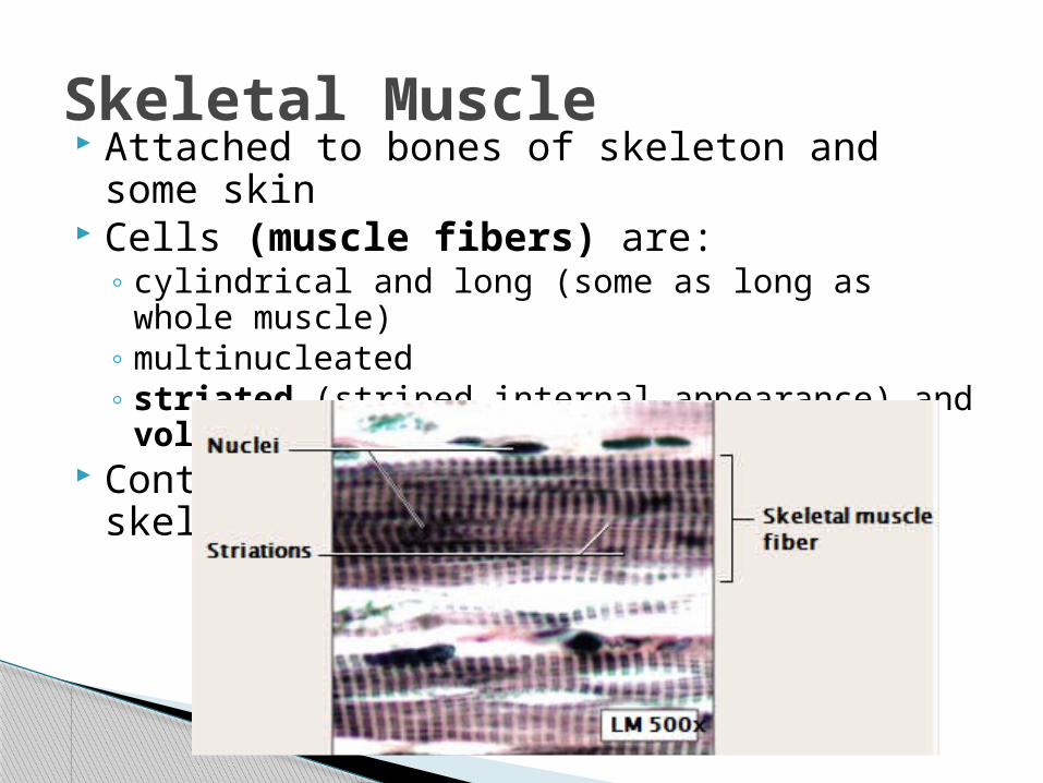
Attached to bones of skeleton and some skin
Cells (muscle fibers) are:◦ cylindrical and long (some as long as whole
muscle)◦ multinucleated◦ striated (striped internal appearance) and
voluntary Contraction causes movement of skeleton
or skin
Skeletal Muscle

Found in the wall of the heart (myocardium) Cells are:
◦ Y-shaped, shorter than skeletal fiber cells◦ striated and involuntary◦ attached by strong gap junctions called
intercalated discs: allow rapid passage of electrical current
Contraction causes movement of blood
Cardiac Muscle
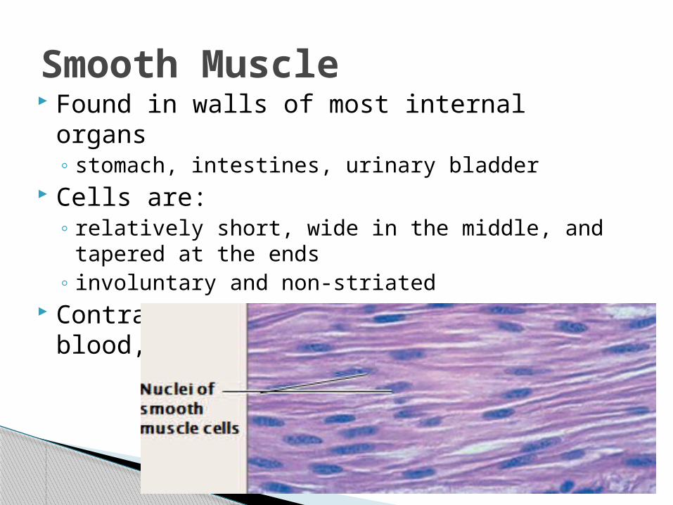
Found in walls of most internal organs◦ stomach, intestines, urinary bladder
Cells are:◦ relatively short, wide in the middle, and tapered
at the ends◦ involuntary and non-striated
Contraction causes movement of food, blood, sperm
Smooth Muscle

Connective TissueProper
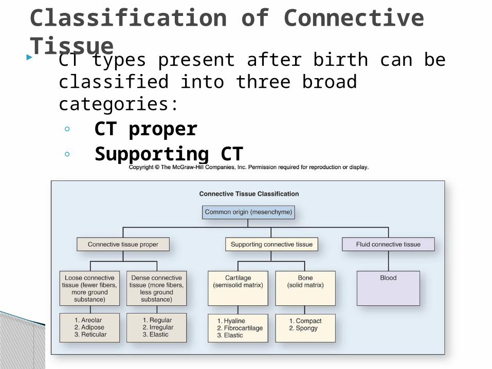
CT types present after birth can be classified into three broad categories:◦ CT proper◦ Supporting CT◦ Fluid CT
Classification of Connective Tissue

Body’s packing material, in spaces around organs; there are three types: Areolar CT: fibroblasts, collagen, and
elastic fibers; distorted without damage; found subcutaneous to skin
Loose Connective Tissue
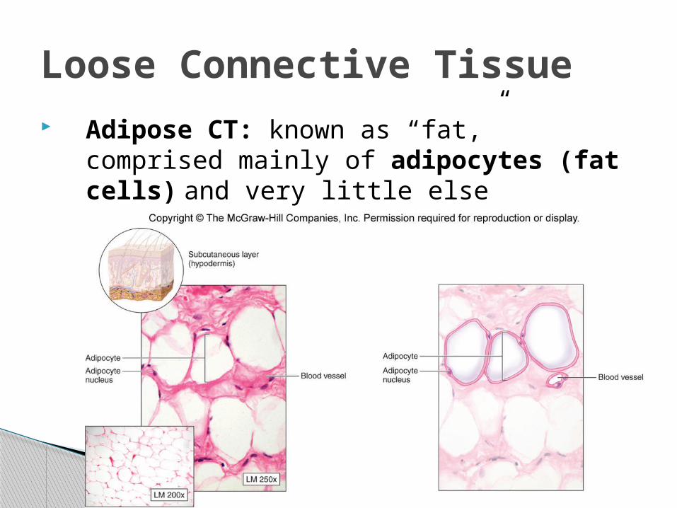
Adipose CT: known as “fat,” comprised mainly of adipocytes (fat cells) and very little else
Loose Connective Tissue
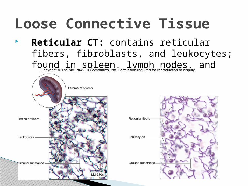
Reticular CT: contains reticular fibers, fibroblasts, and leukocytes; found in spleen, lymph nodes, and bone marrow
Loose Connective Tissue
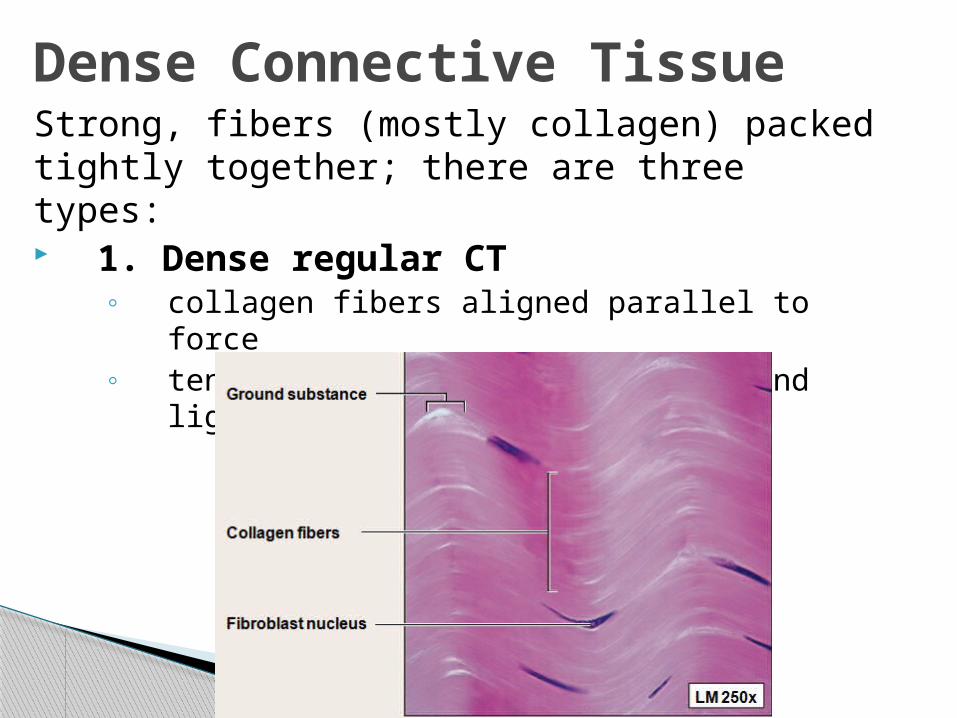
Strong, fibers (mostly collagen) packed tightly together; there are three types: 1. Dense regular CT
◦ collagen fibers aligned parallel to force◦ tendons (attach muscle to bone) and
ligaments (attach bone to bone)
Dense Connective Tissue
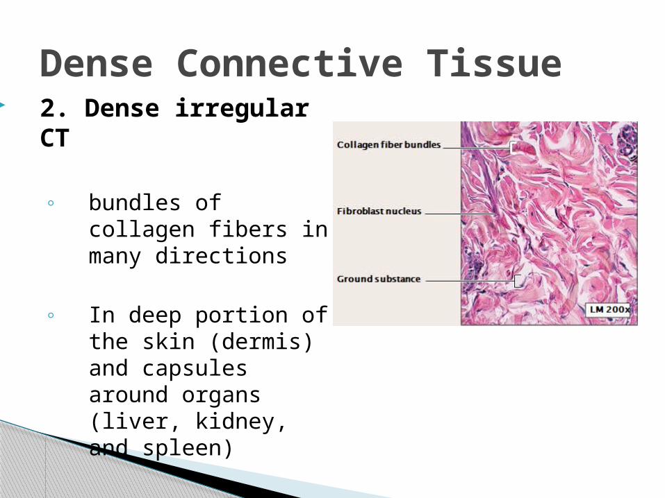
2. Dense irregular CT
◦ bundles of collagen fibers in many directions
◦ In deep portion of the skin (dermis) and capsules around organs (liver, kidney, and spleen)
Dense Connective Tissue
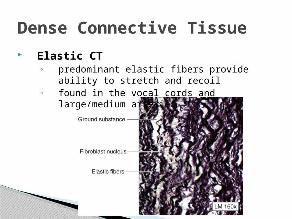
Elastic CT◦ predominant elastic fibers provide ability to
stretch and recoil◦ found in the vocal cords and large/medium
arteries
Dense Connective Tissue
Connective TissueSupportive
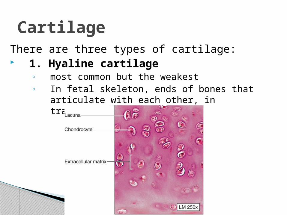
There are three types of cartilage: 1. Hyaline cartilage
◦ most common but the weakest◦ In fetal skeleton, ends of bones that articulate
with each other, in trachea, larynx, and nose
Cartilage
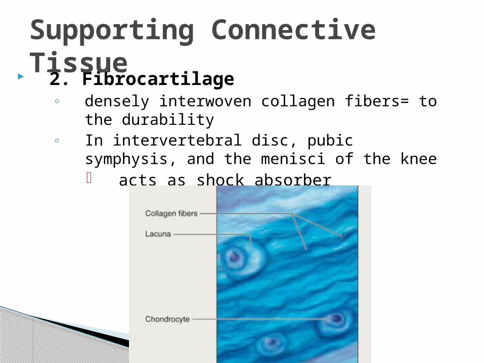
2. Fibrocartilage ◦ densely interwoven collagen fibers= to the
durability◦ In intervertebral disc, pubic symphysis, and the
menisci of the knee acts as shock absorber
Supporting Connective Tissue
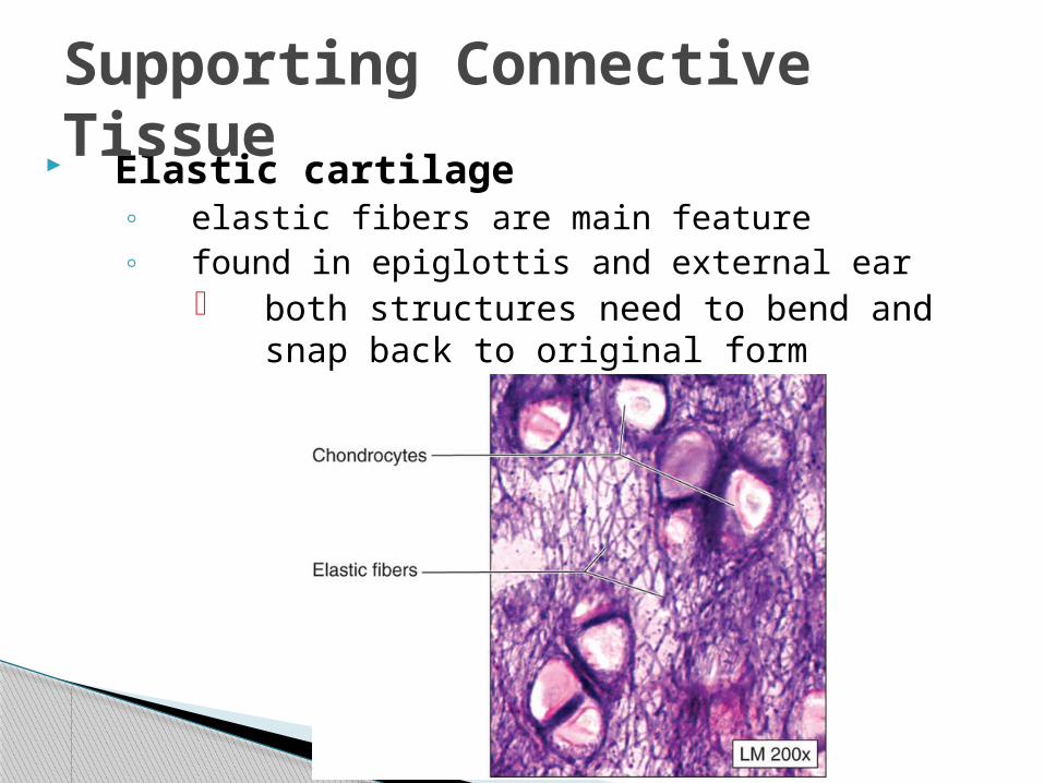
Elastic cartilage◦ elastic fibers are main feature◦ found in epiglottis and external ear
both structures need to bend and snap back to original form
Supporting Connective Tissue
Nervous Tissue
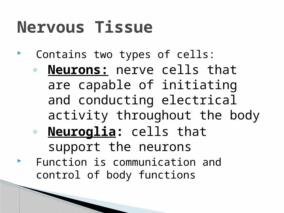
Contains two types of cells:◦ Neurons: nerve cells that are
capable of initiating and conducting electrical activity throughout the body
◦ Neuroglia: cells that support the neurons
Function is communication and control of body functions
Nervous Tissue
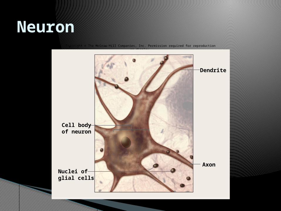
NeuronCopyright © The McGraw-Hill Companies, Inc. Permission required for reproduction or display.
Axon
Dendrite
Cell bodyof neuron
Nuclei ofglial cells