< preliminary report
TRANSCRIPT

Oxidative stress in lens in vivo: Inhibitory effect of caffeine. Apreliminary report
SD Varma,1,2 KR Hegde,1 S Kovtun1
1Department of Ophthalmology & Visual Sciences, University of Maryland School of Medicine, Baltimore, MD; 2Department ofBiochemistry and Molecular Biology, University of Maryland School of Medicine, Baltimore, MD
Purpose: Experiments have been conducted to study the hypothesis that caffeine would inhibit reactive oxygen speciesinduced oxidative stress in the lens in vivo, with implications of attenuating or preventing cataract formation.Methods: Oxidative stress was directly induced by administering 24% galactose diet to young adult rats. The treatedgroup was fed a diet containing 24% galactose + 1% caffeine. Oxidative stress inflicted to the lens was assessed bymeasurement of glutathione (GSH) depletion and observing the status of lens clarity.Results: Caffeine administration was found to minimize the loss of GSH. This was also associated with a bettermaintenance of lens transparency as compared to the untreated galactosemic group.Conclusions: The studies demonstrate that caffeine could be helpful in inhibiting oxidative stress in the lens with theconsequence of attenuating cataract formation.
Caffeine (1,3,7-trimethylxanthine) is one of the commoningredients of many beverages such as coffee, tea and variouscolas. It is also widely used medically, as a CNS, respiratoryand cardiac stimulant, smooth muscle relaxant, analgesic andas a diuretic [1]. The stimulatory effect of caffeine in thenervous system has been attributed to its competitive bindingwith certain pre-synaptic adenosine receptors andconsequently abolishing the inhibitory but regulatory effectof adenosine on release of various neurotransmitters.Adenosine binding to its receptor in the pre-synaptic terminalhas the effect of limiting calcium ion penetration through thecell membrane, and thus inhibiting neurotransmitter release atthe synapses [2]. The basic effect of caffeine binding to theadenosine receptors is therefore to overcome the inhibition ofcalcium passage through the cell membranes andconsequently facilitate the release of stimulatoryneurotransmitters such as epinephrine. Its action onstimulation of respiratory, cardiac and skeletal muscles hasalso been suggested to be exerted by an increase in thecytosolic calcium by causing its de-sequestration from thesarcoplasmic reticulum [3-5]. It does so also by facilitating adirect calcium penetration through the cell membrane [3]. Inaddition to its neural and muscular effects, caffeine has beenshown to have several other physiologic effects such as ageneral metabolic stimulation [6-8] and thermogenesis [9].The latter is considered useful in improving sportsperformance. Most of the latter effects are suggested to be
Correspondence to: Shambhu D. Varma, Ph.D., FARVO, Professor,Department of Biochemistry, Professor and Director of Research,Department of Ophthalmology & Visual Sciences, University ofMaryland School of Medicine, Baltimore, MD, 21201; Phone: (410)706-3395; FAX: (410) 706-7057; email: [email protected]
associated with an increase in the cellular level of c-AMPinduced by virtue of its ability to inhibit adenosine 3′, 5′-monophosphate phosphodiesterase activity [10].
In addition to the above actions, more recent observationssuggest that it can also act as an antioxidant. The suggestionsare largely based on chemical studies showing it to be able toscavenge reactive oxygen species (ROS), particularly thehydroxyl radical (OH·), known to be generated in the body byirradiation with various electromagnetic frequencies such asexposure to UV, as well as by many ambient physiologicreactions involving oxygen utilization [11-13]. Thegeneration of these radicals is also enhanced in the tissues inmany pathological conditions induced by aging and certaindiseases. The interaction of OH· with caffeine results in itsoxidative de-methylation, generating partially N-methylatedxanthines such as theobromine, paraxanthine, andtheophylline [14,15]. In addition, OH· also leads to the fissionreactions at the double bonds producing methylated parabanicacid [16]. However under milder oxidative conditions, asprevalent physiologically, the prominent reaction is thegeneration of 8-hydroxy caffeine (1,3,7-trimethyl-8-hydroxyxanthine), which is structurally analogous to uric acidderived from xanthine [12,17]. The physiologic usefulness ofthe above reactions is strongly indicated by theradioprotective effects of caffeine against radiations such asthe UV irradiation. Additionally, it has been shown to preventFenton’s reaction-induced oxidation of glutathione [11], amajor antioxidant reserve in many tissues, including those ofthe eye. However, studies on examination of the possibleprotective effect of caffeine against ROS induced oxidativestress at the level of cell and tissue culture as well as in vivoare yet very limited, specially in the eye where oxidative stresshas been implicated in the genesis of diseases such as cataracts
Molecular Vision 2010; 16:501-505 <http://www.molvis.org/molvis/v16/a56>Received 11 January 2010 | Accepted 15 March 2010 | Published 23 March 2010
© 2010 Molecular Vision
501

[18-22] and retinal degenerations [23-25]. Recently we haveshown that oxidative damage to the lens in organ cultureinflicted by UV exposure or by trace metals such as iron, canbe significantly prevented by caffeine [26,27]. Preliminarydata presented here strongly suggest the possibility thatcaffeine would be effective in preventing oxidative stress tothe lens in vivo also. Such stress was induced by feeding highgalactose diet to young adult rats. The antioxidant effect ofcaffeine was assessed by measuring its ability to prevent lossof tissue glutathione and preserve lens transparency.
METHODSAll chemicals and reagents were purchased from SigmaChemical Co. (St. Louis, MO) Rats were purchased fromHarlan Laboratories and used in accordance with ARVOguidelines and as approved by the institutional animal careand use committee (IACUC).
Young Sprague Dawley rats weighing about 45 g wereused. The control group was fed a powdered laboratory chowcontaining 24% galactose ad lib. The experimental groupreceived the same diet but also containing 1% caffeine. Sincethe cataract process was found to set in within a couple of daysapparent ophthalmoscopically, they were euthanized on day4, lenses isolated, weighed, photographed, and used fordetermination of glutathione (GSH). Dulcitol was measuredby method of West and Rapoport [28]. The level of bloodgalactose in both the groups was 16±2 mM, determined usinga Boehringer-Manheim kit provided by Roche (Cat # 10 176303 035; Manheim, Germany). The measurement is based onspectrophotometric determination of nicotinamide adenine
dinucleotide, reduced (NADH) produced from nicotinamideadenine dinucleotide (NAD) by D-Galactose dehydrogenasedependent oxidation of galactose. Blood caffeine variedbetween 0.06 to 0.012 mM measured in the hospital clinicallaboratory.
Measurement of GSH and ATP: The isolated lenses werehomogenized in 0.5 ml dH2O and centrifuged. An aliquot ofthe supernatant was used for the determination of ATP asdescribed previously [26]. Subsequently trichloroacetic acidwas added to a final concentration of 5% and supernatantobtained by centrifugation of the sample. This supernatant(100 µl) was then mixed with 300 µl of 0.6 M Na2HPO4. Thiswas followed by the addition of 100 µl DTNB (5, 5′dithio-bis-2-nitrobenzoic acid) reagent [29]. The resulting yellowcolor was read spectrophotometrically at 412 nm. DTNBreagent was prepared by dissolving 4 mg DTNB and 100 mgtrisodium citrate in 10 ml dH2O. GSH standards were also runsimultaneously.
RESULTSThe results are described in Figure 1 and in Table 1, Table 2,and Table 3, the data in each table representing 8, 6, and 8animals, respectively. Since the galactose induced opacitiesin the caffeine untreated animals were similar in both the eyes,they were treated as one observation, treating the contralaterallenses as duplicates for biochemical analysis. Lenses in thecaffeine treated group had no opacity. As summarized inTable 1, caffeine inhibited the decrease in the content of lensGSH in galactose fed animals. The level of this tripeptide inthe control normal rats was about 6 µmoles/g as also reportedpreviously [30]. The level in the galactose fed animals was
Figure 1. Inhibition of cataractformation by caffeine. Trans-illumination photographs ofrepresentative lenses isolated from ratsfed 24% Galactose diet without and with1% Caffeine: Peripheral ring opacity(cortical cataract) was highly apparentin the untreated group (g), representingthe early stage of galactose cataract. Thelenses in the treated group receivingcaffeine were transparent (g+c).
Molecular Vision 2010; 16:501-505 <http://www.molvis.org/molvis/v16/a56> © 2010 Molecular Vision
502
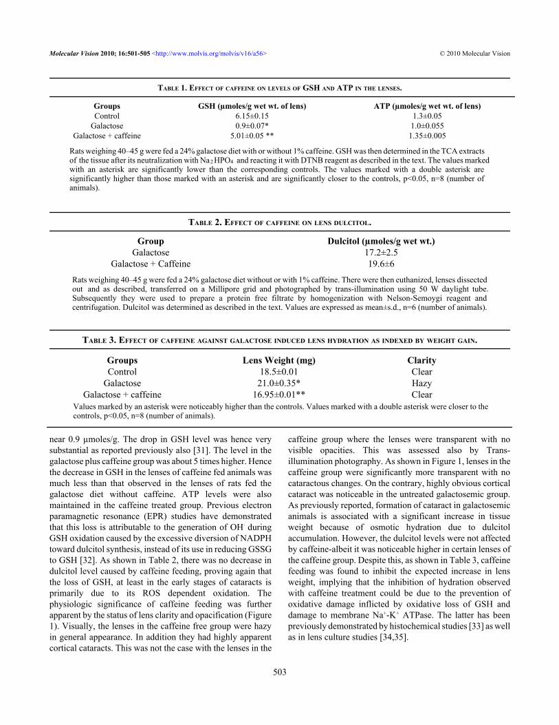
near 0.9 µmoles/g. The drop in GSH level was hence verysubstantial as reported previously also [31]. The level in thegalactose plus caffeine group was about 5 times higher. Hencethe decrease in GSH in the lenses of caffeine fed animals wasmuch less than that observed in the lenses of rats fed thegalactose diet without caffeine. ATP levels were alsomaintained in the caffeine treated group. Previous electronparamagnetic resonance (EPR) studies have demonstratedthat this loss is attributable to the generation of OH· duringGSH oxidation caused by the excessive diversion of NADPHtoward dulcitol synthesis, instead of its use in reducing GSSGto GSH [32]. As shown in Table 2, there was no decrease indulcitol level caused by caffeine feeding, proving again thatthe loss of GSH, at least in the early stages of cataracts isprimarily due to its ROS dependent oxidation. Thephysiologic significance of caffeine feeding was furtherapparent by the status of lens clarity and opacification (Figure1). Visually, the lenses in the caffeine free group were hazyin general appearance. In addition they had highly apparentcortical cataracts. This was not the case with the lenses in the
caffeine group where the lenses were transparent with novisible opacities. This was assessed also by Trans-illumination photography. As shown in Figure 1, lenses in thecaffeine group were significantly more transparent with nocataractous changes. On the contrary, highly obvious corticalcataract was noticeable in the untreated galactosemic group.As previously reported, formation of cataract in galactosemicanimals is associated with a significant increase in tissueweight because of osmotic hydration due to dulcitolaccumulation. However, the dulcitol levels were not affectedby caffeine-albeit it was noticeable higher in certain lenses ofthe caffeine group. Despite this, as shown in Table 3, caffeinefeeding was found to inhibit the expected increase in lensweight, implying that the inhibition of hydration observedwith caffeine treatment could be due to the prevention ofoxidative damage inflicted by oxidative loss of GSH anddamage to membrane Na+-K+ ATPase. The latter has beenpreviously demonstrated by histochemical studies [33] as wellas in lens culture studies [34,35].
TABLE 1. EFFECT OF CAFFEINE ON LEVELS OF GSH AND ATP IN THE LENSES.
Groups GSH (µmoles/g wet wt. of lens) ATP (µmoles/g wet wt. of lens)Control 6.15±0.15 1.3±0.05
Galactose 0.9±0.07* 1.0±0.055Galactose + caffeine 5.01±0.05 ** 1.35±0.005
Rats weighing 40–45 g were fed a 24% galactose diet with or without 1% caffeine. GSH was then determined in the TCA extractsof the tissue after its neutralization with Na2 HPO4 and reacting it with DTNB reagent as described in the text. The values markedwith an asterisk are significantly lower than the corresponding controls. The values marked with a double asterisk aresignificantly higher than those marked with an asterisk and are significantly closer to the controls, p<0.05, n=8 (number ofanimals).
TABLE 2. EFFECT OF CAFFEINE ON LENS DULCITOL.
Group Dulcitol (µmoles/g wet wt.)Galactose 17.2±2.5
Galactose + Caffeine 19.6±6
Rats weighing 40–45 g were fed a 24% galactose diet without or with 1% caffeine. There were then euthanized, lenses dissectedout and as described, transferred on a Millipore grid and photographed by trans-illumination using 50 W daylight tube.Subsequently they were used to prepare a protein free filtrate by homogenization with Nelson-Semoygi reagent andcentrifugation. Dulcitol was determined as described in the text. Values are expressed as mean±s.d., n=6 (number of animals).
TABLE 3. EFFECT OF CAFFEINE AGAINST GALACTOSE INDUCED LENS HYDRATION AS INDEXED BY WEIGHT GAIN.
Values marked by an asterisk were noticeably higher than the controls. Values marked with a double asterisk were closer to thecontrols, p<0.05, n=8 (number of animals).
Molecular Vision 2010; 16:501-505 <http://www.molvis.org/molvis/v16/a56> © 2010 Molecular Vision
503
Groups Lens Weight (mg) ClarityControl 18.5±0.01 Clear
Galactose 21.0±0.35* HazyGalactose + caffeine 16.95±0.01** Clear

DISCUSSIONSeveral previous studies have suggested that intraoculargeneration of oxygen free radicals such as superoxide andhydroxyl radicals and consequent oxidative stress is one of thesignificant factors involved in the genesis of cataractsassociated with aging, UV exposure, and many diseases suchas diabetes. Treatment with certain scavengers of reactiveoxygen is expected to thwart the oxidative stress componentof cataract formation. It is hypothesized that this can beachieved by use of ROS scavengers derived nutritionally, suchas ascorbate [36,37], tocopherols [38,39], and bioflavonoids[40]. However, they get oxidized during food processing aswell as during cooking, unlike caffeine which is present insubstantial amounts even after heating associated with initialprocessing of tea leaves and coffee beans as well as in hotwater used while preparing the drink proper. Hence itsproperty of scavenging ROS and consequent antioxidantactivity still remains maintained. Although substantialnumber of studies already exist on the medicinal andphysiologic effects of caffeine in other systems, such as itseffect in neural and muscular systems and the tissuemetabolism and bioenergetics, studies on its physiologiceffects in the eyes are as yet largely lacking, except our recentstudies showing that it can potentially prevent oxidativedamage to the lens with implications against cataractdevelopment. This has been suggested on the basis of in vitrostudies showing it ability to inhibit oxidative damage to lensinflicted by UV irradiation as well as by the radicals producedby enriching the culture medium with iron [26,27]. Theprimary aim of this investigation was hence to study if it couldbe useful in preventing oxidative stress to the lens in vivo also.This was done in the galactosemic rat model where the linkbetween oxyradical generation with cataract formation hasnow been more convincingly apparent [32,38]. Treatmentwith caffeine was found to inhibit the loss of glutathione aswell as ATP. The decrease in the level of these componentsin the caffeine untreated galactose fed group is similar to thatreported earlier [31,41]. The significance of the effectivenessof caffeine against ROS damage was apparent physiologicallyalso. Apart from the maintenance of lens clarity, other featuresof cataract formation such as lens hydration, was alsoprevented significantly by treatment with caffeine.Interestingly this effect was not found to be associated withany inhibition of lens dulcitol production. In an ongoing studywe have seen that the lenses of galactosemic animals givencaffeine retain their clarity even till three weeks. Furtherexperiments with different levels of caffeine are henceconsidered desirable, including such studies with the diabeticmodel and administering caffeine also topically. The presentpreliminary results showing an in vivo effectiveness ofcaffeine indeed lay the foundation for further mechanisticstudies involving other modes of caffeine effect, such as itsrelationship with possible maintenance of a higher level of
cyclic nucleotides, attributable to its inhibitory effect onphosphodiesterase.
ACKNOWLEDGMENTSThe authors are thankful for the financial support of NEI, NIH.
REFERENCES1. Serafin WE. In: Hardman JG, Limbird LE, Molinoff PB,
Ruddon RW, Goodman Gillman A, editors. Goodman &Gillman’s Pharmacological basis of therapeutics. 9th edition.New York: McGraw Hill; 1996. p. 673–79.
2. Dunwiddie TV. Interaction between the effects of adenosineand calcium in synaptic responses in rat hippocampus in vitro.J Physiol 1984; 350:545-59. [PMID: 6086898]
3. Blinks JR, Olson CB, Jewell BR, Braveny P. Influence ofcaffeine and other methyl xanthines on the mechanicalproperties of isolated mammalian heart muscle. Circ Res1972; 30:367-92. [PMID: 4401230]
4. Degubareff T, Sleator W Jr. Effect of caffeine on mammalianatrial muscle, and its interaction with adenosine and calcium.J Pharmacol Exp Ther 1965; 148:202-14. [PMID: 14301011]
5. Weber A. Mechanism of action of caffeine in sarcoplasmicreticulum. J Gen Physiol 1968; 52:760-72. [PMID: 4176939]
6. Yeo SE, Jentjens RLPG, Wallis GA, Jeukendrup AE. Caffeineincreases exogenous carbohydrate oxidation during exercise.J Appl Physiol 2005; 99:844-50. [PMID: 15831802]
7. Acheson KJ, Markiewicz BZ, Pittet P, Anantharaman K,Jequier E. Caffeine and coffee: their influence on metabolicrate and substrate utilization in normal and obese individuals.Am J Clin Nutr 1980; 33:989-97. [PMID: 7369170]
8. Bauer J, Maier K, Linderkamp O, Hentschel R. Effects ofcaffeine on oxygen consumption and metabolic rate in verylow birth weight infants with idiopathic apnea. Pediatrics2001; 107:660-3. [PMID: 11335740]
9. Belza A, Toubro S, Astrup A. The effect of caffeine, green teaand tryrosine on thermogenesis and energy intake. Eur J ClinNutr 2009; 63:57-64. [PMID: 17882140]
10. Beavo JA, Rogers NL, Crofford OB, Hardman JG, SutherlandEW, Newman EV. Effect of xanthine derivatives on lipolysisand on adenosine 3′,5′-monophosphate phosphodiesteraseactivity. Mol Pharmacol 1970; 6:597-603. [PMID: 4322367]
11. Shi X, Dalal NS, Jain AC. Antioxidant behaviour of caffeine:efficient scavenging of hydroxyl radicals. Food ChemToxicol 1991; 29:1-6. [PMID: 1847890]
12. Stadler RH, Fay LB. Antioxidative reactions of caffeine:formation of 8-oxocaffeine (1,3,7 trimethyl uric acid) incoffee subjected to oxidative stress. J Agric Food Chem 1995;43:1332-8.
13. Devasagayam TP, Kamat JP, Mohan H, Kesavan PC. Caffeineas an antioxidant: inhibition of lipid peroxidation induced byreactive oxygen species. Biochim Biophys Acta 1996;1282:63-70. [PMID: 8679661]
14. Stadler RH, Richoz J, Turesky RJ, Welti DH, Fay LB. Oxidationof caffeine and related methylxanthines in ascorbate andpolyphenol driven Fenton-type oxidation. Free Radic Res1996; 24:225-35. [PMID: 8728124]
15. Chung WG, Chay N. Oxidation of caffeine to therobromine andtheophylline is catalyzed primarily by flavins containing
Molecular Vision 2010; 16:501-505 <http://www.molvis.org/molvis/v16/a56> © 2010 Molecular Vision
504

monooxygenase in liver microsomes. Biochem Biophys ResCommun 1997; 235:685-8. [PMID: 9207220]
16. Dalmazio I, Santos LS, Lopes RP, Eberlin MN, Augusti R.Advanced oxidation of caffeine in water: On-line and real-time monitoring by electrospray ionization massspectrometry. Environ Sci Technol 2005; 39:5982-8. [PMID:16173554]
17. Telo JP, Vieira AJSC. Mechanism of free radical oxidation ofcaffeine in aqueous solution. J Chem Soc Perkin Trans 1997;2:1755-7.
18. Varma SD, Ets TK, Richards RD. Protection against superoxideradicals in rat lens. Ophthalmic Res 1977; 9:421-31.
19. Varma SD, Chand D, Sharma YR, Kuck JF Jr, Richards RD.Oxidative stress on lens and cataract formation: role of lightand oxygen. Curr Eye Res 1984; 3:35-57. [PMID: 6360540]
20. Creighton MO, Ross WM, Stewart-DeHaan PJ, Sanwal M,Trevithick JR. Modelling cortical cataractogenesis VII:Effects of vitamin E treatment on galactose-induced cataracts.Exp Eye Res 1985; 40:213-22. [PMID: 3979462]
21. Zigler JS Jr, Goosey JD. Singlet oxygen as a possible factor inhuman senile nuclear cataract development. Curr Eye Res1984; 3:59-65. [PMID: 6690229]
22. Varma SD, Kumar S, Richards RD. Light-induced damage toocular lens cation pump. Proc Natl Acad Sci USA 1979;76:3504-6. [PMID: 291017]
23. Organisciak DT, Wang HM, Noell WK. Aspects of theascorbate protective mechanism in retinal light damage of ratswith normal and reduced ROS docosahexaenoic acid. ProgClin Biol Res 1987; 247:455-68. [PMID: 2960984]
24. Winkler BS, Boulton ME, Gottsch JD, Sterber P. Oxidativedamage in age related macular degeneration. Mol Vis 1999;5:32. [PMID: 10562656]
25. Age Related Eye Diseases Study Group. A randomized,placebo-controlled, clinical trial of high dosesupplementation with vitamins C and E and beta carotene forage related cataract and vision loss. Ared report no 9. ArchOphthalmol 2001; 119:1439-52. [PMID: 11594943]
26. Varma SD, Hegde KR, Kovtun S. UV-B induced damage to thelens in vitro. Prevention by caffeine. J Ocul Pharmacol Ther2008; 24:439-44. [PMID: 18788993]
27. Varma SD, Hegde KR. Prevention of oxidative damage to lensby caffeine. J Ocul Pharmacol Ther 2010; 26:73-7. [PMID:20148663]
28. West CD, Rapoport S. Modification of colorimetric method ofdetermination of mannitol and sorbitol in plasma and urine.Proc Soc Exp Biol Med 1949; 70:141-2. [PMID: 18109749]
29. Ellman GL. Tissue sulphydryls groups. Arch Biochem Biophys1959; 82:70-7. [PMID: 13650640]
30. Varma SD, Devamanoharan PS, Morris SM. Photoinduction ofcataracts in rat lens in vitro. Preventive effect of pyruvate. ExpEye Res 1990; 50:805-12. [PMID: 2373172]
31. Sippel TO. Changes in water, protein and glutathione contentsof the lens in the course of galactose cataract development inrats. Invest Ophthalmol Vis Sci 1966; 5:568-82.
32. Kubo E, Miyoshi R, Fakuda M, Akagi Y. Cataract formationthough polyol pathway is associated with free radicalproduction. Exp Eye Res 1999; 68:457-64. [PMID:10192803]
33. Unakar NJ, Tsui J. Sodium-potassium-dependent ATPase II.Cytochemical localization during the reversal of galactosecataracts in rat. Invest Ophthalmol Vis Sci 1980; 19:378-85.[PMID: 6244231]
34. Kinoshita JH, Merola L. Hydration of the lens during thedevelopment of galactose cataract. Invest Ophthalmol Vis Sci1964; 3:577-84.
35. Reddy DVN. Amino acid transport in the lens in relation tosugar cataracts. Invest Ophthalmol Vis Sci 1965; 4:700-8.
36. Varma SD. Ascorbic acid and the eye with special reference tolens. Ann N Y Acad Sci 1987; 498:280-306. [PMID:3039891]
37. Vinson JA, Courey BS, Maro NP. Comparison of the two formsof vitamin C on galactose cataract. Nutr Res 1992; 12:915-22.
38. Creighton MO, Trevithick JR. Cortical cataract formationprevented by vitamin E and glutathione. Exp Eye Res 1979;29:689-93. [PMID: 544285]
39. Robertson JM, Donner AP, Trevithick JR. Vitamin E intake andrisk of cataracts in humans. Ann N Y Acad Sci 1989;570:372-82. [PMID: 2629606]
40. Varma SD, Mikuni I, Kinoshita JH. Diabetic cataracts andflavonoids. Science 1977; 195:205-6. [PMID: 401544]
41. Sippel TO. Energy metabolism in the lens during developmentof galactose cataract in rats. Invest Ophthalmol Vis Sci 1966;5:576-82.
Molecular Vision 2010; 16:501-505 <http://www.molvis.org/molvis/v16/a56> © 2010 Molecular Vision
The print version of this article was created on 18 March 2010. This reflects all typographical corrections and errata to the articlethrough that date. Details of any changes may be found in the online version of the article.
505