© springer-verlag 1990 peribuccal placopecten magellanicus ...peter-beninger.com/perribuccal organ...
TRANSCRIPT

Marine Biology 107, 215-223 (•990)
Peribuccal (Mollusca: for feeding
organs Bivalvia):
I. The labial palps *
M a r i n e = = ........... BiOlOgy
© Springer-Verlag 1990
of Placopecten magellanicus and Chlamys varia structure, ultrastructure and implications
P . G . Beninger 1, 2, M. Auffret 1 and M. Le Pennec 1
1 Laboratoire de Biologic Marine, Facult6 des Sciences, Universit6 de Bretagne Occidentale, F-29287 Brest c~dex, France 2 D~partement de Biologic et Centre de Recherches et d'Etudes sur l'Environnement, Facult6 des Sciences et de G~nie, Universit6 de Moncton, Moncton, New Brunswick E1A 3E9, Canada
Date of final manuscript acceptance: June 1, 1990. Communicated by R.O'Dor, Halifax
Abstract. In order to better understand the structure of bivalve peribuccal organs and relate this to existing func- tional paradigms of their role in feeding, the labial palps of two scallop species, PIacopecten magelIanicus from the Bay of Fundy, Canada (1985 and 1986), and ChIarnys varia from the Bay of Brest, France (1986), were examined using histological techniques and electron microscopy. The ridged palp surface displays a uniformly dense cilia- tion with relatively few mucocytes; these are essentially concentrated in the region of the secondary ledge and may, through their secretory activity, determine the fate of particle masses in this area. The mucus secretions of the ridged palp surface are qualitatively different from those of the smooth palp surface. Mucocytes are much more abundant on the smooth palp surface, where it is suggested that their homogeneous secretions attenuate the potentially adverse effects of anteriorly-directed cleansing and swimming currents. Two other cell types are found in the palp epithelia: citiated cells, which are very numerous on the ridged surface and relatively rare on the smooth surface, and non-ciliated epithelial cells, which are very numerous on the smooth surface and rare on the ridged surface, where they are confined to the palp mar- gin. In addition to the mechanical role of the ciliated cells and mucocytes, the ultrastructural characteristics of the ciliated and non-citiated epithelial cells indicate a di- chotomy of function between the ridged and smooth sur- faces. The ridged surface epithelial cells present an ultra- structural specialization in the absorption of dissolved and colloidal matter, suggesting an accessory nutritive role, whereas the smooth surface simple epithelial cells show signs of active molecular synthesis. No specialised sensory cells were observed on the ridged surface; it is therefore not yet possible to conclude whether the labial palps are capable of selection based on individual particle characteristics.
* Please address all correspondence to Dr. Beninger at his Cana- dian address
Introduction
In bivalves (with the exception of the Protobranchia), the principal structures involved in the capture and subse- quent handling of particles prior to ingestion or rejection are the gills, the labial palps, and the lips. Increasing interest in the domestication and aquaculture of commer- dally-important bivalve species such as scallops (family Pectinidae) has created a need to more completely under- stand the mechanisms involved in feeding. The structure and function of the scallop gill are now relatively well known (Owen and McCrae 1976, Owen 1978, Beninger et al. 1988, Le Pennec et al. 1988); however, little atten- tion has hitherto been directed toward the labial palps of pectinids. Similarly, the complex lips have been the object of scant and sporadic study and will be considered in the paper immediately following (Beninger et al. 1990).
Functional observations of the labial palps and stud- ies of particle clearance, gut contents, feces and pseudofe- ces compositon have been performed for several bivalve species, leading to widely-divergent interpretations of their role in particle selection, acceptance or rejection (Nelson 1960, Stasek 1961, Galtsoff 1964, Jorgensen 1966, Bernard 1974, Foster-Smith 1975a, 1978, Hughes 1975, Kiorboe and Mohlenberg 1981, Shumway et al. 1985, Newell et al. 1989). Despite these conflicting views of bivalve palp function, little published data are avail- able concerning their structure, while accounts of their ultrastructure are singularly lacking. To the best of the authors' knowledge, only a very few low-resolution pho- tomicrographs have been punished for non-pectinids (Matthews 1928, Garland et al. 1982a, b, Meehan and Diaz 1985), while none have been published for the Pec- tinidae.
The present study of the structure and ultrastructure of the palps of the scallops Placopecten rnagellanicus and Chlamys varia is intended to serve as a baseline for the evaluation of their role in feeding. Such data are neces- sary to clarify some of the paradigms concerning palp function, notably by modifying those with which the structural data is at variance, by supporting those with

216 P.G. Beninger et al. : Palps of Placopecten and Chlamys
i Q ) g
!1,: : ~ n ' ? 'I~'
,, ,, ; l / , ' /Sq
(~',/~;@"'//~iii <.! ,
: , i ' : l j , j i ,,. '.=, ~ i ~ ) \~7'-~ (,
Fig. 1. Placopecten magellanicus. General topography and anatomical relationships of gill (g), labial palp (lp), and lips (1), showing location of mucus cords (mco) on gill. m: mouth; og: oral groove; rs: ridged surface of palp; ss: smooth surface of palp
which such d a t a is in agreement , and also by reveal ing a dd i t i ona l possibi l i t ies .
Materials and methods
Preliminary scanning electron microscopic observations were per- formed on the labial palps of four adult Placopecten magellanicus collected by divers in May 1985 from Chamcook Bay (Bay of Fundy, New Brunswick, Canada), while detailed electron micro- scopic investigations were carried out on twelve adult P. magellan# cus collected in 1986, using a scallop drag in Passamoquoddy Bay (Bay of Fundy), New Brunswick. All individuals were transported live to the Laboratoire de Biologic Marine of the Universit6 de Bretagne Ocidentale, Brest, France, where they were allowed to acclimate in refrigerated aquaria containing artificial seawater for 10 to 17 d prior to dissection.
Eight adult specimens of Chlamys varia were obtained in 1986, using a scallop drag in the Bay of Brest; three were used for electron microscopic observations, while five were used for histological ob- servations. They were maintained for 7 to 14 d in refrigerated aquaria prior to dissection. The aquarium water was changed daily, and all scallops were fed daily with a mixture of the cultured unicel- lular algae Monochrysis galbana and Dunaliella primolecta. Histo- logical studies were performed on six adult Plactopecten magellan# cus obtained from Passamoquoddy Bay using a scallop drag in May 1986, June 1988 and February 1989. They were maintained in a refrigerated recirculating seawater system at the Universit6 de Moncton (New Brunswick) for several days prior to dissection.
Using microsurgical instruments, the labial palps were carefully removed from all individuals used for study. Histological prepara- tions were fixed in aqueous Bouin's solution, embedded in paraffin, serially sectioned at 7 to 10 #m, and stained using the topological protocol described in Beninger (1987), and also using a modifica- tion of this technique in which the phospho-orange G was replaced by fuchsin-ponceau. This method was particularly useful in reveal- ing the presence of muscle fibres. In order to study the distribution of mucocytes, sections were stained using the Mowry technique of Alcian blue and trioxyhematein (Gabe 1968). Further information concerning the types of mucocyte and their secretions was obtained using the Alcian blue - periodic acid-Schiff (PAS) protocol (Gabe 1968).
A histological search for sensory cells was performed on the labial palps of Placeopecten magellanicus using the Mann-Dominici technique (Gabe 1968, p. 766-767; p. 960-961). Although no his- tochemical procedure is specific to sensory cells, this method does reveal the presence of both acidophyllic and basophyllic secretory granules which resist permanganate oxidation, and is often used to test for the presence of neurosecretions. Clear results have been obtained for known sensory cells in the gill axis (Beninger et al. in preparation), thus providing a reference standard.
The procedure and instrumentation used for scanning and transmission electron microscopy was identical to that described by Beninger et al. (1988) and Le Pennec et al. (1988). Semi-thin sec- tions (0.5 to 1 #m) were stained with toluidine blue and observed directly using clear-field microscopy. Thin sections (< 0.5 gm) were stained with alcoholic uranyl acetate and aqueous lead-citrate solu- tions.
Results
The a n a t o m y and u l t r a s t ruc tu re o f the pa lps o f bo th spe- cies were found to be very similar . The fo l lowing de ta i led desc r ip t ion for Placopecten rnagellanicus is thus appl ica- ble to Chlamys varia; any except ions are no ted . A n a t o m - ical te rms are those o f p rev ious studies, n o t a b l y Ne l son (1960), Ansel l (1961, 1981), F o s t e r - S m i t h (1975a, 1978) and B. M o r t o n (1979).
Gene ra l t o p o g r a p h y
The left and r ight lab ia l pa lps each consis t o f a pa i r o f r h o m b o i d a l - s h a p e d tissue f laps on ei ther side o f the mou th . The mos t l a te ra l f lap o f each pa i r is cal led the ou te r l ab ia l pa lp , while the i nne rmos t f lap is cal led the inner lab ia l palp . The an te r io r ex t remi ty o f the gill inserts be tween the pa lps in a C a t e g o r y I I I r e la t ionsh ip (Stasek
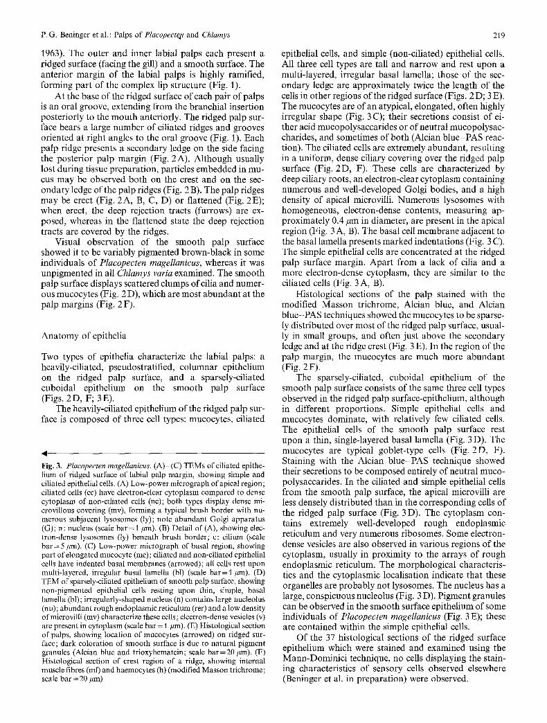
) Fig. 2. Placopecten magellanicus (P. m.: A - D ; F) and Chlamys varia (C. v. : E). (A), (B) SEMs of ridged surface of palp (P. m.) showing (A) erect crest (cr) and secondary ledges (sl) (note uniform, dense ciliation) and (B) mucus-bound particles (p) on secondary ledge and crest (both scale bars = 50 #rn). (C), (D) Light microscopy of transverse histological sections of labial palp (P. m.) showing (C) general organization from centre to margin [note mucus strand (arrowed) at extremity of palp margin; hs: haemolymphatic space; rs: ridged surface; ss: smooth surface; scale bar= 100 #m] and (D) detail of histological section [bl: basal lamella; c: cilia; er: crest; dt: deep rejection tract; mc: mucoeytes; mct: lacunar muscular-connec- tive tissue; sl: secondary ledge (modified Masson trichrome; scale bar = 20 #m)]. (E) SEM of palp (C. v.) showing flattened configura- tion of ridges; cr: crest; sl: secondary ledge (scale bar = 50 #m). (F) Detail of histological section of palp margin (P. m.); note abundant mucus (m) and mucocytes (arrowed); c: cilia (modified Masson trichrome; scale bar = 20 #m)

P.G. Beninger et al. : Palps of Plaeopecten and Chlamys 217
C
Y 5
$ 5
\
218 P, G. Beninger et al.: Palps of P!acopecten and Chtamys
P.G. Beninger et al. : Palps of Placopecte, n and Chlamys
1963). The outer and inner labial palps each present a ridged surface (facing the gill) and a smooth surface. The anterior margin of the labial palps is highly ramified, forming part of the complex lip structure (Fig. 1).
At the base of the ridged surface of each pair of palps is an oral groove, extending from the branchial insertion posteriorly to the mouth anteriorly. The ridged palp sur- face bears a large number of ciliated ridges and grooves oriented at right angles to the oral groove (Fig. 1). Each palp ridge presents a secondary ledge on the side facing the posterior palp margin (Fig. 2 A). Although usually lost during tissue preparation, particles embedded in mu- cus may be observed both on the crest and on the sec- ondary ledge of the palp ridges (Fig. 2 B). The palp ridges may be erect (Fig. 2A, B, C, D) or flattened (Fig. 2E); when erect, the deep rejection tracts (furrows) are ex- posed, whereas in the flattened state the deep rejection tracts are covered by the ridges.
Visual observation of the smooth palp surface showed it to be variably pigmented brown-black in some individuals of Placopecten magellanicus, whereas it was unpigmented in all Chlamys varia examined. The smooth palp surface displays scattered clumps of cilia and numer- ous mucocytes (Fig. 2 D), which are most abundant at the palp margins (Fig. 2 F).
Anatomy of epithelia
Two types of epithelia characterize the labial palps: a heavily-ciliated, pseudostratified, columnar epithelium on the ridged palp surface, and a sparsely-ciliated cuboidal epithelium on the smooth palp surface (Figs. 2 D, F; 3 E).
The heavily-ciliated epithelium of the ridged palp sur- face is composed of three cell types: mucocytes, ciliated
-I
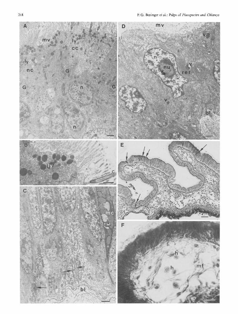
Fig. 3. Plaeopecten magellanicus. (A) (C) TEMs of ciliated epithe- lium of ridged surface of labial patp margin, showing simple and ciliated epithelial cells. (A) Low-power micrograph of apical region; ciliated cells (cc) have electron-clear cytoplasm compared to dense cytoplasm of non-ciliated cells (nc); both types display dense mi- crovillous covering (mv), forming a typical brush border with nu- merous subjacent lysosomes (ly); note abundant Golgi apparatus (G); n: nucleus (scale bar= 1/lm). (B) Detail of (A), showing elec- tron-dense lysosomes (ly) beneath brush border; c: cilium (scale bar = 5 #m). (C) Low-power micrograph of basal region, showing part of elongated mucocyte (mc); ciliated and non-ciliated epithelial cells have indented basal membranes (arrowed); all cells rest upon multi-layered, irregular basal lamella (bl) (scale bar = 1 #m). (D) TEM of sparsely-ciliated epithelium of smooth palp surface, showing non-pigmented epithelial cells resting upon thin, simple, basal lamella (bl); irregularly-shaped nucleus (n) contains large nucleolus (nu); abundant rough endoplasmic reticulum (rer) and a low density of microvilli (mv) characterize these cells; electron-dense vesicles (v) are present in cytoplasm (scale bar = 1 #m). (E) Histological section of palps, showing location of mucocytes (arrowed) on ridged sur- face; dark coloration of smooth surface is due to natural pigment granules (Alcian blue and trioxyhematein; scale bar=20 #m). (F) Histological section of crest region of a ridge, showing internal muscle fibres (mr) and haemocytes (h) (modified Masson trichrome; scale bar = 20 #m)
219
epithelial cells, and simple (non-ciliated) epithelial cells. All three cell types are tall and narrow and rest upon a multi-layered, irregular basal lamella; those of the sec- ondary ledge are approximately twice the length of the cells in other regions of the ridged surface (Figs. 2D; 3 E). The mucocytes are of an atypical, elongated, often highly irregular shape (Fig. 3 C); their secretions consist of ei- ther acid mucopolysaccarides or of neutral mucopolysac- charides, and sometimes of both (Alcian blue-PAS reac- tion). The ciliated cells are extremely abundant, res~llting in a uniform, dense ciliary covering over the ridged palp surface (Fig. 2D, F). These cells are characterized by deep ciliary roots, an electron-clear cytoplasm containing numerous and well-developed Golgi bodies, and a high density of apical microvilli. Numerous lysosomes with homogeneous, electron-dense contents, measuring ap- proximately 0.4/~m in diameter, are present in the apical region (Fig. 3 A, B). The basal cell membrane adjacent to the basal lamella presents marked indentations (Fig. 3 C). The simple epithelial cells are concentrated at the ridged palp surface margin. Apart from a lack of cilia and a more electron-dense cytoplasm, they are similar to the ciliated cells (Fig. 3 A, B).
Histological sections of the palp stained with the modified Masson trichrome, Alcian blue, and Alcian blue-PAS techniques showed the mucocytes to be sparse- ly distributed over most of the ridged palp surface, usual- ly in small groups, and often just above the secondary ledge and at the ridge crest (Fig. 3 E). In the region of the palp margin, the mucocytes are much more abundant (Fig. 2 F).
The sparsely-ciliated, cuboidal epithelium of the smooth palp surface consists of the same three cell types observed in the ridged palp surface-epithelium, although in different proportions. Simple epithelial cells and mucocytes dominate, with relatively few ciliated cells. The epithelial cells of the smooth palp surface rest upon a thin, single-layered basal lamella (Fig. 3 D). The mucocytes are typical goblet-type cells (Fig. 2D, F). Staining with the Alcian blue-PAS technique showed their secretions to be composed entirely of neutral muco- polysaccarides. In the ciliated and simple epithelial cells from the smooth palp surface, the apical microvilli are less densely distributed than in the corresponding cells of the ridged palp surface (Fig. 3 D). The cytoplasm con- tains extremely well-developed rough endoplasmic reticulum and very numerous ribosomes. Some electron- dense vesicles are also observed in various regions of the cytoplasm, usually in proximity to the arrays of rough endoplasmic reticulum. The morphological characteris- tics and the cytoplasmic localisation indicate that these organelles are probably not lysosomes. The nucleus has a large, conspicuous nucleolus (Fig. 3 D). Pigment granules can be observed in the smooth surface epithelium of some individuals of Placopecten magellanicus (Fig. 3 E); these are contained within the simple epithelial cells.
Of the 37 histological sections of the ridged surface epithelium which were stained and examined using the Mann-Dominici technique, no cells displaying the stain- ing characteristics of sensory cells observed elsewhere (Beninger et al. in preparation) were observed.

220
Internal anatomy
The interior of the labial palps consists mainly of lacunar vesicular connective tissue, traversed by smooth-muscle fibres (Figs. 2 D, F; 3 F); it will herein be referred to as muscular-connective tissue. Besides being present in the palp blood-vessels, haemocytes can be observed in the lacunae of the muscular-connective tissue, indicating that the blood circulation within the palps is open. A thin layer of smooth muscle fibres is found beneath the basal lamella of both epithelia. In each palp ridge, a particular- ly dense network of smooth muscle fibres extends from the basal lamella of the upper margin of the secondary ledge to the basal lamella of the epithelium of the smooth palp surface (Fig. 2 D). Such muscle fibres also traverse the crest region of the ridges, both horizontally and obliquely (Figs. 2D; 3 F). Branches of the palp nerve are occasionally observed beneath the smooth muscle layer.
When the scallops are ripe and ready to spawn, acini from the gonad extend into the lacunar palp tissue, giving a secondary pinkish coloration if the subjacent gonad is female. This is most pronounced in Pecten maximus (own personal observations), and much less so in either Pla- copecten magellanicus or Chlamys varia.
Discussion
The ridged surfaces of bivalve labial palps have often been ascribed a function in the selection of particles prior to ingestion or rejection. This interpretation has been derived either from studies based on the behavior of sus- pended particles deposited directly on the separated palp surface 1 (Nelson 1960, Stasek 1961, Jorgensen 1966, B. Morton 1979), or from indirect evidence such as stomach contents (Hughes 1975) or pseudofeces contents (Kiorboe and Mohlenberg 1981, Newell and Jordan 1983, Shumway et al. 1985). However, Bernard's (1974) non-quantitative direct observations indicated that there is no particle selection on appressed ridged surfaces (i.e., in the natural state) in Crassostrea gigas. Furthermore, as emphasized by Foster-Smith (1975 a, 1978), single-parti- cle suspensions do not arrive directly on the palps; rather, groups of particles bound in mucus cords are delivered to the palps from the gills. The arrival of particles bound in mucus cords has been confirmed for all scallop species examined to date (Kellogg 1915, Bernard 1972, Gilmour 1974, Beninger et al. 1988). Under such conditions, selec- tion of individual particles would only be possible if the mucus viscosity were somehow greatly reduced. Newell and Jordan (1983) proposed that mechanical handling by the ciliated ridges would reduce mucus viscosity and al- low individual particles to be extracted.
1 Galtsoff (1964) deposited inert particles directly on the unsepa- rated palps of Crassostrea virginica, i.e., on the smooth palp surface. His subsequent interpretation of particle behavior and palp func- tion failed to recognize that food particles are not deposited on this surface under natural conditions
P.G. Beninger et al. : Palps of Placopecten and Chlamys
Evidence for selection at the labial palps based on studies of contents of gut or pseudofeces may overlook the fact that modification of the proportions of different particle types can also be accomplished by other sites and mechanisms. Guard tentacles (which are present on the mantle edge in scallops and around the inhalent siphons of many other bivalves) may perform a screening or baf- fle function (Walter 1975, Palmer and Williams 1980). Most high-density (i.e., inorganic, e.g. silt) particles > 14/~m probably settle onto the mantle and are rejected as pseudofeces before reaching the gills or palps (Bernard 1974), while certain mucus strings formed on the scallop gill may be rejected as pseudofeces before reaching the palps, especially at high particle concentrations (Kellogg 1915, Owen and McCrae 1976). In addition, mechanical sorting may occur in the stomach after particle ingestion (Purchon 1977: p. 226-227; J. E. Morton 1979: p. 114- 117).
Meticulous functional observations using the shell- window technique have led Foster-Smith (1975 a, 1978) to propose that the palps are not capable of particle selec- tion, but rather should be considered as acceptance or rejection structures, acting on particle-mucus cords, whose behavior is influenced mainly by particle concen- tration in the medium, and not by any intrinsic property of the particles themselves. Although differential treat- ment of particles based on weight, density or volume may be excluded (Bernard 1974, Foster-Smith 1975 a, b, 1978, Hughes 1975), growing evidence indicates that the organ- ic coating or composition of a particle may influence its probability of ingestion (Taghon 1982, Newell and Jor- dan 1983, Ward and Targett 1989). Recent investigations have shown that, in the case of particles coated with algal ectocrines, preferential selection is not due to an in- creased adhesiveness of such particles (Targett et al. in preparation). In any event, the predominance of the mod- erately-sized alga Phaeodactylurn tricornutum in the pseudofeces of scallops fed with moderate concentrations (104 cells ml- 1) of a mixed cell suspension, as reported by Shumway et al. (1985), strongly argues for some sort of selection either at the gills (for which no mechanism has yet been proposed), or at the labial palps.
Assuming that a mechanism exists which would allow the extraction of individual particles from mucus cords at the labial palps, selection on the basis of the recognition of intrinsic particle characteristics would require sensory structures within the ridged surface epithelium, as postu- lated for Crassostrea virginica (Newell and Jordan 1983). Furthermore, given the small size of most food particles, such cells would have to be fairly numerous in order to be encountered by particles before the latter are included in new mucus cords destined for ingestion. The existence of very infrequent putative taste-receptors on the smooth palp surface has been inferred from electrophysiological studies of the labial palps of C. gigas (Dwivedy 1973) but, since this surface does not participate in particle handling under normal circumstances (see footnote at beginning of "Discussion"), such information is of limited relevance. Furthermore, it is uncertain what effect distilled-water rinses of the palp might have had on the recorded poten- tials. Similarly, although putative sensory cells have been
P.G. Beninger et al. : Palps of Placopecten and Chlamys
reported on the ridged surface of the palps of Anodonta sp. (Matthews 1928), this interpretation was based on the comparative thickness of the cilia as viewed under the light microcope, as well as on the uptake of a methylene blue stain. No noticeable differences in cilia thickness were observed in the two species (Placopecten magellani- cus and Chlamys varia) of the present study using either light microscopy, scanning or transmission electron mi- croscopy; in addition, it should be noted that mucocytes also stain readily with methylene blue. As histological examination using the Mann-Dominici technique failed to reveal cells with staining properties similar to those of sensory cells found elsewhere in scallops (Beninger et al. in preparation), the existence of such cells on the labial palps of bivalves therefore remains to be demonstrated unequivocally.
In the absence of conclusive data on sensory struc- tures, it is not yet possible to determine whether or not the labial palps are involved in selection of individual particles based on their intrinsic characteristics. Howev- er, it is clear that particle rejection depends upon ambient particle concentration. Under conditions of low particle concentration, the mucus strings from the gills arriving in the oral groove form a relatively compact cord, which may proceed directly to the mouth (Kellogg 1915, Beninger et al. 1988). Under conditions of increasing par- ticle concentration, the mucus cord is more massive, and tends to be pulled out of the oral groove and onto the ridged palp surface, presumably by the ciliated crest re- jection-tracts (Kellogg 1915). The subsequent handling of this material is quite complex, and may not only change within an individual for a given region of a ciliated tract depending on its location in the ridged surface, but also with the shape of the underlying ridge and palp (Foster- Smith 1978). A generalized description of the behavior of the ridged palp surface has nevertheless been given by Foster-Smith. Handling of the mucus cords (i.e., direc- tion to acceptance or rejection tracts) is conditioned by the degree to which the palp ridges are erected. The dense network of muscle fibres extending from the secondary ledge to the basal lamella of the smooth surface would thus be responsible for this behavior in the two species studied here. The formation of notches in the crests of the ridges, allowing acceptance of the mucus cords when the palps are relatively flattened, would similarly be mediat- ed by the muscle fibres observed in the crest region. Through their highly plastic treatment of mucus strings, the ridged surfaces of the labial palps may thus function to attenuate the potential overload of the bivalve's inges- tive capacity when particle concentration increases.
Several points should be made concerning the distri- bution of mucocytes on the palps. The relatively low den- sity of such cells on the ridged surface (with the exception of the palp margins), attests to its predominantly me- chanical role. This surface probably does not add much mucus to the cords and particle groups on the palps, with the possible exception of the re-sorting tract above the secondary ledge. The addition of mucus at this level may determine to which ciliated tract the particle groups or cords in this region will be directed, and ultimately their acceptance or rejection. Bernard (1974) postulated that
221
the palps function primarily as mucus reducers, concen- trating food particles and rejecting mucus as pseudofeces, thus minimizing the amount of mucus ingested. In addi- tion to the fact that such a continual loss of mucus would be energetically inefficient, this interpretation is not sup- ported by the anatomical evidence of the present study which demonstrates the presence of mucocytes on the ridged palp surface. Furthermore, ongoing research shows that there is a high density of active mucocytes around the mouth and in the oesophagus (Beninger et al. 1990, and in preparation), such that more mucus is actu- ally added at these levels.
The mucus secretions of the ridged surface are quali- tatively different from those of the smooth surface. The existence of two mucus types of different viscosities has been observed on the ridged surface of the labial palps of Crassostrea gigas, and this has been related to particle handling or rejection (Bernard 1974), which is performed by the ridged surface only.
The very high density of mucocytes on the ridged and smooth surfaces of the palp margin is probably related to the rejection of material arriving from the ridged surface into the exhalent current of the pallial cavity. Neutral mucopolysaccharide-secreting mucocytes are quite abun- dant on the general smooth palp surface. Although mu- cocytes are typically present in the molluscan integument (see Bubel 1984), they may serve an important function in this particular location. Several authors have pointed out that the Pectinacea have evolved muscular-driven mecha- nisms for the expulsion of pseudofeces, which conse- quently generate a strong current in the buccal region (Yonge 1967, B. Morton 1979). Such periodic currents would tend to separate the palps and expel potential food material along with the pseudofeces. However, the pres- ence of a well-developed mucus layer on the smooth palp surfaces offers little frictional resistance, such that the cleansing currents could pass over this surface without unduly disturbing the palps.
Previous reports of the presence of sub-epithelial mu- cocytes in five non-pectinid species (Galtsoff 1964, Bernard 1974, Foster-Smith 1975a)were not confirmed for the two species studied here. Indeed, the presence of such cells within the muscular-connective tissue be- neath the basal lamella would constitute an anatomical anomaly.
In contrast to Galtsoff's (1964) observations of paraffin-embedded labial palps of Crassostrea virginica, the data of the present study clearly show that the epithe- lium of the smooth palp surface of Placopecten magellan# cus and Chlamys varia is only sparsely ciliated, and obvi- ously not specialized for the transport of particles. Galt- soff's observations of the movements of particles de- posited on these surfaces in Crassostrea virginica should thus be interpreted with caution (i.e., not confused with normal cleansing behavior of the pallial organ epithelia) until detailed anatomical studies of the smooth palp sur- face have been performed in this species.
The ultrastructural characteristics of the ridged sur- face epithelium strongly suggest an accessory nutritive role. The microvillous brush border, together with the internal ramifications of the basal cell membrane are

222
characteristic o f the absorpt ion o f dissolved substances, whereas the numerous Golgi apparatus , lysosomes and phagosomes indicate the capture and digestion o f large molecular agglomerates and colloidal matter . A similar funct ion has been at t r ibuted to other epithelial regions over which water flow (and hence dissolved organic and colloidal matter) is considerable: the gills (Pasteels 1968, Beninger e ta l . 1988, Le Pennec et al. 1988) and the mantle (Bevelander and N a k a h a r a 1966, N a k a h a r a and Bevelander 1967). In the case o f the appressed ridged surfaces o f the labial palps, water flow would be compar- atively small, but the mechanical handl ing o f the mucus cords would necessarily result in the dissolution o f some of the mucus and the dissociation o f some of the smaller particles. Both o f these substances could be recovered by the epithelial cells o f the ridged surface. Hence, while this surface might no t be a mucus reducer in the sense origi- nally p roposed by Bernard (1974), some mucus reduct ion would result f rom the normal r idged-surface activity; recycling dissociated material originally destined for in- gestion would maximize t rophic efficiency and reduce over load in the digestive system.
The smoo th palp surface, which does no t handle mu- cus cords, does no t present these ul t rastructural charac- teristics. Rather , the simple epithelial cells f rom this sur- face exhibit cytological evidence o f active molecular syn- thesis (well-developed rough endoplasmic reticulum, large nucleolus). Occasional electron-dense vesicles oc- curring close to the arrays o f rough endoplasmic reticu- lum are possible sites for p roduc t accumulat ion; howev- er, it is no t yet clear whether these products serve in the metabol ism of the epithelial cells or are released as secre- tions to the external medium.
Acknowledgements. The authors wish to thank Dr. G. Sinquin for assistance with electron microscopy, Dr. K. Benhalima for assis- tance with histology, and MM. A. Le Mercier and L. Blanchard for their excellent photographic work. Fig. 1 was skillfully drawn by Ms. L. Colpitts, while Fig. 2E was kindly provided by Mine N. Casse. Discussions and comments on earlier versions of the manuscript by Drs. R. L. Foster-Smith, Y. Poussart, S. E. Shumway, and Ms. R. Friedrich greatly contributed to the final result. The patient word-processing of this manuscript by Mine J. Corvest and L. Briard is gratefully acknowledged. Specimens of Placopecten magellanicus were acquired using the facilities of the Huntsman Marine Science Centre, St. Andrews, New Brunswick, Canada. This collaborative study was funded by the CNRS/CNRC France-Canada scientific exchange programme, as well as by Natu- ral Sciences and Engineering Research Council Grant No. 0003658, Facult6 des Etudes Sup&ieures et de la Recherche de l'Universit6 de Moncton Grant No. 20-53202-54-111, and Department of Employ- ment and Immigration Challenge '88 and Challenge '89 pro- grammes.
Literature cited
Ansell, A. D. (1961). The functional morphology of the British species of Veneracea (Eulamellibranchia). J. mar. biol. Ass. U.K. 41:489-515
Ansell, A. D. (1981). Functional morphology and feeding of Donax serra R6ding and Donax sordidus Hanley (Bivalvia: Donacidae). J. mollusc. Stud. 47:59-72
Beninger, P. G. (1987). A qualitative and quantitative study of the reproductive cycle of the giant scallop, Placopecten magellani-
P.G. Beninger et al. : Palps of Placopecten and Chlamys
cus, in the Bay of Fundy (New Brunswick, Canada). Can. J. Zool. 65:495 498
Beninger, P. G., Le Pennec, M., Auffret, M. (1990). Peribuccal organs of Placopecten magellanicus and Chlamys varia (Mol- lusca: Bivalvia): structure, ultrastructure and implications for feeding. II. The lips. Mar. Biol. 107:225-233
Beninger, P. G., Le Pennec, M., Sala/in, M. (1988). New observa- tions of the gills of Plaeopecten magellanieus (Mollusca: Bivalvia), and implications for nutrition. I. General anatomy and surface microanatomy. Mar. Biol. 98:61-70
Bernard, F. R. (1972). Occurrence and function of lip hypertrophy in the Anisomyaria (Mollusca, Bivalvia). Can. J. Zool. 50: 53-57
Bernard, E R. (1974) Particle sorting and labial palp function in the Pacific oyster Crassostrea gigas (Thunberg, 1795). Biol. Bull. mar. biol. Lab., Woods Hole 146:1-10
Bevelander, G., Nakahara, H. (1966). Correlation of lysosomal activity and ingestion by the mantle epithelium. Biol. Bull. mar. biol. Lab., Woods Hole 131:76-82
Bubel, A. (1984). Mollusca: epidermal cells. In: Bereiter-Hahn, J., Matoltsy, A. B., Richards, K. S. (eds.) Biology of the integu- ment. I. Invertebrates. Berlin, Springer-Verlag, p. 400-447
Dwivedy, R. C. (1973). A study of chemo-receptors on labial palps of the American oyster using microelectrodes. Proc. natn. Shell- fish. Ass. 63:20-26
Foster-Smith, R. L. (1975 a). The role of mucus in the mechanism of feeding in three filter-feeding bivalves. Proc. malac. Soc. Lond. 41:571 588
Foster-Smith, R. L. (1975 b). The effect of concentration of suspen- sion on the filtration rates and pseudofaecal production for Mytilus edulis L., Cerastoderma edule (L.) and Venerupis pullas- tra (Montagu). J. exp. mar. Biol. Ecol. 17:1-22
Foster-Smith, R. L. (1978). The function of the pallial organs of bivalves in controlling ingestion. J. mollusc. Stud. 44:83-89
Gabe, M. (1968). Techniques histologiques. Masson & Cie, Paris Galtsofl, P. S. (1964). The American oyster Crassostrea virginica
Gmelin. Fishery Bull. Fish Wildl. Serv. U.S. 64:111-120 Garland, C. D., Nash, G. V., McMeekin, T. A. (1982a). Absence of
surface-associated microorganisms in adult oysters (Cras- sostrea gigas). Appl. envirl Microbiol. 44: 1205-1211
Garland, C. D., Nash, G. V., McMeekin, T. A. (1982b). The preser- vation of mucus and surface-associated microorganisms using acrolein vapour fixation. J. Microscopy 128:307-312
Gilmour, T. H. J. (1974). The structure, ciliation, and function of the lips of some bivalve molluscs. Can. J. Zool. 52:335-343
Hughes, T. G. (1975). The sorting of food particles by Abra sp. (Bivalvia: Tellinacea). J. exp. mar. Biol. Ecol. 20:137 156
Jorgensen, C. B. (1966). The biology of suspension feeding. Perga- mon Press, Oxford
Kellogg, J. L. (1915). Ciliary mechanisms of lamellibranchs, with descriptions of anatomy. J. Morph. 26:625 701
Kiorboe, T., Mohlenberg, E (1981). Particle selection in suspension- feeding bivalves. Mar. Ecol. Prog. Ser. 5:291 296
Le Pennec, M., Beninger, P. G., Herry, A. (1988). New observations of the gills of Plaeopecten magellanicus (Mollusca: Bivalvia), and implications for nutrition. II. Internal anatomy and mi- croanatomy. Mar. Biol. 98:229-237
Matthews, S. A. (1928). The palps oflamellibranchs as autonomous organs. J. exp. Zool. 51:209-258 + 2 plates
Meehan, B. W, Diaz, R. J. (1985). Comparison ofMacoma balthica labial palps from the Eastern and Western North Atlantic: an- other look. J. mollusc. Stud. 50:231-232
Morton, B. (1979). A comparison of lip structure and function correlated with other aspects of the functional morphology of Lima lima, Limaria (Platilimaria) fragilis, and Limaria (Platili- maria) hongkongensis sp. nov. (Bivalvia: Limacea). Can. J. Zool. 57:728-742
Morton, J. E. (1979). Molluscs. Hutchinson & Co., London Nakahara, H., Bevelander, G. (1967). Ingestion of particulate mat-
ter by the outer surface cells of the mollusc mantle. J. Morph. 122:139 145
P.G. Beninger et al. : Palps of Placopecten and Chlamys
Nelson, T. C. (1960). The feeding mechanism of the oyster. II. On the gills and palps of Ostrea virginica and Crassostrea angulata. J. Morph. 107:163-191
Newell, R. I. E., Jordan, S. J. (1983). Preferential ingestion of organ- ic material by the American oyster Crassostrea virginica. Mar. Ecol. Prog. Ser. 13:47 53
Newell, C. R., Shumway, S. E., Cucci, T. L., Selvin, R. (1989). The effects of natural seston particle size and type on feeding rates, feeding selectivity and food resource availability for the mussel Mytilus edulis Linnaeus, 1758 at bottom culture sites in Maine. J. Shellfish Res. 8:187 196
Owen, G. (1978). Classification and the bivalve gill. Phil. Trans. R. Soc. (Set. B) 284:377 385
Owen, G., McCrae, J. M. (1976). Further studies on the latero-fron- tal tracts of bivalves. Proc. R. Soc. (Ser. B) 194:527-544
Palmer, R. E., Williams, L. G. (1980). Effect of particle concentra- tion on filtration efficiency of the bay scallop Argopecten irradi- ans and the oyster Crassostrea virginiea. Ophelia 19:163-174
Pasteels, J. J. (1968). Pinocytose et arthrocytose par l'epith61ium branchial de Mytilus edulis L.: analyse experimentale au micro- scope 61ectronique. Z. Zellforsch. microsk. Anat. 92:339-359
Purchon, R. D. (1977). The biology of the Mollusca. 2nd ed. Per- gamon Press, Oxford
223
Shumway, S. E., Cucci, T. L., Newell, R. C., Yentsch, C. M. (1985). Particle selection, ingestion, and absorption in filter-feeding bivalves. J. exp. mar. Biol. Ecol. 91:77-92
Stasek, C. R. (1961). The ciliation and function of the labial palps of Acila castrensis (Protobranchia, Nuculidae), with an evalua- tion of the role of the protobranch organs of feeding in the evolution of the Bivalvia. Proc. zool. Soc. Lond. 137:511-538
Stasek, C. R. (1963). Synopsis and discussion of the association of ctenidia and labial palps in the bivalved Mollusca. Veliger 6: 91 96
Taghon, G. L. (1982). Optimal foraging by deposit-feeding inverte- brates: roles of particle size and organic coating. Oecologia 52: 295 302
Walter, T. R. (1975). The behavior and tentacle morphology of pteriomorphian bivalves: a motion-picture study. Bull. Am. malac. Un. 1975:7 13
Ward, J. E., Targett, N. G. (1989). Influence of marine microalgal metabotites on the feeding behavior of the blue mussel Mytilus edulis. Mar. Biol. 101:313-321
Yonge, C. M: (1967). Observations on Pedum spondyloideum (Chemnitz) Gmelin, a scallop associated with reef-building corals. Proc. malac. Soc. Lond. 37:311-323