wordpress.clarku.edu€¦ · web viewformative assessment. reinforce order of mitotic phases and...
TRANSCRIPT

Sara SterlingNativity School of Worcester
Seventh Grade SciencePeriod 6: 1:15- 2:01pm
Monday, April 14, 2014
Clark University Master of Arts in Teaching ProgramLearning Activity Plan
“LIVESTRONG: TARGETING TUMORS”
I. Content : Describe what it is you will teach. What is the content?The purpose of today’s lesson is to apply student knowledge of cell division to
cancer diagnosis and treatment. Students will act as doctors and will construct a model of a patient infected with tumors. Students will then diagnose the patient and write a treatment plan with the next point of action. Students will read, write, construct, and discuss the process of cancer diagnosis and treatment.
II. Learning Goals: Describe what specifically students will know and be able to do after the experience of this class.
Describe cancer as uncontrolled cell division Create a model to identify the difference between malignant and benign tumors Evaluate and Synthesize information from the lesson by writing a patient
treatment plan
III. Rationale : Explain how the content and learning goal(s) relate to your Curriculum Unit Plan learning goals.
Last week we discussed the process of mitosis. Students created macaroni models for the phases of mitosis. Students demonstrated a basic understanding of cell division, in that following cell division the parent cell yields two identical daughter cells. This lesson is designed to stimulate group discussion and analysis of a cancer patient. We are investigating the question, “What happens when cell division goes wrong?” I gave students the context of a hospital and working with patients so that they can visualize the practical applications of cell division. Students will create a model of the concept and then use their models to evaluate the patient’s condition. Students are reading, writing, analyzing, defining, and evaluating the material in different ways. I decided to give each group a different patient so that the groups work within their operating room stations. This hopefully will eliminate the need for students to roam from group to group. The model construction is designed to maximize student participation. The activity allows for some group members emerge as leaders and others to be the model constructors. The writing piece then helps students think critically. I provided guiding questions to help students sequence their information and highlight the important information. The discussion at the end of class is meant to give students a voice in a safe space to share about their experience with cancer and to summarize the objective of the lesson: promote awareness of cancer as the result of uncontrolled cell division in the body.
IV. Assessment : Describe how you and your students will know they have reached your learning goals.
Students will create a model following a rubric to show the locations of a benign tumor and a malignant tumor on a patient.
Students will evaluate the patient by synthesizing a guided research packet and applying the research to their own diagnoses in the form of a written treatment plan.
V. Personalization and equity : Describe how you will provide for individual student strengths and needs. How will you and your lesson consider the needs of each student and scaffold learning? How specifically will ELL students and students with learning disabilities gain access and will be supported?

LAB GROUPS:
Operating Room 1: Larry, Misael, Stanley, RandalOperating Room 2: Vincent, Brian, JohnyOperating Room 3: Jack, Devon, DanteOperating Room 4: Julian, Adrian, Omar
Students will first review the lesson material from the previous week. They will then work in groups to create models. The group work is designed to stimulate discussion and to help students, like Misael, visualize the vocabulary. Students will then act like real doctors as they develop a treatment plan for their patients. Each group has been assigned a different patient with different symptoms. This is done so that groups have to work within their operating room to find the solution and to help students feel their group is unique. I gave students a doctor handbook with all the essential information. I also provided students with a vocabulary list, complete with pictures to help them better understand the language used in this lesson.
VI. Activity description and agenda
Duration What the students will be doing
What I will be doing Rationale Co-teacher responsibility
1:15 to 1:20 Filling out Do Now Ticket
Facilitating do now ticket instruction
Formative assessment. Reinforce order of mitotic phases and introduce the day’s lesson on cancer.
Monitor student behavior
1:20 to 1:25 Listen to directions and read instruction sheet. Read new vocabulary terms and the big question: How does a doctor target tumors in a cancer patient?
Introduce students to the doctor handbook and patient models. Students will work in groups of three to create the model of the patient. They will read the patient profile and then use the stickers to represent the cancerous tissue on the model.
Clarify directions and expectations before beginning the activity
Monitor student behavior
1:25 to 1:35 Create models of patient according to the information.
Monitor accuracy of model creation.
Formative assessment.
Monitor student behavior
1:35 to 1:45 Begin patient treatment plan written report. Students can work collaboratively in groups and then finish the report for homework.
Help with instruction for patient treatment plan.
Collaborative group work, highlight science literacy of students, force students to evaluate and synthesize information.
Help struggling students, monitor student behavior
1:45 to 2:00 Conclusion discussion about cancer, cancer treatment, and any new discoveries from the day’s class
Highlight the personal investments students may have with cancer research and treatment. Share personal experience about my aunt who battled cancer for ten years and who lost the battle five months ago.
Promote awareness and hopefully inspire students to seek a cure for cancer. Give students a voice to share (if they feel comfortable sharing with the class) their personal experiences with cancer.
Participate in the discussion and monitor student behavior.

VII. List the Massachusetts Learning Standards this lesson addresses.
LS2 Recognize that all organisms are composed of cells, and that many organisms are single-celled. In these single celled organisms, one cell must carry out all the basic functions of life
LS5 Describe the hierarchical organization of multicellular organisms from cells to tissues to organs to systems to organisms.
VIII. Sources of Information
http://www.cancer.gov/cancertopics/understandingcancer/cancer/
http://www.livestrong.org/
http://www.scholastic.com/browse/article.jsp?id=3751444
IX. Reflection
a. In light of all areas of planning, but especially in terms of your stated purpose and learning goals, in what ways was the activity(ies) successful? How do you know? In what ways was it not successful? How might the activity be planned differently another time?
b. What did you learn from the experience of this lesson that will inform your next LAP?

DO NOW Name: _____________________
PUT THE 5 PHASES OF MITOSIS IN ORDER
1. ________________________
2. ________________________
3. ________________________
4. ________________________
5. ________________________ & CYTOKENISIS
What do you get after a cell divides? (What is the end result of Mitosis?)
___________________________________________________________
___________________________________________________________
What do we call “uncontrolled cell division”? ___________________________

LIVESTRONG: TARGETING TUMORS
CALLING ALL FERNANDO DOCTORS. SEVERAL PATIENTS HAVE ARRIVIED IN THE HOSPITAL AND NEED YOUR HELP WITH A DIAGNOSIS.
BIG QUESTION: How can a doctor target tumors in a cancer patient?
Materials:Folder with Patient InformationPatient: Body OutlineCancer Cells: StickersTreatment Plan SheetDoctor Handbook
Objectives:
1.Create a model, using the materials provided, that shows the location of cancerous tumors in a patient.
2.Write a patient treatment plan using the treatment plan sheet as a guide.
RUBRIC:
ITEM POINTS POSSIBLE POINTS EARNEDPatient model with locations of all tumors. Tumors are represented as a group of stickers.
15
Effort and teamwork 5
Written treatment plan that uses information from the reading and accurately describes the patient’s condition.
25
Correctly identifies patient’s cancer 5


DOCTOR HANDBOOK
VOCABULARY:
CANCER: Uncontrolled cell division
TUMOR: Growing mass of cancer tissue
MALIGINANT: Tumors that are capable of spreading
BENIGN: Tumors that cannot spread
BIOPSY: When a doctor removes a small piece of tissue, looks at it under a microscope, and sees if the cells are cancer.
DID YOU KNOW:
Cancer can occur anywhere in the body
Cancer disrupts the balance between cell division and cell death in the body
A Biopsy will tell the doctor whether a tumor is actually present and, if so, whether it is malignant (i.e., cancer) or benign.
By definition, the term "cancer" applies only to malignant tumors.
Tumors ultimately increase in size because new cells are being produced in greater numbers than needed.
Cancer can be caused by genetics, diet, chemicals (e.g., smoking), radiation, and viruses or bacteria

Have you heard of any of these types of cancer?
Carcinoma: found in cells that cover external and internal body surfaces, including Lung, breast, and colon cancer. (Most common type of cancer)
Sarcoma: found in the supporting tissues of the body such as bone, cartilage, fat, connective tissue, and muscle.
Lymphoma: found in the lymph nodes and tissues of the body’s immune system.
Leukemia: found in immature blood cells that grow in the bone marrow and move through the bloodstream.
Have you ever wondered how scientists name different types of cancer?
“In general, these names are created by using different Latin prefixes that stand for the location where the cancer began its unchecked growth.”
For example, the prefix “osteo” means bone, so a cancer found in bone is called an osteosarcoma.

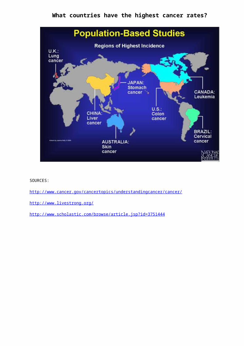
What countries have the highest cancer rates?
SOURCES:
http://www.cancer.gov/cancertopics/understandingcancer/cancer/
http://www.livestrong.org/
http://www.scholastic.com/browse/article.jsp?id=3751444

PATIENT PROFILE
Patient #1: L. HamiltonCountry of Residence: England
Body scan shows a mass of tissue in the lung. Mass appears to be localized in the lung. Does not appear to be growing.
Body scan also shows signs of abnormal tissue growth in the bone marrow of the fibula (lower leg bone). Appears to have spread in the blood stream to the upper thigh, pelvis, and right humorous (upper arm bone). This growth appears to be different than the growth found in the lung.
TASK:
1.Use the materials provided to show where the cancer is located in the patient’s body. (AS A GROUP)
2.Evaluate the body scan and the doctor handbook to give a diagnosis for the patient’s treatment.(AS A GROUP)
3.Answer the patient treatment plan questions on a separate sheet of paper(INDEPENDENTLY)

PATIENT PROFILE
Patient #2: B. KobayashiCountry of Residence: Japan
Body scan shows a mass of tissue in the stomach. Mass appears to be localized in the stomach. Does not appear to be growing.
Body scan also shows signs of abnormal tissue growth in the bone marrow of the fibula (lower leg bone). Appears to have spread in the blood stream to the upper thigh, pelvis, and right humorous (upper arm bone). This growth appears to be different than the growth found in the lung.
TASK:
1.Use the materials provided to show where the cancer is located in the patient’s body. (AS A GROUP)
2.Evaluate the body scan and the doctor handbook to give a diagnosis for the patient’s treatment.(AS A GROUP)
3.Answer the patient treatment plan questions on a separate sheet of paper(INDEPENDENTLY)

PATIENT PROFILE
Patient #3: F. Ricciardo Country of Residence: Australia
Body scan shows a mass of tissue in the stomach. Mass appears to be localized in the stomach. Does not appear to be growing.
Body scan also shows signs of abnormal tissue growth in the pigment cells of the lower arm. Appears to have spread in the blood stream to the upper arm, left shoulder, and abdominal muscles. This growth appears to be different than the growth found in the stomach.
TASK:
1.Use the materials provided to show where the cancer is located in the patient’s body. (AS A GROUP)
2.Evaluate the body scan and the doctor handbook to give a diagnosis for the patient’s treatment.(AS A GROUP)
3.Answer the patient treatment plan questions on a separate sheet of paper(INDEPENDENTLY)
PATIENT PROFILE
Patient #4: J. ButtonCountry of Residence: England
Body scan shows a mass of tissue in the lung. Mass appears to be localized in the lung. Does not appear to be growing.
Body scan also shows signs of abnormal tissue growth in the pigment cells of the lower arm. Appears to have spread in the blood stream to the upper arm, left shoulder, and abdominal muscles. This growth appears to be different than the growth found in the stomach.
TASK:
1.Use the materials provided to show where the cancer is located in the patient’s body. (AS A GROUP)
2.Evaluate the body scan and the doctor handbook to give a diagnosis for the patient’s treatment.(AS A GROUP)
3.Answer the patient treatment plan questions on a separate sheet of paper(INDEPENDENTLY)

PATIENT TREATMENT PLAN
DOCTOR ANALYSIS Doctor ___________Patient #_________
After performing a BIOPSY, what did you discover about your patient’s condition?
How fast do you predict the cancer(s) to be growing?
Has the cancer spread to different parts of the body?
What do you believe is the next step for treatment? WHY?
SURGERY
CHEMOTHERAPY
RADIATION THERAPY
TARGETED THERAPY
Why is cancer potentially dangerous? (Think about the difference between benign and malignant tumors)

CANCER TREATMENT
SURGERY: Surgery can be used to diagnose, treat, or even help prevent cancer in some cases. Most people with cancer will have some type of surgery. It often offers the greatest chance for cure, especially if the cancer has not spread to other parts of the body.
CHEMOTHERAPY:Chemotherapy (chemo) is the use of medicines or drugs to treat cancer.
Chemo may be used to shrink tumors before surgery or radiation.
It may be used after surgery or radiation to help kill any cancer cells that are left.
It may be used with other treatments if the cancer comes back.
Sometimes the goal is to slow the growth of the cancer. Other times the goal may be to reduce tumors so that you feel better. Chemo is often used to fight cancers that have spread to other parts of the body.
Chemo kills cancer cells. These drugs can affect normal cells, too. But most normal cells can repair themselves.
RADIATION THERAPY:Radiation therapy uses high-energy particles or waves to destroy or damage cancer cells. It is one of the most common treatments for cancer, either by itself or along with other forms of treatment.
TARGETED THERAPY:Targeted therapy is a newer type of cancer treatment that uses drugs or other substances to more precisely identify and attack cancer cells, usually while doing little damage to normal cells.
Antibody drugs are man-made antibodies (like fighter cells) that have been designed to attack certain targets on cancer cells.
