thefac.ksu.edu.sa/.../files/microbial_diagnosis_bassam.docx · web viewused for the growth of only...
TRANSCRIPT

Microbial Diagnosis
320 MIC
Prepared by
Bassam Alnafisi
(Practical Microbial Diagnosis)
Abstract
For
Microbial diagnosis

LAB#1
Introduction to Microbial Diagnosis
*Microbial diagnosis: is a study of microbial identification using methods:
1-Microscopic diagnosis: to study the shapes of microorganism use of:
A-Wet prep slide: add one drop of saline water (water contain concentrate of NaCl 1-3%) on slide and examine it under microscope).
B-Stains: to study the shapes of bacteria and fungus by using stain (simple stain use one type of stain or complex stain use two or more of stain).
Examples of simple stain (Lactophenol, safranin, methylene blue).
Examples of complex stain (Gram stain, Zeil Nilsson stain).
2-Cultural diagnosis: to study the morphology of colonies on culture media and use biochemical reaction to differentiate between bacteria and fungus.
3- serological diagnosis: to study the interaction between antigen and antibody to diagnostic viral infection or bacterial infection.
4-Moleculer diagnosis: to study and detect the bacterial infection or viral infection by use molecules diagnosis (example: Polymerase reaction technique(PCR)).
LAB#2
Specimen collection and saving and transport and processing
Microorganisms you can see in clinical microbiology lab:
1-Bacteria 2-Fungi (yeast or mold). 3-Viruses 4-Parasites.
Specimen excepts to see in microbiology lab receiving:
*Label the container of all specimen with name of patient and date of birth and time and date of sample collection on and the doctor test ordered.
1-Urine: Urine has a long history as a specimen for analysis in clinical laboratories.
After blood, urine is the most commonly used specimen for diagnostic testing, monitoring of disease status and detection of drugs.

*Types of urine specimen:
1-Random urine: collect form urine pass and put it in clean container and no time to determine to collect.
2-Mid-stream urine: collect from urine middle pass and put it on clean container.
3-Clean catch urine: collect urine You will use a special kit to collect the urine. It will most likely have a cup with a lid and wipes.
4-Urine catheter : (Example: foley catheter) use a syringe followed by transfer to a specimen tube or cup alternatively, urine can be drawn directly from the catheter to an evacuated tube using an appropriate adaptor.

5-Urine suprapubic:
May be necessary when a non-ambulatory patient cannot be catheterized or where there are concerns about obtaining a sterile specimen by conventional means.
Urine saving:
Don’t keep it in room temperature more than two hours
Save it in fridge for 72 hours after that, through it.
2-Stool(Feaces) specimen: are a specimen use to detect bacterial infection or parasite infection if a patient has symptoms.
Collect specimen to clean container (pass it in toilet in clean site and take by sterile spoon).

Shapes of stool specimen
Stool collection
Stool saving: don’t keep it in room temperature more than two hours.
Save it in fridge for 73 hours.
4-Respiratory samples: a specimen collection from respiratory tract (sputum, Endotracheal aspiration, tracheal aspiration, Nasopharyngeal aspiration, BAL).
Collect samples to clean container.

Save it in fridge for one week.
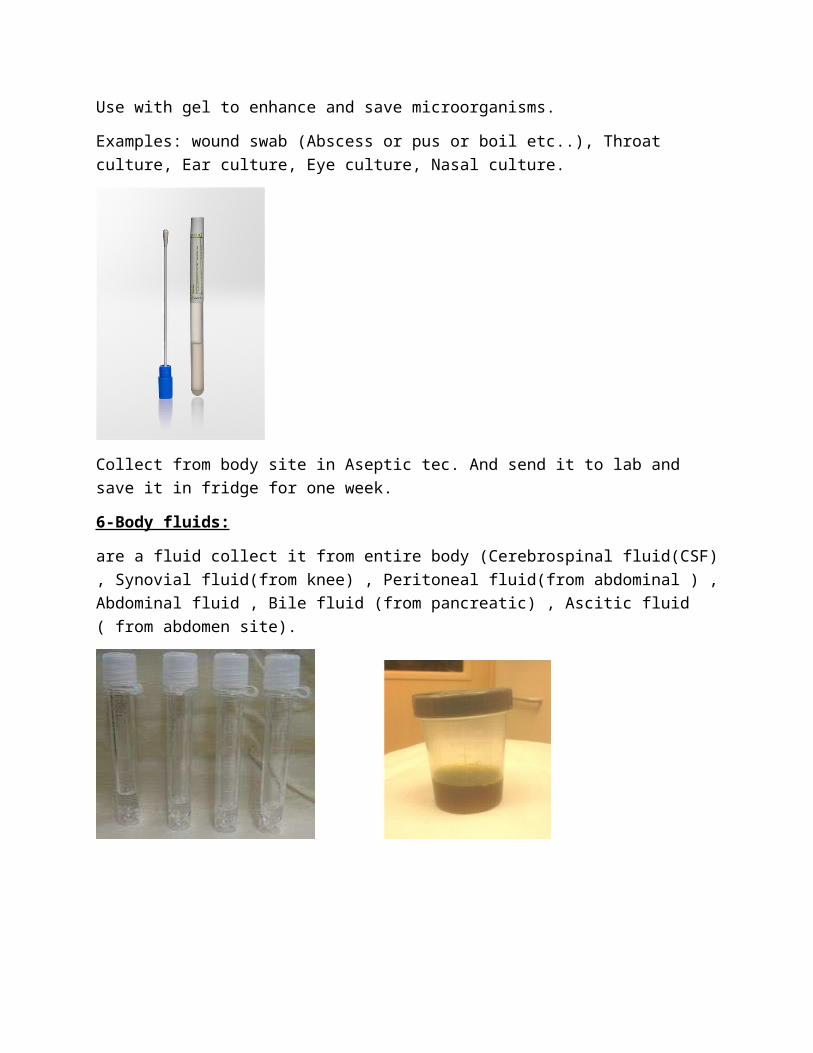
5- Cultural Swab:
Use with gel to enhance and save microorganisms.
Examples: wound swab (Abscess or pus or boil etc..), Throat culture, Ear culture, Eye culture, Nasal culture.
Collect from body site in Aseptic tec. And send it to lab and save it in fridge for one week.
6-Body fluids:
are a fluid collect it from entire body (Cerebrospinal fluid(CSF) , Synovial fluid(from knee) , Peritoneal fluid(from abdominal ) , Abdominal fluid , Bile fluid (from pancreatic) , Ascitic fluid ( from abdomen site).
7-Blood specimen:
Most important clinical samples if the patient have fever, nausea we should to take blood samples to check it for microbiological examination.
Collect blood specimen with aseptic tec.
Save it for 20 days to 30 days.

LAB#3
Microbiological culture media
Microbiological culture media: Use to cultivating of microorganisms contain substances necessary to support microorganism.
Agar : is a solidifying agent use to solidify media and it is a polysaccharide extract from red algae, it melts at 84 c and solidify at 38 c.
Peptones : used as a source of nitrogen.
Meat and plant extracts : used as source of nutrients and contains of vitamins, amino acids, peptides, carbohydrates minerals.
Growth factor : Many organisms have specific growth factor requirement that must be included to the media for successful cultivation.
Example: Blood and yeast extracts
X factor (heme) and V(NAD) factor
Selective components: That inhibit the growth of non-target organisms.
Types of media:
1-General media: used to isolate pathogen and nonpathogen microorganism (examples, Nutrient agar, potatoes dextrose agar).
2-Enriched media: Enriched media contain the nutrients required to support the growth of a wide variety of organisms, including some fastidious ones (Examples, Blood agar, Chocolate agar).
3-Enrichement media: promotes the growth of a particular organism by providing it with the essential nutrients and rarely contains certain inhibitory substance to prevent the growth of normal competitors (example, Selenite F broth).
4-Selective media: used for the growth of only selected microorganisms (example, EMB (Eosin Methylene agar for E. coli, MSA for staph sp.)
5-Differintial media: Differential or indicator media distinguish one microorganism type from another growing on the same medium. (example, Mannitol salt agar, MacConkey agar).
*By the way some of media have both selective and differential in same time
1-Nutrient agar: general-purpose, nutrient medium used for the cultivation of bacteria consists of digestive products of proteins (called peptones) and beef extract.

2-Blood agar: Non-selective Enriched Medium Enriched with 5% sheep blood, suitable for Hemolysis determination and provides X-factor Widely used for the growth of Pathogenic and Non-pathogenic Organisms Contains meat, peptones, beef extract, yeast extract and cornstarch.
3-Chocolte agar: General purpose medium use for the isolation of many organisms including fastidious organisms like Neisseria and Haemophilus
Contain X(Hemin) and V(NAD) factors.
4-MaCckonkey agar: Selective medium and differential
Inhibit gram positive organisms by adding crystal violet
Differentiate between lactose fermenters (coliforms) and Non-lactose fermenters When an organism ferments lactose, pH drops, this causes the colony to become pink due to presence of Neutral red.

5-Sabroud dextrose agar: General clinical media for the isolation of Yeast and Fungi.
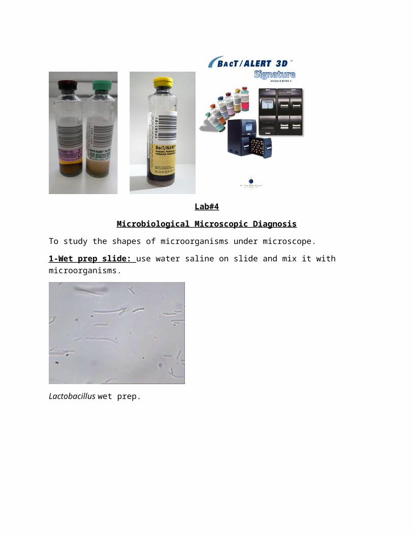
6-Blood culture : Used to determine presence of pathogens (Bacteria-Fungi) in blood stream a minimum of 10 ml of blood is taken through venipuncture and injected into two or more "blood bottles" (from patient and send it to lab) with specific media for aerobic and anaerobic organisms. A common medium used for anaerobes (is thioglycolate). And aerobic used Tryptic soya broth.
Green venipuncture for aerobic and brown venipuncture for anaerobic and yellow venipuncture for pediatric and incubate it in BAcT/ALERT machine.

Lab#4
Microbiological Microscopic Diagnosis
To study the shapes of microorganisms under microscope.
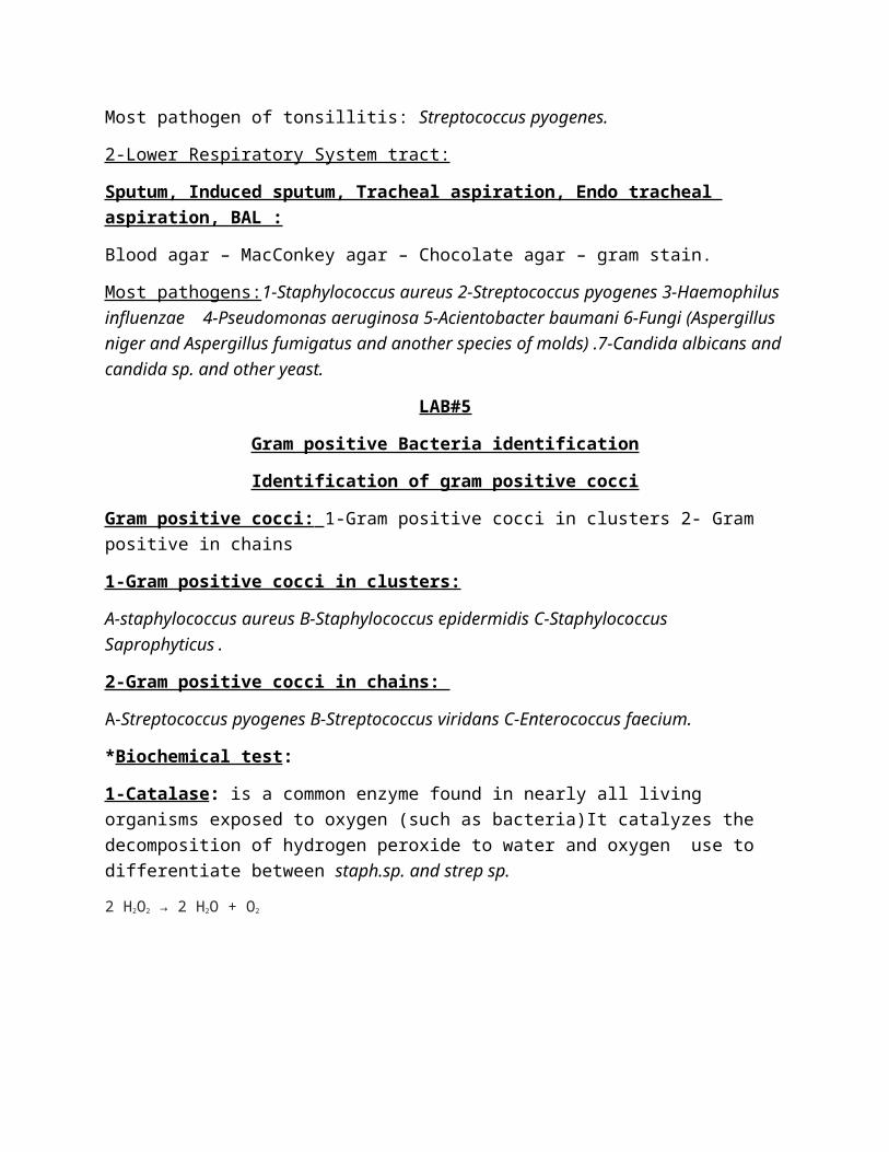
1-Wet prep slide: use water saline on slide and mix it with microorganisms.
Lactobacillus wet prep.
Yeast wet prep
-Diagnostic by stain:
A-gram stain:
Is a differential stain use to divide two large group of bacteria Based on cellular content, Gram stain divides almost all bacteria into two main groups: gram positive- bacteria (Purple) and gram-negative bacteria(Red).
*Procedure of gram stain practical:
*Materials:
1-Crystal violet (primary stain). 2-Gram Iodine solution (mordant or fixation step).
3-Acetone-alcohol(decolorizer). 4-Safranine (Counter stain). 5-New glass slides
6-Fresh bacterial culture.7-Loop.8-Bensin flame.
*methods:
1-Make a smear of bacterial colonies (take one colony and mix it with saline) and fix it under bensin flame till dry.
2-Flood (cover completely) the entire slide with crystal violet for 1 min and wash it by water.
3-Flood the entire slide with Iodine solution for 1 min and wash it by water.
4-This step is best performed while holding the slide at a slant with forceps, rather than while sitting on a slide rack, add decolorizer(Acetone-alcohol) dropwise and allow it run off. Rinse with running tap water. Repeat this step until the blue dye no longer runs off the slide with the decolorizer. Rinse with running tap water and shake off the excess is somewhat tricky because using too much decolorizer could result in a false Gram (-) result. Likewise, not using enough decolorizer may yield a false Gram (+) result.
5-Flood the entire slide with Safranin for 1 min and wash it by water.
6-allow the slide to air dry before viewing under the microscope and add drop of xylene oil (immersion oil) and examine it under 100x eye lens.

Gram positive cocci in clusters gram positive cocci in chains
Gram positive rods Gram negative bacilli

Gram negative coccobacilli Gram negative cocci

Candida albicans gram positive Wound specimen(exaudted)
Staphylococcus aureus (gram
stain of specimen) .
Sputum gram stain
Look at pictures
B-Ziel -Nilsson stain: also known as the acid-fast stain, It is a special bacteriological stain used to identify acid-fast organisms, mainly Mycobacteria. Mycobacterium tuberculosis is the most important of this group because it is responsible for tuberculosis (TB) Acid-fast bacilli will be bright red after staining.

Materials and methods:
1-Sputum specimen 2-fresh culture for TB control 3-Glass slides
4-Carbol-fuchsin 5-acid alcohol 6-methylene blue7-Immersion oil.
Methods:
1-Flood fixed smear with carbolfuchsin for 6 minutes
2-Wash gently
3-Decolorize with 3% acid alcohol for 2 minutes
4-Wash gently
5-Counterstain with methylene blue for 2 minutes
6-Wash gently
7-Examine smear using oil immersion.

Lab#4
Media selection for specimen
1-Urine culture:
Blood agar/MacConkey agar bi plate
Most pathogens of Urinary Tract Infection(UTI):
1-E. coli
2-Enterobacteriaceae (Proteus sp. (marbles and vulagris), Providencia stuartii, Citrobacter sp., Enterobacter sp. (aerogenes), Klebsiella sp.(Pneumonia).
3-Non-Fermenter bacteria (Pseudomonas aeruginosa, Acinetobacter baumni (Check for Multi Drug Resistance(MDR), Stenotrophomonas maltophilia).
4-Enterococcus sp. (Faecium and Faecalis).
5-Staphylococcus aureus (Check for MRSA (Methicillin Resistance Staphylococcus aureus) and Staphylococcus Saprophytic).
6-Candida sp. (Candida albicans and Candida tropicalis and Candida parpasoli...etc.).
*91% E. coli Cause Urinary tract infection (UTI) For female and male
But males have longer ureters than females.

2-Stool culture:
Blood agar – MacConkey agar – Hektoen enteric agar or S.S agar (Salmonella Shigella agar) – Selenite F broth-Campylobacter lacked blood agar 7%
Inoculation of stool by swab in first step and streak it fourth-quarter streak and cut the cotton swab and put it inside selenite f broth.
*Hektoen enteric agar: Selective and differential medium used for enteric pathogens isolation, bile salts inhibit gram positive organisms, differentiation occurs with the use of sodium thiosulfate and ferric ammonium citrate which allows the detection of hydrogen sulfide production, organisms that produces H2s appear black.
Shigella: green.
Salmonella and proteus sp.: green with and without black center.
Coliforms: yellow to orange.

*Selenite F broth : Enrichment medium for the isolation of salmonella and shigella species, inhibit normal enteric flora.
* Campylobacter lacked blood agar 7%: selective media for campylobacter sp. after inoculation This culture has been incubated for 48 hours in an atmosphere containing 5% oxygen and 10% carbon dioxide.
Most pathogens:
1-Salmonella sp. 2-Shegilla sp. 3-Campylobacter sp.4-Virbiro cholera 5-Parasites.

3-Upper Respiratory system tract:
Most pathogens:
1-Staphylococcus aureus 2-Streptococcus pyogenes 3-Haemophilus influenzae 4-Pseudomonas aeruginosa 5-Acientobacter baumani 6-Fungi (Aspergillus niger and Aspergillus fumigatus and another species of molds) .7-Candida albicans and candida sp. and other yeast.
A-Eye culture: Blood agar – Chocolate agar – MacConkey agar – Gram stain.
Most pathogen of eye: 1-Staphylococcus aureus 2-Streptococcus pyogenes 3-Haemophilus influenzae 4-Pseudomonas aeruginosa.
B-Ear culture: Blood agar – Chocolate agar – MacConkey agar – Gram stain.
Most pathogen of Otitis: 1-Staphylococcus aureus 2-Streptococcus pyogenes 3-Haemophilus influenzae 4-Pseudomonas aeruginosa.5-Aspergillus niger and another species of molds.
C-Nasal culture: Blood agar – Salt broth 6.5%
Most pathogen of Nose: Staphylococcus aureus.
D-Mouth culture: Blood agar – Sabroud dextrose agar
Most pathogen of thrush: Candida albicans and another species of candida sp. and other yeast.
E-Throat culture: Blood agar
Most pathogen of tonsillitis: Streptococcus pyogenes.
2-Lower Respiratory System tract:
Sputum, Induced sputum, Tracheal aspiration, Endo tracheal aspiration, BAL :
Blood agar – MacConkey agar – Chocolate agar – gram stain.
Most pathogens:1-Staphylococcus aureus 2-Streptococcus pyogenes 3-Haemophilus influenzae 4-Pseudomonas aeruginosa 5-Acientobacter baumani 6-Fungi (Aspergillus niger and Aspergillus fumigatus and another species of molds) .7-Candida albicans and candida sp. and other yeast.

LAB#5
Gram positive Bacteria identification
Identification of gram positive cocci
Gram positive cocci: 1-Gram positive cocci in clusters 2- Gram positive in chains
1-Gram positive cocci in clusters:
A-staphylococcus aureus B-Staphylococcus epidermidis C-Staphylococcus Saprophyticus.
2-Gram positive cocci in chains:
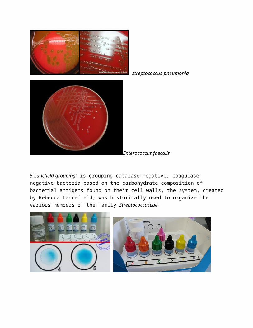
A-Streptococcus pyogenes B-Streptococcus viridans C-Enterococcus faecium.
*Biochemical test:
1-Catalase: is a common enzyme found in nearly all living organisms exposed to oxygen (such as bacteria)It catalyzes the decomposition of hydrogen peroxide to water and oxygen use to differentiate between staph.sp. and strep sp.
2 H2O2 → 2 H2O + O2
2-Coagulase test:
is a protein enzyme produced by several microorganisms that enables the conversion of fibrinogen to fibrin. In the laboratory, it is used to distinguish between different types of Staphylococcus isolates. Importantly, S. aureus is generally coagulase-positive, meaning that a positive coagulase test would indicate the presence of S. aureus. A negative coagulase test would instead show the presence of coagulase negative organisms such as S. epidermidis or S. saprophyticus. However, it is now known that not all S. aureus are coagulase-positive.
A-Coagulase tube : the tube test uses rabbit plasma that has been inoculated with a staphylococcal colony (gram-positive cocci which are catalase positive). The tube is then incubated at 37 °C for 1.5 hours. If negative, then incubation is continued up to 18 hours.
B-Coagulase slide: test is run with a negative control to rule out autoagglutination. Two drops of saline are put onto the slide labeled with sample number, test and control. The two saline drops are emulsified with the test organism using a wire loop, straight wire, or wooden stick, a drop of plasma (rabbit plasma anticoagulated with EDTA is recommended) is placed on the

inoculated saline drop corresponding to test, and mixed well, then the slide is rocked gently for about 10 seconds.
3-Manitol salt agar: is a selective and differential media and have a high concentration of salt, selective for staphylococcus species and inhibited growth of gram positive bacteria and gram negative bacilli and contain of mannitol sugar and phenol red indicator, differential between mannitol fermenter(Staphylococcus aureus) and non-fermenter mannitol(coagulase negative staph.).
Mannitol fermenter Non-fermenter
4-Blood agar differential between Streptococcus species:

A-BETA hemolysis: Complete hemolysis. (Streptococcus pyogenes and agalactiae)
B-Alpha hemolysis: partial hemolysis this is sometimes called green hemolysis because of the color change in the agar. Other synonymous terms are incomplete hemolysis and partial hemolysis. Alpha hemolysis is caused by hydrogen peroxide produced by the bacterium, oxidizing hemoglobin to green biliverdin(Streptococcus pneumonia and viridins).
C-Gamma hemolysis(non-hemolysis):no hemolysis around colonies (Enterococcus faecium and faecalis).
staphylococcus aureus
Streptococcus pyogenes
streptococcus pneumonia
Enterococcus faecalis
5-Lancfield grouping: is grouping catalase-negative, coagulase-negative bacteria based on the carbohydrate composition of bacterial antigens found on their cell walls, the system, created by Rebecca Lancefield, was historically used to organize the various members of the family Streptococcaceae.

Lab#6
Identification of Gram negative bacilli lactose fermenter and non-fermenter bacteria
1-Enterobacteriaceae: gram negative bacilli, lives in gastro tract(colon) called (coliform)some of them pathogens and another not pathogen.

Lactose fermenter(E.coli) Non-lactose(Salmonella)
*Biochemical test:
1-Indole test :

E.coli indole positive
LAB#7
Gram negative bacilli
Non-fermenter bacteria
1-Pseudomonas aeruginosa: gram negative bacilli, produce green pigment (Pyocyanin), oxidase positive, pathogenic bacteria, non-lactose fermenter.
*Biochemical test:
Oxidase test: The oxidase test is used to identify bacteria that produce cytochrome c oxidase, an enzyme of the bacterial electron transport chain. When present, the cytochrome c oxidase oxidizes the reagent (tetramethyl-p-phenylenediamine) to (indophenols) Blue color end product. When the enzyme is not present, the reagent remains reduced and is colorless.
pseudomonas aeruginosa on blood agar
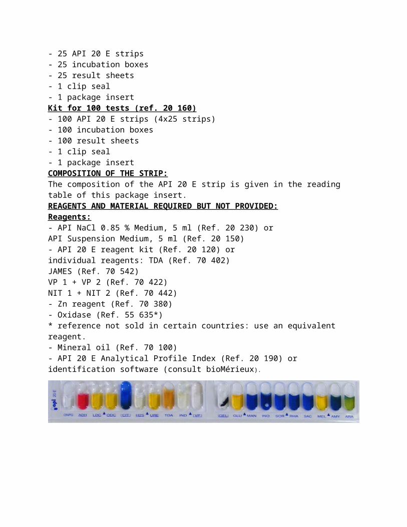
API 20E TEST
Identification system for Enterobacteriaceae and other non-fastidious Gram-negative rods.
SUMMARY AND EXPLANATION:API 20 E is a standardized identification system for Enterobacteriaceae and other non-fastidious, gram negative rods which uses 21 miniaturized biochemical tests and a database, the complete list of those organisms that it is possible to identify with this system is given in the Identification Table at the end of this package insert.PRINCIPLEThe API 20 E strip consists of 20 microtubes containing dehydrated substrates. These tests are inoculated with a bacterial suspension that reconstitutes the media, during incubation, metabolism produces color changes that are either spontaneous or revealed by the addition of reagents, the reactions are read according to the reading Table

and the identification is obtained by referring to the analytical profile Index or using the identification software.CONTENT OF THE KITKit for 25 tests (ref. 20 100)- 25 API 20 E strips- 25 incubation boxes- 25 result sheets- 1 clip seal- 1 package insertKit for 100 tests (ref. 20 160)- 100 API 20 E strips (4x25 strips)- 100 incubation boxes- 100 result sheets- 1 clip seal- 1 package insertCOMPOSITION OF THE STRIP:The composition of the API 20 E strip is given in the reading table of this package insert.REAGENTS AND MATERIAL REQUIRED BUT NOT PROVIDED:Reagents:- API NaCl 0.85 % Medium, 5 ml (Ref. 20 230) orAPI Suspension Medium, 5 ml (Ref. 20 150)- API 20 E reagent kit (Ref. 20 120) orindividual reagents: TDA (Ref. 70 402)JAMES (Ref. 70 542)VP 1 + VP 2 (Ref. 70 422)NIT 1 + NIT 2 (Ref. 70 442)- Zn reagent (Ref. 70 380)- Oxidase (Ref. 55 635*)* reference not sold in certain countries: use an equivalent reagent.- Mineral oil (Ref. 70 100)- API 20 E Analytical Profile Index (Ref. 20 190) oridentification software (consult bioMérieux).

Another gram negative
1- Haemophillus influenzae : gram negative variable (coccobacilli or bacilli) fastidious organism require x and v factor and can grow in chocolate agar and blood agar around staphylococcus aureus, oxidase positive.
Neisseria gonorrhoeae: gram negative cocci, oxidase positive, fastidious organism, can grow on Thayer martin agar (chocolate agar with substances to selective growth of Neisseria).

LAB#8
Diagnosis by susceptibility test

LAB#9
Diagnosis by Serological methods
Serology : is the study of serum and other body fluids. In practice, the term usually refers to the diagnostic identification of antibodies in the serum. Such antibodies are typically formed in response to an infection (against a given microorganism), against other foreign proteins (in response, for example, to a mismatched blood transfusion), or to one's own proteins (in instances of autoimmune disease).
Serological test use to diagnosis viral or bacterial infection.
*ELISA ( The enzyme-linked immunosorbent assay ):
ELISA is a test that uses antibodies and color change to identify a substance.
ELISA is a popular format of "wet-lab" type analytic biochemistry assay that uses a solid-phase enzyme immunoassay (EIA) to detect the presence of a substance, usually an antigen, in a liquid sample or wet sample
1-Indirect ELISA: is a two-step ELISA which involves two binding process of primary antibody (sample antibody) and labeled secondary antibody.
2-Direct ELISA(Sandwich ELISA):the sandwich ELISA quantify antigens between two layers of antibodies (i.e. capture and detection antibody).

Lab#9
Moleculer diagnosis
PCR (Polymerase reaction) is an in-vitro enzymatic reaction used for amplification of a specific DNA fragment that lies between two regions of known nucleotide sequence.
Use to diagnosis of viral infection and bacterial infection
Three steps of PCR principles:
1-Denaturation: the two DNA stands are separated by breaking down the hydrogen bonds between complementary bases at 92-96°C.
2-Annealing: Two specific primers are linked to sequences flanking the target region (37-65°C).
Primers serve as a starting unit for DNA polymerase
3-Extension: Two specific primers are linked to sequences flanking the target region (72°C).
Primers serve as a starting unit for DNA polymerase.
Examples of some machine for PCR technique:1-PCR Thermocycler:
used for amplification of nucleic acids.After that used Gel Electrophoresis to detect the nucleic acid.
*Gel electrophoresis: Is a technique used for allows us to separate of DNA or RNA or Proteins by Agarose gel and some chemicals and visual it under monitor.

2-PCR light cycler:Used for amplification of nucleic acid without Gel electrophoresis
3-Real – Time PCR (RT-PCR):This type is based on the same principle, but the only difference is that it is associated with a device to determine the real time to start the reaction and then the real quantity of copies of DNA. This depends on the presence of free radioactive nitrogen bases to determine this, which makes it easier for researchers to determine the presence of the gene and the amount of the gene without reaching the end of the specific thermocouples and its results chart.
3-GeneXpert:
The GeneXpert test is a molecular test for Genes responsible for Multi drug resistance bacteria which diagnoses MRSA or TB or Carbapenem resistance
by detecting the presence of genes bacteria, as well as testing for resistance to the drug Methicillin.
Lab#10
Yeast identification
1-Germ tube test:
Use to differentiate between Candida albicans and candida sp.
Tube contains of human plasma or horse plasma
*Procedure:
1-suspention one colony of yeast on plasma.
2-incubate at 37 c for 3 hours and read result
germ tube

Germ tube positive Candida albicans
Germ tube negative (+)positive False positive