00 slow vision loss 2013 - stony brook university hospital
TRANSCRIPT

3/30/2013
1
SLOW VISION LOSS
Robert HonkanenMDAssociate Clinical Professor
Department of OphthalmologyStony Brook University
Eye Anatomy
Slow Vision Loss
Definition: Vision loss that occurs gradually over a period of weeks to years, often in a
Causes
Glaucoma
Cataract
Macular Degenerationto years, often in a slow impercetablemanner.
Macular Degeneration
Amblyopia/Strabismus

3/30/2013
2
Glaucoma
Four basic clinical presentations we will review
Primary Open Angle Glaucoma
Primary Angle Closure Glaucoma (Chronic or Acute)
Secondary Glaucoma
Congenital Glaucoma
Glaucoma
Definition – a group of diseases that have in common a characteristic optic neuropathy with associated visual field loss for which elevated IOP is one of the primary risk factors.
Glaucoma
Epidemiology
Second leading cause of irreversible blindness. 60.5 million affected individuals in 2010 increasing to 79.6 million by 2020.
14% of those affected will be blind bilaterally from the disease.
Number one cause of irreversible blindness in African Americans.
3/30/2013
3
Glaucoma
Epidemiology
Up to 50% of affected individuals may not know they have the disease.
Affects approximately 2‐5% of the population over pp y 5 p page forty; with prevalence doubling every decade.
By age 70, approximately 10% of individuals will have glaucoma.
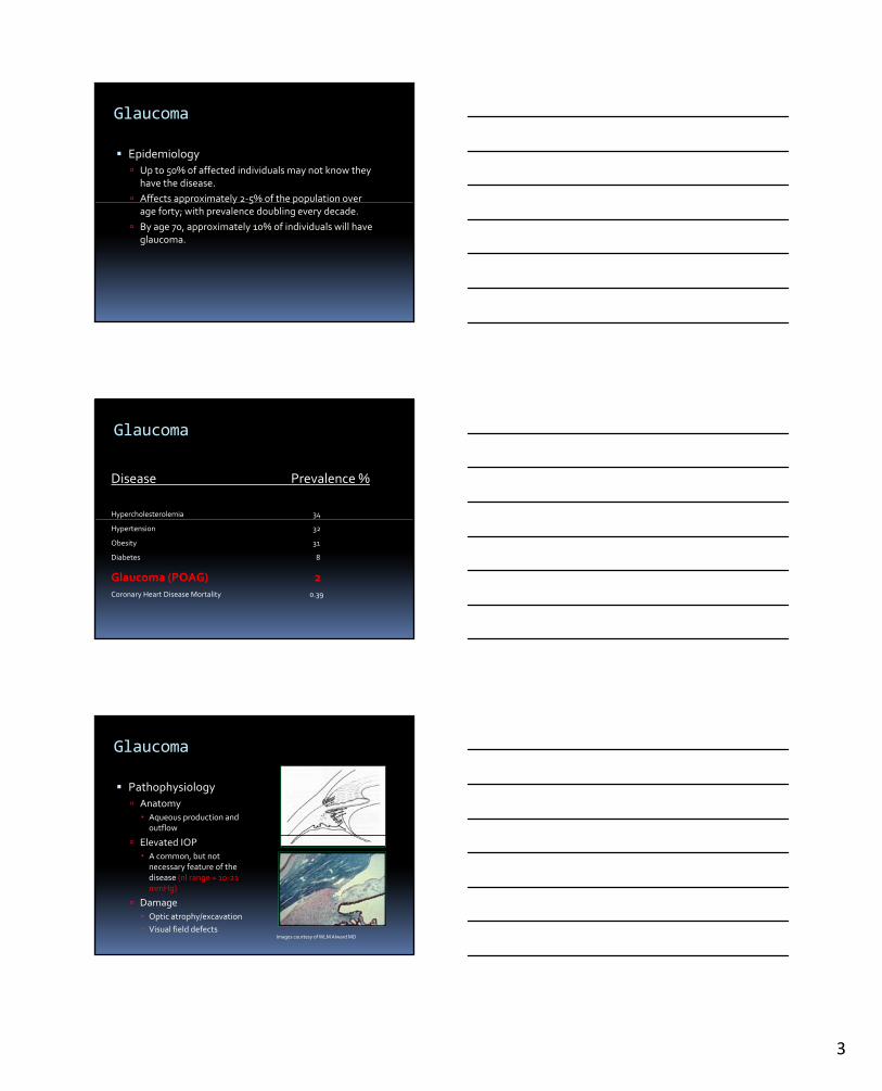
Glaucoma
Disease Prevalence %
Hypercholesterolemia 34
Hypertension 32
Obesity 31
Diabetes 8
Glaucoma (POAG) 2
Coronary Heart Disease Mortality 0.39
Glaucoma
Pathophysiology
Anatomy Aqueous production and outflow
Elevated IOP A common, but not necessary feature of the disease (nl range = 10‐21 mmHg)
Damage
Optic atrophy/excavation
Visual field defectsImages courtesy of WLM AlwardMD

3/30/2013
4
Glaucoma
Pathophysiology
Anatomy Aqueous production and outflow
Elevated IOP A common, but not necessary feature of the disease
Damage Optic atrophy/excavation
Visual field defects
Glaucomatous ONH Progression
Excavation of the disc is due to loss/death of the retinal ganglion cells and their axons which pass through the optic nerve head. The site of damage is at the lamina cribosa of the optic nerve head.
Glaucoma
Pathophysiology
Anatomy Aqueous production and outflow
Elevated IOP A common, but not necessary feature of the disease
Damage Optic atrophy/excavation
Visual field defects

3/30/2013
5
Glaucoma
Pathophysiology
Open Angle
Closed Angle
Limbal blood vessels on cornea
Schwalbe’s line (not really seen)TM
Scleral SpurCiliary Body Band
Cornea
Iris

3/30/2013
6
Glaucoma
Primary Open Angle Glaucoma Most common type (70% of all
glaucoma cases) Familial disease, hereditary Bilateral Caused by acquired impairment Caused by acquired impairment
of acqueous drainage through the trabecular meshwork
Marked by progressive constriction of the field of vision, excavation of the optic nerve head and elevated IOP; painless. (Normal tension glaucoma would be the definition if the IOP was never elevated)
Glaucoma
Primary Closed Angle Glaucoma (“Narrow angle glaucoma”)
Acute Ophthalmologic Emergency Covered in ocular Covered in ocular
emergency lecture (Dr. Haque)
Chronic Often assymptomaticwith
same VF/ONH/IOP findings as POAG.
Differs from POAG because the angle is narrow with PAS (peripheral anterior synechiae).

3/30/2013
7
Glaucoma
Angle closure glaucoma (“Narrow angle”)
Chronic
PAS
Chronic Often assymptomatic with same VF/ONH/IOP findings as POAG.
Differs from POAG because the angle is narrow with PAS (peripheral anterior synechiae).
Glaucoma
Symptoms of POAG
No symptoms until the late phase of disease h d d i i
Symptoms of CACG
May be assymptomatic
May have occasional when advanced vision loss finally becomes symptomatic.
Occasional symptom of decreased vision in dark or at twilight when disease is more advanced.
headache like symptoms especially when reading or in dimly lit rooms
Occasional symptom of decreased vision in dark or at twilight when disease is more advanced.
Glaucoma
Signs of POAG
Slowly progressive loss of vision, usually starting in the periphery
Signs of CACG
Same as those listed for POAG
Will see peripheral anterior the periphery
Increased cupping
Nerve fiber layer defects around the optic nerve
Increased IOP (Not all forms of glaucoma
need to have elevated IOP –however, by definition for POAG it does)
Will see peripheral anterior synechia in the angle.
3/30/2013
8
Glaucoma
Management of POAG and CACG
To be discussed after a few more slides.
Glaucoma
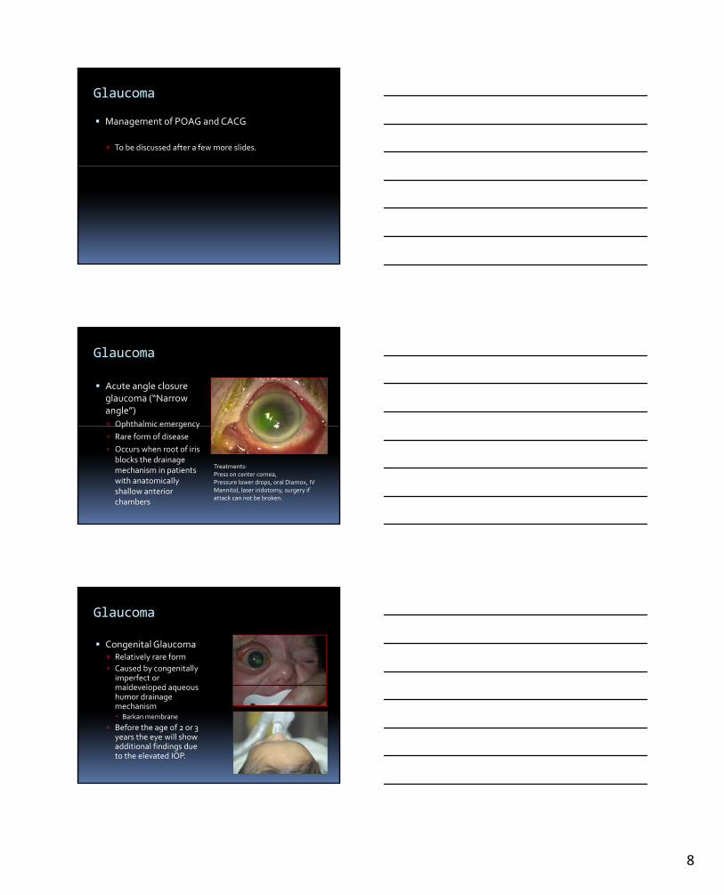
Acute angle closure glaucoma (“Narrow angle”)
Ophthalmic emergencyOphthalmic emergency
Rare form of disease
Occurs when root of iris blocks the drainage mechanism in patients with anatomically shallow anterior chambers
Treatments:Press on center cornea,Pressure lower drops, oral Diamox, IV Mannitol, laser iridotomy, surgery if attack can not be broken.
Glaucoma
Congenital Glaucoma Relatively rare form
Caused by congenitally imperfect or
ld l d maldeveloped aqueous humor drainage mechanism Barkanmembrane
Before the age of 2 or 3 years the eye will show additional findings due to the elevated IOP.

3/30/2013
9
Glaucoma
Congenital Glaucoma
Clinical findings seen in congenital glaucoma: Cloudy / hazy corneas
Breaks in Descemet’smembrane on the cornea –Haab’s striae
Increased axial length – or buphthalmos
Increased corneal diameter
Increased IOP
Glaucoma
Symptoms for Congenital Glaucoma
Tearing (epiphora)
Photophobia (light
Treatment
Surgical for primary cases of congenital glaucomaPhotophobia (light
sensitivity or pain to light)
Blepharospasm (forceful closure of eyelids)
Goniotomy or trabeculotomy surgery
“angle surgery”
Video 01 – f4v
Glaucoma
Secondary Glaucoma
Result of damage to drainage mechanism by other intraocular disorders; e.g. inflammation, after surgery, traumatic, diabetes. Open or closed angle mechanisms

3/30/2013
10
Glaucoma
Secondary Glaucoma
Result of damage to drainage mechanism by other intraocular disorders; e.g. inflammation, after surgery, traumatic, diabetes. Open or closed angle mechanisms
Glaucoma
Symptoms for Secondary Glaucoma
May have more acute onset
Signs for Secondary Glaucoma
Identifiable pathology known to contribute to
May have a painful, red eye
May have peri‐orbital pain
glaucoma – often but not limited to abnormalities in corneal endothelium, angle structures, iris architecture, or lens.
Glaucoma
Examination for Glaucoma
Anterior segment exam / posterior segment exam to r/o secondary causes
Intraocular pressure
Ophthalmoscopy (clinical optic nerve head evaluation)
Visual field testing
Optic Nerve Head Imaging (OCT, HRT, GDx)

3/30/2013
11
Glaucoma
Management (adult onset)
All forms of treatment are currently aimed at ylowering the IOP
Modalities Include: Medication
Laser
Surgery
Glaucoma
Medications Cholinergics /
parasympathomimetics
Beta adrenergic blockers
Bottle cap colors Green
Light blue or yellow Carbonic anhydrase
inhibitors
Alpha‐adrenergic agonists
Prostaglandins
Combination drops
Orange
Green / purple bottle
Tiel
Glaucoma
Laser Treatments
Laser trabeculoplasty ALT
SLT
Laser Iridotomy
For angle closure
3/30/2013
12
Glaucoma
Surgical Treatments
Minimally invasive glaucoma surgery
Filtering proceduresg p
Penetrating procedures
Non‐penetrating procedures
Seton Implants
Ciliary Body Destructive procedures
Glaucoma
Surgical Treatments
Minimally invasive glaucoma surgery
Filtering procedures
Video 02
g p
Penetrating procedures
Non‐penetrating procedures
Seton Implants
Ciliary Body Destructive procedures
Glaucoma
Postoperative appearance after trabeculectomy.

3/30/2013
13
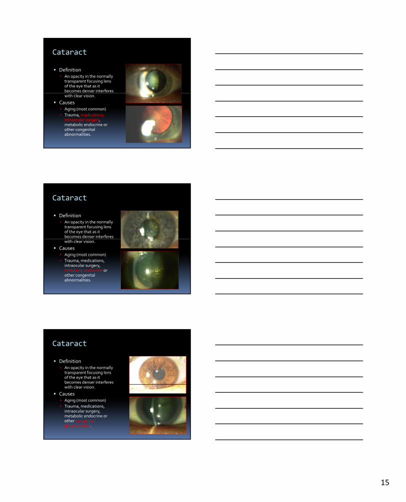
Cataract
Definition An opacity in the normally
transparent focusing lens of the eye that as it becomes denser interferes with clear vision.
Causes Aging (most common)
Trauma, medications, intraocular surgery, metabolic endocrine or other congenital abnormalities.
Cataract
Definition An opacity in the normally
transparent focusing lens of the eye that as it becomes denser interferes with clear vision.
Causes Aging (most common)
Trauma, medications, intraocular surgery, metabolic endocrine or other congenital abnormalities.
Cataract
Definition An opacity in the normally
transparent focusing lens of the eye that as it becomes denser interferes with clear vision.
Causes Aging (most common)
Trauma, medications, intraocular surgery, metabolic endocrine or other congenital abnormalities.
3/30/2013
14
Cataract
Definition An opacity in the normally
transparent focusing lens of the eye that as it becomes denser interferes with clear vision.
Causes Aging (most common)
Trauma, medications, intraocular surgery, metabolic endocrine or other congenital abnormalities.
Cataract
Definition An opacity in the normally
transparent focusing lens of the eye that as it becomes denser interferes with clear vision.
Causes Aging (most common)
Trauma, medications, intraocular surgery, metabolic endocrine or other congenital abnormalities.
Cataract
Definition An opacity in the normally
transparent focusing lens of the eye that as it becomes denser interferes with clear vision.
Causes Aging (most common)
Trauma, medications, intraocular surgery, metabolic endocrine or other congenital abnormalities.
3/30/2013
15
Cataract
Definition An opacity in the normally
transparent focusing lens of the eye that as it becomes denser interferes with clear vision.
Causes Aging (most common)
Trauma, medications, intraocular surgery, metabolic endocrine or other congenital abnormalities.
Cataract
Definition An opacity in the normally
transparent focusing lens of the eye that as it becomes denser interferes with clear vision.
Causes Aging (most common)
Trauma, medications, intraocular surgery, metabolic endocrine or other congenital abnormalities.
Cataract
Definition An opacity in the normally
transparent focusing lens of the eye that as it becomes denser interferes with clear vision.
Causes Aging (most common)
Trauma, medications, intraocular surgery, metabolic endocrine or other congenital abnormalities.

3/30/2013
16
Cataract
Epidemiology
Most common cause of visual loss in the adult population
By age 65, greater than 90% of all people have cataracts
May develop at any age (essential to detect in neonatal period to prevent amblyopia)
Cataract
Treatment is surgical removal
Couching
ICCEICCE
ECCE
Phacoemulsification
Multifocal / pseudo‐accomodating lenses
Astigmatism correcting lenses
Image taken from : www.neatorama.com/2006/11/02/early‐cataract‐surgery/
Cataract
Treatment is surgical removal
Couching
ICCE
Video ECCE
Quicktime #03ICCE
ECCE
Phacoemulsification
Multifocal / pseudo‐accomodating lenses
Astigmatism correcting lenses
3/30/2013
17
Cataract
Treatment is surgical removal
Couching
ICCE
Video Phaco
Quicktime #04ICCE
ECCE
Phacoemulsification
Multifocal / pseudo‐accomodating lenses
Astigmatism correcting lenses
Cataract
Cataract Surgery Often deferred until vision
is decreased and interferes with ADLs.
Surgery is medically Surgery is medically indicated: Neonates
When cataract interferes with diagnosis or management of other eye diseases (eg DM or tumor)
When cataract causes other eye problems such as uveitisor glaucoma
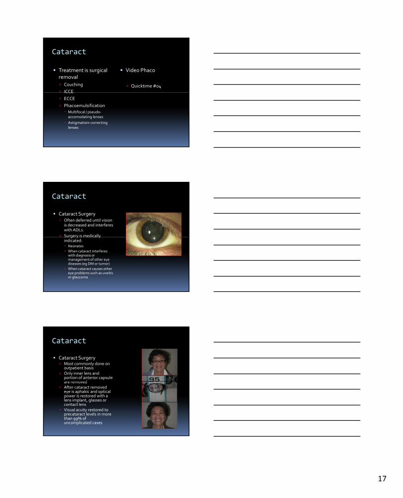
Cataract
Cataract Surgery Most commonly done on
outpatient basis Only inner lens and
portion of anterior capsule are removedare removed
After cataract removed eye is aphakic and optical power is restored with a lens implant, glasses or contact lens
Visual acuity restored to precataract levels in more than 99% of uncomplicated cases
3/30/2013
18
Cataract
Surgical Complications (rare) Endophthalmitis
Retained lens fragments
M l d Macular edema
Retinal Detachment
IOL dislocation
Corneal decompensation
Ptosis
Diplopia
Bleeding
Uveitis
Slow Vision Loss
Definition: Causes
Glaucoma
Cataract
Macular DegenerationMacular Degeneration
Amblyopia/Strabismus
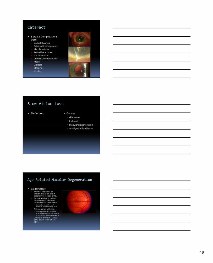
Age Related Macular Degeneration
Epidemiology Number one cause of
irreversible vision loss in people over the age of 50.
Estimated that 15 million people in North America p pcurrently have the disease. Estimates predict a 60%
increase in numbers by 2020.
Risk increases with age. Framingham data showed : 6.4% with signs of AMD age<75 19.7% with signs of AMD age>75
Occurs as dry form (about 85%) or wet form (about 15%).

3/30/2013
19
Age Related Macular Degeneration
Risk Factors Older age, positive family
history, cigarette smoking, hyperopia, light iris color, hypertension, hypercholesterolemia hypercholesterolemia, female gender, and cardiovascular disease
Etiology Complex, but may involve
genetic mutations in the complement pathway. Tyr402His, and Ala69Ser genes
may account for up to 75% of the genetic risk of AMD.
Age Related Macular Degeneration
Exam Drusen – small, round, yellow
lesions located at level of RPE within the macula. Include: Laminar deposits (granular lipid
rich material and collagen) Linear deposits (phoshpolipidLinear deposits (phoshpolipid
vesicles and electron dense granules within inner aspect of Bruch’s membrane.
Typically do not cause symptoms.
Drusen can be described as: Small, Intermediate, or Large Soft,Hard, Confluent
Large, Soft and Confluent drusenmore likely to have vision loss.
Age Related Macular Degeneration
Exam
RPE changes Focal hyperpigmentation
RPE detachments
RPE loss / attenuation (geographic atrophy)
Neovascularization
Subretinal fluid, lipid or blood.
RPE detachments

3/30/2013
20
Age Related Macular Degeneration
Symptoms
Gradual / rapid loss of vision
Metamorphopsiap p
Scotomata
No RAPD
Age Related Macular Degeneration
Clinical exam findings
Decreased central vision/Snellen acuity
Amsler grid gabnormalities.
Flouroscein angiography / Optical coherancetomography (FA/OCT) abnormalities.
Age Related Macular Degeneration
Treatments for Dry ARMD

3/30/2013
21
Age Related Macular Degeneration
Micronutrients Antioxidants
Vitamins 500 mg Vitamin C
400 IU Vitamin E400 IU Vitamin E
15 mg beta carotene
Zinc (80 mg zinc oxide, 2 mg
cupric oxide)
May reduce risk of progression to advanced AMD by up to 25%.
Age Related Macular Degeneration
Other behavior modifications
Avoiding UV light might be beneficial.
Smoking cessation.
Obesity reduction.
Age Related Macular Degeneration
Other behavior modifications
Avoiding UV light might be beneficial.
Smoking cessation.
Obesity reduction.
3/30/2013
22
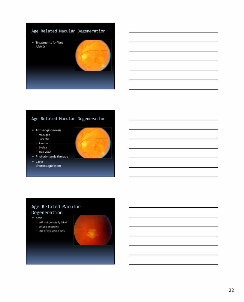
Age Related Macular Degeneration
Treatments for Wet ARMD
Age Related Macular Degeneration
Anti‐angiogenesis
Macugen
Lucentis
AvastinAvastin
Eyelea
Trap VEGF
Photodynamic therapy
Laser photocoagulation
Age Related Macular Degeneration Keys
Will not go totally blind
20/400 endpoint
Use of low vision aidsUse of low vision aids
3/30/2013
23
Slow Vision Loss
Definition: Causes
Glaucoma
Cataract
Macular DegenerationMacular Degeneration
Amblyopia/Strabismus
Amblyopia
Amblyopia Reduced visual acuity
(best‐corrected) not attributable to structural abnormality of eye or abnormality of eye or posterior visual pathway “Lazy eye”
Prevalence: 2‐4% in North America
Onset prior to age 7
Preventable or reversible
Amblyopia
Block in normal visual development
Strabismus
Refractive error Anisometropia
High isometropiag p
Visual deprivation Cataract
Ptosis
Lack of binocular mapping of the environment
Decrease synapses within lateral geniculate body (even atrophy)
Lack of fusion, decreased stereovision
3/30/2013
24
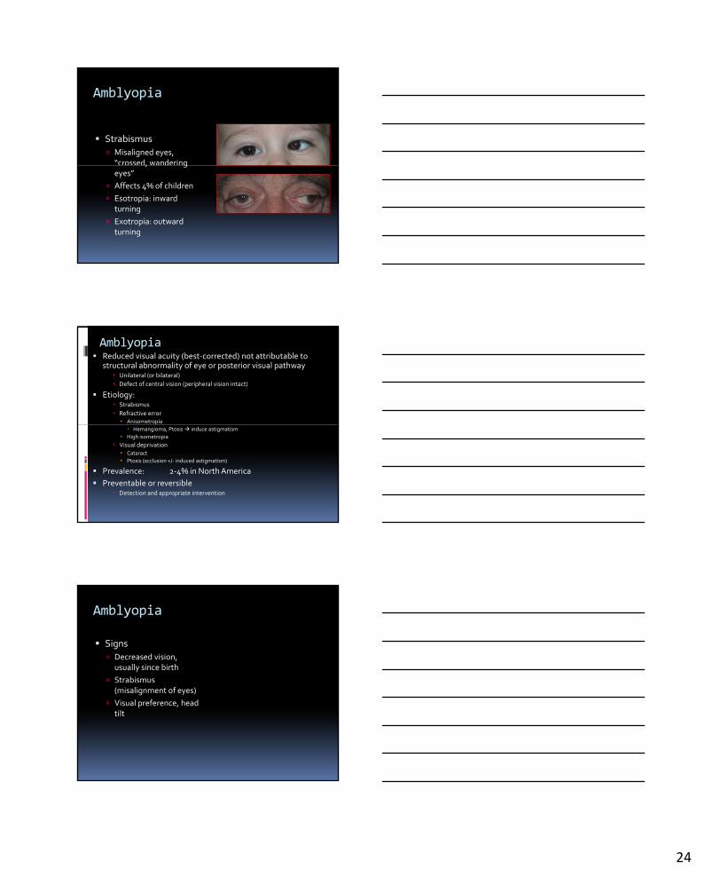
Amblyopia
Strabismus
Misaligned eyes, “crossed, wandering , geyes”
Affects 4% of children
Esotropia: inward turning
Exotropia: outward turning
Amblyopia Reduced visual acuity (best‐corrected) not attributable to
structural abnormality of eye or posterior visual pathway Unilateral (or bilateral)
Defect of central vision (peripheral vision intact)
Etiology: Strabismus
Refractive error Anisometropiap
Hemangioma, Ptosis induce astigmatism
High isometropia
Visual deprivation Cataract
Ptosis (occlusion +/‐ induced astigmatism)
Prevalence: 2‐4% in North America
Preventable or reversible Detection and appropriate intervention
Amblyopia
Signs
Decreased vision, usually since birth
Strabismus (misalignment of eyes)
Visual preference, head tilt

3/30/2013
25
Amblyopia
Treatment
Correct refractive errors / eyeglasses
Treat ocular disease –cataracts / ptosis / glaucoma
Occlusion therapy (patching/penalization of the GOOD eye)
Surgery –move the eye muscle insertions
Amblyopia
Preventable cause of blindness
Critical period
Ri k til Risk until age 10
THE END
Questions