042 introducing a novel model
TRANSCRIPT

Introducing a Novel Model for Studying Macrophage Homing into Atherosclerotic
Plaque in APOE-Deficient Mice
• S. Litovsky, P. Wyde, M. Madjid, A. Akhtar, S.W. Casscells, M. Naghavi
• Texas Heart Institute and University of Texas-Houston, Houston, USA
European Atherosclerosis Society MeetingSalzburg, July 7-10, 2002

Why Study Monocyte Recruitment?
• Monocytes/Macrophages are key players not only in plaque initiation but also in plaque progression and complications through several mechanisms.

Monocyte Recruitment into Atherosclerotic Plaques
Quantification Methods

Review of the studies done by:
• Gerrity et al
• Steinberg et al
• Willerson et al
• The potential role of SPIO
Initial EM (electron microscopy) observation by Gerrity et all (Artery 1980;8:208-14), led to the hypothesis of “macrophage assisted lipid clearancemechanism”.
The swine model of hypercholesterolemia was employed. Yorkshire pigs were fed high cholesterol diets and sacrificed at the ages of 12, 15 and 30 weeks. Samples of aortic arch areas were examined by electron microscopy.

EM studies indicated bi-directional movement of foam cells through the endothelium overlying the fatty streak lesion.
The above mentioned phenomenon may explainthe stability of fatty streaks for a period of time.It also explains that macrophages may initiallyserve as carriers of lipid material out of the plaque.However, focal endothelial damage would resultfrom this process, which may contribute to laterlesion progression by introducing SMC proliferating factors into the wall.

In a 2nd study by Gerrity et al (Atherosclerosis 1988;71:17-25) an additional method to trace macrophages was used:
Monocytes were isolated from peripheral blood of pigs with hypercholesterolemia.
Monocytes were labeled with FITC (fluorocin Isothiocyanate 1-hydrochloride)
Labeled monocytes were reinjected back into the animal.Labeled cells were found in the blood.
Animals were killed after 9 days, followed by histologic examination.
Result
1)FITC-labeled monocytes were found adherent to the thickened site of the intima but not to normal areas.
2)Labelled cells were also found within the atherosclerotic lesions.

In the study by S. Patel, James T. Willerson and Edward Yeh, (Circulation 1998;97:75-81), wild-type mice peritoneal macrophages were labeled with fluorescent latex microspheres and IV injected into ApoE deficient mice.
Animals were sacrificed after 48 hours, and tissuesobtained for histology.
A quantitative survey was done to detect the number of labeled macrophages in the arterial wall.
Antibodies to ICAM-1, integrin and E-selectin were injected 6-8 hours before macrophage injection.
Results
-Study was performed in 2 groups, (control and test) with respect to the use of antibodies.
-After 48 hours macrophages were observed adhering to all stages of plaques.
-The mean number of macrophages in the proximal 1mm of aortic root was estimated to be 143+17.
-Antibodies against ICAM-1 and integrin significantly reduced the number of macrophage homing.
Steinberg et al (ATVB 2000;20:1976-82) reported on a new method of detecting monocytes in the plaque.
The basic idea was the introduction into a recipient animal of leukocytes differing from those of the recipient by virtue of one easily identified and quantified geneticmarker.
Monocytes were transfused from a male donor intoa female recipient and PCR was used to amplifythe Sry gene (testis-determining gene) on the Y chromosome.
Method
The animal model was the LDL receptor deficient mice. Males and females from the same highly inbred strain were used as donors and recipients, so that no significantimmune response occurred.
Females who were to be recipients, were fed an atherogenic diet for 3-6 months, beginning at 6 to 9 months of age.
All animals showed lesions in the aortic arch covering 13% to 49% of the total surface area.
Methods(cont.)
Monocytes collected from male mice were injected into 3 female mice that served as controls. A second group of three female mice also received IP cytokine injection(TNF-α, IL-1β).
Twenty four hours later the animals were euthanized and aorta obtained for the study.
Results
Cytokine-treated mice showed 100% more recruitmentof monocytes in the aortic wall, compared to the controlgroup.
There was no cytokine effect in the animals with >40%of the aortic arch covered by lesions.
The data in the control animals showed that monocyterecruitment continued even when 30% to 50% of the aortic surface was covered by lesions, but the rate ofrecruitment was slower than it was in the earlierstages of the disease.
• Although very informative, all these techniques are invasive and therefore cannot be used sequentially in the same experimental animal.

OBJECTIVE• Work from our group and other laboratories has
shown that the tracer Superparamagnetic Iron Oxide (SPIO) administered intravenously 5 to 7 days before magnetic resonance imaging localizes to aortic atherosclerotic plaques of apoE k/o mice in addition to the reticuloendothelial system (RES).
• In the present study we assessed by histopathology the effect of several cytokines on monocytes homing into aortas of apoE k/o mice.

METHODS• Eleven apoE k/o retired breeders, 11-months old,
were divided in 2 groups. Six received TNF-α 0.2μg, IL-1β 0.2μg and IFNγ 100U/g intraperitoneally, the latter for 5 days; the five control received 0.5 mL saline containing 1% BSA. Three hours later, all the animals were injected with SPIO (Feridex) 1 mMol/kg of iron.
• Seven days later, the recipients were euthanized, the heart and aorta perfused under physiologic pressure and the entire aorta studied histologically.
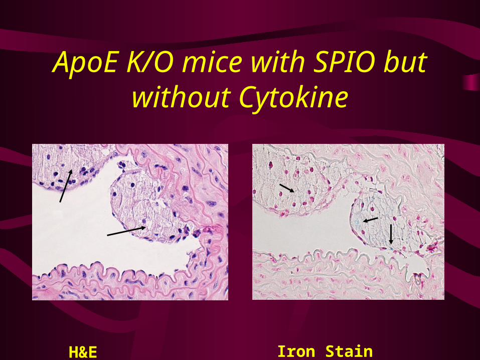
ApoE K/O mice with SPIO but without Cytokine
H&E Iron Stain
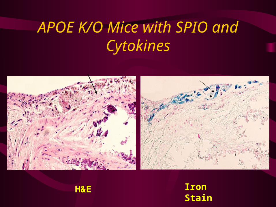
APOE K/O Mice with SPIO and Cytokines
H&E Iron Stain

Iron Deposition on the Edges of Atherosclerotic Plaques
H&E Iron Stain

Iron Deposition in Endothelial Cells

IMMUNOHISTOCHEMISTRY
MAC Stain CD3 Stain
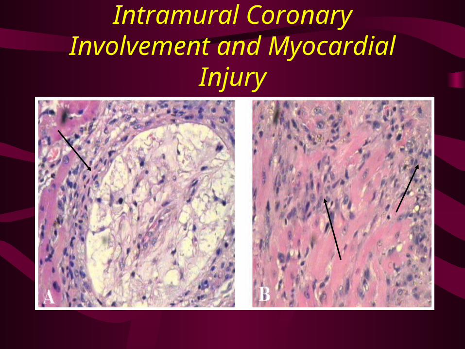
Intramural Coronary Involvement and Myocardial Injury

CONCLUSIONS
• Numerous iron particles are detected subendothelially in the aorta specimens of apoE k/o mice that received cytokines and SPIO intravenously 7 days before sacrifice.
• Aortic iron particle levels are much lower in mice not treated with cytokines.

IMPLICATIONS
• Since SPIO is a contrast agent used in magnetic resonance imaging, this agent offers great hope of studying monocyte recruitment into the atherosclerotic plaque non-invasively.