1 brain - school of medical sciences · cessing in the brain. general semantic deficits (hodges...
TRANSCRIPT

BRAINA JOURNAL OF NEUROLOGY
Neural basis of music knowledge: evidencefrom the dementiasSharpley Hsieh,1,2 Michael Hornberger,1,2 Olivier Piguet1,2 and John R. Hodges1,2
1 Neuroscience Research Australia, Sydney, NSW, 2031, Australia
2 School of Medical Sciences, University of New South Wales, Sydney, NSW, 2031, Australia
Correspondence to: John R. Hodges,
Neuroscience Research Australia,
PO Box 1165,
Randwick,
NSW, 203, Australia
E-mail: [email protected]
The study of patients with semantic dementia has revealed important insights into the cognitive and neural architecture of
semantic memory. Patients with semantic dementia are known to have difficulty understanding the meanings of environmental
sounds from an early stage but little is known about their knowledge for famous tunes, which might be preserved in some
cases. Patients with semantic dementia (n = 13), Alzheimer’s disease (n = 14) as well as matched healthy control participants
(n = 20) underwent a battery of tests designed to assess knowledge of famous tunes, environmental sounds and famous faces,
as well as volumetric magnetic resonance imaging. As a group, patients with semantic dementia were profoundly impaired
in the recognition of everyday environmental sounds and famous tunes with consistent performance across testing
modalities, which is suggestive of a central semantic deficit. A few notable individuals (n = 3) with semantic dementia demon-
strated clear preservation of knowledge of known melodies and famous people. Defects in auditory semantics were mild in
patients with Alzheimer’s disease. Voxel-based morphometry of magnetic resonance brain images showed that the recognition
of famous tunes correlated with the degree of right anterior temporal lobe atrophy, particularly in the temporal pole. This
area was segregated from the region found to be involved in the recognition of everyday sounds but overlapped considerably
with the area that was correlated with the recognition of famous faces. The three patients with semantic dementia with sparing
of musical knowledge had significantly less atrophy of the right temporal pole in comparison to the other patients in the
semantic dementia group. These findings highlight the role of the right temporal pole in the processing of known tunes
and faces. Overlap in this region might reflect that having a unique identity is a quality that is common to both melodies
and people.
Keywords: music; semantic memory; semantic dementia; Alzheimer’s disease; voxel-based morphometry
IntroductionSemantic dementia is a neurodegenerative brain disease within the
family of frontotemporal dementias (Neary et al., 1998) charac-
terized by the progressive and striking loss of semantic memory
(Warrington, 1975; Snowden et al., 1989; Hodges et al., 1992;
Joubert et al., 2006), which is evident across testing modalities,
and involves the loss of knowledge of words, objects and con-
cepts. The degree of semantic loss has been correlated with the de-
gree of atrophy in the anterior and inferior regions of the temporal
lobe, notably the anterior fusiform gyrus (Mummery et al., 2000;
doi:10.1093/brain/awr190 Brain 2011: Page 1 of 12 | 1
Received April 11, 2011. Revised June 10, 2011. Accepted June 15, 2011.
� The Author (2011). Published by Oxford University Press on behalf of the Guarantors of Brain. All rights reserved.
For Permissions, please email: [email protected]
Brain Advance Access published August 21, 2011 at U
niversity of New
South W
ales on October 13, 2011
brain.oxfordjournals.orgD
ownloaded from

Levy et al., 2004; Snowden et al., 2004; Thompson et al., 2004;
Williams et al., 2005; Mion et al., 2010).
Patients with semantic dementia have provided a unique oppor-
tunity to study the cognitive architecture of semantic memory
(Patterson et al., 2007). Early studies focused on knowledge of
objects and people, which were found to partially dissociate, with
loss of knowledge of famous people particularly striking in individ-
uals where there is greater right (than left) temporal involvement
(Thompson et al., 2004) although there are complexities related to
the mode of presentation (faces versus names) of person informa-
tion (Snowden et al., 2004). In the auditory domain, comprehen-
sion of everyday sounds (e.g. croaking of a frog) is compromised
in the early stages of semantic dementia (Bozeat et al., 2000;
Adlam et al., 2006; Goll et al., 2010). Knowledge of well-known
tunes has been reported only in case studies: three individuals
showed preservation of memory for famous melodies (Hailstone
et al., 2009; Omar et al., 2010; Weinstein et al., 2011), whereas
recognition of known tunes was degraded in another patient with
predominantly right-sided temporal atrophy (Gentileschi et al.,
2001). Famous tunes appear to be a category of knowledge
that can be spared in at least some instances of semantic dementia
and might relate to the location of pathology. If confirmed, this
has important implications for the understanding of semantic pro-
cessing in the brain.
General semantic deficits (Hodges and Patterson, 1995; Xie
et al., 2010) and loss of person-specific knowledge (Hodges
et al., 1993; Joubert et al., 2010) is also seen in Alzheimer’s dis-
ease although the magnitude is less than that in semantic demen-
tia (Snowden et al., 2004; Rogers et al., 2006b; Xie et al., 2010).
Within the auditory domain, comprehension of everyday sounds
and famous tunes also appears compromised in Alzheimer’s dis-
ease (Rapcsak et al., 1989; Bartlett et al., 1995). There has been a
single reported study of auditory semantics in Alzheimer’s disease
and semantic dementia, which concerned two individuals, both of
whom were musicians (Omar et al., 2010). This study showed
impairment in the recognition of famous tunes in Alzheimer’s dis-
ease, whereas knowledge of other types of sounds was spared; in
contrast, the pattern of deficits was reversed in the patient with
semantic dementia. The applicability of these intriguing findings in
a larger population of patients with semantic dementia and
Alzheimer’s disease is unclear.
The aims of this study were to: (i) investigate comprehension of
well-known tunes and everyday sounds in a group of patients with
semantic dementia compared with patients with Alzheimer’s dis-
ease and healthy controls; and (ii) to investigate the neural correl-
ates for the recognition of famous tunes using voxel-based
morphometry. It is predicted that the recognition of famous
tunes might be preserved, in at least some cases of semantic de-
mentia, whereas performance should be impaired in the group
with Alzheimer’s disease. It is hypothesized that the recognition
of everyday sounds should be impaired, relative to healthy indi-
viduals, in both dementia groups and also to a milder degree in
Alzheimer’s disease compared with semantic dementia. Exploration
of the neural correlates of famous tune recognition adds to litera-
ture on the neural basis for the anatomical organization of seman-
tic memory.
Methods
ParticipantsA total of 47 subjects participated: 13 patients with semantic demen-
tia, 14 patients with Alzheimer’s disease and 20 healthy controls.
Patients were recruited from the Frontier Frontotemporal Dementia
Research Group, Sydney, Australia where they were diagnosed by a
senior neurologist (J.R.H.). All patients with semantic dementia met
current consensus criteria (Neary et al., 1998) with insidious onset
and gradual progression of a language disorder characterized by im-
paired single word comprehension, as well as non-verbal semantic
deficits (e.g. object and/or face recognition), in the context of relative
preservation of other language skills (phonology, syntax, word repeti-
tion, speech fluency). Structural MRI showed predominant focal atro-
phy in the polar and inferolateral temporal lobes bilaterally. Patients
with Alzheimer’s disease met NINCDS-ADRDA diagnostic criteria
(McKhann et al., 1984) for probable Alzheimer’s disease. Control par-
ticipants were selected from a healthy volunteer panel or were
spouses/carers of patients.
All participants, or their person responsible, provided informed con-
sent for the study according to the Declaration of Helsinki. This study
was approved by the Southern Sydney and Illawarra Area Health
Service and the University of New South Wales ethics committees.
Table 1 provides demographic details. Groups were matched for age
[F(2, 46) = 0.07, non-significant (NS)] and years of education [F(2,
46) = 0.07, NS]. None of the patients with semantic dementia or con-
trol participants were professional musicians. One patient with
Alzheimer’s disease had previously been a member of a popular rock
band. No group difference was observed [H(2) = 1.23, NS] on a
melodic task, which required discrimination between short melodic
phrases, to screen for basic deficits in the perception of musical
tones (Montreal Battery for the Evaluation of Amusia scale subtest;
Peretz et al., 2003).
General cognitive and semantic testsParticipants were given the following neuropsychological tests: the
Mini-Mental State Examination (MMSE) and the Addenbrooke’s
Cognitive Examination—Revised (ACE-R; Mioshi et al., 2006) as gen-
eral measures of cognitive impairment. The Rey-Osterreith Complex
Figure Test (ROCF; Meyers and Meyers, 1995) was administered to
obtain measures of visuospatial drawing ability and visual memory.
Verbal fluency to the category ‘animals’ and the 15-item Boston
Naming Test (BNT-15; Goodglass and Kaplan, 2000) were used as
standardized measures of verbal semantic impairment. A non-verbal
semantic task was also used. Participants were asked to recognize 12
famous faces selected from stimuli compiled by Lambert et al. (2006).
Participants were shown a display of four black and white photo-
graphs; each famous face was paired with three distracter items of
the same age, sex and overall appearance. Participants were not
required to name the famous individual.
Everyday Sounds TestForty-eight sounds were selected from the set provided by Marcell
et al. (2000). Supplementary Table 1 gives a list of the sounds se-
lected. These consisted of 12 sounds in four categories: animals (e.g.
donkey), objects (e.g. camera), musical instruments (e.g. trumpet) and
human vocalizations (e.g. cough). A sound-to-picture matching task
was used as a measure of non-verbal knowledge, which is equivalent
2 | Brain 2011: Page 2 of 12 S. Hsieh et al.
at University of N
ew S
outh Wales on O
ctober 13, 2011brain.oxfordjournals.org
Dow
nloaded from
to the word-to-picture matching task. On different occasions, partici-
pants were presented on each trial with the sound of the object, its
spoken and written name, and asked to match this stimulus to the
target picture from an array including the target and five
within-category distracter items. Coloured pictures were obtained
from the Shutterstock image database (retrieved from 1 June to 31
July 2009 from http://www.shutterstock.com). Different images were
used for target items and the distracters in the two conditions. An
example of the item ‘trumpet’ is provided in Fig. 1.
Famous Tunes Test
Famous Melodies
Thirty famous melodies were selected consisting of Christmas tunes
(e.g. Jingle Bells), folk songs (e.g. Scarborough Fair), instrumental
works (e.g. Beethoven’s Symphony No. 5) and other familiar melodies
(e.g. Pink Panther). Half of these melodies were songs that originally
contained lyrics; others were purely instrumental compositions.
Supplementary Table 2 provides a list of the melodies selected. Each
excerpt consisted of the first melodic phrase of the famous tune. In a
pilot study, these tunes were rated by 16 individuals (mean
age = 38.3 � 14.2; range = 23–63 years) as highly familiar, with a
mean score of 6.43 � 0.58 on a seven-point scale, with 1 meaning
‘unknown’ and 7 meaning ‘very familiar’. A novel tune was created
matched to each famous melody in the manner of Bartlett et al.
(1995). Each novel tune had the same number of notes, pitch inter-
vals, rhythmic units and sound quality to the famous melodies (Fig. 2).
Novel tunes were rated as unfamiliar, with a mean score of
1.84 � 0.48 on the seven-point scale described above.
All tunes were created using the music composition software Finale
2008. Melodies were �7 s in duration. After hearing each tune, par-
ticipants were asked whether or not they recognized it as a famous
tune. They were not required to provide the name of the famous
melodies. A corrected total percentage score of the hits minus false
positives was used to account for response bias. Participants were
tested individually in a quiet room using a laptop with portable speak-
ers and were free to adjust the volume if they wished. Melodies could
be repeated once if requested by the participant.
Famous Titles
Forty song titles were chosen; half were the same as those used in the
Famous Melodies Subtest. Supplementary Table 2 shows a list of the
items chosen. For each famous title, three distracter items were cre-
ated. For example, distracter items for the title ‘Baa Baa Black Sheep’
consisted of ‘Baa Baa Black Cow’, ‘Moo Moo Black Sheep’ and ‘Moo
Moo Black Cow’. These titles were presented in written form and were
read to the patients if required. Participants were required to select
the famous title for each item. The mean score in a pilot study with
15 individuals (mean age = 41.3 � 17.1; range = 22–70 years) was
36.7 � 2.7 out of 40.
Data analysisData were analysed using Predictive Analytics SoftWare Statistics
(Version 18.0.0). Variables were checked for normality of distribution
using the Kolmogorov–Smirnov test. Parametric data were compared
across the three groups (semantic dementia, Alzheimer’s disease and
controls) using one-way ANOVA with post hoc comparisons using the
Tukey Honestly Significant Difference test. The Kruskal–Wallis Test
was used to compare groups where data were non-parametric; post
hoc group comparisons used the Mann–Whitney U Test.
Correlations were used for subtests of the Everyday Sounds Test and
Famous Tunes Test. However, as there is a gradual impairment to a
single central system of conceptual understanding in semantic demen-
tia, impairment should be evident in all tasks that assess the same
concept regardless of whether its input or output modality on testing
is verbal or non-verbal (Rogers, et al., 2004; Patterson, et al., 2007).
Performance on the same items across subtests of the Everyday
Sounds Test and the Famous Tunes Test should not only be correlated
but also show significant consistency across the items when tested
across different modalities.
Item-to-item consistency was assessed using simultaneous logistic
regression analyses (Bozeat et al., 2000) in order to take into account
the familiarity of a concept, which affects performance in semantic
dementia (Hodges et al., 1995; Lambon-Ralph et al., 1998). That is,
performance accuracy is likely to be boosted by the correct identifi-
cation and rejection of items that are highly familiar or unfamiliar,
respectively. Item-to-item consistency was tested by predicting the
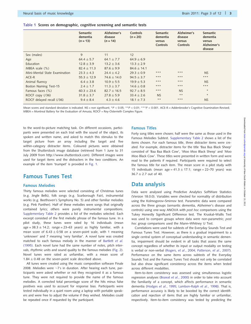
Table 1 Scores on demographic, cognitive screening and semantic tests
Semanticdementia(n = 13)
Alzheimer’sdisease(n = 14)
Controls(n = 20)
SemanticdementiaversusControls
Alzheimer’sdiseaseversusControls
SemanticdementiaversusAlzheimer’sdisease
Sex (males) 9 11 12
Age 64.4 � 5.7 64.1 � 7.7 64.9 � 6.9
Education 12.8 � 3.9 13.2 � 3.6 13.3 � 2.9
MBEA scale (%) 82.4 � 11.3 87.6 � 9.9 84.6 � 14.1
Mini-Mental State Examination 23.3 � 4.3 24.4 � 4.2 29.3 � 0.9 *** *** NS
ACE-R 55.3 � 12.9 74.6 � 14.0 94.5 � 3.7 *** *** ***
Animal fluency 6.4 � 3.8 10.9 � 5.5 19.9 � 5.3 *** *** NS
Boston Naming Test-15 2.4 � 1.7 11.3 � 3.7 14.6 � 0.8 *** *** ***
Famous faces (%) 60.3 � 23.6 82.7 � 16.5 92.7 � 8.5 *** NS *
ROCF copy (/36) 31.8 � 3.7 27.8 � 5.9 33.4 � 2.6 NS ** *
ROCF delayed recall (/36) 9.4 � 8.4 4.3 � 4.6 18.1 � 7.3 ** *** NS
Mean scores and standard deviation is indicated. NS = non significant; *P50.05; **P5 0.01; ***P5 0.001. ACE-R = Addenbrooke’s Cognitive Examination-Revised;MBEA = Montreal Battery for the Evaluation of Amusia; ROCF = Rey-Osterrieth Complex Figure.
Neural basis of music knowledge Brain 2011: Page 3 of 12 | 3
at University of N
ew S
outh Wales on O
ctober 13, 2011brain.oxfordjournals.org
Dow
nloaded from

more difficult task by its easier counterpart. That is, on the Everyday
Sounds Test, accuracy on items of the sound-to-picture condition was
used as the outcome variable and two predictor variables: performance
on the word-to-picture condition and familiarity ratings for the sounds,
which were provided by Marcell et al. (2000). On the Famous Tunes
Test, item consistency was assessed by predicting performance on the
Famous Titles subtest from two predictor variables: recognition accur-
acy on the same items in the Famous Tunes subtest and a measure of
familiarity as indexed by whether or not the tunes contain lyrics (e.g.
‘Rudolph the Red Nose Reindeer’) or not (e.g. ‘Blue Danube Waltz’).
Data from pilot testing indicated that famous titles were better recog-
nized if they contained lyrics.
Voxel-based morphometryVoxel-based morphometry is a technique that identifies grey matter
volume change on a voxel-by-voxel basis from structural MRI data. It
was used to investigate regions of grey matter atrophy between
groups and the neuroanatomical correlates of performance on behav-
ioural measures of interest.
Structural MRIs were available for 11 patients with semantic demen-
tia (seven males; mean age = 64.0 � 6.1; mean years of educa-
tion = 13.4 � 4.0) and 10 patients with Alzheimer’s disease (eight
males; mean age = 64.8 � 7.9; mean years of education = 14.0 � 3.9)
within 7 months of experimental testing. Two patients with semantic
dementia had pacemakers and were unable to be scanned. MRI for
15 control participants were available (nine males; mean age = 64.3 �
6.6; mean years of education = 13.9 � 2.6). Groups were matched for
age [F(2, 35) = 0.04, NS] and education [F(2, 35) = 0.11, NS].
Dementia groups were matched on the Mini-Mental State
Examination [t(19) = �1.86, P4 0.05].
MRI images were obtained using a 3-Tesla Philips scanner with a
standard quadrature head coil. Whole-brain T1-weighted images were
obtained using the following sequences: coronal orientation, matrix
256 � 256, 200 slices, 1 � 1 mm2 in-plane resolution, slice thickness
1 mm, echo time/repetition time = 2.6/5.8 ms, flip angle � = 19�.
MRI data were analysed with FSL-voxel-based morphometry, a
voxel-based morphometry style analysis (Ashburner and Friston,
2000; Mechelli et al., 2005) carried out with the FSL-voxel-based
morphometry tool box (http://www.fmrib.ox.ac.uk/fsl/; Smith et al.,
2004). Structural images were first brain-extracted using BET (Smith,
2002). Tissue-type segmentation was carried out using FAST4 (Zhang
et al., 2001). The resulting grey matter partial volume images were
then aligned to MNI152 standard space using non-linear registration
with FNIRT (Andersson et al., 2007a, b), which uses a b-spline repre-
sentation of the registration warp field (Rueckert et al., 1999). The
resulting images were averaged to create a study-specific template, to
which the native grey matter images were then non-linearly
re-registered. The registered partial volume images were then modu-
lated by dividing by the Jacobian of the warp field. The modulated seg-
mented images were then smoothed with an isotropic Gaussian kernel
with a sigma of 3 mm.
Grey matter intensity differences were investigated via permutation-
based non-parametric statistics (Nichols and Holmes, 2002) with 5000
permutations per contrast. First, differences in cortical grey matter
intensities between patients with semantic dementia and control par-
ticipants were assessed using an unpaired contrast to check the overall
atrophy pattern in the patients. The correlation between behavioural
Figure 1 Example of the item ‘trumpet’ in the Everyday Sounds Test for the (A) sound-picture matching task and (B) word-picture
matching task. Reproduced with permission from Shutterstock.
Figure 2 Example of the melody (A) ‘Happy Birthday’ and (B) the novel melody created as its distracter.
4 | Brain 2011: Page 4 of 12 S. Hsieh et al.
at University of N
ew S
outh Wales on O
ctober 13, 2011brain.oxfordjournals.org
Dow
nloaded from

variables and regions of grey matter atrophy in the patient group
combined were then investigated in separate analyses. The first set
of analyses focused on the regions of atrophy that correlate with ex-
perimental measures of auditory semantics and included scores on the
Famous Melodies subtest and the sound-picture matching task. In a
separate investigation, correlation analyses were conducted on two
standard semantic measures—recognition of famous people and
object naming (i.e. Boston Naming Test)—which have been demon-
strated to be lateralized primarily within the right and left anterior
temporal lobes, respectively, in semantic dementia (Joubert et al.,
2006; Mion et al., 2010). Finally, correlation between scores on the
copy of the Rey-Osterrieth Complex Figure Test and regions of brain
atrophy was analysed as a non-semantic control measure.
For statistical power, a covariate-only statistical model with a [1]
t-contrast was used where poorer performance on a behav-
ioural measure would be associated with decreasing grey matter
volume. Patients with semantic dementia and Alzheimer’s disease
were entered into a single group to increase variance in test perform-
ance. Thresholding of the calculated statistical maps was carried
out using threshold-free cluster enhancement, a method for finding
significant clusters in MRI data, which avoids arbitrary thresholds
(Smith and Nichols, 2009). The reported results were significant at
the P5 0.05, fully corrected for multiple comparisons [Family Wise
Error (FWE)], with the exception of the copy of the Rey-Osterreith
Complex Figure Test where a threshold of P5 0.005 uncorrected
for multiple comparisons was adopted. Clusters 4400 continu-
ous voxels are reported. Anatomical locations of the significant clus-
ters were overlaid on the MNI standard brain. Maximum coordinates
are provided in MNI stereotaxic space. Anatomical labels were de-
termined with reference to the Harvard-Oxford probabilistic cortical
atlas.
Results
General neuropsychology andsemantic testsSummary of performance on standardized cognitive measures is
shown in Table 1. Group differences were found for the Mini-
Mental State Examination [H(2) = 28.5, P50.001] with both de-
mentia groups scoring worse than the controls (semantic demen-
tia: U = 9.50, z = �4.54, P50.001, r = �0.79; Alzheimer’s
disease: U = 16.5, z = �4.43, P50.001, r = �0.76); the patients
with semantic dementia and Alzheimer’s disease did not differ
from each other (U = 74.0, z = �0.83, NS, r = �0.16). In con-
trast, group differences on the Addenbrooke’s Cognitive
Examination-Revised [F(2, 46) = 56.0, P50.001] revealed in
post hoc comparisons that not only dementia groups scored sig-
nificantly lower than controls (P50.001) but also the patients
with semantic dementia were more impaired than patients with
Alzheimer’s disease (P5 0.001).
Group differences on the copy scores [H(2) = 12.2, P50.005]
of the Rey-Osterrieth Complex Figure Test revealed in post hoc
comparisons that the Alzheimer’s disease group scored signifi-
cantly lower than the control (U = 29.0, z = �3.43, P = 0.001,
r = �0.62) and semantic dementia (U = 41.0, z = �2.24,
P5 0.05, r = �0.44) groups on this measure of visuospatial draw-
ing ability, whereas the semantic dementia group did not differ
from controls (U = 85.5, P4 0.10). Group differences on the im-
mediate delayed recall scores [F(2, 42) = 15.0, P5 0.001] of this
task showed that both patient groups scored significantly lower
than the controls in the recall of visual material (Alzheimer’s dis-
ease: P5 0.001; semantic dementia: P5 0.01). No other signifi-
cant comparisons were observed (P40.10).
Not surprisingly, the semantic dementia group were impaired on
standard tests of verbal semantic knowledge. Group differences
for the Boston Naming Test-15 [H(2) = 30.2, P5 0.001] and ani-
mal fluency [F(2, 45) = 29.7, P50.001] revealed, using post hoc
tests, that dementia groups scored worse than controls (semantic
dementia: P50.001 and Alzheimer’s disease: P5 0.001 on both
tests). Patients with semantic dementia were more anomic than
patients with Alzheimer’s disease (U = 10.0, z = �3.73,
P50.001, r = �0.75). Although dementia groups did not differ
in fluency, there was a trend for patients with semantic dementia
to generate fewer animals (P = 0.07). On the famous faces recog-
nition test, a significant group difference was again present
[H(2) = 15.5, P50.001] and on post hoc tests, patients with se-
mantic dementia scored significantly worse than controls
(U = 21.0, z = �3.73, P50.001, r = �0.69) and Alzheimer’s dis-
ease (U = 40.0, z = �2.50, P = 0.01, r = �0.48), whereas patients
with Alzheimer’s disease did not differ from controls (U = 71.5,
z = �1.75, r = �0.32, P = 0.08).
Everyday Sounds TestPerformance on the sound-picture matching task and word-picture
matching task is shown in Fig. 3. Comparisons on the
sound-picture matching task revealed an overall group effect
[F(2,45) = 36.9, P50.001] and post hoc tests revealed that pa-
tients with semantic dementia were most impaired compared with
Alzheimer’s disease and controls (P5 0.001 in both comparisons);
patients with Alzheimer’s disease also showed deficits compared
with controls (P50.05). On the word-picture matching task,
there was also a significant overall group effect [H(2) = 28.7,
P50.001] with marked impairment in the semantic dementia
group compared with the Alzheimer’s disease group (U = 15.5,
z = �3.53, P50.001, r = �0.69) and controls (U = 0.00,
z = �4.67, P5 0.001, r = �0.84). Patients with Alzheimer’s dis-
ease also showed word comprehension deficits compared with
healthy participants (U = 48.0, z = �3.15, P50.005,
r = �0.585).
As expected, performance on these two tests correlated signifi-
cantly for patients with semantic dementia (rp = 0.85, P5 0.001).
Logistical regression analyses indicated that accuracy on the
sound-picture matching task was significantly predicted by per-
formance on the word-picture matching task (Wald value = 18.9;
P50.001) as well as the familiarity of the sound (Wald
value = 12.8; P5 0.001) confirming item consistency across subt-
ests of the Everyday Sounds Test with accuracy, as expected, af-
fected by familiarity.
Famous Tunes TestPerformance on the Famous Tunes Test is shown in Fig. 4. On the
Famous Melodies subtest, there was a significant group effect
Neural basis of music knowledge Brain 2011: Page 5 of 12 | 5
at University of N
ew S
outh Wales on O
ctober 13, 2011brain.oxfordjournals.org
Dow
nloaded from

[F(2, 46) = 18.0, P50.001] and post hoc tests showed that pa-
tients with semantic dementia were impaired compared with pa-
tients with Alzheimer’s disease (P50.001) and controls
(P50.001), whereas performance in patients with Alzheimer’s
disease did not differ from controls (P40.10). The pattern on
the Famous Titles subtest was the same with a significant group
effect [H(2) = 16.2, P50.001] and post hoc analyses revealing
that the patients with semantic dementia were most impaired
(Alzheimer’s disease: U = 26.5, z = �2.62, P5 0.01, r = �0.53;
controls: U = 15.5, z = �3.92, P50.001, r = �0.70), whereas
patients with Alzheimer’s disease did not differ from controls
(P40.10).
Subtests of the Famous Tunes Test were significantly correlated
(rp = 0.74, P = 0.01) for patients with semantic dementia.
Logistical regression analyses indicated that accuracy on the
Famous Melodies subtest was significantly predicted by perform-
ance on the Famous Titles subtest (Wald value = 9.71, P5 0.01)
as well as the index of familiarity used (that is, whether the tune
was a song that contained lyrics or a purely instrumental compos-
ition; Wald value = 17.4, P50.001). As with the Everyday Sounds
Test, there was significant item consistency with modulation of
accuracy by familiarity.
Individual profiles of patients withsemantic dementiaA striking finding on the Famous Tunes Test was the variation
across cases with semantic dementia with some individuals clearly
demonstrating preserved knowledge of well-known melodies and
song titles on testing. Table 2 displays the clinical profiles and test
performances of the patients with semantic dementia in this study
ranked in order according to their scores on the Famous Tunes
Test. Supplementary Table 3 gives information about the patients
with Alzheimer’s disease.
As can be seen, three individuals (Patients D.M., M.S. and V.S.)
performed consistently well on both subtests of the Famous Tunes
Test. Performance on standardized measures of verbal semantic
knowledge and the Everyday Sounds Test was, however, pro-
foundly impaired in these individuals. In contrast, all three patients
performed within the normal range on the recognition of famous
faces. Moreover, the recognition of famous faces correlated sig-
nificantly with both the Famous Melodies (rp = 0.75, P50.01)
and Famous Titles subtests (rp = 0.67, P50.05) in the semantic
dementia group. Correlations with other cognitive measures, such
as the Addenbrooke’s Cognitive Examination-Revised, Boston
Figure 3 Performance on the Everyday Sounds Test for the semantic dementia (SD), Alzheimer’s disease (AD) and control groups on the
(A) Sound-Picture Matching Task and (B) Word-Picture Matching Task. Whiskers represent maximum and minimum values.
Figure 4 Performance on the Famous Tunes Test for semantic dementia (SD), Alzheimer’s disease (AD) and control groups on the (A)
Famous Melodies and (B) Famous Titles subtests. Whiskers represent maximum and minimum values.
6 | Brain 2011: Page 6 of 12 S. Hsieh et al.
at University of N
ew S
outh Wales on O
ctober 13, 2011brain.oxfordjournals.org
Dow
nloaded from

Naming Test-15, Animal Fluency and the Everyday Sounds Test,
were not significant (Table 3).
It is also notable that all 13 patients, including the three indi-
viduals with preserved recognition of famous tunes, showed
marked deficits in the recognition of everyday sounds.
Voxel-based morphometryResults of the voxel-based morphometry analysis are displayed in
Fig. 5 and Table 4. Comparing the patients with semantic demen-
tia and control participants, bitemporal lobe atrophy was present
in the patients with semantic dementia with greater volume loss in
the left rather than the right hemisphere. Within the temporal
lobes, atrophy was most striking in the anterior poles and infer-
olateral temporal regions with the anterior fusiform gyri severely
affected. Areas of brain atrophy also extended into the insular
cortices and the orbitofrontal regions bilaterally. Overall, the
group comparison of semantic dementia and controls replicated
atrophy patterns previously found in semantic dementia
(Mummery et al., 2000; Rosen et al., 2002).
Object naming was associated with atrophy of the left anterior
temporal lobes in a region that extended from the anterior fusi-
form cortex to the temporal poles. In contrast, the recognition of
famous faces was correlated with right-sided anterolateral tem-
poral atrophy, including the posterior fusiform cortex. On a
non-semantic control task, the ability to copy a complex geometric
figure correlated with volume loss in the right superior parietal
lobe and also frontal pole.
Correlation analyses on measures of auditory semantics revealed
that the recognition of famous melodies was significantly corre-
lated with volume loss in the right temporal lobes, particularly in
an area within the temporal pole, which extended into regions
within the insula, amygdala and orbitofrontal cortex. Notably,
this area overlapped considerably with the region that was found
to correlate with famous faces. Patterns of correlations, however,
differed: atrophy of the right posterior fusiform cortex was found
to correlate with the recognition of famous faces but not with the
Table 2 Demographic information and test scores for the patients with semantic dementia
Patients DM MS VS KC JH RW JF GC ST GJ TM JJ PT Controls
Demographic information
Male/female F M F M M M M M F M M F M
Born in Australia? Y N Y Y Y Y Y N N N Y N N
Play (or ever played) amusical instrument?
Y N N N Y Y Y Y N N N Y N
Handedness R R R R R R R R R R L R R
MRI Y Y Y N Y Y Y Y Y N Y Y Y
Test performance
MMSE (/30) 20 24 26 27 23 25 29 27 14 20 28 21 19 29 � 1
ACE-R (/100) 42 55 54 68 57 57 74 79 35 46 61 48 43 95 � 4
Animal fluency 6 9 2 12 6 7 9 13 1 2 5 4 3 20 � 5
Boston Naming Test-15 (/15) 1 2 2 3 3 2 3 7 0 1 1 3 1 15 � 1
Famous face recognition (%) 100 83 83 50 67 83 58 50 25 67 58 25 33 93 � 9
ROCF copy (/36) 27.5 32 31 34 35 35 32 35 29.5 34 35 34 23 33 � 3
ROCF delay (/36) 0.5 0 13.5 2 13.5 23.5 0 21 2 16.5 15 11 4 18 � 7
Everyday Sounds Test
Sound-picture matching (%) 48 40 71 56 56 81 50 65 23 44 44 25 33 89 � 5
Word-picture matching (%) 40 65 71 73 69 88 65 75 NA 48 50 42 40 98 � 2
Famous Tunes Test
Famous melodies subtest (%) 93 90 90 83 63 57 57 53 43 40 37 23 7 91 � 7
Famous titles subtest (%) 88 80 75 53 50 70 83 73 NA 38 55 NA 25 89 � 7
Patients have been ranked according to their scores on the Famous Melodies subtest. NA = patient refused to complete this task. ACE-R = Addenbrooke’s CognitiveExamination-Revised; MMSE = Mini-Mental State Examination; ROCF = Rey-Osterrieth Complex Figure.
Table 3 Correlation between cognitive variables and subtests of the Famous Tunes Test in semantic dementia
ACE-R Animal Fluency Boston Naming Test-15 Everyday Sounds Test FamousFaces
Famous Melodies Subtest 0.22 0.36 0.05 0.48 0.75**
Famous Titles Subtest 0.32 0.42 0.27 0.38 0.67*
ACE-R = Addenbrooke’s Cognitive Examination-Revised. *P50.05; **P5 0.01.
Neural basis of music knowledge Brain 2011: Page 7 of 12 | 7
at University of N
ew S
outh Wales on O
ctober 13, 2011brain.oxfordjournals.org
Dow
nloaded from

recognition of well-known tunes. The recognition of everyday
sounds was, in contrast, correlated with atrophy in an area medial
to the region that was associated with the recognition of famous
tunes. The cluster included primarily the right amygdala and also
extended into the orbitofrontal and anterior fusiform cortex.
Given the significant correlation between right temporal polar
atrophy and the recognition of famous melodies, an additional
analysis contrasting cortical grey matter intensities was conducted
comparing the three individuals with semantic dementia who per-
formed consistently well on both subtests of the Famous Tunes
Figure 5 Voxel-based morphometry analyses showing (A) brain areas that are atrophied in the group with semantic dementia compared
with control participants and (B) brain areas that correlate with the recognition of famous tunes (red), famous faces (blue), everyday
sounds (yellow) and object naming (green). The area represented in purple represents the overlap between the recognition of famous
tunes and famous faces. Coloured voxels show regions that were significant in the analysis with P5 0.05 fully corrected for multiple
comparisons (FWE, t41.7). MNI coordinates: x = 54, y = 4, z = �32. Clusters are overlaid on the MNI standard brain.
Table 4 Voxel-based morphometry results showing regions of significant grey matter atrophy in patients withsemantic dementia and patients with Alzheimer’s disease covarying with behavioural measures of interest
Side BA Cluster size x y z t-value
Famous Melodies Subtest
Anterior inferior temporal gyrus R 21 1994 50 4 �42 2.97
Sound-Picture Matching Task
Anterior parahippocampal gyrus R 28 1231 34 8 �24 3.12
Famous Faces Recognition
Anterior middle temporal gyrus R 21 3250 50 4 �32 3.19
Boston Naming Test
Temporal pole L 38 1279 �38 12 �34 2.74
Rey-Osterrieth Complex Figure copy*
Superior parietal lobe R 7 1286 34 �40 38 4.14
Middle frontal gyrus R 8 1246 30 28 34 4.14
All results are significant for P5 0.05 fully corrected for multiple comparisons (FWE) except for *P5 0.005, uncorrected. BA = Brodmann area.
8 | Brain 2011: Page 8 of 12 S. Hsieh et al.
at University of N
ew S
outh Wales on O
ctober 13, 2011brain.oxfordjournals.org
Dow
nloaded from

Test and the eight remaining patients within the semantic demen-
tia group. Significantly less atrophy of the right temporal lobe was
present in individuals who performed well on the recognition of
famous melodies compared with the other patients with semantic
dementia (Fig. 6). In addition, voxel-based morphometry analyses
in the Alzheimer’s disease group in comparison to controls re-
vealed no significant grey matter atrophy in the Alzheimer’s dis-
ease group in the region that was correlated with the recognition
of famous melodies. Finally, comparison between semantic de-
mentia and Alzheimer’s disease groups showed that the group
with semantic dementia had greater brain atrophy in the region
that was correlated with the recognition of everyday sounds
(Supplementary Figs 1 and 2).
DiscussionThis study is the first to investigate systematically auditory seman-
tics in patients with semantic dementia and Alzheimer’s disease
using both behavioural and neuroimaging tools. Contrary to re-
ports to date, knowledge of famous tunes was generally impaired
in semantic dementia but, importantly, a subgroup showed pres-
ervation despite an equivalent level of impairment on tests of
object-based auditory semantics. Recognition of famous tunes
was found to correlate with the degree of involvement in the
right anterior temporal pole, which was relatively spared in the
subgroup with preserved musical knowledge. Tests of famous
tune and famous face recognition were strongly correlated. On
voxel-based morphometry analyses, both of these measures cor-
related with atrophy of the right polar region but with important
differences: involvement of the right posterior fusiform cortex was
related to the recognition of famous faces but not tunes.
Comparisons between semantic dementia and Alzheimer’s dis-
ease revealed that knowledge of everyday sounds and of famous
tunes was much more impaired in semantic dementia. This is con-
sistent with a large body of work demonstrating that the level of
semantic deficits in Alzheimer’s disease is milder in comparison to
semantic dementia (Snowden et al., 2004; Rogers et al., 2006b;
Xie et al., 2010). These results, however, differ from the pattern
reported by Omar et al. (2010); their patient with Alzheimer’s
disease was impaired when deciding whether melodic fragments
belonged to the same tune, which contrasted to the findings in
the individual with semantic dementia who had relatively spared
music knowledge. One explanation may be that the task used by
Omar et al. (2010) was more difficult than the one that was
employed in this study. An important conclusion, however, is
that dissociations found in individuals may not reflect the general
pattern evident across diseases.
These findings relate to the issue of the underlying deficit in
semantic dementia. Performance across items on both subtests
of the Everyday Sounds Test and the Famous Tunes Test were
highly consistent in semantic dementia, even after controlling for
the effect of familiarity. That is, recognition deficits occurred re-
gardless of whether the same concept was accessed using sound
or verbal testing modalities. Item consistency across different tasks
is a consistent finding in semantic dementia and highlights that
this syndrome is characterized by degradation of conceptual
knowledge rather than of disruption to input or output pathways
(Rogers et al., 2004; Patterson et al., 2007). Auditory perceptual
deficits, for example, appear not to be associated with the level of
impairment in the recognition of everyday sounds in semantic de-
mentia (Goll et al., 2010). Importantly, the recognition impairment
for famous tunes in semantic dementia differs from patients who
have an auditory associative agnosia for music (Eustache et al.,
1990; Peretz, 1996; Dalla Bella, 2009). These patients have a
modality-specific recognition deficit and are unable to identify
famous tunes from the sound of the melody, whereas they are
typically better at recognizing famous lyrics/titles.
Voxel-based morphometry analyses revealed that significant
right temporal polar atrophy differentiated the group of individuals
with semantic dementia, who showed preservation of knowledge
of known tunes, with the rest of the semantic dementia group.
That is, recognition of famous tunes is modulated by the degree of
atrophy in the right temporal pole. In keeping with this
Figure 6 Voxel-based morphometry analysis showing areas of brain atrophy in comparison between the three patients with semantic
dementia who performed well on the Famous Tunes Test and the remaining eight individuals in the semantic dementia group. Coloured
voxels show regions that were significant in the analysis with P50.01, uncorrected (t42.8). MNI coordinates: x = 54, y = 4, z = �32.
Clusters are overlaid on the MNI standard brain.
Neural basis of music knowledge Brain 2011: Page 9 of 12 | 9
at University of N
ew S
outh Wales on O
ctober 13, 2011brain.oxfordjournals.org
Dow
nloaded from
assumption, a single case study of an Italian patient with semantic
dementia with significant right (versus left) temporal atrophy
showed marked impairment in the recognition of previously
known Italian pop songs and areas (Gentileschi et al., 2001).
Regarding the neural basis of different knowledge domains,
voxel-based morphometry analyses revealed strong correlations
between the recognition of famous faces and atrophy in the
right anterior temporal lobes, including the posterior fusiform
gyrus. This finding is consistent with several studies that have
shown that atrophy predominantly on the right in patients with
semantic dementia results in profound and disproportionate loss of
knowledge of people (Evans et al., 1995; Thompson et al., 2004;
Joubert et al., 2006; Busigny et al., 2009). Similarly, patients with-
out semantic dementia with unilateral right temporal lesions show
defects in the recognition and retrieval of person-specific semantic
information from pictures of famous people (Ellis et al., 1989;
Tranel et al., 1997; for a review, see Gainotti, 2007). Functional
neuroimaging studies have implicated a network of activity in the
semantic processing of famous faces (Sergent et al., 1992;
Damasio et al., 1996; Gorno-Tempini and Price, 2001; Brambati
et al., 2010), which includes activity in the right anterior tem-
poral lobe and also the posterior fusiform areas, a region that is
responsible for the visual analysis of faces (Kanwisher and Yovel,
2006).
Turning to theoretical implications, much debate has surrounded
the basis for selective impairment for person knowledge and the
role of the anterior temporal lobes in semantic cognition (for a
review, see Simmons and Martin, 2009). More specifically, the
debate is whether the category-specific impairment for known
individuals, resulting from lesions (more typically) to the right tem-
poral pole, is because this area is a repository for knowledge of
socially relevant concepts (Zahn et al., 2007, 2009; Ross and
Olson, 2010) or for semantically unique entities (Damasio et al.,
2004). Alternatively, it has been argued that the apparent categor-
ical effect of loss of person knowledge reflects the level of pro-
cessing that is needed to identify people at such a specific level. In
this view, the anterior temporal lobes are responsible for the pro-
cessing of all types of concepts (Rogers et al., 2004; Patterson
et al., 2007; Lambon Ralph et al., 2010) but the polar regions
(versus more posterior structures, such as the anterior fusiform
cortex) are increasingly sensitive to more specific levels of semantic
processing (Tyler et al., 2004; Rogers et al., 2006a; Brambati
et al., 2010; Mion et al., 2010). The bias towards the left and
right hemispheres are, according to this model, due primarily to
the nature of modality-specific input or output processes (Lambon
Ralph et al., 2001; Ikeda et al., 2006; Acres et al., 2009; Mion
et al., 2010). Therefore, while conceptual knowledge is distributed
throughout the anterior temporal lobes, tasks that require the pro-
cessing or retrieval of words/names would be strongly biased to-
wards the left as a result of the left-lateralized language system in
most individuals, whereas non-verbal assessments of the same
concept are lateralized to the right (Snowden et al., 2004; Mion
et al., 2010).
Although famous tunes and everyday sounds have not been
considered from this perspective, findings from this study are con-
sistent with this view. Famous tunes share with pictures of famous
faces the quality of being non-verbal unique entities that require
specific levels of processing for recognition. In contrast, everyday
sounds do not require the same level of processing and are iden-
tified at a more basic level. Behavioural findings revealed a signifi-
cant correlation between the recognition of famous tunes and
faces but not with the comprehension of everyday sounds.
Voxel-based morphometry analyses further showed that the ability
to recognize famous tunes was modulated by atrophy of the right
temporal pole. Most interestingly, this region overlapped consid-
erably with the area that was found to correlate with the recog-
nition of famous faces. These novel findings highlight the
relationship between two apparently distinct aspects of cognition.
In contrast, the identification of everyday sounds was correlated
with right-sided atrophy of medial temporal structures (e.g. amyg-
dala) and included the anterior fusiform cortex with little involve-
ment of the temporal pole.
This study highlights the importance of studying cognition in
patients with progressive disorders taking both a group and a
multiple single-case approach. It is particularly informative to com-
pare performance across syndromes using tests designed to probe
related but potentially dissociable processes such as the identifica-
tion of famous faces and tunes. The right temporal pole appears
critical for the processing of both known tunes and faces, and
overlap in this region might reflect that having a unique identity
is a quality that is important and common to both melodies and
people.
AcknowledgementsThe authors thank participants for their support of our research.
We also thank Karalyn Patterson for valuable comments during
the preparation of this article.
FundingNational Health and Medical Research Council, Australia
(NHMRC) (510106); Australian Research Council (ARC) Centre
of Excellence in Cognition and its Disorders (CE110001021).
Australian Postgraduate Award (APA) (to S.H.); ARC Research
Fellowship (DP110104202 to M.H.); NHMRC Clinical Career
Development Award Fellowship (510184 to O.P.); ARC
Federation Fellowship (FF0776229 to J.R.H.).
Supplementary materialSupplementary material is available at Brain online.
ReferencesAcres K, Taylor KI, Moss HE, Stamatakis EA, Tyler LK. Complementary
hemispheric asymmetries in object naming and recognition: a
voxel-based correlational study. Neuropsychologia 2009; 47: 1836–43.
Adlam A-LR, Patterson K, Rogers TT, Nestor PJ, Salmond CH, Acosta-
Cabronero J, et al. Semantic dementia and fluent primary progressive
aphasia: two sides of the same coin? Brain 2006; 129: 3066–80.
10 | Brain 2011: Page 10 of 12 S. Hsieh et al.
at University of N
ew S
outh Wales on O
ctober 13, 2011brain.oxfordjournals.org
Dow
nloaded from

Andersson JLR, Jenkinson M, Smith S. Non-linear optimisation. FMRIB
Technical Report TR07JA1. Oxford: University of Oxford FMRIB
Centre; 2007a. Available from: http://www.fmrib.ox.ac.uk/analysis/
techrep/tr07ja1/tr07ja1.pdf (January 2011, date last accessed).Andersson JLR, Jenkinson M, Smith S. Non-linear registration, aka Spatial
normalisation. FMRIB Technical Report TR07JA2. Oxford: University of
Oxford FMRIB Centre; 2007b. Available from: http://www.fmrib.ox.
ac.uk/analysis/techrep/tr07ja2/tr07ja2.pdf (January 2011, date last
accessed).
Ashburner J, Friston KJ. Voxel-based morphometry–the methods.
Neuroimage 2000; 11: 805–21.Bartlett JC, Halpern AR, Dowling WJ. Recognition of familiar and un-
familiar melodies in normal aging and Alzheimer’s disease. Mem
Cognit 1995; 23: 531–46.
Bozeat S, Lambon-Ralph MA, Patterson K, Garrard P, Hodges JR.
Non-verbal semantic impairment in semantic dementia.
Neuropsychologia 2000; 38: 1207–15.
Brambati SM, Benoit S, Monetta L, Belleville S, Joubert S. The role of the
left anterior temporal lobe in the semantic processing of famous faces.
Neuroimage 2010; 53: 674–81.
Busigny T, Robaye L, Dricot L, Rossion B. Right anterior temporal lobe
atrophy and person-based semantic defect: a detailed case study.
Neurocase 2009; 15: 485–508.Dalla Bella S. Music perception and recognition disorders. In: Cacace AT,
McFarland DJ, editors. Controversies in central auditory processing
disorder. San Diego: Plural Publishing; 2009. p. 243–67.
Damasio H, Grabowski TJ, Tranel D, Hichwa RD, Damasio AR. A neural
basis for lexical retrieval. Nature 1996; 380: 499–505.
Damasio H, Tranel D, Grabowski T, Adolphs R, Damasio A. Neural sys-
tems behind word and concept retrieval. Cognition 2004; 92:
179–229.
Ellis AW, Young AW, Critchley EM. Loss of memory for people following
temporal lobe damage. Brain 1989; 112: 1469–83.
Eustache F, Lechevalier B, Viader F, Lambert J. Identification and discrim-
ination disorders in auditory perception: a report on two cases.
Neuropsychologia 1990; 28: 257–70.Evans JJ, Heggs AJ, Antoun N, Hodges JR. Progressive prosopagnosia
associated with selective right temporal lobe atrophy. A new syn-
drome? Brain 1995; 118: 1–13.
Gainotti G. Different patterns of famous people recognition disorders in
patients with right and left anterior temporal lesions: a systematic
review. Neuropsychologia 2007; 45: 1591–607.
Gentileschi V, Sperber S, Spinnler H. Crossmodal agnosia for familiar
people as a consequence of right infero-polar temporal atrophy.
Cogn Neuropsychol 2001; 18: 439–63.Goll JC, Crutch SJ, Loo JHY, Rohrer JD, Frost C, Bamiou D-E, et al.
Non-verbal sound processing in the primary progressive aphasias.
Brain 2010; 133: 272–85.
Goodglass H, Kaplan E. Boston naming test. 2nd edn. Philadelphia:
Lippincott Williams & Wilkins; 2000.
Gorno-Tempini ML, Price CJ. Identification of famous faces and build-
ings: a functional neuroimaging study of semantically unique items.
Brain 2001; 124: 2087–97.
Hailstone JC, Omar R, Warren JD. Relatively preserved knowledge of
music in semantic dementia. J Neurol Neurosurg Psychiatry 2009;
80: 808–9.Hodges JR, Graham N, Patterson K. Charting the progression in semantic
dementia: implications for the organisation of semantic memory.
Memory 1995; 3: 463–95.
Hodges JR, Patterson K. Is semantic memory consistently impaired early
in the course of Alzheimer’s disease? Neuroanatomical and diagnostic
implications. Neuropsychologia 1995; 33: 803–25.
Hodges JR, Patterson K, Oxbury S, Funnell E. Semantic dementia: pro-
gressive fluent aphasia with temporal lobe atrophy. Brain 1992; 115:
1783–806.Hodges JR, Salmon DP, Butters N. Recognition of famous faces in
Alzheimer’s disease: a cognitive analysis. Neuropsychologia 1993; 31:
775–88.
Ikeda M, Patterson K, Graham KS, Lambon-Ralph MA, Hodges JR. A
horse of a different colour: do patients with semantic dementia rec-
ognise different versions of the same object as the same?
Neuropsychologia 2006; 44: 566–75.
Joubert S, Brambati SM, Ansado J, Barbeau EJ, Felician O, Didic M, et al.
The cognitive and neural expression of semantic memory impairment
in mild cognitive impairment and early Alzheimer’s disease.
Neuropsychologia 2010; 48: 978–88.Joubert S, Felician O, Barbeau E, Ranjeva JP, Christophe M, Didic M,
et al. The right temporal lobe variant of frontotemporal dementia:
cognitive and neuroanatomical profile of three patients. J Neurol
2006; 253: 1447–58.
Kanwisher N, Yovel G. The fusiform face area: a cortical region specia-
lized for the perception of faces. Philos Trans R Soc Lond B Biol Sci
2006; 361: 2109–28.
Lambert NA, Swain MA, Miller LA, Caine D. Exploring the neural organ-
ization of person-related knowledge: lateralization of lesion, category
specificity, and stimulus modality effects. Neuropsychology 2006; 20:
346–54.
Lambon-Ralph MA, Graham KS, Ellis AW, Hodges JR. Naming in seman-
tic dementia–what matters? Neuropsychologia 1998; 36: 775–84.
Lambon Ralph MA, McClelland JL, Patterson K, Galton CJ, Hodges JR.
No right to speak? The relationship between object naming and se-
mantic impairment: neuropsychological evidence and a computational
model. J Cogn Neurosci 2001; 13: 341–56.Lambon Ralph MA, Sage K, Jones RW, Mayberry EJ. Coherent concepts
are computed in the anterior temporal lobes. Proc Natl Acad Sci USA
2010; 107: 2717–22.Levy DA, Bayley PJ, Squire LR. The anatomy of semantic knowledge:
medial vs. lateral temporal lobe. Proc Natl Acad Sci USA 2004; 101:
6710–5.Marcell MM, Borella D, Greene M, Kerr E, Rogers S. Confrontation
naming of environmental sounds. J Clin Exp Neuropsychol 2000; 22:
830–64.
McKhann G, Drachman D, Folstein M, Katzman R, Price D, Stadlan EM.
Clinical diagnosis of Alzheimer’s disease: report of the
NINCDS-ADRDA Work Group under the auspices of Department of
Health and Human Services Task Force on Alzheimer’s Disease.
Neurology 1984; 34: 939–44.
Mechelli A, Price CJ, Friston KJ, Ashburner J. Voxel-based morphometry
of the human brain: methods and applications. Curr Med Imaging Rev
2005; 1: 1–9.
Meyers J, Meyers K. The Meyers Scoring System for the Rey Complex
Figure and the Recognition Trial: professional Manual. Odessa, FL:
Psychological Assessment Resources; 1995.Mion M, Patterson K, Acosta-Cabronero J, Pengas G, Izquierdo-
Garcia D, Hong YT, et al. What the left and right anterior fusiform
gyri tell us about semantic memory. Brain 2010; 133: 3256–68.Mioshi E, Dawson K, Mitchell J, Arnold R, Hodges JR. The
Addenbrooke’s Cognitive Examination Revised (ACE-R): a brief cogni-
tive test battery for dementia screening. Int J Geriatr Psychiatry 2006;
21: 1078–85.
Mummery CJ, Patterson K, Price CJ, Ashburner J, Frackowiak R,
Hodges JR. A voxel-based morphometry study of semantic dementia:
relationship between temporal lobe atrophy and semantic memory.
Ann Neurol 2000; 47: 36–45.
Neary D, Snowden JS, Gustafson L, Passant U, Stuss D, Black S, et al.Frontotemporal lobar degeneration: a consensus on clinical diagnostic
criteria. Neurology 1998; 51: 1546–54.
Nichols TE, Holmes AP. Nonparametric permutation tests for functional
neuroimaging: a primer with examples. Hum Brain Mapp 2002; 15:
1–25.Omar R, Hailstone JC, Warren JE, Crutch SJ, Warren JD. The cognitive
organization of music knowledge: a clinical analysis. Brain 2010; 133:
1200–13.Patterson K, Nestor PJ, Rogers TT. Where do you know what you know?
The representation of semantic knowledge in the human brain. Nat
Rev Neurosci 2007; 8: 976–87.
Neural basis of music knowledge Brain 2011: Page 11 of 12 | 11
at University of N
ew S
outh Wales on O
ctober 13, 2011brain.oxfordjournals.org
Dow
nloaded from

Peretz I. Can we lose memory for music? A case of music agnosia in anonmusician. J Cogn Neurosci 1996; 8: 481–96.
Peretz I, Champod AS, Hyde K. Varieties of musical disorders: the
Montreal Battery of Evaluation of Amusia. Ann N Y Acad Sci 2003;
999: 58–75.Rapcsak SZ, Kentros M, Rubens AB. Impaired recognition of meaningful
sounds in Alzheimer’s disease. Arch Neurol 1989; 46: 1298–300.
Rogers TT, Hocking J, Noppeney U, Michelli A, Gorno-Tempini ML,
Patterson K, et al. Anterior temporal cortex and semantic memory:reconciling findings from neuropsychology and functional imaging.
Cogn Affect Behav Neurosci 2006a; 6: 201–13.
Rogers TT, Ivanoiu A, Patterson K, Hodges JR. Semantic memory inAlzheimer’s disease and the frontotemporal dementias: a longitudinal
study of 236 patients. Neuropsychology 2006b; 20: 319–35.
Rogers TT, Lambon Ralph MA, Garrard P, Bozeat S, McClelland JL,
Hodges JR, et al. Structure and deterioration of semantic memory: aneuropsychological and computational investigation. Psychol Rev
2004; 111: 205–35.
Rosen HJ, Gorno-Tempini ML, Goldman WP, Perry RJ, Schuff N,
Weiner M, et al. Patterns of brain atrophy in frontotemporal dementiaand semantic dementia. Neurology 2002; 58: 198–208.
Ross LA, Olson IR. Social cognition and the anterior temporal lobes.
Neuroimage 2010; 49: 3452–62.
Rueckert D, Sonoda LI, Hayes C, Hill DL, Leach MO, Hawkes DJ.Nonrigid registration using free-form deformations: application to
breast MR images. IEEE Trans Med Imaging 1999; 18: 712–21.
Sergent J, Ohta S, MacDonald B. Functional neuroanatomy of face andobject processing. A positron emission tomography study. Brain 1992;
115: 15–36.
Simmons WK, Martin A. The anterior temporal lobes and the functional
architecture of semantic memory. J Int Neuropsychol Soc 2009; 15:645–9.
Smith SM. Fast robust automated brain extraction. Hum Brain Mapp
2002; 17: 143–55.
Smith SM, Jenkinson M, Woolrich MW, Beckmann CF, Behrens TE,Johansen-Berg H, et al. Advances in functional and structural MR
image analysis and implementation as FSL. Neuroimage 2004; 23:
S208–19.
Smith SM, Nichols TE. Threshold-free cluster enhancement: addressingproblems of smoothing, threshold dependence and localisation in clus-
ter inference. Neuroimage 2009; 44: 83–98.
Snowden JS, Goulding PJ, Neary D. Semantic dementia: a form of cir-
cumscribed cerebral atrophy. Behav Neurol 1989; 2: 167–92.Snowden JS, Thompson JC, Neary D. Knowledge of famous faces and
names in semantic dementia. Brain 2004; 127: 860–72.
Thompson SA, Graham KS, Williams G, Patterson K, Kapur N,
Hodges JR. Dissociating person-specific from general semantic know-ledge: roles of the left and right temporal lobes. Neuropsychologia
2004; 42: 359–70.
Tranel D, Damasio H, Damasio AR. A neural basis for the retrieval ofconceptual knowledge. Neuropsychologia 1997; 35: 1319–27.
Tyler LK, Stamatakis EA, Bright P, Acres K, Abdallah S, Rodd JM, et al.
Processing objects at different levels of specificity. J Cogn Neurosci
2004; 16: 351–62.Warrington EK. Selective impairment of semantic memory. Q J Exp
Psychol 1975; 27: 635–57.
Weinstein J, Koenig P, Gunawardena D, McMillan C, Bonner M,
Grossman M. Preserved musical semantic memory in semantic demen-tia. Arch Neurol 2011; 68: 248–50.
Williams GB, Nestor PJ, Hodges JR. Neural correlates of semantic and
behavioural deficits in frontotemporal dementia. Neuroimage 2005;
24: 1042–51.Xie SX, Libon DJ, Wang X, Massimo L, Moore P, Vesely L, et al.
Longitudinal patterns of semantic and episodic memory in frontotem-
poral lobar degeneration and Alzheimer’s disease. J Int NeuropsycholSoc 2010; 16: 278–86.
Zahn R, Moll J, Iyengar V, Huey ED, Tierney M, Krueger F, et al.
Social conceptual impairments in frontotemporal lobar degeneration
with right anterior temporal hypometabolism. Brain 2009; 132:604–16.
Zahn R, Moll J, Krueger F, Huey ED, Garrido G, Grafman J. Social con-
cepts are represented in the superior anterior temporal cortex. Proc
Natl Acad Sci USA 2007; 104: 6430–5.Zhang Y, Brady M, Smith S. Segmentation of brain MR images through a
hidden Markov random field model and the expectation-maximization
algorithm. IEEE Trans Med Imaging 2001; 20: 45–57.
12 | Brain 2011: Page 12 of 12 S. Hsieh et al.
at University of N
ew S
outh Wales on O
ctober 13, 2011brain.oxfordjournals.org
Dow
nloaded from