1 the chemical principles of rna catalysis
TRANSCRIPT
3
1
The Chemical Principles of RNA CatalysisTimothy J. Wilson and David M. J. Lilley
The University of Dundee, Cancer Research UK Nucleic Acid Structure Research Group, MSI/WTB Complex,Dow Street, Dundee DD1 5EH, UK
1.1 RNA Catalysis
Ribozymes are enzymes that are made of RNA rather than protein. Their functionis to accelerate the rates of chemical reactions. This chapter discusses the chemicalprinciples of catalysis as applied to biological macromolecules. Except for the pep-tidyl transferase reaction of the ribosome, the known natural ribozymes all carryout phosphoryl transfer reactions, either transesterification (including nucleotidyltransfer) or hydrolysis. We shall therefore focus on these reactions principally.However, RNA can bind small molecules with great selectivity, and indeed theriboswitches exploit this ability in many ways to control gene expression. One, theglmS (glucosamine-6-phosphate riboswitch) ribozyme, uses a bound molecule asa coenzyme, and it is not impossible that other ribozymes that use coenzymes ina wider range of chemistry remain to be discovered. Ribozymes that have beenselected in the laboratory demonstrate that a wider range of chemistry can besupported by RNA catalysis [1–3].
One of the most powerful tools that we can use to study the ribozyme mechanismis X-ray crystallography. Having a knowledge of the three-dimensional structure isinvaluable. Yet this is not enough and may even be misleading. This may happen forseveral possible reasons. First, the RNA sequence may have been reduced too far,removing key elements required for proper structure and function. This occurredwith the hammerhead ribozyme [4], where the removal of critical elements thatformed a tertiary interaction led to a remodeling of the active center [5]. The activespecies is rarely studied. The structure of the ribozyme has often been modifiedto prevent activity, and the possible consequences of the modification need to beconsidered. Second, crystal contacts may induce structural changes, as found in thetwister ribozyme where the nucleotide 5′ to the scissile phosphate was pulled out ofthe active site by interaction with guanine in a symmetry-related ribozyme moleculeleading to loss of an in-line geometry [6]. Lastly, of course, a crystal structure cannever capture the transition state because, by definition, it is fleeting. The best we
Ribozymes, First Edition. Edited by Sabine Müller, Benoît Masquida, and Wade Winkler.© 2021 WILEY-VCH GmbH. Published 2021 by WILEY-VCH GmbH.

4 1 The Chemical Principles of RNA Catalysis
can do is to find a related high-energy intermediate if it exists or try to model itwith a transition state analog as was done for the hairpin [7] and hammerhead [8]ribozymes. But we always need to apply chemical insight when looking at crystalstructures of ribozymes, and frequently we must extrapolate what we find to deducethe events occurring in the transition state. Ultimately only kinetic measurementsprobe transition state properties. It is thus the combination of structural analysis,kinetic measurements, and atomic mutagenesis that allow us to approach an under-standing of the catalytic chemical mechanism.
1.2 Rates of Chemical Reactions and Transition StateTheory
The possible trajectory of a chemical reaction is described by a potential energy sur-face plotting the free energy at each point on the reaction landscape. The reactionwill take the path of lowest free energy between the starting material and product,and the highest point along that path is the transition state, generally described as asaddle point on the reaction trajectory. In transition state theory [9–11], we calculatethe rate of reaction as the concentration of the activated complex (i.e. the species atthe point of highest free energy, or transition state) multiplied by the rate of passageout of that state that will be related to bond vibrational frequencies. An equilibriumbetween the ground state and transition state is postulated, leading to an expressionfor the rate (k) as
k = 𝜅 ⋅(
kBT∕h)
exp(−ΔG‡∕RT
)(1.1)
where kB is Boltzmann’s constant, h is Planck’s constant, R is the gas constant, andT is the absolute temperature. 𝜅 is a factor that measures the probability of the tran-sition state proceeding to form the product. The key parameter here is ΔG‡, whichis the free energy of activation that must be supplied to promote the substrate tothe transition state. The population of the activated complex thus governs the rateof reaction that will be determined by the energy barrier according to statisticalmechanics. Parenthetically, we note that it is mechanistically valuable to be able torelate chemical rates to equilibrium properties, and that linear free energy relation-ships are very important in the analysis of catalysis.
Transition state theory shows that chemical catalysis is a question of reducing theenergetic barrier to the formation of the activated complex, i.e. stabilizing the tran-sition state to lower its free energy. This can occur in a variety of ways, includingelectrostatic interactions, stabilization by hydrogen bonding, and formation of cova-lent complexes. We shall discuss this further for the phosphoryl transfer reactionsthat occur in the ribozymes.
The activation free energy ΔG‡ can, of course, be parsed into enthalpy andentropies of activation. The latter can be particularly important in bimolecularreactions, which involve the loss of translational and rotational entropy in theformation of the activated complex. Chemical catalysis occurs in two forms:homogeneous catalysis, where reactants and catalyst are in the same phase, andheterogeneous catalysis, where typically the reaction occurs on some surface. In
1.3 Phosphoryl Transfer Reactions in the Ribozymes 5
a sense, macromolecular catalysis is somewhat intermediate. When two reac-tants bind to a macromolecular catalyst, the loss of translational and rotationalfreedom is partially “paid for” in advance, and the effective concentration of thereactants is increased. Thus in peptidyl transferase reactions, the peptidyl- andaminoacyl-transfer RNAs (tRNAs) are bound to the ribosome and oriented readyto condense to form a new peptide bond. In the group, I intron ribozyme first stagereaction exogenous guanidine reacts when it is bound to the ribozyme. Activationentropy can also appear in more subtle ways, as the reorganization of solvent duringthe reaction, for example.
1.3 Phosphoryl Transfer Reactions in the Ribozymes
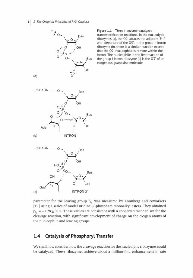
All the known natural ribozymes apart from ribosomal peptidyl transferase catalyzephosphoryl transfer, so we shall focus here on phosphoryl transfer reactions [12].Figure 1.1 shows the reactions catalyzed by the nucleolytic ribozymes, and the groupII and I self-splicing intron ribozymes. These are all transesterification reactions thatinvolve the nucleophilic attack of a ribose hydroxyl group on the phosphodiesterlinkage of RNA. For the nucleolytic and group II ribozymes, the nucleophile is a2′-hydroxyl, on the adjacent or a remote ribose, respectively. For the first stage ofthe group I ribozymes, the nucleophile is the 3′-hydroxyl of a guanosine molecule.In the case of RNaseP, the nucleophile is water, and the group II ribozyme can alsoundergo a hydrolytic reaction.
The phosphorus atom in a phosphodiester is tetrahedral, with four sp3 orbitalsbonded to two bridging and two non-bridging oxygen atoms. The pKa = 1, so there isa negative charge; the bonds to the non-bridging O atoms have partial double bondcharacter by pπ–dπ interaction, and the charge is delocalized. Nucleophilic attackof the oxygen of ROH or HOH requires the participation of the P d orbital, form-ing a phosphorane intermediate with sp3d character that is close to the transitionstate (Figure 1.2a). The phosphorane is trigonal bipyramidal, with three equatorialO atoms and the nucleophilic and leaving group O atoms in the apical positions [14](Figure 1.2b). Therefore to generate the phosphorane, the nucleophile must attackin-line with the P and the leaving group O.
The cleavage reaction for the nucleolytic ribozymes is shown in Figure 1.3. Theproducts of the reaction are a cyclic 2′,3′-phosphate, and a 5′-hydroxyl. For thehammerhead and hairpin ribozymes, it has been demonstrated that the reactionproceeds with inversion of chirality at the phosphorus atom [15, 16]. This isconsistent with the SN2 mechanism shown, proceeding via the phosphorane.Kinetic isotope effects measured for the formation of cyclic 2′,3′-phosphate fromuridine 3′-m′-nitrobenzyl phosphate [17] were indicative of a phosphorane-typetransition state. Using a series of oligonucleotides containing a nucleoside analogwith 2′-C-β-branched substituents with fluoro substitution having a range of pKavalues, Piccirilli and coworkers [18] measured the Brønsted parameter βnuc. Thisgives a measure of the change in charge on the nucleophile approaching thetransition state. Ye et al. obtained βnuc = 0.75± 0.15. The corresponding Brønsted
6 1 The Chemical Principles of RNA Catalysis
5′
3′
O
O
O
O
O OO
O
P
OH
OH
Bas
Bas
3′
5′-EXON O
O
OO
O O
P
OOO
OAde
INTRON
O
OH
OH
Bas
Bas
5′-EXON O
O
O
P
OO
O
O
OOGua
INTRON 3′
OH
OH
OH
HO
Bas
Bas
(a)
(b)
(c)
Figure 1.1 Three ribozyme-catalyzedtransesterification reactions. In the nucleolyticribozymes (a), the O2′ attacks the adjacent 3′-Pwith departure of the O5′. In the group II intronribozyme (b), there is a similar reaction exceptthat the O2′ nucleophile is remote within theintron. The nucleophile in the first reaction ofthe group I intron ribozyme (c) is the O3′ of anexogenous guanosine molecule.
parameter for the leaving group βlg was measured by Lönnberg and coworkers[19] using a series of model uridine 3′-phosphate monoalkyl esters. They obtainedβlg =−1.28± 0.02. These values are consistent with a concerted mechanism for thecleavage reaction, with significant development of charge on the oxygen atoms ofthe nucleophile and leaving groups.
1.4 Catalysis of Phosphoryl Transfer
We shall now consider how the cleavage reaction for the nucleolytic ribozymes couldbe catalyzed. These ribozymes achieve about a million-fold enhancement in rate

1.4 Catalysis of Phosphoryl Transfer 7
O
O
(a) (b)
O
O2′
O5′
Figure 1.2 The geometry of a phosphorane. (a) A representation of phosphorus orbitalhybridization in a phosphorane. The phosphorus atom is sp3d hybridized and has trigonalbipyramidal geometry. (b) The structure of a vanadate transition state analog taken fromthe crystal structure of the hairpin ribozyme [13]. The vanadate mimics the conformation ofthe penta-coordinate phosphorane that is close to the transition state for the ribozymetransesterification reaction. The nucleophile and leaving group are in the apical positions,and three oxygen atoms lie in the central plane.
O
OO
O
O
O O
O
O
O O
OO
O
O5′
Bas
P
PO
O
O OH
OH
OH
H
HOO
P
HO
O
O
AH+
AA
4. General acidcatalysis 3. General
base catalysis
2. Stabilizetransition state
1. In-line
attack
B–B HB
O
O
3′
5′5′
3′3′
O OH
Bas
BasBas
BasBas
∂
∂
∂
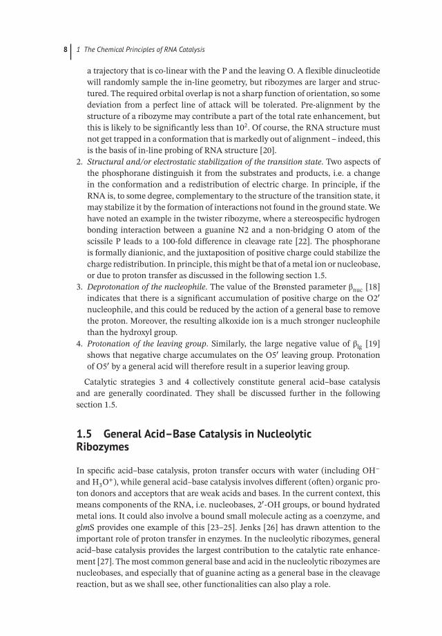
Figure 1.3 The chemical mechanism of the nucleolytic ribozymes and possible catalyticstrategies. Cleavage proceeds left to right, and ligation right to left. The transition state isapproximated by the central structure, with a pentavalent, trigonal-bipyramidalphosphorane structure. Four potential catalytic strategies are indicated. (1) is the in-linetrajectory of attack, (2) stabilization of the transition state structurally or electrostatically,(3) deprotonation of the nucleophile, and (4) protonation of the oxyanion leaving group. Bythe principle of microscopic reversibility, this reaction scheme is symmetrical. For example,B− acts as a general base to deprotonate the O2′ nucleophile in the cleavage reaction, andthus, BH acts as a general acid to protonate the O2′ leaving group in the ligation reaction,where O5′ is now the nucleophile.
over the reaction in a flexible dinucleotide, and a number of catalytic strategies maycontribute to this; several authors have listed the potential contributions [20–22].While this is a useful guide to our chemical thinking, in the end, everything comesdown to stabilization of the transition state, i.e. lowering the activation barrier ΔG‡
in Eq. (1.1). All the contributions are interconnected, and catalysis is multifacto-rial. With that caveat, we can consider four major contributions to catalysis of thephosphoryl transfer reaction (Figure 1.3).
1. Alignment of nucleophilic attack. As noted above, the nucleophile O, P, and leav-ing group O are aligned in the phosphorane, and the nucleophile must attack with
8 1 The Chemical Principles of RNA Catalysis
a trajectory that is co-linear with the P and the leaving O. A flexible dinucleotidewill randomly sample the in-line geometry, but ribozymes are larger and struc-tured. The required orbital overlap is not a sharp function of orientation, so somedeviation from a perfect line of attack will be tolerated. Pre-alignment by thestructure of a ribozyme may contribute a part of the total rate enhancement, butthis is likely to be significantly less than 102. Of course, the RNA structure mustnot get trapped in a conformation that is markedly out of alignment – indeed, thisis the basis of in-line probing of RNA structure [20].
2. Structural and/or electrostatic stabilization of the transition state. Two aspects ofthe phosphorane distinguish it from the substrates and products, i.e. a changein the conformation and a redistribution of electric charge. In principle, if theRNA is, to some degree, complementary to the structure of the transition state, itmay stabilize it by the formation of interactions not found in the ground state. Wehave noted an example in the twister ribozyme, where a stereospecific hydrogenbonding interaction between a guanine N2 and a non-bridging O atom of thescissile P leads to a 100-fold difference in cleavage rate [22]. The phosphoraneis formally dianionic, and the juxtaposition of positive charge could stabilize thecharge redistribution. In principle, this might be that of a metal ion or nucleobase,or due to proton transfer as discussed in the following section 1.5.
3. Deprotonation of the nucleophile. The value of the Brønsted parameter βnuc [18]indicates that there is a significant accumulation of positive charge on the O2′
nucleophile, and this could be reduced by the action of a general base to removethe proton. Moreover, the resulting alkoxide ion is a much stronger nucleophilethan the hydroxyl group.
4. Protonation of the leaving group. Similarly, the large negative value of βlg [19]shows that negative charge accumulates on the O5′ leaving group. Protonationof O5′ by a general acid will therefore result in a superior leaving group.
Catalytic strategies 3 and 4 collectively constitute general acid–base catalysisand are generally coordinated. They shall be discussed further in the followingsection 1.5.
1.5 General Acid–Base Catalysis in NucleolyticRibozymes
In specific acid–base catalysis, proton transfer occurs with water (including OH−
and H3O+), while general acid–base catalysis involves different (often) organic pro-ton donors and acceptors that are weak acids and bases. In the current context, thismeans components of the RNA, i.e. nucleobases, 2′-OH groups, or bound hydratedmetal ions. It could also involve a bound small molecule acting as a coenzyme, andglmS provides one example of this [23–25]. Jenks [26] has drawn attention to theimportant role of proton transfer in enzymes. In the nucleolytic ribozymes, generalacid–base catalysis provides the largest contribution to the catalytic rate enhance-ment [27]. The most common general base and acid in the nucleolytic ribozymes arenucleobases, and especially that of guanine acting as a general base in the cleavagereaction, but as we shall see, other functionalities can also play a role.
1.5 General Acid–Base Catalysis in Nucleolytic Ribozymes 9
The mechanism of the general acid–base-catalyzed cleavage reaction is shownin Figure 1.3. The general base deprotonates the O2′ nucleophile, and the generalacid protonates the 5′-oxyanion leaving group. Some ribozymes catalyze the reverseligation reaction. In that case by the principle of microscopic reversibility, thegeneral acid and base exchange roles, and their required state of protonation.The catalytic power of macromolecular catalysts employing general acid-basecatalysis will be limited by two aspects: the fraction of catalyst that is in anappropriate state of protonation to be active and the reactivity of the general acidand base.
1.5.1 The Fraction of Active Catalyst, and the pH Dependenceof Reaction Rates
In the cleavage reaction depicted in Figure 1.3, the general acid must have a protonto donate, and the general base must be able to accept a proton. In other words,to be active, the ribozyme must have a protonated general acid and a deprotonatedgeneral base. The observed rate of cleavage (kobs) will be lower than the rate whenthe ribozyme is fully active (kcleave) according to:
kobs = kcleave ⋅ fA ⋅ fB (1.2)
where f A is the fraction of protonated acid and f B is the fraction of deprotonated base.f A and f B will be a function of pH, according to the pKa values of the general acid andbase. pH is arguably the most powerful experimental tool we have in probing generalacid–base catalytic mechanisms. For example, the general base can exist either asB− or BH (e.g. for guanine; the corresponding species for adenine would be B andBH+) at high and low pH values, respectively. The fraction of the two forms will begiven by
[B−]∕[BH] = 10(pH−pKa) (1.3)
When the base is operating at a pH that is the same as its pKa, then half of themolecules will be in the required unprotonated form. If the base has a pKa = 10,and the reaction is carried out at pH = 7, then most molecules will be protonated(inactive as a base), and only one molecule in 1000 will be in the active unproto-nated form. However, we shall see later that this is compensated to some degree bya higher reactivity of the base due to its high pKa. Similarly, if this moiety is actingas a general acid, then 999 molecules out of 1000 will be in the protonated form thatcan act as a general acid. But these species will be reluctant to donate that proton,i.e. they will be relatively unreactive.
In the cleavage reactions of the hairpin and Varkud satellite (VS) ribozymes, thegeneral acid and base are the nucleobases of adenine and guanine, respectively (seeChapter 3). The apparent pKa values (i.e. the values measured from the pH depen-dence of cleavage rate) in the context of the VS ribozyme reaction have been mea-sured at 5.2 and 8.4, respectively [28]. Neither is close to physiological pH, and wewould expect that only one ribozyme molecule in 1000 would be active. Bevilacqua[29] carried out a general analysis for the case of a ribozyme using a general acid and

10 1 The Chemical Principles of RNA Catalysis
base (actually formulated for the hairpin ribozyme, but it applies generally), derivinga partition function to calculate f A and f B for given values of pKa:
fA ={
1+10(pKBa −pH)
}∕{
1+10(pKBa −pH)+10(pKB
a −pKAa )+10(pH−pKA
a )}
(1.4)
fB ={
1+10(pH−pKAa )
}∕{
1+10(pKBa −pH)+10(pKB
a −pKAa )+10(pH−pKA
a )}
(1.5)
where the pKa values of the general acid and base are written pKAa and pKB
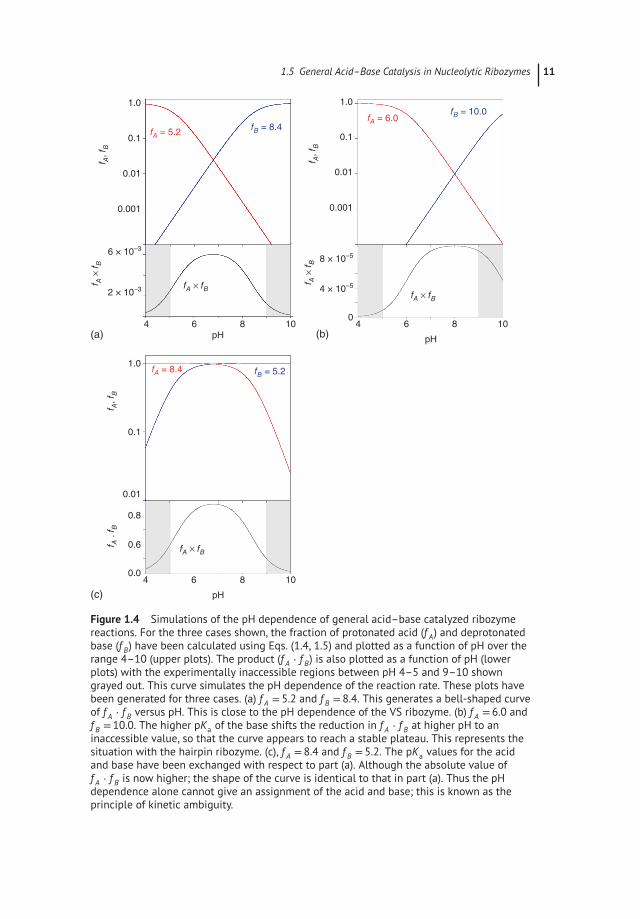
a , respec-tively. Figure 1.4a shows f A and f B plotted as a function of pH for the case of pKavalues of 5.2 and 8.4 (simulating the case of the VS ribozyme). At the low pH end,f A = 1 (fully protonated), and then falls in a log-linear manner as the pH rises. Overthe same range, f B rises until it reaches a plateau (fully deprotonated) toward thehigh pH end. Equation (1.2) shows that the pH dependence of the reaction willreflect the product f A ⋅ f B, that is also plotted in Figure 1.4a. f A ⋅ f B rises at the lowpH end in the regime where f A = 1, but f B is increasing steadily. It then begins totail off as f A begins to fall while f B is still rising. Eventually, f B begins to saturate(f B approaches 1) while f A is steadily falling in a log-linear fashion. The net resultis a bell-shaped curve. It is important to note that the initial rise at low pH is dueto deprotonation of the general base, and the fall at high pH is due to general aciddeprotonation. However, the reducing rise in f A ⋅ f B approaching the peak is due tothe deprotonation of the acid, not the base.
The shape of this curve fits the experimental data for the VS ribozyme very well(see Chapter 3) [28]. Note that because the general acid has a low pKa value, thegeneral base has a high pKa value, and over three units separate the two, the maxi-mum value of f A ⋅ f B is only 6× 10⁻⁴. In protein enzymes, the imidazole side chainof histidine is frequently used in general acid–base catalysis. The pKa of imidazoleis normally close to neutrality, so when two histidine residues are used for generalacid–base catalysis, a considerably higher value of f A ⋅ f B is achieved.
The pKa values for the adenine and guanine nucleobases in the hairpin ribozymeare further apart than those in the VS ribozyme, mostly because the pKa of guaninein the hairpin ribozyme is higher than that in the VS ribozyme. We have computedan f A ⋅ f B simulation for pKa values of 6.0 and 10.0 that corresponds to the hairpinribozyme in Figure 1.4b. Because the pKa values are now separated by four units,a distinct plateau forms, where the deprotonation of acid and base compensate inf A ⋅ f B to create the flat top. Moreover, the eventual fall of f A ⋅ f B does not occur untila high pH is reached at which measurement of cleavage rate is not possible (i.e.the fall lies in the shaded region of the plot). The overall shape is therefore maskedin the experimental profile of rate versus pH, which can therefore be mistaken fora single ionization. This has led to persistent serious errors of interpretation inpast studies.
Returning to the case of nucleobases of pKa values of 5.2 and 8.4, we may ask whatpH profile will result for the reverse (ligation) reaction, where the nucleobase of lowpKa acts as a general base in deprotonated form, and that of high pKa acts are generalacid in its protonated form. This is simulated in Figure 1.4c. Now both f A and f B = 1over much of the range, and the maximum value of f A ⋅ f B = 0.9. Yet the shape of
1.5 General Acid–Base Catalysis in Nucleolytic Ribozymes 11
1.0
0.1
(a)
6 × 10⁻³
2 × 10⁻³
0.01
4 6
f A, f B
fA = 5.2fB = 8.4
f A ×
f B
fA × fBfA × fB
0.001
8 10pH
(c)
fA = 8.4 fB = 5.2
fA × fB
1.0
0.1
0.8
0.6
0.0
0.01
f A, f B
f A .
f B
4 6 8 10
pH
(b)
fA = 6.0fB = 10.0
1.0
0.1
0
8 × 10⁻⁵
4 × 10⁻⁵
0.01
f A, f B
f A ×
f B
0.001
4 6 8 10
pH
Figure 1.4 Simulations of the pH dependence of general acid–base catalyzed ribozymereactions. For the three cases shown, the fraction of protonated acid (f A) and deprotonatedbase (f B) have been calculated using Eqs. (1.4, 1.5) and plotted as a function of pH over therange 4–10 (upper plots). The product (f A ⋅ f B) is also plotted as a function of pH (lowerplots) with the experimentally inaccessible regions between pH 4–5 and 9–10 showngrayed out. This curve simulates the pH dependence of the reaction rate. These plots havebeen generated for three cases. (a) f A = 5.2 and f B = 8.4. This generates a bell-shaped curveof f A ⋅ f B versus pH. This is close to the pH dependence of the VS ribozyme. (b) f A = 6.0 andf B = 10.0. The higher pKa of the base shifts the reduction in f A ⋅ f B at higher pH to aninaccessible value, so that the curve appears to reach a stable plateau. This represents thesituation with the hairpin ribozyme. (c), f A = 8.4 and f B = 5.2. The pKa values for the acidand base have been exchanged with respect to part (a). Although the absolute value off A ⋅ f B is now higher; the shape of the curve is identical to that in part (a). Thus the pHdependence alone cannot give an assignment of the acid and base; this is known as theprinciple of kinetic ambiguity.
12 1 The Chemical Principles of RNA Catalysis
f A ⋅ f B versus pH is identical to that where the general base and acid have high andlow pKa values, respectively. This is termed the principle of kinetic ambiguity, and asa consequence, the pH dependence of the reaction cannot reveal which nucleobaseis acting as general base and which is the general acid. Other approaches must beused to determine this – see Chapter 3.
Nucleobases participating in proton transfer may also respond to the electrostaticinfluence of nearby nucleotides that can add complexity to the shape of the pHdependence of reaction rate. Bevilacqua and coworker [30] have recently derivedpartition functions that take into account cooperative interactions between generalacid and base and the effect of titration of nearby nucleotides.
Nucleobase substitution or atomic mutagenesis is generally carried out in con-junction with rate versus pH measurements to investigate the roles for particularfunctionalities in ribozymes. To take an example, when it was suspected that G630in the VS ribozyme was acting as the general base in the cleavage reaction, it wassubstituted by diaminopurine (i.e. replacing O6 by an amine) with a significantlylower pKa of around 5 [28]. This should shift the curve of f B versus pH stronglyleft (i.e. to lower pH) in our simulations, and so the peak of f A ⋅ f B moves close topH= 5. The experimental curve followed this closely [28], confirming that the higherpKa was indeed due to G630. Note, however, that this experiment by itself does notallow us to determine whether G630 is acting as general acid or general base. Similarexperiments were performed to probe the role of G33 as a general base in the twisterribozyme [6].
Atomic mutagenesis, coupled with rate versus pH measurements, can providecompelling evidence for the involvement of a given nucleobase. In the twisterribozyme, the adenine (A1) immediately 3′ to the scissile phosphate was suspectedto be acting as the general acid in the cleavage reaction, so was subjected to atomicmutagenesis. Replacement of adenine N7 by CH raises its pKa by about 1.5 units,and it was found that this substitution at A1 raised the rate of cleavage by thetwister ribozyme fivefold at pH = 8.5 [22]. While there are a number of waysmutagenesis can lower the catalytic power of a ribozyme, it is much harder toexplain how the rate can be increased. But this can be easily rationalized in termsof the fraction of active ribozyme. The A1N7C substitution displaces the curve off A versus pH toward that of f B, raising the peak value of f A ⋅ f B. In this way, thefraction of active catalyst is higher than for the unmodified ribozyme, resulting in afaster-observed rate.
The mechanism of the twister ribozyme illustrates a factor that is normally disre-garded. A purine nucleobase has three ring-nitrogen atoms that can be protonated:N3, N7, and N1, in order of decreasing acidity. In other ribozymes, it is N1 that partic-ipates in proton transfer, but nucleotide A1 of the twister ribozyme acts as a generalacid by donating a proton from the highly acidic N3 [22]. The shape of the rate ver-sus pH curve is determined by the macroscopic pKa of the nucleobase. However,the relative extent of protonation of the three nitrogen atoms is determined by theirrespective microscopic pKas. For adenosine the extent of protonation is N3 0.7%,N7 3.2%, N1 96.1% [31]. Equations (1.2)–(1.5) above assume a single site of proto-nation, and this is reasonable when N1 participates in proton transfer. However,
1.6 pKa Shifting of General Acids and Bases in Nucleolytic Ribozymes 13
to explain the results of atomic mutagenesis for the twister ribozyme, it is necessaryto modify Eq. (1.2):
kobs = kcleave ⋅ fA ⋅ fB ⋅ fN3H (1.6)
where f A and f B are the fractions of protonated acid and base, respectively, and theextra factor f N3H is the fraction of protonation that occurs specifically at N3 [22].
1.5.2 The Reactivity of General Acids and Bases
We have seen that the nucleobases typically have pKa values that are removed sig-nificantly far from neutrality. Clearly, a disadvantage compared to a protein enzymeusing histidine for general acid–base catalysis. Yet the reactivity of these bases is alsoa function of pKa, and this works in the opposite sense to the population of the activeform, as discussed above. In general base catalysis the rate of ribozyme cleavage isrelated to the pKa of the general base by the Brønsted equation:
log kcleave = B − 𝛽 ⋅ pKa (1.7)
where 𝛽 is the extent of proton transfer in the transition state, and B is a constantspecific to the reaction studied. An analogous linear free energy relation can be writ-ten for acidity. The activity of an hepatitis delta virus (HDV) ribozyme mutated inthe key cytosine nucleotide that acts as the general acid was found to be restoredby bases such as imidazole within the solution [32]. A value of 𝛽 = 0.5 was calcu-lated by measuring the reaction rate as a function of the pKa of the exogenous base[33, 34]. Thus the intrinsic rate for a base of pKa = 10 will be 32 times higher thanone of pKa = 7. This higher reactivity partially compensates for the small populationof unprotonated base that can act in general base catalysis at physiological pH. Inthe HDV ribozyme experiment, the exogenous base is acting as an acid, so the oppo-site relationship applies. The intrinsic rate for an acid of pKa = 10 will be 32 timeslower than one of pKa = 7, and this is partially offset by the higher population ofprotonated acid at physiological pH.
1.6 pKa Shifting of General Acids and Basesin Nucleolytic Ribozymes
The generally accepted pKa values for the adenine and guanine nucleobases are 4.2and 9.5, respectively. If they remained at these values in a ribozyme such as the VSthen only one molecule in about 105 would be in the correct state of protonation tocatalyze the cleavage reaction. This fraction could become more favorable if the pKavalues were shifted closer to neutrality, and for the VS ribozyme, apparent valuesof 5.2 and 8.4 were measured experimentally [28] for the cleavage reaction. Highervalues of the lower apparent pKa have been measured for the hairpin ribozyme as6.3 [35] and 6.9 for the twister ribozyme [6]. In the context of the electronegativeenvironment of RNA, it is relatively easy to explain raised pKa values. The mostremarkable of these is the twister ribozyme. The adenine (A1) that acts as the general
14 1 The Chemical Principles of RNA Catalysis
acid is held in position by a number of hydrogen bonds, two of which involve bothprotons of N6 that are donated to non-bridging O atoms of successive phosphategroups, that carry a negative charge. It is this unusual environment that raises thepKa of the adenine virtually three units. This is necessary because, in the catalyticmechanism, it is the highly acidic N3, not the usual N1, that donates a proton to theO5′ oxyanion leaving group (see Chapter 3) [22].
It is harder to see how the pKa of guanine can be lowered in a ribozyme, and ingeneral, this is less variable. The apparent pKa of G630 in the VS ribozyme has anunusually low value of 8.4, but this was measured in a high Mg2+ concentration. Itis quite likely that there is an ion binding site close to G630 [36], and the positivelycharged ion lowers its apparent pKa.
1.7 Catalytic Roles of Metal Ions in Ribozymes
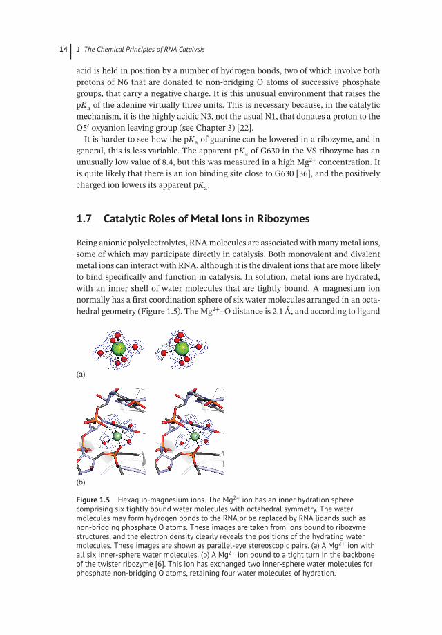
Being anionic polyelectrolytes, RNA molecules are associated with many metal ions,some of which may participate directly in catalysis. Both monovalent and divalentmetal ions can interact with RNA, although it is the divalent ions that are more likelyto bind specifically and function in catalysis. In solution, metal ions are hydrated,with an inner shell of water molecules that are tightly bound. A magnesium ionnormally has a first coordination sphere of six water molecules arranged in an octa-hedral geometry (Figure 1.5). The Mg2+–O distance is 2.1 Å, and according to ligand
(a)
(b)
Figure 1.5 Hexaquo-magnesium ions. The Mg2+ ion has an inner hydration spherecomprising six tightly bound water molecules with octahedral symmetry. The watermolecules may form hydrogen bonds to the RNA or be replaced by RNA ligands such asnon-bridging phosphate O atoms. These images are taken from ions bound to ribozymestructures, and the electron density clearly reveals the positions of the hydrating watermolecules. These images are shown as parallel-eye stereoscopic pairs. (a) A Mg2+ ion withall six inner-sphere water molecules. (b) A Mg2+ ion bound to a tight turn in the backboneof the twister ribozyme [6]. This ion has exchanged two inner-sphere water molecules forphosphate non-bridging O atoms, retaining four water molecules of hydration.
1.7 Catalytic Roles of Metal Ions in Ribozymes 15
field theory, the bond has significant covalent character. These water molecules arenot readily displaced, and the great majority of ions associated with RNA retain afull hydration sphere. These are not specifically bound at one location, and exhibitfast exchange. This is termed outer-sphere binding or “atmospheric” binding. Somewater molecules within the first hydration sphere may be hydrogen-bonded to accep-tors on the RNA, and thus can be located in crystal structures. Monovalent ions virtu-ally always bind as outer-sphere complexes. Most divalent ions such as Mg2+ will alsobe bound in an outer sphere fashion. However, inner-sphere water molecules cansometimes be substituted by one or more RNA ligands (often a non-bridging phos-phate O) if a suitable binding pocket can form. In that case, we call this inner-spherebinding, and the complex is in slow exchange. We observed seven hydrated Mg2+
ions bound to the twister sister (TS) ribozyme structure [37], all with octahedralsymmetry. Two were outer-sphere complexes (retaining full coordination spheresof water molecules), three had exchanged a single inner-sphere water molecule forRNA ligands, and two had exchanged two water molecules. All the RNA ligandswere non-bridging phosphate oxygen atoms apart from one where a cytosine O2was directly bonded to the metal.
Small ions near the top of the periodic table have weakly polarizable orbitals andare classed as “hard” ions. Larger ions with more electrons are more polarizable andare classed as “soft” ions. In general, hard ions bind preferentially to similarly hardanions, so that bound Mg2+ ions will normally be found attached to oxygen ligands.By contrast, Mn2+ or Cd2+ ions bind more avidly to soft atoms like sulfur, and thiscan be the basis of a way to investigate the roles of metal ions in the transition statesof ribozyme reactions.
In general, metal ions are indispensable to the folding of RNA, to lower the electro-static repulsion between the phosphate groups. In most cases, this can be achieved bymonovalent ions, albeit in higher concentration (2 or 3 logs typically) than requiredfor divalent ions, so outer-sphere binding is generally sufficient to achieve foldinginto the active conformation. However, we sometimes observe that very tight turnsin the backbone of RNA may be bridged by an Mg2+ ion as an inner-sphere complex(Figure 1.5).
Site-specifically bound metal ions can directly participate in the chemistry of catal-ysis in a number of different ways:
● They can bind to the reactants and organize and stabilize the structure of the tran-sition state.
● They can stabilize developing negative charge in the transition state electrostati-cally. An atmosphere of outer-sphere metal ions could also achieve some stabiliza-tion of the transition state and, likely, this occurs quite generally in the nucleolyticribozymes.
● Metal ions can act as Lewis acids, binding directly to reactants to activate them.● Lastly, hydrated metal ions can act in general acid–base catalysis. Ions like Mg2+
are weakly acidic, whereby one of the inner-sphere water molecules can lose aproton, i.e.[
Mg(H2O
)6
]2+ + H2O ←−−−−−−−−→[Mg
(H2O
)5OH
]+ + H3O+

16 1 The Chemical Principles of RNA Catalysis
with pKa = 11.4. There is good evidence in the HDV ribozyme that a boundmetal ion acts as a general base to deprotonate the nucleophilic O2′ [38], theTS ribozyme probably acts in a similar manner [37] (see Chapter 3), and wehave recently obtained evidence that a bound metal ion acts as a general acid toprotonate the O5′ leaving group of the pistol ribozyme [55].
There are a number of tests that can point to the direct involvement of metal ionsin ribozyme chemistry. Ribozymes such as the hairpin or twister are active in highconcentrations of monovalent ions, at a rate that is 1/10th of that in Mg2+ ions [39].However, when the metal ion is required to participate directly in the reaction, thatfactor increases to ∼10−5, e.g. as found in the TS ribozyme [37]. The requirement forinner-sphere coordination can be tested by replacing Mg2+ with Co3+(NH3)6 ions.The two ions are structurally similar, but the ammine ligands of the latter exchangeextremely slowly. Thus if inner-sphere ligand exchange is required, Co3+(NH3)6 can-not replace Mg2+ with retention of activity. For example, the HDV ribozyme is essen-tially inactive in the presence of Co3+(NH3)6 ions [38].
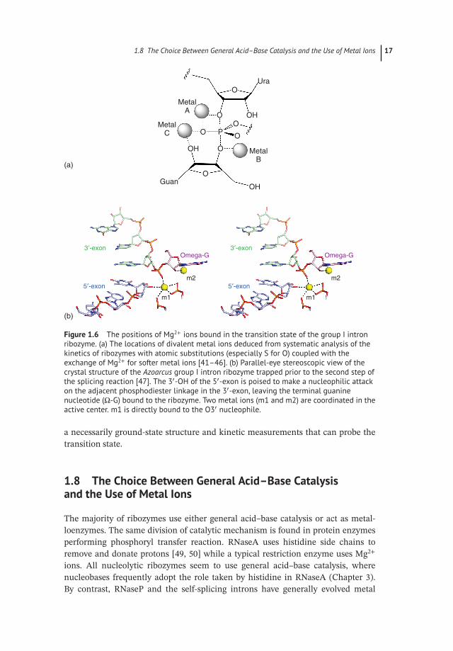
In contrast to the nucleolytic ribozymes, the larger ribozymes, such as theself-splicing introns and RNaseP, seem to have rejected general acid–base catalysis,in favor of acting as metalloenzymes. Many protein enzymes, including nucleasesand polymerases, carry out phosphoryl transfer reactions using two metal ions toactivate the nucleophile, position components, and stabilize the transition state[40]. Over a number of years, Herschlag, Piccirilli, and their coworkers exploredthe role of metal ions in the catalytic mechanism of the group I intron ribozymeusing a combination of atomic mutagenesis and careful reaction kinetics [41–46].Contacts between the metal ions and the transition state were studied by synthesisof sulfur- or amino-substituted substrates (stereo-selectively where relevant), andthen looking for restoration of activity using softer metal ions such as Mn2+ or Cd2+.This is often termed metal ion rescue. These experiments indicated that three metalions are bound to the transition state of the group I intron ribozyme (Figure 1.6a).These are:
● Metal ion A. Bound to the 3′ and proS non-bridging O atoms of the scissile phos-phate. This probably functions by stabilizing the transition state as a negativecharge develops on the leaving group.
● Metal ion B. Bound to the O3′ of the guanosine. This interaction would be expectedto activate the nucleophile, possibly acting as a Lewis acid.
● Metal ion C. Bound to the O2′ of the guanosine plus the proS non-bridging O of thescissile phosphate. Here the role is less obvious, but the metal ion could serve toposition the reacting groups, as well as providing further electrostatic stabilization.
Two metal ions were observed bound in the active site of the Azoarcus group Iintron ribozyme poised at the second stage [47, 48] (Figure 1.6b). One was boundto the 3′ and proS non-bridging O atoms of the scissile phosphate, i.e. as deducedfor metal ion A. The other was bound like a combination of metal ions B and C,interacting with O2′ and O3′ of the guanosine as well as the proS non-bridging O ofthe scissile. The difference between the conclusions from structural and functionalstudies could reflect disorder within the crystal, or perhaps a difference between
1.8 The Choice Between General Acid–Base Catalysis and the Use of Metal Ions 17
MetalA
MetalB
Guan
m2
m1
3′-exon 3′-exon
5′-exon 5′-exon
Omega-G Omega-G
m2
m1
MetalC
(a)
(b)
OH
OH
OH
Ura
O
O
O P
O
O
O
O
Figure 1.6 The positions of Mg2+ ions bound in the transition state of the group I intronribozyme. (a) The locations of divalent metal ions deduced from systematic analysis of thekinetics of ribozymes with atomic substitutions (especially S for O) coupled with theexchange of Mg2+ for softer metal ions [41–46]. (b) Parallel-eye stereoscopic view of thecrystal structure of the Azoarcus group I intron ribozyme trapped prior to the second step ofthe splicing reaction [47]. The 3′-OH of the 5′-exon is poised to make a nucleophilic attackon the adjacent phosphodiester linkage in the 3′-exon, leaving the terminal guaninenucleotide (Ω-G) bound to the ribozyme. Two metal ions (m1 and m2) are coordinated in theactive center. m1 is directly bound to the O3′ nucleophile.
a necessarily ground-state structure and kinetic measurements that can probe thetransition state.
1.8 The Choice Between General Acid–Base Catalysisand the Use of Metal Ions
The majority of ribozymes use either general acid–base catalysis or act as metal-loenzymes. The same division of catalytic mechanism is found in protein enzymesperforming phosphoryl transfer reaction. RNaseA uses histidine side chains toremove and donate protons [49, 50] while a typical restriction enzyme uses Mg2+
ions. All nucleolytic ribozymes seem to use general acid–base catalysis, wherenucleobases frequently adopt the role taken by histidine in RNaseA (Chapter 3).By contrast, RNaseP and the self-splicing introns have generally evolved metal

18 1 The Chemical Principles of RNA Catalysis
ion-based catalytic mechanisms. The reason for the distinction is not clear. Anactive center in which metal ions play the key catalytic role may be more amenableto remodeling in between two-stage reactions like those of the group I and IIintrons. It is perhaps easier to evolve an RNA that uses bound metal ions, givenits polyanionic nature. No ribozyme derived by in vitro selection has proved touse nucleobases, and the close similarity of the active sites of the hairpin andVS ribozymes (Chapter 3) suggests there could be relatively few ways to usenucleobases to catalyze phosphoryl transfer reactions.
1.9 The Limitations to RNA Catalysis
By comparison with proteins, the catalytic resources of RNA are very limited.These are just four rather similar heterocyclic nucleobases, 2′-hydroxyl groups, andhydrated metal ions. This is perhaps reflected in the catalytic rate enhancementsachieved and the limited range of reactions that are catalyzed. Part of the limitationcomes from the pKa values of the nucleobases; for example, only a small fractionof the VS ribozyme carrying out cleavage is active at a given time. If due allowanceis made for that, then the catalytic efficiency becomes comparable to RNaseA[51]. Rate could well be a limitation, and the majority of ribozymes that exist incontemporary cells do not undergo multiple turnovers.
The lack of a wide array of chemical resources may be a greater limitation onthe range of chemistry catalyzed. In the RNA world hypothesis, it is necessarythat RNA would catalyze a much greater range of chemistry. We can speculatethat the chemical repertoire of RNA might be expanded if small moleculescould bind and act as coenzymes. RNA is very good at binding small moleculeligands with great specificity, exemplified by the great range of ligands for theriboswitches [52]. Riboswitches have been identified that bind a number ofcoenzymes, including thiamine pyrophosphate (TPP), flavin mononucleotide(FMN), S-adenosylmethionine (SAM), S-adenosylhomocysteine (SAH), tetrahy-drofolate (THF), Ado-cobalamine. GlmS (Chapter 3) provides a precedent fora ribozyme using a coenzyme, where bound glucosamine-6-phosphate is theprobable general acid in the cleavage reaction. The most abundant group ofriboswitches are those that bind TPP [53]. TPP is a very versatile coenzyme,involved in the formation and breakage of carbon–carbon bonds, e.g. in transke-tolase. As we have discussed previously [54], we should consider the possibilitythat a ribozyme might bind TPP as a coenzyme, to catalyze a new range ofmetabolic interconversions. The discovery of such novel ribozymes would beexciting indeed!
Acknowledgment
Work on RNA catalysis in Dundee is funded by Cancer Research UK under programGrant A18604.

References 19
References
1 Seelig, B. and Jäschke, A. (1999). A small catalytic RNA motif withDiels–Alderase activity. Chem. Biol. 6 (3): 167–176.
2 Sengle, G., Eisenfuh, R.A., Arora, P.S. et al. (2001). Novel RNA catalysts for theMichael reaction. Chem. Biol. 8 (5): 459–473.
3 Tsukiji, S., Pattnaik, S.B., and Suga, H. (2003). An alcohol dehydrogenaseribozyme. Nat. Struct. Biol. 10 (9): 713–717.
4 Khvorova, A., Lescoute, A., Westhof, E., and Jayasena, S.D. (2003). Sequenceelements outside the hammerhead ribozyme catalytic core enable intracellularactivity. Nat. Struct. Biol. 10 (9): 1–5.
5 Martick, M. and Scott, W.G. (2006). Tertiary contacts distant from the active siteprime a ribozyme for catalysis. Cell 126 (2): 309–320.
6 Liu, Y., Wilson, T.J., McPhee, S.A., and Lilley, D.M. (2014). Crystal structureand mechanistic investigation of the twister ribozyme. Nat. Chem. Biol. 10 (9):739–744.
7 Rupert, P.B., Massey, A.P., Sigurdsson, S.T., and Ferré-D, Amaré, A.R. (2002).Transition state stabilization by a catalytic RNA. Science 298 (5597): 1421–1424.
8 Mir, A. and Golden, B.L. (2016). Two active site divalent Ions in the crystalstructure of the hammerhead ribozyme bound to a transition state analogue.Biochemistry 55 (4): 633–636.
9 Evans, M.G. and Polyani, M. (1935). Some applications of the transition statemethod to the calculation of reaction velocities, especially in solution. Trans.Faraday Soc. 31: 875–894.
10 Eyring, H. (1935). The activated complex in chemical reactions. J. Chem. Phys. 3:107–114.
11 Pelzer, H. and Wigner, E. (1935). Über die geschwindigkeitskon stante vonaustauschreaktionen Z. Phys. Chem. B15: 445.
12 Oivanen, M., Kuusela, S., and Lonnberg, H. (1998). Kinetics and mechanisms forthe cleavage and isomerization of the phosphodiester bonds of RNA by Brönstedacids and bases. Chem. Rev. 98 (3): 961–990.
13 Rupert, P.B. and Ferré-D’Amaré, A.R. (2001). Crystal structure of a hairpinribozyme-inhibitor complex with implications for catalysis. Nature 410: 780–786.
14 Westheimer, F.H. (1968). Pseudo-rotation in the hydrolysis of phosphate esters.Acc. Chem. Res. 1: 70–78.
15 van Tol, H., Buzayan, J.M., Feldstein, P.A. et al. (1990). Two autolytic process-ing reactions of a satellite RNA proceed with inversion of configuration. NucleicAcids Res. 18 (8): 1971–1975.
16 Slim, G. and Gait, M.J. (1991). Configurationally defined phosphorothioate-containing oligoribonucleotides in the study of the mechanism of cleavage ofhammerhead ribozymes. Nucleic Acids Res. 19 (6): 1183–1188.
17 Gerratana, B., Sowa, G.A., and Cleland, C.W. (2000). Characterization of thetransition-state structures and mechanisms for the isomerization and cleav-age reactions uridine 3′-m-nitrobenzyl phosphate. J. Am. Chem. Soc. 122 (51):12615–12621.
20 1 The Chemical Principles of RNA Catalysis
18 Ye, J.D., Li, N.S., Dai, Q., and Piccirilli, J.A. (2007). The mechanism of RNAstrand scission: an experimental measure of the Brønsted coefficient, beta nuc.Angew. Chem. 46 (20): 3714–3717.
19 Kosonen, M., Youseti-Salakdeh, E., Strömberg, R., and Lönnberg, H. (1997).Mutual isomerization of uridine 2′- and 3′-alkylphosphatesand cleavage toa 2′,3′-cyclic phosphate: the effect of the alkyl group on the hydronium andhydroxide-ion-catalyzed reactions. J. Chem. Soc. Perkin Trans. 2: 2661–2666.
20 Soukup, G.A. and Breaker, R.R. (1999). Relationship between internucleotidelinkage geometry and the stability of RNA. RNA 5 (10): 1308–1325.
21 Koo, S., Novak, T., and Piccirilli, J.A. (2008). Catalytic mechanism of the HDVribozyme. In: Ribozymes and RNA Catalysis (eds. D.M.J. Lilley and F. Eckstein).Cambridge: Royal Society of Chemistry.
22 Wilson, T.J., Liu, Y., Domnick, C. Kath-Schorr, S. and D. M. J. Lilley, D. M. J.(2016). The novel chemical mechanism of the twister ribozyme. J. Am. Chem.Soc. 138 (19): 6151–6162.
23 McCarthy, T.J. Plog, M. A., Floy, S. A., Jansen, J. A., Soukup, J. K. and Soukup,G. A. (2005). Ligand requirements for glmS ribozyme self-cleavage. Chem. Biol.12 (11): 1221–1226.
24 Klein, D.J. and Ferré-D’Amaré, A.R. (2006). Structural basis of glmS ribozymeactivation by glucosamine-6-phosphate. Science 313 (5794): 1752–1756.
25 Cochrane, J.C., Lipchock, S.V., Smith, K.D., and Strobel, S.A. (2009). Structuraland chemical basis for glucosamine 6-phosphate binding and activation of theglmS ribozyme. Biochemistry 48 (15): 3239–3246.
26 Jenks, W.P. (1987). Catalysis in Chemistry and Enzymology. New York: DoverPublications Inc.
27 Li, Y. and Breaker, R.R. (1999). Kinetics of RNA degradation by specific basecatalysis of transesterification involving the 2′-hydroxyl group. J. Am. Chem. Soc.121: 5364–5372.
28 Wilson, T.J., McLeod, A.C., and Lilley, D.M.J. (2007). A guanine nucleobaseimportant for catalysis by the VS ribozyme. EMBO J. 26 (10): 2489–2500.
29 Bevilacqua, P.C. (2003). Mechanistic considerations for general acid–base cataly-sis by RNA: revisiting the mechanism of the hairpin ribozyme. Biochemistry 42(8): 2259–2265.
30 Frankel, E.A. and Bevilacqua, P.C. (2018). Complexity in pH-dependent ribozymekinetics: dark pKa shifts and wavy rate-pH profiles. Biochemistry 57 (5): 483–488.
31 Kapinos, L.E., Operschall, B.P., Larsen, E., and Sigel, H. (2011). Understandingthe acid–base properties of adenosine: the intrinsic basicities of N1, N3 and N7.Chemistry 17 (29): 8156–8164.
32 Perrotta, A.T., Shih, I., and Been, M.D. (1999). Imidazole rescue of a cytosinemutation in a self-cleaving ribozyme. Science 286 (5437): 123–126.
33 Shih, I.H. and Been, M.D. (2001). Involvement of a cytosine side chain in protontransfer in the rate-determining step of ribozyme self-cleavage. Proc. Natl. Acad.Sci. U.S.A. 98 (4): 1489–1494.

References 21
34 Nakano, S., Proctor, D.J., and Bevilacqua, P.C. (2001). Mechanistic character-ization of the HDV genomic ribozyme: assessing the catalytic and structuralcontributions of divalent metal ions within a multichannel reaction mechanism.Biochemistry 40 (40): 12022–12038.
35 Nahas, M.K. et al. (2004). Observation of internal cleavage and ligation reactionsof a ribozyme. Nat. Struct. Mol. Biol. 11 (11): 1107–1113.
36 Zamel, R. and Collins, R.A. (2002). Rearrangement of substrate secondary struc-ture facilitates binding to the Neurospora VS ribozyme. J. Mol. Biol. 324 (5):903–915.
37 Liu, Y., Wilson, T.J., and Lilley, D.M.J. (2017). The structure of a nucleolyticribozyme that employs a catalytic metal ion. Nat. Chem. Biol. 13: 508–513.
38 Nakano, S., Chadalavada, D.M., and Bevilacqua, P.C. (2000). General acid–basecatalysis in the mechanism of a hepatitis delta virus ribozyme. Science 287:1493–1497.
39 Murray, J.B., Seyhan, A.A., Walter, N.G. et al. (1998). The hammerhead, hairpinand VS ribozymes are catalytically proficient in monovalent cations alone. Chem.Biol. 5: 587–595.
40 Steitz, T.A. and Steitz, J.A. (1993). A general 2-metal-ion mechanism for catalyticRNA. Proc. Natl. Acad. Sci. U.S.A. 90 (14): 6498–6502.
41 Shan, S.O., Yoshida, A., Sun, S.G. et al. (1999). Three metal ions at the activesite of the Tetrahymena group I ribozyme. Proc. Natl. Acad. Sci. U.S.A. 96 (22):12299–12304.
42 Shan, S., Kravchuk, A.V., Piccirilli, J.A., and Herschlag, D. (2001). Defining thecatalytic metal ion interactions in the Tetrahymena ribozyme reaction. Biochem-istry 40 (17): 5161–5171.
43 Forconi, M., Lee, J., Lee, J.K. et al. (2008). Functional identification of ligands fora catalytic metal ion in group I introns. Biochemistry 47 (26): 6883–6894.
44 Forconi, M., Sengupta, R.N., Piccirilli, J.A., and Herschlag, D. (2010). A rear-rangement of the guanosine-binding site establishes an extended network offunctional interactions in the Tetrahymena group I ribozyme active site. Bio-chemistry 49 (12): 2753–2762.
45 Sengupta, R.N., Herschlag, D., and Piccirilli, J.A. (2012). Thermodynamic evi-dence for negative charge stabilization by a catalytic metal ion within an RNAactive site. ACS Chem. Biol. 7 (2): 294–299.
46 Sengupta, R.N. et al. (2016). An active site rearrangement within the Tetrahy-mena group I ribozyme releases nonproductive interactions and allows formationof catalytic interactions. RNA 22 (1): 32–48.
47 Adams, P.L., Stahley, M.R., Wang, J., and Strobel, S.A. (2004). Crystal structureof a self-splicing group I intron with both exons. Nature 430: 45–50.
48 Stahley, M.R. and Strobel, S.A. (2005). Structural evidence for a two-metal-ionmechanism of group I intron splicing. Science 309 (5740): 1587–1590.
22 1 The Chemical Principles of RNA Catalysis
49 Thompson, J.E. and Raines, R.T. (1994). Value of general acid–base catalysis toribonuclease A. J. Am. Chem. Soc. 116: 5467–5468.
50 Raines, R.T. (1998). Ribonuclease A. Chem. Rev. 98 (3): 1045–1066.51 Wilson, T.J. et al. (2010). Nucleobase-mediated general acid–base catalysis in the
Varkud satellite ribozyme. Proc. Natl. Acad. Sci. U.S.A. 107: 11751–11756.52 Breaker, R.R. (2011). Prospects for riboswitch discovery and analysis. Mol. Cell 43
(6): 867–879.53 Winkler, W., Nahvi, A., and Breaker, R.R. (2002). Thiamine derivatives bind mes-
senger RNAs directly to regulate bacterial gene expression. Nature 419 (6910):952–956.
54 Wilson, T.J. and Lilley, D.M.J. (2015). RNA catalysis – is that it? RNA 21 (4):534–537.
55 T. J. Wilson, Y. Liu N. S. Li, Q. Dai, J. A. Piccirilli and D. M. J. Lilley (2019).Comparison of the structures and mechanisms of the pistol and hammerheadribozymes. J. Amer. Chem. Soc 141, 7865–7875.