structural principles of rna catalysis in a 2 5 lariat
TRANSCRIPT
Structural Principles of RNA Catalysis in a 2′−5′ Lariat-FormingRibozymeTeresa Carlomagno,*,‡ Irene Amata,‡ Luca Codutti,‡ Melanie Falb,‡ Jorg Fohrer,§ Pawel Masiewicz,‡
and Bernd Simon‡
‡Structural and Computational Biology Unit, EMBL, Meyerhofstraße 1, D-69117 Heidelberg, Germany§Max Planck Institute for Biophysical Chemistry, Am Faßberg 11, D-37077 Gottingen, Germany
*S Supporting Information
ABSTRACT: RNA-catalyzed lariat formation is present inboth eukaryotes and prokaryotes. To date we lack structuralinsights into the catalytic mechanism of lariat-formingribozymes. Here, we study an artificial 2′−5′ AG1 lariat-forming ribozyme that shares the sequence specificity of lariatformation with the pre-mRNA splicing reaction. Using NMR,we solve the structure of the inactive state of the ribozyme inthe absence of magnesium. The reaction center 5′-guanosineappears to be part of a helix with an exceptionally widenedmajor groove, while the lariat-forming A48 is looped out at theapex of a pseudoknot. The model of the active state built bymutational analysis, molecular modeling, and small-angle X-ray scattering suggests that A48 is recognized by a conservedadenosine, juxtaposed to the 5′-guanosine in one base-pair step distance, while the G1-N7 coordinates a magnesium ion essentialfor the activation of the nucleophile. Our findings offer implications for lariat formation in RNA enzymes including themechanism of the recognition of the branch-site adenosine.
■ INTRODUCTION
Introns are removed from precursor pre-mRNAs in eukaryoticcells during mRNA maturation. The process, called splicing, ishighly regulated and critical to gene expression. The machineryresponsible for intron excision and exon ligation is a largeribonucleoprotein complex, the spliceosome.1 In the spliceo-some, splicing proceeds through two transesterificationreactions: in the first step, the 2′-hydroxyl of an adenosinewithin the intron attacks the 5′ exon−intron junction, resultingin the release of the 5′ exon and formation of a lariat RNA; inthe second step, the 3′ end hydroxyl of the released 5′ exonattacks the intron−3′ exon junction, completing the splicingand releasing a lariat RNA intron. The lariat form contains anunusual triply linked (2′, 3′, and 5′) nucleotide (nt).1
Splicing via formation of lariat RNA is not exclusive toeukaryotes. Bacteria, organelles, and viruses contain self-splicinggroup II introns that catalyze their own excision from precursormRNA through two transesterification reactions resembling thetwo steps of eukaryotic pre-mRNA splicing.2
The observation that both group II introns and nuclearintrons are processed through the formation of lariat RNA hasled to the hypothesis that spliceosomal catalysis might besupported primarily by the spliceosomal (sn) RNA.3−6 Despitethe importance of the intron splicing process via lariatformation, an atomistic picture of the catalytic mechanisms isstill lacking. An atomic-resolution structure of the catalytic coreof the spliceosome is unavailable, and the structural studies of
spliceosomal sub-complexes published in the past decade(reviewed in ref 1) fail to provide such insight.The recently reported crystallographic structure of the group
II intron from the alkaliphile Oceanobacillus iheyensis8 hasrevealed a wealth of information on the tertiary structure andprinciples of RNA−RNA recognition around the active site ofthis ribozyme; however, the catalytic core of the intronremained disordered in the structure. In a more recent study,the structure of the 5′ splice site could be detected in a crystalof the same group II intron in the precatalytic state at 3.65 Åresolution.9
In this work we use a combined approach consisting ofcomplementary biophysical and biochemical techniques tostudy the structure of a model 2′−5′ AG1 lariat-formingribozyme. This ribozyme was identified by an in vitro selectionfrom a library of 2 × 10¹⁴ different sequences based on thesequence of the U6 snRNA7,10 (Figure 1). It contains 59nucleotides including the conserved U6 ACAGAGA boxsequence that is essential for catalytic activity in thespliceosome. Moreover the branch formation has the samesequence specificity as in pre-mRNA, including the attack of the5′-phosphate of a guanosine by the 2′-OH group of an internaladenosine with formation of a 2′−5′ branched lariat. In the caseof the ribozyme, the guanosine is at the 5′ terminus and thereaction proceeds with diphosphate release.
Received: December 10, 2012Published: March 8, 2013
Article
pubs.acs.org/JACS
© 2013 American Chemical Society 4403 dx.doi.org/10.1021/ja311868t | J. Am. Chem. Soc. 2013, 135, 4403−4411
With a combination of NMR, mutational analysis, andmolecular modeling, we uncover the structure of the 2′−5′AG1 lariat-forming ribozyme in the inactive state and provide amodel of the catalytically active form. Our results suggest therecognition of the branch-site adenosine by a N6,N7 base pairwith a conserved adenosine juxtaposed to the 5′-guanosine inone base-pair step distance. The 2′-OH of the branch-pointadenosine may be activated by a magnesium ion coordinatedbetween the α-phosphate and the N7 of the 5′-guanosine. Thecompact fold of the active state of the ribozyme is likelystabilized by magnesium ions bridging phosphate groups ofnearby backbone segments, in agreement with previous datafrom phosphorothioate interference experiments.7
This first atomic view of the catalytic site of a lariat-formingribozyme sets the basis for understanding catalysis in thisimportant class of enzymes and offers new testable models forthe mechanism of group II introns and the spliceosome.
■ MATERIALS AND METHODSSample Preparation and Activity Tests. The RNA was
prepared by in vitro transcription using T7 polymerase produced inhouse. For structural studies a 3′-extended construct was producedand cut in trans by a hammerhead ribozyme to obtain a well-defined 3′end. Formation of the lariat for the wild-type and mutant RNAs wasmonitored by gel electrophoresis after overnight incubation of theRNA with 25 mM [Mg2+] at 30 °C and pH 7.6. Under theseconditions the wild-type RNA produces ∼60% of lariat RNA. TheA39-N7-deaza and N6-Me ribozymes of sequence 5′-GGAGCGCCA-CUGGAAAACUACAGAGACGCCAGUCACUCAGAUAUCCUGG-3′ were purchased by IBA and were tested for activity in combinationwith the substrate 5′pppGGAAAUGCCCAAGCGCUC-3′, as de-scribed in ref 7.NMR Analysis. The resonance assignment of the 2′−5′ AG1
branch-forming ribozyme in the absence of Mg2+ was based on thefollowing experiments: 2D 13C/15N-edited HSQCs, 2D HNN-COSY,11,12 2D imino NOESY, 3D HsCNb/HbCNb,13 3D HCCH−COSY-TOCSY,14 3D 13C-edited/13C-filtered NOESY and 3D 13C-edited/12C-filtered NOESY15 acquired on the following selectivelylabeled samples: (1) 13C,15N A-labeled RNA; (2) 13C,15N G-labeledRNA; (3) 13C,15N C-labeled RNA; (4) 13C,15N U-labeled RNA; and(5) 13C,15N AU-labeled RNA. All experiments were acquired at 800 or900 MHz Bruker spectrometers equipped with cryoprobes. NOEswere measured in 3D 13C-edited/13C-filtered NOESY, 3D 13C-edited/12C-filtered NOESY,15 and 2D imino NOESY spectra. AllNMR experiments were acquired at pH 6.6 in 20 mM sodiumphosphate buffer at 298 K. Samples had concentrations varyingbetween 0.1 and 0.5 mM. After purification, the RNA was extensivelydialyzed against 1 M NaCl, to remove residual bound magnesium, andsubsequently slowly dialyzed in the final buffer, which did not contain
any NaCl. Addition of NaCl did not change the NMR signals. Thechemical shifts have been deposited at the Biological MagneticResonance Bank (BMRB) with code rcsb103224.
Structure Calculations. Structures were calculated using the Aria1.2/CNS 1.1 setup16,17 (details in the Supporting Information). A totalof 1234 unambiguous and 105 ambiguous NOE distances werecategorized as weak (2.0−5.5 Å), medium (2.5−4.0 Å), or strong(1.8−3.0 Å). The ribose conformation of nt’s 1−12, 17−27, 32−34,39−41, 45−47, and 55−57 of helix H1, H2, or H3 was restrained tothe C3′-endo range, as indicated by the analysis of the chemical shiftsof the C1′, C4′, and C5′ carbons.18 The dihedral angles α, β, ε, and ζwere restrained to A-form helix ranges 300° ± 30°, 180° ± 30°, −135°± 30°, and 300° ± 30°, respectively, for nt’s 5, 10−12, 17−19, 23, 32−34, 38−41, 45, 46, and 55−57, involved in canonical WC base pairs,and loosely to the allowed ranges 180° ± 150°, 180° ± 110°, −125° ±75°, and 180° ± 150°, respectively, for all other nucleotides. Thedihedral angle γ was restrained to the gauche+ range for nucleotidesinvolved in canonical base pairs only. The χ angles of 39 nucleotideswere restricted to the anti conformation on the basis of the intensitiesof the intranucleotide H8−H1′ (Pu) and H6/H5−H1′ (Py) NOEs.The structure of the ribozyme in its inactive state has been depositedin the Protein Data Bank (PDB) with code 2m58.
Calculation of the Model for the Active State. The model ofthe active form was calculated in a similar manner as the structure ofthe inactive form. In addition to the NMR derived restraints definingthe structures of helix H1 and of the 3′-terminal pseudoknot, hydrogenbonds restraints were added between A48-N6 and N7 and A26-N7and N6, respectively; co-planarity was imposed for the base rings ofA26 and A48. A distance restraint of 2.5 ± 0.5 Å was imposed betweenthe G1-Pα and the A48-2′-O. NOEs stemming from A31 and G49were eliminated to allow for rearrangements of the relative position ofthe 5′-terminal helix and the 3′-terminal pseudoknot in the active state.Weak restraints were imposed between the bases of A3/A4 and thephosphates of A48/C47.
Positioning of the Mg2+ Ions. Molecular dynamics runs of the2′−5′ AG1 lariat-forming ribozyme were prepared using theAmberTools 11.0 suite and run using AMBER 11.19 The system inanalysis consists of a molecule of ribozyme whose charges are counter-balanced by K+ ions in explicit TIP3P water.20 First, K+ ions wereallowed diffusing in the RNA structure of the active form model during1.0 ns of molecular dynamics (MD) at constant pressure (1 atm) andtemperature (300 K), using a Langevin thermostat. From the K+ ionswith a residence time at the RNA molecule >50%, 8 ions occupyingconserved sites in all runs were selected, corresponding to a finalconcentration of ca. 30 mM, and changed to Mg2+ ions for subsequentMD. The equilibration with Mg2+ was divided in 14 steps, for a totalamount of 0.62 ns, gradually releasing a restraint mask from heavyatoms from 10 to 0.001 kcal mol−1 Å−2.21 A distance restraint withaverage value of 3.0 Å was imposed between G1-Pα and A48-2′-Owith force of 5 kcal mol−1 Å−2 during the simulation; furthermore, co-planarity (15 kcal mol−1 Å−2) and distance restraints (5 kcal mol−1
Å−2) were imposed between bases A48 and A26. Details of theprotocol are in the Supporting Information.
Small-Angle X-ray Scattering (SAXS) Data. SAXS experimentswere recorded on the A48-2′-OCH3 2′−5′ AG1 lariat-formingribozyme. Samples were titrated with increasing amounts of MgCl2at RNA concentrations of 0.5 and 1 mg/mL. Data were collected atthe ESRF BioSAXS beamline ID14EH3.22 Standard data collectionwas used, employing an automated robot, mounting the samples to acapillary, and collection of 10 frames of 10 s duration in flow-throughmode, using a total of 30 μL of sample. The 10 frames were addedautomatically by the data collection software. The buffer scattering wasmeasured before and after each sample and subtracted using theprogram PRIMUS.23 Radius of gyration was obtained by extrapolatingthe values at 0.5 and 1 mg/mL to zero RNA concentration.
■ RESULTS
NMR Analysis of the 2′−5′ Lariat-Forming Ribozyme.To discover the fold of the 2′−5′ AG1 lariat-forming ribozyme
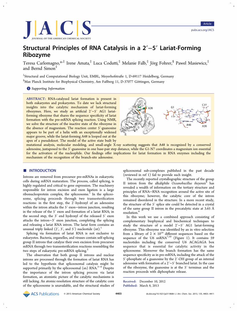
Figure 1. Primary and secondary structure of the 2′−5′ AG1 lariat-forming ribozyme. Bold lines indicate Watson−Crick (WC) base pairspreviously proposed on the basis of covariation experiments7 and hereverified by NMR; thin lines and dots indicate respectively WC andnon-canonical base-pairs found by NMR analysis. The stretch 32−35is base-paired with the stretch 44−47 (in gray), forming a pseudoknotthrough nucleotides 32−58. The G1 and the A48 are involved in thetransesterification reaction leading to lariat RNA (yellow shadows).The conserved ACAGAGA box is marked in red. The three helicalstretches are marked as H1, H2, and H3.
Journal of the American Chemical Society Article
dx.doi.org/10.1021/ja311868t | J. Am. Chem. Soc. 2013, 135, 4403−44114404
in its inactive state (PDB code 2m58), we determined thestructure of this 59mer RNA (Figure 1) in solution in theabsence of Mg2+ ions by NMR. The 1H−13C correlation of the13C/15N uniformly labeled RNA, as well as those of 13C/15Nselective U-, A-, C-, and G-labeled RNAs are heavily overlappedin both the base and the ribose regions (Figure S1). In addition,more than the 59 expected resonances are visible in theH8,H6/C8,C6 and H1′/C1′ correlations. Analysis of 13C-edited NOESY spectra indicates that nt’s 30−59 are present intwo conformations, which slowly exchange with each other(Figure S2). Resonances belonging to the less populatedconformation (∼35%) do not show internucleotide NOEs,indicating that the ribozyme lacks a defined structure from nt’s30 to 59 in this conformation. The structure that we presenthere refers to the major, completely folded conformation.The resonance assignment and the analysis of the NOESY
spectra for this 59mer RNA offered several challenges,including extreme spectral overlap, the presence of 89resonance sets, instead of 59, and the inhomogeneous intensityof nucleotide resonances belonging to different RNA regions.To cope with these challenges, we collected data sets for fiveselectively labeled samples using mostly 13C-edited, 12C-filtered/13C-filtered experiments (see Materials and Methodsand Supporting Information).15 Moreover, to confirm theassignment of ambiguous regions, we synthesized several single-or double-nucleotide mutant RNAs (Figure 2). Finally, morethan 95% of the base resonances and 82% of the riboseresonances could be assigned unambiguously.
13C-edited, 12C-filtered, and 13C-edited, 13C-filtered NOESYspectra15 on 13C/15N selective AU-, U-, A-, C-, and G-labeledRNAs resulted in 1339 assigned NOEs for structure calculation,corresponding to an average of 23 restraints per residue.Covariation experiments had previously identified base-pairednucleotides,7 indicated with bold lines in Figure 1. Weconfirmed the presence of these base pairs by inspection ofthe imino proton region of 15N-HSQC and HNN-COSY12 2Dspectra.A continuous network of strong H8/H6(i)-H2′(i−1) NOEs,
accompanied by medium H8/H6(i)-H1′(i−1) NOEs and weakH8/H6(i)-H8/H6(i−1), suggested the extension of helix H1from nt 1 to nt 27, over five non-canonical base pairs (2·26,3·25, 4·24, 7·21, and 8·20) and three canonical base pairs (1−27, 5−23 and 6−22) (Figure 1).To confirm the presence of an extended helix H1, we
performed mutational analysis (Figure 2), which confirmed theexistence of base pairs 5−23, 6−22, 7·21, and 1−27 by eithersingle-point or compensatory double-point mutations (Figure2). In addition, the mutational analysis showed that the non-canonical base pairs 2·26, 3·25, and 4·24 can be substituted bynucleotide combinations compatible with Watson−Crick (WC)base pairs (U2-A26, A3-U25, and A4-U24), as long as theidentity of nt’s 3, 4, and 26 is preserved. G9 can be removedwithout any impact on catalytic activity (Figure 2) and istherefore not part of the helix, as already predicted by the NOEpattern in the A8-G10 segment. The revised secondarystructure for the stretch 1−27 of the 2′−5′ AG1 lariat-formingribozyme is depicted in Figure 1. Prior to calculating thestructure of the RNA from NOE data, the topology of the non-canonical base pairs 8·20, 7·21, 4·24, 3·25, and 2·26 was derivedas described in the Supporting Information. To summarize, cisWC-WC topology24 was found for all these base pairs (seehydrogen-bonding patterns in Table S1). The cis WC-WC basepairs can be well integrated in the helical geometry suggested
by the NOEs, and are fully consistent with our mutational data(Figure 2).In the stretch containing nt’s 28−59, the analysis of the
imino proton region of the 15N−1H correlation confirmed thepresence of the canonical base pairs encompassing nt 38−41and 55−58, which had been inferred by covariation experi-ments.7 In addition, the 3D NOESY spectra revealed acontinuous network of strong H8/H6(i)−H2′(i−1) NOEs,accompanied by medium H8/H6(i)−H1′(i−1) NOEs andweak H8/H6(i)−H8/H6(i−1) in the stretch 44−47 and, to alesser extent, in the stretch 32−35. Besides, NOEs were presentbetween C47 and A50, between U46 and both U51 and A52,and between A35 and U53. The NOEs are indicative of apseudoknot structure, with two short (4 base pairs) helicalsegments (H2 and H3 in Figure 1) and the stretch 50−53contacting the minor groove of helix H2. The base pairingbetween stretches 44−47 and 32−35 is critical to the structureand was therefore confirmed by compensatory mutations(Figure 2). The six catalytically impaired mutants G32U, A33U,
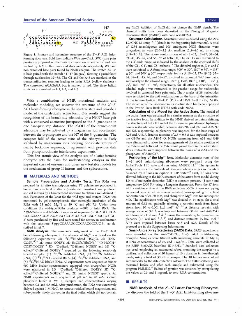
Figure 2. Mutational analysis of the 2′−5′ AG branch-formingribozyme. (a) Single-nucleotide mutants. Red, yellow and green colorsindicate mutants blocking (no lariat), reducing (<20%). and notaffecting (20−70%) the lariat formation, respectively. (b) Double-nucleotide mutants. The color code is as in panel a. (c) Deletion andthree-nucleotide mutants. The color code is as in panel a. The boxedresidues were deleted in each deletion variant, as indicated in thefigure. (d) Example of mutational analysis for the stretch 23−28 and42−45. (−) and (+) indicate the absence and the presence of 25 mMMg2+. Formation of the lariat was monitored after overnightincubation at 30 °C. For the control, a longer RNA, which wasextended at 3′-end by 8 nucleotides, was used. This RNA produces thesame fraction of lariat as the 59mer construct.
Journal of the American Chemical Society Article
dx.doi.org/10.1021/ja311868t | J. Am. Chem. Soc. 2013, 135, 4403−44114405
A33C, and G34C can be rescued by complementary mutationsC47A, U46A, U46G, and C45G, respectively, supporting thenotion that the stretches 44−47 and 32−35 are mutually base-paired (Figure 1). The non-canonical A35·A44 base pair wasfound to be in the trans WC-Hoogsteen topology, as describedin the Table S1.Structure of the 2′−5′ AG1 Lariat-Forming Ribozyme
in the Absence of Magnesium Ions. The structurecalculation (details in Materials and Methods and SupportingInformation; statistics in Table 1) converged to a well-defined
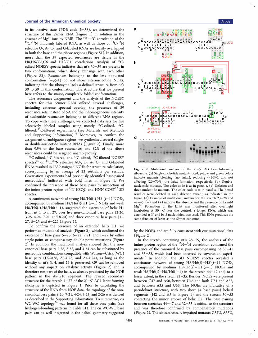
conformation for stretches 1−27 (excluding G9 and the 5′-triphosphate, heavy atom root-mean-square deviation, rmsd =0.83 Å) and helices H2, H3 (heavy atom rmsd = 0.97 Å)(Figure S3).The 5′-terminal stretch folds into a 12 base pair long helix
closed at one apex by the UUCG tetraloop. This helix containsseveral non-canonical base pairs (see above), and thus itsoverall conformation significantly deviates from ideal A-formRNA (Figure 3b, Table S2). The inclination angles of the basepairs with respect to the helix axis are lower than for A-formhelices and approximate those for A′-RNA. The inter base pairrise values (average value 3.1 Å) are between those of A′-RNAand B-DNA. This unwinding of the helix is caused by acontinuous stacking of six purine bases on one strand and threepurine bases on the other strand (Figure 3c). The major grooveis substantially widened (ca. 11 Å) with respect to that of A-RNA (3.8 Å) or even A′-RNA (8.0 Å) and resembles that of B-DNA (11.4 Å) (Table S2). Such opening of the major groovearound base pairs G2·A26, A3·A25, A4·A24, and G5·C23makes this region of the AG1 lariat-forming RNA unusuallyaccessible to interactions with ligands or with other structuralelements of the same RNA. Consistently, our mutationalanalysis of the upper part of the 5′-terminal helix of the RNA
indicates that the widening of the major groove in this region isrelevant for the catalytic function of the RNA. A24U or A25Usingle mutants, which are compatible with canonical basepairing at single sites, are catalytically competent, suggestingthat one WC base pair might be tolerated without loss offunction. On the other hand, A24U/A25U double mutants orthe G2U/A24U/A25U triple mutant lack activity; this iscompatible with the notion that two consecutive WC base pairswould bring the helix closer to A-form, with a deeper andnarrower major groove; this structure is evidently unable tosupport catalysis (Figure 2).The grooves gradually narrow toward the lower part of the
helical segment. Next to the tetraloop (Figure 3b, Table S2),the major groove width is within 7−8 Å close to the value forA′-RNA. This part of the helix displays two stacks of two andthree purine bases each, including a cross-strand stack betweenA20 and G10 (Figure 3c).The 3′-terminal stretch (nt’s 32−59) folds into a pseudoknot
structure (Figure 3d). Two short helices comprising nt’s 32−35and 44−47 (helix H2) and nt’s 38−41 and 55−58 (helix H3)stack upon each other. The nt’s 42 and 43 are bulged out. The
Table 1. Structure Statistics (10 Structures of 200Calculated, PDB code 2m58)
in vacuum water refined
Distance Restraintstotal unambiguous NOEs 1234inter-residue 569sequential (|i − j| = 1) 631long-range (|i − j| > 2) 34total ambiguous NOEs 105dihedral angle restraints 383hydrogen bonds 20
Structure StatisticsDeviations from Idealized Geometrybond lengths (Å) 0.0060 ± 0.0001 0.0042 ± 0.0001bond angles (deg) 0.74 ± 0.01 0.96 ± 0.02impropers (deg) 0.50 ± 0.01 0.70 ± 0.02Violations (Mean and SD)distance restraints rmsd (Å) 0.08 ± 0.01 0.12 ± 0.01distance restraint violations >0.5 Å 0.4 ± 0.7 10.1 ± 1.8dihedral angle restraints rmsd (deg) 1.48 ± 0.09 1.65 ± 0.12dihedral angle violations >5° 4.7 ± 1.3 5.7 ± 1.5Coordinate Precision (Å)backbone (1−27) 1.22 ± 0.30 1.34 ± 0.31heavy atoms (1−27) 1.15 ± 0.26 1.28 ± 0.26backbone (32−58) 1.34 ± 0.48 1.38 ± 0.31heavy atoms (32−58) 1.75 ± 0.50 1.86 ± 0.39
Figure 3. Structure of the 2′−5′ AG1 branch-forming ribozyme. (a)The 2′−5′ lariat-forming ribozyme folds into a 5′-terminal helixcomprising nt’s 1−27 (orange) and a 3′-terminal pseudoknotcomprising nt’s 32−59 (green and gray). The pseudoknot structurebrings A48 (cyan) in proximity of G1 (cyan). The relative position ofthe 5′-terminal helix and the 3′-terminal pseudoknot is variable in theabsence of Mg2+. For clarity only one structure is shown in cross-eyestereoview. A superimposition of all structures of the NMR ensemblecan be found in Figure S3. (b) 5′-terminal helix showing the wide andshallow major groove at the 5′ end of the helix. (c) Stacking of purineresidues in the 5′-terminal helix. Upper panels: Stacking of a six purinestretch on one strand (pale pink) and a three purine stretch on theopposite strand (aubergine) in the first six base pairs of the 5′-terminaldomain. Lower panels: Stacking of two and three purine stretches inthe apical part of the 5′-terminal helix (in pale pink and aubergine).The stack of three purines contains a cross-strand stacking betweenG10 (pale pink) and A20 (aubergine). Pyrimidines are shown in white.For clarity, the bonds between the riboses and the bases have beenomitted in the left half. (d) 3′-terminal pseudoknot. Strand 32−37 is indark green, strand 38−47 in lime and strand 49−59 in mud green; theA48 is in cyan. Two short helical segments are formed by nt’s 32−35,which base-pair with the stretch 47−44, and by nt’s 38−41, whichbase-pair with nt’s 58−55. The stretch 50−53 contacts the minorgroove of the first helical segment.
Journal of the American Chemical Society Article
dx.doi.org/10.1021/ja311868t | J. Am. Chem. Soc. 2013, 135, 4403−44114406
Δ42,43 mutant is catalytically active, confirming that these twonucleotides are not part of any essential structural element. Theexistence of helix H2 was confirmed by mutational analysis:The inactive mutant RNA A33U and the weakly active mutantsG32U and G34C could be rescued by compensatory mutationsU46A, C47A, and C45G (Figure 2). The stretch A50-U53contacts the minor groove of helix H2, with A52 positionedfavorably to form a base triple with the sugar edge face of G34.The pseudoknot structure induces a sharp kink in the RNAbackbone after C47; as a result, nt’s 48 and 49 are looped out.A48 comes close to the 5′ terminus of helix H1 and is availablefor recognition via tertiary interactions with structural elementswithin the helix (Figure 3a).The stretch 28−31, connecting the 5′ helix H1 and the 3′-
terminal 32−58 pseudoknot is not well defined. In moststructures U28 stacks on C27 of helix H1, while A31 stacks onG32 at the start of the 3′-terminal pseudoknot. The NOEs ofA29 and C30 are compatible with more than one conformation,which suggests that in the ground state the relative orientationof helix H1 and the 3′-terminal pseudoknot is not fixed butsamples multiple conformations in a dynamic equilibrium(Figure S3). However, a weak NOE between A29-H2 and G49-H1′ and several NOEs between C47 and G49 confirm thepseudoknot structure, the extrusion of A48 and the proximity ofthe A48-G49 nucleotides with the 3′ end of helix H1.Structural Model of the Active Form of the 2′−5′
Lariat-Forming Ribozyme. The 2′−5′ lariat-forming ribo-zyme performs catalysis in the presence of magnesium. Intheory, the structure of the active form of the ribozyme couldbe determined by trapping the active state via addingmagnesium to a catalytically incompetent form of the RNA(for example, with a 2′-deoxyadenosine at position 48).Unfortunately, the synthesis of such a modified RNA withthe many 13C/15N labeling schemes necessary for structuralinvestigation by NMR, is impractical. In this study, we chooseto build a model of the active form of the ribozyme on the basisof its structure in the inactive form (in the absence ofmagnesium), the enzymatic activity of the mutants andmolecular modeling.The mutant analysis showed that the nature of four bases,
A3, A4, A26, and A48 is essential to sustain catalytic activity. Inthe wild-type ribozyme, A3 and A4 form non-canonical basepairs with A25 and A24, respectively. The single mutationsA24U and A25U are tolerated, while the double mutant A24U/A25U is inactive. These data underline the importance of a
widened major groove around A3 and A4 and suggest thatthese nucleotides are involved in the tertiary recognition ofother RNA structural elements. A26 is also crucial to catalysis;furthermore, the reduced activity of the G2U mutant underlinesthe relevance of a widened major groove at this position. All inall, these data point at a specific recognition of the base of A48by either A3 or A4 or A26 or a combination thereof.The terminal nucleotide carrying the reactive 5′-triphosphate
must form a canonical base pair, as indicated by the loss ofcatalytic activity in the G1A mutant, which can be rescued bythe complementary mutation C27U (Figure 2). Thisconclusion is supported by the loss of activity of the mutantΔ6−8/Δ20−22, where the base pair register in H1 is shifted byone nucleotide. Further deletion of the unpaired G9 in theΔ6−9/Δ20−22 mutant restores activity, indicating that thelength of the 5′-terminal helix H1 is not essential and that therecognition of the catalytic center is confined to the 5′-terminalbase pairs.With this information in our hands we constructed a model
for the active state of the 2′−5′ lariat-forming ribozyme(detailed protocol in Supporting Information). We argued thatthe two secondary structure elements present in the inactivestate, the 5′-terminal helix H1 and the 3′-terminal pseudoknot,are rather rigid structures; however, they can likely move withrespect to each other via the hinge region (around nt’s 28−31).In the active state of the ribozyme the two secondary structureelements must come closer to each other in a way that the 2′-OH of the A48 ribose can reach to the 5′-phosphate of G1.With this restraint in mind, the base of A48 can be
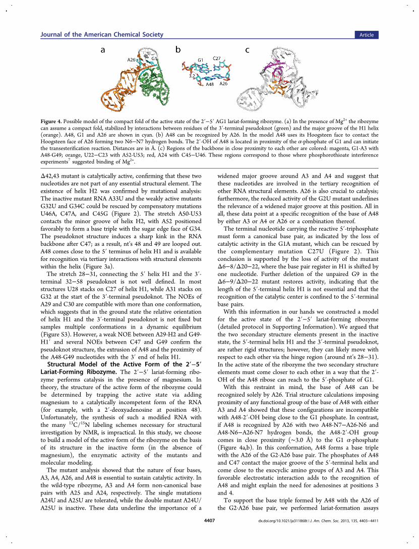
recognized solely by A26. Trial structure calculations imposingproximity of any functional group of the base of A48 with eitherA3 and A4 showed that these configurations are incompatiblewith A48-2′-OH being close to the G1 phosphate. In contrast,if A48 is recognized by A26 with two A48-N7−A26-N6 andA48-N6−A26-N7 hydrogen bonds, the A48-2′-OH groupcomes in close proximity (∼3.0 Å) to the G1 α-phosphate(Figure 4a,b). In this conformation, A48 forms a base triplewith the A26 of the G2·A26 base pair. The phosphates of A48and C47 contact the major groove of the 5′-terminal helix andcome close to the exocyclic amino groups of A3 and A4. Thisfavorable electrostatic interaction adds to the recognition ofA48 and might explain the need for adenosines at positions 3and 4.To support the base triple formed by A48 with the A26 of
the G2·A26 base pair, we performed lariat-formation assays
Figure 4. Possible model of the compact fold of the active state of the 2′−5′ AG1 lariat-forming ribozyme. (a) In the presence of Mg2+ the ribozymecan assume a compact fold, stabilized by interactions between residues of the 3′-terminal pseudoknot (green) and the major groove of the H1 helix(orange). A48, G1 and A26 are shown in cyan. (b) A48 can be recognized by A26. In the model A48 uses its Hoogsteen face to contact theHoogsteen face of A26 forming two N6−N7 hydrogen bonds. The 2′-OH of A48 is located in proximity of the α-phosphate of G1 and can initiatethe transesterification reaction. Distances are in Å. (c) Regions of the backbone in close proximity to each other are colored: magenta, G1-A3 withA48-G49; orange, U22−C23 with A52-U53; red, A24 with C45−U46. These regions correspond to those where phosphorothioate interferenceexperiments7 suggested binding of Mg2+.
Journal of the American Chemical Society Article
dx.doi.org/10.1021/ja311868t | J. Am. Chem. Soc. 2013, 135, 4403−44114407
with ribozymes carrying modified A48. Both the A48-N7-deazaand the A48-N6-Me ribozymes were completely inactive, whichstrongly supports the involvement of the Hoogsteen face ofA48 in the recognition of the branch-site adenosine.The low-field shift of both the C3′ and the C4′ carbons of
A48 in the inactive state (80.2 and 83.4 ppm, respectively),indicate that the ribose populates, to a large extent, the C2′-endo conformation. In addition, the low-field-shifted reso-nances of both the H2′ and H3′ protons (5.30 and 5.07 ppm,respectively) suggest that the base is in the syn conformation.The conformational preferences of the ribose of A48 in theribozyme inactive state are similar to the conformation of ourmodel of the active state, where the A48 ribose assumes aconformation close to C2′-endo. In addition, a small H1′−H8NOE peak suggests that the syn conformation of the A48 base,observed in the model of the active state, is considerablypopulated also in the inactive state.The stretch 28−31 as well as the stretch 49−53 do not form
any base-specific tertiary contacts in our structural model of theactive state. This is in agreement with the notion that anysubstitution at positions 49−53 is compatible with catalyticactivity, as well as any substitution at positions 30 and 31.Intriguingly, the U28C mutant does not support catalysis,
despite the fact that this nucleotide is not involved in anysequence specific contacts in the structure.We reasoned that the lack of activity of the U28C mutant
could be determined by a particular structural feature of thismutant that impedes the recognition of A48, for example byformation of a base pair between U28 and G49. Indeed, thecatalytically active mutant U28G can be turned into acatalytically incompetent mutant by the mutation G49C,while the single mutant G49C is active. This confirms thatformation of a base pair between nt’s 28 and 49 is detrimentalfor activity.These observations suggest that the 28−31 stretch is flexible
and this flexibility is essential for the function of the ribozyme,strongly supporting the notion that it functions as a hinge the5′ and 3′ rigid structural motifs pivot on.Interestingly, in the modeled compact active conformation,
the backbone phosphates of A48-G49 are in close proximity tothe backbone phosphates of G2-A4 and the G1 α-phosphate. Inaddition, the backbone of C45 and U46 approaches thebackbone of A24 on the other strand of the 5′-terminal helix(Figure 4c), and the backbone of A52 and U53 comes close tothe backbone of U22 and C23 (Figure 4c).It is likely that such tertiary contacts require divalent cations
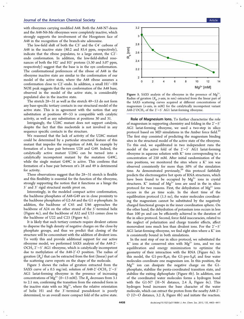
to disperse the high density of negative charges on the close-byphosphate groups, and thus we predict that closing of thestructure will be concomitant with the addition of divalent ions.To verify this and provide additional support for our activeribozyme model, we performed SAXS analysis of the A48-2′-OCH3 2′−5′ AG1 ribozyme, which is catalytically incompetentdue to methylation of the A48-2′-O position. The radius ofgyration (Rg) that can be extracted from the first (linear) part ofthe scattering curve reports on the shape of the molecule.Figure 5 shows the radius of gyration extracted from the
SAXS curve of a 0.5 mg/mL solution of A48-2′-OCH3 2′−5′AG1 lariat-forming ribozyme in the presence of increasingconcentrations of Mg2+ (0−13 mM). The Rg decreases from 3.1to 2.1 nm, confirming the transition from the extended form inthe inactive state with no Mg2+, where the relative orientationof helix H1 and the 3′-terminal pseudoknot is poorlydetermined, to an overall more compact fold of the active state.
Role of Magnesium Ions. To further characterize the roleof magnesium in supporting chemistry and folding in the 2′−5′AG1 lariat-forming ribozyme, we used a two-step in silicoprotocol based on MD simulations in the Amber force field.25
The first step consisted of predicting the magnesium bindingsites in the structural model of the active state of the ribozyme.To this end, we equilibrated in two independent runs themodel of the active fold of the 2′−5′ AG1 lariat-formingribozyme in aqueous solution with K+ ions corresponding to aconcentration of 250 mM. After initial randomization of theions positions, we monitored the sites where a K+ ion wasobserved consistently for more than 50% of the simulationtime. As demonstrated previously,26 this protocol faithfullypredicts the electronegative hot spots of RNA structures, whichhave been found to be occupied by Mg2+ ions in crystalstructures. K+ instead of Mg2+ ions are used in the in silicoprotocol for two reasons. First, the dehydration of Mg2+ ionsoccurs in the μs time scale. In the short time of theequilibration protocol (1.5 ns), the water molecules coordinat-ing the magnesium cannot be substituted by the negativelycharged functional groups in the inner coordination sphere. Onthe other hand, the dehydration of potassium ions occurs in lessthan 100 ps and can be efficiently achieved in the duration ofthe in silico protocol. Second, force field inaccuracies, related tothe neglect of polarization and charge transfer effects, affectmonovalent ions much less than divalent ions. For the 2′−5′AG1 lariat-forming ribozyme, we find eight sites where a K+ ionis consistently bound in both simulations.In the next step of our in silico protocol, we substituted the
K+ ions at the conserved sites with Mg2+ ions, and we ranequilibration and energy minimization to optimize thegeometry of their interaction with the RNA (Figure 6a). Inthis model, the G1-pro-RPα, the G1-pro-SPβ, and four watermolecules coordinate one magnesium ion. In this position, theMg2+ ion can dissipate the negative charge on the G1-phosphate, stabilize the penta-coordinated transition state, andstabilize the exiting diphosphate (Figure 6b). In addition, oneof the coordinated water molecules forms a hydrogen bondwith the G1-N7 (H−N distance, 2.4 Å, Figure 6c). Thishydrogen bond increases the base character of the watermolecule, which can extract the proton from the nearby A48-2′-O (O−O distance, 3.2 Å, Figure 6b) and initiate the reaction.
Figure 5. SAXS analysis of the ribozyme in the presence of Mg2+.Radius of gyration (Rg, y-axis, in nm) extracted from the linear part ofthe SAXS scattering curves acquired at different concentrations ofmagnesium (x-axis, in mM) for the catalytically incompetent variantA48-2′OCH3 of the 2′−5′ AG1 lariat-forming ribozyme.
Journal of the American Chemical Society Article
dx.doi.org/10.1021/ja311868t | J. Am. Chem. Soc. 2013, 135, 4403−44114408
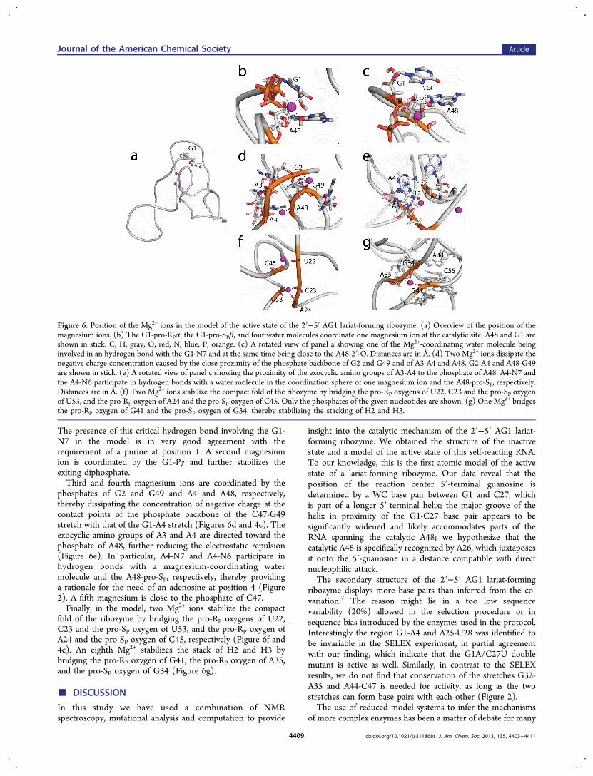
The presence of this critical hydrogen bond involving the G1-N7 in the model is in very good agreement with therequirement of a purine at position 1. A second magnesiumion is coordinated by the G1-Pγ and further stabilizes theexiting diphosphate.Third and fourth magnesium ions are coordinated by the
phosphates of G2 and G49 and A4 and A48, respectively,thereby dissipating the concentration of negative charge at thecontact points of the phosphate backbone of the C47-G49stretch with that of the G1-A4 stretch (Figures 6d and 4c). Theexocyclic amino groups of A3 and A4 are directed toward thephosphate of A48, further reducing the electrostatic repulsion(Figure 6e). In particular, A4-N7 and A4-N6 participate inhydrogen bonds with a magnesium-coordinating watermolecule and the A48-pro-SP, respectively, thereby providinga rationale for the need of an adenosine at position 4 (Figure2). A fifth magnesium is close to the phosphate of C47.Finally, in the model, two Mg2+ ions stabilize the compact
fold of the ribozyme by bridging the pro-RP oxygens of U22,C23 and the pro-SP oxygen of U53, and the pro-RP oxygen ofA24 and the pro-SP oxygen of C45, respectively (Figure 6f and4c). An eighth Mg2+ stabilizes the stack of H2 and H3 bybridging the pro-RP oxygen of G41, the pro-RP oxygen of A35,and the pro-SP oxygen of G34 (Figure 6g).
■ DISCUSSION
In this study we have used a combination of NMRspectroscopy, mutational analysis and computation to provide
insight into the catalytic mechanism of the 2′−5′ AG1 lariat-forming ribozyme. We obtained the structure of the inactivestate and a model of the active state of this self-reacting RNA.To our knowledge, this is the first atomic model of the activestate of a lariat-forming ribozyme. Our data reveal that theposition of the reaction center 5′-terminal guanosine isdetermined by a WC base pair between G1 and C27, whichis part of a longer 5′-terminal helix; the major groove of thehelix in proximity of the G1-C27 base pair appears to besignificantly widened and likely accommodates parts of theRNA spanning the catalytic A48; we hypothesize that thecatalytic A48 is specifically recognized by A26, which juxtaposesit onto the 5′-guanosine in a distance compatible with directnucleophilic attack.The secondary structure of the 2′−5′ AG1 lariat-forming
ribozyme displays more base pairs than inferred from the co-variation.7 The reason might lie in a too low sequencevariability (20%) allowed in the selection procedure or insequence bias introduced by the enzymes used in the protocol.Interestingly the region G1-A4 and A25-U28 was identified tobe invariable in the SELEX experiment, in partial agreementwith our finding, which indicate that the G1A/C27U doublemutant is active as well. Similarly, in contrast to the SELEXresults, we do not find that conservation of the stretches G32-A35 and A44-C47 is needed for activity, as long as the twostretches can form base pairs with each other (Figure 2).The use of reduced model systems to infer the mechanisms
of more complex enzymes has been a matter of debate for many
Figure 6. Position of the Mg2+ ions in the model of the active state of the 2′−5′ AG1 lariat-forming ribozyme. (a) Overview of the position of themagnesium ions. (b) The G1-pro-RPα, the G1-pro-SPβ, and four water molecules coordinate one magnesium ion at the catalytic site. A48 and G1 areshown in stick. C, H, gray, O, red, N, blue, P, orange. (c) A rotated view of panel a showing one of the Mg2+-coordinating water molecule beinginvolved in an hydrogen bond with the G1-N7 and at the same time being close to the A48-2′-O. Distances are in Å. (d) Two Mg2+ ions dissipate thenegative charge concentration caused by the close proximity of the phosphate backbone of G2 and G49 and of A3-A4 and A48. G2-A4 and A48-G49are shown in stick. (e) A rotated view of panel c showing the proximity of the exocyclic amino groups of A3-A4 to the phosphate of A48. A4-N7 andthe A4-N6 participate in hydrogen bonds with a water molecule in the coordination sphere of one magnesium ion and the A48-pro-SP, respectively.Distances are in Å. (f) Two Mg2+ ions stabilize the compact fold of the ribozyme by bridging the pro-RP oxygens of U22, C23 and the pro-SP oxygenof U53, and the pro-RP oxygen of A24 and the pro-SP oxygen of C45. Only the phosphates of the given nucleotides are shown. (g) One Mg2+ bridgesthe pro-RP oxygen of G41 and the pro-SP oxygen of G34, thereby stabilizing the stacking of H2 and H3.
Journal of the American Chemical Society Article
dx.doi.org/10.1021/ja311868t | J. Am. Chem. Soc. 2013, 135, 4403−44114409
years.27,28 Here, we wish to underline that no data are availablevalidating that the catalytic mechanism of the 2′−5′ AG1 lariat-forming ribozyme is similar to that of the spliceosome or ofintrons II. Nevertheless, nature has evolved common pathwaysto perform a given task and it is reasonable to use the lessonlearned from model systems to generate hypotheses. Thevalidity of these hypotheses needs then to be tested for themore complex, native systems either in vitro or in vivo. Here,given the conservation of the attacking adenosine in lariat-forming ribozymes, we wonder if the recognition modeobserved in the 2′−5′ AG1 lariat-forming ribozyme can beextended also to other catalytic RNAs and to the spliceosome.Strikingly, the spliceosome features a U downstream of the 5′splice-site guanosine. This uridine could base pair with anadenosine and support the same mode of recognition for thecatalytic adenosine as in the 2′−5′ AG1 lariat-formingribozyme. In support of this, the G2U mutant, which convertsthe non-canonical G2·A26 base pair to a canonical one in the2′−5′ lariat-forming ribozyme is catalytically competent, even ifmuch less efficient than the wild-type ribozyme. For the role ofthe A26 equivalent adenosine in the spliceosome, a goodcandidate may be the fifth A of the conserved ACAGAGA box.In fact, cross-link between this A and the U next to the splice-site guanosine in the spliceosome has been previouslyreported.29,30 In the spliceosome the catalytic adenosine isbulged out from a helical segment pairing the intron with theU2 RNA.31 Similarly, in the 2′−5′ lariat-forming ribozyme theA48 is extruded from the pseudoknot structure at the tip ofhelix H2.Interestingly, while the above ACAGAGA box is also present
in the 2′−5′ lariat-forming ribozyme (nt’s 29−35), our dataindicate that it plays a different role than in the spliceosome. Inthe ribozyme the ACA sequence is not involved in any basepair, while the GAGA sequence has a critical structural role inthe formation of the 3′ pseudoknot and in positioning thecatalytic A48 close to the 5′ terminus. However, theACAGAGA segment does not provide any functional groupsto the chemical reaction, or Mg2+ binding.7 In contrast, in thespliceosome the ACA segment was shown to base-pair withnucleotides close to the 5′ splice site, while the GAGA segmentbinds magnesium ions that are important for catalysis.32,33
In silico equilibration of the structural model of the activestate of the 2′−5′ AG1 lariat-forming ribozyme in magnesium-containing buffer suggests that one magnesium ion is critical forchemistry. This Mg2+ is coordinated by the G1-N7 (through awater molecule) and the G1 α-phosphate (Figure 6b). Thewater molecule bridging between the Mg2+ and the G1-N7 isactivated by the Mg2+ and the G1-N7 acting as bases and is in afavorable position to extract the A48-2′-O proton, therebyinitiating catalysis. The position of the Mg2+ ion is compatiblewith the role of stabilizing the penta-coordinated phosphorus inthe transition state and the exiting diphosphate. In support ofthis Mg2+ arrangement, the G1A/C27U double mutant, with aconserved purine at the 5′ terminus and the N7 available forMg2+ coordination, is catalytically active, while the G1C/C27Gmutant, which lacks the N7 on the 5′ terminus nucleotide, doesnot support chemistry. Interestingly, the same requirement fora purine residue at the 5′-terminal splice site is present also inthe spliceosome, suggesting a conserved role for the N7position of this nucleotide. However, in contrast to whatproposed for the spliceosome and other ribozymes, which arethought to require two magnesium ions for catalysis,34 themodel of the active state of the 2′−5′ AG1 lariat-forming
ribozyme predicts the involvement of only one Mg2+ at thereaction center. This conclusion has to be taken with caution, asthe inaccuracy of the model of the active state of the 2′−5′ AG1lariat-forming ribozyme, together with the limitation of the insilico modeling of ion binding sites, may be the cause formissing the second ion at the catalytic center.In our model, other magnesium ions bind to the RNA
phosphate backbone and stabilize the compact fold of themodel of the active state of the ribozyme. The tight interactionof the major groove of the 5′-terminal helix with A48 brings thephosphate backbones of the 5′-terminal helix and the 3′-terminal pseudoknot in close proximity in the model. Theelectrostatic repulsion of the negatively charged RNA back-bones is compensated by binding one magnesium ion at thephosphates of C45 and A24, one at the phosphates of U22,C23, and U53, and two at the phosphates of G2, A4, A48, andG49. In support of this model, phosphorothioate interferenceexperiments have shown that binding of magnesium to the pro-RP of G1-A3, C21, U22, A24, A25, and C47-G49 is essential forbranching activity7 (Figure 4c). Moreover, our SAXS data alsoshow that the structure becomes more compact in the presenceof magnesium. Taken together, magnesium ions are likely tohave a double role in the 2′−5′ lariat-forming ribozyme:structural stabilization and involvement in the chemicalreaction.
■ CONCLUSIONSThis study shows how integration of data from differentdisciplines can be combined to obtain structural models ofstates that are inaccessible to standard structural biologytechniques. Mutational analysis data can be converted into aninteraction network that, together with the structure of theaccessible state of an enzyme, can be used in molecularmodeling protocols to model the structure of other states. Inthe case of the 2′−5′ lariat-forming ribozyme, we were able toshow that the branch-point adenosine may be recognized by anadenosine juxtaposed to the 5′-terminal nucleotide, one basepair downstream. The N7 atom of the 5′-terminal purine hasbeen appointed a role in coordinating a magnesium ion, likelyrelevant to the activation of the attacking nucleophile. Thesefindings allow us to draw parallels with the spliceosomal RNAand propose experiments to probe the role of the N7 atom ofthe guanosine at the 5′ splice site and the role of the fifthadenosine of the conserved ACAGAGA box in recognizing thebranch-point adenosine in the first step of pre-mRNA splicing.
■ ASSOCIATED CONTENT*S Supporting InformationSupplemental figures and tables referred to in the text, detailedexperimental procedures, and pdb file for the model in Figure 4.This material is available free of charge via the Internet athttp://pubs.acs.org.
■ AUTHOR INFORMATIONCorresponding [email protected] authors declare no competing financial interest.
■ ACKNOWLEDGMENTSThis work was supported by the EMBL, the MPG, and grantCA 294/2-1 from the DFG. We thank Frank Gabel for
Journal of the American Chemical Society Article
dx.doi.org/10.1021/ja311868t | J. Am. Chem. Soc. 2013, 135, 4403−44114410
discussion of the SAXS data and Claudia Schwiegk for precioushelp in sample preparation.
■ REFERENCES(1) Will, C. L.; Luhrmann, R. Cold Spring Harbor Persp. Biol. 2011, 3,a003707.(2) Pyle, A. M.; Lambowitz, A. M. The RNA World, 3rd ed.; ColdSpring Harbor Laboratory Press: Cold Sping Harbor, NY, 2006.(3) Cech, T. R. Cell 1986, 44, 207−210.(4) Sharp, P. A. Cell 1985, 42, 397−400.(5) Guthrie, C. Science 1991, 253, 157−163.(6) Valadkhan, S.; Manley, J. L. Nature 2001, 413, 701−707.(7) Tuschl, T.; Sharp, P. A.; Bartel, D. P. RNA 2001, 7, 29−43.(8) Toor, N.; Keating, K. S.; Taylor, S. D.; Pyle, A. M. Science 2008,320, 77−82.(9) Chan, R. T.; Robart, A. R.; Rajashankar, K. R.; Pyle, A. M.; Toor,N. Nat. Struct. Mol. Biol. 2012, 10, 555−557.(10) Tuschl, T.; Sharp, P.; Bartel, D. P. EMBO J. 1998, 17, 2637−2650.(11) Hennig, M.; Williamson, J. R. Nucleic Acids Res. 2000, 28, 1585−1593.(12) Dingley, A. J.; Grzesiek, S. J. Am. Chem. Soc. 1998, 120, 8293−8297.(13) Sklenar, V.; Dieckmann, T.; Butcher, S. E.; Feigon, J. J. Magn.Reson. 1998, 130, 119−124.(14) Hu, W. D.; Kakalis, L. T.; Jiang, L. C.; Jiang, F.; Ye, X. M.;Majumdar, A. J. Biomol. NMR 1998, 12, 559−564.(15) Zwahlen, C.; Legault, P.; Vincent, S. J. F.; Greenblatt, J.; Konrat,R.; Kay, L. E. J. Am. Chem. Soc. 1997, 119, 6711−6721.(16) Brunger, A. T.; Adams, P. D.; Clore, G. M.; DeLano, W. L.;Gros, P.; Grosse-Kunstleve, R. W.; Jiang, J. S.; Kuszewski, J.; Nilges,M.; Pannu, N. S.; Read, R. J.; Rice, L. M.; Simonson, T.; Warren, G. L.Acta Crystallogr. Sect. D: Biol. Crystallogr. 1998, 54, 905−921.(17) Linge, J. P.; Habeck, M.; Rieping, W.; Nilges, M. Bioinformatics2003, 19, 315−316.(18) Ohlenschlager, O.; Haumann, S.; Ramachandran, R.; Gorlach,M. J. Biomol. NMR 2008, 42, 139−142.(19) Case, D. A.; Darden, T. A.; Cheatham III, T. E.; Simmerling, C.L.; Wang, J.; Duke, R. E.; Luo, R.; Walker, R. C.; Zhang, W.; Merz, K.M.; et al. AMBER 11; University of California, San Francisco, 2010.(20) Jorgensen, W. L.; Chandrasekhar, J.; Madura, J. D.; Impey, R.W.; Klein, M. L. J. Chem. Phys. 1983, 79, 926.(21) Hashem, Y.; Auffinger, P. Methods (San Diego, Calif.) 2009, 47,187−197.(22) Pernot, P.; Theveneau, P.; Giraud, T.; Fernandes, R. N.;Nurizzo, D.; Spruce, D.; Surr, J.; McSweeney, S.; Round, A.; Felisaz,F.; Foedinger, L.; Gobbo, A.; Huet, J.; Villard, C.; Cipriani, F. J. Phys.:Conf. Ser. 2010, 247, 12009.(23) Konarev, P. V; Volkov, V. V; Sokolova, A. V; Koch, M. H. J.;Svergun, D. I. J. Appl. Crystallogr. 2003, 36, 1277−1282.(24) Leontis, N. B.; Westhof, E. Comp. Funct. Genom. 2002, 3, 518−524.(25) Pearlman, D. a.; Case, D. a.; Caldwell, J. W.; Ross, W. S.;Cheatham, T. E.; DeBolt, S.; Ferguson, D.; Seibel, G.; Kollman, P.;Cheatham Iii, T. E. Comput. Phys. Commun. 1995, 91, 1−41.(26) Auffinger, P.; Bielecki, L.; Westhof, E. Structure 2004, 12, 379−388.(27) Valadkhan, S.; Manley, J. L. RNA 2009, 15, 4−7.(28) Smith, A. J.; Konarska, M. M. RNA 2009, 15, 1−3.(29) Madhani, H. D.; Guthrie, C. Annu. Rev. Genet. 1994, 28, 1−26.(30) Sontheimer, E. J.; Steiz, J. A. Science 1993, 262, 1989−1996.(31) Parker, R.; Siciliano, P. G.; Guthrie, C. Cell 1987, 49, 229−239.(32) Nilsen, T. W. In RNA structure and function; Simons, R. W.,Grunberg-Manago, M., Eds.; Cold Spring Harbor Laboratory Press::Cold Spring Harbor, NY, 1998; pp 303−357.(33) Moore, M. J.; Query, C. C.; Sharp, P. A. In The RNA world;Gestland, R. F., Atkins, J. F., Eds.; Cold Spring Harbor LaboratoryPress:: Cold Spring Harbor, NY, 1993; pp 303−357.
(34) Steiz, T. A.; Steiz, J. A. Proc. Natl. Acad. Sci. U.S.A. 1993, 90,6498−6502.
Journal of the American Chemical Society Article
dx.doi.org/10.1021/ja311868t | J. Am. Chem. Soc. 2013, 135, 4403−44114411