1 the nervous system e. mcintyre ib biology. 2 nervous system central nervous system peripheral...
TRANSCRIPT

1
The Nervous System
E. McIntyre
IB Biology

2
Nervous Nervous SystemSystem
Central Nervous System
Peripheral Nervous System
Parasympathetic
Somatic Nerves
Autonomic Nerves
Spinal Cord
BrainMotorSensory Sympathetic
HindbrainMidbrainForebrain
Cerebellum
Medulla Oblongata
Pons
Hypothalamus
ThalamusOlfactory
Lobes
Corpus Callosum
Cerebrum
The Four
Lobes…
3
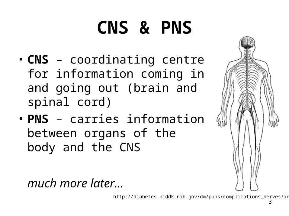
CNS & PNS
• CNS – coordinating centre for information coming in and going out (brain and spinal cord)
• PNS – carries information between organs of the body and the CNS
much more later…http://diabetes.niddk.nih.gov/dm/pubs/complications_nerves/index.htm
4
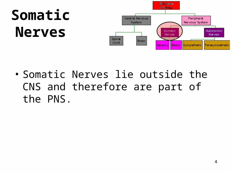
Somatic Nerves
• Somatic Nerves lie outside the CNS and therefore are part of the PNS.
5
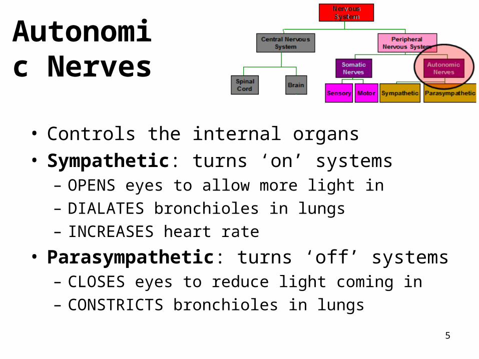
Autonomic Nerves
• Controls the internal organs• Sympathetic: turns ‘on’ systems
– OPENS eyes to allow more light in
– DIALATES bronchioles in lungs
– INCREASES heart rate
• Parasympathetic: turns ‘off’ systems– CLOSES eyes to reduce light coming in
– CONSTRICTS bronchioles in lungs
6
Types of Nerve Cells
There are two types of nerve cells1. Glial cells
– Non-conducting cells: structure/support and metabolism of nerve cells
2. Neurons– Nerve cells that conduct electrical signals called
nerve impulses.– carries impulses in only ONE direction – Basic functional unit of the Nervous System

7

8
Types of Neurons
Somatic nerves control skeletal muscle, bones, skin
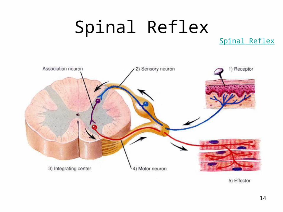
• Sensory (AFFERENT) – relay information from sense organs (eyes, nose, mouth etc) to CNS.
• Interneurons – Connect Sensory and Motor neurons and carry impulses between them. They are found entirely within the Central Nervous System.
• Motor neurons (EFFERENT ) – relay information from CNS to the effectors (muscles, organs, glands)

9
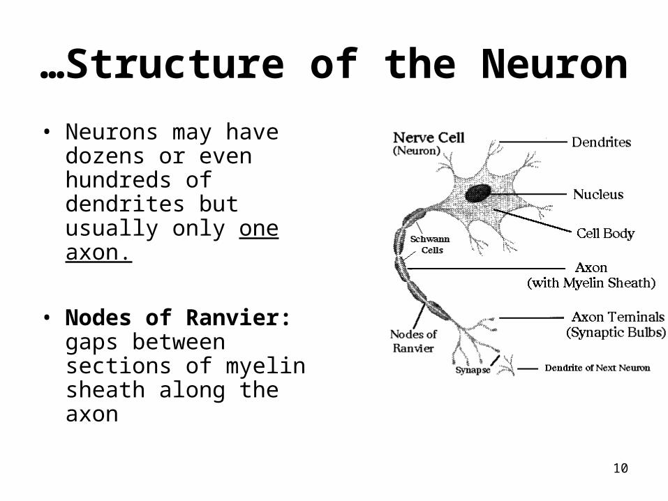
Structure of the NeuronA neuron has three main parts• Body: largest part, contains the nucleus and much
of cytoplasm, most of the metabolic activity of the cell, including the generation of ATP and synthesis of protein.
• Dendrites: Short branch extensions spreading out from the cell body. Dendrites Receive STIMULUS (Action Potentials) and carry IMPULSES from the ENVIRONMENT or from other NEURONS AND carry them toward cell body.
• Axon: A Long Fiber that CARRIES IMPULSES AWAY FROM THE CELL BODY. Each neuron has only ONE AXON. The axon ends in a series of small swellings called AXON TERMINALS.
Structure of Neuron
10
• Neurons may have dozens or even hundreds of dendrites but usually only one axon.
• Nodes of Ranvier: gaps between sections of myelin sheath along the axon
…Structure of the Neuron
11
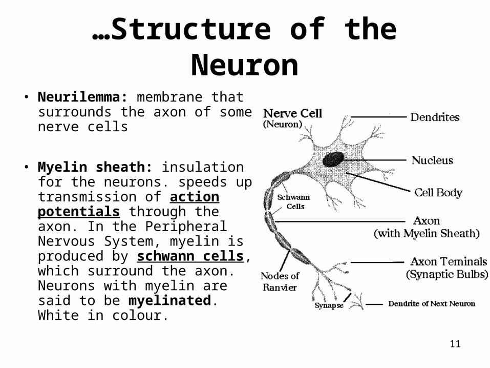
• Neurilemma: membrane that surrounds the axon of some nerve cells
• Myelin sheath: insulation for the neurons. speeds up transmission of action potentials through the axon. In the Peripheral Nervous System, myelin is produced by schwann cells, which surround the axon. Neurons with myelin are said to be myelinated. White in colour.
…Structure of the Neuron
12
…Structure of the Neuron
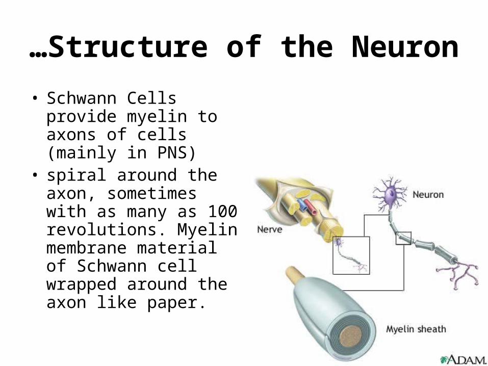
• Schwann Cells provide myelin to axons of cells (mainly in PNS)
• spiral around the axon, sometimes with as many as 100 revolutions. Myelin membrane material of Schwann cell wrapped around the axon like paper.

13
Nerve vs. Neuron
Nerve conduction

16
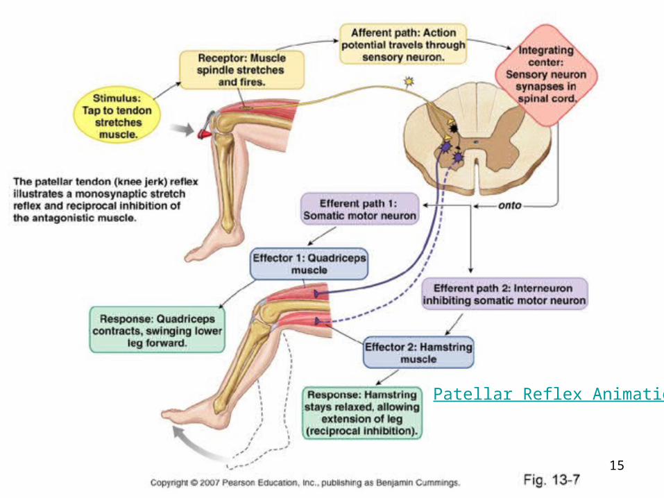
Withdraw Reflex

17
Crossed Extensor Reflex
18
Transmission of Nerve Impulses
19
Overview of Action Potential• Nerve impulses are electrochemical messages created by the movement of
ions through the nerve cell.
• RESTING POTENTIAL– When a neuron is at resting state.– meaning the inside is negative relative to the outside of the axon. There
are many negatively charged proteins on the inside of the neuron.– A resting nerve membrane normally has a potential somewhere near -
70mV on the inside.– The membrane is said to be polarized.
• ACTION POTENTIAL– when a neuron is activated and temporarily reverses the electrical state
of its interior membrane from negative to positive– Inside of neuron is now +40mV.
Sodium Potassium Pump Animation

20
The Sodium Potassium Pump• At resting potential, there is more Na+ on the outside of the nerve
membrane and more K+ on the inside.
• Active transport maintains this – a sodium-potassium pump pumps the Na+ ions out and pumps the K+ ions in.
• The sodium-potassium pump uses ATP.
• The sodium-potassium pump is constantly working
Sodium Potassium Pump
Sodium-Potassium Pump Animation

21
• At rest, the neuron has a ‘resting potential of -70mV (negative on inside).• When a nerve becomes excited as a result of a stimulus, the gated ion channels
on the membrane open and allow the Na+ to rush into the membrane. This is Facilitated diffusion (no energy is required). (Remember that the concentration gradient was established by the sodium potassium pump).
• The Na+ rushes through the membrane causes the inside of the neuron to become positive.
• The neuron is now DEPOLARIZED.
Action Potential

22
• Once the neuron is depolarized, it reaches the ‘all or none’ stage which means that there is no turning back.
• Complete depolarization occurs and the stimulus will be transmitted
Interactive Action Potential Animation
…Action Potential

23
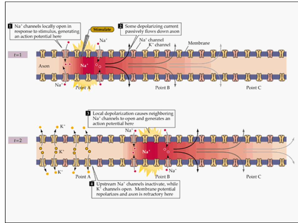
• The Na+ and K+ transport proteins are voltage-gated channels. Depolarization of the neural membrane causes neighboring gates to open
…Action Potential
24
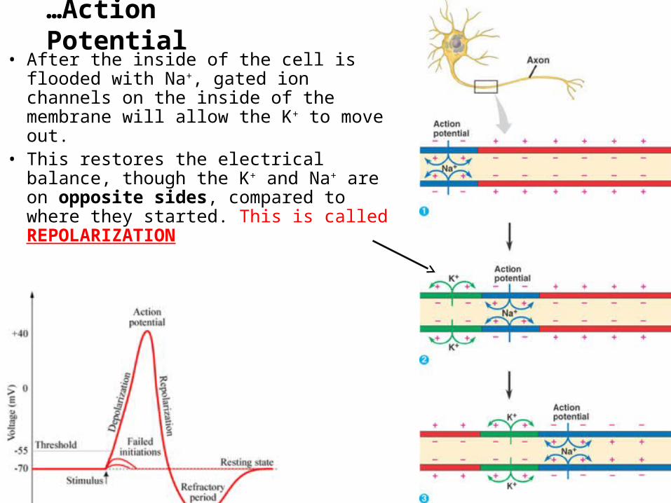
• After the inside of the cell is flooded with Na+, gated ion channels on the inside of the membrane will allow the K+ to move out.
• This restores the electrical balance, though the K+ and Na+ are on opposite sides, compared to where they started. This is called REPOLARIZATION
…Action Potential

25
• The REFRACTORY PERIOD is the time it takes before the neuron can produce another action potential. This is due to the fact that K+ ions channels remain open longer than required to bring the membrane back to the resting potention.
• The Sodium-Potassium pump is responsible for reestablishing resting potential. (Pumping Na+ and K+ ions back acros the membrane)
• While this process is taking place, the nerve cannot respond to any other signals. WHY Not?
Voltage Gated Channels and the Action Potential
…Action Potential
THINK
• Write down what you can remember about the following terms…
• Threshold
• Resting potential
• Depolarizarion
• Repolarization
• Refractory period26
27

28
Synaptic Transmission

29
The Synapses
30
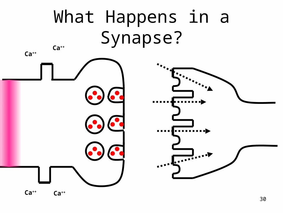
What Happens in a Synapse?Ca++
Ca++
Ca++ Ca++
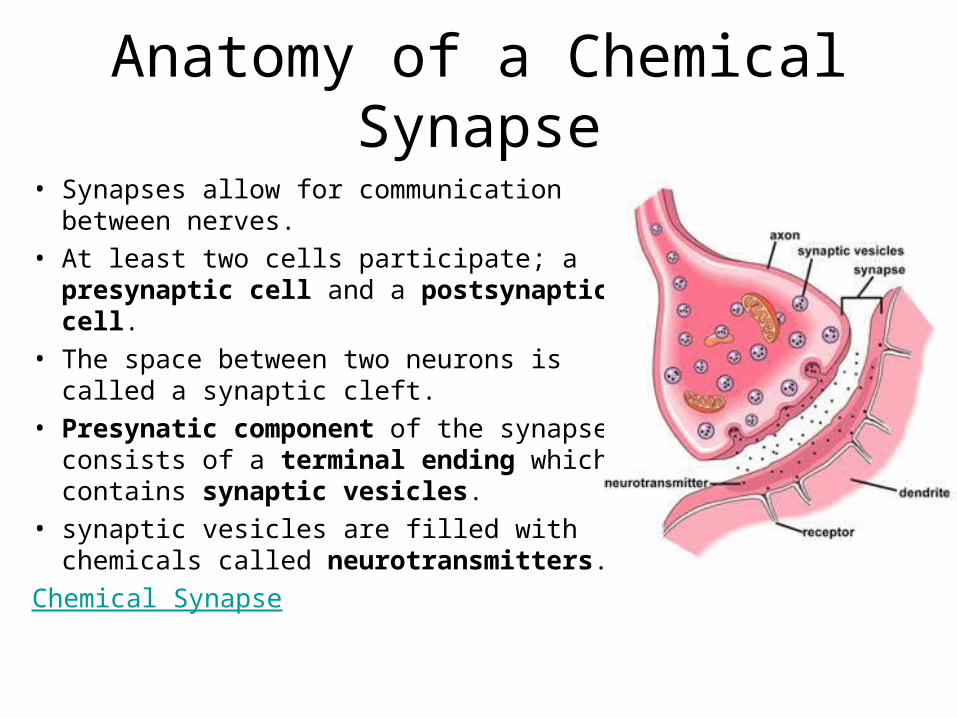
Anatomy of a Chemical Synapse
• Synapses allow for communication between nerves.
• At least two cells participate; a presynaptic cell and a postsynaptic cell.
• The space between two neurons is called a synaptic cleft.
• Presynatic component of the synapse consists of a terminal ending which contains synaptic vesicles.
• synaptic vesicles are filled with chemicals called neurotransmitters.
Chemical Synapse

32
Presynaptic Mechanisms of Chemical Transfer
• Release of neurotransmitters is produced by the action potential when it invades the terminal ending.
• This change in membrane potential activates voltage-sensitive Ca2+ channels, causing influx of Ca2+ ions into the terminal.
• The Ca2+ ions cause the synaptic vesicles to fuse with the pre-synaptic membrane and release neurotransmitter molecules into the synaptic cleft by the process of exocytosis.
Chemical Synapse

33
…Presynaptic Mechanisms of Chemical Transfer
• Each synaptic vesicle releases a fixed number of neurotransmitter molecules.
• The number of synaptic vesicles that dump their transmitters into the synaptic cleft depends on the amount of Ca2+ within the synaptic terminal.
• Amount of Ca2+ ions within the terminal is regulated by the number of action potentials the neuron has generated. So…
• Increased frequency of action potentials more Ca2+ into terminal more neurotransmitters released by vesicles
Chemical Synapse

Think
• What effect do you think neurotransmitters have on the postsynaptic cell?
• Hint: remember the spinal reflex? What must happen to specific muscles when you pull your hand away from a hot stove?
34
35
Postsynaptic Mechanisms of Chemical Transfer
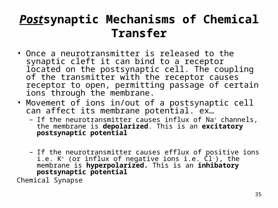
• Once a neurotransmitter is released to the synaptic cleft it can bind to a receptor located on the postsynaptic cell. The coupling of the transmitter with the receptor causes receptor to open, permitting passage of certain ions through the membrane.
• Movement of ions in/out of a postsynaptic cell can affect its membrane potential. ex…– If the neurotransmitter causes influx of Na+ channels, the membrane is
depolarized. This is an excitatory postsynaptic potential
– If the neurotransmitter causes efflux of positive ions i.e. K+ (or influx of negative ions i.e. Cl-), the membrane is hyperpolarized. This is an inhibatory postsynaptic potential
Chemical Synapse

36
…Postsynaptic Mechanisms of Chemical Transfer
• Channels activated by neurotransmitters remain open as long as the neurotransmitter is bound to the receptor.
Chemical Synapse
37
Termination of Synaptic Transmission
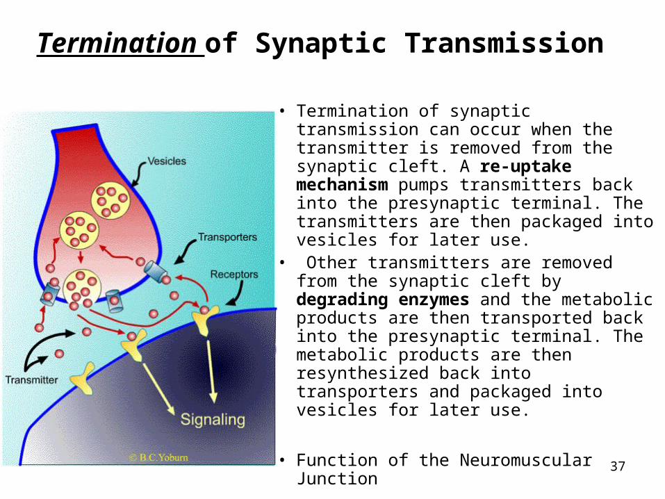
• Termination of synaptic transmission can occur when the transmitter is removed from the synaptic cleft. A re-uptake mechanism pumps transmitters back into the presynaptic terminal. The transmitters are then packaged into vesicles for later use.
• Other transmitters are removed from the synaptic cleft by degrading enzymes and the metabolic products are then transported back into the presynaptic terminal. The metabolic products are then resynthesized back into transporters and packaged into vesicles for later use.
• Function of the Neuromuscular Junction
38
Neurotransmitters
39
What is a neurotransmitter?• a chemical released at the axon terminal of a neuron that
travels across the synaptic cleft
• binds a specific receptor on the far side (postsynaptic membrane), and depending on the nature of the receptor, depolarizes or hyperpolarizes a second neuron or a muscle or a gland.
• The physiological response of a neurotransmitter depends on the neurotransmitter/receptor combination. The same neurotransmitter can have a stimulatory effect in one part of the body and a inhibitory effect on another part of the body.
What is a ‘drug’?
• A very vague term
• all ingested substances alter bodily function
• ‘drug’ is reserved for things that have pronounced effects when ingested in small quantities.
• We will be interested in drugs that affect the CNS and PNS.
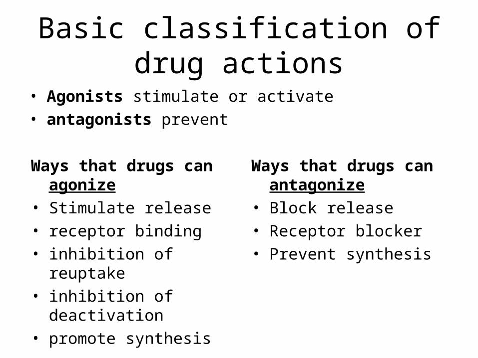
Basic classification of drug actions
• Agonists stimulate or activate
• antagonists prevent
Ways that drugs can agonize
• Stimulate release
• receptor binding
• inhibition of reuptake
• inhibition of deactivation
• promote synthesis
Ways that drugs can antagonize
• Block release
• Receptor blocker
• Prevent synthesis
Amino acids: The workhorses of the neurotransmitter family
• Glutamate– the primary excitatory neurotransmitter in brains
• GABA (Gamma-amino-butyric-acid)– the primary inhibitory neurotransmitter
42
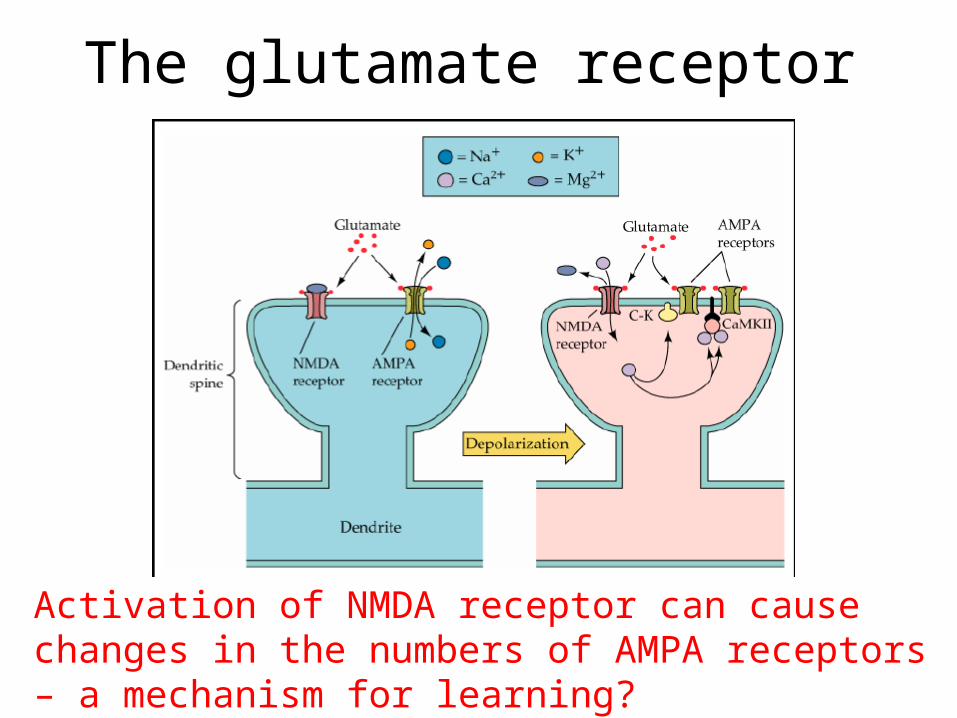
The glutamate receptor
Activation of NMDA receptor can cause changes in the numbers of AMPA receptors – a mechanism for learning?
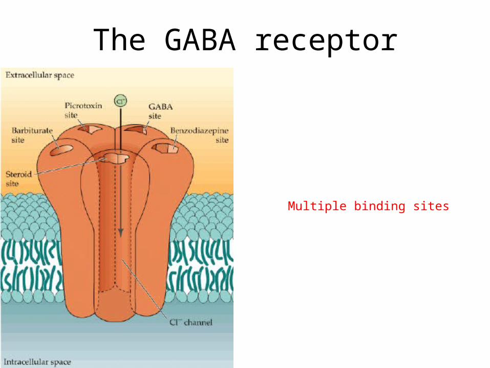
The GABA receptor
Multiple binding sites

45
Acetylcholine (ACh)
Found in CNS and PNS
• PNS
– Can be excitatory in the somatic nervous system (stimulates muscle contraction) by opening Na+ channels and allowing Na+ ions to depolarize postsynaptic neurons.
– Can be inhibitory in the autonomic nervous system (slows down heart rate by increasing the permeability of K+ ions to hyperpolarize the cell).
– Whether Ach is stimulatory or inhibitory depends on the type of receptor used.
• Use of acetylcholine in a Synaptic transmission

A nicotinic receptor is a ligand-gated ion channel that allows for influx of Na+ ions (depolarization)
46
Use it or Lose it!
• Close your Notes…
• How can acetylcholine be inhibitory in the autonomic nervous system and stimulatory in the somatic nervous system?
47

48
Drugs That Affect Acetylcholine• Snake venom
– blocks Acetylcholine receptors so muscles can’t contract paralysis!
• Nerve Gases and local anesthetics (Novocane, Lidocaine) and Cocaine– block sodium channels so pain impulse is not transmitted.
• Botulism toxin– inhibits release of acetylcholine weak muscle contraction.
1 g can kill 20 x 106 people
49
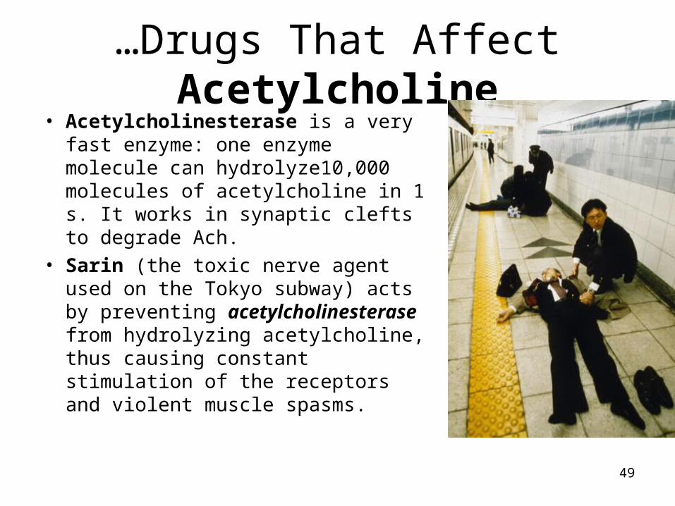
…Drugs That Affect Acetylcholine• Acetylcholinesterase is a very fast
enzyme: one enzyme molecule can hydrolyze10,000 molecules of acetylcholine in 1 s. It works in synaptic clefts to degrade Ach.
• Sarin (the toxic nerve agent used on the Tokyo subway) acts by preventing acetylcholinesterase from hydrolyzing acetylcholine, thus causing constant stimulation of the receptors and violent muscle spasms.

50
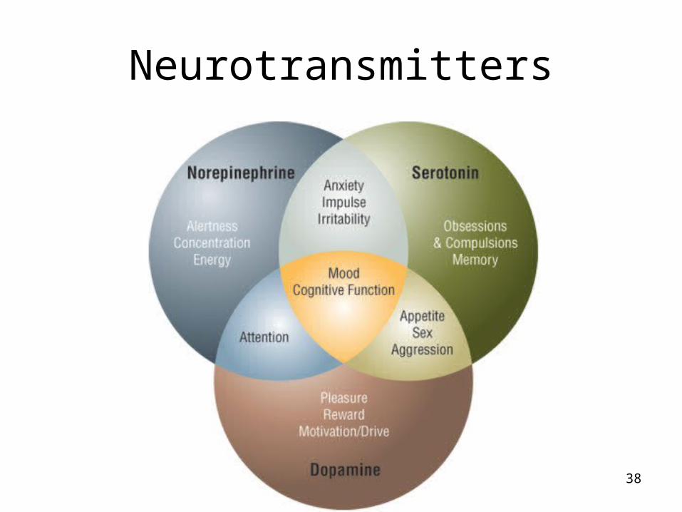
Epinephrine and Norepinephrine
• AKA adrenalin and noradrenalin
• Fight or flight stimulates heart contractions, inhibits muscles in digestive tract.
• Awakening from sleep
• No effect on skeletal muscles (no receptors)

51
Drugs That Affect Epinephrine and Norepinephrine
• Amphetamines mimic these transmitters (heart stimulant)
• Cocaine– moves into synapse and blocks absorption of
epinephrine – it remains in synapse heart is continually
stimulated heart failure– Decreased appetite
52
Dopamine
• Regulates emotions (pleasure), skeletal muscle motion.
• Deficiency Parkinson’s disease. Michael J. Fox (uncontrolled tremors and twitches)
• Too much schizophrenia
53
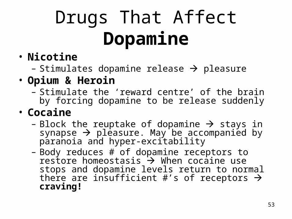
Drugs That Affect Dopamine
• Nicotine– Stimulates dopamine release pleasure
• Opium & Heroin– Stimulate the ‘reward centre’ of the brain by forcing
dopamine to be release suddenly• Cocaine
– Block the reuptake of dopamine stays in synapse pleasure. May be accompanied by paranoia and hyper-excitability
– Body reduces # of dopamine receptors to restore homeostasis When cocaine use stops and dopamine levels return to normal there are insufficient #’s of receptors craving!
54
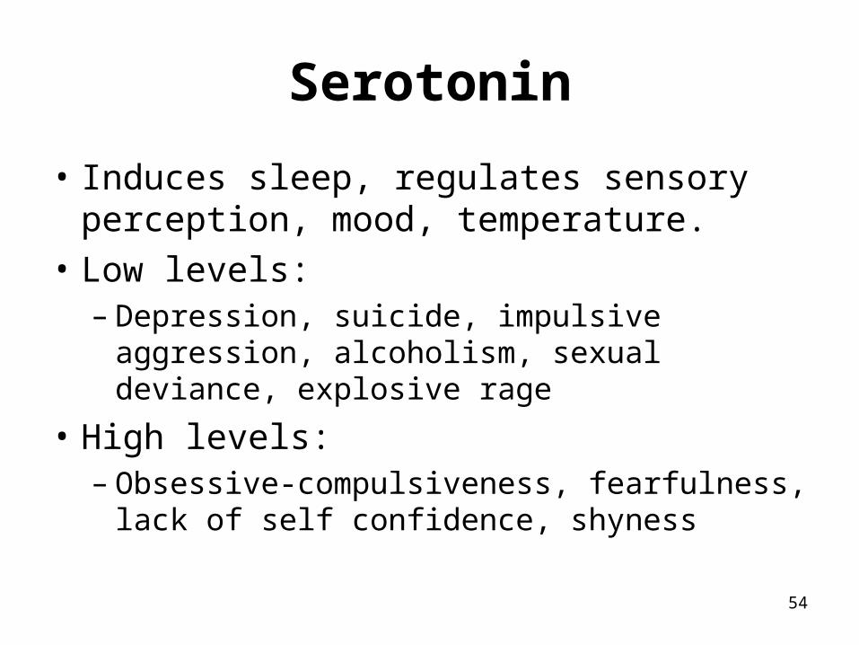
Serotonin
• Induces sleep, regulates sensory perception, mood, temperature.
• Low levels:– Depression, suicide, impulsive aggression,
alcoholism, sexual deviance, explosive rage
• High levels:– Obsessive-compulsiveness, fearfulness, lack of
self confidence, shyness
55
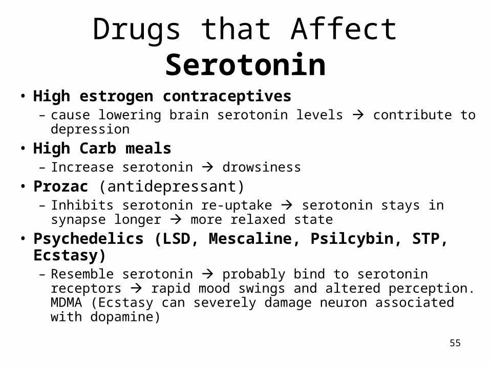
Drugs that Affect Serotonin
• High estrogen contraceptives– cause lowering brain serotonin levels contribute to depression
• High Carb meals– Increase serotonin drowsiness
• Prozac (antidepressant)– Inhibits serotonin re-uptake serotonin stays in synapse longer
more relaxed state
• Psychedelics (LSD, Mescaline, Psilcybin, STP, Ecstasy)– Resemble serotonin probably bind to serotonin receptors
rapid mood swings and altered perception. MDMA (Ecstasy can severely damage neuron associated with dopamine)
56
Endorphins, Enkephalins
• Natural pain killers block pain receptors
• Released by brain stimulate natural opiate receptors

57
Opiates (Opium, Heroin, Morphine, Codeine)
• These mimic the body’s natural pain killers
• Intake of opiates decrease in endorphins.
• When the drug stops receptors are vacant due to decreased endorphin levels painful withdrawal
58
Questions for Homework
• What is a polarized membrane?• What does ‘depolarize’ and ‘repolarize’ mean?• K+ has a positive charge. Why then is there an
overall negative charge inside the membrane?• What are the three pumps used in this process?• What is the purpose of the sodium-potassium
pump at the end of the process? Is this active or passive transport?

59
http://highered.mcgraw-hill.com/sites/0072437316/student_view0/chapter45/animations.html

60
A medical problem related to what you now know about nerves……….
61
Multiple Sclerosis - Symptoms
• Its first symptom is usually a visual disturbance blurred or double vision, red-green confusion, or even temporary blindness in one eye.
• Soon, muscle fatigue, pain, numbness, weakness, stiffness, or 'pins and needles' develop.
• As the disease progresses, patients may lose coordination, balance, hearing, or bladder/bowel control.
• Some endure mild concentration or memory problems, while others experience depression, manic-depression, and paranoia.
• Other possible symptoms include sexual dysfunction, tremors, dizziness, slurred speech, trouble swallowing, urinary problems, and episodes of facial pain or inappropriate emotions.
62

63
Multiple Sclerosis
• MS is caused by a chemical attack on the brain's neurons that destroys the myelin.
• Areas of scarred myelin are called lesions
• They disrupt the transmission of messages that cause the symptoms of MS.

64
MS - Animation
http://www.hcnr.med.harvard.edu/visitorInfo/MS.php
http://www.neurodiscovery.harvard.edu/

65
Now for something a little different….

66
Mirror Neurons
• http://www.pbs.org/wgbh/nova/sciencenow/3204/01.html
67
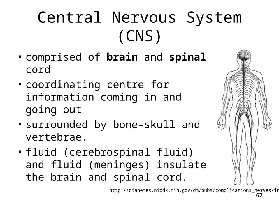
Central Nervous System (CNS)
• comprised of brain and spinal cord
• coordinating centre for information coming in and going out
• surrounded by bone-skull and vertebrae.
• fluid (cerebrospinal fluid) and fluid (meninges) insulate the brain and spinal cord.
http://diabetes.niddk.nih.gov/dm/pubs/complications_nerves/index.htm

68
…Central Nervous System (CNS)
• The brain is composed of three main parts:
– Cerebrum (seat of consciousness)
– Cerebellum: muscle coordination and maintains normal muscle tone and posture. The cerebellum coordinates balance
– medulla oblongata regulation of heartbeat, breathing, vasoconstriction (blood pressure), and reflex centers for vomiting, coughing, sneezing, swallowing, and hiccupping

69
Peripheral Nervous System (PNS)
• carries information between organs of the body and the CNS