10-color flow cytometry for the efficient diagnosis of
TRANSCRIPT

10-Color Flow Cytometry for the Efficient Diagnosis of
Lymphoid NeoplasmsSteven J. Kussick, MD, PhD
Associate Medical Director/Director of Flow CytometryPhenoPath Laboratories
October 1, 2011
Overview• Rationale for 10-color flow cytometry (FC)
• Keys to setting up a 10-color FC laboratory
• 9- and 10-color immunophenotyping, with an emphasis on challenging cases• Immature lymphoid neoplasms• Mature lymphoid neoplasms• Plasma cell neoplasms
Why 10-Color Flow?• Gets much more information from small samples (FNA, CSF)
- 10-color yields 7.5x more info. than 4-color flow (45 vs. 6 dot-plots)
- 10-color yields 3x more info. than 6-color flow (45 vs. 15 dot-plots)
- 10-color yields 1.6x more info. than 8-color flow (45 vs. 28 dot-plots)
• Routinely collect > 100,000 viable cells (> 300,000 viable cells for MRD work), so identify small abnormal populations missed by lesser flow
• In even small biopsies, can almost always make definitive 2008 WHO diagnoses when combine flow with histology and limited IHC/FISH
• Streamlined specimen setup / reporting = FASTER turnaround
• Reference: Wood BL. 9-Color and 10-Color Flow Cytometry in the Clinical Laboratory. Arch Pathol Lab Med. 2006;130:680–690.

9/10-Color FC Implementation-1: Choice of Flow Cytometer
• Signed out 9-/10- color clinical flow data from BD LSRIIs at the U. of WA from 2004-5, so wanted at least 6 colors, with definite ability to expand to 8 colors, for lab efficiency and better handling of small specimens and MRD
• Not possible with the 6-color FACSCanto in May 2005
• Chose 3-laser/9-color LSRII in summer 2005, and expanded to 10 colors in January 2010 with availability of V500 fluor.
• Beckman-Coulter 3-laser/10-color Gallios available now
3-Laser / 10-Color LSRII
Lasers
Detectors
Flow Control Sample
FlowCell
Fiber-optics

From Becton-Dickinson FACSCanto Flow Cytometer pdf (www.bdbioscience.com)
LSRII / FACSCanto Optics
Implementation-2: Choice of Antigens (27) to Evaluate Routinely
B-cell Ags: Kappa, Lambda, CD19, CD20, CD10, CD38, CD45
T & NK Ags: CD2, CD3, CD4, CD5, CD7, CD8, CD56, CD16
Pan-myeloid Ags: CD13, CD33, CD45
Myeloid blast-associated Ags: CD34, CD117, HLA-DR, CD38
Granulocyte-associated Ags: CD10, CD14, CD15, CD16, CD117, CD11b
Monocyte/dendritic cell-associated Ags: HLA-DR, CD4, CD14, CD15, CD38, CD64, CD123, CD36, CD11b
Erythroid-associated Ags: CD38, CD71, CD117

Implementation-3: Principles of Antibody Panel Design
• Goal: Maximize information obtained per unit of specimen, unit of cytometry staff effort, and unit of cytometer time
• Maintain across-tube comparability (e.g., CD45/SS gating in all) while minimizing antibody redundancy
• Pair strongly expressed antigens (e.g., neutrophil CD16) with dimmer fluorochromes (e.g., FITC, Alexa700), and vice versa
• Combine antibodies to maximize ability to identify abnormal cellpopulations (e.g., pairing CD34 and CD7 antibodies to look at blasts)
• All things being equal, choose IgG1 over IgG2 antibodies (see Wood BL & Levin GR. Cytometry B Clin Cytom. 2006;70:321-8)
Implementation-4: Fluorochrome Choice
From Baumgarth N, Roederer M. A practical approach to multicolor flow cytometry for immunophenotyping. J Immunol Methods 2000;243-77-97.
- Availability: Single vs. tandem dyes, stock vs. custom reagents
- Excitation of dye: function of excitation spectra (related to size of dye) and laser type
- Measuring fluorescence of dye: function of emission spectra and optics of instrument

Implementation-5: Ab Panels in Practice
• Sought to screen B and T cells with 1 tube each, and myeloid cells with 2 tubes
• Sought a single main add-on tube for each of the following: CLL/MCL, HCL/MZL, plasma cells, B-ALL, T-ALL, AML
• Sought to maximize use of ASRs and off-the-shelf reagents, and minimize custom antibodies, hence original choice of Alexafluor 700 as “ninth” color in 2005
• Compromises often required for antibodies & fluorochromes
Lymphoid Panel in 2000: 4-Color Flow, Single Laser (13 Ags)
FITC PE PE-TR PE-Cy5Kappa / Lambda/ CD19 / CD5CD20 / CD10 / CD19 / CD45CD2 / CD7 / CD3 / CD4CD5 / CD56 / CD3 / CD8

Lymphoid Panel in 2010: 10-Color Flow (15 Ags + Viability Dye)
VIOLET BLUE REDLASER LASER LASER
PBlue / V500 // FITC / PE /PE-TR / PE-Cy5 / PE-Cy7 // APC /APC-A700 /APC-A750
DAPI / CD45 // CD2 / CD7 / CD3 / CD34 / CD56 // CD5 / CD4 / CD8
DAPI / CD45 // Kap. / Lam. /CD19 / CD5 / CD20 // CD10 / CD56 / CD38
Implementation-6: Creating Antibody Cocktails
• Encourages use of standardized reagents within laboratory
• Time savings for technical staff
• Enhances assay-to-assay reliability by:
– Reducing chances of pipetting incorrect antibody
– Reducing chances of pipetting incorrect volume of antibody, as relatively large volumes used in cocktails
• Recent FDA limitations limit available ASR cocktails

Ab Optimization / Cocktail Validation
• Titrate antibodies individually, on appropriate cell population(s), to optimize signal:noise ratios
• Confirm similar antibody-associated fluorescence on singly-stained cells vs. cells stained with 9/10-color cocktails
• If already using fewer-color cocktails, confirm similar performance vs. 9/10-color cocktails
Implementation-7: Assay Validation During Lab Set-up
• Used ~50 normal peripheral bloods and ~20 well-defined hematolymphoid cell lines (from ATCC and DMSZ) to confirm expected immunoreactivity
• Cross validation on ~30 clinical specimens kindly provided by several outside laboratories, with uniform concordance with available outside flow interpretations
• Initial validation period: ~4 months from LSRII set-up

Data Analysis-1: General Gating
FS/SS Gating WBC GatingDoublet Elimination
B cells: CD19/SS
T cells: CD3/SS
Blasts:CD34/SS
Data Analysis-2: Lineage Gating
Viable CellsCD45/SS

Data Analysis-3: Compensation and Data Scaling
From Flow Cytometry in Clinical Diagnosis, 3rd Ed. ASCP Press, 2001 p. 40 (JP McCoy chapter).
Case Studies:Thoroughly Understand the
Normal in Order to Recognize the Abnormal

B-lymphoidNeoplasms
Normal Bone Marrow: B Cells and Plasma Cells

Case Study 1: 73 y.o. male with lymphocytosis
Case Study 1: 73 y.o. male with lymphocytosis
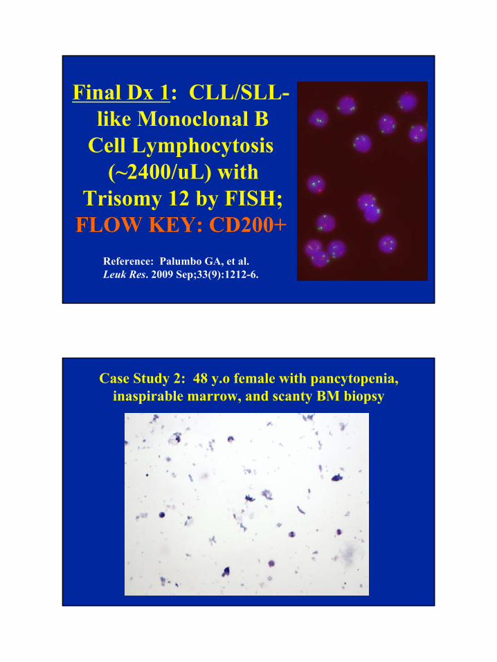
Final Dx 1: CLL/SLL-like Monoclonal B
Cell Lymphocytosis(~2400/uL) with
Trisomy 12 by FISH;FLOW KEY: CD200+
Reference: Palumbo GA, et al. Leuk Res. 2009 Sep;33(9):1212-6.
Case Study 2: 48 y.o female with pancytopenia, inaspirable marrow, and scanty BM biopsy

Case Study 2: 48 y.o female with pancytopenia, inaspirable marrow, and scanty BM biopsy
Final Dx 2: Hairy cell leukemia;FLOW KEY:
CD103+, CD25+

Case Study 3: 72 y.o. female with right axillary lymphadenopathy, small biopsy
Case Study 3: 72 y.o. female with right axillary lymphadenopathy, small biopsy

Final Dx 3: Follicular
lymphoma, Grade 1-2;
FLOW KEY:bcl-2+, low CD10+
Case Study 4: 21 y.o female with omental mass and scanty specimen

Case Study 4: 21 y.o female with omental mass and scanty specimen
Final Dx 4: Burkitt
lymphoma, MYC+
by FISH;FLOW KEY:
CD10+, CD38+, Bcl-2-negative

Case Study 5: 58 y.o. male with hx of myeloma
Case Study 5: 58 y.o. male with hx of myeloma

Final Dx 5: t(11;14)+plasma cell myeloma, positive for
CD20 & surface light chain;FLOW KEY: CD138+,
bright CD38+, low CD45+ low CD19+, CD22-negative
(Reference: Lohrsbach RB, et al. Am J Clin Pathol. 2011;136:168-82)
Case Study 6: 25 y.o. female with C7-T4 paraspinal mass
Initial IHC in SBRCT Workup

Case Study 6: 25 y.o. female with C7-T4 paraspinal mass
Second round of IHC to Establish Lineage (but CD45-)
Case Study 6: 25 y.o. female with C7-T4 paraspinal mass
BM
CSF

Final Dx 6: B-Lymphoblastic Lymphoma with Positive CSF and Minimal Marrow
Involvement;FLOW KEY:
CD45-neg., CD19+
Case Study 7: 35 y.o. female, s/p SCT for CHL

Case Study 7: 35 y.o. female, s/p SCT for CHL
Final Dx 7: Relapsed CHL, CD4+;
FLOW KEY: CD45-negReference: Asano N, et al.
J Clin Oncol. 2006;24:4626-33

T-lymphoidNeoplasms
Normal Thymus: Immature T Cells

Normal Bone Marrow: Mature T Cells
Case Study 8: 54 y.o. male with marked peripheral lymphocytosis

Case Study 8: 54 y.o. male with marked peripheral lymphocytosis
Final Dx 8: T-Prolymphocytic
Leukemia, (inv[14]+);FLOW KEY: Dual CD4+/
CD8+ with MATURE CD34-/TdT-/CD1a- phenotype

Case Study 9: 62 y.o. female, R/O AML
Case Study 9: 62 y.o. female, R/O AML

Final Dx 9: Anaplastic large cell lymphoma, ALK+;
FLOW KEY:CD30+, bright CD45+, CD7+,
cyto CD3+, aberrant CD13+
Reference: Bovio IM, Allan RW. Am J Clin Pathol. 2008 Oct;130(4):628-3
Case Study 10: 80 y.o. female with adenopathy

Case Study 10: 80 y.o. female with adenopathy
Final Dx 10: Angio-immunoblastic T Cell Lymphoma
(missed by 6-color FC
elsewhere);FLOW KEY: CD4+, CD5+, surface CD3-& TCR-α/β-negative

Case Study 11: 38 y.o. male, pleural fluid
Case Study 11: 38 y.o. male, pleural fluid

Final Dx 11: T-Lymphoblastic Lymphoma
(cytokeratin-negative mediastinal mass);
FLOW KEY: Abnormally low CD1a and high CD7 for
benign thymocytes
Non-Hematopoietic Neoplasms That Can Mimic Lymphoma

Case Study 12: Lung biopsy in 68 y.o. female
Case Study 12: Lung biopsy in 68 y.o. female

Final Dx 12: Small CellNeuroendocrine Carcinoma;
FLOW KEY: CD45-negative,strongly CD56+ population
(Reference: Chang A, et al. Am J Clin Pathol. 2003;119:643-55)
Acknowledgements• �Donna Ceniza, MT (ASCP)
• Barbara Oppenlander, MT (ASCP)
• Earline Carlone, MT (ASCP)
• Sara Duesterhoeft, MD
• Sandra Bohling, MD

The End