1096 publication tamoxifen as a novel chemotherapeutic agent treating anaplastic thyroid cancer
TRANSCRIPT
316 Proffered Papers
with chemotherapy. Each patient received 3 cycles of treatment before continuing to radiotherapy of 70 Gy (fractionated) as local treatment. Results: Between January 2000 and December 2003, a total of 69 patients were enrolled and assessable for response and toxicity analysis. The median age was 55 years (age range 45-70 years). The anatomical sites were oral cavity (45%), nasopharynx (35%) and larynx (20%). Overall response rate was 96% with complete response seen in 42 patients (61%) and partial response in 24 patients (35%). The remaining patients (4%) showed stable disease. The median time for follow-up was 45 months. The most common hematological side effects were neutropenia (35 patients; 51%) out of which G3/4 was found in 10 patients. The non-hematological events were vomiting (28 patients; 41%) and stomatitis (10 patients; 14%). There were no treatment related deaths. Conclusion: For treatment of locally advanced SCCHN, docetaxel in combination with cisplatin and 5-FU may be an effective regimen with a manageable toxicity profile.
1096 PUBLICATION Tamoxifen as a novel chemotherapeutic agent treating anaplastic thyroid cancer.
J. O'Neill, B. O'Neill, C. Condron, M. Walsh, D. Bouchier-Hayes. The Royal College of Surgeons in Ireland, Dept of Otolaryngology and Surgery, Dublin, Ireland
Introduction: Anaplastic Thyroid Cancer (ATC) is a highly aggressive rare neoplasm with a dismal prognosis. It represents 2% of all thyroid cancers with a mean survival of 3-7 months. The majority of ATC patients develop metastases during their illness hence there is an essential role for systemic chemotherapy. Doxorubicin, cisplatin and paxlitaxel to date offer poor chemotherapeutic response. Method: We have investigated the anti-proliferative effects using coloro- metric dimethyl-thiazol-diphenyltetrazolium bromide (MTT), pro-apoptotic effects was investigated using flow cytometry and annexin V and anti- invasive properties using Matrigel invasion assays at varying concentra- tions of tamoxifen on anaplastic thyroid carcinoma cell line Cal-62.
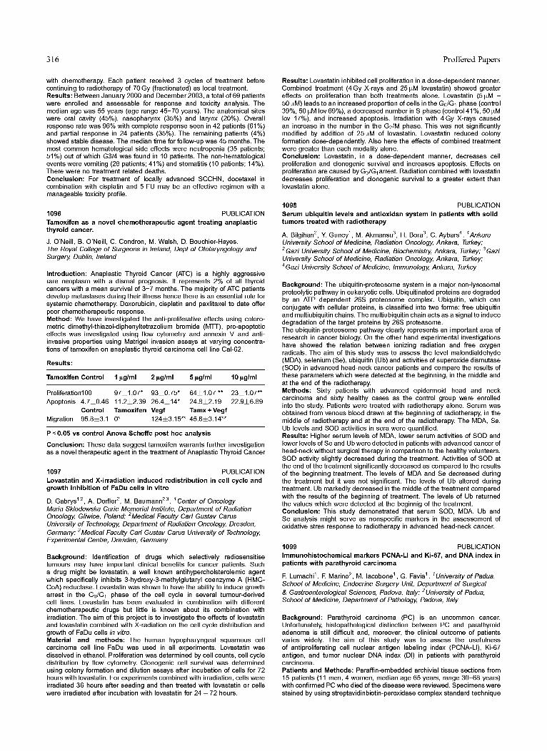
Results:
Tamoxifen Control 1 ~g/ml 2 ~g/ml 5 ~g/ml 10 ~g/ml
Proliferation100 97±1.07" 93±0.75* 64±1.07 ** 23±1.07"* Apoptosis 4.7±0.46 11.2±2.39 26.4±14" 24.8±2.19 22.9±6.89
Control Tamoxifen Vegf Tamx + Vegf Migration 95.8±3.1 0* 124±3.15"* 45.6±3.14"*
P <0.05 vs control Anova Scheffe post hoc analysis
Conclusion: These data suggest tamoxifen warrants further investigation as a novel therapeutic agent in the treatment of Anaplastic Thyroid Cancer
1097 PUBLICATION Lovastatin and X-irradiation induced redistribution in cell cycle and growth inhibition of FaDu cells in vitro
D. Gabrys 1'2, A. Dorfler 2, M. Baumann 2'3. 1Center of Oncology Maria Sklodowska-Curie Memorial Institute, Department of Radiation Oncology, Gliwice, Poland; 2Medical Faculty Carl Gustav Carus University of Technology, Department of Radiation Oncology, Dresden,
3 Germany; Medical Faculty Carl Gustav Carus University of Technology, Experimental Centre, Dresden, Germany
Background: Identification of drugs which selectively radiosensitise tumours may have important clinical benefits for cancer patients. Such a drug might be Iovastatin, a well known antihypercholsterolemic agent which specifically inhibits 3-hydmxy-3-methylglutaryl coenzyme A (HMG- CoA) reductase. Lovastatin was shown to have the ability to induce growth arrest in the G0/G1 phase of the cell cycle in several tumour-derived cell lines. Lovastatin has been evaluated in combination with different chemotherapeutic drugs but little is known about its combination with irradiation. The aim of this project is to investigate the effects of Iovastatin and Iovastatin combined with X-radiation on the cell cycle distribution and growth of FaDu cells in vitro. Material and methods: The human hypopharyngeal squamous cell carcinoma cell line FaDu was used in all experiments. Lovastatin was dissolved in ethanol. Proliferation was determined by cell counts, cell cycle distribution by flow cytometry. Clonogenic cell survival was determined using colony formation and dilution assays after incubation of cells for 72 hours with Iovastatin. For experiments combined with irradiation, cells were irradiated 36 hours after seeding and then treated with Iovastatin or cells were irradiated after incubation with Iovastatin for 24 - 72 hours.
Results: Lovastatin inhibited cell proliferation in a dose-dependent manner. Combined treatment (4Gy X-rays and 25 ~M Iovastatin) showed greater effects on proliferation than both treatments alone. Lovastatin (5~M - 50 ~M) leads to an increased proportion of cells in the G0/G1 phase (control 39%, 50 ~M Iov 69%), a decreased number in S-phase (control 41%, 50 ~M Iov 17%), and increased apoptosis. Irradiation with 4Gy X-rays caused an increase in the number in the G2/M phase. This was not significantly modified by addition of 25~M of Iovastatin. Lovastatin reduced colony formation dose-dependently. Also here the effects of combined treatment were greater than each modality alone. Conclusion: Lovastatin, in a dose-dependent manner, decreases cell proliferation and clonogenic survival and increases apoptosis. Effects on proliferation are caused by G0/G1 arrest. Radiation combined with Iovastatin decreases proliferation and clonogenic survival to a greater extent than Iovastatin alone.
1098 PUBLICATION Serum ubiquitin levels and antioxidan system in patients with solid tumors treated with radiotherapy
A. Bilgihan 2, Y. Guney 1 , M. Akmansu 3, H. Born 3, C. Aybars 4. 1Ankara University School of Medicine, Radiation Oncology, Ankara, Turkey; 2 Gazi University School of Medicine, Biochemistry, Ankara, Turkey; 3 Gazi University School of Medicine, Radiation Oncology, Ankara, Turkey; 4 Gazi University School of Medicine, Immunology, Ankara, Turkey
Background: The ubiquitin-proteosome system is a major non-lysosomal proteolytic pathway in eukaryotic cells. Ubiquitinated proteins are degraded by an ATP dependent 26S proteosome complex. Ubiquitin, which can conjugate with cellular proteins, is classified into two forms: free ubiquitin and multiubiquitin chains. The multiubiquitin chain acts as a signal to induce degradation of the target proteins by 26S proteasome. The ubiquitin-proteosome pathway clearly represents an important area of research in cancer biology. On the other hand experimental investigations have showed the relation between ionizing radiation and free oxygen radicals. The aim of this study was to assess the level malondialdehyde (MDA), selenium (Se), ubiquitin (Ub) and activities of superoxide dismutase (SOD) in advanced head-neck cancer patients and compare the results of these parameters which were detected at the beginning, in the middle and at the end of the radiotherapy. Methods: Sixty patients with advanced epidermoid head and neck carcinoma and sixty healthy cases as the control group were enrolled into the study. Patients were treated with radiotherapy alone. Serum was obtained from venous blood drawn at the beginning of radiotherapy, in the middle of radiotherapy and at the end of the radiotherapy. The MDA, Se, Ub levels and SOD activities in sera were quantified. Results: Higher serum levels of MDA, lower serum activities of SOD and lower levels of Se and Ub were detected in patients with advanced cancer of head-neck without surgical therapy in comparison to the healthy volunteers. SOD activity slightly decreased during the treatment. Activities of SOD at the end of the treatment significantly decreased as compared to the results of the beginning treatment. The levels of MDA and Se decreased during the treatment but it was not significant. The levels of Ub altered during treatment. Ub markedly decreased in the middle of the treatment compared with the results of the beginning of treatment. The levels of Ub returned the values which were detected at the beginnig of the treatment. Conclusion: This study demonstrated that serum SOD, MDA, Ub and Se analysis might serve as nonspecific markers in the assessement of oxidative stres response to radiotherapy in advanced head-neck cancer.
1099 PUBLICATION Immunohistochemical markers PCNA-LI and Ki-67, and DNA index in patients with parathyroid carcinoma
F. Lumachi 1 , E Marino 2, M. lacobone 1 , G. ravin 1 . 1University of Padua, School of Medicine, Endocrine Surgery Unit, Department of Surgical & Gastroenterological Sciences, Padova, 2 Italy; University of Padua, School of Medicine, Department of Pathology, Padova, Italy
Background: Parathyroid carcinoma (PC) is an uncommon cancer. Unfortunately, histopathological distinction between PC and parathyroid adenoma is still difficult and, moreover, the clinical outcome of patients varies widely. The aim of this study was to assess the usefulness of antiproliferating cell nuclear antigen labeling index (PCNA-LI), Ki-67 antigen, and tumor nuclear DNA index (DI) in patients with parathyroid carcinoma. Patients and Methods: Paraffin-embedded archivial tissue sections from 15 patients (11 men, 4 women, median age 65 years, range 30-68 years) with confirmed PC who died of the disease were reviewed. Specimens were stained by using streptavidinbiotin-peroxidase complex standard technique