12-1 nervous tissue controls and integrates all body activities within limits that maintain life...
TRANSCRIPT

12-1
Nervous Tissue
• Controls and integrates all body activities within limits that maintain life
• Three basic functions– sensing changes with sensory receptors
• fullness of stomach or sun on your face
– interpreting and remembering those changes– reacting to those changes with effectors
• muscular contractions
• glandular secretions
12-2
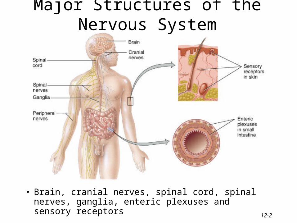
Major Structures of the Nervous System
• Brain, cranial nerves, spinal cord, spinal nerves, ganglia, enteric plexuses and sensory receptors

12-3
Nervous System Divisions
• Central nervous system (CNS) – consists of the brain and spinal cord
• Peripheral nervous system (PNS)– consists of cranial and spinal nerves that contain both
sensory and motor fibers– connects CNS to muscles, glands & all sensory
receptors

12-4
Subdivisions of the PNS• Somatic (voluntary) nervous system (SNS)
– neurons from cutaneous and special sensory receptors to the CNS
– motor neurons to skeletal muscle tissue
• Autonomic (involuntary) nervous systems– sensory neurons from visceral organs to CNS– motor neurons to smooth & cardiac muscle and glands
• sympathetic division (speeds up heart rate)
• parasympathetic division (slow down heart rate)
• Enteric nervous system (ENS)– involuntary sensory & motor neurons control GI tract– neurons function independently of ANS & CNS

12-5
Neurons• Functional unit of nervous system• Have capacity to produce action potentials
– electrical excitability
• Cell body– single nucleus with prominent nucleolus– Nissl bodies (chromatophilic substance)
• rough ER & free ribosomes for protein synthesis
– neurofilaments give cell shape and support– microtubules move material inside cell– lipofuscin pigment clumps (harmless aging)
• Cell processes = dendrites & axons

12-6
Nucleus with Nucleolus
Parts of a Neuron
Axons or Dendrites
Cell body
Neuroglial cells
12-7
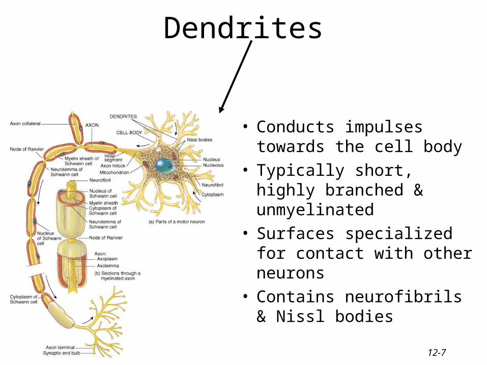
Dendrites
• Conducts impulses towards the cell body
• Typically short, highly branched & unmyelinated
• Surfaces specialized for contact with other neurons
• Contains neurofibrils & Nissl bodies
12-8
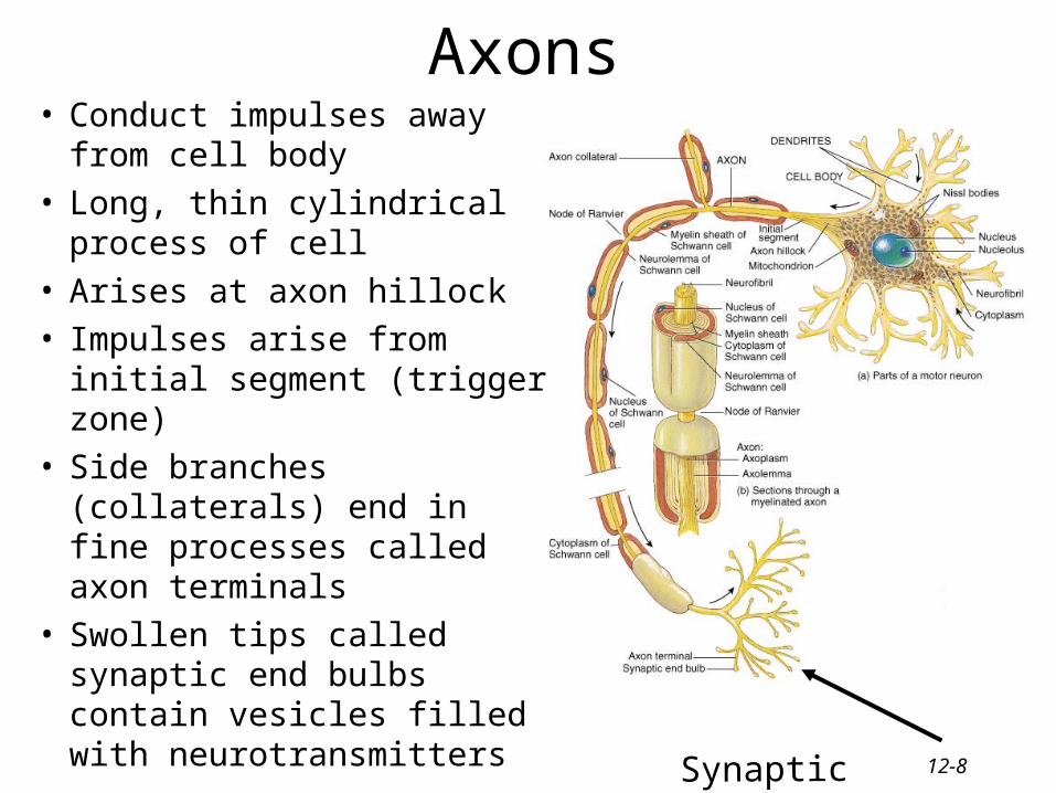
Axons• Conduct impulses away from
cell body• Long, thin cylindrical
process of cell • Arises at axon hillock • Impulses arise from initial
segment (trigger zone)• Side branches (collaterals)
end in fine processes called axon terminals
• Swollen tips called synaptic end bulbs contain vesicles filled with neurotransmitters Synaptic boutons
12-9
Axonal Transport
• Cell body is location for most protein synthesis– neurotransmitters & repair proteins
• Axonal transport system moves substances – slow axonal flow
• movement in one direction only -- away from cell body
• movement at 1-5 mm per day
– fast axonal flow• moves organelles & materials along surface of microtubules
• at 200-400 mm per day
• transports in either direction
• for use or for recycling in cell body

12-10
Axonal Transport & Disease
• Fast axonal transport route by which toxins or pathogens reach neuron cell bodies– tetanus (Clostridium tetani bacteria)– disrupts motor neurons causing painful muscle spasms
• Bacteria enter the body through a laceration or puncture injury– more serious if wound is in head or neck because of
shorter transit time

12-11
Functional Classification of Neurons
• Sensory (afferent) neurons– transport sensory information from skin,
muscles, joints, sense organs & viscera to CNS
• Motor (efferent) neurons– send motor nerve impulses to muscles & glands
• Interneurons (association) neurons– connect sensory to motor neurons– 90% of neurons in the body

12-12
Structural Classification of Neurons
• Based on number of processes found on cell body– multipolar = several dendrites & one axon
• most common cell type
– bipolar neurons = one main dendrite & one axon• found in retina, inner ear & olfactory
– unipolar neurons = one process only(develops from a bipolar)• are always sensory neurons
12-13
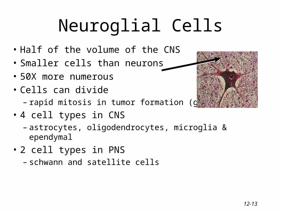
• Half of the volume of the CNS
• Smaller cells than neurons
• 50X more numerous
• Cells can divide – rapid mitosis in tumor formation (gliomas)
• 4 cell types in CNS– astrocytes, oligodendrocytes, microglia & ependymal
• 2 cell types in PNS– schwann and satellite cells
Neuroglial Cells

12-14
Astrocytes
• Star-shaped cells• Form blood-brain
barrier by covering blood capillaries
• Metabolize neurotransmitters
• Regulate K+ balance• Provide structural
support

12-15
Oligodendrocytes
• Most common glial cell type
• Each forms myelin sheath around more than one axons in CNS
• Analogous to Schwann cells of PNS
12-16
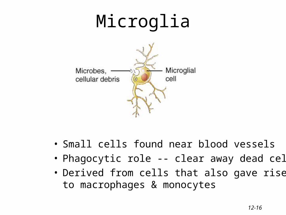
Microglia
• Small cells found near blood vessels• Phagocytic role -- clear away dead cells• Derived from cells that also gave rise to
macrophages & monocytes
12-17
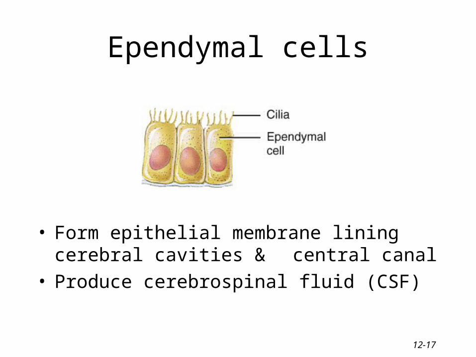
Ependymal cells
• Form epithelial membrane lining cerebral cavities & central canal
• Produce cerebrospinal fluid (CSF)

12-18
Satellite Cells
• Flat cells surrounding neuronal cell bodies in peripheral ganglia
• Support neurons in the PNS ganglia

12-19
Schwann Cell
• Cells encircling PNS axons• Each cell produces part of the myelin sheath
surrounding an axon in the PNS

12-20
Axon Coverings in PNS• All axons surrounded by a lipid & protein covering (myelin
sheath) produced by Schwann cells• Neurilemma is cytoplasm & nucleus
of Schwann cell– gaps called nodes of Ranvier
• Myelinated fibers appear white– jelly-roll like wrappings made of
lipoprotein = myelin
– acts as electrical insulator
– speeds conduction of nerve impulses
• Unmyelinated fibers– slow, small diameter fibers
– only surrounded by neurilemma but no myelin sheath wrapping

12-21
Myelination in PNS
• Schwann cells myelinate (wrap around) axons in the PNS during fetal development
• Schwann cell cytoplasm & nucleus forms outermost layer of neurolemma with inner portion being the myelin sheath
• Tube guides growing axons that are repairing themselves

12-22
Myelination in the CNS• Oligodendrocytes myelinate axons in the CNS • Broad, flat cell processes wrap about CNS axons,
but the cell bodies do not surround the axons• No neurilemma is formed• Little regrowth after injury is possible due to the
lack of a distinct tube or neurilemma

12-23
Gray and White Matter
• White matter = myelinated processes (white in color)• Gray matter = nerve cell bodies, dendrites, axon terminals,
bundles of unmyelinated axons and neuroglia (gray color)– In the spinal cord = gray matter forms an H-shaped inner core
surrounded by white matter
– In the brain = a thin outer shell of gray matter covers the surface & is found in clusters called nuclei inside the CNS