12| signal transductionclasses.biology.ucsd.edu/bibc100.fa16/documents/... · chapter 12 signal...
TRANSCRIPT

12| Signal Transduction
© 2013 W. H. Freeman and Company

CHAPTER 12 Signal Transduction
– General features of signal transduction – Structure and function of G‐protein coupled receptors– Structure and function of enzyme‐linked receptors– Structure and function of gated ion channels– Physiological processes using signal transduction
Key topics:

Biological Role of Signal Transduction• Cells receive signals from the environment beyond the plasma membrane– Antigens– Hormones– Neurotransmitters– Light– Touch– Pheromones
• These signals cause changes in the cell’s composition and function– Differentiation and antibody production– Growth in size or strength– Sexual vs. asexual cell division

Receptors Receptor: A membrane‐bound or soluble protein or protein complex, which exerts a physiological effect (intrinsic effect) after binding its natural ligand.
• G‐protein coupled receptors – Epinephrine receptor
• Enzyme‐linked receptors– Insulin receptor
• Ligand‐gated ion channels– Nicotinic acetylcholine receptor
• Other membrane receptors – Integrin receptors
• Nuclear receptors– Steroid receptors

Five Features of Signal‐Transducing Systems

Receptors Bind Specific Ligands
Typical ligands are:• Small ions
– ferric ion: bacterial ferric receptor• Organic molecules
– Adrenalin: epinephrine receptor• Polysaccharides
‒ Heparin: fibroblast growth factor• Peptides
– Insulin: insulin receptor• Proteins
– Vascular endothelial growth factor: VEGF receptor

Receptors Bind Specific Ligands

Receptor Binding Studies: Filter Assay• Rationale:
– Equilibrium binding of labeled ligand with the receptor– R + L RL– The bound complex becomes radioactive– Free receptor remains non‐radioactive – Free ligand can pass through the filter – Complex cannot pass because the protein binds to the filter
• Steps:– Isolate membranes– Add ligand to membranes– Pass through a filter– Wash off unbound ligand– Measure bound radioactivity, which is proportional to [COMPLEX]

Receptor Binding Studies: Nonspecific Binding
Problem: Hydrophobic ligands are nonspecifically soaked into the membrane.
Solution:• Measure total binding• Measure nonspecific binding (NSB) in the absence
of receptors• Subtract NSB from Total to get specific binding• Analyze specific binding data
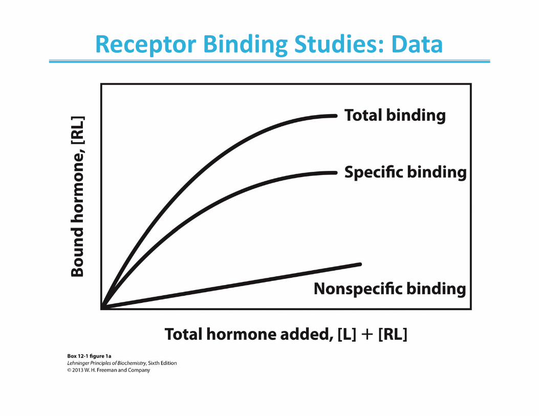
Receptor Binding Studies: Data

Receptor Binding Studies: Data Analysis
• Determine– Binding constant– Number of receptors– Number of sites– Cooperativity
]ligand Bound[1]ligand Free[]ligand Bound[ max
dd KKB

Receptor Binding Studies: Data Analysis

Signaling Through the Membrane

G‐Protein Coupled Signaling
• G‐Protein Coupled Receptors (GPCRs) are ‐helicalintegral membrane proteins
• G‐proteins are heterotrimeric () membrane‐associated proteins that bind GTP
• G‐proteins mediate signal transduction from GPCRs to other target proteins

Prototypical G‐protein: Ras

GPCRs: The Receptors

Epinephrine: The Fight or Flight Hormone
•Hormone made in adrenal glands (pair of organs on top of kidneys)
•Mediates stress response: mobilization of energy•Binding to receptors in muscle or liver cells induces breakdown of glycogen
•Binding to receptors in adipose cells induces lipid hydrolysis
•Binding to receptors in heart cells increases heart rate

Epinephrine and Analogs

Sensing the Epinephrine Signal via a G‐Protein Coupled Receptor

Synthesis of cAMP• cAMP is a secondary messenger
– Allosterically activates cAMP‐dependent protein kinase A (PKA)– PKA activation leads to activation of enzymes that produce glucose from glycogen

Signal Amplification in Epinephrine Cascade
• Activation of few GPCRs leads to the activation of few adenylyl cyclase enzymes
• Every active adenylyl cyclase enzyme makes several cAMP molecules, thus activating several PKA enzymes
• These activate thousands of glycogen‐degrading enzymes in the liver tissue
• At the end, tens of thousands of glucose molecules are released to the bloodstream

Signal Amplification

Self‐Inactivation in G‐protein Signaling
• Epinephrine is meant to be a short‐acting signal
• The organism must stop glucose synthesis if there is no more need to fight or flee
• Down‐regulation of cAMP occurs by the hydrolysis of GTP in the subunit of the G‐protein
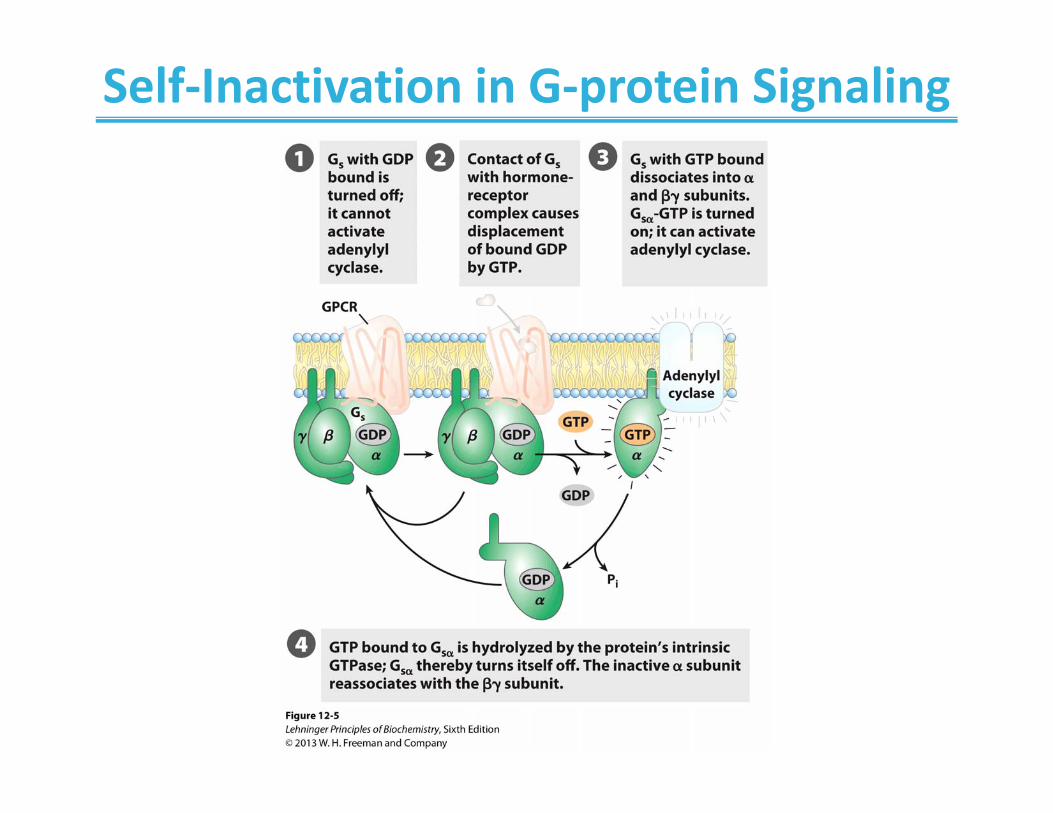
Self‐Inactivation in G‐protein Signaling
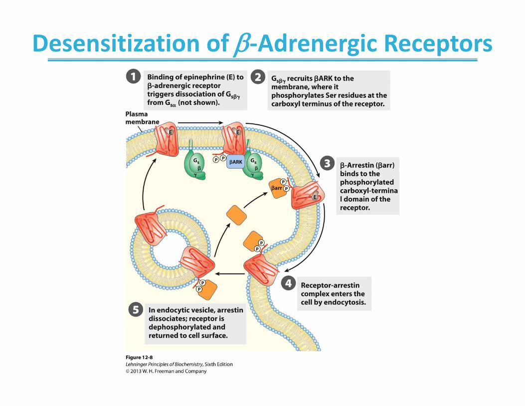
Desensitization of ‐Adrenergic Receptors
cAMP is a common secondary messenger
• A large number of GPCRs mediate their effects via cAMP– Both activating and inhibiting cAMP synthesis
• The human genome encodes about 1000 GPCRs– With ligands such as hormones, growth factors, and neurotransmitters
• There are also hundreds of different GPCRs that can be responsible for similar processes– Such as taste or smell
• Ligands for many GPCRs have yet to be identified


cAMP is able to mediate multiple signals due to localization of protein kinase A • PKA is localized to particular structures by anchoring protein• Different anchors are expressed in different cell types to
determine the downstream affect of cAMP

Some bacterial toxins are enzymes that inactivate G‐proteins
• Adenylate cyclase is now always (constitutively) active and produces too much cAMP from ATP
• Cholera toxin and pertussis toxin function this way

GPCRs can use other secondary messenger molecules
• i.e., Inositol‐1,4,5‐triphosphate (IP3) and/or Calcium
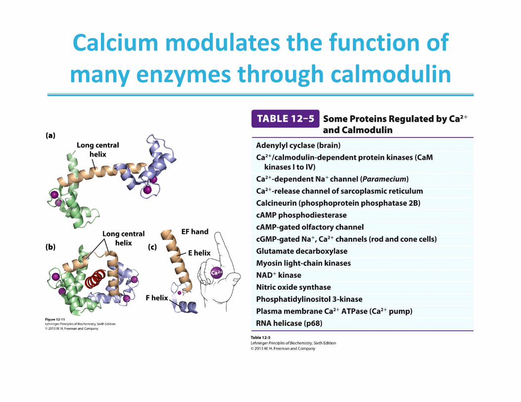
Calcium modulates the function of many enzymes through calmodulin

Enzyme‐linked Membrane Receptors• Many membrane receptors consists of:
– extracellular ligand‐binding domain, and of – intracellular catalytic domain
• The most common catalytic domains have the tyrosine kinase activity– Adds a phosphate group to itself; auto‐phosphorylationleads to a conformational change allowing binding and catalytic phosphorylation of specific target proteins
– Adds a phosphate group to a tyrosine in specific target proteins
• Some catalytic domains have guanylyl cyclase activity– Convert GTP to cGMP, a secondary messenger

Receptor Tyrosine Kinases

Insulin: The Hormone for Glucose Uptake and Metabolism
• Insulin is a peptide hormone that is produced by the ‐cells of islets of Langerhans in the pancreas
• Insulin is produced and released from the pancreas in response to nutrients such as glucose
• Insulin reaches target cells, such as liver, muscle, or fat tissue cells via bloodstream
• Binding of insulin to the insulin receptor initiates a cascade of events that leads to increased glucose uptake and metabolism
• Inability to make or sense insulin diabetes
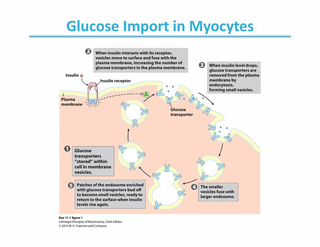
Glucose Import in Myocytes
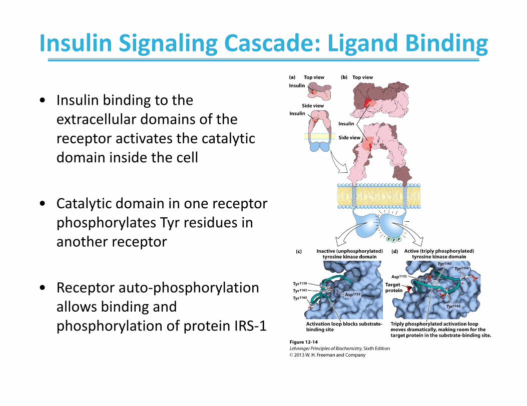
Insulin Signaling Cascade: Ligand Binding
• Insulin binding to the extracellular domains of the receptor activates the catalytic domain inside the cell
• Catalytic domain in one receptor phosphorylates Tyr residues in another receptor
• Receptor auto‐phosphorylation allows binding and phosphorylation of protein IRS‐1

• Indirect interaction of phosphorylated IRS with protein Ras initiates a series of protein phosphorylations
• ERK, one of the phosphorylated protein kinases, enters the nucleus
• A transcription factor Elk1 becomes phosphorylated and stimulates the expression of specific genes– glucose transporter (GLUT4)
Insulin Signaling Cascade

Insulin Signaling Cascade

Another Tyrosine Kinase
• JAK: a protein kinase• STATs: signal transducers and activators of transcription
The JAK‐STAT signaling system

Crosstalk between a Tyrosine Kinase Receptor and a GPCR

Receptor Guanylyl Cyclases• Catalytic domain converts GTP to cGMP• Works through activation of protein kinase G

SH2‐domains bind proteins with phosphotyrosine residue

Modular Structure of Signaling Proteins

Regulation of signaling by a scaffold protein

Gated Ion Channels
• Regulate transport of ions across cell membranes • Responds to:
– Changes in the membrane potential– Ligand binding to specific receptor sites
• Many important roles in the nervous system– Voltage‐gated sodium channels– Nicotinic acetylcholine receptor– Ionotropic glutamate receptor– Gamma aminobutyric acid receptor A

Membranes are electrically polarized• The inside of the cell is typically negatively charged compared to the outside: Vm –50 to –70 mV
• The membrane potential is largely due to electrogenic Na+K+ ATPase– 3 Na+ out– 2 K+ in
• Flow of ionic species across the membrane depends on its concentration gradient and overall electrical potential

Membranes are electrically polarized

Voltage‐Gated and Ligand‐Gated Ion Channels in Nerve Signaling
• Nerve signals within nerves propagate as electrical impulses
• Propagation of the impulse involves opening of voltage gated Na+‐channels
• Opening of voltage‐gated Ca++ channels at the end of the axon triggers the release of neurotransmitter acetylcholine
• Acetylcholine opens the ligand‐gated ion channel on the receiving cell

Ion Channels in Nerve Signaling

Voltage‐Gated Sodium Channel

Voltage‐Gated Sodium Channel

The Acetylcholine Receptor• Nicotinic acetylcholine receptor:
– Ion channel for influx of Na+, Ca2+– Gate opened by acetylcholine

The Acetylcholine Receptor

Integrins mediate cell adhesion
• Extracellular domain interacts with Arg‐Gly‐Asp–containing proteins:– Collagen, fibrinogen, fibronectin, and others
• This triggers cytoskeleton rearrangement and gene expression
• Newly expressed genes bind to intracellular domain triggering extracellular response

Integrins mediate cell adhesion

Direct Regulation of Transcription by Hormones

Bacterial chemotaxis is controlled by enzyme‐coupled receptors


Plants and animals use structurally similar signaling molecules

Plants and animals use similar signal transduction pathways

Sensory perception is mediated by GPCRs

Generation of a nerve impulse in response to light
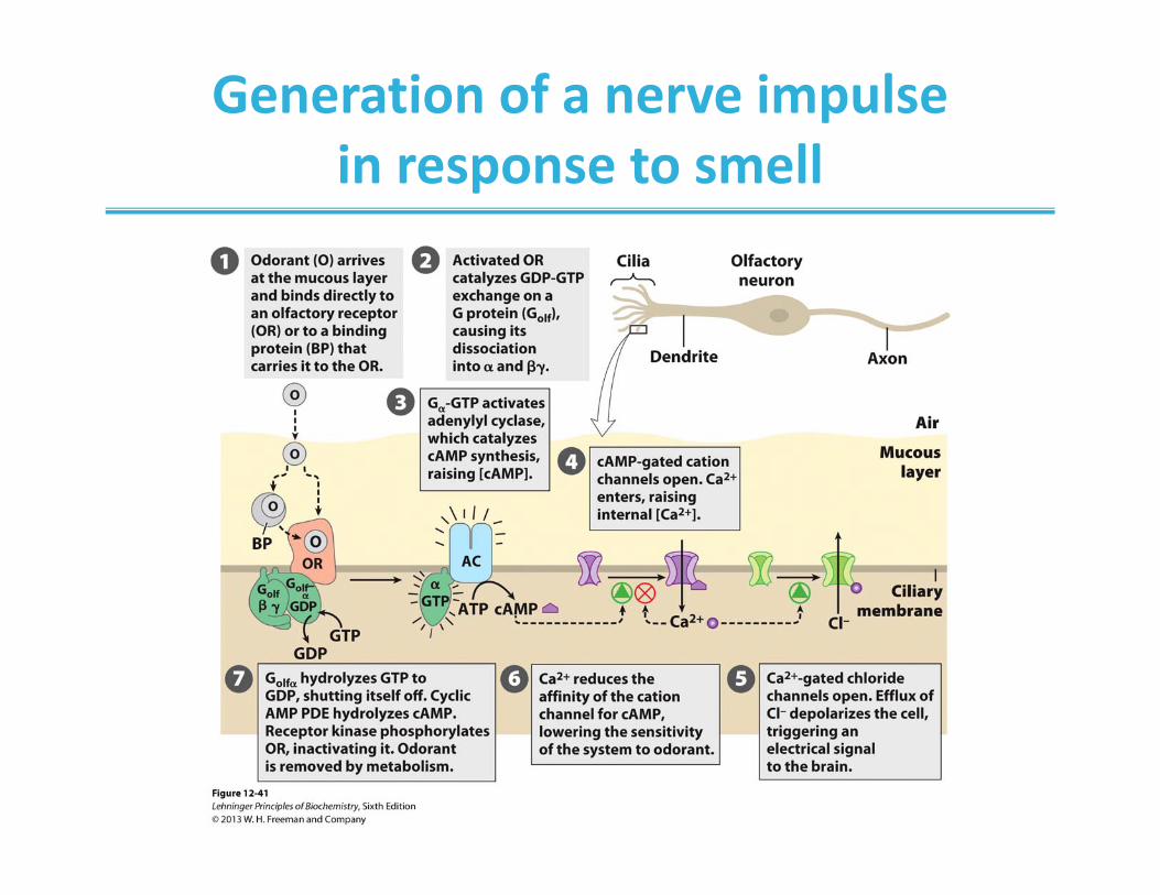
Generation of a nerve impulse in response to smell

Generation of a nerve impulse in response to taste

Cell cycle is regulated intracellularly by cyclin‐dependent protein kinases
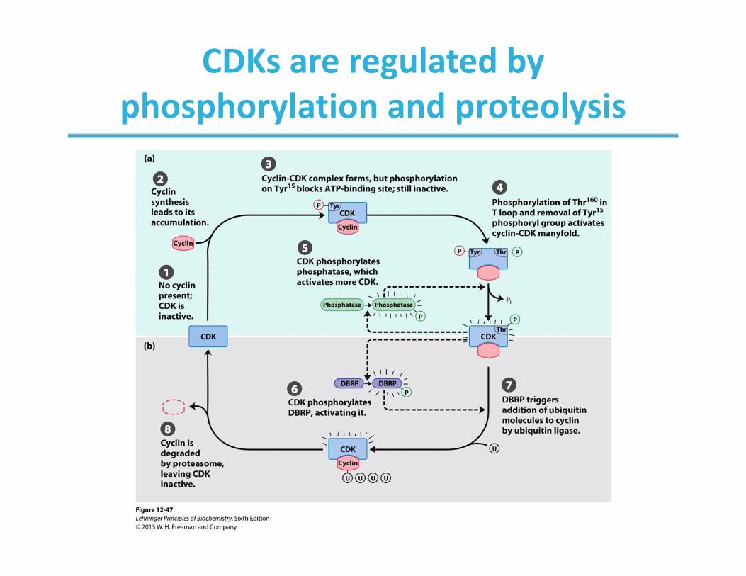
CDKs are regulated by phosphorylation and proteolysis

Growth factors trigger transcriptional regulation of CDKs

Constitutive activation of CDKs can lead to cancer
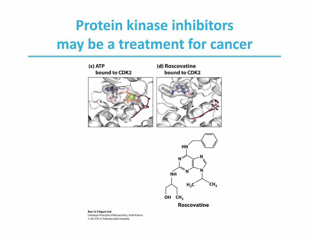
Protein kinase inhibitors may be a treatment for cancer

Damage to macromolecules can trigger programmed cell death (apoptosis)

Chapter 12: Summary
• Cell signaling is triggered by receptors that sense the extracellular environment– Binding tightly to specific messenger molecules
• GPCRs bind GTP and activate interacting proteins• Receptor tyrosine kinases activate protein kinases with auto‐
phosphorylation • Receptor guanylyl cyclases generate the secondary messenger
cGMP• Voltage‐gated ion channels generate and propagate nerve
impulses • Vision, smell, and taste are sensed by GPCRs• Disregulation of intracellular signaling cascades can lead to cancer
In this chapter, we learned: