17 11 sun - association for biology laboratory education · are oxidase and catalase positive, ......
TRANSCRIPT

Association for Biology Laboratory Education (ABLE) ~ http://www.zoo.utoronto.ca/able 167
Chapter 11
Identification of Microorganisms Encountered in the Upper Respiratory Tract
Iris L. Sun
Department of Biology Purdue University
West Lafayette, Indiana 47907-1392 (317) 494-4984, FAX (317) 494-0876
Iris received her Ph.D. in Cell Biology from the Department of Biological Sciences, Purdue University. She then stayed in the department as a research associate working on the action mechanism of anti-cancer drugs and studying the effect of cytokines on transformed cells. Besides doing research, she also teaches Lab of Medical Microbiology and Introduction of Microbiology in the department.
© 1996 Purdue University

Sun, I. L. 1996. Identification of microorganisms encountered in the upper respiratory tract. Pages 167-184, in Tested studies for laboratory teaching, Volume 17 (J. C. Glase, Editor). Proceedings of the 17th Workshop/Conference of the Association for Biology Laboratory Education (ABLE), 255 pages.
• Copyright policy: http://www.zoo.utoronto.ca/able/volumes/copyright.htm
Although the laboratory exercises in ABLE proceedings volumes have been tested and due consideration has been given to safety, individuals performing these exercises must assume all responsibility for risk. The Association for Biology Laboratory Education (ABLE) disclaims any liability with regards to safety in connection with the use of the exercises in its proceedings volumes.

168 Microorganisms in URT Contents
Background Information.............................................................................168 Materials .....................................................................................................174 Note for the Instructor ................................................................................176 Student Outline ...........................................................................................176 Literature Cited...........................................................................................183 Appendix A:
A Flow Chart of Expected Results for Staphylococcal and Streptococcal Species ......................................................................184
Background Information
Differentiation of Potential Pathogens from the Upper Respiratory Tract
A. Gram Positive Aerobic Cocci: Staphylococci
Staphylococci occur typically as grape-like clusters because cell division occurs in three planes and daughter cells tend to remain attached. They grow well on basic or enriched media at temperatures ranging from 25°C to 35°C forming opaque, smooth, and glistening colonies. On sheep blood agar, colonies of S. aureus appear gold or bronze.
Staphylococci produce the enzyme catalase which decomposes hydrogen peroxide (H2O2) into oxygen (O2) and water (H2O). They are most resistant to extreme conditions, and can survive temperatures of 60°C for 1 hour or be carried on dust particles and live for months on dried pus or sputum. Another mechanism for their survival is a tolerance to high salt concentration, such as salty food. Staphylococci are resistant to phenol, sulfonamides and many antibiotics, but are very sensitive to basic dyes. They produce a high number of toxins which account for the variety of diseases, in addition to infections of the respiratory tract.
Hemolysins are hemolytic exotoxins acting on the cell membrane and lysing several kinds of blood and tissue cells.
Leucocydins are non hemolytic exotoxins which lyse lymphocytes, neutrophils and macrophages.
Enterotoxin is produced by about 50% of coagulase positive strains of Staphylococci. It is a common cause of food poisoning.
Exfoliatin damages skin cells resulting in peeling of large sheets of lining cells. It affects primarily young infants causing the scalded skin syndrome.
Coagulase causes the blood plasma to clot. It is only present in virulent strains and its property is the base for the coagulase test.
Lipase breaks down lipids in skin structure and lipoproteins in the blood. Staphylococci utilizes the metabolites derived from lipid breakdown, and this may explain the high number of Staphylococci on the skin surface.
Hyaluronidase increases tissue permeability to Staphylococci. Staphylokinase dissolves clots and contributes to the spread of local infections.
Pathogenicity:
The abscess is the type lesion caused by Staphylococci, a consequence of its pus-forming ability and limited capacity for spread.

Microorganisms in URT 169 S. aureus is the most common pathogen while S. epidermidis is not pathogenic but may cause
infections in some circumstances. Since both are inhabitants of the skin, most lesions are superficial and develop as boils, pustules, pimples, furuncles, carbuncles, or impetigo contagiosa. Staphylococcal pneumonia results from multiple abscesses in the lungs. The invasion of the body by Staphylococci (septicemia) results from secondary invasion of the bloodstream by organisms from a localized skin infection.
Bacteriological Diagnosis:
Gram-stained smears and cultures on blood agar are the first step toward Staphylococcal identification. The enzyme catalase distinguishes Staphylococci (catalase +) from Streptococci (catalase –). The Coagulase test distinguishes pathogenic staphylococci (coagulase +) from nonpathogenic (coagulase –). Virulent Staphylococci produce yellow pigment visible in cultures, ferment the sugar mannitol and produce deoxyribonucleases (DNAses).
Phage typing: Bacteriophages are viruses that specifically attach to bacteria, invade them and cause their lysis. When an agar plate is inoculated with bacteria from a pure culture and subsequently covered with a specific phage suspension, the organisms susceptible to the phage will be lysed and cause agar areas of no growth known as plaques.
S. aureus of human origin have been “typed” using this technique and classified into types of increasing pathogenicity. Bacteriophages are given identification numbers which also designate strains of Staphylococci related to them.
B. Gram Positive Facultative Anaerobic Cocci: Streptococci
Streptococci are facultative anaerobic saprophytes and parasites that normally occur on a wide variety of body surfaces. Because of their ability to ferment lactose, the Streptococci are also important agents of milk spoilage. Streptococci may be distinguished from Staphylococci as follows: (1) cocci are arranged in short chains or in pairs rather than as irregular clusters; (2) colonies exposed to the air are small, drop-like, mucoid, and unpigmented; (3) catalase is not produced; (4) NaCl concentration exceeding 6.5% is not tolerated; and (5) growth is not inhibited by sodium azide (Streptococci lack cytochromes).
With regard to differentiation of strains of Streptococci:
Pyogenic Streptococci such as S. pyogenes are ß-hemolytic. ß-Hemolytic Streptococci may become virulent pathogens under certain conditions, digesting deep tissues and destroying phagocytes. Some strains synthesize a hyaluronic acid which is identical to that in human connective tissue. The bacteria, therefore, are not coated with antibodies and are able to evade phagocytes. Most of the clinically important ß-hemolytic Streptococci are in serologic Group A. Group A strains are uniformly sensitive to low concentrations (0.02 units) of bacitracin. Only a small percentage of other serologic groups are susceptible. In practice, a disc impregnated with the drug is placed on a lawn of bacteria. Group A Streptococci fail to grow around the disc where bacitracin has diffused.
Hemolytic Streptococci are typed according to two immunologically active components of the cell wall: the “M” protein and an extractable glucan, N-acetylglucosamine (the “Lancefield carbohydrate”). The Lancefield scheme (Table. 4) is most widely used. Groups A, C, and G are implicated in a wide variety of infections. Group B Streptococci may be carried asymptomatically by adults, but they can cause meningitis and other infections in the newborn. They may also be pathogenic in diabetic women, often as part of a syndrome with thromboembolism and necrosis of

170 Microorganisms in URT the skin. Identification of Group A Streptococci can be confirmed by assaying serum for one of several streptococcal enzymes: streptolysin O, streptococcal hyaluronase, or streptococcal DNAse.
Viridans Streptococci are hemolytic. They cause partial hemolysis of red blood cells resulting in greening of colony surroundings. Some strains produce large polysaccharide capsules that probably have significance in pathogenesis. The capsules of S. mutans result in deposits around teeth (dental plaque) that enhance tooth decay. The capsules of S. pneumoniae (sometimes called diplococci) help it adhere to the epithelium of the lung. Because of the role of S. pneumoniae in lobar pneumonia, middle-ear infections, meningitis, and other infections, its diagnostic characteristics are clinically important. Pneumococci may form extremely mucoid colonies, are lysed by bile salts or by sodium desoxycholate, and are inhibited by the antibiotic optochin (ethylhydrocupreine hydrochloride). Biotypes of pneumococci are distinguished serologically. The polysaccharides making up the capsule have specific antigenic properties, and reaction with homologous antisera results in capsular swelling (Quellung reaction).
C. Gram-positive rods: Coryneform Bacteria
Typical Corynebacteria are small, Gram positive, club shaped rods arranged in V-shaped pairs, “Chinese letters”, or palisade arrangements. They are all nonmotile and aerobic. The distinctive arrangements of Corynebacteria may be attributed to the fact that the cell walls do not completely separate and the cells remain attached to each other at an angle. Other distinctive characteristics of Corynebacteria are bluish-purple metachromatic granules that are visible after staining cells with methylene blue, and the formation of black colonies on blood agar containing potassium tellurite. The granules are negatively charged particles of polyphosphate which absorb the methylene blue cations more strongly than other components of the cell. They are more visible in Corynebacterium diphtheriae than other Corynebacteria. The potentially pathogenic C. diphtheriae can be distinguished from nonpathogenic species on the basis of fermentation reactions: C. diphtheriae ferments maltose whereas other species do not. Virulence is generally verified by in vitro tests for toxin production such as the Elek virulence test. For this test, a strip or filter paper is soaked in diphtheria antitoxin then is embedded in an agar plate. Once the agar has hardened, the organism is streaked perpendicular to the filter paper. A positive test is indicated by a zone of precipitation.
D. Gram-Negative Cocci: The Family Neisseriaceae
The Neisseriaceae include four parasitic genera: Neisseria, Branhamella, Moraxella and Acinetobacter. All are Gram negative cocci or coccobacilli, nonspore forming, catalase positive and nonmotile. Members of the genus Neisseria are kidney shaped diplococci with flat adjacent sides. Aerobic or facultatively anaerobic, they are oxidase positive.
The Gram stain shows typical extra- or intracellular diplococci. In some instances, they stain very deep pink and in clinical specimens are present with a high number of polymorphonuclear cells. Neisseria meningitidis grows well on enriched media such as chocolate agar at 37°C, with 6% CO2 and 50% humidity. After 18 hours of incubation on chocolate agar, colonies of N. meningitidis are round, grey and convex with a smooth moist glistening surface and an entire edge. The medium around the colonies may exhibit a greenish appearance.
The genus Branhamella contains one species important for human: Branhamella catarrhalis. Gram stain and colonies are identical to N. meningitidis. B. catarrhalis can be differentiated from N. meningitidis in the following way: B. catarrhalis grows at room temperature on blood or chocolate agar while N. meningitidis does not. In addition, B. catarrhalis does not ferment glucose, maltose, lactose, sucrose nor fructose while N. meningitidis ferments glucose and maltose.
Microorganisms in URT 171 E. Small Gram Negative Bacilli: The Genus Haemophilus
Bacterial species of genus Haemophilus require special growth factors that are found only in blood and other blood fluid. The name Haemophilus means “blood-loving” (Greek, haemo = blood; philus = loving). Haemophilus influenzae is the major disease-producing species of this genus. Bergey’s classification of bacteria lists Haemophilus among the facultative anaerobic Gram negative rods. Haemophilus are nonmotile, non-sporeforming bacteria surrounded by a capsule. Most of them are oxidase and catalase positive, ferment glucose and require additional factors such as Hemin (called Factor X) and/or NAD (called Factor V) to grow and develop colonies on trypticase soy agar. They grow well on chocolate agar since it contains both factors.
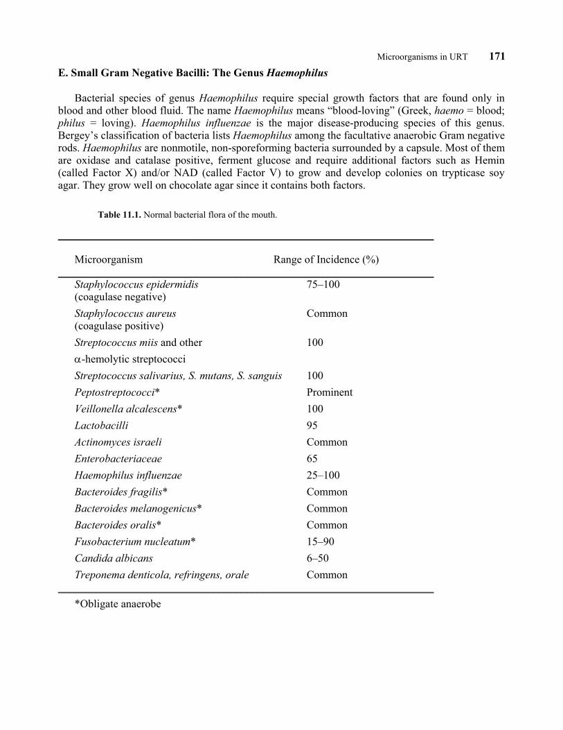
Table 11.1. Normal bacterial flora of the mouth.
____________________________________________________________________ Microorganism Range of Incidence (%) ____________________________________________________________________ Staphylococcus epidermidis 75–100 (coagulase negative) Staphylococcus aureus Common (coagulase positive) Streptococcus miis and other 100 α-hemolytic streptococci Streptococcus salivarius, S. mutans, S. sanguis 100 Peptostreptococci* Prominent Veillonella alcalescens* 100 Lactobacilli 95 Actinomyces israeli Common Enterobacteriaceae 65 Haemophilus influenzae 25–100 Bacteroides fragilis* Common Bacteroides melanogenicus* Common Bacteroides oralis* Common Fusobacterium nucleatum* 15–90 Candida albicans 6–50 Treponema denticola, refringens, orale Common ____________________________________________________________________ *Obligate anaerobe

172 Microorganisms in URT
Table 11.2. Normal bacterial flora of the nasopharynx.
________________________________________________________________________ Microorganism Range of Incidence (%) ________________________________________________________________________ Staphylococcus aureus 20–85 Staphylococcus epidermidis 90 Aerobic Corynebacteria (diphtheroids) 5–80 Streptococcus pneumoniae 0–17 Streptococcus pyogenes (Group A) 0.1–5 Branhamella catarrhalis 12 Haemophilus influenzae 12 Gram negative bacteria (Enterobacteriaceae) Rare ________________________________________________________________________
Table 11.3. Normal bacterial flora of the oropharynx (throat).
________________________________________________________________________ Microorganism Range of Incidence (%) ________________________________________________________________________ Staphylococcus aureus 35–40 Staphylococcus epidermidis 30–70 Aerobic corynebacteria (diphtheroids) 50–90 Streptococcus pyogenes (Group A) 0–9 Streptococcus pneumoniae 0–50 α- and nonhemolytic Streptococci 25–99 Branhamella catarrhalis 10–97 Neisseria meningitidis 0–15 Haemophilus influenzae 5–20 Haemophilus parainfluenzae 20–35 _________________________________________________________________________

Microorganisms in URT 173 Table 11.4. Lancefield groupings of streptococci.
______________________________________________________________________________ Group Species Hemolysis Significance ______________________________________________________________________________ A Streptococcus pyogenes β Important human diseases; group sensitive to penicillin B Streptococccus agalactiae β (?) Bovine mastitis C Streptococcus equi β Animal diseases; mild human Streptococcus zooepidemicus β Respiratory infections Streptococcus equisimilis β Streptococcus dysgalactiae – D Streptococcus faecalis Enterococci; genitourinary tract Streptococcus faecalis subsp. liquefaciens infections, endocarditis, human Streptococcus faecalis subsp. zymogenes wound infections, Streptococcus faecium found in dairy products E Disease of swine, found in normal milk F Streptococcus minutus Found in human respiratory tract G Streptococcus anginosus Mild respiratory infections in human; genital infections in dogs H Streptococcus sanguis α Found in human respiratory tract K Streptococcus salivarius α Found in human respiratory tract L Genital tract infections in dogs M Genital tract infections in dogs N Streptococcus lactis Lactic group; found in dairy Streptococcus cremoris products α Viridans group; subacute bacterial endocarditis; found in human respiratory tract; Microaerophilic streptococci; Anaerobic streptococci–13 species
_____________________________________________________________________________

174 Microorganisms in URT Materials
A. BSS The formula for the 10 ∞ stock of phosphate balanced salts solution (BSS) is: K2HPO4 0.40 g KH2PO4 0.10 g KC1 8.0 g Distilled H2O 100 ml Filter sterilize and store 4°C – 8°C B. Bile Esculin Agar 1,000 ml H20 64 g bile esculin agar C. Blood & Chocolate Blood Agar 1,000 ml H20 40 g T.S.A (Trypticase Soy Agar, see below). Pour 570 ml of media into 1 liter flask with stir bar. Autoclave it then add: –For chocolate blood: Add 30 ml of sheep blood into the media right after autoclaving. –For Blood: Add 30 ml of sheep blood into the media when the media cool down to 50°C. D. C.T.A. (cystine tryptic agar) with glucose, lactose, maltose or sucrose 1,000 ml H20 29.5 g C.T.A. To each 1,000 ml of media, add: –For glucose: 10 g glucose/liter –For lactose: 10 g lactose/liter –For maltose: 5 g maltose/liter –For sucrose: 10 g sucrose/liter E. Coagulase Plasma EDTA Difco Laboratories P.O. Box 331058 Detroit, Michigan 48232 1-800-521-0851 F. DNAse Test Agar 1,000 ml H2O 42 g DNAse test agar G. Gelatin Agar 1,000 ml distilled H20 8 g nutrient broth 30 g gelatin 15 g agar

Microorganisms in URT 175 H. ID2 Triplate and ID QUAD Plate REMEL P.O. Box 14428 12076 Santa Fe Drive Lenexa, Kansas 66215 1-800-255-6730 I. Mannitol Salt Agar 1,000 ml H2O 111 g mannitol salt agar J. Nutrient Agar 1,000 ml H2O 31 g nutrient agar or: 8 g nutrient broth 15 g agar K. 6.5% NaCL Broth 1,000 ml H2O 65 g NaCl (sodium chloride) 25 g heart infusion broth 3 g glucose 5 ml bromocresol purple indicator (0.2 g bromocresol purple + 12.5 ml ETOH) L. Trypticase Soy Agar (T.S.A.) 1,000 ml H2O 30 g trypticase soy broth 15 g agar M. Tellurite Chocolate Blood Agar 1,000 ml H2O 20 g proteose peptone #3 2 g dextrose 5 g NaCl 13 g agar Pour 540 ml of media into 1 liter flask, autoclave it, cool down to 50°C., then add: 30 ml of sheep blood 30 ml tellurite solution (1%)

176 Microorganisms in URT Note for the Instructor
This experiment is mainly for students to learn the procedures of identifying each type of bacteria encountered in the upper respiratory tract. Due to the serious pathogenicity of some of the bacteria, such as N. meningitidis, S. aureus and C. diphtheriae, it is suggested to give students unknown species with no or minimal pathogenicity, such as S. epidermidis, S. agalactiae, N. lactamica and H. parainfluenzae. Students need to go through all the procedures in order to identify their unknown species. You have not given them pathogenic species, but they have learned the procedures for the identification.
Student Outline
Objective
The objective of this exercise is to become familiar with the diversity of the microorganisms residing in the mouth, nose and throat. You will attempt to identify potential pathogens by comparing them with known cultures, and performing several tests to determine various physical and physiological properties of the organisms. These will allow you to identify most of the genera encountered. You will also be introduced to simple, presumptive procedures for determining the presence of Corynebacteria and Neisseriae.
1. Observation of known organisms
A. Observe on the microscope, under 400∞ magnification as well as oil-immersion (1,000∞), stained slides of Staphylococci, Streptococci, Branhamella, Neisseria, and Haemophilus. For each of them, record in your notebook, all characteristics which would allow you to recognize them and differentiate them from each other, i.e., their color, shape, arrangement, presence of granules, etc. Rinse slides thoroughly with xylene to remove immersion oil.
B. Observe each known culture under direct illumination and under the dissecting scope, and record their characteristics in your notebook. The objective of this part is to become familiar with the appearance and characteristics of several important groups of bacteria you will later encounter in unknown cultures.
2. Differentiation of major groups of bacteria based upon the observation of Gram stained organisms and the performance of biochemical tests
To become familiar with the use of simple biochemical procedures, I suggest that you perform each test using first a known species, then repeating it using your unknown and known organisms as positive or negative controls. The unknown may be either your URT isolate or one provided to you. These exercises will allow you to verify that manipulations are performed correctly and that they give the expected result.
A. Gram Staining Choose two known and two unknown pure cultures. On each plate, look for a well isolated
colony and locate it by circling and numbering the bottom of the plate around it. Gram stain a loopful of organisms, and observe them under oil immersion.

Microorganisms in URT 177 Procedure:
a. Cover a heat-fixed smear with crystal violet for one minute. b. Briefly wash off the excess stain with water from the squeeze bottle after the one minute
above. c. Cover with Gram’s iodine for one minute. d. Wash very briefly with water. e. Hold the slide at a 45°angle. Gently pour alcohol over the slide and let it run off the slide just
until it runs colorless. This is the critical step! Do not use the disinfecting alcohol for this. f. Quickly wash with water again for a brief period. g. Counter stain with safranin for one minute. h. Wash briefly. i. Carefully blot dry with paper towels. Be very careful not to press down on top of the
bacteria. Simply let the paper towel absorb the excess water. j. View the slide at 1000∞ magnification again.
B. Biochemical Testing
a. Differentiation of Staphylococci from other gram positive cocci, namely Streptococci, by means of the catalase test
Principle: Catalase is an enzyme that decomposes hydrogen peroxide (H2O2) into oxygen (O2) and water (H2O), bubbling out the oxygen. Excluding the Streptococci, most aerobic and facultative anaerobic bacteria possess catalase activity.
Procedures: Put a loopful of bacteria on a slide. Add a few drops of 3% hydrogen peroxide to the bacterial sample. The presence of catalase is shown by effervescence (a “fizzing” or bubbling of the liquid).
b. Differentiation of S.aureus from S.epidermidis
Further differentiation between two common staphylococcal species can be achieved by inoculating them into rabbit plasma, streaking on mannitol salt agar, gelatin slant and DNA slant. The results are shown in the following table.
Table 11.5. Differentiation of S. aureus from S. epidermidis.
________________________________________________________ Test S. aureus S. epidermidis _________________________________________________________ Coagulase + – Acid from mannitol + – Liquefies gelatin + – DNAse positive + –
_________________________________________________________

178 Microorganisms in URT Gelatin Liquefaction. In order to determine whether an organism produces gelatinase, a gelatin stab can be inoculated with the organism. A gelatin stab contains nutrient broth with 10–20% gelatin added. This amount of gelatin causes the material to “gel” or become semi-solid at temperatures below 25°C. The bacterial culture to be tested is “stabbed” into the nutrient gelatin tube with an inoculating needle. If the organism produces gelatinase, then the enzyme hydrolyzes the gelatin, and the gelatin subsequently loses its ability to gel. The DNAse Test. DNA test agar contains DNA which can be precipitated by 1N HCL. Where an organism has degraded the DNA, no precipitation will occur. The result is a clear halo around the colonies on the agar. Other formulations use indicator dyes such as methyl green or toluidine blue in the medium. Methyl green is green when complexed with DNA and colorless when the DNA has been hydrolyzed. Toluidine blue is blue with the DNA and pink when it is hydrolyzed. The mannitol salt agar can differentiate between mannitol fermenters and non-fermenters. The fermenter produces acids and changes the PH indicator, phenol red, from red to yellow. The non-fermenter does not change the red color of the agar plate. The Coagulase Test: Principle: Coagulase is an enzyme converting fibrinogen into fibrin and resulting in the formation of a visible clot. The test can be performed either on a slide with organisms taken from an agar colony, or in a tube with cells from a broth culture. Procedure: Tube test: Aseptically add 0.5 ml of rabbit plasma in a small test tube by means of a Pasteur pipette. With a 1 ml pipet, add 0.5 ml of a broth culture of the organisms. Mix gently (no vortex!) and place in a 37°C incubator for at least 4 hours. If positive, a clot will be present in the bottom. Never forget your controls, especially when working with an unknown, perform the test simultaneously with S. aureus (positive control) and S. epidermidis (negative control).
c. Subsequent tests and observations to differentiate among Streptococci
The pathogenic Streptococci grow readily in the laboratory, but the best development requires blood or serum media and body temperature, namely 37°C. Their colonies on blood agar reveal several types of hemolysis around them when examined by reflected light. α-hemolytic Streptococci leave a zone of partially lysed RBC (red blood cells) around the colony, surrounding it by a darker halo (brown or greenish). ß-hemolytic Streptococci lyse all RBC around and are thus surrounded by a clear yellowish or transparent halo-hemolysis, represents the absence of hemolysis around the colony. To distinguish between those different types of hemolysis, look at the plates either under the dissecting microscope where you may be able to see RBC in blood agar although it may be difficult to focus, or simply through the light microscope. Compare the tint of the halo: S. pneumoniae is α-hemolytic, S. pyogenes, ß-hemolytic. Compare unknowns with control plates.
d. Differentiation of Streptococcus pneumoniae by means of the bile solubility test
Principle: Bile salts like sodium deoxycholate have the capability to selectively lyse Streptococcus pneumoniae when added to actively growing bacterial cells in a culture medium. The bile solubility test can be performed either with a broth culture (pH = 7.0) or with colonies on agar. Only the latter test will be explained here. Procedure: To a well isolated colony of the test organism on blood agar, add a drop of 5% sodium deoxycholate and place the plate at 37°C for half an hour. Bile soluble colonies of

Microorganisms in URT 179 S. pneumoniae disappear leaving a hemolyzed area. Negative controls, which would be any other α-hemolytic Streptococci, will remain intact.
e. Differentiation of group A Streptococci from other serological types
All ß-hemolytic Streptococci can be divided into 18 or more serological groups, (Table 11.4) called Lancefield groups (after their discoverer Rebecca Lancefield) on the basis of carbohydrates present in the cell wall. Most pathogenic ß-hemolytic Streptococci belong to group A, among them S. pyogenes. Group B are for the most part harmless to humans, Group C and G are less pathogenic. To distinguish Group A Streptococci from the other groups, antibiotic susceptibility tests are used. The procedure is to pick up cells from a colony we wish to identify and streak the entire surface of a plate to create a lawn of colonies. A disc impregnated with a particular antibiotic is then placed on the agar and the culture incubated for 24 hours at 37°C in the CO2 incubator. The antibiotic contained in the paper disc will diffuse into the medium and if the bacteria are sensitive to it, a zone of no growth will appear around the disc. Group A Streptococci are sensitive to Bacitracin (labeled commercially here as Taxo-A discs), Streptococcus pneumoniae (which is α-hemolytic) is sensitive to Optochin (Taxo-P-discs). Streak a few plates with unknown organisms and with known controls. Place the two discs at a reasonable distance from each other. Press the disc gently on agar with a flamed forceps or a needle. Label your plates, incubate them at 37°C and check them tomorrow, or the day after. Remember to keep a record of your manipulations. Another way to differentiate S. pneumoniae is the bile solubility test explained before.
f. Identification of group B Streptococci by means of the CAMP test
A test allowing identification group B ß-hemolytic Streptococci is called the CAMP test (an acronym of its four discoverers). Principle: Group B Streptococci secrete a factor, the CAMP factor, which enhances the hemolytic activity of Staphylococcus aureus ß-lysin on erythrocytes. Wherever the two reactants overlap on a sheep blood agar plate, accentuation of the ß-hemolytic reaction occurs. Procedure: On a blood agar plate, streak with the flamed needle a single line of Staphylococcus aureus. Flame the needle. Then pick up your unknown Streptococci and streak similarly a single line perpendicular to the previous one and with 5 mm distant from each other. The two lines must not touch each other. The plates are incubated at 37°C in CO2 incubator for 24 hours. A positive test will show a triangular zone of enhanced clearing in the zone of closest proximity between the two streaks. Streptococcus agalactiae is the positive control.
g. Differentiation of group D Streptococci by means of Bile-esculin test
Principle: Bile-esculin (BE) test (for the identification of Group D Streptococci): Group D Bile-esculin medium Esculetin + Glucose Streptococci Esculetin + Ferric Citrate Black complex 174 (positive)
Procedure: Stab the bacteria into the bile-esculin slant with the flamed loop, then streak a zig-

180 Microorganisms in URT zag line on the slant surface. The group D Streptococci will turn the slant into dark black. A positive BE test tells you that you have a group D Streptococcus; differentiation of the two types of group D streptococci depends on the salt-tolerance test.
3. Gram negative cocci: Neisseria and Branhamella
The upper respiratory tract may harbor various species of Neisseria which being morphologically similar, are distinguished by carbohydrate utilization. Neisseria gonorrhoeae normally parasitizes the genito-urinary tract but may be recovered from the URT in rare occasions. Neisseria lactamitca is a commensal. Both are to be distinguished from Neisseria meningitidis and Branhamella catarrhalis. All species develop small, opaque, gray-white colonies with a glistening surface after 24 to 48 hour incubation on chocolate agar in a moist atmosphere at 35–37°C in 5% CO2.
Exercise Procedure:
a. Observe known colonies of Neisseria sp. and Branhamella in cultures and on Gram stained slides.
b. Take some unknowns from chocolate agar and try to identify possible Neisseria by observing colony morphology and performing a Gram stain.
c. After having recognized at least two Neisseria sp. try to identify the species using the Brown test or CTA media for carbohydrate utilization.
Brown Test Procedure for Carbohydrate Utilization
For each species to be identified, take 5 tubes with 0.1 ml balanced salt solution with 1% phenol red (BSS tubes), four solutions of carbohydrates namely: glucose, maltose, sucrose and lactose and pure colonies of unknown and known controls. Harvest a few loopfuls of colonies in one of the BSS tubes and mix well as to obtain a heavy suspension. Label the four other tubes for each carbohydrate. Add one drop of each respective carbohydrate to each BSS tube with a Pasteur pipet, using a different pipet for each tube, then one drop of bacterial suspension. Mix well. Close the tubes and incubate in a 35°C waterbath for 4 hours. Record the results on a chart. Positive tests in which the bacteria have utilized the sugar have turned yellow (acidic). Negative tests remain red-orange, the color of phenol red indicator at neutral pH. Compare your results with Table 11.6 below.
Procedure for CTA Carbohydrate-Containing Media
CTA (Cystine Tryptic Agar) is a semisolid basal medium used for fermentation studies with the addition of various carbohydrates. Also, its low agar content makes it well suited for motility studies. In this exercise, inoculate the following CTA carbohydrate-containing media immediately below the surface: glucose, lactose, maltose, and sucrose. Tighten lids and incubate without increasing CO2. Examine daily up to five days or until acid production is seen. A positive result may be yellow color for acid production or diffuse growth for motility.

Microorganisms in URT 181
Table 11.6. Results of carbohydrate utilization tests.
_______________________________________________________________ Species Glucose Maltose Sucrose Lactose _______________________________________________________________ N. gonorrhoeae + – – – N. meningitidis + + – – N. lactamica + + – + B. catarrhalis – – – – _______________________________________________________________
4. Gram positive rods, Corynebacterium diphtheriae
Corynebacterium diphtheriae are aerobic, non-sporeforming, nonmotile Gram positive rods with a typical microscopic appearance: the cells contain granules and frequently show club shaped swellings typical of the genus. Snapping divisions result in a typical palisade (picket-fence) and angular arrangement of the cells.
Corynebacteria grow on blood agar where they form tiny grayish colonies but are mostly isolated on highly selective media such as tellurite agar plates or Loeffler’s Serum Slants. Non-toxigenic Corynebacerium diphtheriae are used in our cultures. Observe the morphology of Gram stained Corynebacteria noting the club-shaped end, palisade arrangement (or “Chinese letters”) and possible granules.
Exercise Procedure:
Streak the bacteria on the blood tellurite agar plates and observe the colony phenotype.
5. Gram negative rods, Haemophilus
Identification of Haemophilus species by streaking the bacteria on ID2 Triplate and ID Quad Plate.
a. ID2 Triplate
Section 1 contains horse blood and will determine hemolytic activity of Haemophilus. Section 2 (chocolate agar) contains reagent necessary for the porphyrin test. Section 3 contains hemin.
b. ID Quad Plate:
The Haemophilus ID Quad Plate is intended to demonstrate growth factor requirements of species within genus Haemophilus. The plate contains Hemin, Beta-DPN (NAD), Horse Blood and Hemin-Beta DPN combination, each sectioned into various compartments. The combination of Hemin and Beta-DPN will speciate Haemophilus by means of “Growth” or “No Growth.” Horse Blood added to Quadrant Four will detect the hemolytic activity of the Haemophilus species.

182 Microorganisms in URT
Table 11.7. Identification of Haemophilus species on ID2 Triplate.
_____________________________________________________________________________ Organism Hemolysis Porphyrin* Hemin (sect. 1) (sect. 2) (sect. 3) _____________________________________________________________________________
H. influenzae none no fluorescence no growth
H. parainfluenzae none orange fluorescence no growth
H. hemolyticus beta no fluorescence no growth
H. parahemolyticus beta orange fluorescence no growth
H. aphrophilus none orange fluorescence growth
_____________________________________________________________________________ *Examine the plate under UV light
Table 11.8. Identification of Haemophilus species on ID Quad Plate.
___________ Organism Growth in Quadrant Growth Factor Hemolysis Requirements (Horse Blood) ___________ I II III IV X-Factor V-Factor (Hemin) (β-DPN) ______________________ ___________________ ______________
H. influenza – – + + + + –
H. parainfluenza – + + + – + –
H. haemolyticus – – + + + + +
H. parahaemolyticus – + + + – + +
H. aphrophilus + – + + V – –
H. aeqypticus – – + + + + –

Microorganisms in URT 183
Literature Cited (arranged by diagnostic test)
1. Bacitracin (Taxo-A) Inhibition Test. Baker, J. S. 1984. Comparison of various methods for differentiation of staphylococci and
micrococci. Journal of Clinical Microbiology, 19: 875–879. Falk, D. and S. J. Guering. 1983. Differentiation of Staphylococcus and Micrococcus sp. with
the Taxo A bacitracin disk. Journal of Clinical Microbiology, 18:719–720. 2. The Bile Solubility Test.
Baron, E. J. and S. M. Finegold. 1990. Diagnostic Microbiology (eighth edition). The C.V. Mosby Company. 861 pages.
3. The CAMP Test. Christie, R., N. E. Atkins, and E. Munch-Petersen. 1944. A note on a lytic phenomenon shown
by group B Streptococci. Australian Journal of Experimental Biology For Medical Sciences, 22:197–200.
4. The CTA Carbohydrate Utilization Test. Koneman, E. W., S.D. Allen, M.W. Janda, P.C. Schreckenberger, and W.C. Winn, Jr. 1992.
Color atlas and textbook of diagnostic microbiology (fourth edition), J.B. Lippincott Company, 1154 pages.
5. The Coagulase Test. Hebert, G.A., C.G. Growder, and G.A. Hancock. 1988. Characteristics of coagulase-negative
staphylococci that help differentiate these species from other members of the family Micrococcaceae.Journal of Clinical Microbiology, 26:1939–1946.
6. Diagnostic Tests for Group A S. Pyogenes. Blanchette, L.P. 1967. Group A Streptococcus screening with neomycin blood agar. American
Journal of Clinical Pathology, 48:441–443. Vincent, W.R., W.E. Gibbons, and H.A. Gaafer. 1971. Selective mediums for the isolation of
Streptococci from clinical specimens. Applied Microbiology, 22:942–943. 7. The DNAse Test.
Gudding, R. 1983. Differentiation of Staphylococci on the basis of nuclease properties. Journal of Clinical Microbiology, 18:1098–1101.
Langlois, B. E.., R. J. Harmon, and K. Akers. 1989. Comparison of method for determining DNASE and phosphatase activities by Staphylococci. Journal of Clinical Microbiology, 27:1127–1129.
8. Optochin (Taxo-P) Inhibition Test. Balows, A., W.J. Hansler, Jr., K.L. Herrmann, H.D. Isenberg, and H.J. Shadomy. 1991. Manual
of Clinical Microbiology (fifth edition), American Society for Microbiology. Washington D.C., 1364 pages.
9. The Oxidase Test. Faller, A., K.H. Schleifer. 1981. Modified oxidase and benzidine tests for separation of
Staphylococci and Micrococci. Journal of Clinical Microbiology, 13:1031–1035.

184 Microorganisms in URT