2 veterinary practice today spring 2014 · in neuropharmacology about the underlying mechanisms of...
TRANSCRIPT

VOLUME TWO I ISSUE TWO I SUMMER 2014
@VPTODAY | WWW.VETERINARYPRACTICETODAY.COM
Stress in practice:are we choosingthe right peoplefor the job?Is there a case for acloser look at point ofselection methods forveterinary students?
Managing epilepsy in the dogA practical case study
The nurse’s role in tackling pet obesity Nursing clinics: educating the pet owner
Rumen healthExploring ways to safeguard rumen function
Contracts and job descriptionsConfusion about their different roles

VETERINARY PRACTICE TODAY SPRING 20142

@VPTODAY WWW.VETERINARYPRACTICETODAY.COM 3
WelcomePublisher
Published quarterly by Vision Media, a division of Central Veterinary Services Ltd.
Elmtree Business ParkElmswell
Bury St EdmundsSuffolk IP30 9HR
Tel: 01359 245310Fax: 01359 245253
veterinarypracticetoday.com
Tel: 01359 245310
Tel: 01359 245310
Editorial Editor
Maggie [email protected]
01359 245310
Associate editor Sarah Kidby
[email protected] 245310
Design and photographyGraphic designer
Gemma BakerChris Kerslake
ProductionPublications manager
Carol [email protected]
Tel: 01359 245310
Tel: 01359 245310
©2014 Vision Media
All rights reserved. Reproduction, in part or in full, is strictly prohibited without the prior consent of the publisher. The
content of this magazine is based on the best knowledge and information available at the time of publication. Every effort
has been made to ensure that all advertisements and editorial are correct at the time of going to press. The views expressed
by the authors are not necessarily those of the publisher, proprietor, or others associated with its production.
ISSN: 2053-440X
The paper used for the publication is a recyclable and renewable product. It has been produced using wood sourced
from sustainably managed forests and elemental or total chlorine free bleached pulp.
This magazine can be recycled.
Welcome to the summer edition of Practice Today, The Journal for Personal and Professional Development.
This is my first issue as editor of Practice Today and I am very much looking forward to working with David Watson and the rest of the team on future editions of what I think is a really useful and informative journal for all those in the veterinary profession.
It’s a stressful world, nowhere more so than in veterinary practice where the pressures of a working day can sometimes be overwhelming. Our “big issue” cover story looks at the pressures of life as a practising veterinary surgeon and what help can be provided to support not only those who are struggling in practice, but also how the undergraduate can be prepared for life in “the fast lane” once they have graduated. Perhaps even more importantly we ask the question “How good is our selection process?” and we give you, our readers, the opportunity to send us your comments on this really important issue. See page 7 to find out how to do this.
Growth and continuing professional development are the core values of this journal and we continue to provide you with excellent CPD articles with questions and answers from all disciplines – small animal, exotics and wildlife, large animal, equine and practice management.
MRCVSonline and its sister website VNonline provide you with the most current and relevent veterinary, nursing and management topics on a daily basis, while Practice Today compliments these two resources by providing you with in-depth articles on specific subjects and issues. In this way we aim to keep you up-to-date and well informed whatever your specialist discipline or interests may be.
We aim to always provide you with practical, useful articles that you can use in an everyday working environment. So in this issue we cover topics ranging from epilepsy in the dog, nurse training for equine theatre and cattle rumen health, to a management article explaining contracts of employment and job descriptions.
Subscription to Practice Today is free to MRCVSonline and VNonline members – simply visit either MRCVS.co.uk or VNonline.co.uk to register your details. If you are already a registered member, please use either of the websites to check your details are correct and, if you have not done so already, sign up for the journal.
If you enjoy reading Practice Today, please spread the word to your colleagues. Suggestions for subjects you would like covered in future issues are very welcome. Similarly, if you would like to contribute to our journal by sharing your own expertise and knowledge in an article, please email [email protected]
Maggie Shilcock, Editor
You can receive Veterinary Practice Today for free by registering your details at MRCVS.co.uk and/or VNonline.co.uk. Alternatively you can subscribe for £120 per year by emailing [email protected]
Printed in Great Britain by Swallowtail Print Ltd, NorwichTel: 01603 868862swallowtail.co.uk
©2014 Vision Media. No part of this publication may be reproduced in any form without the written permission of the publisher. Veterinary Practice Today is a trade mark of Vision Media. All other trade marks are acknowledged.

VETERINARY PRACTICE TODAY SUMMER 20144
Contents
6
13
29
206
Comments
COVER STORYCombatting stress in practice: the elephant in the roomWe have various support mechanisms in place to help combat stress in practice but could we do more?
8
9
13
18
20
Small animal
RoundupCats and dogs in the news.
Anaesthesia for geriatric cats and dogsAge is not a contraindication for anaesthesia.
CPDCOVER STORY
Managing epilepsy in the dogA case study of epilepsy in the dog - the most common neurological condition seen in first opinion practice.
Poisons Bees and barbecues: the hazards of summer.
CPDCOVER STORY
The evolution of the nurse’s role in tackling pet obesity Nursing clinics are a valuable source of education for pet owners.
Large animal
34
35
Equine
24
25
29
RoundupAll that’s new within the equine profession.
CPDLaminitisA common equine disease - what are the risk factors and what treatment and prevention measures can be taken?
CPDEquine theatre practice – what is different in an equine theatre?The considerations involved in equine theatre design and equine theatre practice.
RoundupNews from the food-producing animal sector.
CPDCOVER STORY
Rumen health - tips to help farmers get it rightExploring ways to safeguard rumen function without compromising production.

@VPTODAY WWW.VETERINARYPRACTICETODAY.COM 5
Victoria BowesRVN Dip. RSA MIfL QTLS
Victoria is a qualified veterinary nurse with
15 years’ experience in both small animal and
emergency practice environments. She has
been a lecturer at Warwickshire College for the past 10 years and is currently
course manager for veterinary nursing. As a practical examiner for the RCVS, Central
Qualifications and City and Guilds she also has the pleasure of assessing the next
generation of veterinary nurses.
Marie RippingdaleBSc(Hons) REVN clinical
coach G-SQP DipHE CVN DipAVN(Equine)
Marie is head equine nurse and a clinical
coach at XLVets practice Scarsdale Vets. She is also a lecturer on
the veterinary nursing diploma course at Bottle Green Training.
Tom DuttonBVM&S MRCVS Resident
ECZM (avian)
Tom is a 2011 graduate of the University
of Edinburgh. After finishing his veterinary degree he completed a
one-year rotating internship at Northwest Surgeons in preparation for his residency training. He started his European College of Zoological Medicine (ECZM) residency training in avian medicine and surgery at
Vets Now Referrals in May 2012.
Professor Holger A. VolkDVM DipECVN PhD PGCAP
FHEA MRCVS
Holger is clinical director of the RVC Small Animal
Referral Hospital, the head of the neurology
and neurosurgery service, and Professor of Veterinary Neurology and
Neurosurgery at the Royal Veterinary College. He is also currently the vice-president of the
European College of Veterinary Neurology (ECVN). He graduated from the Veterinary School of Hanover in June 2001. Following
this, he started a German National Academic Foundation funded three-year PhD programme
in neuropharmacology about the underlying mechanisms of pharmacoresistant epilepsy,
which was rewarded with the Domagk Prize. He then completed an internship and a residency
in neurology and neurosurgery at The Royal Veterinary College in September 2007.
Management
46
48
50
54
57
RoundupA management view of the latest veterinary news.
Social media: back to basics Using social media for your marketing.
CPDCOVER STORY
Employment contracts and job descriptions: what’s the difference?There is still confusion about their different roles and exactly what should go into each.
How is your cash flow?Cash flow is an integral part of the day-to-day running of a business.
Industry InsightsDr Peter Graham, managing director of NationWide Laboratories, on his company’s role as a service provider and how they support the veterinary profession.
Featuredcontributors
35
41
54Wildlife and exotics
40
41
RoundupDevelopments from the exotics sector.
CPDCommon avian medical conditionsAn exploration of four commonly encountered medical conditions: Aspergillosis, zinc toxicity, Proventicular Dilatation Disease (PDD) and Avian Bornavirus (ABV).
VETERINARY PRACTICE TODAY SUMMER 20146
STRESS IN PRACTICE
COMMENT | STRESS IN PRACTICE
Combatting stress in practice:the elephant in the room
Significantly, as noted in a recent blog on the website of the American Institute of Stress, “Women are twice as likely to experience major depression than men. They are also up to three times more apt to suffer from anxiety disorders or to attempt suicide”. This is an important fact, bearing in mind the high proportion of women now in the veterinary profession.
Difficult clinical encounters are a major source of stress for the clinician. In the case of younger vets there is often frustration owing to unexpected or unwanted clinical outcomes, misaligned expectations between vet and client, and the ever-present worry about litigation.
These negative issues tend to colour judgement and prey on the mind. While older more experienced vets have had time to devote to developing coping mechanisms, this is usually not the case with their younger colleagues. Last but not least are the disappointments and disillusionment of veterinary life – missed diagnoses, disappointing clinical outcomes, relationships with colleagues, difficult clients and the list goes on.
Vets are “A” type people who expect to succeed. So often their expectations do not match the reality of day-to-day veterinary work and they feel they have underachieved.
On its website, the Veterinary Surgeons’ Health Support Programme (VSHSP) asks the question “Young vets – do they have the equipment they need?” and states that although students graduate from vet schools with an excellent set of clinical skills, these are only 25 per cent of the skills needed for a fulfilled and successful veterinary career.
It goes on to say, “It is not enough just to be able to relate to animals – we have to relate to people too”. It is probably fair to say that poor communication is a big factor in many of the issues mentioned above. Good communication skills and empathy are essential equipment in any veterinary surgeon’s tool box.
Help and supportThere are numerous veterinary and other helplines available, some of which are listed at the end of this article. In addition, veterinary practice managers are much more proactive in considering the health and well-being of staff.
Although it is excellent that there is so much external and practice support for those in need, there is still truth in the old proverb – fore-warned is fore-armed. So what is being done to help reduce the potential stress factors before they appear rather than treat them once they are evident?
In the past, with the limited time available on such an intensive course, veterinary schools struggled to provide emotional support for the potential new vet; but things have changed and now all UK vet schools recognise the issues that face newly qualified graduates and provide support within their curricula.
The veterinary workplace has greater potential for stressful situations than many other work environments, and veterinary bodies such as the BVA, BSAVA and Vetlife, have long provided helplines for vets who are suffering from stress and depression.
Why is the veterinary world so stressful?There is no doubt that the levels of psychological distress are elevated in the veterinary profession compared with the general population. Vets also have an elevated risk of suicide, with a proportional mortality ratio for suicide of around three to four times that of the general population, while younger veterinary surgeons experience higher levels of stress than older colleagues.
Historically, we have always tried to help and support those working in the veterinary profession who are suffering from stress and depression. We do a good job and this support is vital. However, this is the end game; attacking the problem earlier has to be the way forward – by instigating good working practice and a culture of well-being, by providing training and support in vet school curricula and perhaps during the selection process.

@VPTODAY WWW.VETERINARYPRACTICETODAY.COM 7
STRESS IN PRACTICE | COMMENT
What new graduates sayWhen asked what they felt were the most stressful aspects of life in practice, the most common issues mentioned by recent graduates were:
� No mentoring system within the practice � Being on call � Large animal visits � The sudden transition from life in vet school to an
environment where they feel cut off from support � Complex medical cases � The responsibility of lone decision making � Reluctance to ask too many clinical questions of their
colleagues.
as well as those posed by a blame-orientated and finance-driven culture. Ultimately we want students to be excellent clinicians or researchers and be fulfilled and happy in their veterinary lives.”
The Bristol Veterinary School runs a “professional studies” stream throughout the five-year course, which covers the areas of communication skills, professional conduct and the law, business management, health and safety, and learning and study skills.
Feedback on the courses is invited through online surveys and is generally very positive. Responses are also obtained from recent graduates via the new graduate seminars run for the school by the Veterinary Defence Society (VDS) – subsequent courses are being tailored in response to this feedback.
The elephant in the roomThere may also be a case for a closer look at point of selection methods for veterinary students. Is enough really being done to give potential young vets a proper insight into what the job in practice actually entails? We currently select the most academically qualified students to enter vet school. We need gifted academics for veterinary study and research, but it can be the case that the highly gifted academic is not always the most practical person. As the VSHSP suggest, if students graduate from vet schools with an excellent set of clinical skills, but these are only 25 per cent of the skills needed for a fulfilled and successful veterinary career, then 75 per cent of the skills required are a little less academic, covering communication, empathy, practical application and so on.
How can we give potential veterinary students a better understanding of what life in veterinary practice involves before they commit to a five-year degree course? What can vet schools, career advisors and veterinary practices do to help the potential veterinary surgeons of the future? Should we, as is now done when selecting student doctors, carry out psychometric tests at selection interviews in order to choose the most resilient students?
SummaryWorking as a veterinary surgeon is a hugely challenging role; and for both newly qualified vets and those who have been in practice for a number of years, support is essential.
For many there is very good in-house support and veterinary helplines play a vital part; but perhaps the most important role is that of the veterinary schools in selecting and preparing potential vets for the rigors of life in practice.
Veterinary support linesVet Helpline – a 24/7 telephone helpline 07659 811118Vetlife website – www.vetlife.org.ukVeterinary Surgeons Health Support Programme (VSHSP) on 07946 634220 or [email protected]
Role of the vet schoolsAll UK vet schools now recognise the importance of preparing their graduates for veterinary life. Examples of support provided by some of the UK’s vet schools are given below.
The Nottingham Veterinary School has included personal and professional development within its curriculum since its inception in 2006. This covers topics such as communication, work/life balance, ethical reasoning, decision making, business skills, human-animal bond considerations, euthanasia and dealing with bereaved clients (including self care), working with others and managing difficult situations.
Liz Mossop BVM&S MRCVS, associate professor of veterinary education at Nottingham, says: “Although acknowledging that no organisation can fully prepare students for what practice might throw at them, by working on their development as professionals, we hope we can prepare them in some respects.”
Support is provided in a range of formats – from theoretical lecture material and facilitated small group sessions, through to interactive sessions with trained medical actors. Nottingham also runs workshops on topics such as managing stress and dealing with procrastination.
Liz admits that, as might be expected, students often struggle to see the relevance of some of the topics covered in these courses. It can be difficult for them to imagine what might happen in the future and, therefore, to apply themselves to learning about stress. However, she feels the benefits outweigh any disadvantages and in particular the communication skills and business teaching gets excellent feedback.
Feedback is an important factor in designing course structure and graduates are surveyed once they have been in practice for six months. Even by this stage in their working life, the newly qualified vets totally recognise why the development and communication skills teaching is so important.
The training is considered a very important part of the undergraduate curriculum because, as Liz says: “It doesn’t matter how much you know about clinical issues, if you can’t manage your clients and work with others effectively you will not be a very good vet!”
Dr Alison Blaxter BVM&S (Edin) BA (Open) PhD (Bristol) MRCVS, clinical teaching fellow at Bristol Veterinary School considers that the inclusion of communication and coping skills are a vital part of the vet school curriculum. She says: “Our students face huge challenges in the face of a veterinary world that is changing rapidly in terms of business models and structures, together with the expectations of clients and employers. Students are going to face ethical challenges posed by medical advances and welfare issues,
Send us your views on “The elephant in the room”
If you have thoughts on how to select or better prepare potential veterinary surgeons please let us know by contacting us on [email protected]. We will publish a selection of views on our sister website MRCVS.co.uk.
References
Rosch PJ. (2014) Why do women suffer more from depression and stress? Available
from: http://www.stress.org/why-do-women-suffer-more-from-depression-and-stress

VETERINARY PRACTICE TODAY SUMMER 20148
Humans and pets share MRSA bacteria
Cambridge scientists have found that humans and companion animals harbour the same types of MRSA infections.
Fourty six MRSA samples from cats and dogs were compared to a global collection of human samples and it was found that the infections fell into the same family – epidemic MRSA 15 (EMRSA-15) (sequence type ST22).
This suggests that companion animal bacteria originated in humans.In addition, scientists discovered that the animal MRSA they studied was significantly
less likely than human MRSA to be resistant to erythromycin, which is rarely used in UK practices. MRSA in the animal samples was more likely to contain mutations causing resistance to clindamycin which is widely used in veterinary medicine in the UK.
MRSA infection in dogs and cats is however rare and there is very little risk of owners contracting it from their pets. Likewise healthy pets are unlikely to pick up MRSA from humans.
In this section
Anaesthesia for geriatric cats and dogsAge is not a contraindication for anaesthesia.Turn to P9
CPD
Managing epilepsy in the dogA case study of epilepsy in the dog - the most common neurological condition seen in first opinion practice. Turn to P13
Poisons Bees and barbecues: the hazards of summer.Turn to P18
CPD
The evolution of the nurse’s role in tackling pet obesity Nursing clinics are a valuable source of education for pet owners.Turn to P20
SMALL ANIMAL | ROUNDUP
In briefBattersea Dogs and Cats Home is campaigning on behalf of its older feline residents, who are left without loving homes as the “kitten season” begins.
Charity workers are trying to raise awareness of the benefits of an older companion, compared to the more high maintenance kittens.
“Older cats are calmer, cleaner and more independent: you can leave an adult cat while kittens require constant attention”, says Sharon Weller, rehoming and welfare assistant.
Owners face tougher sentences if their dogs injure or killNew laws have come into force as part of a Government crackdown on dangerous dogs. Owners will now face tougher sentences if their dogs injure or kill a person or assistance dog.
Some welfare charities have concerns that the new laws deal with the consequences rather than the cause of dog attacks.
Under the Anti-social Behaviour, Crime and Policing Act 2014, jail sentences have been extended, meaning owners face a maximum of 14 years in prison if their dog kills somebody.
If a dog attacks and injures a person, owners can be jailed for up to five years, or three years if the dog injures or kills an assistance dog.
A further amendment to the Dangerous Dogs Act 1991 means owners can now be prosecuted if their dog attacks on private property. Previously, the law only applied to public spaces.
An exception exists where a dog is dangerously out of control when a trespasser is in the house, or the owner of the dog believes the person to be a trespasser. Gardens are not covered by the exception.
The Government will be looking at future dog bite statistics to determine the effectiveness of the new dog laws.
Recently released figures show that children under the age of 10 account for the highest number of hospital admissions through dog bites in the UK.

@VPTODAY WWW.VETERINARYPRACTICETODAY.COM 9
ANAESTHESIA
Anaesthesia for geriatriccats and dogsAge is not a contraindication for anaesthesia. If an animal has the appropriate genetics, has not been exposed to many environmental pollutants or toxins (this includes previous anaesthetics), has had a trauma free life and has good exercise tolerance then it is likely to be “fit” for anaesthesia until it is well over 75 per cent of its expected life span (a common definition of geriatric).
What does “fit for anaesthesia” mean? Everyone probably has their own interpretation of this phrase. For most people this means that the anaesthetic is more likely to kill the animal than the condition it is being anaesthetised for. However, this depends on the skill and knowledge base of the vet anaesthetising the animal, the nurse monitoring the patient and the vet carrying out the procedure.
Drugs used in anaesthesia and analgesia today are safer than previously, but they are still “reversible poisons” and can maim or kill if used inappropriately. Thus we must know about the drugs
we are using, what the monitors are telling us and how to react to a situation, which may exacerbate damage to vital organs in the anaesthetised patient.
Genetics and lifespan“Appropriate genetics” partially determines lifespan. This has been shown in both a roundworm and the zebra finch − and is likely to be true in all animals. However this is not the whole story, genetics also determine susceptibility to disease e.g. flat coated retrievers are prone to soft tissue sarcomas which may result in premature death. The longer an animal lives, the greater the likelihood of exposure to damaging factors
in the environment. These generally affect the liver as this is the major detoxifying organ in the body, but lungs, kidneys and neuronal tissue may also be affected depending on the damaging material and the duration of exposure.
Trauma increases the likelihood of muscular skeletal damage. This may be dramatic, as in a dislocated joint or fracture, or more insidious with repetitive use trauma. Thus older animals are more likely to have arthritis and resulting chronic pain states than younger animals.
In plain language our bodies wear out with age and the same is true for cats and dogs. What we need to determine prior to anaesthesia is exactly how worn-out our patient is and not to assume that the number of years the animal has existed determines its reaction to an anaesthetic.
Preoperative assessmentThe organ systems that we are most interested in under anaesthesia tend to be the cardiovascular system and respiratory systems as malfunction in either system can result in failure of oxygen to be delivered to tissues
“Do not assume that the number of years the animal has existed determines its reaction to an anaesthetic”
ANAESTHESIA | SMALL ANIMAL
Dr Jackie BrearleyMA VetMB DVA DipECVA
MRCA MRCVS
Jackie Brearley graduated from the University of Cambridge
in 1983 and since has largely worked in academic referral clinics as a clinical
anaesthetist. She in currently senior lecturer in Veterinary
Anaesthesia at the University of Cambridge. In between
graduating and the present she has worked at Glasgow and Liverpool Veterinary Schools and the Animal Health Trust.
She is very aware that academic anaesthesia and
anaesthesia in general practice may not always be similar
and the aim of the Cambridge anaesthesia team is to prepare
new graduates to understand and use drugs available to them in practice in the safest manner.
She is particularly interested in the effects of hypothermia
in the anaesthetised and recovering patient.
Ginger cat covered with forced hot air blanket.

VETERINARY PRACTICE TODAY SUMMER 201410
SMALL ANIMAL | ANAESTHESIA
and waste products to be removed. The most vulnerable organs to such hypoxic damage are the brain and the kidneys, but other organs are also prone to damage e.g. the gastrointestinal system. The heart of preoperative assessment is a good history and clinical examination, which should include body condition scoring, temperament assessment, mentation as well as the routine cardiovascular, pulmonary and locomotor assessment.
HistoryParticular areas to pay attention to in the history are exercise tolerance, changes in habits, appetite and drinking, mobility and anaesthetic history. Decreased exercise tolerance may be due to arthritis and particularly to cardiac or pulmonary disease. This is where questioning about general mobility is important. Can the animal still get up stairs, jump onto furniture or into the car as it used to when younger? Arthritis is a great and undiagnosed source of
chronic pain, which may alter the animal’s temperament and reaction to anaesthetics and analgesics. If the anaesthetic history includes prolonged recoveries from anaesthesia, liver disease may be indicated if the drugs have been used appropriately.
Body condition scoringThis is important when taken with body mass in order to estimate the lean body mass, which is more appropriate for drug dosing. Dogs between six and 10 years of age are more likely to be overweight than very young or, interestingly, very old animals. Restricting the diet of golden retrievers has been found to increase their life span and decrease chronic disease incidence. A large amount of fat over the ribcage and intra-abdominally will also affect respiratory function increasing the work of breathing by limiting diaphragmatic movement and decreasing thoracic compliance. Geriatric animals will have a tendency to decreased muscle function. This is partially due to increased fibrosis within skeletal muscle, but also to
a decrease in neuromuscular function. Thus artificial ventilation should be incorporated into planning the anaesthetic at an early stage when and if the animal shows signs of decreased function.
TemperamentAs with any animal, temperament assessment will partially determine the degree of sedation that may be required for handling and decreasing stress in the perioperative period prior to anaesthesia. It is important to assess mentation as distinct from temperament as this gives some indication of mental acuity. Post anaesthetic “cognitive dysfunction” is well recognised in human anaesthesia and anecdotally occurs in geriatric animals as well. This is probably due to neuronal death under anaesthesia reducing neuronal mass below the amount required for normal mentation. Just as in human society, senility in geriatric animals in an ageing pet population is increasingly recognised. It is well to assess this aspect prior to anaesthesia to determine if there is any deterioration afterwards.
Cardiopulmonary functionThis should be carefully assessed and should include exercise tolerance as well as auscultation (cardiac murmurs, arrhythmias, pulmonary extraneous noise), percussion (areas of dullness or hyper resonance), palpation (to assess pulse quality and consistency with heart beat) and observation (abdominal component of respiration, colour of mucous membranes, respiratory effort and pattern).
Ancillary testsThese should be indicated from the basis of the basic physical assessment. They may include haematology, biochemistry, clotting prof ile, blood gas analysis, x-ray (in particular chest x-rays), electrocardiogram and/or echocardiography. However, imaging often requires sedation to be performed safely and so the risks of sedation must be balanced against the potential use of the information the modality will elicit. See Table 1 for potential tests and their uses in geriatric patients.
Table 1. Potential tests and their uses in geriatric patients
Test Indication Action
� Haematocrit or haemoglobin � Pale mucous membranes � Tachycardia, tachypnoea � Heart murmur, history of bleeding,
melena
� Anaemia, reduced oxygen carrying capacity, may require prolonged supplemental oxygen and/or transfusion
� Biochemistry – urea, total plasma protein, creatinine, liver enzymes
� Blood glucose
� Polydypsia, polyurea (PUPD), weight loss, history of prolonged anaesthetics, polyphagia
� Fluid administration to offset inability to concentrate, maintenance of hydration, choice of anaesthetic drugs which are less liver dependent for metabolism
� Urine specific gravity � PUPD, decreased water intake � Fluid administration
� Chest x-ray � Respiratory signs, heart murmur with exercise tolerance, primary tumour elsewhere
� Signs of congestive heart failure require treatment prior to anaesthesia to optimise cardiac function, respiratory disease ditto to optimise respiratory function
� ECG � Cardiac arrhythmia on auscultation or pulse palpation and exercise intolerance
� Diagnosis of arrhythmia, determine if interfering with cardiac output and requiring treatment

@VPTODAY WWW.VETERINARYPRACTICETODAY.COM 11
ANAESTHESIA | SMALL ANIMAL
Anaesthesia and analgesia AnalgesiaAdequate analgesia starting in the preoperative period will allow lower doses of anaesthetic agents to be administered and so reduce the side effects of these depressant drugs (hypotension, respiratory depression). Local techniques are extremely useful as they avoid the systemic side effects of systemic analgesics. Thus local nerve blocks, splash block and epidural blocks are all useful. If these are administered in conjunction with systemic alpha-2 agonist agents, prolongation of the local anaesthetic effects are seen. Systemic analgesics should be considered carefully in the geriatric patient. Many will already be on non-steroidal anti-inflammatory agents for arthritic conditions. These should be continued in the perioperative period to improve pain control and promote mobility after the anaesthetic.
Decreased postoperative mobility is a cause of significant morbidity e.g. respiratory dysfunction, hypothermia, gut stasis. The side effects of these drugs, however, means that extra vigilance should be undertaken to prevent hypotension under
anaesthesia and meticulous attention to haemostasis given. Renal perfusion in hypotensive states is dependent on prostaglandin function. If prostaglandin production is inhibited by NSAIDs, then the kidneys are prone to hypotensive damage. In the elderly this may tip the patient over the edge of compensation if renal damage is already present. Similarly NSAIDs inhibit thromboxane expression on platelets, which decreases their function and makes these patients more prone to prolonged bleeding during surgery.
Sedation and anaesthesiaThe actual drugs used for anaesthesia are probably of secondary importance to the monitoring of the patient and rapid correction of any deviations from normal. The adage of the safest drugs are the ones you are most familiar with is pertinent to the geriatric patient. However, the author suggests that acepromazine is avoided or only used at very low doses (less than 0.01mg/kg) due to its long duration of action, vasodilatory effects (promoting hypothermia and hypotension) and sedative effects (reducing post operative mobility).
Opiates (buprenorphine or methadone) will often produce a mild sedation adequate to allow intravenous catheterisation. If additional sedation is required and there are no contraindications (e.g. mitral valve disease), low doses of medetomidine (0.0005-0.001mg/kg) or dexmedetomidine (0.000025-0.005mg/kg) intramuscularly, can be used. These drugs have the advantage of a licensed reversal agent. Benzodiazepines (midazolam, diazepam) often have a more marked sedative effect in geriatric patients than in younger adults and so could also be considered.
Intravenous catheterisation This should always be performed to allow fluid administration and maintain an “open” vein during anaesthesia. This is even more important in the elderly when venous access is often difficult to gain when the animal is already anaesthetised and intravenous drug administration is required in an emergency.
MonitoringTemperatureOne of the most important parameters to monitor is the animal’s temperature. Geriatric
animals are often in poor body condition and so lack any thermal insulation. They may also have limited ability to generate heat if they have limited liver function. Once cold, patients have decreased anaesthetic requirements to maintain the same depth of anaesthesia which is not often recognised. Thus they become more depressed for the same amount of anaesthetic administered.
They will metabolise drugs more slowly and so have prolonged recoveries. Their clotting cascade will be slowed due to reliance on temperature dependent enzymes and so will bleed more. Finally, cardiac arrhythmias are more common in very cold animals. It is easier to prevent animals getting cold than to reverse an established hypothermia. Heated pads, warmed intravenous fluids,
“Intravenous catheterisation is even more important in the elderly when venous access is often difficult to gain”
Black cat showing various easy methods to maintain body heat insulation with blanket, ‘hot hands’ around which fluid lines can be wrapped warming intravenous fluids, and a heat and moisture exchange device in the breathing system to conserve exhaled moisture and to conserve heat of evaporation.
Taking a peripheral pulse in an elderly animal.
Elderly crossbreed whose stance suggests multiple arthritic joint which will require careful handling under anaesthesia to reduce post anaesthetic discomfort.

VETERINARY PRACTICE TODAY SUMMER 201412
SMALL ANIMAL | ANAESTHESIA
limited wetting and minimising anaesthetic time will all help with this.
Other monitorsOther monitors which will provide useful information are capnographs and pulse oximeters. These are the only two monitors which have been shown to have a positive influence on patient outcome in human anaesthesia. The capnograph gives information on depth of anaesthesia, presence of pulmonary circulation (to deliver carbon dioxide to the lungs and hence into the respiratory gases), presence of rebreathing, presence of anaesthetic breathing system disconnections and respiratory obstructions as well as respiratory rate and pattern.
The pulse oximeter should read well over 90 per cent in anaesthetised animals breathing at least 50 per cent oxygen and so most of the time will be of limited use. However, they are good at warning the person monitoring the anaesthetic of impending hypoxaemia before the presence of cyanosis, which is only appreciable at haemoglobin saturations of less than 80 per cent. They are of tremendous use in the recovery period particularly in geriatric patients who have an increased incidence of postoperative hypoxaemia compared to younger patients. Monitors which display a wave form allow some appreciation of the quality of the signal and so the reliability of the information given.
Electrocardiograms only indicate the electrical activity in the heart and not function. This means they are essential
References
Adams J (2008) Genetic control of aging and life span. Nature Education 1(1): 130
Britt J. Heidinger, doi: 10.1073/pnas. 1113306109
Boerkamp KM van der Kooij M et al. (2013) Gene Expression Profiling of
Histiocytic Sarcomas in a Canine Model: The Predisposed Flatcoated Retriever
Dog. PLoS ONE 8(8): e71094. doi:10.1371/journal.pone.0071094
Bennett D Zainal Ariffin SM et al. (2012) J Feline Med Surg. 14(1):65-75. doi:
10.1177/1098612X11432828.Osteoarthritis in the cat: 1. how common is it and
how easy to recognise?
Wiseman-Orr M Lesley, et al. (2006)Validation of a structured questionnaire
as an instrument to measure chronic pain in dogs on the basis of effects on
health-related quality of life. American Journal of Veterinary Research 67 (11):
1826-1836.
Lund EM et al. (2006) Prevalence and risk factors for obesity in adult dogs from
private US veterinary practices. International Journal of Applied Research in
Veterinary Medicine 4(2):177.
Kealy RD et al. (2002) Effects of diet restriction on life span and age-related
changes in dogs. Journal of the American Veterinary Medical Association 220
(9): 1315-1320.
Brooks SV and Faulkner JA (1994) Skeletal muscle weakness in old age:
underlying mechanisms. Medicine and science in sports and exercise 26 (4):
432-439.
Landsberg G (2005) Therapeutic agents for the treatment of cognitive
dysfunction syndrome in senior dogs. Progress in Neuro-Psychopharmacology
and Biological Psychiatry 29 (3): 471-479.
Abdallah FW and Brull R (2013) Br. J Anaesth. Jun;110(6):915-25. doi:
10.1093/bja/aet066. Epub 2013 Apr 15. Facilitatory effects of perineural
dexmedetomidine on neuraxial and peripheral nerve block: a systematic review
and meta-analysis.
Morray JP et al. (1993)A comparison of pediatric and adult anesthesia closed
malpractice claims. Anesthesiology 78 (3): 461-467.
Liu Mingzheng, Xiaoqin Hu et al. (2001) The effect of hypothermia on isoflurane
MAC in children. Anesthesiology 94 (3): 429-432.
Comroe Jr JH and Botelho S (1947) The unreliability of cyanosis in the recognition
of arterial anoxemia. The American Journal of the Medical Sciences 214 (1): 1-6.
Oliveira Filho Getúlio Rodrigues de,et al. (2001) Factors associated to
hypoxemia in the immediate postoperative period. Revista Brasileira de
Anestesiologia 51(3): 185-195.
for diagnosing arrhythmias, but not much use for routine monitoring of the circulation.
Fluid therapyHow much intravenous fluids to give intraoperatively and postoperatively is a commonly asked question. As long as cardiac function is good, then a balanced polyionic crystalloid solution (e.g. Hartmann’s solution) can be administered to replace any intraoperative losses, including insensible losses until voluntary intake is re-established postoperatively. Crystalloids do not stay in the circulation very long (about one third of the administered dose will still be present after 20 minutes with the rest redistributed extravascularly) and so are of limited use to maintain blood pressure due to relative or absolute hypovolaemia. Colloids would be a better choice for this indication.
If cardiac function is poor then careful administration of fluids should be undertaken. Central venous pressure is often used to gauge the rate of administration to prevent cardiac overload in the failing heart. If in doubt it is probably better to err on less rather than more in the geriatric patient, with careful monitoring being paramount.
SummaryAge should not preclude anaesthesia. The suffering caused by, for example, severe dental disease in animals which are “too old to have an anaesthetic” to my mind outweighs the risk of anaesthesia. Careful assessment and preparation with judicious use of local anaesthetic blocks should allow the vast majority of geriatric patients to be anaesthetised safely.
“Electrocardiograms only indicate the electrical activity in the heart and not function”

CPD | EPILEPSY IN THE DOG | SMALL ANIMAL
@VPTODAY WWW.VETERINARYPRACTICETODAY.COM 13
CPD
Epilepsy in the dog: a case studyEpilepsy, characterised by the propensity of recurrent seizures, remains the most common neurological condition seen in first opinion practice. Here, we describe a clinical reasoning approach to the diagnosis of canine epilepsy, based on a clinical case formerly presented to the author’s clinic.
Professor Holger A. Volk DVM DipECVN PhD PGCAP FHEA
MRCVS
Holger is clinical director of the RVC Small Animal
Referral Hospital, the head of the neurology and neurosurgery
service, and Professor of Veterinary Neurology and Neurosurgery at the Royal
Veterinary College.
He is also currently the vice-president of the European
College of Veterinary Neurology (ECVN). He graduated from the
Veterinary School of Hanover in June 2001. Following this,
he started a German National Academic Foundation funded
three-year PhD programme in neuropharmacology about
the underlying mechanisms of pharmacoresistant epilepsy,
which was rewarded with the Domagk Prize.
He then completed an internship and a residency in
neurology and neurosurgery at The Royal Veterinary College in
September 2007.
Seizures can easily be confused with other episodic events such as syncope, transient vestibular attacks, movement disorders and compulsive behaviour episodes. In the diagnosis of canine epilepsy, the history should focus on confirming that the episodic events do represent epileptic seizures. The diagnosis of idiopathic epilepsy is a diagnosis of exclusion. The investigation of the interictal period should focus on excluding any underlying causes.
Fundamental for the diagnosis of idiopathic epilepsy in dogs is an unremarkable interictal physical and neurological examination, routine blood and urine tests that are within normal limits and an age of onset of the seizures between six months and six years of age. If a dog with generalised tonic-clonic seizures meets all these criteria then the dog has a very high probability of having idiopathic epilepsy. As part of this diagnostic investigation, advanced diagnostic imaging is often not required, but should be considered if the dog does not respond to treatment or the presentation of the dog changes indicating intracranial disease.
Clinical presentation “Poppy”, a three-year-old female spayed Labrador retriever presents to your clinic with a history of episodes of collapsing, laying on her side and looking fearful. The dog
had three episodes in the last two months. The dog appears normal when it presents in your clinic.
What is the presenting complaint? As veterinary surgeons, we often have to rely on the owner who witnesses the episodic events, and therefore a detailed history is vital. Before you can consider diagnostics, the presenting complaint (the problem) needs to be better defined; syncope, narcolepsy/cataplexy, pain, vestibular attacks, movement disorders, neuromuscular weakness and seizures are episodic events that share some similarities (Chandler and Volk, 2008).
SyncopeSyncope (“fainting”) suggests a transient, sudden disruption of oxygen (respiratory or cardiovascular disease) or disruption of energy (glucose) supply to maintain normal brain function. Syncopal episodes are characterised by a sudden, short, transient loss of consciousness and postural tone. The dog will usually show no pre or post-episodic signs, as is the case with seizures. Syncope is also more likely to be associated with exercise than rest, unlike seizures, and the recovery of consciousness is usually instant.
NarcolepsyNarcolepsy is a sleeping disorder with an altered sleep-wake cycle. In dogs, familial (for example, Doberman
pinscher and Labrador retriever) and sporadic forms have been described (Toth and Bhargava, 2013). The disorder has been linked to a defect in the hypocretin receptor 2 gene or reduced CSF hypocretin levels. Apart from an altered sleep pattern, some dogs also suffer from cataplectic attacks, which can be elicited by excitement, stress, food and pharmacologically (such as physostigmine). Following the stimulus, the affected dogs experience an atonic collapse with a complete loss of muscle tone.
PainImpinged nerve roots can elicit episodic pain behaviours. The impingement of the nerve root can be intermittent depending on movements of the spine. We have seen patients with nerve root compression that present with a history of abrupt freezing when walking, neck spasms, muscle fasciculations and intermittent limb flexion (nerve root signature). Certain compulsive behaviour changes also need to be differentiated from seizures, such as tail-chasing or episodes of aggression. The dogs tend to be normal in between episodes, have no change in muscle tone, have a normal level of consciousness and usually a behavioural trigger can be identified. A detailed history, and thorough physical and neurological examination, can help differentiate these episodes from seizures.

VETERINARY PRACTICE TODAY SUMMER 201414
SMALL ANIMAL | EPILEPSY IN THE DOG | CPD
Transient vestibular attacksThese can look very similar to seizures. Usually they are accompanied with the cardinal signs of vestibular disease, such as head tilt, nystagmus and ataxia. Most of the patients will have no altered consciousness during an episode. Transient vestibular attacks can be challenging to diagnose and generally do not respond to standard anti-epileptic drug treatment.
Movement disordersMovement disorders are becoming more frequently recognised in animals. Most of the movement disorders are elicited or aggravate with stress, rarely occur out
of rest or sleep, are episodic and involve an increase in muscle tone (dystonia), do not affect consciousness and are usually shorter than a seizure (Chandler and Volk, 2008). Movement disorders can be stereotypical, remain static or resolve over time, and only affect certain body parts; for example, idiopathic head tremors in Doberman pinschers and English bulldogs (Guevar et al, 2014, Wolf et al, 2011). Genetic profiling has helped to characterise and diagnose these often breed specific disorders further (for example, episodic falling in the cavalier King Charles spaniel – episodes of tetany, hypertonicity, “deer-stalking” (Forman et al, 2012, Gill et al, 2012).
If one is presented with a pure breed dog that does not respond as well to anti-epileptic drugs, a search of the relevant internet databases should be considered. Some disorders, which were formerly considered by some as focal seizure disorders, are now thought to be more likely movement disorders; for example, Canine Epileptoide Cramping Syndrome or “Spikes Disease” in border terriers - characterised by episodic occurring of mild tremors, dystonia and difficulties walking - has more similarities to a paroxysmal dyskinesia reported in people (paroxysmal dystonic choreoathetosis) than to seizures (Black et al, 2013).
These disorders can be differentiated from focal motor seizures by a clear observation of the character of the episodes, absence of identifiable preceding aura (sensory seizure activity usually lasting a couple of minutes just prior to the motor seizure activity), absence of autonomic signs (such as hypersalivation) and no generalisation of motor activity (for example, generalised tonic or tonic-clonic seizure) or impairment of consciousness. The most useful question to ask an owner when judging if an animal does not have an impaired consciousness is can the animal look in their eyes during an event.
Figure 1. Clinical reasoning in canine epilepsy. Epilepsy can be caused by a plethora of diseases. Following a step-by-step approach can help you to tackle even the most challenging clinical presentation.
Seizures
Generalised onset seizure/Seizure affects both sides symmetrically
Symmetrical interictal neurological deficits
Extracranial causesSignificant blood and/or
urine changes
Reactive seizures, e.g.:- Portosystemic shunt- Hypoglycaemia- Intoxication- Iconic imbalances
Generalised onset seizureNo interictal neurological deficits
Intracranial causesBlood panel and urinalysis unremarkable
MRI*CSF analysis
Abnormal Normal Normal
Symptomatic Epilepsy/Structural intracranial causes, e.g.:- Neoplasia- Inflammation/Infection
Cryptogenic Epilepsy(Focal lesion suspected,
but not identified)
Idiopathic epilepsy/Functional intracranial causes
AbnormalNormal
Focal onset seizure/Seizure affects one side more than the other?Asymmetrical interictal neurological deficits
*If the dog does not adequately respond to antiepileptic medication OR the dog’sclinical examination and clinical pathology is unremarkable and the dog is <6 monthsor >6 years a brain MRI and CSF analysis should be considered

@VPTODAY WWW.VETERINARYPRACTICETODAY.COM 15
CPD | EPILEPSY IN THE DOG | SMALL ANIMAL
Increased muscle tone is far more common in seizures than decreased tone. Ask the owner if the animal was stiff or floppy during the episode. Classically in a tonic-clonic generalised seizure, the animal becomes first rigid (tonic phase), loses proprioception and lays in lateral recumbency. Seizures typically last around one minute, but do often exhibit several stages (pre-ictal behaviour changes (prodrome [hours to days] and/or aura [minutes]), ictus, post-ictal behaviour or neurological deficits [hours to days]). Seizures usually occur out of sleep or rest, and are not exercise related. Most of the seizure disorders will at least initially respond to anti-epileptic treatment. The golden standard to differentiate a seizure from other episodic events remains to be an ictal electroencephalographic recording (EEG); however, this is rarely possible, although inter-ictal recordings can also be useful.
In brief Dog normal in between episodes
� Normal - syncope, seizure (idiopathic epilepsy), myasthenia gravis, narcolepsy, movement disorders, compulsive behaviour episodes, transient ischaemic vestibular attacks
� Abnormal - seizure (symptomatic epilepsy), vestibular disease, pain behaviours
Characterise the episodes Precipitating event
� Exercise or excitement (syncope, narcoplexy/cataplexy, neuromuscular weakness, movement disorders)
� Food (narcolepsy/cataplexy [instant after] or pancreatic island tumour or portosystemic shunt [30 to 60 minutes after])
� Frustration or fear/anxiety (compulsive behaviour episodes)
� Flashing lights (seizure)
Shortly before the episode � Any behaviour change
� Can the owner identify a day or multiple hours before that their animal will have an event? Seizure more likely (prodrome)
� Behaviour changes, such as starring, freezing, sniffing in corners or attention seeking. Seizure more likely (sensory seizure = aura)
Event � Consciousness - loss of
consciousness (syncope, seizure, narcolepsy)
� Impairment of consciousnes: “Can your pet look in your eyes?” (complex-focal seizure)
� Disorientated or normal (vestibular event)
� Normal (neuromuscular weakness, movement disorders, compulsive behaviour disorder)
� Motor activity/muscle tone � Hypertonicity or
convulsions (seizure) � Flaccid (syncope,
narcolepsy/cataplexy, neuromuscular weakness)
� Dystonia (movement disorders)
� Tremors (movement disorders, seizure)
� Normal (neuromuscular weakness, compulsive behaviour changes)
� Increased autonomic function
� Salivation, urination and defaecation (seizure)
� Duration - seconds to minutes (syncope, narcolepsy/cataplexy, vestibular, seizure, movement disorders)
� Five to ten minutes (status epilepticus, movement disorders, vestibular, compulsive behaviour disorders)
� Lateralising signs (seizure, movement disorder, vestibular)
� Post-event - behaviour change and/or neurological deficits (seizure)
� Normal (syncope, vestibular, movement disorder,
narcolepsy/cataplexy, compulsive behaviour disorder)
FindingsJust prior to the ictus, Poppy seeks attention. She then has a generalised tonic-clonic seizure. After the seizure, Poppy is disorientated and appears blind for a couple of minutes. The physical and the neurological examination, serum biochemical profile and haematology are completely normal. You suspect that the dog might have “idiopathic epilepsy” and start the dog on standard anti-epileptic drug treatment with phenobarbitone or imepitoin. You inform the owner of the potential side effects, ask them to return in one to two months time for a re-check and to contact you if Poppy’s condition deteriorates.
Idiopathic epilepsyDogs with recurrent epileptic seizures, and where no interictal neurological deficits or abnormalities on routine diagnostic tests are evident, have traditionally been defined as having “idiopathic epilepsy”. Idiopathic epilepsy is not one single disease, but a disease category describing dogs with epilepsy for which the underlying cause was not identified and that results from a complex interaction between epilepsy susceptibility genes (genetic predisposition), intrinsic metabolic and extrinsic environmental factors.
A hereditary and familial basis for idiopathic epilepsy has been proposed in a number of breeds, including the golden retriever, Labrador retriever, Australian, German and Belgian shepherd (Tervueren), Bernese mountain dog, Irish wolfhound, English springer spaniel, keeshond, Hungarian vizsla, standard poodle, border collie and lagotto romagnolo (Ekenstedt and Oberbauer, 2013). Based on this information, it may be more likely that Poppy has idiopathic epilepsy. In first opinion practice,
epilepsy in which a cause cannot be identified is reported as the most common chronic neurological presentation in dogs with a prevalence of around 0.6 per cent (Kearsley-Fleet et al, 2013). Interestingly, the case study denotes a Labrador retriever - a common dog breed - as diagnosed with idiopathic epilepsy. The only breeds that remained statistically significant in the multivariate statistic analysis as having an increased risk were the German shepherd dog and Border terrier.
The importance of lateralisation of the clinical signsYou are now confident that Poppy does have seizures. Seizures might result secondary to extracranial (can be accompanied with symmetrical clinical signs and neurological deficits) or intracranial causes. Intracranial causes can be further subdivided into functional disorders (idiopathic epilepsy; no gross structural changes of the brain and therefore unremarkable interictal neurological examination) and structural diseases (presence of gross structural changes of the brain causing asymmetrical neurological deficits; for example, neoplasms, inflammatory/infectious causes, vascular accidents, cerebral anomalies).
Seizures are caused by a disorder affecting the forebrain. Your neurological examination will therefore need to focus on evaluation of forebrain function. However, it is important not to ignore the rest of the neurological examination as the identification of multifocal or widespread neurological disease will alter your clinical reasoning approach.
Seizures can also affect one side of the body more than the other (lateralisation). Generalised
“A hereditary and familial basis for idiopathic epilepsy has been proposed in a number of breeds”

VETERINARY PRACTICE TODAY SUMMER 201416
SMALL ANIMAL | EPILEPSY IN THE DOG | CPD
onset (symmetrical) seizures are more common with idiopathic epilepsy, metabolic, toxic and degenerative causes and with hydrocephalus. Focal onset (usually asymmetrical) seizures are more often reported in dogs with structural brain disease, such as inflammatory/infectious causes, neoplasms, cerebral anomalies and vascular accidents. In Poppy’s case, the seizures were generalised and so a disease process that would affect the brain symmetrically or diffusely is the most likely.
The presence of interictal neurological deficits and lateralisation of the seizure activity will guide your clinical reasoning to perform further investigations (Figure 1). If no lateralisation can be identified and especially if symmetrical interictal neurological deficits can be identified, then your work-up should initially focus on extracranial causes, whereas asymmetrical neurological deficits or seizures are more suggestive of an intracranial cause. One exception is hydrocephalus, which may cause symmetrical clinical deficits. An important take home message is that a normal interictal neurological examination is one of the most important criteria for the diagnosis of idiopathic epilepsy (Smith et al, 2008).
One can also observe neurological deficits post-ictally lasting hours to days. These changes can especially be noted in patients with cluster seizures or status epilepticus. You should therefore repeat the neurological examination if you found deficits shortly after the seizure. Seizures may cause transient post-ictal changes visible on brain MRI (Mellema et al, 1999) and a mild increase in the cerebrospinal fluid cell count (Goncalves et al, 2010).
Idiopathic epilepsy � Idiopathic epilepsy is the most
common epilepsy type in the dog
� It is a diagnosis of exclusion � Seizure onset usually
between six months and six years of age (one to three years most common)
� Dogs older than six years can be diagnosed with idiopathic epilepsy, but structural diseases become more likely (Arrol et al, 2012, Smith et al, 2008)
� Certain breeds are more commonly affected (check familial history)
� Generalised tonic-clonic seizures are the most likely
� Interictal neurological examination is unremarkable
� Unremarkable haematology, serum biochemical profile, dynamic bile acids and urinalysis (note, epilectic seizures can cause a transient increase in creatine kinase activity secondary to muscle exertion)
Extracranial causes to be excluded for the diagnosis of idiopathic epilepsyThe main extracranial causes to be excluded are metabolic disorders, intoxication or changes in blood perfusion. The most important metabolic diseases are electrolyte disturbance, hypoglycaemia and hepatic insufficiency (in particular porto-systemic shunts in young animals and acquired hepatic
disease in older dogs). Perfusion changes can be secondary to polycythaemia, hyperlipidaemia and hypertension. These differentials can be relatively easily assessed by blood and urine test (complete blood cell count, serum biochemical profile, fasting blood glucose and potentially triglycerides and blood pressure measurements).
Toxic causes of seizures usually rely on a history of toxin exposure. Acute intoxications are usually characterised by a rapid onset of seizures (cluster seizure and status epilepticus) in dogs that have no prior history of seizures and are known to scavenge for food.
Intracranial structural diseasesThere are many structural intracranial diseases that can cause seizures. If you suspect an intracranial structural disease then magnetic resonance imaging of the brain and cerebrospinal f luid analysis needs to be considered.
Age of onset of clinical signs can help to differentiate the various diseases. Younger dogs tend to have anomalies. Inflammatory/infectious diseases are more common
in dogs between six months to six years. Infectious causes - such as Toxoplasma gondii, Neospora caninum and canine distemper virus - can be tested for in blood. Dogs older than six years of age are more likely to have neoplasia or vascular disease. Head trauma can occur in dogs of any age. Seizures can occur either directly post-trauma or it can take several months to years (post-traumatic epilepsy)(Steinmetz et al, 2013).
Follow upPoppy represents after one month for her re-check appointment. Since the last visit Poppy not only had three seizures, but the seizures also did change in appearance. The seizure affects first only the left side of the face (left-sided facial muscle twitching), before the seizure generalised to a tonic-clonic seizure. The interictal neurological examination revealed a left-sided menace response and postural reaction deficits. You localise the lesion to the right forebrain.
Does this change anything in your clinical decision making? Based on the lateralising signs of the seizures and the neurological deficits, an intracranial disease process becomes more
Figure 2. Magnetic resonance imaging changes in the right occipital lobe.The magnetic resonance images of the brain reveal multifocal lesions mainly affecting the grey matter of the right occipital lobe, which are hyperintense to grey matter on T2 weighted images and enhance with contrast on T1 weighted images. Image A: sagittal (the line in image A indicates the plane for the transverse image). Image B: transverse T2 weighted image. Image C: T1 weighted post contrast transverse image.

@VPTODAY WWW.VETERINARYPRACTICETODAY.COM 17
CPD| EPILEPSY IN THE DOG | SMALL ANIMAL
CPD Questions1. Which is the most common epilepsy type in dogs?
2. Which of the paroxysmal episodes can be triggered by food?
3. Idiopathic epilepsy is a diagnosis of exclusion. True or False?
4. Dogs with a normal physical and interictal neurological examination, generalised onset seizures, unremarkable routine blood and urine tests, and an age of seizure onset between ___ and ___ of age have a very high chance of having idiopathic epilepsy. Complete the age range.
5. Is idiopathic epilepsy likely if the dog does not respond to treatment, the age of onset is outside “the” (see question 4.) age range and/or the dog has lateralised seizures and interictal neurological deficits?
Answers1. Idiopathic epilepsy 2. Narcolepsy/cataplexy [instant after] or pancreatic island tumour or portosystemic shunt [30-60 minutes after] 3. True 4. Six months to six years 5. No (structural epilepsy is most likely)
References
Arrol L, Penderis J et al. (2012). Aetiology and long-term outcome of juvenile
epilepsy in 136 dogs. The Veterinary record, 170, 335.
Black V, Garosi L et al. (2013). Phenotypic characterisation of canine epileptoid
cramping syndrome in the Border terrier. Journal of Small Animal Practice 55
(2):120
Chandler K, and Volk HA (2008). Seizures: Intracranial or extracranial disease?
In Practice 30: (7)
Ekenstedt KJ, and Oberbauer AM. (2013). Inherited epilepsy in dogs. Topics in
Companion Animal Medicine. 28: 51-8.
Forman OP, Penderis J et al. (2012). Parallel mapping and simultaneous
sequencing reveals deletions in BCAN and FAM83H associated with discrete
inherited disorders in a domestic dog breed. PLoS Genetics, 8: e1002462.
Gill JL, Tsai KL et al. (2012). A canine BCAN microdeletion associated with
episodic falling syndrome. Neurobiology of Disease, 45: 130-6.
Goncalves R, Anderson T et al. (2010). Effect of seizures on cerebrospinal fluid
analysis in dogs with idiopathic epilepsy. Veterinary Record, 166: 497-8.
Guevar J, De Decker S et al. (2014). Idiopathic head tremor in English bulldogs.
Movement Disorders, 29: 191-4.
Kearsley-Fleet L, O’Neill DG et al. (2013). Prevalence and risk factors for canine
epilepsy of unknown origin in the UK. Veterinary record, 172: 338.
Mellema LM, Koblik PD et al. (1999). Reversible magnetic resonance imaging
abnormalities in dogs following seizures. Veterinary Radiology and Ultrasound,
40: 588-95.
Smith PM, Talbot CE and Jeffery ND, (2008). Findings on low-field cranial MR
images in epileptic dogs that lack interictal neurological deficits. Veterinary
Journal, 176: 320-5.
Steinmetz S, Tipold A and Loscher W, (2013). Epilepsy after head injury in dogs:
A natural model of post-traumatic epilepsy. Epilepsia, 54: 580-8.
Toth LA and Bhargava P, (2013). Animal models of sleep disorders. Comp Med,
63: 91-104.
Wolf M, Bruehschwein A et al. (2011). An inherited episodic head tremor
syndrome in Doberman pinscher dogs. Movement Disorders, 26: 2381-6.
likely. You advise the owner to consider an intracranial work-up. The magnetic resonance imaging reveals multiple focal brain lesions (Figure 2). A cerebrospinal fluid analysis shows a mononuclear pleocytosis. The aforementioned tests for infectious organisms are negative and you make a presumptive diagnosis of an inflammatory meningoencephalomyelitis of unknown aetiology.
SummaryEpilepsy is the most common chronic neurological disease in dogs, with idiopathic epilepsy being the most prevalent form. The diagnosis of idiopathic epilepsy in the majority of cases does not require advanced diagnostic imaging. Dogs with a normal physical and interictal neurological examination, generalised onset seizures, unremarkable routine blood and urine tests, and an age of seizure onset between six months and six
years of age have a very high chance of having idiopathic epilepsy (Smith et al, 2008). If the dog does not respond to treatment, the age of onset is outside the aforementioned age range and/or the dog has lateralised seizures and interictal neurological deficits, intracranial structural disease needs to be considered.

NURSING CPD
SMALL ANIMAL | POISONS
Declaration of interests:
Jane is an employee of VPIS.
Bees and barbecues: the hazards of summerSummer heralds the arrival of circumstances involving toxic agents that are less commonly seen during the other months. Examples of these are wasp or bee stings and agents associated with barbecues.
Each year the VPIS receives numerous calls regarding these agents and a wealth of case data has been collected, which acts as a valuable source of additional information when answering such enquiries.
Wasp and bee stingsThese are common during summer and each year we receive a small number of cases - mainly dogs - who have been stung. In 2013, we were only consulted about five cases, but this would not necessarily be an accurate reflection of the actual number of incidences.
Hymenoptera stings may result in localised pain and swelling, and only very rarely cause severe toxicity. Severe anaphylactic reactions are reported to occur in animals sensitive to the insect venom, occasionally resulting in fatality. Deaths have occurred as a result of upper airway blockage through oedema, caused by stings in the mouth or on the neck or head.
Normal reactions to a sting include immediate pain, redness, and inflammation. The animal may bark or yelp continuously. The area local to the site of the sting may become irritated and swollen. If the sting is on the tongue or in the mouth the local swelling may potentially cause respiratory distress.
Treatment of local reactions need only be supportive, with the use of antihistamines if deemed necessary. Steroids may be given for severe local swelling or if the sting is to the mouth or facial regions.
Toxic reactions normally occur with multiple stings. The clinical effects include
local reactions as above, with vomiting, diarrhoea, hypotension, fever and very rarely, transient drowsiness, convulsions, anaemia and renal failure have been reported. Cardiovascular and respiratory collapse of rapid onset have been reported. These reactions will probably only require symptomatic and supportive care as for local reactions, with intravenous fluid support if required. Antibiotic cover is recommended.
in liquid form this could be an aspiration risk. Fire lighter blocks have the fuel in a wax or similar matrix; but these may still present an aspiration risk or vomiting.
The systemic toxicity of kerosene and other paraffin hydrocarbons is low and therefore vomiting should not be induced; however, if the dog has a cough or is tachypnoeic, lung sounds should be checked. Activated charcoal is of limited use and treatment is therefore supportive and symptomatic. If there is evidence of buccal irritation, a bland diet would be recommended and care should be taken to ensure adequate hydration and nutrition. Ideally, an observation period of 12 hours would be recommended.
It may also worth noting that where there are barbecues there are often onions. Dogs on their ceaseless quest for food will be ready to tidy up any dropped food, care should be taken to ensure onions are kept well away from their eager mouths.
Poisons CPD courses are available from the VPIS website vpisuk.co.uk/portal/CPD/CPDCourses/tabid/150/Default.aspx VPIS also runs 10 online CPD modules, available at vetacademy.org/CE-CPD-Providers/VPIS-veterinary-poisons-information-service
If the sting was from a bee the stinger should be removed by flicking or scraping with a fingernail, piece of card or knife blade. The stinger should not be squeezed or pulled from the skin since this may release more venom into the wound.
Allergic reactions may also be seen; in sensitised animals a single sting may produce serious, potentially fatal, anaphylactic reactions usually within 30 minutes. Clinical effects include bronchospasm and collapse.
These anaphylactic reactions require prompt treatment of bronchospasm and hypotension that may result. The use of adrenaline (1:1000 strength) is advocated for severe reactions.
BarbecuesThere are a number of issues to be aware of when barbecue weather arrives. Clearly there are fire risks, but also the use of firelighters/accelerants can present a risk to dogs and other companion animals. Most of these products will contain a fuel hydrocarbon – kerosene or white spirit – and
VETERINARY PRACTICE TODAY SUMMER 2014
POISONS
Jane Ellison BSc (Hons)
Jane is an information scientist who has worked for the
Veterinary Poisons Information Service (VPIS) and the human
poisons service at Guy’s Hospital, on and off since 1984,
and has also worked in the pharmaceutical industry. Jane
was a founder of the veterinary service in the 1980s and has
recently returned to work for the service in the 24-hour rota team.
18

CPD | SMALL ANIMAL
@VPTODAY WWW.VETERINARYPRACTICETODAY.COM 19
For more information and an application packcall 01359 243405 or email [email protected]
www.ccoas.org.uk
Enrol on one of our newly available behaviour courses:
Diploma in Companion Animal Training Level 4 (65 credits)
Advanced Diploma in Companion Animal Training Level 5 (115 credits)
Professional Diploma in Clinical Animal Behaviour Level 6 (220 credits)
Our courses allow you to:
• Start at a time that is convenient to you• Study while you work• Learn from leading practitioners
Make behaviour advicecentral to your practice

VETERINARY PRACTICE TODAY SUMMER 201420
SMALL ANIMAL | PET OBESIT Y | CPD
CPD
Victoria BowesRVN Dip. RSA MIfL QTLS
Victoria is a qualified
veterinary nurse with 15 years’ experience in both small
animal and emergency practice environments. She has been
a lecturer at Warwickshire College for the past 10 years
and is currently course manager for veterinary nursing.
As a practical examiner for the RCVS, Central Qualifications and City and Guilds she also has the pleasure of assessing
the next generation of veterinary nurses.
Pet obesity: evolution of the veterinary nurse’s roleThrough nursing clinics, RVNs can play an essential role in helping owners tackle the rising tide of pet obesity. Clinics allow pet owners to talk openly to the veterinary nurse on the importance of dietary management and obesity related issues, without fear of being judged as irresponsible by a veterinary surgeon. Education and awareness are the key to helping pet owners create a healthier diet and lifestyle for their animals.
ClassificationObesity is defined as an accumulation of excessive amounts of adipose tissue in the body. An animal can be classified as obese when its bodyweight exceeds 10 to 20 per cent of its recommended weight (German, 2006). Obesity is becoming one of the most common nutritional disorders in companion animals, often as the result of either excessive dietary intake or inadequate exercise, which “causes a state of positive energy balance” (German, 2006).
Clinical signs The clinical signs of pet obesity in dogs and cats may include:
� Slower body movement � Lethargy � Panting � Hesitant to jump or climb
stairs
� Exercise intolerance � Excessive fat over rib cage � In cats, belly dragging on
ground � Inability to groom efficiently � Dyspnoea
Rabbit obesity has been anecdotally associated with several health disorders of rabbits such as myiasis, pododermatitis, pregnancy toxaemia, gastointenstinal stasis and ileum (Harcourt-Brown, 2002).
Evolution of the problem An escalating global problem (German, 2006), obesity is now a recognised welfare concern within the pet population (Ellis, 1990, German, 2006). Veterinary surgeons estimate that up to 45 per cent of all pets they treat are overweight or obese according to the Pet Food
Manufacturers Association (PFMA, 2014) and weight control is as relevant to pets as it is to humans (Mintel, 2011). The PFMA (2014) indicates owners’ awareness of pet obesity has improved by 30 per cent since 2009, yet the PDSA’s research estimates that 25 per cent of dogs seen at its hospitals in the UK are overweight (Lund et al, 2006). This suggests owners are failing to recognise and acknowledge signs of obesity in their own animals.
Companion animals are often regarded as a valued family member (McNicholas, 2005). Owners often misinterpret cat-led interaction as a request for food even when they are not hungry. Furthermore, if food is provided at such times, the cat soon learns that initiating
Figure 1. Owner compliance is key to helping dogs lose weight.

@VPTODAY WWW.VETERINARYPRACTICETODAY.COM 21
CPD | PET OBESIT Y | SMALL ANIMAL
Table 1. Using a 5 point scale
Body Condition Scoring Appearance Classification
1 Ribs and bony protrusions, especially spine and pelvic regions, are visible from a distance. No body fat can be observed. Usually concurrent with a loss in muscle bulk.
Emaciated
2 Above protrusions are easily palpable, ribs have minimal fat cover. Present abdominal tuck and waist viewable. The animal is lean in appearance.
Thin
3 The spine and ribs are easy to palpate, although not necessarily seen. Abdominal tuck and waist evident.
Ideal
4 The spine and ribs are not easy to palpate, fat cover evident on examination. Abdominal tuck lost with more barrel type appearance. Some fat deposits may be viewable.
Overweight
5 Unable to palpate superficial bony prominences. Abdominal tuck and waist lost with complete barrel appearance, possibly a visibly distended chest and abdomen. Fat deposits viewable.
Obese
contact results in a food reward (German, 2006).
In contrast, eating is a social function for humans and dogs. It is important for veterinary nurses to consider a number of factors, including the owner’s observation of the length of time it takes for the dog to eat. The argument here is that the longer the dog is at the food bowl, the more likely he is to eat and the more likely he is to become obese. It is also worth speaking to the owner about his/her understanding of this, their interest in pet nutrition
and if they are conscious about their own health. If the owner is obese, their pet is more likely to become obese as well. Finally, German (2006) says lower income of the owner will also play a role in pet obesity.
A major problem in the prevention and treatment of canine obesity is compliance. Owners argue that they love their pet so much they cannot deny it treats. Just like cat owners, they tend to interpret their dog’s every need as a request for food (see figure 1.) In part, this could be due to
a transfer of their own health and eating habits (Kienzle et al, 1998), thus disregarding warnings on possible health risks when it comes to portion size and normal feeding behaviours.
Such feeding behaviour may also be evident in the owners of pet birds. Instead of weighing out food, they may instead top up food bowls when they appear empty regardless of the amount that has been consumed and without taking into account whether the bird has been confined to the cage for long periods of the day, reducing their potential for exercise.
Owners commonly use human based foods as treats and do not truly acknowledge the caloric impact on the animal. The PFMA reports a 28 per cent increase over five years in cat and dog owners feeding their pets table leftovers. Its 2014 report said these acts of apparent kindness are a leading cause of pet weight gain (78 per cent for dogs).
It is easy to want to express affection for a much-loved pet by rewarding them with treats, however, “titbits” should generally mean smaller main meals to compensate for the
caloric intake. PFMA (2014) findings show that nearly half (48 per cent) of pet owners are treating pets more than twice a day. The fast paced demanding lifestyles of pet owners create a double edged sword. Not only do they not have sufficient time to make sure their pet gets enough exercise, they are also left feeling guilty at the lack of attention they have been able to provide to their pet, and so compensate by offering tasty treats.
Advertisements are becoming more prevalent, thus encouraging the feeding of high fat, high salt diets and treats, with two in three (68 per cent) pet owners admitting to not following professional guidelines (PFMA, 2014) when deciding portion size, and 30 per cent taking a cavalier approach relying purely on instinct.
Figure 2. Neutering has been linked to obesity.
Figure 3. Rabbit owners in particular may need RVN advice on obesity.
“Owners commonly use human based foods as treats and do not truly acknowledge the calorific impact on the animal”

VETERINARY PRACTICE TODAY SUMMER 201422
SMALL ANIMAL | PET OBESIT Y | CPD
Surgery and metabolismWhile owners are responsible for the provision of food given to the pet, other factors such as endocrinopathology and recovery from illness, can alter the metabolic and physical state of an animal and should be considered. Neutering is an important risk factor for obesity in all species as it leads to a decrease in the metabolic rate (see Figure 2). Fettman et al, (1997) and Jeusette et al, (2004) suggest a widespread acceptance that neutering is associated with obesity. Furthermore, many veterinary surgeons and most owners appear to treat this as an unavoidable association. It is imperative that veterinary nurses educate owners when they come in to have their pets neutered, that the procedure can result in the animal’s energy expenditure being reduced by up to a third. Careful management of food intake will be required to reduce the risk of the pet becoming obese. Many recognised pet food manufacturers now offer specific nutritionally balanced diets to meet the needs of neutered pets.
Gender itself is also a predisposing factor in indoor, middle age and apartment-dwelling cats (German, 2006).
Certain dog breeds are also more prone to obesity, such as Labrador retrievers (Raffan, 2014). It is thought the obesity problem in this breed has been fuelled by its changing role, from a working gundog to a popular household pet. Breed predisposition also suggests that genetics are also an important factor in dog obesity, which has led to specific diet foods for certain breeds and ages (Mintel, 2011).
Management The PDSA apply a 30 per cent overweight figure to the entire UK dog population meaning that around 1.95 million UK dogs are overweight (Dog Nutrition, 2014). Others have suggested prevalence to be even higher, with that figure rising to almost half in dogs and cats between five and 10 years old (Lund et al, 2005).
During nursing clinics, RVNs are able to monitor obesity on a one-to-one basis with the owner and track weight loss using graphical data. It can be useful to involve the owner, making them take responsibility for what is happening to their pet. Taking photographs before and during a weight loss programme can have a significant impact particularly when compared to body condition scoring.
Additionally, RVNs can provide guidance on dietary and exercise management, as well as advice on interpreting pet food labels to help contribute to an organised weight loss programme. Nurses can educate owners how to calculate daily requirements by following recommended feeding guidelines from the food packet in order to meet the desired target weight. The owner should also be given the option to switch their pet’s food intake to a specific veterinary recommended obesity diet.
The PFMA (2014) have found that only one in three (37 per cent) know how to check their pet’s weight, leading to misconception of the overall body condition. Rabbit owners in particular may not appreciate what is a normal or ideal bodyweight (Meredith, 2012) as no current validated body condition scoring for rabbits exists (Cardinali et al, 2008)(see Figure 3). Body condition scoring can be measured against a recognised scoring system produced by most recognised pet food manufacturers. However, if at any stage it becomes evident that it is not a diet or owner based issue, then the veterinary nurse should refer the matter to the relevant veterinary surgeon.
Body condition scoring (BCS)Body condition scoring can be carried out by RVNs during obesity clinics or general appointments (see Table 1). Scoring should be completed by viewing the animal from the side and from above to classify appearance by palpation of
the ribs and bony prominences e.g. spine. Most food manufacturing companies provide BCS classification tools online with pictorial references for owners.
Obesity related problemsExcess weight can result in increased wear on the joints which may result in the early onset or worsening of osteoarthritis and skeletal disorders, thus compromising life enjoyment and quality of life due to impaired movement (see Figure 4). Dental, gastrointestinal disease and behavioural problems (Meredith, 2012) may also be associated with inadequate diets but other obesity related issues may include respiratory and cardiac related problems with increased predisposition to other metabolic diseases, ultimately resulting in a shorter life span.
Conclusion Pet obesity is on the rise and will continue to increase unless the education of owners is improved substantially. Literature plays an important role with waiting rooms being the ideal location for pet obesity displays. There is a need to increase awareness within the veterinary profession that obesity in companion animals is a serious medical concern (German, 2006). Many pet owners are open to support and would welcome guidance provided by nurse clinics to promote the health needs of their animals (McNicholas, 2005). “Prevention is better than cure” and veterinary professionals can actively play an invaluable part in educating owners on the weight management of their pets.
“Rabbit owners in particular may not appreciate what is a normal or ideal body weight”
Figure 4. Pet obesity can lead to increased wear on joints.

@VPTODAY WWW.VETERINARYPRACTICETODAY.COM 23
CPD | PET OBESIT Y | SMALL ANIMAL
References
Bren L (2001). Pet Food: The Lowdown on Labels (online) www.fda.gov/fdac/
features/2001/301_pet.html (accessed March 23, 2014)
Cardinali R et al (2008). Connection between body condition score, chemical
characteristics of body and reproductive traits of rabbit does, Livestock Science
116: 209–215
Dog Nutrition (2014). Dog Obesity Warning: Overweight Dogs Die Young
(online) www.dognutritionguide.co.uk/dog-obesity/#dog-obesity-map
(accessed March 23, 2014)
Fettman M et al (1997). Effects of neutering on bodyweight, metabolic rate and
glucose tolerance of domestic cats, Research in Veterinary Science 62: 131-136
German A (2006). The Growing Problem of Obesity in Dogs and Cats, Journal
of Nutrition 136 http://jn.nutrition.org/content/136/7/1940S.short (accessed
March 23, 2014)
Harcourt-Brown F (2002) Textbook of Rabbit Medicine, Butterworth-Heinemann
Kienzle E, Bergler R and Mandernach A (1998). A Comparison of the Feeding
Behavior and the Human–Animal Relationship in Owners of Normal and
Obese Dogs, The Journal of Nutrition 128 http://nutrition.highwire.org/
content/128/12/2779S.short (accessed March 23, 2014)
Lund EM et al (2005). Prevalence and risk factors for obesity in adult cats from
private US veterinary practices, International Journal of Applied Research in
Veterinary Medicine 3; 88-95
Lund EM et al (2006). Prevalence and risk factors for obesity in adult dogs from
private US veterinary practices, International Journal of Applied Research in
Veterinary Medicine 4: 177-186
McNicholas J (2005). Pet ownership and human health: a brief review of
evidence and issues, British Medical Journal 331, www.bmj.com/content/331/75
27/1252?ehom#alternate (accessed March 23, 2014)
Mintel (2011). Pet Food and Supplies, http://store.mintel.com/pet-food-and-
supplies-uk-march-2011?cookie_test=true (accessed March 24, 2014)
Meredith A (2012). Is Obesity in Pet Rabbits a Problem?, The Veterinary Record
171 http://veterinaryrecord.bmj.com/search?fulltext=is+obesity+a+problem+in
+rabbits+&submit=yes&x=0&y=0 (accessed March 23, 2014)
PDSA (2014). PDSA reveals pet obesity map of the UK and urges owners to take
the “weight off” their portly pets www.pdsa.org.uk/about-us/media-pr-centre/
news/801_pdsa-reveals-pet-obesity-map-of-the-uk-and-urges-owners-to-take-the-
’weight-off’-their-portly-pets (accessed March 23, 2014).
Raffan A (2014). The big problem: battling companion animal obesity,
Veterinary Record: 287-291
CPD answers1. B 2. B 3. C 4. D
CPD QuestionsA.
1. List a clinical sign of obesity
A. Obvious rib cageB. Exercise-induced dyspnoeaC. Exercise-induced tachycardiaD. Glossy coat
2. What percentage of animals in the UK are classified as obese according to the PDSA?
A. 15 per centB. 25 per centC. 35 per centD. 50 per cent
3. What is a body score?
A. A length around the waist by a tape measureB. Scoring the body shape to a specific speciesC. Scoring the body shape to a chartD. Analysing all components of the body and getting a
number
4. What body score would class an animal as obese?
A. 1B. 2C. 4D. 5

VETERINARY PRACTICE TODAY SUMMER 201424
In this sectionCPD
LaminitisA common equine disease - what are the risk factors and what treatment and prevention measures can be taken.Turn to P25
CPD
Equine theatre practice – what is different in an equine theatre?The considerations involved in equine theatre design and equine theatre practice.Turn to P29
EQUINE | ROUNDUP
In briefA Government survey has been launched to find out more about equine healthcare services and whether the care being given by non-vets is of a high enough standard.
Findings from the survey will form the basis of discussions about how to manage the services provided by non-vets, in order to protect horse health and welfare.
David Mountford, chief executive of the BEVA, who are co-ordinating the survey, said: “There is concern that in 2014, many horse owners aren’t aware of the level of skills, knowledge and protection that they are paying for and that this could open the door for unscrupulous individuals (or even well-meaning individuals without appropriate training), to set up in business and put horse health and welfare at risk”.
Horse owners are being asked to provide information on dental care, hoof care and musculoskeletal therapies.
The survey forms part of Defra’s Review of Minor Procedures and the deadline is June 15th, visit: surveymonkey.com/s/RMPRHorseOwner
New horse passport “doomed to fail”More than a year on from the horse meat scandal, equine vets are concerned that a new horse passport system would place an “unworkable obligation” on them to check horse owners have filed the correct paperwork.
Under the new proposals, vets could be breaking the law if they do not realise a horse owner has forgotten to get the animal’s ID endorsed by the passport issuer, and rectify the mistake.
This would require the vet to know which of the 70 plus passport issuers in the UK - or many others elsewhere in the EU - had registered the horse.
BEVA chief executive David Mountford explained: “Vets should be accountable for the medicines they prescribe, but the responsibility for the drug residues in the individual horse and the horse’s passport documentation should logically lie with the horse owner or keeper as it does with every other species that may end up in the human food chain.”
It is felt the new laws could discourage horse owners from making responsible end-of-life decisions. Under the new system, ID is required to accompany the carcase for destruction. Collection vehicle drivers who take the carcase away without ID would be breaking the law.
However in many cases, the ID will not be available, or the horse will never have been issued with ID - for example if it is under a year old, is a stray, has been abandoned, comes off a moor or is a road casualty.
Horse owners reminded to carry out faecal worm egg counts
According to a recent survey, more than a third of horse owners never carry out faecal worm egg counts (FWECs).
Nearly 600 horse owners participated in the survey by the British Riding Clubs early in 2014. It consisted of 21 questions on general horse health, care and management.
Animal health company Zoetis, who commissioned the survey, have spoken out to remind horse owners of the value of FWECs for controlling worms in mature horses.
In mature horses, the animal health company recommends FWECs should be carried out every six to eight weeks during May to September, however 36 per cent of participants admitted to never employing this method.
According to Zoetis FWECs are useful to differentiate between horses that need dosing for redworm and those that don’t. The counts also show how many eggs each horse is contributing to the contamination of the pasture.
The FWEC approach protects the health of horses that are infected with worms, and saves the owner from the cost of worming uninfected horses unnecessarily.
It is important to weigh horses before worming, Zoetis says, to ensure the correct dose is administered, as under-dosing could lead to increased resistance of the horse to the wormer.
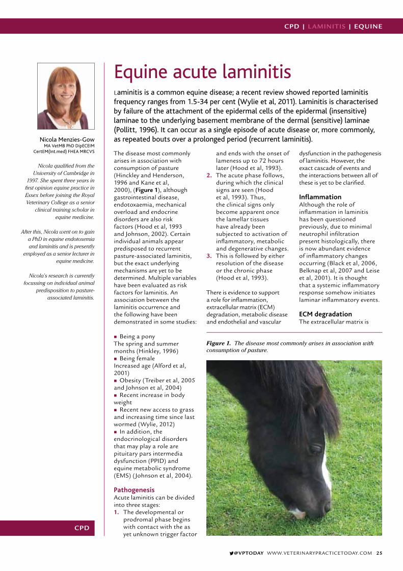
@VPTODAY WWW.VETERINARYPRACTICETODAY.COM 25
CPD | L AMINITIS | EQUINE
CPD
Nicola Menzies-GowMA VetMB PhD DipECEIM
CertEM(Int.med) FHEA MRCVS
Nicola qualified from the
University of Cambridge in 1997. She spent three years in
first opinion equine practice in Essex before joining the Royal Veterinary College as a senior
clinical training scholar in equine medicine.
After this, Nicola went on to gain a PhD in equine endotoxemia and laminitis and is presently
employed as a senior lecturer in equine medicine.
Nicola’s research is currently focussing on individual animal
predisposition to pasture-associated laminitis.
Equine acute laminitisLaminitis is a common equine disease; a recent review showed reported laminitis frequency ranges from 1.5-34 per cent (Wylie et al, 2011). Laminitis is characterised by failure of the attachment of the epidermal cells of the epidermal (insensitive) laminae to the underlying basement membrane of the dermal (sensitive) laminae (Pollitt, 1996). It can occur as a single episode of acute disease or, more commonly, as repeated bouts over a prolonged period (recurrent laminitis).
The disease most commonly arises in association with consumption of pasture (Hinckley and Henderson, 1996 and Kane et al, 2000), (Figure 1), although gastrointestinal disease, endotoxaemia, mechanical overload and endocrine disorders are also risk factors (Hood et al, 1993 and Johnson, 2002). Certain individual animals appear predisposed to recurrent pasture-associated laminitis, but the exact underlying mechanisms are yet to be determined. Multiple variables have been evaluated as risk factors for laminitis. An association between the laminitis occurrence and the following have been demonstrated in some studies:
� Being a pony The spring and summer months (Hinkley, 1996)
� Being female Increased age (Alford et al, 2001)
� Obesity (Treiber et al, 2005 and Johnson et al, 2004)
� Recent increase in body weight
� Recent new access to grass and increasing time since last wormed (Wylie, 2012)
� In addition, the endocrinological disorders that may play a role are pituitary pars intermedia dysfunction (PPID) and equine metabolic syndrome (EMS) (Johnson et al, 2004).
PathogenesisAcute laminitis can be divided into three stages: 1. The developmental or
prodromal phase begins with contact with the as yet unknown trigger factor
and ends with the onset of lameness up to 72 hours later (Hood et al, 1993).
2. The acute phase follows, during which the clinical signs are seen (Hood et al, 1993). Thus, the clinical signs only become apparent once the lamellar tissues have already been subjected to activation of inflammatory, metabolic and degenerative changes.
3. This is followed by either resolution of the disease or the chronic phase (Hood et al, 1993).
There is evidence to support a role for inflammation, extracellular matrix (ECM) degradation, metabolic disease and endothelial and vascular
dysfunction in the pathogenesis of laminitis. However, the exact cascade of events and the interactions between all of these is yet to be clarified.
Inflammation Although the role of inflammation in laminitis has been questioned previously, due to minimal neutrophil infiltration present histologically, there is now abundant evidence of inflammatory changes occurring (Black et al, 2006, Belknap et al, 2007 and Leise et al, 2001). It is thought that a systemic inflammatory response somehow initiates laminar inflammatory events.
ECM degradationThe extracellular matrix is
Figure 1. The disease most commonly arises in association with consumption of pasture.

VETERINARY PRACTICE TODAY SUMMER 201426
EQUINE | L AMINITIS | CPD
a diverse structure formed of structural proteins, proteoglycans, regulatory proteins, proteases, and protease inhibitors responsible for the maintenance of structural support, movement, growth, remodelling and healing along with the modulation of cytokines, inflammation, healing, and cell migration. Dysregulation of the protease enzymes known as matrix metalloproteases (MMPs) was previously thought to be the initiating event in laminitis (Johnson et al, 1998, Pillit, 1998 and Kyaw-Tanner, 2004). However, recent research has shown laminar separation may instead occur following a dysregulation of cell adhesion (Black, 2009), most likely caused by inflammatory and/or hypoxic cellular injury.
Metabolic diseaseLaminitis is more common in ponies and certain individual animals appear predisposed to recurrent pasture-associated laminitis. Animals at greatest risk of pasture-associated laminitis have a metabolic phenotype which includes obesity (Figure 2) and insulin resistance (IR), similar to that seen in human metabolic syndrome (HMS) (Treiber et al, 2006). Thus the same pathologic mechanisms that underlie the cardiovascular diseases associated with
HMS − including changes in insulin signalling, inflammatory cytokines and endothelial dysfunction caused by adipose tissue-derived mediators − could contribute to laminitis.
However, it must be remembered that not all laminitis prone animals are IR and/or obese and not all IR animals are laminitis prone. Thus it remains unclear whether IR per se plays a direct role in the development of laminitis, or whether other factors associated with IR increase susceptibility to laminitis when the animal is exposed to conditions known to trigger development of the condition.
Endothelial and vascular dysfunctionVascular events identified in the early stages of laminitis include digital venoconstriction and consequent laminar oedema (Hood et al, 1993, Hood et al, 2001 and Adair et al, 2000). This venoconstriction may be caused by platelet activation and platelet-neutrophil activation resulting in the release of the vasoactive mediators. In addition, normally the gastrointestinal flora produces high concentrations of various dietary amines by fermenting ingested amino acids, which remain within the gastrointestinal lumen. Grass contains soluble
carbohydrate in the form of fructans, a polysaccharide that is minimally digested by the small intestines and passes through to the hindgut. Fermentation of large amounts of carbohydrate by the hindgut bacteria is associated with increased numbers of certain Gram positive bacteria that produce increased amounts of these vasoactive dietary amines which may enter the circulation and directly and indirectly contribute to digital venoconstriction.
Finally, IR in other species alters endothelial function creating a pro-inflammatory condition, leading to platelet and leucocyte activation, increased production of the potent vasoconstrictor endothelin-1 and production of mediators of inflammation and oxidant stress21. Thus, alterations in digital vascular haemodynamics associated with inflammation, platelet activation and the action of vasoactive amines absorbed from the intestinal tract may contribute to lamellar injury.
DiagnosisDiagnosis is usually based on the clinical signs which include lameness affecting two or more limbs, characteristic stance of leaning back on the heels and taking weight off the toes, increased digital
pulses, increased hoof wall temperature, pain on hoof tester pressure at the region of the point of the frog and possibly a palpable depression at the coronary band. The lameness can vary in severity, from that only perceptible at the trot through to spending prolonged periods recumbent. Repeated episodes of disease result in divergent rings on the hoof wall (Figure 3).
Further tests are performed in those cases where an underlying endocrinological abnormality is suspected and radiographs are taken in those cases where movement of the pedal bone is suspected.
TreatmentTreatment should be initiated as soon as possible after the onset of the clinical signs. By the time the clinical signs become overt, the lamellar tissues have already been damaged and so therapy should be aimed at providing analgesia and foot support. The use of therapies to alter digital perfusion remains controversial.
AnalgesiaNon-steroidal anti-inflammatory drugs (NSAIDs) given either orally or intravenously are the first choice for analgesia as they have the additional beneficial effect of inhibiting part of the inflammatory cascade. There is no evidence to suggest that any one specific NSAID is superior to the next (Menzies-Gow et al, 2010). If they do not provide sufficient pain relief, then opiates can additionally be used.
Foot supportSupporting the foot is an essential part of the
Figure 2. Animals at greatest risk have a metabolic phenotype which includes obesity.
Figure 3. Divergent rings on the hoof wall.
“Treatment should be initiated as soon as possible after the onset of the clinical signs”

@VPTODAY WWW.VETERINARYPRACTICETODAY.COM 27
CPD | L AMINITIS | EQUINE
management. The horse naturally adopts a stance that bears most of the weight over the caudal part of the foot rather than the painful toe region. Additional support should be supplied to this region of the foot in order to provide pain relief and minimise the mechanical forces on the laminae.
The simplest method is to increase the depth of the bedding and ensure that it extends to the door (Figure 4). Extra support can be applied directly to the frog and/or the caudal two thirds of the foot itself. Frog-only support can be achieved using rolled up bandaging material of the same length as the frog, placed along the length of the frog and secured in place with adhesive tape. Alternatively, a commercially available product such as the lily pad or TLC frog support can be used. Combined frog and sole support can be provided using, for example, dental impression material that is moulded to the contours of the caudal two thirds of the foot or styrofoam pads that are crushed by the weight of the horse. There is no evidence to suggest that any one foot support method is superior (Menzies-Gow, 2010).
Vasodilator or vasoconstrictor therapyThe use of vasodilator or vasoconstrictor therapy remains controversial due to contradictory scientific evidence relating to whether digital perfusion is increased or decreased in the prodromal stages of the disease. Vasodilator therapy is frequently used, based on laminitis being a consequence of digital hypoperfusion. Acepromazine is the most effective digital vasodilator available (Leise et al, 2007). However, even if the pathophysiology involves vasoconstriction, this has normally resolved once the clinical signs become apparent. Nevertheless,
the sedative effect of acepromazine may have the additional beneficial effect of reducing movement or even resulting in increased periods of time spent recumbent with the weight taken off the feet.Cryotherapy has been shown to prevent experimentally-induced laminitis, possibly due to prevention of vasodilation (Van Eps et al, 2012).
However, cryotherapy will also protect against ischaemia by reducing cellular metabolic demands and inflammation by decreasing enzymatic activity. Regardless of the mechanism of action, continuous adequate and controlled chilling of the digits over a prolonged period of time may be difficult to achieve in the clinical situation and is probably of most benefit in the prodromal stage before the clinical signs are seen.
Additional therapiesAdditional therapies are indicated if an underlying endocrinological disorder is diagnosed.
PreventionPrevention of pasture-associated laminitis centres on achieving and maintaining an optimum body condition and on limiting intake of pasture non-structural carbohydrates (NSC). A diet based on grass hay (or hay substitute) with low (up to 10 per cent) NSC content should be fed and cereals avoided. Soaking hay in water for 16 hours before feeding will leach water soluble carbohydrates, however this does not reliably decrease the NSC content to 10 per cent or less in every case and ideally forage should be analysed.
If weight loss is required, hay or hay substitute should initially be provided at up to 1.5 per cent of current body weight per day. It is preferable not to decrease forage provision to less than one per cent of target body weight as this may
increase the risk for hindgut dysfunction, stereotypical behaviours, ingestion of bedding, or coprophagy. The forage should be divided into three to four feeds per day and strategies to prolong feed intake time considered, such as the use of haynets with multiple small holes. This diet low in NSC also has the additional benefit of improving insulin sensitivity in IR animals.
The NSC content of pasture fluctuates widely, decreasing when the plant is growing − as the NSC is used to provide energy for growth − and increasing when the plant continues to photosynthesis without growing e.g. when there is high light intensity, low temperatures or lack of water. In addition, the NSC content is affected by season, being low in early spring and increasing in late spring; thus, zero grazing should be considered. If a laminitis prone animal is to be turned out, steps should be taken to minimise NSC intake.
The pasture should be managed to encourage growth, but it should be regularly topped. Animals should be turned out from late night to early morning, when the NSC content of the pasture is lowest. Grazing should be particularly limited in spring and autumn when the grass is growing. Turn out should be avoided if there has been a frost with bright sunshine or a drought, as this restricts growth while photosynthesis continues, resulting in NSC accumulation. Paddocks should be rotated in order to keep them at the ideal height, as grass that is too short will be stressed and have a higher NSC content, while grass that is too long means a greater amount is available for consumption. Methods to restrict intake such as strip grazing and grazing muzzles can be employed.
Forage-only diets do not provide adequate protein, minerals, or vitamins and so a low-calorie commercial ration balancer product containing sources of high-quality protein and a mixture of vitamins and minerals to balance the low vitamin E, copper, zinc, selenium, and other minerals typically found in mature grass hays is recommended.
Figure 4. Ensure that the bedding extends to the door.
“Exercise is also essential in the preventionof laminitis”

VETERINARY PRACTICE TODAY SUMMER 201428
EQUINE | L AMINITIS | CPD
ReferencesWylie CE, Collins SN, Verheyen KL et al. (2011) Frequency of equine laminitis: A systematic review with quality appraisal of published evidence. Vet J
Pollitt CC. (1996) Basement membrane pathology: a feature of acute equine laminitis. Equine Vet J 28 :38-46.
Hinckley KA, Henderson IW. (1996) The epidemiology of equine laminitis in the UK. 35th Congress of the British Equine Veterinary Association 62.
Kane AJ, Traub-Dargatz JL, Garber LP et al. (2000) Lameness and Laminitis in US Horses. USDA:APHIS:VS, CEAH, National Animal Health Monitoring System. Fort Collins, CO,
Hood DM, Grosenbaugh DA, Mostafa MB et al.(1993) The role of vascular mechanisms in the development of acute equine laminitis. J Vet Intern Med 7228-234.
Johnson PJ. (2002) The equine metabolic syndrome peripheral Cushing’s syndrome. Vet Clin North Am Equine Pract, 918: 271-293.
Alford P, Geller S, Richardson B et al. (2001) A multicenter, matched case-control study of risk factors for equine laminitis. Prev Vet Med 49: 209-222.
Treiber KH, Hess TM, Kronfeld DS et al. (2005) Insulin resistance and compensation in laminitis-predisposed ponies characterized by the Minimal Model. Equine Nutrition Symposium 91-92.
Johnson PJ, Messer NT, Slight SH et al. (2003) Endocrinopathic laminitis in horses. Clinical Techniques in Equine Practice 93: 45-56.
Wylie CE. Epidemiological investigations of equine laminitis in Great Britain 2009-2011: University of London (Royal Veterinary College), 2012; 389.
Black SJ, Lunn DP, Yin C et al. (2006) Leukocyte emigration in the early stages of laminitis. Vet Immunol Immunopathol 109: 161-166.
Belknap JK, Giguere S, Pettigrew A et al. (2007) Lamellar pro-inflammatory cytokine expression patterns in laminitis at the developmental stage and at the onset of lameness: innate vs. adaptive immune response. Equine Vet J, 39: 42-47.
Leise BS, Faleiros RR, Watts M et al. (2011) Laminar inflammatory gene expression in the carbohydrate overload model of equine laminitis. Equine Vet J 43: 54-61.
Johnson PJ, Tyagi SC, Katwa LC et al. (1998) Activation of extracellular matrix metalloproteinases in equine laminitis. Vet Rec 142: 392-396.
Pollitt CC, Pass MA, Pollitt S. Batimastat (1998) (BB-94) inhibits matrix metalloproteinases of equine laminitis. Equine Vet J Suppl 26: 119-124.
Kyaw-Tanner M, Pollitt CC. (2004) Equine laminitis: increased transcription of matrix metalloproteinase-2 (MMP-2) occurs during the developmental phase. Equine Vet J 36: 221-225.
Black SJ. (2009) Extracellular matrix, leukocyte migration and laminitis. Vet Immunol Immunopathol 129: 161-163.
Treiber KH, Kronfeld DS, Hess TM et al. (2006) Evaluation of genetic and metabolic predispositions and nutritional risk factors for pasture-associated laminitis in ponies. J Am Vet Med Assoc 228: 1538-1545.
Hood DM, Wagner IP, Brumbaugh GW.(2001) Evaluation of hoof wall surface temperature as an index of digital vascular perfusion during the prodromal and acute phases of carbohydrate-induced laminitis in horses. Am J Vet Res 62: 1167-1172.
Adair HS, 3rd, Goble DO, Schmidhammer JL et al.(2000) Laminar microvascular flow, measured by means of laser Doppler flowmetry, during the prodromal stages of black walnut-induced laminitis in horses. Am J Vet Res 61: 862-868.
Potenza MA, Addabbo F, Montagnani M. (2009) Vascular actions of insulin with implications for endothelial dysfunction. Am J Physiol Endocrinol Metab 297: E568-577.
Menzies-Gow NJ, Stevens K, Barr A et al. (2010) Severity and outcome of equine pasture-associated laminitis managed in first opinion practice in the UK. Vet Rec 1670: 364-369.
Leise BS, Fugler LA, Stokes AM et al. (2007) Effects of intramuscular administration of acepromazine on palmar digital blood flow, palmar digital arterial pressure, transverse facial arterial pressure, and packed cell volume in clinically healthy, conscious horses. Vet Surg 36: 717-723.
Van Eps AW, Leise BS, Watts M et al. (2012) Digital hypothermia inhibits early lamellar inflammatory signalling in the oligofructose laminitis model. Equine Vet J 44: 230-237.
CPD Questions1. What are the risk factors for laminitis?
2. During which stage of laminitis do the clinical signs become evident?
3. What are the clinical signs frequently associated with laminitis?
4. When might cryotherapy be of most benefit in the treatment of laminitis?
5. Ideally, what should the non-structural carbohydrate content of the hay be to try and prevent laminitis?
Answers1. Being a pony, the spring and summer months, being female, increased age, obesity, recent increase in body weight, recent new access to grass and increasing time since last wormed 2. Acute 3. Lameness affecting two or more limbs, characteristic stance of leaning back on the heels and taking weight off the toes, increased digital pulses, increased hoof wall temperature, pain on hoof tester pressure at the region of the point of the frog and possibly a palpable depression at the coronary band 4. In the prodromal stage before the clinical signs are seen 5. Up to 10 per cent
If weight gain is required or the animal is undertaking a large amount of exercise, then a forage-based diet may not meet energy requirements and caloric intake can be increased by adding unmolassed soaked sugar beet pulp (0.2-0.7kg/day) or by feeding vegetable oil (100-225ml sid or bid up to a maximum of 100ml/100kg of body weight).
Several supplements containing magnesium, chromium or cinnamon are marketed with claims for improved insulin sensitivity but scientific evidence demonstrating their efficacy is lacking.
Exercise is also essential in the prevention of laminitis as it reduces IR, suppresses inflammation and decreases food intake. Light exercise appears sufficient to improve insulin sensitivity, but
this probably needs to be maintained on a regular and possibly even daily basis. This is only possible once an animal has recovered and is sound. There should be a gradual increase in the intensity and duration of the exercise undertaken.

@VPTODAY WWW.VETERINARYPRACTICETODAY.COM 29
CPD | THEATRE | EQUINE
CPD
Marie RippingaleBSc(Hons) REVN clinical coach
G-SQP DipHE CVN DipAVN(Equine)
Marie is head equine nurse and a clinical coach at XLVets
practice Scarsdale Vets in Derby. Marie is also a lecturer on the
veterinary nursing diploma course at Bottle Green Training,
Melbourne, Derbyshire.
Equine theatre practice – what is different in an equine theatre?The risk of mortality or serious morbidity is particularly high in horses undergoing general anaesthesia (GA) (Taylor and Clarke, 2007). For this reason, any registered veterinary nurse (RVN) wanting to be involved with this process will require equine specific knowledge. This article will discuss the main considerations involved in equine theatre design and practice.
The principles of theatre practice remain the same whether nursing equine patients or small animals. The application, however, is often slightly different when dealing with the equine species.
Equine theatre designA well-designed surgical suite (SS) facilitates the establishment and maintenance of good aseptic techniques, reducing the chance of surgical site infections (SSIs) (McHugh, 1991). The ideal equine SS consists of several defined areas, each with specific requirements.
Induction/recovery boxThis must be large enough to accommodate a recovering horse, but not so large that it allows a recovering horse to gain momentum and speed during recovery. It is important to reduce the risk of patient injury, as Bettschart and Johnston, (2011) attributed a 23.3 per cent equine anaesthesia mortality rate to fractures attained during
recovery. Auckburally and Flaherty, (2009) suggested four-by-four metres as an adequate size for an equine induction/recovery box.The induction/recovery box should be equipped with dimmable lights, heat lamps, convex mirrors, viewing ports, closed circuit television (CCTV), an oxygen supply, anaesthetic waste, gas scavenging and slinging facilities for supportive treatment of complications (Greet, 2008).
The walls and floor should be lined with padded mats, which are essential to facilitate a safe induction, and are easy to clean (Figure 1) (Greet,
2008). A rubberised floor will reduce the risk of slipping during induction which is an important safety measure for the patient (Greet, 2008). The induction/recovery box requires an overhead hoist to move the anaesthetised horse to the operating theatre and visa versa (Figure 2).
A recovery box should also be fitted with rope recovery systems to allow assisted recovery by supporting the horse to stand and thereby reducing the risk of injury (Auckburally and Flaherty, 2009). Small windows should be present in the recovery box door to allow the assisted recovery to be controlled
Figure 1. An equine induction/recovery box should have padded walls and a padded floor.
Figure 2. An equine theatre needs a hoist so horses can be moved from the induction box into theatre and visa versa.

VETERINARY PRACTICE TODAY SUMMER 201430
EQUINE | THEATRE | CPD
by handlers outside the box, minimising risk to staff.
Clinics with a high surgical caseload will usually have one induction box and two or more recovery boxes to allow a second horse to be anesthetised while the first horse is recovering (McHugh, 2012).
Preparation area (PA)This is often a separate area outside of theatre where the horse is prepared while standing and conscious. This is an important consideration as Johnston, (2005) found that in surgical procedures lasting more than 90 minutes, equine patients were 10 times more likely to develop a post anaesthetic myopathy (PAM). This is thought to result from inadequate blood perfusion of muscle tissue, producing ishaemic muscle damage (Murrell and Ford-Fennah, 2012). The bodyweight of the horse alone is enough to cause PAM during GA in weight bearing muscles, such as the quadriceps during dorsal recumbency.
Clinical signs of PAM include hot, swollen, painful muscles and recumbency. Unfortunately these symptoms are only apparent after GA. Therefore to reduce anaesthetic time and anaesthetic risk, surgical site clipping (Figure 3) and the initial skin scrub (Figure 4) are commonly performed with the patient standing in a PA. This is in contrast to small animal surgery where patients are commonly anaesthetised in the
PA (McHugh et al, 2011). In some clinics the PA is situated within the main operating suite and anaesthetised horses are positioned, clipped and have their first surgical skin scrub carried out on a mobile operating table that can then be pushed into theatre (McHugh, 2012). This reduces contamination in the operating theatre (OT) and therefore reduces the risk of SSIs for the patient.
The operating theatre Larger equine clinics will have two OTs: one for elective surgery such as orthopaedic surgery and one for contaminated surgery such as colic, airway and dental surgery. The standard of asepsis should be the same in both theatres (McHugh, 2012).
Greet, (2008) stated that an optimal equine OT should measure more than 6 by 5.5 metres. The OT must be large enough to accommodate a horse positioned in dorsal or lateral recumbency and to allow for bulky surgical equipment such as an arthroscopy monitoring trolley (McHugh, 2012). Walls and floors must be made of smooth, impervious, non-staining material and floors should be hard wearing and
non-slip (McHugh, 2012). Drains, although often considered as a potential source of bacteria, are essential in an equine operating theatre and should be included to facilitate effective cleaning (McHugh, 2012).
A positive pressure ventilation system which allows changes of air in the OT should be installed to facilitate removal of airborne microorganisms and help maintain asepsis (Stick, 2012).
The temperature for an equine OT should be maintained at 20°C with a relative humidity of 50 per cent (Stick, 2012). This is for patient benefit, as hypothermia is a common side effect of GA in horses (Mayerhofer et al, 2005). This is an important perioperative consideration as resultant morbidity may not become apparent until the postoperative period (Harvey, 2006). Adverse effects of hypothermia can include delayed wound healing and increased risk of wound infection (Harvey, 2006). It is essential in terms of perioperative nursing to prevent hypothermia if high standards of patient care are to be achieved.
Other areasAn equine SS should also contain a scrubbing up area, sterile storage area, a washing and sterilising room and changing rooms just like in a small animal theatre.
Layout of an equine SSIdeally there should be a one-way or circular traffic system so that the surgical team and sterile supplies enter through one door and leave by another to minimise cross-contamination (McHugh, 2012). The theatre suite should be divided into three zones: contaminated, clean and aseptic. The aseptic zone should be at the centre of the SS and would contain the OT, sterile store and scrubbing up area. This should be surrounded by a clean zone on either side and this would contain the induction and recovery boxes, washing and sterilising room and changing rooms. The contaminated zone or PA would be on the outside of the clean zone.
The different zones should be colour coded and strict dress protocols should be put in place. Effective education and management of staff would be important to ensure that specific protocols are adhered to (McHugh, 1991).
Figure 3. Initial clipping is commonly performed with the patient standing in the preparation area to reduce anaesthetic time.
Figure 4. Initial skin scrubbing is commonly performed with the patient standing in the preparation area to reduce anaesthetic time.
“The body weight of the horse alone is enough to cause post anaesthetic myopathy during general anaesthetic in weight bearing muscles”

@VPTODAY WWW.VETERINARYPRACTICETODAY.COM 31
CPD | THEATRE | EQUINE
The operating tableThe equine operating table should have the following features:
� Adjustable to allow the horse to be placed in any position and at any height
� Easily moveable to any position in the room
� Easily cleaned, maintained and rust resistant
� Have battery or mains-operated hydraulics to assist with patient positioning
� Be equipped with limb and head supports and other positioning aids (McHugh, 2012).
There are many different types of equine operating table including fixed hydraulic, mobile hydraulic and inflatable. Whatever type of table is used, attention must be paid to providing good padding and even positioning to avoid PAM, which occurs in one per cent of healthy equines undergoing anaesthesia (Johnston, 2005).
Equine theatre practiceThe aim of good theatre practice is to provide a safe and sterile environment in which to perform a variety of surgical procedures, as this minimises risk to the patient (Rutland, 2011). Effective equine theatre practice includes correct patient preparation, positioning the horse correctly for surgery, clipping, and correct skin preparation. These procedures are discussed in more detail below.
Patient preparation The following list may not always be possible in emergency situations, however, for elective procedures all components should be completed.
The patient should be starved for up to 12 hours before surgery as this reduces the weight of the gastrointestinal tract (GIT) on the diaphragm and increases the functional residual capacity of the lung (Taylor and Clarke, 2007). Preoperative starvation should not exceed 12-18 hours or the
horse will become agitated and detrimental metabolic changes will develop (Taylor and Clarke, 2007). The patient should be groomed to minimise contamination of the SS. If the patient is particularly dirty it should be bathed to further reduce contamination of the aseptic areas of the SS (McHugh, 2012).
Shoes should be removed to minimise the risk of self-inflicted injury to the patient during induction and recovery (Auckburally and Flaherty, 2009). This will also reduce the risk of damage to the induction/recovery box floor (Murrell and Ford-Fennah, 2012).
If surgery is to be carried out on the foot, scrupulous preparation is required. Once the shoe has been removed the foot must be pared and scrubbed thoroughly. A solution of strong iodine (10 per cent) should then be applied using gauze and cotton pads (McHugh, 2012). The foot should be covered with a poultice and left overnight to allow the iodine to soak into the hoof wall and sole. Petroleum jelly should be applied to the coronary band to prevent it being irritated by the iodine (McHugh, 2012).
The patient’s mouth should be rinsed out with warm water to prevent food debris being pushed down the trachea during intubation (Murrell and Ford-Fennah, 2012). An intravenous (IV) catheter should be placed to provide ready venous access should drugs need to be administered in an emergency (Taylor and Clarke, 2007). The IV catheter also allows access for the administration of IV fluids during GA.
Clipping the surgical site is necessary for most surgical procedures. This is mostly performed in the PA with the horse standing and conscious. Preoperative clipping improves asepsis
by removing loose hairs and by allowing access for the surgical site to be washed and pre-cleaned prior to surgery (McHugh, 2012). Risk of post anaesthetic complications is lowered through the reduction of GA time.
Care should be taken not to damage the skin when clipping as this may increase the risk of the patient developing an SSI (McHugh, 2012).
Once the horse is positioned on the table the feet and tail must be covered to minimise contamination (McHugh, 2012).
Positioning for surgeryThe horse should be placed on the operating table in a position that does not put any part of the body under
strain. Limbs should settle naturally and be secured without force. When the horse is lying in lateral recumbency both non-dependant limbs should be supported parallel to the ground. The dependant forelimb should always be pulled forwards to take pressure off the lower triceps and prevent PAM (Taylor and Clarke, 2007).
In dorsal recumbency the hindlimbs should never be drawn back with the patellae locked as this can lead to the horse being unable to stand postoperatively. If this position is necessary for surgery it should be restricted to one limb at a time and limited to 20 minutes in duration (Taylor and Clarke, 2007). Padding helps to spread the load of the horse’s weight over as large an area of body surface as possible (Taylor and Clarke, 2007). This helps to reduce concentrated pressure at any one site. Padding is usually achieved with foam beds and pads. Inflatable beds and cushions can be used but the beds should not be
Figure 5. Final skin preparation is performed in the operating theatre with the horse anaethetised.
“Padding helps to spread the load of the horse’s weight over as large an area of body surface as possible”

VETERINARY PRACTICE TODAY SUMMER 201432
EQUINE | THEATRE | CPD
fully inflated as this will make them hard and more likely to contribute to the development of PAM.
Preparation of the skinTheatre practice in the form of effective perioperative nursing care includes surgical site preparation, first by hair removal and then by skin cleansing (Figure 5). This removes dirt and oil as well as reducing resident skin flora populations, which can potentially cause SSIs (Stick, 2012).
Which skin disinfectant is best? The RVN must decide what type of skin disinfectant to use for preparing the surgery site. The two most commonly used skin disinfectants in veterinary practice are chlorhexidine and povidone-iodine (Gibson et al, 1997).
Povidone-iodine is inactivated by organic material (Gibson et al, 1997) and this could be considered undesirable for use in equine patients who live in a heavily contaminated environment (McHugh, 2012). Osuna et al, (1990) found a significantly increased rate of skin reactions with the use of povidone-iodine, which would be undesirable as this could lead to wound breakdown and reduced healing.
Chlorhexidine is however, believed to have a residual antiseptic effect lasting up to six hours (Desrochers et al, 1996). This would be particularly useful in patients undergoing emergency colic surgery, as the abdominal discomfort could have caused them to roll and significantly contaminate the surgical site.
Wilson et al, (2011) recommended the use of chlorhexidine as a preoperative skin preparation for ponies after comparing it with povidone-iodine. This would support the use of a four per cent chlorhexidine solution for
the preparation of surgical sites in horses.
Skin preparation techniqueLarge lint-free swabs are best for skin preparation and the following technique works well:
� Position the horse for surgery � The RVN should wear sterile
surgical gloves to prevent skin contamination being passed from scrub nurse to the patient
� Using sterile lint-free swabs, the surgical site should be scrubbed with four per cent chlorhexidine solution and a small amount of sterile water. Scrubbing should begin at the proposed incision site and work outwards towards the periphery. On reaching the outside, the swab should be discarded and a fresh one taken
� This procedure is continued until the area is clean (McHugh, 2012)
It has been traditionally recommended that a solution of 70 per cent alcohol should be sprayed over the surgical site after the skin has been cleansed. However Osuna et al, (1990) discovered that the residual activity of chlorhexidine is decreased by the use of alcohol after the surgical scrub. Osuna et al, (1990) recommended that chlorhexidine be used for the correct contact time and then rinsed with sterile saline to preserve the residual antimicrobial activity. Coolman et al, (1998) also demonstrated a residual antimicrobial effect in canine IV catheter sites prepared with chlorhexidine and sterile saline. Further research is required to fully validate the claims made by Osuna et al, (1990) and Coolman et al, (1998) as the majority of the literature concerning the preparation of equine surgical sites still advocate the use of alcohol after scrubbing with chlorhexidine.
Conclusion The main aims of equine theatre design and equine theatre practice are to provide
an aseptic environment in which surgery can take place and to prevent associated complications such as SSIs, fractures in recovery, PAM and hypothermia.
The RVN should be aware of correct equine theatre design, and also help to enforce correct theatre practice including ensuring a one-way system is in place, and correct theatre apparel is worn. Effective perioperative patient care includes ensuring a correct environment for induction and recovery, correct patient preparation before induction, correct positioning and padding of the horse on the operating table, and correct clipping and skin preparation.
With detailed knowledge of equine theatre design and equine theatre practice, the RVN can ensure a safe and effective environment for horses undergoing surgery and provide the highest standards of nursing care.

@VPTODAY WWW.VETERINARYPRACTICETODAY.COM 33
CPD | THEATRE | EQUINE
CPD answers1. Four-by-four metres 2. Dimmable lights, heat lamps, viewing ports, convex mirrors, CCTV, an oxygen supply, anaesthetic waste, gas scavenging and slinging facilities for supportive treatment of complications 3. 6 by 5.5 metres or larger 4. They facilitate effective cleaning of the theatre facilities 5. The temperature for an equine OT should be maintained at 20°C with a relative humidity of 50 per cent 6. This is for patient benefit, as hypothermia is a common side effect of GA in horses 7. The patient’s mouth should be rinsed out with warm water to prevent food debris being pushed down the trachea during intubation 8. A solution of strong iodine (10 per cent) should then be applied using gauze and cotton pads to the foot and this should covered with a poultice and left overnight 9. 20 minutes maximum 10. Clinical signs of PAM include hot, swollen, painful muscles and recumbency
References
Auckburally A and Flaherty D (2009). Recovery from anaesthesia in horses. 2.
Avoiding complications. In Practice 31: 362-369.
Bettschart R and Johnston M (2011). Confidential enquiry into perioperative equine
fatalities: CEPEF 4 – a chance to gain new evidence about the risks of equine
general anaesthesia. Equine Veterinary Journal Vol 44(1): 7.
Coolman BR, Marretta SM, Kakoma I, Wallig MA, Coolman SL and Paul AJ (1998).
Cutaneus antimicrobial preparation prior to intravenous catheterisation in healthy
dogs: Clinical, microbial, and histopathological evaluation. Canadian Veterinary
Journal 39: 757-763.
Desrochers A, St-Jean G, Anderson DE, Rogers DP and Chiengappa MM (1996).
Comparative Evaluation of Two Surgical Scrub Preparations in Cattle. Veterinary
Surgery 25: 336-341.
Gibson KL, Donald AW, Hariharan H and McCarville C (1997). Comparison of Two
Pre-Surgical Skin Preparation Techniques. Canadian Journal of Veterinary Research
61: 54-156.
Greet T (2008). Hospital design and organisation. In Corley K and Stephen J (eds),
The Equine Hospital Manual, West Sussex, Wiley-Blackwell.
Harvey RC (2006). Thermoregulation. In Doherty T and Valverde A (eds), Manual
of Equine Anaesthesia and Analgesia, Oxford, Blackwell Publishing Ltd.
Johnston GM (2005). Findings from the CEPEF epidemiological studies into equine
perioperative complications, Equine Veterinary Education 7: 64-68.
Mayerhofer I, Scherzer S, Gabler C and Van Den Hoven R (2005). Hypothermia in
horses induced by general anaesthesia and limiting measures. Equine Veterinary
Education 17(1): 53-56.
McHugh D (1991). Planning an equine operating theatre. Equine Veterinary
Education 3(4): 235-238.
McHugh D, Young A and Johnson J (2011). Theatre practice. In Cooper B,
Mullineaux E, Turner L and Greet T (eds), BSAVA Textbook of Veterinary Nursing
(5th edn), Gloucester, BSAVA.
McHugh DP (2012). Theatre practice. In Coumbe K (Ed) (2012). Equine Veterinary
Nursing Manual (2nd edn), Oxford, Blackwell Science.
Murrell JC and Ford-Fennah V (2012). Anaesthesia. In Coumbe K (Ed) (2012).
Equine Veterinary Nursing Manual (2nd edn), Oxford, Blackwell Science.
Osuna DJ, DeYoung DJ and Walker RL (1990). Comparison of Three Skin
Preparation Techniques Part Two: Clinical Trial in 100 Dogs. Veterinary Surgery
19(1): 20-23.
Rutland G (2011). Optimising the theatre environment. Veterinary Nursing Journal
26(4): 121-125.
Stick JA (2012). Preparation of the Surgical Patient, the Surgery Facility, and
the Operating Team. In Auer JA and Stick JA (eds), Equine Surgery (4th edn),
Missouri, Elsevier.
Taylor PM and Clarke KW (2007). Handbook of Equine Anaesthesia (2nd edn),
Philadelphia, Saunders Elsevier.
Wilson DG, Hartmann F, Carter VR, Klohnen A and MacWilliams PS (2011).
Comparison of three preoperative skin preparation techniques in ponies, Equine
Veterinary Education 23(9): 462-465.
CPD questions1. What is the recommended size for an induction/
recovery box for equine patients?
2. An induction/recovery box should have padded walls. Name three other accessories that should be included.
3. What is the optimal recommended size for an equine operating theatre?
4. Why are drains essential to have in an equine theatre?
5. What is the optimal temperature and humidity for an equine theatre?
6. Why is it so important to maintain this temperature and humidity?
7. Why should a horse’s mouth be rinsed out before GA?
8. What strength of iodine should be used in a foot poultice the night before surgery?
9. Sometimes when the horse is anaesthetised in dorsal recumbency the hindlimbs need to be drawn back with the patellae locked for surgery to take place. How long should the hindlimbs be left in this position before being flexed again?
10. Name three symptoms of PAM in horses

VETERINARY PRACTICE TODAY SUMMER 201434
L ARGE ANIMAL | ROUNDUP
In brief
In this sectionCPD
Rumen health - tips to help farmers get it rightExploring ways to safeguard rumen function without compromising production.Turn to P35
An informal consultation has been launched to seek views on fee changes for statutory services delivered by AHVLA.
Informal views are being sought ahead of a full consultation in summer 2014.
Fee increases are planned with a view to moving to full cost recovery (FCR) for services delivered in relation to bovine embryos, bovine semen, porcine semen (collectively known as artificial breeding controls (ABCs)), poultry health scheme, border inspection posts (BIPs) and salmonella national control programme (NCP).
Phased increases were agreed in June 2013 for ABCs, poultry health scheme and BIPs. A move to FCR is planned for this year.
After a revised FCR model was introduced - based on HM Treasury guidance - AHVLA says the majority of further revisions to fees will be lower than the FCR amounts forecast in 2012. However, the agency says it cannot be specific about revisions to individual fees and charges.
AHVLA is seeking views on how stakeholders feel the changes will affect businesses.
Further details about revisions to individual fees and charges are expected to be available by summer 2014, when a public consultation will be launched.
AHVLA aims to introduce revised fees during the final quarter of this year.
New service offers testing for exclusion of notifiable avian diseases (NAD)
AHVLA has begun offering poultry keepers and vets a new service, testing for the exclusion of NAD.
The service started on May 23 and will initially be piloted for one year. It is available to poultry keepers and vets from the National Reference Laboratory, AHVLA Weybridge.
Testing is intended specifically for cases where NAD is not suspected but cannot be ruled out entirely.
Initially only turkey and chicken flocks will be tested but if the service is successful this may be rolled out to other species in the future.
In a letter to the Veterinary Record on May 24, chief veterinary officer Nigel Gibbens et al said: “This service may help to detect NAD at the earliest opportunity in those cases where the clinical signs do not give rise to a level of suspicion of a NAD that justifies either a consultation case or statutory notification and official enquiry in the first instance.”
Private veterinary surgeons can submit samples to AHVLA after a telephone discussion with an AHVLA duty vet, where it is agreed that NAD is not suspected.
AHVLA testing will be carried out using PCR methods, taking swabs from the oropharynx and cloaca. The agency will charge the full cost for testing.
Results will usually be available within 48 hours, where samples are submitted on weekdays. Arrangements for out-of-hours submission and testing will also be available. Positive test results will trigger an official AHVLA investigation.
Industry drive to promote control of major diseasesA new initiative aims to promote better control of four major diseases affecting dairy cattle - BVD, Johne’s disease, lameness and mastitis.
“The project represents a progression from previous regionally based health and welfare projects, towards more co-ordinated delivery,” explained Ray Keatinge, head of research and development at DairyCo, which is leading the project.
Not-for-profit organisation DairyCo will work on the campaign with Duchy College, SAC Consulting and XLVets.
The campaign will deliver technical advice to groups of farmers or one-to-one. Advice will be given to those undertaking control measures against BVD and Johne’s disease, as well as farmers looking for information about what steps to take for the first time.
A number of events and vet visits will also be funded, including 90 herd health seminars, 100 DairyCo mastitis control plans and 180 farm vet visits under the DairyCo Healthy Feet Programme. On-farm veterinary advice will receive 70 per cent funding from the Regional Development Programme for England (RDPE). Some other events will be free of charge.

@VPTODAY WWW.VETERINARYPRACTICETODAY.COM 35
make it, plus another 65MJ for maintenance; so 220MJ in total.
If we were to rely on rough forage, what the rumen was arguably designed for, with an metabolisable energy (ME) density of around 9MJ/kg dry matter, our average yielding cow would need to eat a huge 24kg dry matter intake (DMI) - equivalent to around 100kg of silage, or 160kg of fresh grass. The maximum even the largest cow can eat in a day is around 60kg fresh weight.
Of course, high yielding cows do indeed eat huge intakes, and DMIs of a 24kg magnitude are not uncommon, but this is only achieved by feeding a processed diet that can be hoovered up quickly and that doesn’t over-bulk the rumen with long fibre or high moisture content.
A cow fed a forage-only diet, or grazing, could only achieve around 17-18kg DMI, because of the bulk and the length of time such a diet would be retained in the rumen.
So, two things are happening. To feed enough nutrients for milk production, we need to increase the energy density of the diet, and we need to achieve high DMIs. This is usually done by feeding concentrates and forages that are very digestible and energy dense. Maize silage, for example, although a forage, is a long way from the rough grass for which cows’ rumens have evolved. Indeed, modern grass varieties only have a distant resemblance to their evolutionary forebears and grazing cows are therefore not necessarily immune to rumen health problems, including acidosis.
CPD | RUMEN HEALTH | L ARGE ANIMAL
Rumen health:tips to help farmers get it rightThis third and final article in the series on rumen health explores ways to safeguard rumen function while not compromising production. Although based around the dairy cow and her requirements, the principles apply to any ruminant.
The feed balance dilemmaIn essence, maintaining a healthy rumen is easy; simply feed a forage based diet, little and often, just as the cow and her rumen has evolved over eons to cope with. However, life is rarely so simple and the outputs of even a moderately productive dairy cow mean that a forage based diet alone will be insufficient to provide her requisite nutrients.
Consider a cow producing 30 litres per day of a standard quality milk (4 per cent fat, 3.2 per cent protein, 4.8 per cent lactose). Each day she produces:
Fat: 30 x 0.04 = 1.2 kg
Protein: 30 x 0.032 = 1 kg
Lactose: 30 x 0.048 = 1.5 kg
All this contains around 75MJ of energy, but it takes 155MJ to
Owen Atkinson BVSc DCHP MRCVS
Owen has worked in farm
animal practice since 1994, mainly with dairy cows. He
began his professional life in North Yorkshire, before moving on to the University of Liverpool to teach farm animal veterinary practice, and then to Wrexham
in North Wales. He settled in Cheshire with his family where
he became a partner in farm-only vet practice, Lambert, Leonard
and May, part of XLVets.
Owen’s interests in rumen health, cattle foot care and lameness
reduction led him to do an increasing amount of training and advisory work. In 2009,
Owen was awarded a Trehane Trust/Nuffield travel scholarship to look into the role of the dairy
vet in knowledge transfer.
In 2013, Owen left the practice to found Dairy Veterinary Consultancy Ltd, to better
pursue his vision for preventive health management and
strategy. He works with dairy farmers, other vet practitioners
and the wider dairy industry and supply chain. Owen’s
goal is to help farmers make changes on their farms that
benefit the cows, the quality of life and the bank balance.
CPD
The feeding balance dilemma
Problem: insufficient intakes; low energy
production; cow in negative energy balance, unless very low production
Very slow digestion: lots of undigestible fibre;
high rumen retention times;slow release of volatile fatty acids
Optimalfermentationrate for high
producingcow
Problem:poor rumen health likely; acidic rumen
conditions (lactic acid); enzymatic starch digestion in small intestine andsecondary fermentation in hindgut;
poor fermentation of complexcarbohydrates (forage) as rumen
function impaired energy deficit
Ration digested very quickly:sugars, rapidly degradable starch;
quick rumen flora producing lactic acid;quick rumen transit time undigested
carbohydrates exit rumen
Rum
en f
erm
enta
tion
rat
e
Digestion rate of carbohydrates
Graph 1. Illustrates the balance required between inducing a slow or quick rumen fermentation rate. The blue dashed line denotes the rumen fermentation rate increasing in tandem with carbohydrate digestibility.

VETERINARY PRACTICE TODAY SUMMER 201436
L ARGE ANIMAL | RUMEN HEALTH | CPD
The solution to this dilemma is either to expect less of our cows - an approach adopted by lower-output grazing herds, which requires a breed of cow that has a good grazing capacity yet rarely yields
Principles for healthy feedingConcentrating on how the cow is fed, rather than what the cow is fed, there are seven principle areas to examine. A foody acronym to remember these points by is “waffles”:1. WaterWater promotes healthy rumen microbial function and dilutes acids. Good water supply also promotes better intakes - one of the key components of success when wishing to achieve sufficient energy intake for high production.2. AcclimatisationThe surface area of the rumen papillae is directly related to the rate of absorption of VFAs. Higher starch diets promote larger papillae, but it takes time (around 14 days) for this to occur. In addition, acclimatisation of rumen microflora to changes in diet is needed to achieve optimum fermentation.3. FibreThe form of the fibre is at least as important as the amount. Fibre will stimulate cudding and salivation (hence buffering) only if it is “physically effective”. It forms the fibre mat, essential for a healthy rumen microbiome, but also slows passage of rumen contents, reducing the exposure of the small and large intestines to non-fibrous carbohydrates (starches, sugars and VFAs). Too much long fibre and undigestible fibre can slow rumen retention excessively, limiting intakes and hence energy intake.4. Feed fenceGood access to the feed, and for as great a portion of the day as possible, influences total intakes and the number of individual feeding bouts. There should be at least one feeding space per cow, and easy spacious access to the fence to reduce conflicts. The barrier should be designed not to cause discomfort or pain (for example, no neck callouses), allow adequate reach, and not hinder the ventral aspect of the neck while swallowing.5. Little and often feedingCows should be encouraged to split their feeds into equal aliquots throughout a 24 hour period, and avoid “slug” feeds. Good access and space encourage this, as well as regular push-ups.6. EaseCows should be able to feed without stress, conflict or pain (for example, lameness). The impact of stress on rumen health is complex, but a stressed or bullied cow is more likely to eat fewer meals quicker, and perhaps ruminate for less time. Cows experiencing conflicts at the feed barrier are likely to eat less overall. This might manifest itself in less forage intakes (yet eats full concentrate ration in parlour or out-of-parlour feeder), or in eating a less energy-dense pre-sorted TMR or PMR ration. Adequate space, both at the feed barrier/trough and throughout the whole shed, is a key component of reducing stress and conflicts (Figure 1).7. SortingThis is always bad, because some cows inevitably get too much concentrate (non-fibrous carbohydrates), and others too much fibre. It also means the nutrient supply to the rumen is not the same throughout the day. Aim for every mouthful to be the same for every cow.
greater than 20 litres per day - or to manage the feeding of a higher energy-density diet in such a way that rumen health is safeguarded. This article is largely concerned with ways to achieve the latter. It is
imperative to feed the cow for what she is - a ruminant. The cow does have limited ability for enzymatic starch digestion in the small intestine, like simple-stomached animals, and unfortunately this is sometimes over-exploited in a drive for production, but at the expense of good health.
Characteristics of feeding boutsAs grazers, cows like to eat little and often throughout the day. This “trickle-feeding” behaviour is shared by most herbivores. Typically, grazing cows will feed for a total of 6 to 9 hours in a 24 hour period (Philips and Denne,
1988), although there is much variation depending on breed, pasture characteristics, environment and day length (Phillips and Leaver, 1987). This is split between approximately 12 equal grazing bouts. In between, the cow ruminates, while lying down.
This repeated behaviour - standing to eat, lying down to ruminate - is what we need to replicate when feeding cows from a trough or feed platform. Many regular meals mean a steady supply of nutrients to the rumen microbiome and a more level rumen pH. Conversely, “slug feeding” will give clear peaks and troughs in rumen pH.
Slug feeding should not be thought of as just the obvious twice a day concentrate meal, which many cows receive in the parlour during milking, although this is clearly an important example. Cows
“Typically, grazing cows will feed for a total of 6 to 9 hours in a 24 hour period”
Figure 1. Rumen health is influenced by the whole environment. Cows should have plenty of space in the shed to move around freely without conflict, and good access to feed which should be present all day long with one feeding space per cow.

@VPTODAY WWW.VETERINARYPRACTICETODAY.COM 37
CPD | RUMEN HEALTH | L ARGE ANIMAL
fed a total mixed ration (TMR) or partial mixed ration (PMR) are equally capable of slug feeding, as this form of diet can be eaten quickly and in large quantities. It is not unusual for cows to eat around a third of their daily meal intakes in the first feed of the day within one hour of fresh feed being delivered (personal observations).
Assuring adequate fibreOn a very basic level, forage - containing structural carbohydrates - is traditionally thought of as the main source of fibre for the cow. This has led to guidelines based on forage:concentrate ratios. Historically, a rule of thumb of not exceeding 60:40 forage:concentrate has been used. An alternative guideline is the 55:30:15 rule, where at least 55 per cent of the diet is forage, 30 per cent is concentrate and the remaining 15 per cent is made up of either, depending on production requirements.
However, as forages are not all alike, and the fibre contents vary enormously, these guidelines are very blunt instruments. In addition, many feed ingredients are not easily classified as forage or concentrate - for example, the so-called “forage replacers”,
which include by-products such as Brewers Grains, or reprocessed by-product ingredients, such as Trafford Gold.
Far better is to assess fibre, not forage. Fibre is measured principally by neutral detergent fibre (NDF), which is the amount of structural carbohydrate in a feed, consisting of cellulose, hemi-cellulose, lignin and non-starch polysaccharides. NDF is a measure of total fibre, and is present in all feedstuffs.
Higher NDF generally means lower intakes (the “bulk” effect). However, not all NDF behaves the same; there is a big difference between lignin (undigestible) and non-starch polysaccharides (highly digestible), for example. Nevertheless, guidelines are suggested for minimum NDF levels. In the USA, this is typically quoted as at least 25-28 per cent (National Research Council (NRC) guidelines), but in the UK, a safer level of ≥33 per cent is most commonly used.
Again, NDF is a blunt instrument, because of the wide variability of its form and digestibility. Therefore, three other fibre measures are commonplace:
� Acid detergent fibre (ADF) � Forage NDF (FNDF), which
is the NDF that only comes from forage
� Physically effective NDF (PeNDF)
ADF consists of lignin (the “woody”, undigestible parts of plants) and cellulose from plant cell walls, which is digestible. Minimum ADF guidelines are sometimes used, and the NRC suggest ≥21 per cent for early lactation cows and ≥19 per cent for high producing cows. In grass, lignin is found more in stems. After mid-summer, grass flowers less and the shorter days also account for less lignification, so autumn grass and leafy
spring grass have less lignin, and consequently often lower ADF. A high ADF means less energy is available, but it does promote more chewing behaviour during rumination.
NRC guidelines suggest that at least 75 per cent of NDF should come from forage - that is, FNDF. This is because forage derived fibre is more likely to have the physical structure and particle size necessary for good rumen function. However, as forage chopping/processing can alter this physical nature to such a large extent, a more practical method of ensuring the fibre has enough physical structure is to measure the peNDF.
The PeNDF is related to physical characteristics of the fibre - primarily particle size. The concept is based on the principle that only the fibre that is large enough to require chewing should be considered “physically effective” (Figure 2). Physical effectiveness will vary from zero (no chewing stimulated) to one, where the NDF is fully effective in promoting chewing, such as with long hay.
peNDF is not easy to measure. The originally established method (Mertens, 1997) involved measuring fibre length in a
Figure 3. The latest design Penn State Particle Separator (2013) has three sieves with 19mm, 8mm and 4mm diameter apertures and a bottom tray with a solid floor.
Figure 2. Assessing a PMR or TMR with a particle separator divides the diet into different particle lengths. This is useful to assess sorting, and the diet’s physically effective fibre (peNDF).
Figure 4. Simply adding straw to a TMR or PMR with a view to aid rumen health will have limited effect if cows are able to easily selectively eat it.

VETERINARY PRACTICE TODAY SUMMER 201438
L ARGE ANIMAL | RUMEN HEALTH | CPD
three-dimensional vibrating sieve and used 1.18mm as the critical length below which fibre was considered not physically effective. peNDF was calculated by multiplying the NDF value of a feedstuff by the proportion of particles retained on a 1.18mm sieve.
More recent research has modified this method, having found that the critical length of particles escaping the rumens of high producing cows is closer to 4mm (Heinrichs, 2013). A Penn State Particle Separator (PSPS) can be used to roughly assess the physical effectiveness of a TMR or individual forage (Figure 3). For a TMR, it is suggested that at least 60-70 per cent of the diet should be retained above the lower (4mm) sieve, being considered physically effective. Of course, not all of this will be NDF. The peNDF can be roughly estimated as the proportion retained >4mm x the diet overall NDF. The target is ≥22 per cent peNDF.
A particle separator is also very useful to check for longer fibres. The top sieve of the latest PSPS (2013 onwards) is 19mm, and retains the fibre that requires the most substantial cud chewing -
good for saliva production and rumen buffering. It is suggested that at least 3-8 per cent of a TMR diet is retained above this sieve. In the UK, where grass silage is predominant, in practice the amount retained in the top sieve is usually much greater than this, typically 20-25 per cent (personal observation). It is useful to check this longer fibre and estimate the amount which is ≥60mm. As little as possible should be longer than this in a TMR if sorting is to be avoided. Sampling and sieving a TMR at feed out and comparing with several hours later is useful to assess the degree of sorting.
To complicate matters further, one cow does not respond the same as another in terms of her fibre digestion. Rumen retention times, for example, can affect NDF digestibility, with cows that eat more, perhaps in early lactation, having faster transit times than cows eating less.
Particle buoyancy in the rumen may be another factor affecting digestibility. Particles are buoyant when they are actively fermenting. Carbon dioxide and methane gas produced during fermentation of feed particles make them
float in the rumen. Buoyant particles become suspended in the fibre mat. As the fermentable fibre fraction of feed particles decreases, less gas is produced and particles become less buoyant and sink, passing out of the rumen sooner. A cow being fed a higher proportion of non-fibre carbohydrate, for example, might have a lower rumen pH, favouring an alternative microbiome, less conducive to fibre fermentation. This cow will have lower NDF digestibility, and possibly faster rumen transit times, than a cow whose microbiome is more favoured towards fibre fermentation. In this way, poor rumen health can become a perpetuating phenomenon.
In summary, rations improperly balanced or managed for carbohydrates
Figure 6. Forage forms the basis of any diet. Poor silage or sloppy clamp management is a recipe for poor palatability, leading to poor intakes and probably increased sorting.
and fibre can have a profound affect on rumen health and animal performance. Not only is the chemical nature of the fibre important, but the physical form of the fibre is just as critical. For all the measurements that are possible, a degree of guess-work and experience is still required.
Reducing sortingWhere poor rumen health is a concern, it can be tempting to advise the farmer to add long forage or chopped straw to the diet. This is a blunt tool and can have very limited effectiveness, largely due to the effect of “sorting” or selective feeding (Figure 4). Cows are able to sort any forage longer than approximately 6cm long, and, like humans, given the choice of sweeties or a high fibre cereal, they will usually choose the sweeties, even if they are not good for them!
Reducing sorting should always be a priority. Diets containing straw are particularly susceptible, for example pre-calving diets. The following measures will all help reduce sorting, or the effects of sorting, particularly where a PMR or TMR diet is fed:
� Ensure all feed is palatable. This reduces the incentive to sort
� Chop forage so fibre lengths are <6cm. This is easier said
“To complicate matters further one cow does not respond the same as another in terms of her fibre digestion”
Figure 5. If adding straw to a diet, ensure it is pre-chopped. Cows can sort fibre lengths greater than 6 cm long, leading to selective feeding.
Figure 7. Regular push-ups help to stimulate little and often feeding bouts. 5-10 times per day is best, and it can be done by robot or pitch fork.

@VPTODAY WWW.VETERINARYPRACTICETODAY.COM 39
CPD | RUMEN HEALTH | L ARGE ANIMAL
References
Heinrichs J (2013). The Penn State
Particle Separator. Penn State
Department of Animal Science
Cooperative Extension document,
DSE 13-186.
Mertens D R (1997). Creating
a system for meeting the fibre
requirements of dairy cow. Journal
of Dairy Science, 80: 1463-1481.
Phillips C J C and Leaver J D
(1987). Seasonal and diurnal
variations in the grazing behaviour
of dairy cows. Herbage Utilization.
Phillips C J C and Denne S K P J
(1988). Variation in the grazing
behaviour of dairy cows measured
by a vibrarecorder and bite count
monitor. Applied Animal Behavior
Science 21: 329.
than done and depends on silage chop lengths, and mixer wagon efficiency and proper use. Pre-chopping straw is always a good idea, and more and more farmers are doing this or employing a contractor to do it, using specialised machinery, or indeed a forage harvester (Figure 5). Chopped straw stores well, even outside
� Load and use mixer wagon properly to ensure the diet is well mixed, yet not macerated to a pulp
� Feed from a smooth, clean manger surface and ensure leftovers are cleaned up. This reduces feed spoilage and palatability problems (Figure 6)
� For rations with high dry matter (≥50 per cent), introduce a wet product or water to reduce the DM content and make a mix that is less easily sorted. Molasses, being sticky, are particularly useful to reduce sorting. Many farmers are reluctant to add water believing dry matter intakes will fall, but in fact best intakes will occur when the mix is approximately 40-45 per cent DM
� Feed more often, moving to twice daily fresh feed delivery, for example, and certainly avoid every other day delivery. This ensures better feed palatability, but also reduces the effects of sorting where it is occurring
� Push-up feed five to ten times per day. More push-ups not only stimulate lots of smaller feeding bouts, but give any second wave feeders more chance of access to a an unsorted diet (Figure 7). Feeding from troughs reduces the effects of sorting as the feed is constantly remixed as cows feed. However, troughs are harder to keep clean and so feed taint can be more common (poor palatability), and there is less scope to artificially stimulate more feeding bouts with push-ups.
SummaryFeeding cows is a complex, but rewarding aspect of dairy veterinary medicine. Concentrating on how, rather than just what, is fed, often
CPD Questions:1. What is the surest way to reduce risk of acidosis?
A. Increase fibre - more chewingB. Prevent sorting of the dietC. Add a rumen buffer to dietD. Restrict intakes
2. Observing an animal cud-balling (throwing the cud) most likely means what?
A. The cow is suffering from acidosisB. The silage is butyric/tastes badC. There is a high risk of mycotoxicosisD. The cow has a tooth or tongue lesion
3. peNDF is:
A. The fibre which drives ruminationB. The fibre which drives salivationC. With a Penn State Forage Separator (PSFS): (the top
tray x 4) + (second tray) / 5D. With a PSFS: top three trays x overall diet NDF
Answers1. D. This is a bit of a trick question. The first three answers will all potentially reduce acidosis risk, but on the premise that the only thing which drives low pH in the first instance is nutritional substrate (due to fermentation acids and/or inherent acid loading), answer (D) is correct as it is the surest. The practical significance of this is that pathologically acidotic cows will tend to reduce intakes, and acidosis will correct as acids are absorbed into the blood, exit via the rumen outflow or are otherwise buffered.
2. D. Cud-balling is too often used as an indicator of acidosis and yet there is no scientific evidence for this (or even anecdotal evidence from reliable sources). The most likely reason in an individual animal (which is the most usual circumstance) is that the animal has a tongue or tooth lesion which interferes with the normal cudding and re-swallowing reflex. Where the behaviour is seen as a herd sign (which it occasionally is), the best anecdotal evidence is that this is due to poor tasting (e.g. butyric) silage.
3. D. Physically effective neutral detergent fibre (peNDF) is best described as the NDF which is likely to be retained in the rumen and might be expected to form part of the rumen fibre mat. Smaller fibre particles are likely to exit the rumen outflow and not form part of the fibre mat and these are the particles which pass through the 1.8mm sieve and fall to the bottom tray.
sees huge improvements in both health and productivity. Remembering that the cow will be able to tell you more about her diet than any computer model should make nutrition a less daunting subject area for vets. A team approach often works best, using the unique skills and knowledge of farmer, vet and nutritionist. In such cases, establish common goals and work methodically to get there together.

VETERINARY PRACTICE TODAY SUMMER 201440
Scientists say the risk of Campylobacter infection should be considered by those keeping reptiles as companion animals.
Research published in the Veterinary Record (Vol 174 No 19) suggests pet reptiles could be potential reservoirs of Campylobacter fetus and C hyointestinalis and therefore may be a source of human infection.
Scientists from the University of Padua, Italy, collected 109 cloacal swabs from captive reptiles during 2011. Samples were taken from Chelonians, lizards and snakes kept in a zoo and a further 67 animals from private households in northern Italy.
According to the report in the Vet Record, 7.3 per cent of reptiles tested positive for the C fetus sub-species fetus, or C hyointestinalis sub-species hyointestinalis. All snakes tested negative.
These species are known to be pathogenic to people, and C fetus is reported to cause gastroenteritis, septicaemia and disseminated infections.
The report states that C hyointestinalis has been isolated from humans and animals and is known to cause gastroenteritis and septicaemia in humans.
Scientists say more research is needed to determine the distribution of the Campylobacter species, the rate of excretion in reptiles and the risk to humans.
EXOTICS and WILDLIFE | ROUNDUP
Top 100 most unique and endangered birds published
Experts at Yale University and the Zoological Society of London (ZSL) reveal the world’s 100 most unique and endangered birds in a paper published in Current Biology.
The list was produced by ranking the birds according to how evolutionary distinct and globally endangered (EDGE) they are.
Scientists assessed numerous species, not only those threatened with extinction, but those highly unique in the way they look, live and behave. These range from the ankle-high sandpiper to the prehistoric-looking greater adjutant, which stands as tall as an adult human.
The northern bald ibis from Morocco is one of the most critically endangered birds listed. This bird suffered a severe population crash following the introduction of pesticides in the 1950s, and it is believed there are less than 300 adult birds remaining in the wild.
The Philippine eagle is another example of a bird becoming ever more endangered. It preys on monkeys and flying lemurs, but despite its strength, it is now under threat from deforestation. It requires an area bigger than the city of Oxford to rear a single chick.
Government “failing” to crack down on wild animal tradeOne of the world’s leading reptile biologists has warned that the Government is failing to crack down on the wild animal trade, resulting in global animal suffering, human disease and threats to wildlife.
Clifford Warwick’s major new article “The morality of the reptile pet trade” has been published in the Journal of Animal Ethics. The Animal Protection Agency has commented on details of the report.
It compares the exotic pet trade to other industries, pointing out that traders of exotic pets do not have to prove to consumers that their animals are safe before selling them.
Mr Clifford uses the example of a cuddly toy for children. The product has to comply with a variety of safety standards before being marketed.
This is compared to a live wild animal that can be delivered straight into a family home without assessing its safety, despite the fact that it could cause disease or injury to its keeper and pose a threat to wildlife if it escapes.
“The real problem,” Mr Clifford says, “arises because supposedly competent governmental authorities continuously fail to regard the overwhelming scientific evidence and rationale that warrants a ban on all commerce and private keeping of reptiles, among other wild animals.”
In this sectionCPD
Common avian medical conditionsAn exploration of four commonly encountered medical conditions, Aspergillosis, zinc toxicity, Proventicular dilatation disease (PDD) and Avian bornavirus (ABV)Turn to P41
In brief

@VPTODAY WWW.VETERINARYPRACTICETODAY.COM 41
CPD | PSITTACINES | EXOTICS and WILDLIFE
CPD
Figure 1. Syringeal aspergilloma as viewed via a 2.7mm 0° rigid endoscope.
Tom DuttonBVM&S MRCVS Resident
ECZM(avian)
Tom is a 2011 graduate of the University of Edinburgh. After finishing his veterinary
degree he completed a one-year rotating internship at Northwest Surgeons in preparation for his
residency training. He started his European College of Zoological
Medicine (ECZM) residency training in avian medicine and
surgery at Vets Now Referrals in May 2012.
Neil ForbesBVetMed DipECZM(avian) FRCVS
Neil qualified from the RVC in 1983. He gained his RCVS
Specialist Status (Zoo and Wildlife [avian]) in 1992.
Neil received his FRCVS by examination in exotic bird
medicine in 1996 and became a Diplomate of the ECAMS in 1997.
He has lectured internationally and contributed to more than 25 books. Neil has received a wide range of awards for his
work and is senior vice-president of the ECZM and senior vice-
president of the European Board of Veterinary Specialisation. Neil
heads the avian and exotic department at Great Western
Exotic Vets (part of the Vets Now group) in Swindon, where he
runs the only ECZM-approved avian residency in the UK.
Common medical conditions in pet psittacinesHere, Tom Dutton and Neil Forbes provide an exploration of four commonly encountered medical conditions in pet psittacines presented to their practice.
AspergillosisAspergillosis is a fungal disease affecting pet aviary birds. Aspergillus fumigatus is by far the most commonly isolated species. Aspergillus niger is the next most common isolate. The fungus is ubiquitous in the environment. A healthy bird in a normal environment will cope with the expected day to day level of Aspergillus spp. spores that they encounter. One or two scenarios, however, result in clinical disease:1. If the bird is immune-
compromised due to stress or concurrent disease (for example, PBFD) their immune defences can often no longer cope with normal Aspergillus spp. levels. Traditionally, this was most commonly seen in imported wild caught parrots, many of whom would die en route or soon after arrival due to the stress of confinement and transportation.
2. Clinical disease can occur when a parrot is exposed to abnormally high levels of Aspergillus spp. spores. The fungus grows on damp or decaying vegetable material. The worst possible source of Aspergillus spp. spores is hay. However, with regards to parrots, in the author’s opinion, the commonest source is sunflower or other seeds that have been harvested or stored damp (with a moisture content of over 16 per cent).
This is most often a problem when the shipment of sunflower seeds has suffered weevil attack, so that some seeds have a small hole bored
into them. Moisture gains access into the seed and fungal growth can proliferate. Any owner feeding a seed based diet is advised to select a good quality seed. On purchasing any new batch, it is advisable to take half a mug full of seed, break open the shells and check the seeds inside. The inner seed should be clean, white and dry. If there is any sign of brown, green or black dust or any other deposits, then the batch of food must be discarded.
Clinical SignsClinical disease affects three different anatomic loci. Aspergillosis can be classified as:
Type 1. Syringeal/tracheal formLocalised to the distal trachea and syrinx. This is characterised by a change in voice, inspiratory, expiratory or biphasic respiratory stridor. These lesions are generally only visible on tracheal endoscopy (tracheoscopy). Unlike other forms of Aspergillosis, haematology is typically unremarkable. Suspect cases should be
submitted for tracheoscopy as an emergency (Figure 1). Where significant dyspnoea is present, an air sac breathing cannula should be placed to stabilise the patient while diagnosis is confirmed and specific treatment is instigated (Figure 2).
Type 2. Pneumonic formMost commonly seen with excessive environmental spore contamination. Severe respiratory distress, cyanosis and a grave prognosis.
Type 3. Air sac formAspergillosis is contained within the air sacs. The caudal air sacs are most likely to be affected. In cases where the air sacs are involved, typically no respiratory signs are observed. Instead, patients tend to suffer from non-specific clinical signs; weight loss, inappetence, green urates/faeces. Haematology reveals a severe leukocytosis. Biochemistry often shows a derangement in the liver enzymes. Focal air sac consolidation will be seen on the radiographs. A diagnosis is confirmed with coelomic
Figure 2. Placement of an air sac breathing cannula between ribs seven and eight on the left hand side.
Declaration of interests:
Tom Dutton and Neil Forbes both work
for the Vets Now group in Swindon.

SMALL ANIMAL | CPD
VETERINARY PRACTICE TODAY SUMMER 201442
EXOTICS and WILDLIFE | PSITTACINES | CPD
endoscopy and cytology/culture. Visible yellow/green masses are associated with Aspergillis fumigatus. Brown masses are associated with Aspergillis niger. In-house cytology of biopsy samples reveals tubular filaments with regular septa and irregular branching at a 45 degree angle (Figures 3 and 4).
TreatmentTraditional medical therapy for Aspergillosis involved the use of itraconazole (toxicity reported in grey parrots) or amphotericin B (decreased efficacy has been reported with time). Voriconazole, a second generation triazole, has significantly improved reported recovery rates in birds with Aspergillosis (Di Somma et al, 2007). In a study using voriconazole in human patients with systemic mycosis, where treatment had failed with amphotericin B or itraconazole, nine out of thirteen patients had a favorable response. Despite advances in medical treatment, pulmonic or systemic Aspergillosis are associated with a very guarded prognosis.
In some cases of trachea Aspergillosis, resection and anastomosis of a portion of the trachea is required to excise a well-adhered granuloma (Figures 5 and 6). In most psittacines, up to six tracheal rings can be safely removed. Some soft granulomas in the trachea or syrinx can be removed by endoscopic guided suction
following the placement of an air sac breathing cannula. Medical therapy with antifungal agent should be maintained for two months. Haematology should be repeated at the end of the treatment course and at one, three and six months after completing therapy so any relapse can be identified at an early stage of the disease.
PsittacosisPsittacosis is caused by the obligate intracellular bacterium Chlamydophila. Prevalence of infection among pet psittacines has been reported to range from between 10 and 30 per cent. It is a zoonotic infection; human cases are commonly associated with exposure to a pet bird. Disease severity in humans varies from subclinical infection to sepsis and organ failure.
Typical clinical signs include persistent flu-like symptoms, headache, respiratory distress, fever, confusion, or cough. Dyspnoea progressing to pneumonia, and elevated hepatic enzymes are also seen. Most immune-competent people infected show only mild signs. Atypical presentations with greater severity include renal disease, hepatitis, pancreatitis and inflammatory arthritis. Cardiac manifestation of the disease is seen with mortality approaching 50 per cent. Carers/owners of suspected cases should be provided with written advice
about the zoonotic potential of the infection.
Chlamydophila psittaci’s elementary bodies are environmentally stable and can survive for three months in soil and one month in avian faeces. Infection follows inhalation of an elementary body, which attaches itself to a eukaryotic cell – most frequently a respiratory epithelial cell. The incubation period is very variable (three days to several weeks).
Vertical transmission does occur and the young are very susceptible to clinical disease, and may die soon after hatching or while still in the nest. Asymptomatic infection is common especially in pigeons, waterfowl and passerines, but also in psittacines (particularly cockatiels). Stress (reproduction, nursing young, deficient husbandry, etc) all increase the chance of clinical disease resulting from infection.
Clinical signsClinical signs in pet psittacines are non-specific. None of the clinical signs are unique to Chlamydiosis or pathognomonic. Common signs reported include:
� Ocular and nasal irritation and discharge
� Reduced appetite, anorexia � Dyspnoea – clouded air sacs
on radiographs and endoscopy � Lethargy � Weakness and depression � Diarrhoea � Bright green urates – due to
hepatic disease � No signs at all
DiagnosisA number of tests are available for diagnosing Chlamydophila psittaci in pet psittacines. Antibody detections have a high sensitivity but low specificity. Antigen detection has a low sensitivity and high specificity with a high incidence of false negatives. The most common post-mortem findings are splenomegaly, hepatomegaly and clouding of the air sacs (Figures 7 and 8). None of these findings are pathognomonic. Cytological examination of impression smears may reveal organisms. If culture is to be performed, the liver and spleen are the most commonly sampled organs. Antibody detection only conf irms exposure to the bacteria, not infection, and can therefore lead to false positives.
In recently infected birds (up to two weeks), seroconversion may not have occurred. However, due to intermittent shedding of bacteria (particularly in asymptomatic carriers), antigen detection can lead to false negatives. The National Association of State Public Health Veterinarians in the USA has recently published guidelines that advise against prophylactic treatment.
To date no antibiotic resistance has been reported
Figure 3. Air sac viewed during coelomic endoscopy with a typical aspergillus granuloma.
Figure 4. Air sac aspergillomas on a post-mortem specimen.
Figure 5. Resection and anastomosis of tracheal aspergilloma.
Figure 6. The resected portion of trachea with its associated aspergilloma.

@VPTODAY WWW.VETERINARYPRACTICETODAY.COM 43
concerns, the number of pet avian cases has also decreased.
Common sources of avian zinc toxicity include (please note, this list is not exhaustive):
� Galvanized wire cages � Galvanized toys - the
process of galvanization may include coating with a metal alloy that is more than 98 per cent zinc and can contain 1 per cent lead
� Food and water dishes (especially when new)
� Children’s toys � Metallic fixtures and fittings
(C-clamps, padlocks, etc) � Zinc batteries � Pennies - ingestion of
pennies is often quoted in the literature but this occurs in the USA where pennies contain a high percentage of zinc
Clinical signsClinical signs of zinc toxicity may include:
� Lethargy/weakness � PUPD � Diarrhoea, sometimes
haemorrhagic (Figure 9) � Regurgitation – crop stasis � Neurological signs � Haemoglobinurea � Breeders may see decreased
fertility � Routine blood work will
often reveal an anaemia and/or heterophilia
� Feather colour changes
CPD | PSITTACINES | EXOTICS and WILDLIFE
@VPTODAY WWW.VETERINARYPRACTICETODAY.COM 43
with Chlamydophila psittici, but resistance is reported in other Chlamydophila species.
TreatmentThe recommended treatment for Chlamydophila psittaci has traditionally been doxycycline for 45 days. Chlamydophila is not susceptible to antimicrobials while intracellular and therapeutic duration greater than the macrophage lifespan is required. A long acting form of injectable doxycycline (Vibravenous) can be imported from Europe under licence, and is the treatment of choice for pet birds. Large collections and aviary birds can be treated with medicated food or water.
Pharmacokinetic studies looking at a shorter course of treatment have shown initial success in experimentally infected birds but have yet to be evaluated in naturally infected psittacines. As the pathogen is spread in feather dander and faeces, all the birds sharing the same air space should be treated.
Prevention and biosecurityInfected or potentially infected birds should be isolated. Good husbandry and a stress free environment are important. Veterinary facilities treating birds should have adequate isolation to avoid spread from
infected or untested patients. Personal protective equipment (PPE) should be worn by all staff members in contact with potentially infected birds. Nutritional and supportive care should be provided in clinically sick birds.
Prior to its removal, faecal material and contaminated substrate/toys should be sprayed down to prevent aerolisation of the bacteria. Full disinfection and cleaning, including fogging of the environment with a suitable disinfectant (such as F10SC), should be performed regularly throughout treatment.
Heavy metal toxicityIn veterinary medicine, the term “heavy metal” is generally defined as any metal that is potentially toxic. In the true chemical sense, a heavy metal is a metallic substance that does not naturally occur in the body. Trace elements such as iron, copper and zinc are therefore not technically heavy metals, although are usually covered under the heading of heavy metal.
Zinc is by far the most common metal toxicity in psittacine birds. Lead was traditionally the most commonly diagnosed toxicity, but as lead has been removed from the home (paint, pipes, etc) due to human toxicity
DiagnosisSurvey radiographs should be performed to evaluate for metallic densities within the gastrointestinal tract (Figure 10). However, in many cases no metallic foreign objects are found. Plasma zinc concentrations up to 32mmol/l are suggestive of toxicosis. All blood samples collected for zinc assays must be collected in syringes without a rubber top. Haemolysis of the blood sample can lead to falsely elevated zinc levels on many in-house biochemistry analysers. The liver, kidney and pancreas are the most useful tissues to be analysed at post-mortem examination.
TreatmentTreatment is based around removing ingested heavy metal and chelation therapy. If large metallic objects are present, techniques such as endoscopic removal, gastric lavage or surgical removal can be utilised.
If small non-retrievable particles are present, cathartics can be used to expedite their removal. The chelation agent of choice is CaEDTA. Fluid therapy should accompany chelation therapy to prevent possible nephrotoxicity. Treatment should continue for five days after the zinc source has been removed, although longer treatment regimes are occasionally required.
Penicillamine is another chelation agent that can be used in milder toxicities. It can be given via an oral route – regurgitation is the most common side affect. Supportive therapy, nutritional and fluid support should be provided.
Proventricular dilatation disease (PDD)PDD is predominantly a disease of pet psittacines and is a common cause of morbidity and mortality in parrots in the UK. Species
“Zinc is by far the most common metal toxicity in psittacine birds”
Figure 7. Ventro-dorsal radiographic projection from a grey parrot with Chlamydophila psittaci showing bilateral diffuse cloudy opacity of the air sacs.
Figure 8. Post-mortem specimen with marked splenomegaly as commonly seen with psittacosis.

SMALL ANIMAL | CPD
VETERINARY PRACTICE TODAY SUMMER 201444
EXOTICS and WILDLIFE | PSITTACINES | CPD
other then psittacines have also been affected and the role of wildlife acting at a reservoir is still being investigated. PDD is a fatal neurologic disease that uniquely affects the enteric nervous system.
Avian bornavirus (ABV) has been identified as the cause of PDD in psittacines. The clinical signs relate to an immune mediated reaction to the virus. Gangliocides are produced that cause changes to the nerves and disease. Many healthy birds are infected with ABV, and the development of PDD in such cases is not understood and is unpredictable.
Detecting ABV in a sick bird is not confirmation that it is suffering from PDD and detecting ABV in a healthy bird does not indicate it will become sick. ABV is not restricted to psittacines and a PDD-like disease has been diagnosed in canaries. Bornavirus has been shown to have a high prevalence in North American waterfowl, however, no evidence has been produced showing that these waterfowl genotypes can cause disease in psittacines.
The faecal/urate oral route of disease transmission is considered the most significant. The respiratory tract has also been suggested as a route of transmission. ABV has been isolated from the lung of infected birds and
high-volume air sampling has detected ABV in the air of infected aviary environments. ABV has also been detected in feather calami, and feather dander is a suspected source of the air contamination.
Clinical signsInitial clinical signs are generally non-specific. Birds present as lethargic and off-colour. Insidious weight loss is often noticed and undigested food can occasionally be found in the faeces. Crop impaction, delayed crop emptying, vomiting, regurgitation and coelomic distension are the most common GI signs. Birds often have a good appetite but occasionally anorexia is observed. In a small percentage of cases acute peripheral and central nervous system signs are seen including; blindness, fits, seizures and falling from the perch.
Within a collection of birds, isolated cases may be diagnosed but equally epidemics of infection affecting a large percentage of the collection can occur with a fast progression of disease from birds being acutely ill to death within 11 days. Other birds will show a more
insidious disease progression with gradual weight loss and gastrointestinal signs. Some birds in the collection will be asymptomatic but, if endoscopy or radiography is performed, will have mild/moderate signs of proventricular dilation. Other birds within the collection will be unaffected.
Cockatoos, grey parrots and macaws appear most susceptible to PDD. However, the disease has been seen in over 50 species of bird, so all psittacines must be considered susceptible. All age groups can be affected. Incubation period is from as little as 11 days to more than seven years. The disease is not considered to be highly infectious and the pathogen is labile – not surviving outside the host longer than 48 hours.
Diagnosis Proventricular dilatation disease is difficult to diagnose. Clinical signs are non-specific. If a clinician suspects PDD then radiographs (ideally with contrast instilled in the proventriculus in the anaesthetised bird) or fluoroscopy can help detect proventricular dilation (Figures 11 and 12). Dilation of the proventriculus is present if the depth of the proventriculus is greater then 48 per cent of the greatest depth of the carina of the sternum, when viewed
on a lateral projection. Traditionally, a crop biopsy was performed (examined for signs of myenteric ganglioneuritis) as part of the diagnostic protocol; however, this test is only 55 to 76 per cent sensitive.
Differential diagnosis for PDD include:
� Foreign body ingestion � Heavy metal toxicity � Gastrointestinal parasitism � Fungal/bacterial
gastrointestinal infection � Any other cause of GIT
obstruction or atony
To make an informed diagnosis of PDD a clinician should rule out potential differential diagnosis and obtain evidence of ABV infection. It is, however, important to remember that many clinically healthy birds shed ABV, adding to the challenge of making an accurate ante-mortem diagnosis.
Studies at Texas A&M University have suggested reverse transcriptase–PCR as an efficient method of determining the presence of ABV RNA. Four serotypes of ABV are currently recognised and many laboratories do not currently test for all serotypes. The selection of appropriate samples for PCR testing is vital to obtaining a meaningful result - urates/faeces as well as conjunctival, choanal
“Cockatoos, grey parrots and macaws appear most susceptible to PDD”
Figure 9. Haemorrhagic diarrhoea in an Amazon parrot with leaf poisoning.
Figure 10. Metallic density foreign body within the intestinal tract of a parrot with zinc toxicity.
Figure 11. A dilated proventriculus is identified on lateral projection.
Figure 12. Barium contrast instilled under anaesthetic allows more accurate identification of the proventricular wall.

@VPTODAY WWW.VETERINARYPRACTICETODAY.COM 45
CPD | PSITTACINES | EXOTICS and WILDLIFE
References
Cong W, Huang SY, Zhang XX et al
(2014). Chlamydia psittaci exposure
in pet birds. J Med Microbiol 63(4):
578-81.
Santos F, Leal DC, Raso TF et al
(2014). Risk factors associated
with Chlamydia psittaci infection
in psittacine birds. J Med Microbiol
63(3): 458-63.
Evans E (2011). Zoonotic Diseases
of Common Pet Birds: Psittacine,
Passerine, and Columbiform
Species. Veterinary Clinics of North
America: Exotic Animal Practice
14(3): 457-476.
Lane RF (2003). Diagnostic testing
for fungal diseases. Veterinary Clinics
of North America: Exotic Animal
Practice. 6(2): 301-314.
Orosz SE (2003). Antifungal drug
therapy in avian species. Veterinary
Clinics of North America: Exotic
Animal Practice.. 6 2): 337-350.
Gigolashvili T (1999). Update on
antifungal therapy. Cancer Practice
7: 157-59.
Lightfoot T and Yeager J (2008).
Pet Bird Toxicity and Related
Environmental Concerns. Veterinary
Clinics of North America: Exotic
Animal Practice 11(2): 229-259.
Hoppes SM, Tizard I and
Shivaprasad HL (2013). Avian
bornavirus and proventricular
dilatation disease: diagnostics,
pathology, prevalence, and control.
Veterinary Clinics of North America:
Exotic Animal Practice 16(2): 339-55.
Dennison SE, Adams WM, Johnson
PJ et al (2009). Prognostic accuracy
of the proventriculus: keel ratio for
short-term survival in psittacines with
proventricular disease. Vet Radiol
Ultrasound 50(5): 483-86.
Dahlhausen R, Aldred S and Colaizzi
E (2002). Resolution of clinical
proventricular dilatation disease by
cyclooxygenase 2 inhibition. Proc
Annu Conf Association Avian Vet. 9-12.
Di Somma A, Bailey T, Silvanose C
and Garcia-Martinez C (2007). The
use of voriconazole for the treatment
of aspergillosis in falcons (Falco
species). J Avian Med Surg 21(4): 8-12.
Humane euthanasia of fish: (Vol 2 Issue 1) UpdateThere is now a product launched by Vetark called Aqua-Sed. The active agent is phenoxyethanol and the product is used under the exception scheme for small pet animals.
Specialist disposal of the product is required from trade premises (such as, veterinary practice); however, in domestic situations burial of the domestic waste is permitted. The dose rate for anaesthesia is 500-700ppm and for euthanasia 2000-2800ppm.
For euthanasia the fish is placed in a small volume of water and Aqua-Sed is added to make a 2000-2800ppm solution. The fish is left in the solution for one hour, which is sufficient time for euthanasia. The data sheet advises the fish then be placed in the freezer until it can be disposed of appropriately.
and cloacal swabs. ABV is shed in greatest volumes in urates and faeces, however shedding is intermittent and false negatives are therefore a problem. Serology has the potential of being a useful diagnostic test as there appears to be correlation between antibody titres and disease development. Also, sudden seroconversion has been seen to occur just prior to the onset of clinical disease.
TreatmentTreatment has historically been based around the use of anti-inflammatory medication. This was based on the observed histopathologic lesions being inflammatory in nature. The author’s treatment of choice is celocoxib (Dalhausen et al, 2002). Birds were treated for six to twelve weeks with celocoxib at 10mg/kg bid, and showed marked clinical improvement. Fluoroscopy can be used to monitor treatment success and the dose of celocoxib titrated down to the minimum affective dose. Many birds that first present with PDD have
secondary bacterial/fungal crop infections and/or enteritis, which when treated lead to significant clinical improvement.
Many have progressed to using meloxicam, however, a recent study using this drug demonstrates it may be contraindicated. Early trials with alternative drugs including ribavirin (antiviral) and cyclosporine have not been encouraging.
Prevention and biosecurity Preventing disease in collections is based around good hygiene and biosecurity. Any new birds, sick birds or ABV-positive birds should be isolated or quarantined. Healthy ABV-negative birds should be visited f irst and traff ic from infected/ill birds to the rest of the facility should be avoided. In an effort to keep a disease free f lock, all newly presenting birds should be tested using multiple PCR tests and ideally also serology. If it is the owners aim to
CPD Questions1. A grey parrot (Psittacus erithacus) is presented in
severe respiratory distress and a brief clinical history reveals the bird has lost its voice. What is your suspected diagnosis?
A. Syringeal obstructionB. Air sac diseaseC. Pharyngitis
2. True or False. A negative Bornavirus PCR rules out proventricular dilatation disease as a cause of regurgitation in a pet macaw?
3. What specific precaution should be taken when obtaining a blood sample for analysis of zinc levels?
A. Sample must be frozen immediatelyB. Sample must be taken into syringe without a rubber
bungC. Sample must be collected in EDTA
eradicate ABV from their collection, all birds should be tested using repeated PCR tests and serology. Birds should be grouped and, if required, isolated based on the results of this testing. It has been reported that within a positive group of birds, a small number of infected individuals are persistent high-level shedders of the virus. These birds should be targeted and removed as a priority. Due to the intermittent shedding and inconclusive serology testing, testing and separating birds may require years to obtain ABV-negative aviaries.
An
swers
1. A 2. False 3. B

SMALL ANIMAL | CPD
VETERINARY PRACTICE TODAY SUMMER 201446
In this section
Social media: back to basics Using social media for your marketing.Turn to P48
CPD
Employment contracts and job descriptions: what’s the difference?There is still confusion about their different roles and exactly what should go into each.Turn to P50
How is your cash flow?Cash flow is an integral part of the day-to-day running of a business.Turn to P54
Industry InsightsDr Peter Graham, managing director of NationWide Laboratories, on his company’s role as a service provider and how they support the veterinary profession. Turn to P57
MANAGEMENT | ROUNDUP
In brief
New guidance on employment allowance
Employers can now claim up to £2,000 a year in employment allowance, reducing the amount they pay in Class 1 National Insurance Contributions (NICs).
Available to most employers, the scheme means employers who usually pay less than £2,000 a year will generally not have to pay any NICs at all.
Businesses or charities that pay Class 1 NICs on employee or directors’ earnings can claim the allowance. Some employers are excluded however, such as those who carry out functions mainly or wholly of a public nature (unless they have charitable status).
For businesses and charities that are part of a group or structure, one company or charity can claim the allowance.
In addition, the allowance can only be claimed against one PAYE scheme.To claim, you can use your 2014 to 2015 payroll software or HMRC’s basic PAYE
tools for 2014 to 2015.Once you have made your claim, HMRC will automatically carry it forward each tax
year, so check your circumstances remain the same at the beginning of each year.For HMRC’s guide to employment allowance, visit: hmrc.gov.uk/pensionschemes/
pensionflexibility.htm
The National Minimum Wage will go up to £6.50 per hour in October 2014, with bigger increases planned for the future.
The move is based on recommendations from the independent Low Pay Commission (LPC). This is the biggest cash increase low paid workers have seen since 2008.
The National Minimum Wages rates from October 1, 2014, will be:
� A 19p (3 per cent) increase in the adult rate (from £6.31 to £6.50 per hour)
� A 10p (2 per cent) increase in the rate for 18 to 20 year olds (from £5.03 to £5.13 per hour)
� A 7p (2 per cent) increase in the rate for 16 to 17 year olds (from £3.72 to £3.79 per hour)
� A 5p (2 per cent) increase in the rate for apprentices (from £2.68 to £2.73 per hour)
Practice manager recognition on the riseA significant hike was seen in the number of nominations for practice managers and in this year’s Petplan Veterinary Awards.
Practice manager nominations saw a boost of 40 per cent, while nurse nominations rose by just under 25 per cent.
Former VPMA president Helen Sanderson said she was “delighted” at the upsurge and wants to encourage even more members to vote next year.
“Practice managers and nurses deserve this recognition,” she said. “Both work very hard to support practice but don’t always get the acknowledgement for it…”
BVNA and VMPA launched a joint initiative this year, encouraging practice managers to nominate their nurses and nurses to nominate their manager for the awards.
Affectionately known as the Veterinary Oscars, the Petplan Awards are now in their fifteenth year. Sharon Lane-Kieltyka won practice manager of the year at the event in early April.
Congratulating the winners, Ms Sanderson said: “As an association, we strive to support excellence in practice management and give managers the tools they need to do their job effectively”.

CPD | SMALL ANIMAL
@VPTODAY WWW.VETERINARYPRACTICETODAY.COM 47

VETERINARY PRACTICE TODAY SUMMER 201448
SOCIAL MEDIA
MANAGEMENT | SOCIAL MEDIA
Social media: back to basicsIf you haven’t discovered the merits of social media for your veterinary practice, there’s no better time than the present – which in the digital world may indeed already be too late! This article is taking you back to basics, providing an essential guide for getting started with social media.
Lucinda KingMA BA (Hons)
Lucinda is marketing manager at Vision Media – a media
agency specialising in marketing, communications
and design services for the veterinary industry, and a publisher of veterinary
journals, news websites and consumer magazines.
With over half of all UK adults using social media (Office for National Statistics, 2013), the popularity and power of this communication platform can be harnessed by your business to attract new customers, inspire loyalty among current clients and, fundamentally, increase practice profitability.
Why use social media?In August 2013, Facebook published its figures for the number of daily users in the UK – 24 million, more than a third of the UK population (The Guardian, 2014). Similarly, the Office for National Statistics’ 2013 findings revealed that social media usage is not limited to the younger generations. Although 93 per cent of adults aged 16 to 24 used social media in 2013, 50 per cent of 45 to 54-year-olds and nearly 30 per cent of 55 to 64-year-
olds also reported partaking in social networking. More than 70 per cent of adults in the UK used the internet to find out information about goods or services (Office for National Statistics, 2013).
Your current and potential clients are using social media to discover new services, influence their purchasing decisions, discuss experiences of businesses and recommend them to their friends; therefore, it is imperative that your practice is in full “social” view and that you are aware of the conversations surrounding your business.
Getting startedDefine your business objectivesSocial media marketing is a commitment that demands time, planning and resources. But before setting up your
profile and making that all-important first post, it is important to know why you are using social media and what you are hoping to achieve.
Whether you are looking to attract new clients; reinforce relationships with current clients; increase sales by promoting services, products or client benefits; or drive traffic to your website, a clear and defined strategy for social media use is paramount in providing a guide as to what to communicate, to whom and using what language.
Whose responsibility is it?If you want your messages to be heard and to develop meaningful relationships with clients via social media, you need to define your company voice and who will be responsible for projecting it. The author recommends assigning the task of managing and monitoring your social media profile to no more than two people in the practice; while collaboration of ideas and gathering of information from all members of staff is beneficial for creating informed social broadcasts, multiple users provide a greater challenge in ensuring your practice is speaking with one voice.
Those who are managing your social media strategy will also ideally be front-line staff who can allocate time to monitoring your profile daily,
Heading the creative team, Lucinda
has a wealth of marketing and PR
experience, and ensures that Vision
Media is at the forefront of the
changing media landscape.
“Over a third of the UK population use Facebook on a daily basis”
@VPTODAY WWW.VETERINARYPRACTICETODAY.COM 49
SOCIAL MEDIA | MANAGEMENT
who understand the company ethos and who can build relationships with clients both on and offline. It is additionally important that these staff members have an ear-to-the-ground, understanding what is relevant to your clients and recognising what questions are being asked in practice so as to pre-empt these online.
Choosing your platform When initiating social media marketing, be concentrated in your efforts – start small, conquer one social networking platform and nurture your community there. As Facebook remains the most prominent social networking site (Pew Research, 2013), the remainder of this article will concentrate on this platform. Nonetheless, when defining your business objectives it is important to identify which social media channel your audience uses to ensure your business is seen and heard by the greatest number of (potential) clients.
Communicating via your Facebook pageYou’ve defined your business objectives, allocated time, assigned staff and set up your practice profile page, now for the fun part – communicating.
Be interestedAsking questions about your Facebook community’s experiences and pet-related problems allows you to identify concerns and in turn provide opportunities to expand the service offered by your practice. For example, you may ask clients to share any difficulties they have cleaning their dogs’ teeth or administering worming tablets, in which case you could offer free demonstrations by a veterinary nurse in practice or even upload a video tutorial.
Similarly, you may identify concerns among cat owners about the anxiety caused to their pet when visiting the practice, which may prompt the questioning of your
Facebook community: “How can we make the process less stressful?” The insights gained from these interactions can in turn be used to develop your business strategy in the offline world in order to both cater for current client needs and to set your practice apart from your competitors; introducing separate waiting areas, consultation rooms and/or entrances for cats and dogs, for example.
Be interestingEducate, rather than sell. In order to reap the rewards of social media for your bottom line, you need to build a relationship with your “fan” base. A gung-ho, hard-sell approach instructing users to “BUY NOW” or highlighting products that are “DISCOUNTED” may run the risk of alienating, frustrating and encouraging clients to switch-off. Rather, be aware of issues in the wider pet world that affect your clients – dog aggression, compulsory microchipping, puppy farming, the horse meat scandal.
Similarly, utilise your social media platform for raising awareness of commonly missed or misunderstood illnesses – changes in behaviour or unwanted behaviour may disguise underlying health issues, for example. Further, explain to clients the need for certain procedures and products in preventing illnesses – vaccinations, flea and worming, puppy/senior diets, teeth brushing – and top tips for seasonal issues, in particular relating to pet poisoning (xylitol, anti-freeze, lilies, use of canine flea products on cats) can prove invaluable.
Be human Your Facebook page needs to provide your clients with an enjoyable experience, with valuable and entertaining content, where users feel their time (albeit brief) has been well-spent. Don’t be too formal or bamboozle with complicated terminology; rather, be insightful and unique.
Share what veterinary surgeons and nurses do in practice on a day-to-day basis – “A day in the life of”. After all, most people at one time in their lives have wanted to work with animals. Perhaps your surgery has in-house animals, an amusing observation from a furry practice insider may differentiate your posts from your competitors. With owners’ permission, unusual clinical cases can also prove fascinating reads, and rescue stories are always well-received.
In addition to ref lecting life inside the practice, it is important to show how your practice is part of the wider community. Facebook provides a valuable platform for showcasing your involvement in local events, but also in building relationships with local businesses – “like” the business pages of local behaviourists and trainers, groomers, pet shops, animal charities, schools, hospitals, care homes – who in turn have a following that may be potential clients.
Be clear and targetedKeep posts short and concise, and post the type of content that is relevant to your audience (as defined in your business objectives). At all times, don’t lose sight of what you’re trying to achieve. Finally, don’t fixate on your number of fans; rather, think quality not quantity by making sure that your followers are engaged and interacting with your practice.
Don’t be frightened of Facebook!Above all else, don’t be frightened of Facebook. Clients will vocalise their feelings about and experiences of your practice (both positive and negative) via social media whether you have a profile or not, so it is better to be included in the conversation than kept in the dark.
Open communication via social media is a valuable tool for your practice; it allows you to keep tabs on clients’ wants and needs, and how these change over time. As in the offline world, you will need to have a procedure in place for tackling complaints. Ensure that your Facebook account is monitored daily so that any comments, questions or complaints posted on your wall are responded to swiftly and effectively, with care and compassion. Do not delete or ignore negative comments as this may exacerbate the problem; rather, take this as an opportunity to build client relations and turn unhappy customers into brand ambassadors (Ciccotelli, 2014). Once the complaint has been acknowledged, you can also always offer to continue the conversation in person, over the telephone, or via email.
Social media is here to stay, so log on and get posting!
References
Ciccotelli C (2014). How to turn
negative social media into a
positive. (www.marketingprofs.
com/articles/2014/24824/how-to-
turn-negative-social-media-into-a-
positive?adref=nlt040414)
Office for National Statistics (2013).
Internet Access – Households and
inviduals, 2013. (www.ons.gov.uk/ons/
dcp171778_322713.pdf)
Sedghi A (2014). Facebook: 10 years of
social networking, on numbers. (www.
theguardian.com/news/datablog/2014/
feb/04/facebook-in-numbers-statistics)
“A clear and defined strategy for social media use is paramount”

VETERINARY PRACTICE TODAY SUMMER 201450
CPD
MANAGEMENT | EMPLOYMENT CONTRACTS | CPD
Employment contracts and job descriptions: what’s the difference?Employment contracts and job descriptions make up a large part of my work. A few years ago, when I was on an Employment Law course, the lecturer asked “what is the use of an employment contract?” Her answer was, “not much, unless something goes wrong”, and in many ways she was right. Sylvia Wilson
BVSc MBA MRCVS
Sylvia qualified from the University of Bristol in 1980.
After a period in general practice, she joined the State
Veterinary Service (SVS), working in Derby, Chelmsford,
Worcester and Cardiff. In 1995, she moved to SVS
headquarters, Tolworth, as a veterinary adviser with
responsibility for SVS’s personnel unit.
Sylvia received an MBA from Kingston University in 2001
and completed a Postgraduate Diploma in the Management
of Human Resources at the University of Gloucestershire in 2005. She has been working as
a human resources consultant to practices since May 2001.
While most practices now understand the need for these sometimes cumbersome documents, there is still confusion about their different roles and exactly what should go into each. Essentially the contract covers all the terms and conditions of the employment relationship, while the job description is exactly what it says, a description of the tasks the employee is expected to perform.
A version of the job description is one of the required elements of the contract of employment (see below), but if it is to be more than a box-ticking exercise it needs to be an active document that is updated regularly.
Contracts of employmentWe may think of a contract
as a bulky paper document written in very formal “legal language”, but in fact you are entering into a contract with a prospective employee as soon as a job offer is made and accepted, even if nothing is put in writing. It is, however, always best to make the offer of employment in writing, if
only to avoid later confusion and arguments over what was promised. The job advert, promises made at interview, in the offer letter or when a verbal offer is made all contribute to the contract of employment.
The law does require employers to give employees some basic written information about their job. This is known as the Written Statement of Terms and Conditions of Employment. You have two calendar months from the date the employee starts work to provide the information, but it should be provided as soon as possible. It can be included in a contract of employment, written into a letter or you can download a form that guides you through the required
“When people produce their own job descriptions, they are more likely to “take ownership” of the document, rather than feeling that it has been imposed on them”

@VPTODAY WWW.VETERINARYPRACTICETODAY.COM 51
CPD| EMPLOYMENT CONTRACTS | MANAGEMENT
Figure 1. Written Statement of Terms and Conditions of Employment.
The following information should be included in one single document known as the principle statement:
� Name of employer and employee � Date employment and continuous employment started � Job location � Pay and whether it’s weekly, monthly pay etc � Working hours � Holiday entitlement � Job description/job title � Details of any collective agreements that directly affect
the employee’s conditions of employment
Additional information can be provided in other documents such as staff handbooks, intranet sites on:
� Sick leave and pay entitlements � Pensions and pension schemes � Disciplinary and grievance procedures � Appeals procedure
information from the Advisory, Conciliation and Arbitration Service (ACAS) website (acas.org.uk). Figure 1 details the information you have to provide in the Written Statement.
Even if you issue a written contract, there are other elements in the contract of employment that you may not have considered. There are implied terms, that is, things that are too obvious to write down (such as honesty on the part of the employee), terms essential for the contract to function (such as a driver holding a valid licence), “custom and practice” (such as the timing of breaks) and, of course, the requirements of employment law.
Terms and conditions of employment that apply to all or most of your staff can be held in a staff handbook. It must be readily available to all employees and can be a convenient place to hold information about, for example, the disciplinary and grievance procedures, arrangements for taking holidays and the practice’s pension scheme. It has the advantage of making updates and changes to terms and conditions easier to disseminate, but it is essential that it is written with your practice in mind. Many human resource companies will offer a staff handbook “off the shelf”. While these can be useful and using a ready prepared handbook can reduce your set up costs, they are usually written for larger organisations working in offices, shops or factories and they either contain
elements that are completely irrelevant to your practice or fail to include important elements that you need in your employment contracts.
If you want to make changes to employment contracts, you must agree them with your staff. If the changes are merely to reflect changes in employment law or are improving the terms and conditions, simply informing the staff of the changes should be enough, but if you need to make more significant changes, you will need to make more of an effort to keep your employees informed of the need for the changes. Employees are often asked to sign their agreement to the changes, but it is not necessary for a signature to be obtained. A staff member who does not raise any objections and continues to turn up for work has, in effect, accepted the changes, even if they have not signed a new contract.
Any employee not agreeing to the changes may be deemed to have resigned, but there is the danger that they could claim unfair (constructive) dismissal if they have worked for you for more than two years, so it is well worth making the effort to communicate changes to the team and to negotiate
with them to achieve an outcome acceptable to all.
A special type of contract change happens when a practice changes ownership. Particularly when the practice has been absorbed into a bigger organisation, the new owners will want to harmonise the employment contracts with their existing terms and conditions. The Transfer of Undertakings (Protection of Employment) Regulations 1981 (TUPE) (as amended) protect
the rights of employees in this situation. The new employer cannot impose less favourable conditions on employees unless there is an “economic, technical or organisational reason” for the change. In practice, cases involving TUPE are often very complex and professional advice should be sought.
Job descriptionsAt one level, a job description is exactly what it says on the tin; a description of the tasks expected of each member of
“You are entering into a contract with a prospective employee as soon as a job offer is made and accepted”

VETERINARY PRACTICE TODAY SUMMER 201452
MANAGEMENT | EMPLOYMENT CONTRACTS | CPD
staff. It also generally includes details of management and financial responsibilities. You have to include a basic job description in the Written Statement that you have to give to every new employee, and if you are a member of the Practice Standards Scheme (PSS), all your employees should already have written job descriptions. It would be very easy to produce the documents once, file them away and forget about them until the next PSS inspection, but used properly, the job description can be a useful tool in your performance management programme and in ensuring that everyone in the practice is working towards the same goals.
When producing job descriptions, I would strongly recommend that each person should produce
the f irst draft of their own job description. There are several reasons for this. Firstly, it is a time consuming job to write large numbers of job descriptions. Secondly, by asking individuals to write down exactly what they do each day you can get valuable insights into how your employees f ill their days. It is amazing how often you will discover that people spend large amounts of time doing something other than what you thought they did! When people produce their own job descriptions, they are more likely to “take ownership” of the document, rather than feeling that it has been imposed on them.
Once you have a comprehensive view of the job, it can be used as a basis for discussion in your appraisal interviews. Is this person doing the work they were employed to do? Are they overloaded or is there slack time in their week? What are the highest priority tasks? Do they have the skills, resources and support needed to complete the work efficiently? Is further training needed? Have things changed since the last appraisal? Looking forward, how do the employee and the manager see the job changing
Answers1. As the offer forms part of the contract of employment, it is safest to put it in writing to ensure that both sides are clear on the details and to avoid later disputes 2. Two calendar months 3. It is best if employees write their own job descriptions 4. You cannot change a contract unilaterally, but an employee who does not agree to the changes may be deemed to have resigned. However, they may be able to claim unfair dismissal
CPD Questions1. Why should you always make offers of employment
in writing?
2. How long do you have to give a new employee their terms and conditions of employment?
3. Who should write job descriptions?
4. Can you change a contract of employment without the employee’s consent?
SummaryAs we said at the beginning, contracts and job descriptions can just be thought of as bulky documents that sit in a file gathering dust, only to be brought out for PSS inspections or when a problem arises. Alternatively, job descriptions can be a vital tool in performance management, staff and work planning.When considering contracts of employment, remember that other elements than the paper document influence your employees’ terms and conditions and always seek to keep staff informed of proposed changes.
You can find more information at acas.org.uk.
in the future? Will the changes require extra support or training? Change over time is inevitable and job descriptions should change to reflect the realities of the situation.
From a management point of view, a review of the completed job descriptions can help to ensure that all staff are working towards the corporate goals set by the practice and can point out areas where staffing and work practices need to be reviewed.
“Once you have a comprehensive view of the job, it can be used as a basis for discussion in your appraisal interviews”

CPD | SMALL ANIMAL
@VPTODAY WWW.VETERINARYPRACTICETODAY.COM 53
To subscribe, call 0800 137 201 or visit www.magsubscriptions.com/vetnurse
The Veterinary Nurse is a clinical, professional and educational journal created especially for enthusiastic veterinary nurses looking to extend their knowledge in the field.
This international peer-reviewed journal written by fellow veterinary nurses covers the full range of therapeutic areas and topics, including education, clinical, practical and management issues.
Subscribe to The Veterinary Nurse today and benefit from:
• High-quality peer-reviewed clinical articles on all aspects of veterinary nursing and best practice
• Articles written by veterinary nurses for veterinary nurses, creating a platform for learning and sharing best practice worldwide
• All the latest jobs and course information to help you develop within your career and different levels of support to match your particular needs
The Veterinary Nurse is passionate about supporting and championing veterinary nursing, which is reflected in the high-quality content and topical articles inside every issue. Articles within The Veterinary Nurse encourage veterinary practitioners to discuss, debate and improve their clinical practice.
The only international, peer-reviewed journal
for all veterinary nurses.
VETNurse subs no coupon_215x292.indd 2 28/02/2014 15:53VN_Subs-NC_A4.indd 1 28/02/2014 16:12

VETERINARY PRACTICE TODAY SUMMER 201454
CASH FLOW
MANAGEMENT | CASH FLOW
Cash FlowCash flow is an integral part of the day-to-day running of a business. Having a good understanding of what cash is coming in and going out of the bank account is essential for the smooth running and management of the practice.
Declaration of interests
Holly Antill is the finance
manager at Central Veterinary
Services, a company providing a
complete management service for
veterinary practices.
Holly AntillMAAT, ACCA
Holly has worked in the accountancy field for nine years. She is currently the
finance manager at Central Veterinary Services.
Implications of poorcash flowPoor cash f low becomes an issue when negative cash f low occurs on a regular basis. If positive cash f low can no longer be achieved, outgoing cash is continually exceeding incoming cash and the business can no longer generate the funds to support itself. This can lead to damaged reputations and relationships with suppliers and staff, which in turn negatively affects the ability to generate income and cash to rectify the situation.
External sources of funding may be required, but the business will need to demonstrate that it is able to repay these. How long a business would be able to survive with poor cash f low is dependent on the individual circumstances, but the end result is that funds and options would eventually cease.
Knowing how to manage cash flow, and having good controls in place, reduces the chance of poor cash flow and helps identify any problem areas in advance.
Management of cash flowIt is very important to be aware of timing differences in your cash flow. Having high profit or turnover does not necessarily equal high cash. For example, if you have £100,000 of invoices in a month, but you give your customers a 30 day credit term, you are unlikely to receive that cash until the following month. Similarly, low profit and turnover does not necessarily mean
What is cash flow?Cash flow is the movement of money into or out of a business. It is usually measured during a specified time period and is of vital importance to the health of a business.
Cash comes into a business mostly through sales of goods or services – this is called cash inflow – and flows out to pay for costs such as raw materials, transport, labour, etc – this is called outflow. The difference between the two is the net cash flow.
Positive cash flow is where the incoming cash exceeds the outgoing cash. This can then be re-invested into the working capital of the business to generate more income.
Negative cash flow is where the outgoing cash exceeds the incoming cash. This should be prevented from happening on a regular basis and would only ideally occur where there is a known reason (e.g. a one-off purchase that has been budgeted for).
“Poor cash flow becomes an issue when negative cash flow occurs on a regular basis”
A simple measurement of actual cash flow would be:
X: Cash balance at the beginning of the period (opening balance)
Y: Money paid in
Z: Money paid out
X + Y - Z =Cash balance at the end
of the period (closing balance)

@VPTODAY WWW.VETERINARYPRACTICETODAY.COM 55
CASH FLOW | MANAGEMENT
low cash. For example, if you secure a loan to buy a piece of equipment that will help generate income, you will have a high cash balance before the turnover is generated. This is why the management of cash flow is very important and it is likely to be a separate exercise from the day-to-day bookkeeping or monthly/annual reports.
How to improve cash flowTo improve cash flow, either incoming cash needs to be increased, or outgoing cash needs to be reduced, or a mixture of the two. Some tips on how to do this are listed below:
Increasing cash: � Bank cash takings on a
regular basis – Having a regular routine for banking cash takings ensures that the cash in the bank account is increased on a steady and predictable basis. This makes the cash flow easier to manage and reduces the cash kept on the premises.
� Regularly review the debtors balance – Debtors are those people who owe the business money. To keep a good cash f low, the debtors balance should be kept as low as possible to ensure that the cash is received in a timely manner. Chasing customers who exceed credit terms will encourage payment, and will help reduce the chance of bad debts.
� Review who you give credit to – It is important to mark on your system which customers have trouble paying so that the business doesn’t continue to provide services that won’t be paid for. For example, if there is a customer who has several outstanding bills and isn’t
making any contributions to reduce that, you may decide to enforce payment up front for any additional services and not offer any further credit terms until the existing balance has been paid.
� Increase prices – It is important to review prices on a regular basis to make sure they are covering the business expenses, while remaining competitive.Increasing prices where possible, without deterring customers, will increase the cash and turnover of the business.
Reduce costs: � Cut costs – Review the
business expenses to see if there are any unnecessary costs or costs that could be reduced by changing to a different provider. Reviewing direct debits and standing orders on a regular basis is also good practice, as these automatic payments can be overlooked.
� Negotiate renewals – When contracts or expenses come up for renewal, make sure that the business is still receiving the best deal. Check what competitors are offering and negotiate with existing suppliers.
� Review credit terms with suppliers – If you process a monthly supplier payment run, for example, it could be that you are paying some suppliers earlier than their credit terms request. Making full use of the credit terms available on invoices will improve cash flow. In some cases you may be able to negotiate longer credit terms or payment plans.
� Be aware of impending large payments – Budgeting for large payments ensures that you have time to accumulate enough funds to pay the expense and cover any other costs that will be paid around that time. For example, if there is £20,000 in the bank and you have
“Having high profit or turnover does not necessarily equal high cash”
a VAT bill of £15,000 due, you know that £15,000 has already been allocated and you will need to adjust other payments to fit around this.
ConclusionFor a business to operate successfully it must carefully monitor and analyse its cash flow on a regular basis and take remedial measures should negative cash flow become a regular occurrence.

VETERINARY PRACTICE TODAY SUMMER 201456
Seeing is believing
www.vetsystems.com/iris | 01359 243400 | available from
TM
Talk to us about the benefits of networking your Digital X-ray and Ultrasound systems.
• Safe and secure PACS storage.• IRIS is compatible with modern DR and CR imaging systems.• Installed systems include: Agfa, Fuji, GE, Kodak, Konica, Philips.• Range of options to suit individual practice requirements.
Access DICOMs through multiple terminals.

CPD | SMALL ANIMAL
@VPTODAY WWW.VETERINARYPRACTICETODAY.COM 57
INSIGHT | MANAGEMENT
Industry InsightsYour name: Dr Peter Graham
Position: Managing director
Company: NationWide Laboratories
Number of employees: 80
Your role As managing director and senior clinical pathologist I provide overall business management and support for the clinical pathology team. I also spend time assisting veterinary surgeons with case management and laboratory interpretation.
Other roles Journal of Small Animal Practice (JSAP) associate editor and previously secretary of the European Society of Veterinary Clinical Pathology for nine years, I am also grant co-ordinator for the Society of Comparative Endocrinology and EQA scheme co-ordinator for European Society for Veterinary Endocrinology.
Biography I have contributed to book chapters on the subject of endocrinology and have been a first or co-author on peer-reviewed publications. I have also been an invited speaker at veterinary conferences and both presenter and co-author of research abstracts.
How has the past 12 months been for you?In August last year the ownership of our business changed from Dechra Pharmaceuticals to Patterson Companies Inc. This is a very exciting opportunity to dramatically strengthen the service and support we give our customers. The process of changing ownership has been really very painless, but as you can imagine there is a small amount of unavoidable paperwork involved in such a change.
What have been the highs and lows?Our change of ownership and the positive messages we receive from our customers, particularly when we have the opportunity to meet face-to-face at congresses, are the “highs”. I’m not sure that we have had any lows. It is always disappointing if a customer of ours decides to change to another provider and because we care so much about our relationship with our customers, it is hard for our team not to take it personally. That said, it is uncommon for our customers to change to another provider and many of those who do, come back to us after only a short period of time.
What are the biggest challenges/opportunities facing your industry sector?Laboratory testing in support of diagnosis and patient management is and will continue to be an essential part of quality veterinary practice. Recent economic events aside, there has been a trend over the last several decades to move laboratory testing earlier along the patient pathway. Laboratory data now commonly forms a part of the minimum work-up database for a significantly greater proportion of
companion animal visits than was the case 20 years ago. Changes to the Government approach to disease surveillance in the farm animal sector have the potential to be both an opportunity and a challenge.
What do you see as the biggest challenges facing UK vets?I’m not aware of significant increases in demand for companion animal veterinary services or of any macro-economic or cultural shift on the horizon. However, the number of veterinary graduates appear to be on the rise, as is the number of veterinary clinic locations, diluting the available demand across an increasing choice of service locations.
How can vets overcome these challenges?Veterinary businesses need to employ their customer service skills and supportive supplier relationships to create a customer experience that ensures their business is one of the “winners”, in what is heading towards an increasingly competitive business environment.
“The profession needs to charge for professional services as other professions do”
Do you believe the current model of vets splitting fees between services and products is successful and/or sustainable?The profession needs to charge for professional services as other professions do. The availability of product from outlets other than veterinary clinics makes this approach even more important. That said, internet pharmacies need not be the threat they are perceived to be to veterinary practice income; manufacturers, buying groups and wholesalers have tools available for their customers to help make rational product pricing decisions to stay in the supply market.
The RCVS provides specific guidance on how charges for external services, such as laboratory work, should be invoiced to animal owners. This guidance does not appear to be uniformly followed which unfortunately has the potential to cause pricing distortion in the laboratory services sector.
In what other ways does your company support the veterinary profession?NationWide Laboratories provide bi-monthly face-to-face free CPD in the North West of England and less frequently in Cambridgeshire. We also provide laboratory refresher courses for veterinary nurses preparing for practical examinations.
What do you think is the public’s perception of the products and services offered by vets?Much improved in the last couple of decades. Their perception of vets as “expensive” is likely only to be re-enforced by the dilution of demand for services across increasing numbers of clinic locations.
What new innovations do you see coming to your sector of the industry over the next year?Enhancements in the use of IT by both our customers and pet owners.
Seeing is believing
www.vetsystems.com/iris | 01359 243400 | available from
TM
Talk to us about the benefits of networking your Digital X-ray and Ultrasound systems.
• Safe and secure PACS storage.• IRIS is compatible with modern DR and CR imaging systems.• Installed systems include: Agfa, Fuji, GE, Kodak, Konica, Philips.• Range of options to suit individual practice requirements.
Access DICOMs through multiple terminals.

VETERINARY PRACTICE TODAY SUMMER 201458
MANAGEMENT | INSIGHT
As practices invest in testing equipment, how do you see the balance between your role as a service and supplier changing?We see our role as providing access to the best quality in laboratory testing so that clinical decisions based on lab results can be made reliably. Whether that is in-clinic or in a reference lab, the most important thing is that the results are correct. In-clinic equipment fulfills a clinical need and to some extent a convenience demand, but simple arithmetic combined with efficiencies of scale indicates that it can never be more cost effective to test in several thousand clinic locations rather than 10-20 central referral laboratories.
What can be done to improve the speed and accuracy of samples?Speed always needs to be balanced against quality. Is it more important to have an incorrect T4 today in-clinic on which to base your clinical decision, or more important that the result and your clinical decision is correct, but that you have to wait until tomorrow? External laboratories with ISO17025 accreditation go to enormous lengths to ensure the results they provide are correct and that if anything ever does go wrong, it is fixed and communicated to you. When thinking about laboratory testing it is worthwhile to ask yourself, would I use a lab that does not calibrate its equipment each day before running my sample? Would I use a lab that does not run quality control with my samples to check the result is likely to be correct? Would I use a lab that doesn’t participate in regular external quality assessment to check that its results are comparable to other labs? Finally, would I use a lab
that does not have written SOPs and continuously updated documentation to confirm the proficiency of the staff, quality of reagents and recall procedures if something ever went wrong? If you believe the answer to all those questions should be “no” then recheck those questions against what happens in your in-clinic lab. If missing some of these aspects is not acceptable from an external laboratory provider, why is it acceptable for in-clinic laboratories if the same seriousness of clinical decisions is being made on the basis of in-clinic results?
External laboratory providers have worked over the years to get lab results to clinics as quickly as possible. Many offer courier services, although Royal Mail still has an excellent next day delivery rate and in partnership with practice management system providers, labs have arranged for results to get straight to patient records as soon as they are available. It is now possible for VetXML/VetEnvoy customers to submit their lab requests electronically, thereby reducing any delay to sample processing when the sample arrives at the lab.
What would help you to provide an even better service to vets?As clinical pathologists, we are a referral partner to veterinary surgeons. Most of our veterinary staff are holders of advanced qualifications such as Diplomate of European College of Veterinary Clinical Pathology, or Fellow of the Royal College of Pathologists. Involving us in your cases is the equivalent of involving European Diploma holding internal medicine, radiology, cardiology or orthopaedic specialists. It helps us to help vets if they provide us with as much clinical information as they would give to other specialist colleagues they refer to. This is particularly important in cytology and histopathology but applies equally to clinical haematology, chemistry or microbiology. Similarly, the better quality sample we receive the more likely we are going to be able to add value in the case. We are always more than happy to give advice on how to maximise the diagnostic potential in vets’ samples.
“We see our role as providing access to the best quality in laboratory testing so that clinical decisions based on lab results can be made reliably”

@VPTODAY WWW.VETERINARYPRACTICETODAY.COM 59
www.petsmill.co.uk
Premium Pet Food
Become a registered Petsmillstockist today
Offer your clients a completenutritional service with our range
of specially formulated food.

@VPTODAY WWW.VETERINARYPRACTICETODAY.COM 60
Modern • Reliable • Easy to use• Fully compatible with Apple's most modern operating system, OS X Mavericks*
• Like OS X, Spectrum is built on a rock-solid, time-tested UNIX foundation that provides unparalleled stability and industry leading support for internet standards
• SR5, like its predecessors, can be used on any combination of Windows and Apple Mac Terminals
Contact us today to find out more about Spectrum Release 5.
*OS X Mavericks was launched October 2013. Apple, OS X and Mavericks are trademarks of Apple Inc. Spectrum is a trademark of AT Veterinary Systems