20110720 s3 ll polytrauma dgu eng f
TRANSCRIPT

English Version of the German Guideline S3 – Leitlinie Polytrauma/Schwerverletzten-Behandlung
(AWMF-Registry No. 012/019)
Publisher: German Trauma Society (DGU) (lead) Office in Langenbeck-Virchow House Luisenstr. 58/59 10117 Berlin
German Society of General and Visceral Surgery German Society of Anesthesiology and Intensive Care Medicine German Society of Endovascular and Vascular Surgery German Society of Hand Surgery German Society of Oto-Rhino-Laryngology, Head and Neck Surgery German Society of Oral and Maxillofacial Surgery German Society of Neurosurgery German Society of Thoracic Surgery German Society of Urology German Radiology Society
Addresses for correspondence: Prof. Dr. Klaus Michael Stürmer Head of the Guidelines Committee at the DGU Director of the Clinic for Trauma Surgery, Plastic and Reconstructive Surgery University Hospital Göttingen – Georg-August-Universität Robert-Koch Str. 40 37075 Göttingen
Prof. Dr. Prof. h.c. Edmund Neugebauer Head of the Steering Group for the S3 Guideline on Polytrauma Chair of Surgical Research Institute for Research in Operative Medicine (IFOM) University of Witten, Herdecke Ostmerheimerstr. 200 51109 Cologne
S3 – Guideline on Treatment of Patients with Severe and Multiple Injuries
S3 Guideline on Treatment of Patients with Severe and Multiple Injuries
- ii -
Overall coordination
Prof. Dr. rer. nat. Prof. h.c. Edmund Neugebauer Institute for Research in Operative Medicine (IFOM) University of Witten, Herdecke Ostmerheimerstr. 200 51109 Cologne
Coordination of sections
Prehospital
Prof. Dr. med. Christian Waydhas University Hospital Essen Clinic for Trauma Surgery Hufelandstr. 55 45147 Essen
Emergency room
PD Dr. med. Sven Lendemans University Hospital Essen Clinic for Trauma Surgery Hufelandstr. 55 45147 Essen
Prof. Dr. med. Steffen Ruchholtz University Hospital Giessen/Marburg Clinic for Trauma, Hand & Reconstructive Surgery Baldingerstrasse 35043 Marburg
Emergency surgery phase
Prof. Dr. med. Bertil Bouillon Cologne City Hospitals gGmbH Merheim Hospital Clinic for Trauma Surgery, Orthopedics & Sports Injuries 51058 Cologne
Prof. Dr. med. Dieter Rixen Clinic for Trauma Surgery & Orthopedics BG Trauma Hospital Duisburg Grossenbaumer Allee 250 47249 Duisburg

S3 Guideline on Treatment of Patients with Severe and Multiple Injuries
- iii -
Organization, methods advice and support
Dr. med. Michaela Eikermann (from 07/2010) Institute for Research in Operative Medicine (IFOM) University of Witten, Herdecke Ostmerheimerstr. 200 51109 Cologne Christoph Mosch Institute for Research in Operative Medicine (IFOM) University of Witten, Herdecke Ostmerheimerstr. 200 51109 Cologne Ulrike Nienaber Institute for Research in Operative Medicine (IFOM) University of Witten, Herdecke Ostmerheimerstr. 200 51109 Cologne
PD Dr. med. Stefan Sauerland (until 12/2009) Institute for Research in Operative Medicine (IFOM) University of Witten, Herdecke Ostmerheimerstr. 200 51109 Cologne Dr. med. Martin Schenkel Cologne City Hospitals gGmbH Merheim Hospital Clinic for Trauma Surgery, Orthopedics & Sports Injuries 51058 Cologne Maren Walgenbach Institute for Research in Operative Medicine (IFOM) University of Witten, Herdecke Ostmerheimerstr. 200 51109 Cologne

S3 Guideline on Treatment of Patients with Severe and Multiple Injuries
- iv -
Medical societies and their delegates who participated in the consensus process
Dr. med. Michael Bernhard (German Society of Anesthesiology and Intensive Care Medicine) Fulda Hospital gAG Central Accident & Emergency Pacelliallee 4 36043 Fulda Prof. Dr. med. Bernd W. Böttiger (German Society of Anesthesiology and Intensive Care Medicine) University Hospital Cologne Clinic for Anesthesiology and Operative Intensive Care Kerpener Str. 62 50937 Cologne Prof. Dr. med. Thomas Bürger (German Society of Endovascular and Vascular Surgery) Kurhessisches Diakonissenhaus Department of Vascular Surgery Goethestr. 85 34119 Kassel Prof. Dr. med. Matthias Fischer (German Society of Anesthesiology and Intensive Care Medicine) Klinik am Eichert Göppingen Clinic for Anesthesiology and Operative Intensive Care, Emergency Treatment & Pain Therapy Eichertstr. 3 73035 Göppingen Prof. Dr. med. Dr. med. dent. Ralf Gutwald (German Society of Oral and Maxillofacial Surgery) University Hospital Freiburg Clinic for Oral and Maxillofacial Surgery Hugstetterstr. 55 79106 Freiburg Prof. Dr. med. Markus Hohenfellner (German Society of Urology)
Prof. Dr. med. Ernst Klar (German Society of General and Visceral Surgery) University Hospital Rostock Department of General, Thoracic, Vascular & Transplantation Surgery Schillingallee 35 18055 Rostock
Prof. Dr. med. Eckhard Rickels (German Society of Neurosurgery) Celle General Hospital Clinic for Trauma Surgery, Orthopedics & Neurotraumatology Siemensplatz 4 29223 Celle
Prof. Dr. med. Jürgen Schüttler (German Society of Anesthesiology and Intensive Care Medicine) University Hospital Erlangen Clinic for Anesthesiology Krankenhausstr. 12 91054 Erlangen
Prof. Dr. med. Andreas Seekamp (German Trauma Society) University Hospital Schleswig-Holstein (Kiel Campus) Clinic for Trauma Surgery Arnold-Heller-Str. 7 24105 Kiel
Prof. Dr. med. Klaus Michael Stürmer (German Trauma Society) University Hospital Göttingen – Georg-August University Department of Trauma Surgery, Plastic and Reconstructive Surgery Robert-Koch Str. 40 37075 Göttingen
Prof. Dr. med. Lothar Swoboda German Society of Thoracic Surgery Eissendorfer Pferdeweg 17a 21075 Hamburg
University Hospital Heidelberg Urology Clinic Im Neuenheimer Feld 110 69120 Heidelberg

S3 Guideline on Treatment of Patients with Severe and Multiple Injuries
- v -
Prof. Dr. med. Thomas J. Vogl (German Radiology Society) University Hospital Frankfurt Institute of Diagnostic & Interventional Radiology Theodor-Stern-Kai 7 60590 Frankfurt/Main Dr. med. Frank Waldfahrer (German Society of Oto-Rhino-Laryngology, Head and Neck Surgery) University Hospital Erlangen Oto-Rhino-Laryngology Clinic Waldstrasse 1 91054 Erlangen
Prof. Dr. med. Margot Wüstner-Hofmann (German Society of Hand Surgery) Klinik Rosengasse GmbH Rosengasse 19 89073 Ulm/Donau
S3 Guideline on Treatment of Patients with Severe and Multiple Injuries
- vi -
Authors/co-authors of individual chapters
Dr. med. MSc. Ulf Aschenbrenner University Hospital Dresden Clinic for Trauma, Hand & Reconstructive Surgery Fetscherstr. 74 01307 Dresden Ch. 1.9, 2.15 PD Dr. med. Hermann Bail South Nuremberg Hospital Clinic for Trauma & Orthopedic Surgery Breslauer Str. 201 90471 Nuremberg Ch. 1.4, 1.7, 2.5 Dr. med. Marc Banerjee Cologne City Hospitals gGmbH Merheim Hospital Clinic for Trauma Surgery, Orthopedics & Sports Injuries 51058 Cologne Ch. 3.10 Dr. med. Mark Bardenheuer Landshut Hospital gGmbH Clinic for Orthopedics & Trauma Surgery Robert-Koch Str. 1 84034 Landshut Ch. 1.4 Dr. med. Christoph Bartl University Hospital Ulm Clinic for Trauma, Hand, Plastic & Reconstructive Surgery Steinhövelstr. 9 89070 Ulm Ch. 3.2 Dr. med. Michael Bayeff-Filloff Rosenheim Hospital Central Accident & Emergency Pettenkoferstr. 10 83022 Rosenheim Ch. 1.4, 1.6, 2.10, 3.8
Prof. Dr. med. Alexander Beck Juliusspital Würzburg Department of Orthopedics, Trauma & Reconstructive Surgery Juliuspromenade 19 97070 Würzburg Ch. 1.4, 1.6, 1.10 Dr. med. Michael Bernhard Fulda Hospital gAG Central Accident & Emergency Pacelliallee 4 36043 Fulda Ch. 1.2, 2.15, 2.16 PD Dr. med. Achim Biewener University Hospital Dresden Clinic for Trauma & Reconstructive Surgery Fetscherstr. 74 01307 Dresden Ch. 1.4, 1.9 Prof. Dr. med. Jochen Blum Worms Hospital Clinic for Trauma Surgery Gabriel-von-Seidl-Strasse 81 67550 Worms Ch. 3.8 Prof. Dr. med. Bernd W. Böttiger University Hospital Cologne Clinic for Anesthesiology and Operative Intensive Care Kerpener Str. 62 50937 Cologne Ch. 1.2, 2.15, 2.16 Prof. Dr. med. Bertil Bouillon Cologne City Hospitals gGmbH Merheim Hospital Clinic for Trauma Surgery, Orthopedics & Sports Injuries 51058 Cologne Ch. 1.4, 3.10
S3 Guideline on Treatment of Patients with Severe and Multiple Injuries
- vii -
Dr. med. Jörg Braun DRF Stiftung Luftrettung gemeinnützige AG, Medical Division Rita-Maiburg-Str. 2 70794 Filderstadt Ch. 1.9 Prof. Dr. med. Volker Bühren BG Trauma Hospital Murnau Department of Trauma Surgery & Sports Orthopedics Prof. Küntscher-Str. 8 82418 Murnau am Staffelsee Ch. 2.9, 3.7 Dr. med. Markus Burkhardt
University Hospital of the Sauerland Clinic for Trauma, Hand & Reconstructive Surgery Kirrberger Strasse 100 66424 Homburg/Saar Ch. 2.7 Prof. Dr. med. Klaus Dresing University Hospital Göttingen – Georg-August University Clinic for Trauma, Plastic & Reconstructive Surgery Robert-Koch Str. 40 37075 Göttingen Ch. 2.2 Prof. Dr. med. Axel Ekkernkamp Trauma Hospital Berlin Clinic for Trauma Surgery & Orthopedics Warener Str. 7 12683 Berlin Ch. 3.3, 3.4 Christian Fiebig University Hospital Frankfurt Institute of Diagnostic & Interventional Radiology Theodor-Stern-Kai 7 60590 Frankfurt/Main Ch. 2.17 Dr. med. Marc Fischbacher University Hospital Essen Clinic for Trauma Surgery Hufelandstr. 55 45147 Essen Ch. 1.2, 1.4
Prof. Dr. med. Markus Fischer ATOS Clinic Practice Bismarckstr. 9-15 69115 Heidelberg Ch. 2.14 Prof. Dr. med. Matthias Fischer Klinik am Eichert Göppingen Clinic for Anesthesiology and Operative Intensive Care, Emergency Treatment & Pain Therapy Eichertstr. 3 73035 Göppingen Ch. 1.2, 2.15 Dr. med. Mark D. Frank University Hospital Dresden Clinical for Anesthesiology & Intensive Treatment Fetscherstr. 74 01307 Dresden Ch. 1.9 Prof. Dr. med. Florian Gebhard University Hospital Ulm Clinic for Trauma, Hand, Plastic & Reconstructive Surgery Steinhövelstr. 9 89070 Ulm Ch. 3.2 Prof. Dr. med. Dr. med. dent. Ralf Gutwald University Hospital Freiburg Clinic for Oral and Maxillofacial Surgery Hugstetterstr. 55 79106 Freiburg Ch. 2.13, 3.12 Prof. Dr. med. Norbert P. Haas Charité – Campus Virchow Clinic Clinic for Trauma & Reconstructive Surgery Augustenburger Platz 1 13353 Berlin Ch. 2.5 Dr. med. Sebastian Hentsch German Federal Military Hospital Koblenz Department of Trauma & Reconstructive Surgery Rübenacher Str. 170 56072 Koblenz Ch. 1.4

S3 Guideline on Treatment of Patients with Severe and Multiple Injuries
- viii -
Prof. Dr. med. Karl Hörmann University Hospital Mannheim Oto-Rhino-Laryngology Clinic Theodor-Kutzer-Ufer 1-3 68167 Mannheim Ch. 2.14, 3.13 Prof. Dr. med. Markus Hohenfellner University Hospital Heidelberg Urology Clinic Im Neuenheimer Feld 110 69120 Heidelberg Ch. 1.8, 2.8, 3.6 PD Dr. med. Dr. med. dent. Bettina Hohlweg-Majert University Hospital Freiburg Clinic for Oral and Maxillofacial Surgery Hugstetterstr. 55 79106 Freiburg Ch. 2.13, 3.12
Dr. med. Ewald Hüls Celle General Hospital Clinic for Trauma Surgery, Orthopedics & Neurotraumatology Siemensplatz 4 29223 Celle Ch. 1.4
Dr. med. Björn Hußmann University Hospital Essen Clinic for Trauma Surgery Hufelandstr. 55 45147 Essen Ch. 2.10
Prof. Dr. med. Christoph Josten University Hospital Leipzig Clinic & Outpatient Clinic for Trauma & Reconstructive Surgery Liebigstr. 20 04103 Leipzig Ch. 2.15
PD Dr. med. Karl-Georg Kanz Munich University Hospital Surgery Clinic & Outpatient Clinic Nussbaumstr. 20 80336 Munich Ch. 1.2, 1.4
Prof. Dr. med. Lothar Kinzl University Hospital Ulm Clinic for Trauma, Hand, Plastic & Reconstructive Surgery Steinhövelstr. 9 89070 Ulm Ch. 3.2
Dr. med. Christian Kleber Charité – Campus Virchow Clinic Clinic for Trauma & Reconstructive Surgery Augustenburger Platz 1 13353 Berlin Ch. 1.7
Prof. Dr. med. Markus W. Knöferl University Hospital Ulm Clinic for Trauma, Hand, Plastic & Reconstructive Surgery Steinhövelstr. 9 89070 Ulm Ch. 3.2
PD Dr. med. Christian A. Kühne University Hospital Giessen/Marburg Clinic for Trauma, Hand & Reconstructive Surgery Baldingerstrasse 35043 Marburg Ch. 2.2, 2.3
Prof. Dr. med. Christian K. Lackner Munich University Hospital Institute for Emergency Medicine & Medicine Management Schillerstr. 53 80336 Munich Ch. 1.4
PD Dr. med. Sven Lendemans University Hospital Essen Clinic for Trauma Surgery Hufelandstr. 55 45147 Essen Ch. 2.1, 2.10
Dr. med. Dr. med. dent. Niels Liebehenschel University Hospital Freiburg Clinic for Oral and Maxillofacial Surgery Hugstetterstr. 55 79106 Freiburg Ch. 2.13, 3.12

S3 Guideline on Treatment of Patients with Severe and Multiple Injuries
- ix -
PD Dr. med. Ulrich C. Liener Marienhospital Stuttgart Clinic for Orthopedics & Trauma Surgery Böheimstr. 37 70199 Stuttgart Ch. 3.2 Dr. med. Heiko Lier
University Hospital Cologne Clinic for Anesthesiology and Operative Intensive Care Kerpener Str. 62 50937 Cologne Ch. 2.16 Dr. med. Tobias Lindner Charité – Campus Virchow Clinic Clinic for Trauma & Reconstructive Surgery Augustenburger Platz 1 13353 Berlin Ch. 1.7, 2.5 Thomas H. Lynch St. James’s Hospital Trinity College James’s Street Dublin 8 (Ireland) Ch. 1.8, 2.8, 3.6 Prof. Dr. med. Martin G. Mack University Hospital Frankfurt Institute of Diagnostic & Interventional Radiology Theodor-Stern-Kai 7 60590 Frankfurt/Main Ch. 2.17 Dipl.-Med. Ivan Marintschev University Hospital Jena Clinic for Trauma, Hand & Reconstructive Surgery Erlanger Allee 101 07747 Jena Ch. 1.4 PD Dr. med. Gerrit Matthes Trauma Hospital Berlin Clinic for Trauma Surgery & Orthopedics Warener Str. 7 12683 Berlin Ch. 1.2, 1.4, 3.3, 3.4
Dr. med. Hubert Mayer Surgical Group Practice am Vincentinum Franziskanergasse 14 86152 Augsburg Ch. 1.4 Dr. med. Yoram Mor Dept. of Urology The Chaim Sheba Medical Center Tel Hashomer, Ramat Gan, 52621 (Israel) Ch. 1.8, 2.8, 3.6 Prof. Dr. med. Udo Obertacke University Hospital Mannheim Orthopedics Trauma Surgery Center Theodor-Kutzer-Ufer 1-3 68167 Mannheim Ch. 2.4 Prof. Dr. med. Hans-Jörg Oestern Celle General Hospital Clinic for Trauma Surgery, Orthopedics & Neurotraumatology Siemensplatz 4 29223 Celle Ch. 3.10 Prof. Dr. med. Jesco Pfitzenmaier Protestant Hospital Bielefeld Clinic for Urology Schildescher Strasse 99 33611 Bielefeld Ch. 1.8, 2.8, 3.6 Luis Martínez-Piñeiro University Hospital La Paz Paseo de la Castellana, 261 28046 Madrid (Spain) Ch. 1.8, 2.8, 3.6 Eugen Plas City Hospital Lainz Wolkersbergenstrasse 1 1130 Vienna (Austria) Ch. 1.8, 2.8, 3.6
Prof. Dr. med. Tim Pohlemann
University Hospital of the Sauerland Clinic for Trauma, Hand & Reconstructive Surgery Kirrberger Strasse 100 66424 Homburg/Saar Ch. 2.7

S3 Guideline on Treatment of Patients with Severe and Multiple Injuries
- x -
PD Dr. med. Stefan Rammelt University Hospital Dresden Clinic & Outpatient Clinic for Trauma & Reconstructive Surgery Fetscherstr. 74 01307 Dresden Ch. 2.12, 3.11
Dr. med. Marcus Raum Helios Clinic Siegburg Department of Orthopedics & Traumatology Ringstr. 49 53721 Siegburg Ch. 1.3, 1.4
Prof. Dr. med. Gerd Regel Rosenheim Hospital Clinic for Trauma, Hand & Reconstructive Surgery Pettenkoferstr. 10 83022 Rosenheim Ch. 2.10
Dr. med. Alexander Reske
University Hospital Dresden Clinic & Outpatient Clinic for Anesthesiology & Intensive Treatment Fetscherstr. 74 01307 Dresden Ch. 2.15
Dr. med. Andreas Reske
University Hospital Dresden Clinic & Outpatient Clinic for Anesthesiology & Intensive Treatment Fetscherstr. 74 01307 Dresden Ch. 2.15
Prof. Dr. med. Eckhard Rickels Celle General Hospital Clinic for Trauma Surgery, Orthopedics & Neurotraumatology Siemensplatz 4 29223 Celle Ch. 1.5, 2.6, 3.5
Prof. Dr. med. Dieter Rixen Clinic for Trauma Surgery & Orthopedics BG Trauma Hospital Duisburg Grossenbaumer Allee 250 47249 Duisburg Ch. 3.1, 3.10
Prof. Dr. med. Steffen Ruchholtz University Hospital Giessen/Marburg Clinic for Trauma, Hand & Reconstructive Surgery Baldingerstrasse 35043 Marburg Ch. 2.2 Richard A. Santucci Detroit Receiving Hospital Wayne State University School of Medicine Detroit, Michigan (USA) Ch. 1.8, 2.8, 3.6 PD Dr. med. Stefan Sauerland Institute for Research in Operative Medicine (IFOM) University of Witten, Herdecke Ostmerheimerstr. 200 51109 Cologne Ch. 1.4, 1.8, 2.8, 2.15, 3.6, 3.10 Dr. med. Ulrich Schächinger University Hospital Regensburg Department of Trauma Surgery Franz-Josef-Strauss-Allee 11 93053 Regensburg Ch. 1.4 Prof. Dr. med. Michael Schädel-Höpfner University Hospital Dusseldorf Clinic for Trauma & Hand Surgery Moorenstrasse 5 40225 Düsseldorf Ch. 2.11, 3.9 Dr. med. Bodo Schiffmann Muthstrasse 22 74889 Sinsheim Ch. 2.14, 3.13 Mechthild Schiffmann St.Maria Hospital Frankfurt Richard-Wagner-Str. 14 60318 Frankfurt/Main Ch. 2.14, 3.13
Prof. Dr. med. Thomas Schildhauer BG Trauma Hospital Bergmannsheil Surgical University Hospital and Outpatient Clinic Bürkle-de-la-Camp-Platz 1 44789 Bochum Ch. 1.4

S3 Guideline on Treatment of Patients with Severe and Multiple Injuries
- xi -
Prof. Dr. med. Dr. med. dent. Rainer Schmelzeisen University Hospital Freiburg Clinic for Oral and Maxillofacial Surgery Hugstetterstr. 55 79106 Freiburg Ch. 2.13, 3.12 Dr. med. Dierk Schreiter University Hospital Dresden Clinic for Visceral, Thoracic and Vascular Surgery Fetscherstr. 74 01307 Dresden Ch. 1.9, 2.15 PD Dr. med. Karsten Schwerdtfeger University Hospital of the Sauerland Clinic for Neurosurgery Kirrbergerstrasse 66421 Homburg/Saar Ch. 1.5, 2.6, 3.5 Prof. Dr. med. Andreas Seekamp University Hospital Schleswig-Holstein (Kiel Campus) Clinic for Trauma Surgery Arnold-Heller-Str. 7 24105 Kiel Ch. 1.4, 2.7 PD. Dr. med. Julia Seifert Trauma Hospital Berlin Clinic for Trauma Surgery & Orthopedics Warener Str. 7 12683 Berlin Ch. 3.3, 3.4 Dr. med. Daniel Seitz University Hospital Ulm Clinic for Trauma, Hand, Plastic & Reconstructive Surgery Steinhövelstr. 9 89070 Ulm Ch. 3.2 Efraim Serafetinides 417 NIMTS Athens (Greece) Ch. 1.8, 2.8, 3.6
Prof. Dr. med. Hartmut Siebert Diakonie Hospital Schwäbisch-Hall Department of Surgery II Diakoniestr. 10 74523 Schwäbisch-Hall Ch. 2.11, 3.9 PD Dr. med. Christian Simanski Cologne City Hospitals gGmbH Merheim Hospital Clinic for Trauma Surgery, Orthopedics & Sports Injuries 51058 Cologne Ch. 3.10 PD Dr. med. Dirk Stengel Trauma Hospital Berlin Center for Clinical Research Warener Str. 7 12683 Berlin Ch. 3.3, 3.4 Dr. med. Erwin Stolpe Gartenseeweg 8 82402 Seeshaupt Ch. 1.4 Prof. Dr. med. Johannes Sturm Schlüterstr. 32 48149 Münster Ch. 1.4 Prof. Dr. med. Klaus Michael Stürmer University Hospital Göttingen – Georg-August University Department of Trauma Surgery, Plastic and Reconstructive Surgery Robert-Koch Str. 40 37075 Göttingen Ch. 2.2 Prof. Dr. med. Lothar Swoboda German Society of Thoracic Surgery Eisendorfer Pferdeweg 17a 21075 Hamburg Ch. 3.2 PD Dr. med. Georg Täger University Hospital Essen Clinic for Trauma Surgery Hufelandstr. 55 45147 Essen Ch. 2.10
S3 Guideline on Treatment of Patients with Severe and Multiple Injuries
- xii -
Dr. med. Thorsten Tjardes Cologne City Hospitals gGmbH Merheim Hospital Clinic for Trauma Surgery, Orthopedics & Sports Injuries 51058 Cologne Ch. 3.10 Levent Türkeri Marmara University School of Medicine Department of Urology 34688 Haydarpaşa – Istanbul (Turkey) Ch. 1.8, 2.8, 3.6 Prof. Dr. med. Gregor Voggenreiter Kösching Clinic Orthopedic Traumatological Center, Hospital im Naturpark Altmühltal Ostenstr. 31 85072 Eichstätt Ch. 2.4 Prof. Dr. med. Thomas J. Vogl University Hospital Frankfurt Institute of Diagnostic & Interventional Radiology Theodor-Stern-Kai 7 60590 Frankfurt/Main Ch. 2.17 PD Dr. med. Felix Walcher University Hospital Frankfurt Clinic for Trauma, Hand & Reconstructive Surgery Theodor-Stern-Kai 7 60590 Frankfurt Ch. 1.4 Dr. med. Frank Waldfahrer University Hospital Erlangen Oto-Rhino-Laryngology Clinic, Head & Neck Surgery Waldstrasse 1 91054 Erlangen Ch. 2.14, 3.13 Prof. Dr. med. Christian Waydhas University Hospital Essen Clinic for Trauma Surgery Hufelandstr. 55 45147 Essen Ch. 1.1, 1.2, 1.4
Dr. med. Michael Weinlich Medconteam GmbH Gerhard-Kindler-Str. 6 72770 Reutlingen Ch. 1.4 Dr. med. Christoph Georg Wölfl BG Trauma Hospital Ludwigshafen Clinic for Trauma Surgery & Orthopedics Ludwig-Guttmann-Str. 13 67071 Ludwigshafen Ch. 1.4 Prof. Dr. med. Alexander Woltmann BG Trauma Hospital Murnau Department of Trauma Surgery Prof. Küntscher-Str. 8 82418 Murnau am Staffelsee Ch. 2.9, 3.7 Dr. med. Nedim Yücel Practice for Orthopedics & Trauma Surgery Dülmener Str. 60 48653 Coesfeld Ch. 3.10 Prof. Dr. med. Gerald Zimmermann Theresienkrankenhaus Mannheim Trauma Surgery Bassermannstr. 1 68165 Mannheim Ch. 1.4 Prof. Dr. med. Hans Zwipp University Hospital Dresden Clinic & Outpatient Clinic for Trauma & Reconstructive Surgery Fetscherstr. 74 01307 Dresden Ch. 1.9, 2.12, 3.11
S3 Guideline on Treatment of Patients with Severe and Multiple Injuries
- xii -
Contents
Page
Contents...................................................................................................................................xii
List of tables........................................................................................................................... xiv
List of figures .......................................................................................................................... xv
List of abbreviations.............................................................................................................. xvi
A Rationale und goals .......................................................................................................... 1
A.1 Publisher/experts/medical associations/authors ............................................ 4
A.2 Target user group............................................................................................. 6
B Methods ............................................................................................................................. 7
B.1 Literature search and selection of evidence................................................... 7
B.2 Formulating the recommendation and finding consensus............................ 9
B.3 Distribution and implementation.................................................................. 10
B.4 Quality indicators and evaluation................................................................. 10
B.5 Validity and updating of guideline ............................................................... 11
B.6 Funding of the guideline and disclosure of potential conflicts of interests11
1 Prehospital ...................................................................................................................... 14
1.1 Introduction .................................................................................................... 14
1.2 Airway management, ventilation and emergency anesthesia..................... 17
1.3 Volume replacement ...................................................................................... 40
1.4 Thorax ............................................................................................................. 51
1.5 Traumatic brain injury.................................................................................. 86
1.6 Spine ................................................................................................................ 93
1.7 Extremities .................................................................................................... 104
1.8 Genitourinary tract ...................................................................................... 114
1.9 Transport and designated hospital ............................................................. 116
1.10 Mass casualty incident (MCI) ..................................................................... 122
2 Emergency room .......................................................................................................... 131
2.1 Introduction .................................................................................................. 131
2.2 The emergency room - personnel and equipment resources.................... 135
2.3 Criteria for emergency room activation..................................................... 144
2.4 Thorax ........................................................................................................... 152
2.5 Abdomen ....................................................................................................... 176
2.6 Traumatic brain injury................................................................................189
2.7 Pelvis.............................................................................................................. 197
2.8 Genitourinary tract ...................................................................................... 211
S3 Guideline on Treatment of Patients with Severe and Multiple Injuries
- xiii -
2.9 Spine .............................................................................................................. 226
2.10 Extremities .................................................................................................... 240
2.11 Hand .............................................................................................................. 248
2.12 Foot ................................................................................................................ 251
2.13 Mandible and midface ................................................................................. 253
2.14 Neck ............................................................................................................... 256
2.15 Resuscitation ................................................................................................. 260
2.16 Coagulation system ...................................................................................... 269
2.17 Interventional control of bleeding .............................................................. 291
3 Emergency surgery phase............................................................................................ 297
3.1 Introduction .................................................................................................. 297
3.2 Thorax ........................................................................................................... 300
3.3 Diaphragm .................................................................................................... 309
3.4 Abdomen ....................................................................................................... 311
3.5 Traumatic brain injury................................................................................336
3.6 Genitourinary tract ...................................................................................... 341
3.7 Spine .............................................................................................................. 354
3.8 Upper extremity............................................................................................ 363
3.9 Hand .............................................................................................................. 368
3.10 Lower extremity ........................................................................................... 382
3.11 Foot ................................................................................................................ 402
3.12 Mandible and midface ................................................................................. 412
3.13 Neck ............................................................................................................... 418
S3 Guideline on Treatment of Patients with Severe and Multiple Injuries
- xiv -
List of tables
Table 1: AWMF table of levels for guideline development [4]......................................................1
Table 2: CEBM evidence classification [9] ....................................................................................8
Table 3: Prehospital volume replacement - mortality...................................................................41
Table 4: Diagnostic valency of a pathologic auscultation finding with regard to a hemo/pneumothorax ................................................................................................54
Table 5: Diagnostic valency of dyspnea and tachypnea with regard to hemo/pneumothorax......54
Table 6: Diagnostic valency of thoracic pain with regard to hemo/pneumothorax ......................55
Table 7: Statistical probabilities for the presence of a clinically relevant hemopneumothorax in various combinations of findings after blunt chest injury (basic assumption: 10% prevalence as pretest probability and independence of test) ...................................55
Table 8: Incidence of pneumothorax in the presence of chest injury............................................56
Table 9: Complication rates for pleural drains inserted in the prehospital versus in-hospital phase.................................................................................................................................64
Table 10: Complications when inserting a pleural drain...............................................................83
Table 11: Composition and presence of specialist grade physicians in the enlarged emergency room team in relation to the care level ..................................................................141
Table 12: Glasgow Outcome Scale (GOS) [8]:...........................................................................260
Table 13: Drug options for coagulation therapy .........................................................................285
Table 14: Midline laparotomy versus transverse upper abdominal laparotomy in abdominal trauma ....................................................................................................................312
Table 15: Damage Control versus definitive management .........................................................314
Table 16: Methods for abdominal wall closure...........................................................................316
Table 17: Second look after packing...........................................................................................318
Table 18: Angioembolization......................................................................................................321
Table 19: Angiography................................................................................................................322
Table 20: Interventions after blunt splenic injuries.....................................................................323
Table 21: Interventions after blunt or penetrating splenic injuries .............................................326
Table 22: Primary anastomosis versus ileostomy after penetrating colon injury .......................329
Table 23: Hand suture versus stapler after penetrating colon injury...........................................330
Table 24: Hand suture versus stapler after penetrating colon injury...........................................331
Table 25: Grading classification of renal trauma according to the American Association for the Surgery of Trauma (AAST) [117] .........................................................................342

S3 Guideline on Treatment of Patients with Severe and Multiple Injuries
- xv -
List of figures
Figure 1: Operational algorithm for mass casualty incident (MCI) [7] ......................................125
Figure 2: Triage of injured persons at mass casualty incident [7] ..............................................127
Figure 3: Treatment algorithm for complex pelvic trauma [49] .................................................206
Figure 4: CPR algorithm according to the ERC Guidelines [36]................................................264
Figure 5: Algorithm on the diagnostic and therapeutic procedure for suspected renal injuries..347

S3 Guideline on Treatment of Patients with Severe and Multiple Injuries
- xvi -
List of abbreviations
A. Artery
a. p. Anteroposterior
AAST American Association for the Surgery of Trauma
ABC Assessment of blood consumption
ABCD Airway/Breathing/Circulation/Disability
ACS Abdominal compartment syndrome
ACS COT American College of Surgeons Committee on Trauma
ACTH Adrenocorticotropic hormone
AIS Abbreviated Injury Scale
AJ Ankle joint
ALI Acute lung injury
ALS Advanced Life Support
APC Apheresis platelet concentrate
aPTT Activated partial thromboplastin time
ArbStättV Workplace Regulation
ARDS Acute respiratory distress syndrome
ASIA-IMSOP American Spinal Injury Association – International Medical Society of Paraplegia
ASR Workplace Directive
ASS Acetyl salicylic acid
AT Antithrombin
ATLS® Advanced Trauma Life Support
AUC Area under the curve
AWMF Association of Scientific Medical Societies in Germany
ÄZQ Medical Center for Quality in Medicine
BÄK German Medical Association
BE Base excess, base deviation
BG Berufsgenossenschaftliches [Statutory Accident Insurance Company]
BGA Blood gas analysis
BLS Basic Life Support
BP Blood pressure
BS Body surface
BW Body weight
C 1-7 Cervical spine
CA Contrast agent

S3 Guideline on Treatment of Patients with Severe and Multiple Injuries
- xvii -
Ca++ Calcium
CCT Cranial computed tomography/tomogram
CEBM Oxford Centre for Evidence Based Medicine
CI Confidence interval
CK-MB Creatine kinase MB
COPD Chronic obstructive pulmonary disease
CPAP Continuous positive airway pressure
CPP Cerebral perfusion pressure
CPR Cardiopulmonary resuscitation
CRASH Clinical Randomization of Antifibrinolytics in Significant Hemorrhage
CS Cervical spine
CST Cosyntropine stimulation test
CT Computed tomography/tomogram
CTA CT angiography
DC Damage control
DDAVP Desmopressin
DGAI German Society of Anesthesiology and Intensive Care Medicine
DGNC German Society of Neurosurgery
DGU German Trauma Society
DIC Disseminated intravasal coagulopathy
DIVI German Interdisciplinary Association for Intensive and Emergency Care
DL Definitive laparotomy
DO2I Oxygen delivery index
DPL Diagnostic peritoneal lavage
DSA Digital subtraction angiography
DSTC Definitive surgical trauma care
EAES European Association for Endoscopic Surgery
EAST Eastern Association for the Surgery of Trauma
ECG Electrocardiogram
EL
EMS
Evidence level
Emergency medical systems
EMT Emergency Medical Technician
ENT Otorhinolaryngology therapy
ER Emergency room
ERC European Resuscitation Council
S3 Guideline on Treatment of Patients with Severe and Multiple Injuries
- xviii -
ERG Electroretinogram
ETC European Trauma Course
FÄ/FA Specialist physician
FAST Focused assessment with ultrasonography for trauma
FFP Fresh frozen plasma
FR French (equals 1 Charrière [CH] and thus ⅓ mm)
GCS Glasgow Coma Scale/Score
GoR Grade of Recommendation
GOS Glasgow Outcome Scale
h Hour
Hb Hemoglobin
HES Hydroxy ethyl starch
HFS Hannover fracture scale
ICP Intracranial pressure
ICU Intensive care unit
IFOM Institute for Research in Operative Medicine (IFOM)
INR International Normalized Ratio (subsequent standardization for Quick value)
INSECT Interrupted or Continuous Slowly Absorbable Sutures – Evaluation of
Abdominal Closure Techniques
ISS Injury severity score
ICU Intensive care unit
IU International unit
IVP Intravenous pyelography
L 1-5 Lumbar spine
LÄK German regional medical association
LEAP Lower Extremity Assessment Project
LISS Less invasive stabilization system
LoE Level of Evidence
LS Lumbar spine
LSI Limb Salvage Index
MAL Mean axillary line
MCI Mass casualty incident
MCL Medioclavicular line
MESS Mangled Extremity Severity Score
MILS Manual in-line stabilization

S3 Guideline on Treatment of Patients with Severe and Multiple Injuries
- xix -
MPH Miles per hour
mrem Millirem (equals 0.01 millisievert)
MRI Magnetic resonance imaging
MRT Medical radiologic technologist
MSCT Multi-slice helical computed tomography
NaCl Sodium chloride
NASCIS National Acute Spinal Cord Injury Study
NASS CDS National Automotive Sampling System Crashworthiness Data System
NEF Emergency physician vehicle
NISSSA Nerve injury, Ischemia, Soft-tissue injury, Skeletal injury, Shock and Age of patient
NS
n. s.
Paranasal sinuses
Not significant
OMS Oral and maxillofacial surgery
OP Operation/surgery
OPSI Overwhelming Postsplenectomy Syndrome
OR Odds ratio
pAOD Peripheral arterial occlusive disease
PASG Pneumatic anti-shock garment
PC Platelet concentrate
PCC Prothrombin complex concentrate
PHTLS® Prehospital Trauma Life Support
PMMA Polymethyl methacrylate
POVATI Postsurgical Pain Outcome of Vertical and Transverse Abdominal Incision
PPV Positive predictive value
PRBC Packed red blood cells
PSI Predictive Salvage Index
PTFE Polytetrafluorethylene
PTS Polytrauma Score
PTT Partial thromboplastin time
QM Quality management
RCT Randomized controlled trial
RISC Revised Injury Severity Classification
ROSC Return of spontaneous circulation
ROTEM Rotational thromboelastometry
S3 Guideline on Treatment of Patients with Severe and Multiple Injuries
- xx -
RöV German X-ray Ordinance
RR Relative risk
RSI Rapid sequence induction
RTA Road traffic accident
RTH Rescue helicopter
RTS Revised trauma score
RTW Ambulance
RX X-ray
S Spine
SAGES Society of American Gastrointestinal and Endoscopic Surgeons
SBP Systolic blood pressure
SCIWORA Spinal Cord Injury Without Radiographic Abnormality
SIRS Systemic inflammatory response syndrome
START Simple Triage And Rapid Treatment
T 1-12 Thoracic vertebrae
TARN Trauma audit and research network
TASH-Score Trauma Associated Severe Hemorrhage Score
TBI Traumatic brain injury
TEE Transthoracic/transesophageal echocardiography
TEG Thromboelastography
TIC Trauma-induced coagulopathy
tPA Tissue-specific plasminogen activator
Trali Transfusion-associated acute lung failure
TRGS Technical Rules for Hazardous Substances
TRIS Tris(hydroxymethyl)aminomethane
TRIS Trauma Injury Severity Score Method
TS Thoracic spine
TTAC Trauma Team Activation Criteria
i. v. Intravenous
VEP Visually evoked potential
WMD Weighted mean difference
S3 Guideline on Treatment of Patients with Severe and Multiple Injuries
- 1 -
A Rationale und goals
Introduction
Medical guidelines are systematically developed decision aids for service providers and patients on the appropriate method applicable in specific health problems [1]. Guidelines are important tools for providing a rational and transparent basis for decisions in medical care [2]. Through imparting knowledge, they are intended to contribute towards improving care [3].
The process of developing guidelines must be systematic, independent and transparent [2]. Guideline development for Level 3 guidelines follows the criteria according to the AWMF/ÄZQ [German Medical Center for Quality in Medicine] specifications including all elements of systematic development [4].
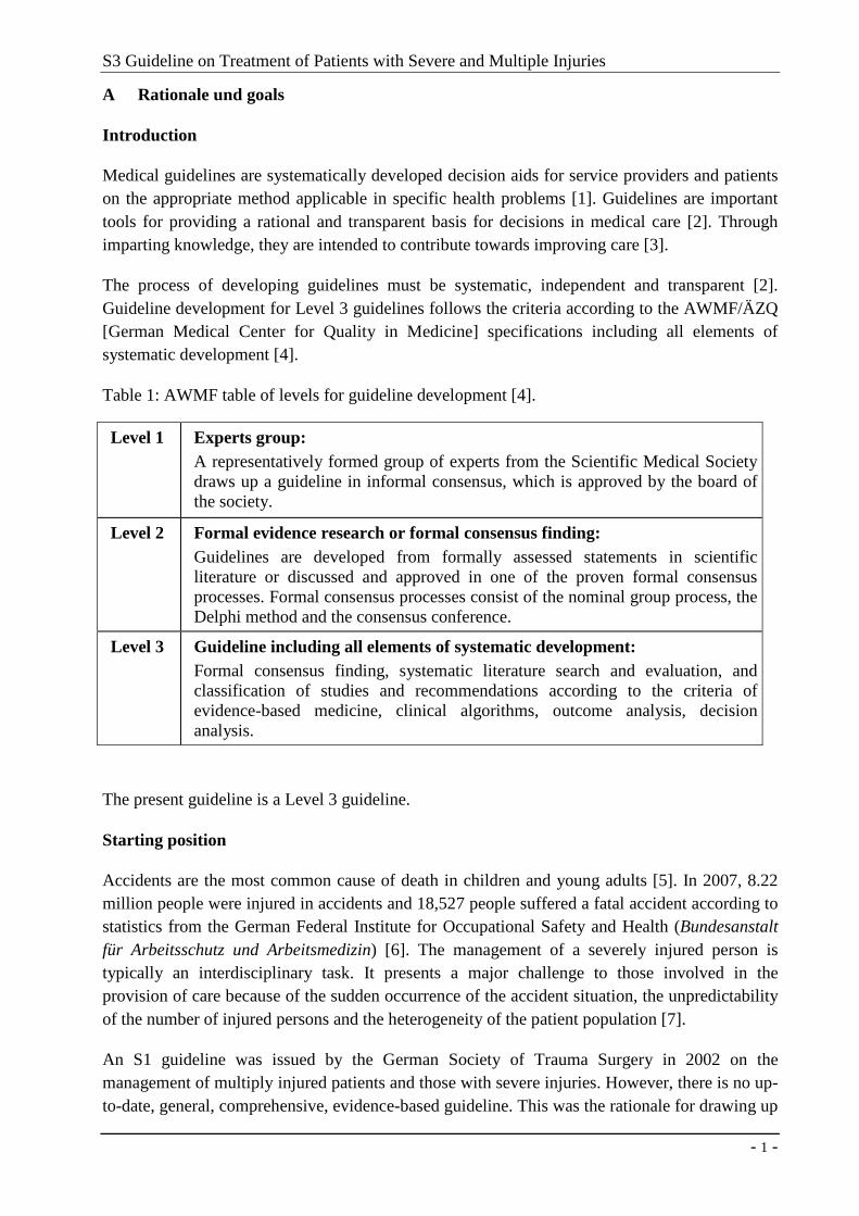
Table 1: AWMF table of levels for guideline development [4].
Level 1 Experts group: A representatively formed group of experts from the Scientific Medical Society draws up a guideline in informal consensus, which is approved by the board of the society.
Level 2 Formal evidence research or formal consensus finding: Guidelines are developed from formally assessed statements in scientific literature or discussed and approved in one of the proven formal consensus processes. Formal consensus processes consist of the nominal group process, the Delphi method and the consensus conference.
Level 3 Guideline including all elements of systematic development: Formal consensus finding, systematic literature search and evaluation, and classification of studies and recommendations according to the criteria of evidence-based medicine, clinical algorithms, outcome analysis, decision analysis.
The present guideline is a Level 3 guideline.
Starting position
Accidents are the most common cause of death in children and young adults [5]. In 2007, 8.22 million people were injured in accidents and 18,527 people suffered a fatal accident according to statistics from the German Federal Institute for Occupational Safety and Health (Bundesanstalt für Arbeitsschutz und Arbeitsmedizin) [6]. The management of a severely injured person is typically an interdisciplinary task. It presents a major challenge to those involved in the provision of care because of the sudden occurrence of the accident situation, the unpredictability of the number of injured persons and the heterogeneity of the patient population [7].
An S1 guideline was issued by the German Society of Trauma Surgery in 2002 on the management of multiply injured patients and those with severe injuries. However, there is no up-to-date, general, comprehensive, evidence-based guideline. This was the rationale for drawing up
S3 Guideline on Treatment of Patients with Severe and Multiple Injuries
- 2 -
an interdisciplinary guideline for the management of multiply injured patients and those with severe injuries.
Requirements of the guideline
The guideline must meet the following fundamental requirements:
� Guidelines for the treatment of polytrauma and patients with severe injuries are aids in decision-making in specific situations, based on the current state of scientific knowledge and on procedures proven in practice.
� Due to its complexity, there is no single ideal concept for the treatment of polytrauma and patients with severe injuries.
� Guidelines need to be constantly monitored and adapted to the current state of knowledge.
� Using the recommendations in this guideline, it should be possible to treat the vast majority of severely injured/multiply injured patients.
� Routine monitoring of treatment and monitoring the effect of treatment are necessary.
� Regular discussion with all involved (physicians, nursing staff, patients, if possible patients’ families) should make the goals and methods of treatment of polytrauma and patients with severe injuries transparent.
Aims of the guideline
This interdisciplinary S3 guideline is an evidence-based and consensus-based tool with the aim of improving the management of multiply injured patients and those with severe injuries. The recommendations are intended to contribute towards the optimization of structural and process quality in hospitals and in prehospital management and, through their implementation, help to improve outcome quality in terms of case fatality rate or quality of life.
The guideline, which is based on the current state of scientific knowledge and on procedures proven in practice, is intended to provide a decision-making aid in specific situations. The guideline can be used not only in the acute treatment situation and in the debriefing but also in discussions about local protocols by the quality circles in individual hospitals. Legal (and insurance) aspects and those relevant to billing are not explicitly dealt with in this guideline. The regulations of the German Social Code Book (Sozialgesetzbuch) (SBG VII) apply.
The guideline should be an interdisciplinary decision-making aid. For this reason, it is also suitable for drawing up new treatment protocols in individual hospitals and for revising protocols already in existence.
The aim of the guideline is to support the care of the vast majority of severely injured persons. Individual patients with defined pre-existing concomitant diseases or specific injury patterns may not all be adequately covered due to their specific problems.

S3 Guideline on Treatment of Patients with Severe and Multiple Injuries
- 3 -
The guideline is intended to stimulate further discussion to optimize the care of severely injured persons. Constructive criticism is therefore expressly welcomed. Ideally, any amendments should be briefly summarized, backed up by references and forwarded to the publisher.
Apart from the terms of reference of this guideline, it is intended to draw up interdisciplinary recommendations on the ongoing process management of severely injured persons during the acute and post-acute phase.
S3 Guideline on Treatment of Patients with Severe and Multiple Injuries
- 4 -
A.1 Publisher/experts/medical societies/authors
The responsibility for this guideline lies with the German Trauma Society (Deutsche Gesellschaft für Unfallchirurgie e. V.).
The following medical societies were involved in drawing up the guideline:
German Society of General and Visceral Surgery (Deutsche Gesellschaft für Allgemein- und
Viszeralchirurgie e. V.)
German Society of Anesthesiology and Intensive Care Medicine (Deutsche Gesellschaft für
Anästhesiologie und Intensivmedizin e. V )
German Society of Endovascular and Vascular Surgery (Deutsche Gesellschaft für
Gefäßchirurgie und Gefäßmedizin e.V.)
German Society of Hand Surgery (Deutsche Gesellschaft für Handchirurgie e.V.)
German Society of Oto-Rhino-Laryngology, Head and Neck Surgery (Deutsche Gesellschaft für
HNO-Heilkunde, Kopf- und Hals-Chirurgie e.V.)
German Society of Oral and Maxillofacial Surgery (Deutsche Gesellschaft für Mund-, Kiefer-
und Gesichtschirurgie e.V.)
German Society of Neurosurgery (Deutsche Gesellschaft für Neurochirurgie e.V.)
German Society of Thoracic Surgery (Deutsche Gesellschaft für Thoraxchirurgie e.V.)
German Trauma Society (Deutsche Gesellschaft für Unfallchirurgie e.V.)
German Society of Urology (Deutsche Gesellschaft für Urologie e.V.)
German Radiology Society (Deutsche Röntgengesellschaft e.V.)
Moderation, coordination and project management
The German Trauma Society as the lead medical association has devolved central coordination for this guideline to the Institute for Research in Operative Medicine (Institut für Forschung in der Operativen Medizin) (IFOM). The tasks were:
� coordination of the project group
� methods support and quality assurance
� systematic literature search
� procurement of literature
� data administration
� structural and editorial harmonization of the guideline texts

S3 Guideline on Treatment of Patients with Severe and Multiple Injuries
- 5 -
� coordination of necessary discussions, meetings, and consensus conferences
� administration of financial resources
S3 Guideline on Treatment of Patients with Severe and Multiple Injuries
- 6 -
Main treatment phase responsibilities
The guideline was divided into 3 main treatment phases: prehospital, emergency room, and emergency surgery. Coordinators were assigned responsibility for each of these treatment phases. The tasks were:
� establishing the contents of the guideline
� screening and evaluating the literature on the different treatment strategies for multiply injured patients and those with severe injuries, drawing up and coordinating the guideline texts
The AWMF, represented by Professor I. Kopp, provided methods guidance in drawing up the guideline.
A.2 Target user group
The guideline’s target user group is primarily the physicians and all other medical professionals involved in the management of a multiply injured patient or one with severe injuries. The recommendations relate to adult patients. Recommendations on the care of children and adolescents are only given occasionally in the guideline.
S3 Guideline on Treatment of Patients with Severe and Multiple Injuries
- 7 -
B Methods
The guideline project was first announced in December 2004 and again in May 2009.
The guideline on the “Treatment of multiply injured patients and those with severe injuries” was developed according to a binding process with a structured plan. It is the result of a systematic literature search and critical evaluation of the evidence from available data using scientific methods as well as discussion with experts in a formal consensus procedure.
B.1 Literature search and selection of evidence
The key questions for the systematic literature search and evaluation were formulated on the basis of preliminary work during 2005. The literature searches were carried out in the MEDLINE database (via PubMed) using medical keywords (Medical Subject Headings/MeSH), partly supplemented by a free text search. The filter recommended in PubMed was used to identify systematic reviews. Supplementary searches were conducted in the Cochrane Library (CENTRAL) (in this case with keywords and text words in the title and abstract). The publication period selected was 1995-2010, and German and English as the publication languages.
The literature searches were carried out partly by the Institute for Research in Operative Medicine (IFOM) and partly by the authors themselves. The results of the literature searches, sorted according to topic, were forwarded to the individual authors responsible for each topic.
The underlying key questions, the literature searches carried out with date and number of hits and, if applicable, search limitations were documented and can be found in the appendix to the separate Methods Report.
Selection and evaluation of the relevant literature
The authors of each chapter selected and evaluated the literature included in the guideline. This was carried out according to the criteria of evidence-based medicine. Sufficient randomization, allocation concealment, blinding and the statistical analysis were taken into account.
The evidence statement for the recommendations was based on the evidence classification of the Oxford Center of Evidence-Based Medicine (CEBM), March 2009 version. In formulating the recommendations, priority was given to studies with the highest level of evidence available (LoE).

S3 Guideline on Treatment of Patients with Severe and Multiple Injuries
- 8 -
Table 2: CEBM evidence classification [9]
Level Studies on therapy/prevention/etiology
1a
1b
1c
Systematic review of randomized controlled trials (RCT)
An RCT (with narrow confidence interval)
All or none principle
2a
2b
2c
Systematic review of well-planned cohort studies
A well-planned cohort study or a low-quality RCT
Outcome studies, ecological studies
3a
3b
Systematic review of case-control studies
Individual case-control study
4. Case-series or low-quality cohort/case-control studies
5. Expert opinion without explicit critical appraisal of the evidence or based on physiology/bench research
Three grades of recommendation (GoR) were possible (A, B, O). The wording of the key recommendation employs “must” “should” or “can” as appropriate. In determining the GoR, in addition to the underlying evidence, benefit-risk evaluations were also taken into account, as were the directness and homogeneity of the evidence along with clinical expertise [2].

S3 Guideline on Treatment of Patients with Severe and Multiple Injuries
- 9 -
B.2 Formulating the recommendation and finding consensus
The medical societies involved each nominated at least one delegate who, as a representative of that subject discipline, participated in drawing up the guideline. Each medical society had a vote in the consensus process.
The recommendations and the grades of recommendation were approved in 5 consensus conferences (April 18-19, 2009, June 30, 2009, September 8, 2009, November 26-27, 2009 and February 01, 2010):
The course of action at these conferences, assisted by the TED (electronic voting) system, was in 6 steps:
� the opportunity to review the guideline manuscript before the conference and to compile notes on the proposed recommendations and grades;
� presentation and explanation from each author responsible on the pre-formulated proposals for recommendations;
� registration via moderators of participants’ opinions and alternative proposals on all recommendations, with speaker contributions solely for clarification;
� voting on all recommendations and grades of recommendation and on the cited alternatives;
� discussion of the points on which no “strong consensus” could be reached in the first round;
� final voting.
Most of the recommendations were approved with “strong consensus” (agreement of > 95% of participants). Areas in which no strong consensus could be reached are marked in the guideline and the various positions are expounded. In classifying the consensus strength, the following consensus grades were decided on in advance [9]:
� strong consensus: > 95% of participants agreed
� consensus: > 75-95% of participants agreed
� majority consensus: > 50–75% of participants agreed
� no consensus: < 50% of participants agreed
The records of the meetings can be viewed at the Institute for Research in Operative Medicine (IFOM). The Delphi method was then applied to recommendations for which no consensus could be reached in the consensus conferences. A detailed methods report is available for viewing on the AWMF website and has been filed at the Institute for Research in Operative Medicine (IFOM).

S3 Guideline on Treatment of Patients with Severe and Multiple Injuries
- 10 -
B.3 Distribution and implementation
The guideline is to be distributed in the following ways:
� via the internet: AWMF website (http://www.awmf-online.de) and the websites of the medical societies and professional organizations involved in the guideline
� via printed media:
− Publication of the guideline as a manual/book by the DGU. A copy will be made available to all hospitals involved in the DGU Trauma Network. In addition, all hospitals involved will be notified in writing about where and how the guideline can be viewed on the AWMF homepage.
− Publication of extracts of the guideline and of implementation strategies in journals of the medical societies involved.
− To simplify use of the guideline, a summary of the guideline containing the key recommendations is also to be published in the Deutsches Ärzteblatt [German medical journal].
� via conferences, workshops, professional training courses offered by the medical societies involved.
Various complementary measures are to be implemented in this guideline. In addition to the presentation of the recommendations at conferences, a link to topic-specific professional training courses is planned.
In addition, implementation at all the German hospitals involved in the trauma network is to be evaluated approximately one year after publication of the guideline. In particular, it should be recorded how the guideline has been used and what practical suggestions the participants have gained from their experience to pass on to other users.
B.4 Quality indicators and evaluation
The audit filters were developed as criteria for quality management for the DGU Trauma Registry. Based on the available audit filters, the following criteria were established for this guideline:
Process quality for evaluation in the prehospital phase:
� duration of prehospital time between accident and hospital admission for severely injured patients with ISS ≥ 16 [∅min ± SD]
� intubation rate in patients with severe chest injury (AIS 4-5) by the emergency physician [%, n/total]
� intubation rate in patients with suspected traumatic brain injury (unconscious, Glasgow Coma Scale [GCS] ≤ 8) [%, n/total]

S3 Guideline on Treatment of Patients with Severe and Multiple Injuries
- 11 -
Process quality for evaluation of emergency room management:
� time between hospital admission and performance of a chest X-ray in severely injured patients (ISS ≥ 16) [∅ min ± SD]
� time between hospital admission and performance of an ultrasound scan of the abdomen/chest in cases of severe trauma (ISS ≥ 16) [∅ min ± SD]
� time until performance of a computed tomography (CT) scan of the cranium (CCT) in pre-hospital unconscious patients (GCS ≤ 8) [∅ min ± SD]
� time until performance of a full-body CT scan on all patients, if carried out [∅ min ± SD]
� time from emergency admission arrival to completion of diagnostic study in severely injured persons if this has been completed normally (ISS ≥ 16) [∅ min ± SD]
� time from emergency admission arrival to completion of diagnostic study in severely injured persons if this has been interrupted due to emergency (ISS ≥ 16) [∅ min ± SD]
Outcome quality for overall evaluation:
� standardized mortality rate: observed mortality divided by the expected prognosis based on RISC (Revised Injury Severity Classification) in severely injured persons (ISS ≥ 16)
� standardized mortality rate: observed mortality divided by the expected prognosis based on TRISS (Trauma Injury Severity Score Method) in severely injured persons (ISS ≥ 16)
The routine recording and evaluation of these data offer a vital opportunity to monitor improvements in quality in the management of multiply injured patients and those with severe injuries. It is not possible to ascertain from this which effects are due to the guideline. Quality indicators should continue to be developed based on the aforementioned criteria.
B.5 Validity and updating of guideline
This guideline is valid until December 2014. The German Trauma Society is responsible for introducing an updating process. The cooperation of the German Society of Plastic, Reconstructive and Esthetic Surgeons (Deutsche Gesellschaft der Plastischen, Rekonstruktiven und Ästhetischen Chirurgen) and of the German Society of Burns Medicine (Deutsche Gesellschaft für Verbrennungsmedizin) and the thematic inclusion of burns, large skin/soft tissue defects and nerve defect injuries (including plexus injuries) is additionally planned for this updating.
B.6 Funding of the guideline and disclosure of potential conflicts of interests
Reimbursement monies for the methods support, costs for literature acquisition, costs for organizing the consensus conferences, and costs of materials were provided by the German Trauma Society and the Institute for Research in Operative Medicine (IFOM) of the University of Witten/Herdecke. The participants’ travel costs arising from the consensus process were

S3 Guideline on Treatment of Patients with Severe and Multiple Injuries
- 12 -
covered by those medical societies/organizations sending representatives or by the participants themselves.
All participants in the consensus conference disclosed potential conflicts of interest in writing. A summary of declarations of potential conflicts of interest from all coordinators, medical society delegates, draft authors, and organizers can be found in the appendix to the separate Methods Report of this guideline. Furthermore, the forms used to disclose potential conflicts of interest can be requested from the Institute for Research in Operative Medicine (IFOM).
Grateful thanks are extended to the coordinators of the individual subsections, the authors and participants in the consensus process for their wholly voluntary work.

S3 Guideline on Treatment of Patients with Severe and Multiple Injuries
- 13 -
References
1. Field, M.J. and K.N. Lohr, eds. Clinical Practice Guidelines: Directions for a New Program. 1990, National Academy Press: Washington, D.C.
2. Council of Europe, Developing a Methodology for drawing up Guidelines on Best Medical Practices: Recommendation Rec(2001)13 adopted by the Committee of Ministers of the Council of Europe on 10 October 2001 and explanatory memorandum. 2001, Strasbourg Cedex: Council of Europe.
3. Kopp, I.B., [Perspectives in guideline development and implementation in Germany.]. Z Rheumatol, 2010.
4. Arbeitsgemeinschaft der Wissenschaftlichen Medizinischen Fachgesellschaften. 3-Stufen-Prozess der Leitlinien-Entwicklung: eine Klassifizierung. 2009; Available from: http://www.uni-duesseldorf.de/AWMF/ll/ll_s1-s3.htm.
5. Robert Koch-Institut, ed.; Gesundheit in Deutschland. Gesundheitsberichterstattung des Bundes. 2006, Robert Koch-Institut: Berlin.
6. Bundesanstalt für Arbeitsschutz und Arbeitsmedizin. Unfallstatistik: Unfalltote und Unfallverletzte 2007 in Deutschland. 2007; Available from: www.baua.de/cae/servlet/content blob/672542/publicationFile/49620/Unfallstatistik-2007.pdf;jsessionid=CC8B45BA699EE9E4E11AC1EAD359CB34.
7. Bouillon, B., et al., Weißbuch Schwerverletzten-Versorgung. Empfehlungen zur Struktur, Organisa-tion und Ausstattung stationärer Einrichtungen zur Schwerverletzten-Versorgung in der Bundesrepu-blik Deutschland., ed. D.G.f.U.e.V. (DGU). 2006, Berlin: Dt. Gesellschaft für Unfallchirurgie e.V.
8. Oxford Centre of Evidence-based Medicine (CEBM): Levels of Evidence (March 2009); Available from: www.cebm.net/index.aspx?o=1025.
9. Schmiegel, W., et al.: S3-Leitlinie “Kolorektales Karzinom: Available from: www.krebsgesellschaft.de/download/s3_ll_kolorektales_karzinom_2008.pdf.
S3 Guideline on Treatment of Patients with Severe and Multiple Injuries
Prehospital - Introduction 14
1 Prehospital
1.1 Introduction
Within the structured emergency services, the professional treatment of severely injured patients starts right at the accident scene. The subsequent course can be set at this stage. So, even for this initial treatment phase, it is expedient and necessary to develop the clearest priorities and strategies for dealing with the situation. Due to the difficult environmental conditions in the prehospital emergency situation, the evidence level is low yet the full diversity of experience and expert knowledge is considerable. Moreover, the benefit-risk evaluation is disputed in a number of interventions, not least in considering the point at which an essentially indicated intervention should be carried out, for example, in the prehospital phase or only once admitted to hospital. Finally, the polarization between “stay and treat” and “load and go” also plays a role here. In addition, a large amount of scientific knowledge has been gained from different emergency systems and its transferability to specific situations in Germany is often ambiguous.
Those active on the spot want a highly specific recommendation with broad validity but this desire is in conflict with the unfortunately often weak data available and the resulting unreliable conclusions. This desire can only be met by achieving a consensus among the experts, on the understanding that scientific uncertainty continues to exist in such areas and that there are differences between different emergency systems and cultures.
The structuring of the prehospital guideline section is based on several considerations. Basically, the management of a (potentially) severely injured patient involves a sequence of actions that follow certain priorities. Every detail and individual step of the sequence itself cannot be evidence-based with proof of general validity. Moreover, many individual circumstances relating to the actual patient have to be considered so that not all possible sequence models can be depicted. The contents of the guideline were therefore not oriented to a specific sequence algorithm but focused instead on individual aspects. These sections concentrate on anatomic regions (head, chest, abdomen, spine, extremities, and pelvis). In the prehospital phase, very few invasive interventional options are available; of these the most important (volume replacement, airway management, chest drain) are dealt with in terms of indications and implementation.
The individual aspects, interventions, and guidelines must be embedded in a general pathway of action that sets priorities and prescribes action pathways and sequences. A framework of this kind can be provided by programs such as Prehospital Trauma Life Support (PHTLS), Advanced Trauma Life Support (ATLS), European Trauma Course (ETC), and others. As such programs already exist and the individual steps cannot be individually scientifically proven, as indicated above, the attempt was not made to develop such a program in this guideline package. The individual guidelines are not intended to replace these programs but to represent the aspects embedded in them.
Besides directly treating the individual patient, general aspects also play a role in the prehospital phase. On the one hand, a decision must be made about the designated hospital. It must be able to treat all acute, life-threatening injuries immediately and independently. The initial-treating hospital must have clear, well-ordered transfer strategies for injuries which require a special structure or expertise. In addition to the increasing number of trauma networks being set up, the

S3 Guideline on Treatment of Patients with Severe and Multiple Injuries
Prehospital - Introduction 15
recommendations in the White Paper of the German Trauma Society [1] may be of great benefit here [2]. The resulting local and regional regulations can provide the emergency physician with additional support when selecting a suitable designated hospital. Besides the hospital structure, however, organizational and logistical circumstances, weather and road conditions or the time of day can also be significant in addition to purely medical considerations. Inextricably linked to this is the question of whether the patient is in fact severely injured. Criteria for this purpose are defined which are aligned to actual detected or suspected injuries, impairment of vital functions or mechanisms of injury. Finally, a balance must be found between the desire to underestimate as few patients as possible and the consequence of classifying too many patients unnecessarily as severely injured (overtriage). Conversely, although undertriage reduces the number of unnecessary emergency room alerts, it is at the cost of having underestimated more genuinely severely injured patients. The latter is viewed by many as the more critical model. Every trauma center should come to an agreement about this within its network or with the emergency services in its area.
The mass casualty incident represents a rare yet particularly challenging situation. Until the arrival of the on-duty lead emergency physician, the emergency physician who arrives on the scene first must take over this function. The switch from individual medical care to triage represents a special challenge and the algorithm should provide support here.
Many important, central domains are dealt with in the present edition of the prehospital polytrauma guideline. But some major topics, for example, pain therapy or prehospital management of traumatic brain injury, are not included. These are to be drawn up in future stages of guideline development, as well as other topics that are requested by the users.
Overall, the rapid, smoothly running medical care of (severely) injured patients is the focus of all action. In this context, the emergency services must work hand-in-hand with the hospitals. To this end, the 2008 Key Points Paper [3] on emergency medical management of patients in hospital and prehospital demands that definitive clinical treatment shall be achieved within 90 minutes for major emergency medical clinical pictures such as a severely injured patient. To make this possible, a time of 60 minutes from emergency call to hospital admission must be achieved. The scope of emergency physician care must be aimed at these targets.

S3 Guideline on Treatment of Patients with Severe and Multiple Injuries
Prehospital - Introduction 16
References
1. Bouillon, B., V. Bühren, et al. (2006). Weißbuch Schwerverletzten-Versorgung. Empfehlungen zur Struk-tur,Organization und Ausstattung stationärer Einrichtungen zur Schwerverletzten-Versorgung in der Bundesrepublik Deutschland. German Society for Trauma Surgery e.V.
2. Eckpunktepapier zur notfallmedizinischen Versorgung der Bevölkerung in Klinik und Präklinik (2008) Arbeitsgemeinschaft Südwestdeutscher Notärzte (agswn), Institut für Notfallmedizin und Medizinmanagement (INM), Bundesärztekammer (BÄK), Bundesvereinigung der Arbeitsgemeinschaften der Notärzte Deutschlands (BAND), Deutsche Gesellschaft für Anästhesiologie und Intensivmedizin (DGAI), Deutsche Gesellschaft für Chirurgie (DGCH), Deutsche Gesellschaft für Kardiologie (DGK), Deutsche Gesellschaft für Neurochirurgie (DGNC), Deutsche Gesellschaft für Unfallchirurgie (DGU), Deutsche Gesellschaft für Neonatologie und Pädiatrische Intensivmedizin (GNPI), Arbeiter Samariter Bund (ASB), Unternehmerverband privater Rettungsdienste (BKS), Deutsches Rotes Kreuz (DRK), Johanniter-Unfall-Hilfe (JUH), Malteser Hilfsdienst (MHD), Ständige Konferenz für den Rettungsdienst (SKRD). Notfall und Rettungsmedizin 11:421-422

S3 Guideline on Treatment of Patients with Severe and Multiple Injuries
Prehospital – Airway management, ventilation and emergency anesthesia 17
1.2 Airway management, ventilation and emergency anesthesia
Summary
Endotracheal intubation and ventilation, and hence definitive securing of the airways, with the aim of the best possible oxygenation and ventilation of the patient, is a central therapeutic measure in emergency medicine [80]. The basic vital functions directly linked to survival have to be secured. The “A” for airway and “B” for breathing are First Aid measures found in established standards on trauma care and therefore have a particular value in terms of weighting in both the prehospital and the early hospital management [3, 74, 107].
Variations in the emergency medical services (EMS) internationally pose a problem. Whereas paramedics are often used in the Anglo-American region, the emergency physician system is widely used in continental Europe. But even here there are differences. In Germany, (specialist) physicians in all disciplines can be involved in the emergency service after acquiring an appropriate additional qualification but in Scandinavian countries this is mainly the prerogative of anesthesiologists [9]. Consequently, the evaluation of international studies on the topic of securing the airway in the prehospital phase reveals that emergency services personnel have different levels of training. Depending on the personnel employed and how commonly they perform intubation, a high rate of esophageal intubations is found in up to 12% of cases in the literature [20]. In addition, there is a high rate of failed intubations (up to 15%) [99]. In paramedic systems, non-guideline-compliant airway management is more common [39]. Due to the different clinical routine of the users, negative outcomes in particular cannot be transferred directly from paramedic systems to the German emergency services and emergency physician system [60, 89]. In the Federal Republic of Germany, the agreed minimum qualification of “Additional qualification in emergency medicine” and the introduction of emergency anesthesia in the emergency physician system offers a different scenario compared to the Anglo-American paramedic system.
The following features of the prehospital setting can and must influence the establishing of indications and planning of anesthesia, intubation and ventilation:
� level of experience and routine training of emergency physician
� circumstances of the medical emergency (e.g., patient is trapped, rescue time)
� type of transport (land-based versus air support)
� transport time
� concomitant injuries around the airway and anything (assessable) that impedes intubation
Depending on the individual case, the indication to carry out or not to carry out prehospital anesthesia, intubation/airway management and ventilation ranges between the extremes of “advanced training level, long transport time, simple airway” and “little experience, short transport time, predicted difficult airway management”. In any event, sufficient oxygenation must be secured by appropriate measures.

S3 Guideline on Treatment of Patients with Severe and Multiple Injuries
Prehospital – Airway management, ventilation and emergency anesthesia 18
If no methodologically high-quality studies were available, a recommendation would still be issued by a consensus of experts if clinically relevant. The following recommendations cover emergency anesthesia, airway management and ventilation in the prehospital phase and emergency room management.
Key recommendations
Emergency anesthesia, endotracheal intubation, and ventilation must be carried out in the prehospital phase in multiply injured patients with apnea or gasping (<6 breaths per minute).
GoR A
Emergency anesthesia, endotracheal intubation, and ventilation should be carried out in the prehospital phase in multiply injured patients with the following indications:
� hypoxia (SpO2 < 90%) despite oxygenation and after exclusion of tension pneumothorax
� severe traumatic brain injury (GCS < 9)
� trauma-associated hemodynamic instability (SYS-BP < 90 mmHg)
� severe chest injury with respiratory insufficiency (breathing rate > 29 breaths per minute)
GoR B
The multiply injured patient must be preoxygenated before anesthesia. GoR A
The in-hospital endotracheal intubation, emergency anesthesia and ventilation must be carried out by trained, experienced anesthesiologists.
GoR A
Explanation:
Severe multiple injuries have a serious effect on the integrity of the human body in its entirety. In addition to the acute trauma consequences for the individual body sections, it causes a mediator-mediated whole-body reaction, i.e. Systemic Inflammatory Response Syndrome (SIRS) [26, 54]. Tissue oxygenation takes on special significance in this damage cascade. Tissue oxygenation can only be achieved if uptake, transport and release of oxygen are guaranteed. Oxygen uptake is only possible if the airway is secured, and endotracheal intubation is the gold standard according to the current European and non-European guidelines [32, 73, 74]. A severe impairment of consciousness due to a traumatic brain injury with a Glasgow Coma Score (GCS) < 9 is regarded as an intubation indication [8]. Endotracheal intubation for the consciousness-impaired trauma patient with a GCS ≤ 8 is also recommended both prehospital and in-hospital according to the guideline of the Eastern Association for the Surgery of Trauma (EAST) [32] and other training programs (e.g., ATLS® [3]). Hypoxia and hypotension are the “lethal duo” which induces secondary damage particularly in polytrauma with traumatic brain injury [18, 19, 52, 87, 90]. It must be further pointed out that even patients with a GCS of 13 or 14, who were intubated
S3 Guideline on Treatment of Patients with Severe and Multiple Injuries
Prehospital – Airway management, ventilation and emergency anesthesia 19
endotracheally in the prehospital phase, displayed abnormal cerebral computed tomography (38%) and intracranial bleeding (28%) [36]. In a prehospital cohort study, it was shown that endotracheal intubation has a positive effect on survival following severe traumatic brain injury [56]. Another retrospective study showed a reduced case fatality rate for children with severe traumatic brain injury who were intubated by emergency physicians in the prehospital phase as compared to those receiving care on Basic Life Support (BLS) and delayed intubation in regional trauma centers [91]. If consideration is limited to a pediatric patient population, the prehospital endotracheal intubation in this study was carried out by emergency medical personnel with good transferability to the German emergency physician system. Using the Trauma and Injury Severity Score (TRISS) method, another study also confirms that prehospital endotracheal intubation leads to improved outcomes in survival and neurologic function [38]. Another paper further showed an improvement in measured systolic blood pressure, oxygen saturation and end-tidal carbon dioxide (etCO2 compared to the baseline values prior to prehospital intubation in patients with severe traumatic brain injury [11].
Current review papers, however, refer to heterogeneous patient collectives, differing emergency services systems and differently trained users and therefore do not always come to a positive conclusion about intubation [9, 12, 25, 31, 60, 62, 69, 74, 98, 100]. The EAST guideline group also tackled this problem. In the “Guidelines for Emergency Intubation immediately following traumatic injury”, it was claimed that there are no randomized controlled trials on this research question. On the other hand, however, the authors of the EAST Guideline also found no studies that could present an alternative treatment strategy proven to be effective. In summary, endotracheal intubation was assessed overall as such an established procedure in hypoxia/apnea that, despite a lack of scientific evidence, a Grade A recommendation was formulated [73]. Other indications for endotracheal intubation (e.g., chest injury) are controversial issues in the literature [78]. There was evidence that hypoxia and respiratory insufficiency were a consequence of severe chest injury (multiple rib fractures, pulmonary contusion, unstable chest wall). Endotracheal intubation is recommended if the hypoxia cannot be remedied by oxygenation, by the exclusion of tension pneumothorax, and by basic airway management procedures [32]. Prehospital endotracheal intubation in patients with severe chest injury is suitable for preventing hypoxia and hypoventilation, which are associated with secondary neurologic damage and extremely severe consequences for the rest of the body. However, with difficult, prolonged intubation attempts and the associated hypoventilation and danger of hypoxia, endotracheal intubation itself can cause procedure-related secondary harms or even death. A database analysis of the Trauma Registry of the German Trauma Society showed no advantage in prehospital endotracheal intubation in patients with chest injury without respiratory insufficiency [78]. However, severe chest injury with respiratory insufficiency does present an indication for prehospital endotracheal intubation whereby the decision to intubate should be dependent on the respiratory insufficiency and not on the (suspected) diagnosis of severe chest injury, which is associated with a certain degree of uncertainty [7].
Endotracheal intubation is included as an “Advanced Life Support” procedure in the prehospital action algorithms of various training programs (e.g., PHTLS® [71]. Using a scoring system to evaluate management problems plus the relevant autopsy reports, a series of fatal traffic accidents were retrospectively analyzed to characterize the effectiveness of prehospital care and potentially avoidable fatal incidents [76]. This flagged up an extended “prehospital and early in-

S3 Guideline on Treatment of Patients with Severe and Multiple Injuries
Prehospital – Airway management, ventilation and emergency anesthesia 20
hospital care period” and a “lack of airway securing using intubation” as factors which led to the incidence of avoidable fatal incidents [76].
A retrospective cohort study of 570 intubated patients compared to 8,137 non-intubated patients showed that the prehospital intubated patients had a prehospital phase which was between 5.2-10.7 minutes longer than the non-intubated patients [24]. The effect of early intubation within 2 hours following trauma on the incidence of subsequent organ failure was evaluated in a prospective non-randomized study [97]. Despite a significantly higher degree of injury, there was a lowered incidence of organ failure and lower case fatality rate in the group of patients who were endotracheally intubated “early” within 2 hours following trauma, compared to the “later” intubated patients. The following factors must therefore be taken into account in selecting the best time to introduce anesthesia and endotracheal intubation: injury pattern, personal experience of the emergency physician/anesthesiologist, environmental conditions, transport distance, available equipment and complications associated with the procedure. Taking these points into consideration, the definitive care that the multiply injured patient should receive is emergency anesthesia with endotracheal intubation and ventilation. With the appropriate indication and appropriate training level, endotracheal intubation should be performed prehospital but, at the latest, during emergency room management. According to the analysis of data from the Trauma Registry of the German Trauma Society, out of 24,771 patients, 31% were unconscious at the accident scene (GCS < 9), 19% had severe hemodynamic instability (systolic blood pressure < 90 mmHg) and, overall, 55% of patients were endotracheally intubated by the emergency physician during the prehospital phase [77]. According to this analysis, in the case of 9% of multiply injured patients, it was necessary to discontinue the emergency room phase in hospital in favor of an emergency intervention/surgery; a total of 77% of multiply injured patients received surgery and 87% needed intensive care [77]. Due to a traumatic brain injury and/or chest injury, many multiply injured patients required intensive care ventilation and invasive ventilation therapy and all required adequate pain relief. In the study mentioned, the mean ventilation period for multiply injured patients was 9 days [77].
In order to prevent the harmful effects of hypoxia and hypoventilation, emergency anesthesia and endotracheal intubation and ventilation should be introduced prehospital or, at the latest, during emergency room care for the appropriate indication and with the appropriate training level. A large retrospective study using a trauma registry from a Level I trauma center studied 6,088 patients who received endotracheal intubation within the first hour following hospital admission [88]. In addition, according to this trauma registry, a further 26,000 trauma patients were endotracheally intubated after the first hour of hospital care on the day of admission. In the hands of experienced anesthesiologists, the “rapid sequence induction” proved in these cases to be an effective, safe procedure in hospital care: no patient died as a result of endotracheal intubation. Of 6,088 patients, 6,008 were successfully intubated orotracheally (98.7%) and a further 59 nasotracheally (0.97%). Only 17 patients (0.28%) had to have a cricothyroidotomy and 4 patients (0.07%) received an emergency tracheotomy. Following the endotracheal intubation, 3 more patients received an emergency tracheotomy during the course [88]. In another retrospective study of a monocenter trauma registry, 1,000 trauma patients (9.9% out of 10,137 patients) who had been endotracheally intubated within 2 hours of admission to the Trauma Center were studied [85]. At < 1%, the incidence of surgically securing the airway was uncommon in this study as well. Aspiration occurred in 1.1% of cases of endotracheal intubation.
S3 Guideline on Treatment of Patients with Severe and Multiple Injuries
Prehospital – Airway management, ventilation and emergency anesthesia 21
Early intubation was seen as safe and effective by the authors [85]. These data also confirm that endotracheal intubation of trauma patients is a safe procedure in the hands of trained personnel. Another retrospective study from a paramedic-supported system with 175 endotracheally intubated patients showed a success rate of 96.6% with a markedly higher cricothyroidotomy rate of 2.3% [37]. In 1.1% of cases, the patient was ventilated by bag-valve-mask during transfer to hospital. There were 5 instances found of right mainstem dislocation (2.9%) and 2 cases of tube dislocation (1.1%). No case of failed intubation was documented.
In a retrospective study of a trauma registry, 3,571 prehospital endotracheal intubations in trauma patients were compared with 746 in the emergency room phase [6]. The endotracheal intubation first carried out during emergency admission was associated with a higher risk of a fatal course compared both to non-intubated patients (odds ratio [OR] 3.1; 95% confidence interval [-CI]; 2.1–4.5, p < 0.0001) and to patients who had already been endotracheally intubated in the prehospital phase (OR 3.0; 95% CI: 1.9–4.9, p < 0.0001) [6]. In addition, it was shown that patients who had been endotracheally intubated in the prehospital phase did not have a higher risk of dying than non-intubated patients in the emergency room phase (OR: 1.1; 95% CI: 0.7–1.9; p = 0.6). The authors concluded that the patients who were endotracheally intubated during emergency admission should have already been intubated in the prehospital phase [6].
In a prehospital cohort study with comparable injury severity (ISS 23 versus 24) and similar duration of care (27 versus 29 min, p = n.s.), 60 patients were treated by emergency services personnel (emergency medical technician [EMT], intubation rate 3%) and 64 patients in Advanced Life Support mode by emergency physicians (intubation rate 100%). Oxygen saturation was significantly improved upon arrival in hospital (SaO2: 86 versus 96; p = 0.04) and systolic blood pressure was significantly higher (105 versus 132 mmHg, p = 0.03). There was no difference in all-cause case fatality rate (42% versus 40%, p = 0.76). However, a sub-group analysis showed a significant survival advantage for those patients with a GCS between 6 and 8 who had been treated by an emergency physician (case fatality rate: 78 versus 24%, p < 0.01; OR 3.85, 95% CI: 1.84–6.38, p < 0.001). The authors concluded that the case fatality rate is reduced by a prehospital emergency physician system offering rapid sequence induction, sufficient oxygenation and circulation drug therapy particularly for patients with clouded consciousness [56].
In the German-speaking emergency physician system, pediatric and adult emergency patients can be endotracheally intubated with a very high success rate if this procedure is carried out by experienced and trained personnel. In a prospective study over a period of 8 years, 4% of all pediatric emergency patients (82 out of 2,040 children) were endotracheally intubated [35]. Pediatric emergency callouts made up 5.6% of all emergency calls (2,040 out of 36,677 emergency physician callouts). Anesthesiologists carried out 58 of the pediatric endotracheal intubations with a success rate of 98.3%. Based on the incidence, the known number of emergency physicians employed each year, and their absolute number of callouts, it was calculated that each emergency physician in the emergency physician service has a gap of, on average, 3 years between pediatric endotracheal intubations and 13 years between infant endotracheal intubations. These results show that endotracheal intubation in childhood is rare outside the hospital setting and special attention must therefore be paid to maintaining specialist

S3 Guideline on Treatment of Patients with Severe and Multiple Injuries
Prehospital – Airway management, ventilation and emergency anesthesia 22
expertise and appropriate training outside the emergency services and emergency physician service.
A prospective study of a cohort of 16,559 patients managed in the prehospital phase included 2,850 trauma patients of which 259 (9.1%) were endotracheally intubated. More than 2 attempts were required in 3.9% of cases before endotracheal intubation was successful, and there was a failed intubation in 3.9% of cases. A difficult airway was described in 18.2% of cases. In comparison, patients with cardiac arrest had a difficult airway in only 16.7% of cases. This study also showed a success rate of 98.0% by anesthesia-trained emergency physicians [94]. Another prospective study of an emergency physician system showed a success rate of 98.5% in 598 patients (of which 10% were trauma patients ) [92]. In another prospective study, endotracheal intubation by anesthesia-trained emergency physicians achieved a 100% success rate in a collective of 342 patients [n = 235 (68.7%) trauma patients]. In this case, endotracheal intubation was successful at the first attempt in 87.4% of cases, at the second attempt in 11.1% and at the third attempt in 1.5% [48]. Another study of the German emergency physician system showed a success rate of 97.9% in prehospital endotracheal intubation of trauma patients [1].
In a retrospective cohort study with 194 patients with traumatic brain injury, there was a significant difference in the case fatality rate between patients treated with basic life support (BLS) procedures in the land-based emergency services and patients who were treated with advanced life support (ALS) procedures by anesthesiologists in the air-borne emergency services (25 versus 21 %, p < 0.05). In this study, the survival rate of patients with traumatic brain injury who were treated highly significantly with more invasive measures in the air rescue group (intubation 92 versus 36%, chest drain 5 versus 0%) was better than the survival rate of patients treated in the land-based emergency services (54 versus 44%, p < 0.05) [10].
Procedural-related complications
Regarding procedural-related complications, a retrospective study using data from a trauma registry showed that there was no higher risk of pneumonia developing in 271 prehospital and 357 in-hospital endotracheally intubated trauma patients [96]. Regarding epidemiological data, prehospital intubated patients showed a lower GCS (4 versus 8, p < 0.001) and a higher injury severity according to the ISS (25 versus 22, p < 0.007) but otherwise no differences in the patient characteristics. Nevertheless, although it was to be expected, there was no difference in the length of hospital stay for both patient collectives (15.7 versus 15.8 d), in the length of intensive care stay (7.6 versus 7.3 d), in the number of days on a ventilator (7.8 versus 7.2 d), in the case fatality rate (31.7 versus 28.2%), and in the rate of resistant bacteria (46% in each case). On average, it took 3 days until the onset of pneumonia in both groups and the pneumonia rate was also not significantly different in both groups [96]. However, a significantly increased rate of pneumonia following prehospital intubation compared to in-hospital intubation was observed in another study [86]. However, this had no influence on the 30-day case fatality rate and the number of days in intensive care. Moreover, the group of prehospital intubated patients had an increased injury severity. In another study, frequency of pulmonary complications was found to be related to injury severity but not to intubation mishaps [84]. It cannot be definitely proven that prehospital endotracheal intubation is related to the incidence of pulmonary complications. In a retrospective study of 244 patients endotracheally intubated in the prehospital phase by an
S3 Guideline on Treatment of Patients with Severe and Multiple Injuries
Prehospital – Airway management, ventilation and emergency anesthesia 23
emergency physician, desaturation with an SpO2 < 90% was documented in 18% of cases and hypotension with systolic blood pressure < 90 mmHg in 13% of cases. The two complications did not occur in parallel in any of the cases [72]. Overall, a low complication rate can be accordingly assumed.
Preoxygenation
To avoid a fall in oxygen saturation during the introduction of anesthesia and endotracheal intubation, the multiply injured patient should, if practicable, be preoxygenated for up to 4 minutes with an oxygen concentration of 100% via a face mask with reservoir [74]. In a non-randomized controlled study of 34 intensive-care patients, the mean paO2 was (T0) 62 ± 15 mmHg at the start of preoxygenation, (T4) 84 ± 52 mmHg after 4 minutes, (T6) 88 ± 49 mmHg after 6 minutes and (T8) 93 ± 55 mmHg after 8 minutes. The differences in paO2
were significantly different between T0 and T4–8 , but no statistical differences could be obtained between the paO2 between T4, T6 and T8. 24% of patients even showed a reduction in the paO2
between T4 and T8. A longer period of preoxygenation for 4 to 8 minutes did not lead to any further marked improvement in arterial oxygen partial pressure and delayed securing the airway in critical patients [67, 68]. Accordingly, sufficient preoxygenation for 4 minutes has special importance in securing the airway in multiply injured patients.
Training
Key recommendation:
Emergency medical personnel must be regularly trained in emergency anesthesia, endotracheal intubation, and alternative ways of securing an airway (bag-valve-mask, supraglottic airway devices, emergency cricothyroidotomy).
GoR A
Explanation:
In a survey recently carried out among prehospital trained emergency physicians, they were questioned on their knowledge of and experience in endotracheal intubation and the alternative methods for securing an airway [93]. This survey included the responses from 340 anesthesiologists (56.1%) and 266 non-anesthesiologists. It revealed that all anesthesia-trained emergency physicians could demonstrate more than 100 endotracheal intubations performed in hospital in contrast to only 35% of non-anesthesiologists performing more than 100 in-hospital intubations. A similar picture emerges for alternative methods for securing an airway as well. 97.8% of anesthesia-trained emergency physicians had used alternative methods for securing an airway on more than 20 occasions while only 11.1% of non-anesthesia-trained emergency physicians had equivalent experience (p < 0.05). In addition, it emerges that only 27% of emergency equipment was equipped with CO2 monitors. From this study it can be concluded that there is an urgent training need for non-anesthesia-trained emergency physicians in endotracheal intubation, capnography and alternative methods for securing an airway [74]. Studies on first-year anesthesiology residents showed that more than 60 intubations were
S3 Guideline on Treatment of Patients with Severe and Multiple Injuries
Prehospital – Airway management, ventilation and emergency anesthesia 24
necessary in order to achieve a success rate of 90% within the first two endotracheal intubation attempts under standardized, optimum conditions in surgery [57]. However, as the success of alternative methods for securing an airway (e.g., supraglottic airways: laryngeal mask, laryngeal tube) can only be as good as the corresponding training level in this procedure and current evidence indicates that a corresponding training level is not available everywhere [93], endotracheal intubation continues to be the gold standard. This knowledge also illustrates that emergency medical personnel should be regularly trained in endotracheal intubation and alternative ways of securing an airway [74].
S3 Guideline on Treatment of Patients with Severe and Multiple Injuries
Prehospital – Airway management, ventilation and emergency anesthesia 25
Alternative methods for securing an airway
Key recommendations
A difficult airway must be anticipated when endotracheally intubating a trauma patient.
GoR A
Alternative methods for securing an airway must be provided when anesthetizing and endotracheally intubating a multiply injured patient.
GoR A
Fiberoptic intubation must be available as an alternative when anesthetizing and endotracheally intubating in-hospital.
GoR A
If difficult anesthetization and/or endotracheal intubation are expected, an anesthesiologist must carry out or supervise this procedure in-hospital provided this does not cause a delay in an emergency life-saving measure. Suitable measures must be in place to ensure that an anesthesiologist is normally on site in time
GoR A
After more than 3 intubation attempts, alternative methods must be considered for ventilation and securing an airway.
GoR A
Explanation:
Due to the framework conditions, the endotracheal intubation of an emergency patient is markedly more difficult in the prehospital environment than in-hospital. A difficult airway must therefore always be anticipated when endotracheally intubating a trauma patient [74]. In a large study of 6,088 trauma patients, risk factors and difficulties in endotracheal intubation consisted of foreign bodies in the pharynx or larynx, direct injuries to the head or neck with loss of normal anatomy in the upper airway, airway edema, pharyngeal tumors, laryngospasms and a difficult pre-existing anatomy[88]. In another study, trauma patients presented difficult airway securing markedly more frequently (18.2%) than, for example, patients with cardiac arrest (16.7%) and patients with other diseases (9.8%). Reasons described for difficult airway management were the position of the patient (48.8% of cases), difficult laryngoscopy (42.7% of cases), secretion or aspiration in the oropharynx (15.9% of cases) and traumatic injuries (including bleeding/burns) in 13.4% of cases [94]. Technical problems occurred in 4.3% and other causes in 7.3% of cases. Further studies show a similar frequency of causes of difficult intubation (blood 19.9%, vomit 15.8%, hypersalivation 13.8%, anatomy 11.7%, changes in anatomy caused by trauma 4.4%, position of patient 9.4%, lighting conditions 9.1%, technical problems 2.9% [48]. In a prospective study with 598 patients, adverse events and complications occurred significantly more frequently in patients with severe injuries than non-traumatized patients (p = 0.001) [92]. At least one event was documented in 31.1% of traumatized patients. The number of attempts required for intubation was also significantly increased in traumatized patients (p = 0.007) [92].
S3 Guideline on Treatment of Patients with Severe and Multiple Injuries
Prehospital – Airway management, ventilation and emergency anesthesia 26
An increased risk of difficult intubation exists particularly in patients with severe maxillofacial trauma (OR 1.9, 95% CI: 1.0–3.9, p = 0.05) [22]. Maxillofacial trauma even represents an independent factor for difficult airway management (OR 2.1, 95% CI: 1.1–4.4, p = 0.038). A retrospective analysis of a trauma registry over a period of 7 years identified 90 patients with severe maxillofacial injuries. Of these, 93% initially received definitive airway securing, in 80% of cases by means of endotracheal intubation and in 15% of cases through surgical airway securing [21]. On the basis of these available data, the trauma patient must definitely be assumed to be non-fasting. In addition, blood, vomit or other fluids associated with a more difficult intubation situation must be expected in the oropharynx to a greater extent. A high-performance suction unit must therefore be available as a matter of course. For structure- and process-related reasons, the possibility of a back-up procedure with an experienced anesthesiologist often does not exist in the prehospital setting but in-hospital the gold standard is generally to involve an anesthesiologist in the management when difficult intubations and anesthesia are expected. In a prospective cohort study, it was therefore shown that if an attending physician in anesthesiology was present at in-hospital emergency intubations, significantly fewer complications occurred (6.1 versus 21.7%, p < 0.0001) [81]. However, there was no difference in the ventilation-free days and the 30-day case fatality rate.
If endotracheal airway securing fails, an appropriate algorithm must be followed, reverting back to bag-valve-mask ventilation and/or alternative methods of securing an airway [4, 15, 49, 73, 74]. In a prospective study, intubation success was evaluated in 598 patients in an emergency physician system solely staffed by anesthesiologists. Endotracheal intubation was successful at the first attempt in 85.4% of all patients. Only 2.7% required more than two attempts, and 1.5% (n = 9) had supralaryngeal aids such as the Combitube (n = 7), laryngeal mask (n = 1) or an emergency cricothyroidotomy (n = 1) after the third unsuccessful intubation attempt [92]. The study illustrates that alternative methods must be provided even in highly professional systems [55].
In a retrospective study of 2,833 patients endotracheally intubated in-hospital at a Level I trauma center, it was shown that the risk of airway-associated complications was markedly increased with more than 2 intubation attempts: hypoxemia 11.8 versus 70%, regurgitation 1.9 versus 22%, aspiration 0.8 versus 13%, bradycardia 1.6 versus 21%, cardiac arrest 0.7 versus 11% [65]. Another study, which was prospective and multi-center, examined over an 18-month period how many intubation attempts (inserting the laryngoscope into the oral cavity) were necessary for successful endotracheal intubation in emergency patients [101]. In 94% of cases, endotracheal intubation was carried out by paramedics and in a further 6% by nurses or emergency physicians. Overall, 1,941 endotracheal intubations were carried out, of which 1,272 (65.5%) were in patients with cardiac arrest, 463 (23.9%) as intubation without drug administration in patients without cardiac arrest, 126 (6.5%) as intubations under sedation in patients without cardiac arrest, and 80 (4.1%) by means of rapid sequence induction using a hypnotic agent and a muscle relaxant. Over 30% of patients required more than one intubation attempt to achieve successful endotracheal intubation. More than 6 intubation attempts were not reported in any case. The cumulative success rate during the first, second and third intubation attempt was 70%, 85% and 90% in patients with cardiac arrest. It was thus markedly higher than in the other 3 patient subgroups with intact circulatory function (intubation without drugs: 58%, 69% and 73%; intubation under sedation: 44%, 63% and 75%; intubation by means of rapid sequence induction:

S3 Guideline on Treatment of Patients with Severe and Multiple Injuries
Prehospital – Airway management, ventilation and emergency anesthesia 27
56%, 81% and 91%). The specific success rates of endotracheal intubation by paramedics, nurses and emergency physicians were not broken down further. The results of this study [101] show that the cumulative success rate of endotracheal intubation in a paramedic system is markedly below that of emergency physician systems staffed solely by anesthesiologists, whose rate is 97-100% [48, 92, 94]. However, the administration of drugs, as they are used during a rapid sequence induction (including muscle relaxants), helps to facilitate endotracheal intubation in patients without cardiac arrest and thus leads to a markedly higher intubation success. Both are frequently vital for survival in an emergency situation. According to the above-cited study results, alternative methods should be considered for securing an airway after more than 3 intubation attempts [4, 65]. Although fiberoptic procedures are only available in isolated cases in the prehospital phase, fiberoptic intubation must be available in-hospital. In all common guidelines and recommendations on emergency airway securing, (awake) fiberoptic intubation is considered a possible procedure for securing an airway if there is appropriate experience and appropriate environmental conditions [33, 46, 49, 59].
In contrast, emergency cricothyroidotomy is simply the last resort in a “cannot ventilate - cannot intubate” situation to secure ventilation and oxygenation in an emergency. In national and international recommendations and guidelines, emergency cricothyroidotomy has a firm place in prehospital and hospital phases and is indicated if alternative methods for securing an airway and bag-valve-mask ventilation are not successful [9, 46, 49, 70].

S3 Guideline on Treatment of Patients with Severe and Multiple Injuries
Prehospital – Airway management, ventilation and emergency anesthesia 28
Monitoring emergency anesthesia
Key recommendation:
ECG, blood pressure measurement, pulse oxymetry and capnography must be used to monitor the patient for anesthesia induction, endotracheal intubation and emergency anesthesia.
GoR A
Explanation:
The German Society of Anesthesiology and Intensive Care Medicine (DGAI) lays down certain features for a “standard workplace” in its update to the directives on equipping the anesthesiology workplace [27]. Special attention must be paid to the often difficult prevailing circumstances (e.g., physical confines, unfavorable lighting conditions, limited resources) in prehospital emergency medicine and particularly in the care of trauma patients.
The following items of equipment should be available in the prehospital phase for carrying out and monitoring emergency anesthetization [74]: electrocardiogram (ECG), non-invasive blood pressure measurement, pulse oxymetry, capnography/capnometry, defibrillator, emergency respirator, and suction unit. Appropriate equipment must be provided based on the guideline “Airway Management” of the German Society of Anesthesiology and Intensive Care Medicine [15] and the German DIN standards for emergency physician vehicle (NEF) [28], rescue helicopter (RTH) [29] and ambulance (RTW) [30].
In-hospital, the directives of the DGAI must be followed in the emergency room and in the other hospital wards [27].
Emergency ventilation and capnography
Key recommendations
During endotracheal intubation in the prehospital and in-hospital phases, capnometry/capnography must be used for monitoring tube placement and then for monitoring dislocation and ventilation.
GoR A
Normoventilation must be carried out in endotracheally intubated and anesthetized trauma patients.
GoR A
From emergency room treatment onwards, ventilation must be monitored and controlled by frequent arterial blood gas analyses.
GoR A
S3 Guideline on Treatment of Patients with Severe and Multiple Injuries
Prehospital – Airway management, ventilation and emergency anesthesia 29
Explanation:
In the prehospital and in-hospital phases, capnometry/capnography must always be used during endotracheal intubation for monitoring the placement of the tube and then to reduce incidence of dislocation and monitor ventilation. Capnography is an essential component here in monitoring the intubated and ventilated patient [74]. Normoventilation should be carried out in endotracheally intubated and anesthetized trauma patients. From emergency room treatment onwards, ventilation must be monitored and controlled by frequent arterial blood gas analyses.
Capnography for monitoring tube placement and dislocation
The most serious complication in endotracheal intubation is an unrecognized esophageal intubation, which can lead to the death of the patient. This is why, both prehospital and in-hospital, all methods must be applied to recognize esophageal intubation and remedy it immediately.
The percentage of esophageal intubations reported in the literature starts at less than 1% [100, 106] spanning 2% [40], 6% [75], and reaching almost 17% [53]. Moreover, a high case fatality rate was shown as a result of tube misplacement in the hypopharynx (33%) or in the esophagus (56%) [53]. Esophageal intubation is thus not a rare event and, particularly in recent years, various studies have examined this catastrophic complication of endotracheal intubation in Germany as well. In a prospective observational study, helicopter emergency physicians trained in anesthesiology identified an esophageal tube placement in 6 out of 84 (7.1%) trauma patients, who had been intubated by land-based emergency physicians before arrival of the helicopter, and an endobronchial tube placement in 11 (13.1%) [95]. The case fatality rate of esophageally intubated patients was 80% in this study. In another prospective study with 598 patients in a German emergency physician system, the rate of esophageal intubations by non-medical personnel or physicians before arrival of the actual emergency physician system was 3.2% [92]. Another prospective observational study revealed esophageal intubation in 5.1% of 58 patients, who had been intubated by the land-based emergency service or emergency physician before arrival of the helicopter emergency physician trained in anesthesiology [43]. In a study focusing on the admitting emergency room team, esophageal intubation was found in 4 out of 375 prehospital intubated and ventilated patients (1.1%) [41].
In a prospective observational study of 153 patients, evidence showed that none of the patients who had been monitored by capnography had an unrecognized misplaced intubation, but 14 out of the 60 patients (23.3%) not monitored by capnography had [83]. Capnography therefore belongs in the standard equipment of the anesthesiology workplace and has dramatically increased the safety of anesthesia.
In a prospective observational study with 81 patients (n = 58 severe traumatic brain injury [TBI], n = 6 maxillofacial trauma, n = 17 multiple injuries), markedly greater sensitivity and specificity was demonstrated by monitoring tube placement by capnography compared to auscultation only (sensitivity: 100 versus 94%; specificity: 100 versus 66%, p < 0.01) [44]. These data prove that capnography must always be used for monitoring tube placement.
S3 Guideline on Treatment of Patients with Severe and Multiple Injuries
Prehospital – Airway management, ventilation and emergency anesthesia 30
A survey found that in Baden-Wurttemberg only 66% out of 116 emergency physician sites had capnography available in 2005 [42]. There is an urgent need here for optimization. In addition, it is unknown how often available capnography is actually used in prehospital endotracheal intubation, tube position verification, and emergency ventilation monitoring. The goal must be to reach a capnography rate of 100% in the prehospital and in-hospital phases. On the basis of the guideline “Airway Management” of the German Society of Anesthesiology and Intensive Care Medicine and the German DIN standards for emergency physician vehicles (NEF) [28], rescue helicopters (RTH) [29] and ambulances (RTW) [30] on the mandatory availability of capnography, the lack of appropriate equipment basically constitutes organizational negligence [42].
Capnography for normoventilation
The introduction of emergency anesthetization is not only used to maintain adequate oxygenation but also effective ventilation and thus the elimination of carbon dioxide (CO2), which accumulates in the human metabolism. Both an accumulation of CO2 (hypercapnia and hyperventilation) and hyperventilation with consecutive hypocapnia can cause damage particularly in patients with traumatic brain injury and must be avoided in the first 24 hours [14, 16]. This results in a vicious circle of elevated intracranial pressure, hypercapnia, hypoxemia, further cellular swelling/edema and subsequent further increase in intracranial pressure.
In a retrospective analysis of prehospital care data from 100 prehospital intubated and ventilated patients, it was shown that an etCO2 > 30 mmHg was attained in 65 patients and an etCO2 ≤ 29 mmHg in 35 patients. A lower case fatality rate was noticeably more likely in normoventilated patients (case fatality rate: 29 versus 46%; OR 0.49, 95% CI: 0.1–1.1, p = 0.10) [17].
In a prospective observational study, only 155 out of 492 patients intubated and ventilated in the prehospital phase showed a paCO2 between 30 and 35 mmHg in the initial arterial blood gas analysis (BGA) in the emergency room and were thus (according to the study protocol) normoventilated [102]. Eighty patients (16.3%) who were hypocapnic (paCO2 < 30 mmHg), 188 patients (38.2%) who were mildly hypercapnic (paCO2 36–45 mmHg) and 69 patients (14.0 %) who were severely hypercapnic (paCO2 > 45 mmHg) were ventilated. The injury severity of the severely hypercapnic patients (paCO2 > 45 mmHg) was markedly higher and these patients also had hypoxia, acidosis or hypotension significantly more frequently compared to the other 3 groups. The case fatality rate of prehospital intubated and ventilated trauma patients both with and without TBI was specifically lowered by normoventilation (OR: 0.57, 95% CI: 0.33–0.99), with patients with isolated TBI gaining more markedly from normoventilation (OR: 0.31, 95% CI: 0.31–0.96). According to the available results, hyperventilation with consecutive hypocapnia (paCO2 < 30 mmHg) in particular appears to be harmful in severely injured patients. These results make clear that, from emergency room treatment onwards, ventilation should be monitored and controlled by frequent arterial blood gas analyses.
In a prospective study of 97 patients, it was shown that patients monitored by capnography had a significantly higher rate of normoventilations (63.2 versus 20%, p < 0.0001) and significantly fewer hypoventilations (5.3 versus 37.5%, p < 0.0001) compared to patients who were ventilated without capnography monitoring but by using the 10-10 rule [47]. Capnography is thus an
S3 Guideline on Treatment of Patients with Severe and Multiple Injuries
Prehospital – Airway management, ventilation and emergency anesthesia 31
orientating procedure in emergency ventilation. When capnography is used for controlling ventilation, it must be taken into account that the correlation between etCO2 and paCO2 is nevertheless weak (r = 0.277) [104]. As a result of a prospective observational study with 180 patients, 80% of patients with an etCO2 of 35–40 mmHg were indeed hypoventilated (paCO2 > 40 mmHg). In a prospective study of 66 intubated and ventilated trauma patients, those patients in particular with high injury severity according to the ISS, hypotension, severe chest injury, and metabolic acidosis revealed a larger difference between etCO2 and paCO2 [61]. Thus, the arterial CO2 (paCO2) cannot always be directly inferred from the CO2 (etCO2) obtained by capnography [74]. This is due to the fact that good correlation between etCO2 and paCO2 under physiologic conditions is negatively affected by pulmonary shunt fractions in pulmonary contusions, atelectasis, hypotension and metabolic acidosis.
Thus, capnography primarily serves to evaluate tube placement and to monitor on-going ventilation, with ventilation control a secondary use. This was also briefly demonstrated in a retrospective cohort study with 547 trauma patients: all trauma patients and especially patients with severe TBI gained from paCO2-controlled ventilation (OR: 0.33, 95% CI: 0.16–0.75). There was a significant survival advantage if paCO2 was already between 30 and 39 mmHg on admission to the emergency room (OR 0.32, 95% CI: 0.14–0.75). In patients whose paCO2 could only be brought into the target range during their stay in the emergency room, there was only a non-significant trend towards a lower case fatality rate (OR 0.48, 95% CI: 0.21–1.09). A markedly worse survival rate was shown by those trauma patients who initially had a paCO2 of 30–39 mmHg but were then hypoventilated (paCO2> 39 mmHg) or hyperventilated (paCO2 < 30 mmHg) or who never attained the target goal of a paCO2 of 30–39 mmHg during their stay in the emergency room. This study also illustrates that paCO2 must not be freely inferred from etCO2 [102].
Using capnography to check tube placement and to detect tube dislocations is advisable and essential. Capnography is the gold standard in standard anesthesia, and ventilation management is markedly better with capnography than without this procedure. There are limitations to ventilation management using capnography due to unpredictable shunt fractions. For this reason, ventilation must be managed by blood gas analysis as early as possible, in other words immediately upon admission to the emergency room.
Lung protective ventilation
In a prospective randomized study, ventilation with a small tidal volume (6 ml/kg BW) in patients with acute respiratory distress syndrome (ARDS) led to a significantly reduced case fatality rate and a lower incidence of barotrauma and improved oxygenation compared to ventilation with high tidal volume [2]. The multi-center randomized, controlled trial conducted by the ARDS network confirmed these results in a ventilation with low tidal volume and limiting plateau pressure to ≤ 30 cm H2O in patients with ARDS [5]. Chest injuries are observed in around 60% of multiply injured patients with the corresponding consequences (e.g., pulmonary contusions, ARDS), and the development of an acute lung injury (ALI) as an independent factor is associated with the case fatality rate (case fatality rate of trauma patients with ALI [n = 93]: 23.7 versus without ALI [n = 190]: 8.4%, p < 0.01) [82]. Thus, lung protective ventilation with a
S3 Guideline on Treatment of Patients with Severe and Multiple Injuries
Prehospital – Airway management, ventilation and emergency anesthesia 32
tidal volume of 6 ml/kg BW and with the lowest possible peak pressures must be implemented as early as possible following endotracheal intubation [45].
Emergency anesthesia
Key recommendations
For endotracheal intubation in multiply injured pat ients, emergency anesthesia must be carried out as rapid sequence induction due to the usual lack of a fasting state and risk of aspiration.
GoR A
Etomidate should be avoided as an induction agent due to the associated side effects on adrenal function (ketamine is usually a good alternative here).
GoR B
Explanation:
Emergency anesthesia is frequently an unavoidable component of the proper care of a multiply injured patient. Anesthesia induction must be carried out in a structured way; if carried out improperly, it is associated with an increased risk of morbidity and case fatality rate [74]. In a retrospective study, compared to non-emergency intubation (n = 2,136), emergency intubation (n = 241) was linked to a markedly higher risk of severe hypoxemia (SpO2 < 70%: 25 versus 4.4%, p < 0.001), regurgitation (25 versus 2.4%, p < 0.001), aspiration (12.8 versus 0.8%), bradycardia (21.3 versus 1.5%, p < 0.001), arrhythmia (23.4 versus 4.1%, p < 0.001) and cardiac arrest (10.2 versus 0.7%, p < 0.001) [66].
In trauma patients, an airway is secured and anesthesia induction is normally carried out as rapid sequence induction (RSI) (ileus or crash induction) to secure an airway in the shortest possible time without aspiration if possible. In a prospective study, an evaluation was conducted over an 18-month period on how many intubation attempts (inserting the laryngoscope into the oral cavity) were necessary for successful endotracheal intubation in 1,941 emergency patients. The cumulative intubation success in patients with intact circulatory function differed greatly in the first 3 intubation attempts between patients who received intubation entirely without drugs (58%, 69% and 73%), intubation only under sedation (44%, 63% and 75%) or intubation by means of rapid sequence induction (56%, 81% and 91%) [101]. There was a high rate of failed intubations in other studies as well where no muscle relaxants were administered to optimize the intubation conditions during anesthesia induction for endotracheal intubation [34]. Drug-supported anesthesia induction in terms of rapid sequence induction is therefore vital for the success of endotracheal intubation.
Depending on the hemodynamic state of the patient, the injury pattern, and the personal experience of the physician, various induction hypnotic agents can be used here (e.g., etomidate, ketamine, midazolam, propofol, thiopental). Each of these drugs has its own pharmacologic profile and associated side effects (e.g., etomidate: superficial anesthesia, affects the adrenal function, ketamine: arterial hypertension, midazolam: slower onset of effect, superficial anesthesia, propofol: arterial hypotension, thiopental: releases histamine and triggers asthma,

S3 Guideline on Treatment of Patients with Severe and Multiple Injuries
Prehospital – Airway management, ventilation and emergency anesthesia 33
necrosis due to extravasation). Ketamine in particular can be used, also in combination with midazolam or low-dose propofol, for rapid sequence induction in patients with marked hemodynamic instability [51, 64, 74]. For analgesics, fentanyl or sufentanil is suitable for patients with stable circulation and ketamine for patients with unstable circulation [51, 64, 74].
Etomidate
Etomidate will be looked at in more detail below because important side effects have been discussed of late. In a retrospective analysis of the data from a trauma registry, the potentially negative effects from using etomidate in severe trauma were shown [105]. Etomidate was given to 35 out of 94 trauma patients (37%) during rapid sequence induction. There were no differences between the patients treated with and without etomidate in the demographic data (age: 36 versus 41 years), the cause of trauma, and the injury severity (injury severity score: 26 versus 22). After adjustment of the data (according to physiology, injury severity and transfusion), etomidate was linked to an increased risk of ARDS and multiple organ failure (adjusted OR: 3.9, 95% CI: 1.24–12.0). The trauma patients anesthetized with a single dose of etomidate also had a longer hospital stay (19 versus 22 d, p < 0.02), more ventilation days (11 versus 14 d, p < 0.04) and a longer intensive care stay (13 versus 16 d, p < 0.02).
In another retrospective study of a US trauma registry, the results of the cosyntropin stimulation test (CST) on 137 trauma patients in intensive care units were examined [23]. 61% of the trauma patients were non-responders. Age (51 ± 19 versus 50 ± 19 years), sex (male: 38 versus 57%), trauma mechanism and injury severity (injury severity score: 27 ± 10 versus 31 ± 12, Revised Trauma Score: 6.5 ± 1.5 versus 5.2 ± 1.8) did not differ significantly between responders and non-responders. In addition, the rate of sepsis/septic shock (20 versus 34%, p = 0.12), the need for mechanical ventilation (98 versus 94%, p = 0.38) and the case fatality rate (10 versus 19%, p = 0.67) did not differ between the two groups. However, there were significant differences in the incidence of hemorrhagic shock (30 versus 54%, p < 0,005), the need for vasopressors (52 versus 78%, p < 0.002), the incidence of coagulopathies (13 versus 41%, p < 0.001), the period in intensive care (13 ± 12 versus 19 ± 14, p < 0.007), the number of ventilation days (12 ± 13 versus 17 ± 17, p < 0.006) and the use of etomidate as an induction hypnotic agent (52 versus 71%, p < 0.03). The authors concluded that etomidate is one of the few modifiable risk factors for the development of adrenocortical insufficiency in critically ill trauma patients.
In another prospective, randomized study, after arriving in a Level I trauma center, trauma patients received either etomidate and succinylcholine or fentanyl, midazolam and succinylcholine for rapid sequence induction [50]. The baseline serum cortisol concentration was recorded before anesthesia induction and an ACTH (adrenocorticotropic hormone) test was carried out. Altogether, 30 patients were examined. The 18 patients in the etomidate group showed no significant differences compared to the 12 patients treated with fentanyl/midazolam with regard to the following patient characteristics (age: 42 ± 25 versus 44 ± 20 years, p = 0.802; Injury Severity Score: 27 ± 10 versus 20 ± 11 years, p = 0.105; baseline serum cortisol concentration: 31 ± 12 versus 27 ± 10 µg/dl, p = 0.321). The patients treated with etomidate showed a slight rise in serum cortisol concentration after the ACTH test compared to the patients treated with fentanyl/midazolam (4.2 ± 4.9 µg/dl versus 11.2 ± 6.1 µg/dl, p < 0.001). The patients treated with etomidate had a longer stay in intensive care (8 versus 3 d, p = 0.011), a

S3 Guideline on Treatment of Patients with Severe and Multiple Injuries
Prehospital – Airway management, ventilation and emergency anesthesia 34
longer period of ventilation (6.3 versus 1.5 d, p = 0.007) and longer hospital treatment (14 versus 6 d, p = 0.007). Two trauma patients in this study collective died, and both had been treated with etomidate. The authors concluded that other induction hypnotic agents instead of etomidate should be used for trauma patients.
Overall, the current data status shows rather unfavorable results for the use of etomidate in trauma patients. Thus, etomidate should only be used with great care and deliberation in the induction of trauma patients.
S3 Guideline on Treatment of Patients with Severe and Multiple Injuries
Prehospital – Airway management, ventilation and emergency anesthesia 35
Procedure for endotracheal intubation with suspected cervical spine injury
Key recommendation:
Manual in-line stabilization should be carried out for endotracheal intubation with the cervical spine immobilization device temporarily removed.
GoR B
Explanation:
Normally, trauma patients, particularly multiply injured patients, are immobilized with a neck brace until a cervical spine fracture can be excluded by imaging technology. However, a correctly positioned cervical spine immobilization device restricts the mouth opening and thus the ability to insert a laryngoscope during an intubation maneuver. The cervical spine immobilization device prevents reclination of the head. Thus, it was possible in a prospective multi-center study to identify cervical spine immobilization as a cause of a more difficult endotracheal intubation [58]. For this reason, some users are replacing the cervical spine immobilization device in endotracheal intubation by manual in-line stabilization (MILS). In this case, the cervical spine is immobilized by another assistant using both hands to immobilize the cervical spine manually. The subsequent direct laryngoscopy under MILS was the standard of care in emergency situations for many years. However, there is controversy surrounding MILS and partially negative effects have been described [63, 79]. As an alternative to direct laryngoscopy, fiberoptic intubation as the gold standard can be performed on alert and spontaneously breathing patients in a stable cardiopulmonary condition by an experienced user in-hospital [13, 74].
S3 Guideline on Treatment of Patients with Severe and Multiple Injuries
Prehospital – Airway management, ventilation and emergency anesthesia 36
References
1. Albrecht E, Yersin B, Spahn DR et al. (2006) Success Rate of Airway Management by Residents in a Pre-hospital Emergency Setting: a Retrospective Study Eur J Trauma 32:516-522 [LoE 3b]
2. Amato MB, Barbas CS, Medeiros DM et al. (1995) Beneficial effects of the "open lung approach" with low distending pressures in acute respiratory distress syndrome. A prospective randomized study on mechanical ventilation. Am J Respir Crit Care Med 152:1835-1846
3. Anonymous (2008) American College of Surgeons Committee on Trauma ATLS® Student Course. Manual 8 Edition [LoE 5]
4. Anonymous (2003) Practice guidelines for management of the difficult airway: an updated report by the American Society of Anesthesiologists Task Force on Management of the Difficult Airway. Anesthesiology 98:1269-1277 [Evidenzbasierte Leitlinie]
5. Anonymous (2000) Ventilation with lower tidal volumes as compared with traditional tidal volumes for acute lung injury and the acute respiratory distress syndrome. The Acute Respiratory Distress Syndrome Network. N Engl J Med 342:1301-1308
6. Arbabi S, Jurkovich GJ, Wahl WL et al. (2004) A comparison of prehospital and hospital data in trauma patients. J Trauma 56:1029-1032 [LoE 2b]
7. Aufmkolk M, Ruchholtz S, Hering M et al. (2003) [The value of subjective estimation of the severity of thoracic injuries by the emergency surgeon]. Unfallchirurg 106:746-753
8. Badjatia N, Carney N, Crocco TJ et al. (2008) Guidelines for prehospital management of traumatic brain injury 2nd edition. Prehosp Emerg Care 12 Suppl 1:S1-52 [Evidenzbasierte Leitlinie]
9. Berlac P, Hyldmo PK, Kongstad P et al. (2008) Prehospital airway management: guidelines from a task force from the Scandinavian Society for Anaesthesiology and Intensive Care Medicine. Acta Anaesthesiol Scand 52:897-907 [Evidenzbasierte Leitlinie]
10. Berlot G, La Fata C, Bacer B et al. (2009) Influence of prehospital treatment on the outcome of patients with severe blunt traumatic brain injury: a single-centre study. Eur J Emerg Med 16:312-317 [LoE 4]
11. Bernard S, Smith K, Foster S et al. (2002) The use of rapid sequence intubation by ambulance paramedics for patients with severe head injury. Emerg Med (Fremantle) 14:406-411 [LoE 4]
12. Bochicchio GV, Ilahi O, Joshi M et al. (2003) Endotracheal intubation in the field does not improve outcome in trauma patients who present without an acutely lethal traumatic brain injury. J Trauma 54:307-311 [LoE 5]
13. Bonhomme V, Hans P (2009) Management of the unstable cervical spine: elective versus emergent cases. Curr Opin Anaesthesiol 22:579-585
14. Bratton SL, Chestnut RM, Ghajar J et al. (2007) Guidelines for the management of severe traumatic
brain injury. XIV. Hyperventilation. J Neurotrauma 24 Suppl 1:S87-90 [Evidenzbasierte Leitlinie]
15. Braun U, Goldmann K, Hempel V et al. (2004) Airway Management. Leitlinien der Deutschen Gesellschaft für Anästhesiologie und Intensiv-medizin. Anästh Intensivmed 45:302-306 [LoE 5]
16. Bullock MR, Povlishock JT (2007) Guidelines for the management of severe traumatic brain injury. Editor's Commentary. J Neurotrauma 24 Suppl 1:2 p preceding S1 [Evidenzbasierte Leitlinie]
17. Caulfield EV, Dutton RP, Floccare DJ et al. (2009) Prehospital hypocapnia and poor outcome after severe traumatic brain injury. J Trauma 66:1577-1582; discussion 1583 [LoE 3b]
18. Chesnut RM (1997) Avoidance of hypotension: conditio sine qua non of successful severe head-injury management. J Trauma 42:S4-9
19. Chesnut RM, Marshall LF, Klauber MR et al. (1993) The role of secondary brain injury in determining outcome from severe head injury. J Trauma 34:216-222
20. Cobas MA, De la Pena MA, Manning R et al. (2009) Prehospital intubations and mortality: a level 1 trauma center perspective. Anesth Analg 109:489-493
21. Cogbill TH, Cothren CC, Ahearn MK et al. (2008) Management of maxillofacial injuries with severe oronasal hemorrhage: a multicenter perspective. J Trauma 65:994-999 [LoE 4]
22. Combes X, Jabre P, Jbeili C et al. (2006) Prehospital standardization of medical airway management: incidence and risk factors of difficult airway. Acad Emerg Med 13:828-834 [LoE 3b]
23. Cotton BA, Guillamondegui OD, Fleming SB et al. (2008) Increased risk of adrenal insufficiency following etomidate exposure in critically injured patients. Arch Surg 143:62-67; discussion 67 [LoE 2b]
24. Cudnik MT, Newgard CD, Wang H et al. (2007) Endotracheal intubation increases out-of-hospital time in trauma patients. Prehosp Emerg Care 11:224-229
25. Davis DP, Hoyt DB, Ochs M et al. (2003) The effect of paramedic rapid sequence intubation on outcome in patients with severe traumatic brain injury. J Trauma 54:444-453 [LoE 5]
26. Dewar D, Moore FA, Moore EE et al. (2009) Postinjury multiple organ failure. Injury 40:912-918
27. DGAI (1997) Richtlinie der Deutschen Gesellschaft für Anästhesiologie und Intensivmedizin und des Berufsverbandes Deutscher Anästhesisten. Ausstattung des anästhesiologischen Arbeitsplatzes. Anästh Intensivmed 39:470-474 [LoE 5]
28. DIN (2009) Notarzt-Einsatzfahrzeuge (NEF) - Begriffe, Anforderungen, Prüfung. Beuth, Berlin
29. DIN (2004) Patiententransportmittel in der Luft, auf dem Wasser und in schwierigem Gelände - Teil 2:. Beuth, Berlin
30. DIN (2004) Rettungsdienstfahrzeuge und deren Ausrüstung - Krankenkraftwagen. Beuth, Berlin
S3 Guideline on Treatment of Patients with Severe and Multiple Injuries
Prehospital – Airway management, ventilation and emergency anesthesia 37
31. DiRusso SM, Sullivan T, Risucci D et al. (2005) Intubation of pediatric trauma patients in the field: predictor of negative outcome despite risk stratification. J Trauma 59:84-90; discussion 90-81 [LoE 5]
32. Dunham CM, Barraco RD, Clark DE et al. (2003) Guidelines for emergency tracheal intubation immediately after traumatic injury. J Trauma 55:162-179 [Evidenzbasierte Leitlinie]
33. Dupanovic M, Fox H, Kovac A (2010) Management of the airway in multitrauma. Curr Opin Anaesthesiol 23:276-282
34. Eckstein M, Chan L, Schneir A et al. (2000) Effect of prehospital advanced life support on outcomes of major trauma patients. J Trauma 48:643-648
35. Eich C, Roessler M, Nemeth M et al. (2009) Characteristics and outcome of prehospital paediatric tracheal intubation attended by anaesthesia-trained emergency physicians. Resuscitation 80:1371-1377 [LoE 2a]
36. Ellis DY, Davies GE, Pearn J et al. (2007) Prehospital rapid-sequence intubation of patients with trauma with a Glasgow Coma Score of 13 or 14 and the subsequent incidence of intracranial pathology. Emerg Med J 24:139-141 [LoE 2b]
37. Fakhry SM, Scanlon JM, Robinson L et al. (2006) Prehospital rapid sequence intubation for head trauma: conditions for a successful program. J Trauma 60:997-1001
38. Frankel H, Rozycki G, Champion H et al. (1997) The use of TRISS methodology to validate prehospital intubation by urban EMS providers. Am J Emerg Med 15:630-632 [LoE 4]
39. Franschman G, Peerdeman SM, Greuters S et al. (2009) Prehospital endotracheal intubation in patients with severe traumatic brain injury: guidelines versus reality. Resuscitation 80:1147-1151 [Evidenzbasierte Leitlinie]
40. Gausche M, Lewis RJ, Stratton SJ et al. (2000) Effect of out-of-hospital pediatric endotracheal intubation on survival and neurological outcome: a controlled clinical trial. JAMA 283:783-790
41. Genzwuerker H, Apfel B, Finteis T et al. (2008) Intubation by emergency physicians in the field: Rate of unrecognized oesophageal intubations upon admission to the trauma room. In: Euroanaesthesia, The European Anaesthesiology Congress. Copenhagen, Denmark
42. Genzwuerker H, Lessing P, Ellinger K et al. (2007) [Infrastructure of emergency medical services. Comparison of physician-staffed ambulance equipment in the state of Baden-Wuerttemberg in 2001 and 2005]. Anaesthesist 56:665-672 [LoE 4]
43. Gries A, Sikinger M, Hainer C et al. (2008) [Time in care of trauma patients in the air rescue service: implications for disposition?]. Anaesthesist 57:562-570 [LoE 2a]
44. Grmec S, Mally S (2004) Prehospital determination of tracheal tube placement in severe head injury. Emerg Med J 21:518-520 [LoE 3b]
45. Hager DN, Krishnan JA, Hayden DL et al. (2005) Tidal volume reduction in patients with acute lung
injury when plateau pressures are not high. Am J Respir Crit Care Med 172:1241-1245
46. Heidegger T, Gerig HJ, Henderson JJ (2005) Strategies and algorithms for management of the difficult airway. Best Pract Res Clin Anaesthesiol 19:661-674 [LoE 5]
47. Helm M, Hauke J, Lampl L (2002) A prospective study of the quality of pre-hospital emergency ventilation in patients with severe head injury. Br J Anaesth 88:345-349 [LoE 1a]
48. Helm M, Hossfeld B, Schafer S et al. (2006) Factors influencing emergency intubation in the pre-hospital setting--a multicentre study in the German Helicopter Emergency Medical Service. Br J Anaesth 96:67-71 [LoE 2b]
49. Henderson JJ, Popat MT, Latto IP et al. (2004) Difficult Airway Society guidelines for management of the unanticipated difficult intubation. Anaesthesia 59:675-694 [Evidenzbasierte Leitlinie]
50. Hildreth AN, Mejia VA, Maxwell RA et al. (2008) Adrenal suppression following a single dose of etomidate for rapid sequence induction: a prospective randomized study. J Trauma 65:573-579 [LoE 1b]
51. Jabre P, Combes X, Lapostolle F et al. (2009) Etomidate versus ketamine for rapid sequence intubation in acutely ill patients: a multicentre randomised controlled trial. Lancet 374:293-300 [LoE 1b]
52. Jeremitsky E, Omert L, Dunham CM et al. (2003) Harbingers of poor outcome the day after severe brain injury: hypothermia, hypoxia, and hypoperfusion. J Trauma 54:312-319
53. Katz SH, Falk JL (2001) Misplaced endotracheal tubes by paramedics in an urban emergency medical services system. Ann Emerg Med 37:32-37
54. Keel M, Eid K, Labler L et al. (2006) Influence of Injury Pattern on Incidence and Severity of Posttraumatic Inflammatory Complications in Severely Injured Patients. Eur J Trauma 32:387-395
55. Keul W, Bernhard M, Volkl A et al. (2004) [Methods of airway management in prehospital emergency medicine]. Anaesthesist 53:978-992
56. Klemen P, Grmec S (2006) Effect of pre-hospital advanced life support with rapid sequence intubation on outcome of severe traumatic brain injury. Acta Anaesthesiol Scand 50:1250-1254 [LoE 4]
57. Konrad C, Schupfer G, Wietlisbach M et al. (1998) Learning manual skills in anesthesiology: Is there a recommended number of cases for anesthetic procedures? Anesth Analg 86:635-639 [LoE 3b]
58. Lackner CK, Reith MW, Ruppert M (2002) Präklinische Intubation und Verifikation der endotrachealen Tubuslage – Eine prospektive multizentrische Studie zum Stellenwert der Kapnografie. Notfall- und Rettungsmedizin 5:430-440
59. Lavery GG, McCloskey BV (2008) The difficult airway in adult critical care. Crit Care Med 36:2163-2173
60. Lecky F, Bryden D, Little R et al. (2008) Emergency intubation for acutely ill and injured patients. Cochrane Database Syst Rev:CD001429 [LoE 5]
S3 Guideline on Treatment of Patients with Severe and Multiple Injuries
Prehospital – Airway management, ventilation and emergency anesthesia 38
61. Lee SW, Hong YS, Han C et al. (2009) Concordance of end-tidal carbon dioxide and arterial carbon dioxide in severe traumatic brain injury. J Trauma 67:526-530 [LoE 2a]
62. Liberman M, Roudsari BS (2007) Prehospital trauma care: what do we really know? Curr Opin Crit Care 13:691-696 [LoE 5]
63. Manoach S, Paladino L (2007) Manual in-line stabilization for acute airway management of suspected cervical spine injury: historical review and current questions. Ann Emerg Med 50:236-245 [LoE 5]
64. Morris C, Perris A, Klein J et al. (2009) Anaesthesia in haemodynamically compromised emergency patients: does ketamine represent the best choice of induction agent? Anaesthesia 64:532-539
65. Mort TC (2004) Emergency tracheal intubation: complications associated with repeated laryngoscopic attempts. Anesth Analg 99:607-613, table of contents
66. Mort TC (2005) Esophageal intubation with indirect clinical tests during emergency tracheal intubation: a report on patient morbidity. J Clin Anesth 17:255-262 [LoE 3b]
67. Mort TC (2005) Preoxygenation in critically ill patients requiring emergency tracheal intubation. Crit Care Med 33:2672-2675 [LoE 2b]
68. Mort TC, Waberski BH, Clive J (2009) Extending the preoxygenation period from 4 to 8 mins in critically ill patients undergoing emergency intubation. Crit Care Med 37:68-71 [LoE 2b]
69. Murray JA, Demetriades D, Berne TV et al. (2000) Prehospital intubation in patients with severe head injury. J Trauma 49:1065-1070 [LoE 5]
70. Mutzbauer TS, Bernhard M, Doll S et al. (2008) Die notfallmäßige Koniotomie. Notfall- und Rettungsmedizin 11:310-316
71. NAEMT (2009) Präklinisches Traumamanagement: Das PHTLS®-Konzept. Elsevier, München [LoE 5]
72. Newton A, Ratchford A, Khan I (2008) Incidence of adverse events during prehospital rapid sequence intubation: a review of one year on the London Helicopter Emergency Medical Service. J Trauma 64:487-492 [LoE 4]
73. Nolan JP, Deakin CD, Soar J et al. (2005) European Resuscitation Council guidelines for resuscitation 2005. Section 4. Adult advanced life support. Resuscitation 67 Suppl 1:S39-86 [Evidenzbasierte Leitlinie]
74. Paal P, Herff H, Mitterlechner T et al. (2010) Anaesthesia in prehospital emergencies and in the emergency room. Resuscitation 81:148-154 [LoE 5]
75. Pelucio M, Halligan L, Dhindsa H (1997) Out-of-hospital experience with the syringe esophageal detector device. Acad Emerg Med 4:563-568
76. Rivara FP, Maier RV, Mueller BA et al. (1989) Evaluation of potentially preventable deaths among pedestrian and bicyclist fatalities. JAMA 261:566-570
77. Ruchholtz S, Lefering R, Paffrath T et al. (2008) Reduction in mortality of severely injured patients in Germany. Dtsch Arztebl Int 105:225-231
78. Ruchholtz S, Waydhas C, Ose C et al. (2002) Prehospital intubation in severe thoracic trauma without respiratory insufficiency: a matched-pair analysis based on the Trauma Registry of the German Trauma Society. J Trauma 52:879-886 [LoE 3b]
79. Santoni BG, Hindman BJ, Puttlitz CM et al. (2009) Manual in-line stabilization increases pressures applied by the laryngoscope blade during direct laryngoscopy and orotracheal intubation. Anesthesiology 110:24-31 [LoE 3b]
80. Schlechtriemen T, Reeb R, Ensle G et al. (2004) Überprüfung der korrekten Tubuslage in der Notfallmedizin. Notfall- und Rettungsmedizin 7:231-236
81. Schmidt UH, Kumwilaisak K, Bittner E et al. (2008) Effects of supervision by attending anesthesiologists on complications of emergency tracheal intubation. Anesthesiology 109:973-977 [LoE 4]
82. Shah CV, Localio AR, Lanken PN et al. (2008) The impact of development of acute lung injury on hospital mortality in critically ill trauma patients. Crit Care Med 36:2309-2315
83. Silvestri S, Ralls GA, Krauss B et al. (2005) The effectiveness of out-of-hospital use of continuous end-tidal carbon dioxide monitoring on the rate of unrecognized misplaced intubation within a regional emergency medical services system. Ann Emerg Med 45:497-503 [LoE 3b]
84. Sing RF, Rotondo MF, Zonies DH et al. (1998) Rapid sequence induction for intubation by an aeromedical transport team: a critical analysis. Am J Emerg Med 16:598-602
85. Sise MJ, Shackford SR, Sise CB et al. (2009) Early intubation in the management of trauma patients: indications and outcomes in 1,000 consecutive patients. J Trauma 66:32-39; discussion 39-40 [LoE 2b]
86. Sloane C, Vilke GM, Chan TC et al. (2000) Rapid sequence intubation in the field versus hospital in trauma patients. J Emerg Med 19:259-264
87. Stahel PF, Smith WR, Moore EE (2008) Hypoxia and hypotension, the "lethal duo" in traumatic brain injury: implications for prehospital care. Intensive Care Med 34:402-404
88. Stephens CT, Kahntroff S, Dutton RP (2009) The success of emergency endotracheal intubation in trauma patients: a 10-year experience at a major adult trauma referral center. Anesth Analg 109:866-872 [LoE 4]
89. Stiell IG, Nesbitt LP, Pickett W et al. (2008) The OPALS Major Trauma Study: impact of advanced life-support on survival and morbidity. CMAJ 178:1141-1152
90. Stocchetti N, Furlan A, Volta F (1996) Hypoxemia and arterial hypotension at the accident scene in head injury. J Trauma 40:764-767 [LoE 4]
91. Suominen P, Baillie C, Kivioja A et al. (2000) Intubation and survival in severe paediatric blunt head injury. Eur J Emerg Med 7:3-7 [LoE 4]
92. Thierbach A, Piepho T, Wolcke B et al. (2004) [Prehospital emergency airway management

S3 Guideline on Treatment of Patients with Severe and Multiple Injuries
Prehospital – Airway management, ventilation and emergency anesthesia 39
procedures. Success rates and complications]. Anaesthesist 53:543-550 [LoE 2a]
93. Timmermann A, Braun U, Panzer W et al. (2007) [Out-of-hospital airway management in northern Germany. Physician-specific knowledge, procedures and equipment]. Anaesthesist 56:328-334 [LoE 4]
94. Timmermann A, Eich C, Russo SG et al. (2006) Prehospital airway management: a prospective evaluation of anaesthesia trained emergency physicians. Resuscitation 70:179-185 [LoE 3b]
95. Timmermann A, Russo SG, Eich C et al. (2007) The out-of-hospital esophageal and endobronchial intubations performed by emergency physicians. Anesth Analg 104:619-623 [LoE 3b]
96. Tracy S, Schinco MA, Griffen MM et al. (2006) Urgent airway intervention: does outcome change with personnel performing the procedure? J Trauma 61:1162-1165 [LoE 2b]
97. Trupka A, Waydhas C, Nast-Kolb D et al. (1995) [Effect of early intubation on the reduction of post-traumatic organ failure]. Unfallchirurg 98:111-117
98. von Elm E, Schoettker P, Henzi I et al. (2009) Pre-hospital tracheal intubation in patients with traumatic brain injury: systematic review of current evidence. Br J Anaesth 103:371-386 [LoE 5]
99. Wang HE, Cook LJ, Chang CC et al. (2009) Outcomes after out-of-hospital endotracheal intubation errors. Resuscitation 80:50-55
100. Wang HE, Sweeney TA, O'Connor RE et al. (2001) Failed prehospital intubations: an analysis of emergency department courses and outcomes. Prehosp Emerg Care 5:134-141 [LoE 5]
101. Wang HE, Yealy DM (2006) How many attempts are required to accomplish out-of-hospital endotracheal intubation? Acad Emerg Med 13:372-377 [LoE 1b]
102. Warner KJ, Cuschieri J, Copass MK et al. (2008) Emergency department ventilation effects outcome in severe traumatic brain injury. J Trauma 64:341-347 [LoE 4]
103. Warner KJ, Cuschieri J, Copass MK et al. (2007) The impact of prehospital ventilation on outcome after severe traumatic brain injury. J Trauma 62:1330-1336; discussion 1336-1338 [LoE 2a]
104. Warner KJ, Cuschieri J, Garland B et al. (2009) The utility of early end-tidal capnography in monitoring ventilation status after severe injury. J Trauma 66:26-31 [LoE 4]
105. Warner KJ, Cuschieri J, Jurkovich GJ et al. (2009) Single-dose etomidate for rapid sequence intubation may impact outcome after severe injury. J Trauma 67:45-50
106. Wayne MA, Friedland E (1999) Prehospital use of succinylcholine: a 20-year review. Prehosp Emerg Care 3:107-109
107. Wolfl CG, Gliwitzky B, Wentzensen A (2009) [Standardised primary care of multiple trauma patients. Prehospital Trauma Life Support und Advanced Trauma Life Support]. Unfallchirurg 112:846-853
S3 Guideline on Treatment of Patients with Severe and Multiple Injuries
Prehospital – Volume replacement 40
1.3 Volume replacement
Key recommendations:
Volume replacement should be introduced in severely injured patients, at a reduced level if there is uncontrollable bleeding, in order to keep the circulation at a low stable level and not exacerbate the bleeding.
GoR B
Volume replacement should be carried out with the aim of restoring normotension in hypotensive patients with traumatic brain injury.
GoR B
Normotensive patients do not require volume replacement but venous lines should be placed.
GoR B
Explanation:
Due to underperfusion from hemorrhaging and consecutively occurring traumatic-hemorrhagic shock, there is an imbalance between oxygen supply and demand in the tissue [83]. This disturbed microcirculation is held responsible for the occurrence of secondary damage following hemorrhagic shock. The goal of volume replacement should be to improve microcirculation and thus organ perfusion. Expert opinion therefore holds that aggressive volume replacement has a favorable effect on the outcome for acutely bleeding patients [1, 28, 53, 56]. This rationale for the prehospital phase has not been confirmed in randomized controlled trials. In a randomized controlled trial [83], prehospital patients were randomized either to receive or not receive volume replacement. 1,309 patients were included. There was no difference between the groups in terms of mortality, morbidity and long-term outcome [83].
In a retrospective study by Balogh et al. [8], 156 patients in shock receiving supranormal resuscitation were compared to patients receiving less treatment which was terminated at the oxygen delivery index (DO2I) . Raised intraabdominal pressure, which is supposed to be associated with increased organ failure, was observed in the aggressive intervention group.
Another study by Bickell et al. [11] found a negative survival effect from volume replacement after bleeding. However, this study only included patients with penetrating torso injuries. 1,069 patients were included in the study. In this selected patient group, the introduction of volume replacement in the prehospital phase led to an increase in mortality from 30% to 38% and to an increase from 23% to 30% in post-operative complications in the group with prehospital volume replacement. The authors concluded from this that prehospital volume replacement should not be carried out and that surgical treatment should be started as quickly as possible. Many authors go along with this conclusion in reviews or experimental studies [9, 45, 48, 60, 67, 77, 82]. However, the authors always highlight there being uncontrollable intrathoracic or intraabdominal bleeding. In such a situation, surgery should be performed as rapidly as possible and not be delayed by prehospital interventions. The goal should be moderate volume replacement with “controlled hypotension” and a systolic blood pressure of about 90 mmHg should be aimed for
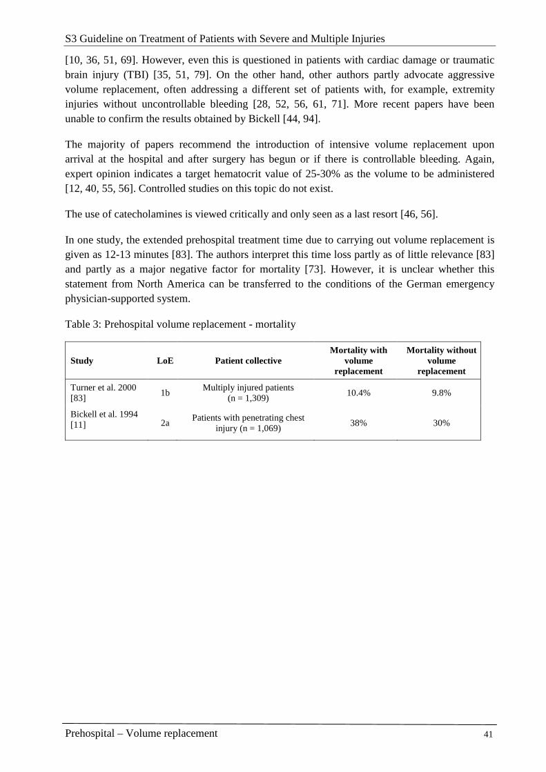
S3 Guideline on Treatment of Patients with Severe and Multiple Injuries
Prehospital – Volume replacement 41
[10, 36, 51, 69]. However, even this is questioned in patients with cardiac damage or traumatic brain injury (TBI) [35, 51, 79]. On the other hand, other authors partly advocate aggressive volume replacement, often addressing a different set of patients with, for example, extremity injuries without uncontrollable bleeding [28, 52, 56, 61, 71]. More recent papers have been unable to confirm the results obtained by Bickell [44, 94].
The majority of papers recommend the introduction of intensive volume replacement upon arrival at the hospital and after surgery has begun or if there is controllable bleeding. Again, expert opinion indicates a target hematocrit value of 25-30% as the volume to be administered [12, 40, 55, 56]. Controlled studies on this topic do not exist.
The use of catecholamines is viewed critically and only seen as a last resort [46, 56].
In one study, the extended prehospital treatment time due to carrying out volume replacement is given as 12-13 minutes [83]. The authors interpret this time loss partly as of little relevance [83] and partly as a major negative factor for mortality [73]. However, it is unclear whether this statement from North America can be transferred to the conditions of the German emergency physician-supported system.
Table 3: Prehospital volume replacement - mortality
Study LoE Patient collective Mortality with
volume replacement
Mortality without volume
replacement
Turner et al. 2000 [83]
1b Multiply injured patients
(n = 1,309) 10.4% 9.8%
Bickell et al. 1994 [11] 2a
Patients with penetrating chest injury (n = 1,069)
38% 30%

S3 Guideline on Treatment of Patients with Severe and Multiple Injuries
Prehospital – Volume replacement 42
Crystalloids versus colloids
Key recommendations:
Crystalloids should be used for volume replacement in trauma patients. GoR B
Isotonic saline solution should not be used; preference should be given to Ringer’s malate, or alternatively Ringer’s acetate or lactated Ringer's solution.
GoR B
Human albumin must not be used in prehospital volume replacement. GoR A
If colloidal solutions are used in hypotensive trauma patients, preference should be given to HES 130/0.4.
GoR B
Explanation:
There is still controversy surrounding the choice of infusion solution to be used. The majority of data is taken from animal experiments or from operations and is limited in its evidential value. Different results are produced from the meta-analyses carried out. In 1989, Velanovich et al. showed a reduction of 12.3% in mortality of trauma patients when crystalloid volume replacement was given [85]. In 1999, Choi et al. confirmed this result and hypothesized a lower mortality after trauma when crystalloids were used [23]. A Cochrane analysis of 2008 yielded no difference between colloids and crystalloids after trauma [19, 20, 21]. The authors concluded from this that colloids could be dispensed with as a volume replacement drug as there was no evidence of an advantage from colloids, and crystalloids were cheaper. A proviso should be mentioned here that old, very large-molecule solutions were used in these reviews and the informative value of these reviews is limited. According to the available studies, the newer colloid hydroxy ethyl starch (HES) 1301/0.4 no longer seems to have the disadvantages of older starch solutions [39, 53]. However, a marked deterioration in coagulation was observed in one in-vitro study even for more modern volume solutions including hypertonic saline solution. This effect was not observed when using lactated Ringer's solution or 0.9% saline solution [14].
Lactated Ringer’s solution is the preferred choice of crystalloid over isotonic saline solution [30, 41, 43, 78]. Experimental papers showed evidence of dilutional acidosis occurring after infusion of large amounts of isotonic saline solution [62, 63]. The addition of lactate to a Ringer’s balanced electrolyte solution prevents dilutional acidosis through the metabolization of the lactate to bicarbonate and water, thus buffering the bicarbonate pool. More recent experimental papers have found evidence of disadvantages in lactated Ringer's solution. According to these papers, lactated Ringer's solution triggers the activation of neutrophil granulocytes, thus causing more lung damage [4, 5, 6, 66]. The rate of granulocyte apoptosis is also apparently increased [32]. There is no evidence of this in clinical studies.
S3 Guideline on Treatment of Patients with Severe and Multiple Injuries
Prehospital – Volume replacement 43
Plasma lactate is used as a shock parameter in diagnosis. Lactated Ringer's solution leads to an iatrogenic increase in plasma lactate level and can thus interfere with the diagnosis [64, 65]. Ringer’s malate or Ringer’s acetate can be used instead. In animal experiments there was evidence of lower mortality with Ringer’s malate. In conclusion, the use of lactated Ringer's solution no longer appears to be worthy of recommendation.
A Cochrane Review did not identify any evidence that one colloid is significantly superior to the other in the choice of colloid to be used [18]. The risk of an anaphylactic reaction to a colloid can be classified as minimal. In 1997, Ring [68] published his findings in The Lancet on the probability of an immune reaction to HES as 0.006%, to dextran as 0.0008% and to gelatine as 0.038%. Individual research papers seem to want to see an advantage of HES over the other colloids [1, 54, 74]. In a large series in France, the tolerance of different colloids was studied in 19,593 patients. 48.1% received gelatine solutions, 27.6% starch solutions, 15.7% albumin, and 9.5% dextrans. Overall, 43 anaphylactic reactions were observed (0.219%). The distribution between the different volume solutions was as follows: 0.345% for gelatine, 0.273% for dextrans, 0.099% for albumin, and 0.058% for starch. 20% of all allergic reactions were serious to very serious (grade III and IV). In a multivariate analysis, independent risk factors were identified as the administration of gelatine (OR 4.81), dextran (OR 3.83), a medical history of allergy to drugs (OR 3.16), and male sex (OR 1.98). Thus, a 6-fold smaller risk of anaphylaxis was observed for starch solutions compared to gelatine and a 4.7 times lower risk for dextran [54].
In 1990, Hankeln et al. tested HES 200/0.6 compared to human albumin in a randomized study of 40 patients with vascular interventions and were able to establish an optimum volume effect for HES 200/0.6 [41]. According to other studies as well, human albumin as a colloid seems to be associated with increased mortality and is not to be recommended [41]. The influence of colloids on coagulation seems negligible [53]. Albumin does not appear to play a role in volume replacement [37].

S3 Guideline on Treatment of Patients with Severe and Multiple Injuries
Prehospital – Volume replacement 44
Hypertonic solutions
Key recommendation:
Hypertonic solutions can be used in multiply injured patients with hypotensive circulation after blunt trauma.
GoR 0
Hypertonic solutions should be used in penetrating trauma if prehospital volume replacement is carried out.
GoR B
A hypertonic solution can be used in hypotensive patients with severe traumatic brain injury.
GoR 0
Explanation:
In recent years, the hypertonic 7.5% saline solution has increasingly gained in importance, especially in prehospital volume replacement. As already described above, the microcirculatory disturbance is the harmful factor in hemorrhagic-traumatic shock.
The mechanism of action of the hypertonic solution is based on mobilizing intracellular and interstitial fluid into the intravasal space and thus on improving microcirculation and total rheology.
The dosage of hypertonic solution is limited in order to counteract harmful hypernatremia. Based mainly on experimental papers, the optimum dosage has been established at 4 ml/kg body weight (BW). A single administration is prescribed.
Microcirculatory disturbance during hemorrhaging is the main factor for late complications occurring. Hypertonic saline solution leads to interstitial and intracellular volume rapidly mobilizing into the intravasal space and thus to consecutively improving rheology and thereby the microcirculatory system [49]. No significant advantages of hypertonic solution have been found in controlled studies. Bunn et al. (2004) studied hypertonic versus isotonic solutions in a Cochrane Review [20]. The authors came to the conclusion that the available data was still insufficient to make a final judgment on hypertonic solution. In two controlled randomized trials, Mattox et al. (1991) and Vassar et al. (1991) argued an advantage of hypertonic solution for survival especially after traumatic brain injury [59, 84]. A paper by Alpar et al. (2004) follows the same line where an improvement in outcome is described in 180 patients especially after traumatic brain injury [2]. However, another controlled study from 2004 revealed that there was no significant difference to be observed in 229 patients in the long-term outcome after traumatic brain injury [29]. Moreover, Vassar et al. (1993) reported that the addition of dextrans did not bring any benefit for survival after trauma and bleeding [84]. This finding is contradicted by several papers which found a clear benefit from the addition of dextran [28, 49, 86, 87].
A positive effect on the clinical treatment of traumatic brain injury has been found in other studies. Wade (1997) and Vassar (1993) showed an effect on mortality after traumatic brain
S3 Guideline on Treatment of Patients with Severe and Multiple Injuries
Prehospital – Volume replacement 45
injury and initial treatment with hypertonic solution [84, 90]. Vassar found that the mortality rate dropped from 49 to 60% and Wade’s mortality rate from 26.9 to 37.0% with hypertonic solution. In the follow-up treatment for increased intracranial pressure, a lowering effect is described particularly for the combination of hypertonic solution/HES [42, 47, 75, 91, 92, 93]. However, this effect could not be confirmed in a controlled clinical trial [76]. Nor could any advantage from the hypertonic solution be detected in another current paper by Bulger et al. published in JAMA with the result that the study was discontinued after 1,313 patients [15]. Wade et al. conducted a comparative study in terms of a short meta-analysis of 14 papers on hypertonic saline solution with and without the addition of dextran and found no relevant advantage in hypertonic solutions [90]. In a paper from 2003, the same author describes a positive effect of hypertonic solutions in penetrating traumas. In a double-blind study with 230 patients, the patients initially received either hypertonic sodium chloride (NaCl) or isotonic solution. The mortality of the patients who were treated with hypertonic NaCl solution was 82.5% versus 75.5%, which was a significant improvement. The surgery rate and bleeding rate were equal. The authors thus concluded that hypertonic solutions improve the survival rate in penetrating traumas without increasing bleeding [89].
In a current study by Bulger et al. [17], lactated Ringer's solution was compared to hypertonic NaCl solution with dextran in a group of 209 multiply injured patients with blunt trauma. The endpoint of this study was ARDS-free survival. As there were no differences, the study was discontinued after an intention-to-treat analysis. In a subgroup analysis, an advantage for hypertonic NaCl solution with dextran was only found after massive transfusion. Even the most recent publication from this working group did not show any advantages for hypertonic solutions after hemorrhagic shock [16]. In fact, a higher mortality rate was observed in patients not requiring transfusion after being given hypertonic solution [28-day mortality-- hypertonic solution with dextrans: 10%; hypertonic solution: 12.2% and 0.9% saline solution: 4.8%, p < 0.01) [16].
Immunologic effects are likewise ascribed to the hypertonic solution. Thus, experimental papers describe a reduction in neutrophil activation and in the pro-inflammatory cascade [4, 5, 6, 7, 26, 27, 31, 33, 66, 81]. No evidence has yet been found on the clinical importance of these effects.
Hypertonic solutions lead to a rapid rise in blood pressure and a reduction in volume requirement [3, 13, 22, 24, 38, 50, 57, 58, 93]. How far this influences the treatment outcome cannot be found in the literature.
With regard to the dosage, Rocha and Silva (1990) showed in dogs that a 7.5% solution with 4 ml/kg BW corresponds to the optimum dosage [70]; this was confirmed again by Wade et al. (2003) [88].
Anti-shock trousers
Key recommendation:
Anti-shock trousers must not be used for circulatory support in multiply GoR A
S3 Guideline on Treatment of Patients with Severe and Multiple Injuries
Prehospital – Volume replacement 46
injured patients.
Explanation:
The anti-shock trousers (pneumatic anti-shock garment [PASG]) were promoted particularly in the 1980s and were often used in the military sector. Soft tissue damage and compartment syndromes brought their use into question. The current Cochrane Review from the year 2000 no longer recommends the use of anti-shock trousers. There are indications that the PASG increases mortality and extends the period of intensive treatment and hospital treatment [34]. Relevant complications after using anti-shock trousers have been described in several reviews and original papers [25, 80]. According to the literature available, the PASG should no longer be recommended.
S3 Guideline on Treatment of Patients with Severe and Multiple Injuries
Prehospital – Volume replacement 47
References
1. Adams Ha, Piepenbrock S, Hempelmann G (1998) [Volume replacement solutions--pharmacology and clinical use]. Anasthesiol Intensivmed Notfallmed Schmerzther 33:2-17 [LoE 5]
2. Alpar Ek, Killampalli Vv (2004) Effects of hypertonic dextran in hypovolaemic shock: a prospective clinical trial. Injury 35:500-506 [LoE 1b]
3. Anderson Jt, Wisner Dh, Sullivan Pe et al. (1997) Initial small-volume hypertonic resuscitation of shock and brain injury: short- and long-term effects. J Trauma 42:592-600; discussion 600-591 [LoE 5]
4. Angle N, Cabello-Passini R, Hoyt Db et al. (2000) Hypertonic saline infusion: can it regulate human neutrophil function? Shock 14:503-508 [LoE 2b]
5. Angle N, Hoyt Db, Cabello-Passini R et al. (1998) Hypertonic saline resuscitation reduces neutrophil margination by suppressing neutrophil L selectin expression. J Trauma 45:7-12; discussion 12-13 [LoE 5]
6. Angle N, Hoyt Db, Coimbra R et al. (1998) Hypertonic saline resuscitation diminishes lung injury by suppressing neutrophil activation after hemorrhagic shock. Shock 9:164-170 [LoE 5]
7. Assalia A, Bitterman H, Hirsh Tm et al. (2001) Influence of hypertonic saline on bacterial translocation in controlled hemorrhagic shock. Shock 15:307-311 [LoE 5]
8. Balogh Z, Mckinley Ba, Cocanour Cs et al. (2003) Supranormal trauma resuscitation causes more cases of abdominal compartment syndrome. Arch Surg 138:637-642; discussion 642-633 [LoE 2a]
9. Banerjee A, Jones R (1994) Whither immediate fluid resuscitation? Lancet 344:1450-1451 [LoE 5]
10. Beekley AC. Damage control resuscitation: a sensible approach to the exsanguinating surgical patient. Crit Care Med. 2008;36:S267-S274 [LoE 5]
11. Bickell Wh, Wall Mj, Jr., Pepe Pe et al. (1994) Immediate versus delayed fluid resuscitation for hypotensive patients with penetrating torso injuries. N Engl J Med 331:1105-1109 [LoE 2a]
12. Brinkmeyer Sd (1983) Fluid resuscitation: an overview. J Am Osteopath Assoc 82:326-330 [LoE 5]
13. Brock H, Rapf B, Necek S et al. (1995) [Comparison of postoperative volume therapy in heart surgery patients]. Anaesthesist 44:486-492 [LoE 1b]
14. Brummel-Ziedins K, Whelihan Mf, Ziedins Eg et al. (2006) The resuscitative fluid you choose may potentiate bleeding. J Trauma 61:1350-1358 [LoE 5]
15. Bulger EM, May S, Brasel KJ, Schreiber M, Kerby JD, Tisherman SA, Newgard C, Slutsky A, Coimbra R, Emerson S, Minei JP, Bardarson B, Kudenchuk P, Baker A, Christenson J, Idris A, Davis D, Fabian TC, Aufderheide TP, Callaway C, Williams C,
Banek J, Vaillancourt C, van Heest R, Sopko G, Hata JS, Hoyt DB; ROC Investigators. Out-of-hospital hypertonic resuscitation following severe traumatic brain injury: a randomized controlled trial. JAMA. 2010 Oct 6;304(13):1455-64 [LoE 1b]
16. Bulger EM, May S, Kerby JD, Emerson S, Stiell IG, Schreiber MA, Brasel KJ, Tisherman SA, Coimbra R, Rizoli S, Minei JP, Hata JS, Sopko G, Evans DC, Hoyt DB; ROC investigators. Out-of-hospital hypertonic resuscitation after traumatic hypovolemic shock: a randomized, placebo controlled trial. Ann Surg. 2011 Mar;253(3):431-41 [LoE 1b]
17. Bulger Em, Jurkovich Gj, Nathens Ab et al. (2008) Hypertonic resuscitation of hypovolemic shock after blunt trauma: a randomized controlled trial. Arch Surg 143:139-148; discussion 149 [LoE 1b]
18. Bunn F, Kwan I, Roberts I et al. (2001) Effectiveness of pre-hospital trauma care. Report to the World Health Organisation Pre-Hospital Care Steering Committee. . In:
19. Bunn F, Roberts I, Tasker R et al. (2002) Hypertonic versus isotonic crystalloid for fluid resuscitation in critically ill patients. Cochrane Database Syst Rev:CD002045
20. Bunn F, Roberts I, Tasker R et al. (2004) Hypertonic versus near isotonic crystalloid for fluid resuscitation in critically ill patients. Cochrane Database Syst Rev:CD002045 [LoE 1a]
21. Bunn F, Trivedi D, Ashraf S (2008) Colloid solutions for fluid resuscitation. Cochrane Database Syst Rev:CD001319 [LoE 1a]
22. Chiara O, Pelosi P, Brazzi L et al. (2003) Resuscitation from hemorrhagic shock: experimental model comparing normal saline, dextran, and hypertonic saline solutions. Crit Care Med 31:1915-1922 [LoE 5]
23. Choi Pt, Yip G, Quinonez Lg et al. (1999) Crystalloids vs. colloids in fluid resuscitation: a systematic review. Crit Care Med 27:200-210 [LoE 1b]
24. Christ F, Niklas M, Kreimeier U et al. (1997) Hyperosmotic-hyperoncotic solutions during abdominal aortic aneurysm (AAA) resection. Acta Anaesthesiol Scand 41:62-70
25. Christensen Ks (1986) Pneumatic antishock garments (PASG): do they precipitate lower-extremity compartment syndromes? J Trauma 26:1102-1105 [LoE 3b]
26. Coimbra R, Hoyt Db, Junger Wg et al. (1997) Hypertonic saline resuscitation decreases susceptibility to sepsis after hemorrhagic shock. J Trauma 42:602-606; discussion 606-607 [LoE 5]
27. Coimbra R, Loomis W, Melbostad H et al. (2005) Role of hypertonic saline and pentoxifylline on neutrophil activation and tumor necrosis factor-alpha synthesis: a novel resuscitation strategy. J Trauma 59:257-264; discussion 264-255 [LoE 5]
S3 Guideline on Treatment of Patients with Severe and Multiple Injuries
Prehospital – Volume replacement 48
28. Conte Ma (1997) Fluid resuscitation in the trauma patient. CRNA 8:31-39 [LoE 5]
29. Cooper Dj, Myles Ps, Mcdermott Ft et al. (2004) Prehospital hypertonic saline resuscitation of patients with hypotension and severe traumatic brain injury: a randomized controlled trial. JAMA 291:1350-1357 [LoE 1b]
30. Coran Ag, Ballantine Tv, Horwitz Dl et al. (1971) The effect of crystalloid resuscitation in hemorrhagic shock on acid-base balance: a comparison between normal saline and Ringer's lactate solutions. Surgery 69:874-880 [LoE 5]
31. Corso Co, Okamoto S, Ruttinger D et al. (1999) Hypertonic saline dextran attenuates leukocyte accumulation in the liver after hemorrhagic shock and resuscitation. J Trauma 46:417-423 [LoE 5]
32. Deb S, Martin B, Sun L et al. (1999) Resuscitation with lactated Ringer's solution in rats with hemorrhagic shock induces immediate apoptosis. J Trauma 46:582-588; discussion 588-589 [LoE 5]
33. Deitch Ea, Shi Hp, Feketeova E et al. (2003) Hypertonic saline resuscitation limits neutrophil activation after trauma-hemorrhagic shock. Shock 19:328-333 [LoE 5]
34. Dickinson K, Roberts I (2000) Medical anti-shock trousers (pneumatic anti-shock garments) for circulatory support in patients with trauma. Cochrane Database Syst Rev:CD001856 [LoE 1a]
35. Dries Dj (1996) Hypotensive resuscitation. Shock 6:311-316 [LoE 5]
36. Dutton Rp, Mackenzie Cf, Scalea Tm (2002) Hypotensive resuscitation during active hemorrhage: impact on in-hospital mortality. J Trauma 52:1141-1146
37. Finfer S, Bellomo R, Boyce N et al. (2004) A comparison of albumin and saline for fluid resuscitation in the intensive care unit. N Engl J Med 350:2247-2256
38. Fischer M, Hossmann Ka (1996) Volume expansion during cardiopulmonary resuscitation reduces cerebral no-reflow. Resuscitation 32:227-240 [LoE 5]
39. Gandhi Sd, Weiskopf Rb, Jungheinrich C et al. (2007) Volume replacement therapy during major orthopedic surgery using Voluven (hydroxyethyl starch 130/0.4) or hetastarch. Anesthesiology 106:1120-1127 [LoE 1b]
40. Giesecke Ah, Jr., Grande Cm, Whitten Cw (1990) Fluid therapy and the resuscitation of traumatic shock. Crit Care Clin 6:61-72 [LoE 5]
41. Hankeln K, Lenz I, Hauser B (1988) [Hemodynamic effect of 6% hydroxyethyl starch (HES 200,000/0.62)]. Anaesthesist 37:167-172 [LoE 2b]
42. Hartl R, Medary Mb, Ruge M et al. (1997) Hypertonic/hyperoncotic saline attenuates microcirculatory disturbances after traumatic brain injury. J Trauma 42:S41-47 [LoE 2b]
43. Healey Ma, Davis Re, Liu Fc et al. (1998) Lactated ringer's is superior to normal saline in a model of
massive hemorrhage and resuscitation. J Trauma 45:894-899 [LoE 5]
44. Holte K, Kristensen Bb, Valentiner L et al. (2007) Liberal versus restrictive fluid management in knee arthroplasty: a randomized, double-blind study. Anesth Analg 105:465-474 [LoE 1b]
45. Hyde J, Graham T (1999) Pre-hospital fluid resuscitation for thoracic trauma. Pre-hospital Immediate Care 3:99-101 [LoE 5]
46. Kalbe P, Kant Cj (1988) [First aid at the scene of the accident from the viewpoint of the trauma surgeon]. Orthopade 17:2-10 [LoE 5]
47. Kempski O, Obert C, Mainka T et al. (1996) "Small volume resuscitation" as treatment of cerebral blood flow disturbances and increased ICP in trauma and ischemia. Acta Neurochir Suppl 66:114-117 [LoE 5]
48. Krausz Mm, Bashenko Y, Hirsh M (2001) Crystalloid and colloid resuscitation of uncontrolled hemorrhagic shock following massive splenic injury. Shock 16:383-388 [LoE 5]
49. Kreimeier U, Christ F, Frey L et al. (1997) [Small-volume resuscitation for hypovolemic shock. Concept, experimental and clinical results]. Anaesthesist 46:309-328 [LoE 5]
50. Kreimeier U, Messmer K (1992) [Use of hypertonic NaCl solutions in primary volume therapy]. Zentralbl Chir 117:532-539 [LoE 5]
51. Kreimeier U, Prueckner S, Peter K (2000) Permissive hypotension. Schweiz Med Wochenschr 130:1516-1524 [LoE 5]
52. Kroll W, Gassmayr Se, Izmail S et al. (1997) Prehospital fluid resuscitation. Acta Anaesthesiol Scand Suppl 111:301-302 [LoE 5]
53. Langeron O, Doelberg M, Ang Et et al. (2001) Voluven, a lower substituted novel hydroxyethyl starch (HES 130/0.4), causes fewer effects on coagulation in major orthopedic surgery than HES 200/0.5. Anesth Analg 92:855-862 [LoE 1b]
54. Laxenaire MC, Charpentier C, Feldman L (1994) Anaphylactoid reactions to colloid plasma substitutes: incidence, risk factors, mechanisms. A French multicenter prospective study. Ann Fr Anesth Reanim. 1994;13(3):301-10 [LoE 1b]
55. Levison M, Trunkey Dd (1982) Initial assessment and resuscitation. Surg Clin North Am 62:9-16 [LoE 5]
56. Marzi I (1996) [Hemorrhagic shock]. Anaesthesist 45:976-992 [LoE 5]
57. Matsuoka T, Hildreth J, Wisner Dh (1996) Uncontrolled hemorrhage from parenchymal injury: is resuscitation helpful? J Trauma 40:915-921; discussion 921-912 [LoE 5]
58. Matsuoka T, Wisner Dh (1996) Resuscitation of uncontrolled liver hemorrhage: effects on bleeding, oxygen delivery, and oxygen consumption. J Trauma 41:439-445
59. Mattox Kl, Maningas Pa, Moore Ee et al. (1991) Prehospital hypertonic saline/dextran infusion for post-traumatic hypotension. The U.S.A. Multicenter Trial. Ann Surg 213:482-491 [LoE 1b]
S3 Guideline on Treatment of Patients with Severe and Multiple Injuries
Prehospital – Volume replacement 49
60. Nolan J (1999) Fluid replacement. Br Med Bull 55:821-843 [LoE 5]
61. Pargger H, Studer W, Ruttimann U (2000) [Volume therapy in hypotensive trauma patients]. Schweiz Med Wochenschr 130:1509-1515 [LoE 5]
62. Pitts R, Lotspeich W (1946) Bicarbonate and the renal regulation of acid base balance. Am J Physiol 147:138-157
63. Pitts Rf, Ayer Jl, Schiess Wa (1949) The renal regulation of acid-base balance in man; the reabsorption and excretion of bicarbonate. J Clin Invest 28:35-44
64. Raum M, Holzgraefe B, Rixen D et al. (2000) Der Einfluß von exogenem Laktat auf gemessene Plasmalaktatspiegel im hämorrhagischen Schock- eine kontrollierte Studie am Schwein. Chir. Forum Band 29:543-547
65. Raum M, Rixen D, Linker R et al. (2002) [Influence of lactate infusion solutions on the plasma lactate concentration]. Anasthesiol Intensivmed Notfallmed Schmerzther 37:356-358 [LoE 5]
66. Rhee P, Burris D, Kaufmann C et al. (1998) Lactated Ringer's solution resuscitation causes neutrophil activation after hemorrhagic shock. J Trauma 44:313-319 [LoE 5]
67. Riddez L, Johnson L, Hahn Rg (1998) Central and regional hemodynamics during crystalloid fluid therapy after uncontrolled intra-abdominal bleeding. J Trauma 44:433-439 [LoE 5]
68. Ring J, Messmer K (1977) Incidence and severity of anaphylactoid reactions to colloid volume substitutes. Lancet 1:466-469
69. Roberts K, Revell M, Youssef H et al. (2006) Hypotensive resuscitation in patients with ruptured abdominal aortic aneurysm. Eur J Vasc Endovasc Surg 31:339-344 [LoE 1a]
70. Rocha E Silva M, Velasco It (1989) Hypertonic saline resuscitation: the neural component. Prog Clin Biol Res 299:303-310 [LoE 5]
71. Rossi R (1997) [Early care or quick transport? The effectiveness of preclinical treatment of emergency patients]. Anaesthesist 46:126-132 [LoE 5]
72. Sackett Dl, Richardson Ws, Rosenberg W et al. (1997) Evidence-based medicine: How to practice and teach EBM. Churchill Livingstone, London
73. Sampalis Js, Tamim H, Denis R et al. (1997) Ineffectiveness of on-site intravenous lines: is prehospital time the culprit? J Trauma 43:608-615; discussion 615-607
74. Schmand Jf, Ayala A, Morrison Mh et al. (1995) Effects of hydroxyethyl starch after trauma-hemorrhagic shock: restoration of macrophage integrity and prevention of increased circulating interleukin-6 levels. Crit Care Med 23:806-814 [LoE 5]
75. Schwarz S, Schwab S, Bertram M et al. (1998) Effects of hypertonic saline hydroxyethyl starch solution and mannitol in patients with increased intracranial pressure after stroke. Stroke 29:1550-1555 [LoE 2b]
76. Shackford Sr (1997) Effect of small-volume resuscitation on intracranial pressure and related cerebral variables. J Trauma 42:S48-53 [LoE 5]
77. Shah N, Palmer C, Sharma P (1998) Outcome of raising blood pressure in patients with penetrating trunk wounds. Lancet 351:648-649 [LoE 5]
78. Shires Gt (1977) Pathophysiology and fluid replacement in hypovolemic shock. Ann Clin Res 9:144-150 [LoE 5]
79. Singbartl G (1985) [Significance of preclinical emergency treatment for the prognosis of patients with severe craniocerebral trauma]. Anasth Intensivther Notfallmed 20:251-260 [LoE 2a]
80. Taylor Dc, Salvian Aj, Shackleton Cr (1988) Crush syndrome complicating pneumatic antishock garment (PASG) use. Injury 19:43-44 [LoE 3a]
81. Tokyay R, Zeigler St, Kramer Gc et al. (1992) Effects of hypertonic saline dextran resuscitation on oxygen delivery, oxygen consumption, and lipid peroxidation after burn injury. J Trauma 32:704-712; discussion 712-703 [LoE 5]
82. Trunkey Dd (2001) Prehospital fluid resuscitation of the trauma patient. An analysis and review. Emerg Med Serv 30:93-95, 96, 98 passim [LoE 5]
83. Turner J, Nicholl J, Webber L et al. (2000) A randomised controlled trial of prehospital intravenous fluid replacement therapy in serious trauma. Health Technol Assess 4:1-57 [LoE 1b]
84. Vassar Mj, Perry Ca, Holcroft Jw (1993) Prehospital resuscitation of hypotensive trauma patients with 7.5% NaCl versus 7.5% NaCl with added dextran: a controlled trial. J Trauma 34:622-632; discussion 632-623 [LoE 1b]
85. Velanovich V (1989) Crystalloid versus colloid fluid resuscitation: a meta-analysis of mortality. Surgery 105:65-71 [LoE 1b]
86. Velasco It, Pontieri V, Rocha E Silva M, Jr. et al. (1980) Hyperosmotic NaCl and severe hemorrhagic shock. Am J Physiol 239:H664-673 [LoE 5]
87. Velasco It, Rocha E Silva M, Oliveira Ma et al. (1989) Hypertonic and hyperoncotic resuscitation from severe hemorrhagic shock in dogs: a comparative study. Crit Care Med 17:261-264 [LoE 5]
88. Wade Ce, Dubick Ma, Grady Jj (2003) Optimal dose of hypertonic saline/dextran in hemorrhaged swine. J Trauma 55:413-416 [LoE 5]
89. Wade Ce, Grady Jj, Kramer Gc (2003) Efficacy of hypertonic saline dextran fluid resuscitation for patients with hypotension from penetrating trauma. J Trauma 54:S144-148 [LoE 1b]
90. Wade Ce, Kramer Gc, Grady Jj et al. (1997) Efficacy of hypertonic 7.5% saline and 6% dextran-70 in treating trauma: a meta-analysis of controlled clinical studies. Surgery 122:609-616 [LoE 1a]
91. Waschke Kf, Albrecht Dm, Van Ackern K et al. (1996) Coupling between local cerebral blood flow and metabolism after hypertonic/hyperoncotic fluid resuscitation from hemorrhage in conscious rats. Anesth Analg 82:52-60 [LoE 5]

S3 Guideline on Treatment of Patients with Severe and Multiple Injuries
Prehospital – Volume replacement 50
92. Waschke Kf, Frietsch T (1999) Selection of adequate substitutes for intravascular volume replacement. Int J Intens Care 2:135-143 [LoE 5]
93. Weinstabl C, Hammerle A (1992) Hypertone, hyperonkotische Hydroxyäthylstärke-Lösung zur Senkung des intrakraniellen Druckes. Fortschr Anaesth 6:105-107 [LoE 2b]
94. Yaghoubian A, Lewis Rj, Putnam B et al. (2007) Reanalysis of prehospital intravenous fluid administration in patients with penetrating truncal injury and field hypotension. Am Surg 73:1027-1030 [LoE 2a]

S3 Guideline on Treatment of Patients with Severe and Multiple Injuries
Prehospital – Volume replacement 51
1.4 Thorax
Diagnostic tests
The decision whether to carry out drainage or decompression of the pleural space is based on examination, assessment of findings (diagnosis), and benefit-risk evaluation (diagnosis certainty with limited diagnostic options, time factor, concomitant circumstances, and risks attached to the method itself).
Examination
Key recommendation:
A clinical examination of the thorax and respiratory function must be carried out.
GoR A
The examination should include as a minimum the measurement of the respiratory rate and auscultation of the lungs. The examination should be repeated.
GoR B
The following can be helpful: inspection (bilaterally unequal in respiratory excursion, unilateral bulging, paradoxical respiration), palpation (pain, crepitations, subcutaneous emphysema, instability) and percussion (hyper-resonant percussion) of the thorax together with pulse oxymetry and, in ventilated patients, monitoring ventilation pressure.
GoR 0
Explanation:
Initial examination
The physical examination of the patient is required for establishing a diagnosis, which in turn is a prerequisite for treatment interventions. An acute life-threatening disorder can only be recognized by examination. Even without scientific proof, it appears to be absolutely essential [89].
Scientific studies on the type and scope of physical examination are mainly only on auscultation, measuring respiratory rate and on clarifying spontaneous pain and tenderness. Thus, only experience can define the required scope of the physical examination in the prehospital emergency examination.
In the emergency situation at the accident scene, the initial examination of the thorax should include (after checking and securing the vital functions) checking the respiratory rate and auscultation (presence of breath sounds, bilaterally equal breath sounds) [14, 36, 39, 40]. All these signs are correlated to significant pathologies or have a direct influence on medical decisions. Other useful examinations can be inspection (for signs of injury, symmetry of the
S3 Guideline on Treatment of Patients with Severe and Multiple Injuries
Prehospital – Volume replacement 52
thorax, symmetry of respiratory excursion, paradoxical respiration, dyspnea, distended neck veins) and palpation (subcutaneous emphysema, pain points, crepitations, instabilities in the bony structure of the thorax). Monitoring ventilation pressure and pulse oxymetry can be added in the course of further management [55].
All the above-mentioned examinations are used to detect relevant, threatening or potentially threatening disorders and injuries, which altogether can make it necessary to administer immediate and specific treatment or make a logistical decision on the spot. All diagnostic procedures that can be introduced prehospital are without specific risk, the only disadvantage being the loss of time, which is usually minimal.
The different findings are to some extent greatly dependent on the examiner, the patient and the environment. For instance, noise can make auscultation more difficult or impossible. Such circumstances must be taken into account when selecting and interpreting the primary diagnostic study [36, 40, 80, 135].
Several researchers showed that, under in-hospital conditions, ultrasound examination (lung sliding, lung point, comet tail, etc.) allows good and accurate detection of pneumothorax and hemothorax (review in [87]). However, there is no experience in prehospital application so a general recommendation cannot be made.
Patient monitoring
The respiratory rate and, if applicable, ventilation pressure should be checked and auscultation and pulse oxymetry performed during the course since a disorder in the respiratory system, a misplacement of the tube, tension pneumothorax or acute respiratory insufficiency can develop dynamically. The follow-up examination can serve as a performance check of the treatment administered.

S3 Guideline on Treatment of Patients with Severe and Multiple Injuries
Prehospital – Volume replacement 53
Diagnosing pneumothorax
Key recommendations:
A suspected diagnosis of pneumothorax and/or hemothorax must be made if breath sounds are weaker or absent on one side (after checking correct placement of the tube). Absence of such auscultation findings largely rules out a larger pneumothorax, especially if the patient is normopneic and has no chest pain.
GoR A
The potential progression of a small pneumothorax which cannot initially be diagnosed in the prehospital phase should be taken into consideration.
GoR B
Explanation:
There are currently no methods available for definite prehospital detection or exclusion of pneumothorax. This is only clinically possible by computed tomography (exclusion).
Auscultation
The studies on diagnostic accuracy of auscultation are summarized in
Table 4. The specificity of a unilateral weakened or absent breath sound for the presence of pneumothorax is very high at 93-98%. The positive predictive value, i.e. the probability of there actually being a pneumothorax in the presence of a weakened breath sound, is also very high at 86-97% [35, 135]. Pneumothoraces not detected by auscultation had a mean volume of 378 ml (max. 800 ml), non-detected hemothoraces had a mean volume of 277 ml (max. 600 ml). No large, acutely threatening lesions were missed [36, 80]. In another prospective series, auscultation was the most reliable method of detecting a pneumohemothorax compared to evidence of pain or tachypnea [24]. Conversely, a hemo-/pneumothorax was virtually excluded in the presence of normopnea and normal auscultation and palpation findings [24].
The prerequisite is the correct placement of the endotracheal tube (as available), which must be ensured beforehand if possible. A proviso must be given here that the cited studies were not conducted at the emergency site but on emergency admission in the hospital. However, they appear to be easily transferable as numerous confounders (e.g., high noise level, disturbance) can also predominate in a comparable manner in emergency admission. False positive findings can occasionally be present (4.5% of cases in [88]) in tube misplacements, diaphragmatic rupture [1, 4] or ventilation disorders (large atelectases, shifting of deeper respiratory tracts).
If there are severe bilateral chest injuries, the presence of a bilateral pneumothorax should be considered. Atypical examination findings may occur in this case.
Data for differentiating between a pneumothorax and a hemothorax or mixed types are unavailable. Percussion can be helpful here but in the prehospital setting seems to be only of

S3 Guideline on Treatment of Patients with Severe and Multiple Injuries
Prehospital – Volume replacement 54
subordinate relevance as the differentiation between pneumo- and hemothorax has no provable effects on treatment requirements (see below).
Table 4: Diagnostic valency of a pathologic auscultation finding with regard to a hemo/pneumothorax
Study LoE Patient collective Sensitivity Specificity
Hirshberg et al. 1988 [80] 1 Sharp trauma (n = 51) 96% 93%
Wormland et al. 1989 [143]
3 Sharp trauma (n = 200) 73.3% 98.6%
Thomson et al. 1990 [135] 1 Sharp trauma (n = 102) 96% 94%
Chen et al. 1997 [36] 3 Sharp trauma (n = 118) 58% 98%
Chen et al. 1998 [35] 1 Mainly blunt trauma (n = 148)
84% 97%
Bokhari et al. 2002 [24] 2 Blunt trauma (n = 523) 100% 99.8%
Bokhari et al. 2002 [24] 2 Sharp trauma (n = 153) 50% 100%
Dyspnea
Even if the symptoms of dyspnea and tachypnea are difficult to quantify in consciousness-clouded patients, evidence of normopnea (respiratory rate between 10-20/min) can be put to good use in clinical practice. Several studies revealed that normopnea is a very reliable sign that a larger hemo/pneumothorax can be excluded after blunt trauma (high specificity). In contrast, the presence of dyspnea in no way indicates the reverse, that pneumothorax is present (low sensitivity).
Table 5: Diagnostic valency of dyspnea and tachypnea with regard to hemo/pneumothorax
Study LoE Patient collective Sensitivity Specificity
Wormland et al. 1989 [143] 3 Sharp trauma (n = 200 patients) 75.6% 84.1%
Hing et al. 2001 [79] 4 Sharp trauma (n = 153 patients) 72.7% 95.5%
Bokhari et al. 2002 [24] 2 Blunt trauma (n = 523 patients) 42.8% 99.6%
Bokhari et al. 2002 [24] 2 Sharp trauma (n = 153 patients) 31.8% 99.2%
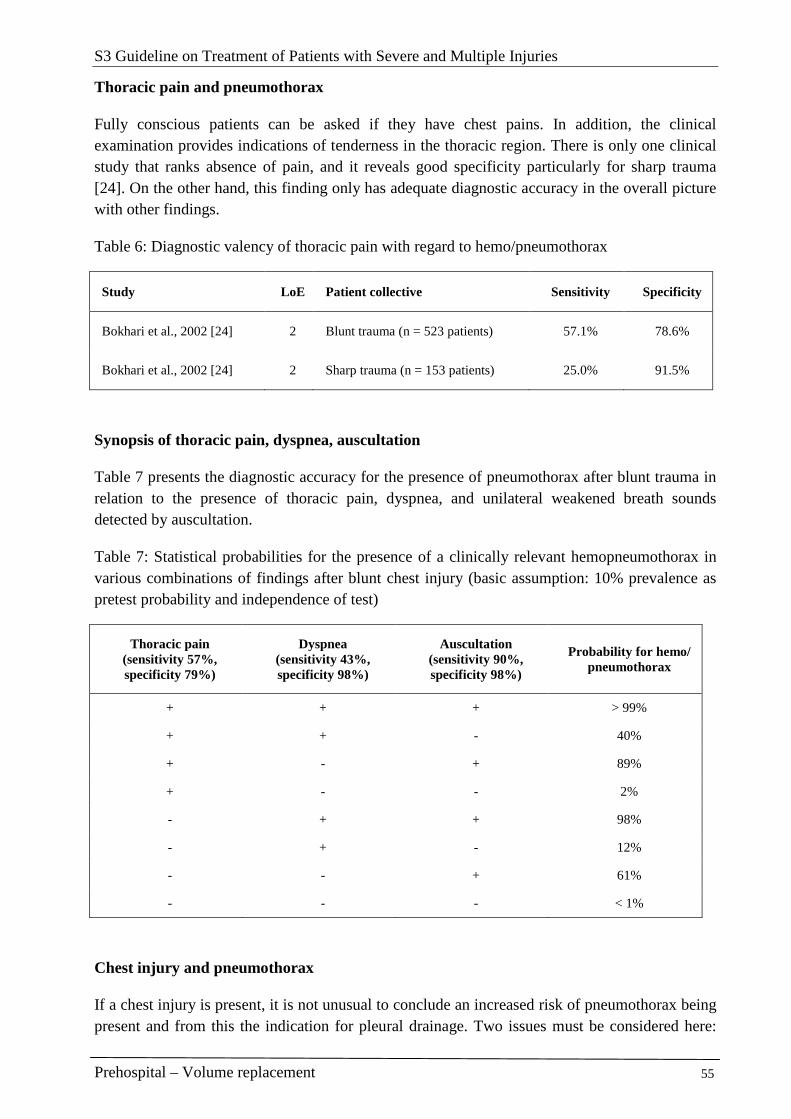
S3 Guideline on Treatment of Patients with Severe and Multiple Injuries
Prehospital – Volume replacement 55
Thoracic pain and pneumothorax
Fully conscious patients can be asked if they have chest pains. In addition, the clinical examination provides indications of tenderness in the thoracic region. There is only one clinical study that ranks absence of pain, and it reveals good specificity particularly for sharp trauma [24]. On the other hand, this finding only has adequate diagnostic accuracy in the overall picture with other findings.
Table 6: Diagnostic valency of thoracic pain with regard to hemo/pneumothorax
Study LoE Patient collective Sensitivity Specificity
Bokhari et al., 2002 [24] 2 Blunt trauma (n = 523 patients) 57.1% 78.6%
Bokhari et al., 2002 [24] 2 Sharp trauma (n = 153 patients) 25.0% 91.5%
Synopsis of thoracic pain, dyspnea, auscultation
Table 7 presents the diagnostic accuracy for the presence of pneumothorax after blunt trauma in relation to the presence of thoracic pain, dyspnea, and unilateral weakened breath sounds detected by auscultation.
Table 7: Statistical probabilities for the presence of a clinically relevant hemopneumothorax in various combinations of findings after blunt chest injury (basic assumption: 10% prevalence as pretest probability and independence of test)
Thoracic pain (sensitivity 57%, specificity 79%)
Dyspnea (sensitivity 43%, specificity 98%)
Auscultation (sensitivity 90%, specificity 98%)
Probability for hemo/ pneumothorax
+ + + > 99%
+ + - 40%
+ - + 89%
+ - - 2%
- + + 98%
- + - 12%
- - + 61%
- - - < 1%
Chest injury and pneumothorax
If a chest injury is present, it is not unusual to conclude an increased risk of pneumothorax being present and from this the indication for pleural drainage. Two issues must be considered here:

S3 Guideline on Treatment of Patients with Severe and Multiple Injuries
Prehospital – Volume replacement 56
the success rate of emergency physicians for diagnosing chest injury and the correlation between a chest injury and a concomitant pneumothorax.
However, the diagnostic accuracy of the emergency physician is greatly limited. An analysis of data from the DGU Trauma Registry showed that the emergency physician had grossly overestimated the chest injury in 18% of cases, i.e. the emergency physician assumed a severe chest injury which was not actually there [7].
In 9-50% of patients with confirmed chest injury, there was also a pneumothorax. It should be noted with these figures that the diagnosis of chest injury in all these studies had been made after a full set of diagnostic tests including imaging.
In the majority of studies, between 37 and 59% of patients with a relevant chest injury diagnosed in hospital had a pneumothorax [23, 55, 66, 137]. If occult pneumothoraces - in other words, those which can only be detected in CT but not clinically and not in standard radiography - are not included, then the proportion of patients with chest injury who have a relevant pneumothorax is actually only 17-25% [23, 137]. However, the incidence of pneumothorax secondary to chest injury was markedly lower at 8.9% in individual studies [52].
Table 8: Incidence of pneumothorax in the presence of chest injury
Study Incidence of pneumothorax (radiologic diagnostic test without CT)
Blostein et al. 1997 [23] 25% of chest injuries
Demartines et al. 1990 [52] 8.9% of chest injuries
Di Bartolomeo et al. 2001 [55] 21% of all critically injured
Gaillard et al. 1990 [66] 41% of chest injuries
Trupka et al. 1997 [137] 17% of chest injuries
Other examinations and pneumothorax
Evidence of subcutaneous emphysema is viewed as an indication of the presence of pneumothorax. However, there are no good diagnostic studies to support this. The specificity and positive predictive value are not known. However, the sensitivity is low and is between 12 and 25% [47, 126]. In a 30-year old study, it was reported that subcutaneous emphysema in intensive care patients had 100% sensitivity for the presence of tension pneumothorax. However, these data are possibly not transferable to acutely ill trauma patients in the prehospital phase [130].
Taking into account the relatively high rate of false findings, the findings of an unstable thorax and of crepitations are indications of the presence of a chest injury but not of pneumothorax.
S3 Guideline on Treatment of Patients with Severe and Multiple Injuries
Prehospital – Volume replacement 57
Pneumothorax and progression
The potential progression of an initially asymptomatic pneumothorax is important, particularly in air rescue as well. The progression of pneumothoraces can vary considerably among individuals, and basically the full range from in-hospital finding to rapid progression is possible. The observation of small pneumothoraces can provide certain clues. In a small retrospective series, 13 patients with occult pneumothorax were conservatively treated, 6 of whom were being mechanically ventilated. It was subsequently necessary in 2 cases to insert a chest drain due to a progressive pneumothorax on the second and third day, respectively, after admission [38]. In a prospective randomized study, 8 out of 21 patients with an occult pneumothorax which was under observation developed progressive pneumothorax and, in 3 cases, tension pneumothorax. All these patients were ventilated [60]. The 3 tension pneumothoraces occurred in the operating room, post-operative after admission to the intensive care unit, and during a prolonged stabilization phase; exact times in hours after trauma are not available. A period of at least 30-60 minutes after hospital admission can at least be assumed. In another prospective randomized study of the treatment of occult pneumothoraces, the progression of pneumothorax in the group of conservatively treated patients (12.5%) was not greater than those on a pleural drain (21%) [25]. Details concerning the duration of pneumothorax progression were not recorded. In a series of 44 newborn, mostly intubated children, the time between the probable start of pneumothorax and the clinical diagnosis being made was on average 127 minutes with a scatter between 45 and 660 minutes [99].
In 3 studies, the maximum size of pneumothorax was indicated as 5 x 80 ml (400 ml), beyond which pleural drainage was indicated [25, 60, 70]. However, as pneumothoraces of this size can usually already be clinically diagnosed by auscultation (see above), it can be assumed that the progression of pneumothoraces with normal auscultation finding meets the above-mentioned conditions.
Most experts believe that the progression of pneumothorax to tension pneumothorax is greater in patients who are on positive pressure ventilation [13] but this cannot be quantified.
To summarize, the data suggest that small, clinically non-diagnosable pneumothoraces generally progress relatively slowly and thus do not require any emergency decompression in the prehospital phase.

S3 Guideline on Treatment of Patients with Severe and Multiple Injuries
Prehospital – Volume replacement 58
Diagnosing tension pneumothorax
Key recommendation:
A suspected diagnosis of tension pneumothorax should be made if auscultation of the lung reveals no breath sounds unilaterally (after checking correct placement of the tube) and, in addition, typical symptoms are present, particularly severe respiratory disorder or upper inflow congestion combined with arterial hypotension.
GoR B
Explanation:
Good scientific data on diagnostic accuracy of examination findings for tension pneumothorax are few and far between. Practically all conclusions are based on case reports, animal experiments or expert opinion. There is no uniform definition on exactly what tension pneumothorax means. The definitions range from pneumothorax with threatening disorders to vital functions, hiss of escaping air during needle decompression, mediastinal shift on the chest X-ray, raised ipsilateral intrapleural pressure, and hemodynamic compromise [91]. For obvious reasons, the ad-hoc diagnosis in the prehospital phase can only be made on the basis of clinical examination findings.
The vast majority of experts consider the diagnosis of tension pneumothorax as given if life-threatening hemodynamic or respiratory disorders are present. Cyanosis, breathlessness, tachypnea, contralateral tracheal deviation, and a drop in oxygen saturation, elevated respiratory excursion and bulging hemithorax with hyper-resonant percussion on the diseased side are possible respiratory signs. Hemodynamic indicators can include distended neck veins, tachycardia, and ultimately a drop in blood pressure through to cardiac arrest (pulseless electrical activity). However, many of these signs can only be detected on closer examination and have not been systematically examined to date. There are few data on trauma patients; most information has been gained from observing tension pneumothoraces in intensive care medicine [90].
Experimental examinations show that, in the alert patient, respiratory impairment and paralysis of the respiratory center secondary to hypoxia precede cardiac arrest, and hypotension, the endpoint of which is cardiac arrest, is a late sign of tension pneumothorax [13, 124]. These experimental findings were recently confirmed by a patient with accidental tension pneumothorax, who became dyspneic, cyanotic and finally unconscious before respiratory arrest occurred. However, the carotid pulse could be felt throughout [131]. The patient’s condition normalized rapidly after decompressing the elevated intrapleural pressure. A tension pneumothorax in the radiograph (mediastinal shift to the contralateral side) without signs of impaired circulation has been described in another case report [101]. This patient’s circulation remained stable in the 30-minute period between making the diagnosis and inserting the chest drain. In another case report, the tension pneumothorax manifested itself clinically by cyanosis, an increase in respiratory and heart rate and impaired consciousness (GCS 10) while other signs were absent. However, a careful inspection revealed ipsilateral overextension and hypomobility in the chest wall. In a review article from 2005, the two symptoms, breathlessness and
S3 Guideline on Treatment of Patients with Severe and Multiple Injuries
Prehospital – Volume replacement 59
tachycardia, were presented as the typical and most frequent signs of tension pneumothorax in the alert patient [91].
However, the same authors also showed that in ventilated patients the cardio-circulatory symptoms of tension pneumothorax occurred earlier and the respiratory symptoms and fall in blood pressure often manifested themselves simultaneously. In the ventilated patient, very elevated or rising airway pressures are an important additional symptom that can be found in approximately 20% of patients with hemo/pneumothorax [14, 40]. However, systematically collected data concerning diagnostic accuracy are not available. According to expert opinion, the combination of (unilaterally) absent breath sound (with tube placement monitored) and life-threatening respiratory and circulatory function disorders makes the presence of tension pneumothorax so probable that the diagnosis should be made and the necessary therapeutic consequences followed. The consequences of a false diagnosis of tension pneumothorax appear to be subordinate compared to failing to carry out necessary decompression.
Indications for pleural decompression
Key recommendations:
Clinically suspected tension pneumothorax must be decompressed immediately.
GoR A
Pneumothorax diagnosed on the basis of an auscultation finding in a patient on positive pressure ventilation should be decompressed.
GoR B
Pneumothorax diagnosed on the basis of an auscultation finding in patients not on ventilation should usually be managed by close clinical observation.
GoR B
Explanation:
Comparative studies between conservative and interventional treatment are not available. The treatment recommendations are based on expert opinion and consideration of the probabilities.
Tension pneumothorax
Tension pneumothorax is an acute life-threatening situation and, if untreated, generally leads to death. Death can occur within a few minutes of the onset of signs of restricted pulmonary and circulatory function. There is no alternative to decompression. The experts are of the opinion that immediate emergency decompression should be carried out particularly on onset of circulatory or respiratory impairment and that the time lost through transferring the patient to a hospital, even one situated in the immediate vicinity, represents an unjustifiable delay. In a study of 3,500 autopsies, there were 39 cases of tension pneumothorax (incidence 1.1%), half of whom had not been diagnosed while still alive. Among soldiers from the Vietnam war, 3.9% of all patients with chest injuries and 33% of all soldiers with fatal chest injury had tension pneumothorax [100]. An analysis of 20 patients who had been categorized as unexpected survivors based on the TRISS
S3 Guideline on Treatment of Patients with Severe and Multiple Injuries
Prehospital – Volume replacement 60
prognosis showed that tension pneumothorax had been treated by decompression in 7 of them in the prehospital phase [29].
Diagnosed pneumothorax
A large pneumothorax, which can be assumed if a typical auscultation finding is collected, essentially presents an indication to evacuate the pleural cavity. Whether this has to take place in the prehospital phase or once admitted to hospital is difficult to decide in the individual case as the risk of progression from simple pneumothorax to tension pneumothorax and the amount of time that such a development can take are variable and difficult to estimate. The literature contains neither general data nor risk factors on this topic. There are indications that intubated patients with a chest injury when admitted to hospital are more likely to have tension pneumothorax than non-intubated patients. Overall, it still appears plausible to the experts that a pneumothorax diagnosed by auscultation in a ventilated patient has a markedly higher risk of developing into tension pneumothorax; thus, prehospital decompression is indicated.
If a patient with pneumothorax diagnosed by auscultation is not ventilated, then the risk of progression to tension pneumothorax appears to be markedly lower. In a series of 54 pneumothoraces after trauma, 29 were treated conservatively, i.e. without inserting a pleural drain. These were non-ventilated patients, mostly without concomitant injuries. A pleural drain was inserted in only 2 cases 6 hours after admission to hospital because of radiologic progressive pneumothorax [85]. Prehospital decompression does not appear to be necessary here and close clinical observation should be carried out. If appropriate clinical monitoring is an issue, e.g., during helicopter transfer, then there is a certain, unquantifiable risk that tension pneumothorax will develop which will not be recognized in time and/or which cannot be adequately treated due to space limitations. In such situations, if relevant clinical signs are present and after individual assessment, decompression of the pneumothorax can be carried out before transfer even in non-intubated patients.
Chest injury without direct pneumothorax diagnosis
If no pneumothorax is diagnosed (i.e. if the auscultation finding shows no lateral difference), then there is also essentially no indication for prehospital decompression or pleural evacuation.
The presence of clear signs of severe chest injury means that between 10 and 50% of these patients may have a pneumothorax (see above) and thus pleural drainage could be indicated in every second to tenth patient. Conversely, this means that at least every second patient and up to 9 out of every 10 patients would, under these conditions, receive unnecessary invasive treatment. This also coincides with the findings that air released from the pleural space was observed in only 32-50% of cases of prehospital decompression [14, 125], and decompression was not indicated in 9-25% of cases of pleural drains inserted in the prehospital phase as there was no pneumothorax or chest injury [7, 8, 125].
In addition, it should be considered that the radiologic findings have not been correlated to the clinical findings in the studies on pneumothorax incidence in chest injury. It can be assumed that numerous radiologically detectable pneumothoraces could also have been diagnosed by auscultation. The rate of pneumothoraces which cannot be diagnosed clinically but are present in

S3 Guideline on Treatment of Patients with Severe and Multiple Injuries
Prehospital – Volume replacement 61
chest injury can thus be assumed to be much lower [38]. As occult pneumothoraces were also included in a series of these studies, i.e. pneumothoraces which were first detectable at least 30 minutes after hospital admission only by computed tomography but not by standard radiography [23, 137], the proportion of prehospital relevant pneumothoraces falls even further. The risk of a pneumothorax, which at the time of primary survey was small and yielded a normal auscultation finding, progressing to tension pneumothorax has already been discussed above and should be viewed as minor in the prehospital timeframe.
Thus, in a justified individual case, decompression can be carried out in ventilated patients with clear signs of chest injury but normal auscultation finding prior to long road or helicopter transfer with limited clinical monitoring or treatment options. The high rate of false positive diagnoses of chest injury by the emergency physician must be taken into consideration.
Under these conditions, decompression is not indicated in non-ventilated patients.
Other indications
Pneumothorax and hemothorax represent the only typical indications for pleural decompression or pleural drainage in prehospital acute medicine. The therapeutic management of pneumothorax has already been presented above. Although hemothorax is essentially an indication for evacuating the blood located in the pleural space, there is generally no direct danger of compression from the blood and there is no indication for evacuation of the blood to the outside in the prehospital phase. Emergency decompression can only be indicated in cases of massive bleeding, possibly with problems developing in terms of tension hemothorax. However, this situation is generally associated with a pathologic auscultation finding and will thus make it necessary to proceed as per the situation with a pneumothorax, especially as it is generally always difficult to differentiate between a hemothorax and a hemopneumothorax in the prehospital phase.
Treatment
Methods
The aim of the treatment is decompression of positive pressure in tension pneumothorax or tension hemothorax. The second treatment goal to be considered is the prevention of a simple pneumothorax developing into a tension pneumothorax. The permanent and, if possible, complete evacuation of air and blood is of no importance in the prehospital emergency.
Key recommendations:
Tension pneumothorax should be decompressed by needle
decompression, followed by surgery to open the pleural space with or without a chest drain.
GoR B

S3 Guideline on Treatment of Patients with Severe and Multiple Injuries
Prehospital – Volume replacement 62
Pneumothorax should be treated with a chest drain provided the indication exists.
GoR B
Explanation:
As there are no suitable comparative data on the 3 methods, no recommendation for a method of choice can be made based on the data. (Predominantly retrospective) data, case series and case histories are available for all 3 methods and they demonstrate that successful decompression of tension pneumothorax is possible by each of these methods.
In view of the low evidence level on choice of method and benefit-risk profile in the direct comparison of methods, the individual ability of the treating emergency physician should be considered for reasons of practicability and risk potential. In one study, a significant difference in complication rate for insertion of a chest drain was observed between emergency admission physicians and surgeons [62]. In a more recent study, a lower complication rate associated with insertion by surgical compared to non-surgical residents was also observed in North America [11]. Due to a lack of reliable data, the extent to which these results are transferable to the German emergency physician system cannot be evaluated.
Chest drain: Efficacy and complications
Insertion of a chest drain is a highly effective, suitable but not complication-free procedure for decompressing a tension pneumothorax, which must be used particularly when the alternative interventions have failed or are insufficiently effective. Generally, it also represents the definitive treatment and has the highest success rate. In 79-95% of cases, pleural drains inserted in the prehospital phase were the successful, definitive treatment intervention [10, 52, 117, 125].
On the other hand, pleural drainage has a failure rate of 5.4-21% (mean of 11.2%) due to misplacements or insufficient efficacy. With this frequency, it was necessary to insert an additional pleural drain [10, 34, 45, 52, 62, 75, 117, 125]. This involved retained pneumothoraces and hemothoraces to more or less the same extent. Individual cases of persistent tension pneumothoraces were also observed with pleural drains inserted in the prehospital phase [10, 31, 98].
The pooled complication rates for pleural drainage are shown in

S3 Guideline on Treatment of Patients with Severe and Multiple Injuries
Prehospital – Volume replacement 63
Table 9 and in Table 10 (see appendix). There do not appear to be any relevant differences between pleural drains inserted in the prehospital and in-hospital phases. However, there are only 2 studies in which the complication rates for prehospital and in-hospital treatment were directly compared within the same establishment [128, 144]. They revealed comparable infection rates (9.4 versus 11.7%) and misplacements (0 versus 1.2%). The number of days in situ was comparable in both groups in each study.

S3 Guideline on Treatment of Patients with Severe and Multiple Injuries
Prehospital – Volume replacement 64
Table 9: Complication rates for pleural drains inserted in the prehospital versus in-hospital phase
Complication Only prehospital pleural drains * Only in-hospital pleural drains *
Subcutaneous misplacements
2.53% (1.55–3.33%)
n = 730, 9 studies
[10, 14, 47, 52, 88, 117, 125, 126, 144]
0.39% (0.08–1.13%) n = 772, 6 studies
[9, 19, 45, 46, 77, 144]
Intra-pulmonary misplacements
1.37% (0.63–2.58%) n = 657, 7 studies
[10, 14, 47, 52, 88, 125, 126]
0.63% (0.27-1.23%) n = 1,275, 7 studies
[9, 19, 45, 46, 54, 77, 107]
Intraabdominal misplacements
0.87% (0.32–1.88%) n = 690, 8 studies
[10, 14, 47, 52, 88, 117, 125, 126]
0.73% (0.29-1.50%) n = 956, 5 studies
[9, 45, 46, 77, 107]
Infections (pleural empyema)
0.55% (0.11–1.59%) n = 550, 5 studies
[10, 14, 52, 125, 144]
1.74% (1.47-2.05%) n = 8,102, 13 studies
[9, 19, 34, 46, 54, 62, 107, 144] [59, 76, 77, 94, 129]
* Mean values obtained from simply adding together the complications given in studies (confidence interval in brackets)
The case histories for the puncture site of the anterior to midaxillary line also report on injury to the intercostal arteries [32], lung perforations [65], perforations of the right atrium [33, 104, 127], the right ventricle [118] and the left ventricle [49], subclavian artery stenosis due to pressure from drain tip from inside [109], Horner syndrome due to pressure on the stellate ganglion from the drain lying in the apex [21, 31], an intraabdominal placement [64], a liver puncture [47], a perforation of the stomach [4] and of the colon [1] due to diaphragmatic hernia, a lesion in the subclavian vein, perforation of the inferior vena cava [61], and triggering of atrial fibrillation [12].
An arteriovenous fistula [43] as well as perforation of the cardiac wall [56] and perforation of the right atrium [104] were reported when the puncture was performed in the mid-clavicular line.
In addition, other known complications are perforations of the esophagus, of the mediastinum triggering a contralateral pneumothorax, an injury to the phrenic nerve among others.
Simple surgical opening: efficacy and complications
The simple surgical opening of the pleural space is a suitable, effective and relatively simple intervention to decompress a tension pneumothorax. However, it is only suitable for patients on positive pressure ventilation as only they have constant positive intrapleural pressure. Negative intrapleural pressure develops in a spontaneously breathing patient and can cause air to be sucked in through the thoracotomy into the thorax.
Clinical experience shows that air is released out when the pleural space is opened in a mini-thoracotomy to insert a pleural drain for a pneumothorax or tension pneumothorax. This release of air can be sufficient to critically improve the clinical symptoms in the case of a

S3 Guideline on Treatment of Patients with Severe and Multiple Injuries
Prehospital – Volume replacement 65
hemodynamically active tension pneumothorax. This technique was examined in a case series of 45 patients in prehospital use and proved itself to be effective without any major complications [50]. In a prospective observational study of a helicopter emergency rescue service over a 2-year period, 55 patients with 59 suspected pneumothoraces underwent a simple surgical opening. As a result of the procedure, arterial oxygen saturation increased on average from 86.4% to 98.5%. A pneumothorax or a hemopneumothorax was found in 91.5% of the cases. No cases of recurrent pneumothorax were observed by the authors, likewise no serious complications (major bleeding, pulmonary laceration, pleural empyema) [97].
However, in another series, relevant complications were observed in 9% of patients involving non-decompressed tension pneumothoraces in just under half the cases [8].
The insertion of a pleural drain via the existing mini-thoracotomy is then indicated in hospital.
Needle decompression: efficacy and complications
Needle decompression is a drainage procedure which is frequently effective, suitable and simple but not complication-free. Surgical decompression and the insertion of a drain must be carried out immediately if efficacy is lacking or insufficient.
In a prehospital study, 47% of needle decompressions discharged air. A clinical improvement was observed in 32% of patients who underwent needle decompression[14]. In a similar study [48] , a release of air was observed during needle decompression in 32% of 89 patients with no difference between pulseless patients and those with maintained circulation. However, the release of air in ventilated patients was more frequently documented than in non-ventilated patients (34.9 versus 25.0%). However, the total rate of 60% clinical improvements remains unexplained as it is unclear how needle decompression is supposed to lead to an improvement in vital functions if no tension pneumothorax has been decompressed (i.e. no release of air). In another prospective series of 114 needle decompressions [58], there was an improvement in vital parameters or in dyspnea in 12% of patients.
In contrast, in a prospective series of 14 patients (a further 5 patients died in the emergency room and were not suitable for analysis) who underwent needle decompression, in 8 patients there was no indication of there having been a pneumothorax, in 2 patients there was an occult pneumothorax, in 2 patients a persistent pneumothorax, only in one case a successfully decompressed tension pneumothorax, and in one patient a persistent tension pneumothorax [44] with the result that only one out of 14 patients had unequivocally gained from needle decompression.
In the study by Barton [14], needle decompression had to be supplemented by a drain in 40% of the cases (32 out of 123) due to insufficient efficacy. In other prehospital studies [37, 48], a chest drain was additionally inserted in the prehospital phase in 53–67% of all patients undergoing needle decompression.
In 4.1% of cases of detected pneumothorax, needle decompression did not work at all as the needle could not be placed far enough in. In 2.4% of cases there was a secondary dislocation of the needle and in 4.1% of punctures the needle was difficult to position. No injuries to organs

S3 Guideline on Treatment of Patients with Severe and Multiple Injuries
Prehospital – Volume replacement 66
were observed [14]. In another study, needle decompression was unsuccessful in 2% of patients as the puncture was not deep enough. It was not indicated in a further 2% and an iatrogenic pneumothorax was the result. Infections and vessel injuries were not observed [58]. However, other researchers report on individual cases of lung injury [48] or cardiac tamponade [30]. In the latter case, breath sounds were absent in an unrecognized intubation of the right main bronchus. Another group reported on 3 patients with severe bleeding which necessitated a thoracotomy [119]. In addition, several case histories and case series reported a failure of needle decompression [28, 84, 110]. The most probable reason is that the needle was too short. In individual cases, a unilateral or bilateral tension pneumothorax was not identified in patients with chronic obstructive pulmonary disease (COPD) or asthma where the entire lung was not deflated [81, 108].
A known problem is the length of the needle used in relation to the chest wall thickness (for details, see II.2.2.1). A commonly used 4.5 cm long cannula is not sufficient in at least a quarter of patients for reaching the pleural fissure and is therefore unsuitable for decompressing a tension pneumothorax. It is not known by how much success rates could be increased if longer cannulas were used and to what extent the complication rate might increase through the longer cannula length. Thus, the use of longer needles cannot be recommended.
Needle decompression versus pleural drain
In 2 studies, needle decompression required a significantly shorter treatment time (about 5 minutes less) at the scene compared to a pleural drain (20.3 versus 25.7 min) [14, 48].
Air evacuation was achieved with needle decompression in 47% of cases, but after insertion of a pleural drain it was achieved in 53.7% of patients [14].
However, in a randomized study of patients with spontaneous pneumothorax (traumatic pneumothoraces were excluded here), drainage by means of a pleural drain showed a significantly higher success rate with 93% compared to simple needle aspiration (68.5%) [5]. In another prospective randomized study [112] on the same research question, 59.3% of needle aspirations and 84.9% of pleural drains were immediately successful. In 33% of patients with needle aspiration, another puncture or insertion of a pleural drain was necessary. The transferability of these data to the traumatic pneumothorax is open.
Some experts do not consider needle decompression an indication unless as a last resort [63].
If puncture by means of a needle is ineffective, surgical opening of the pleural space, if necessary with insertion of a drain, should be undertaken without delay or, in the case of obese patients, should be the first-line choice.
Conduct
Puncture site
Some authors recommend needle decompression in the 2nd-3rd intercostal space in the midclavicular line [14, 40, 44, 58], whereas others recommend the anterior to midaxillary line at the level of the 5th intercostal space [22, 39, 119]. It is postulated that the thickness of the ventral
S3 Guideline on Treatment of Patients with Severe and Multiple Injuries
Prehospital – Volume replacement 67
chest wall is greater than at the axillary line but this could not be confirmed in a study of cadavers (chest wall thickness in the midclavicular line [MCL]: 3.0 cm, in the midaxillary line [MAL]: 3.2 cm) [26].
On the other hand, the danger of a lung injury due to adhesions is considered greater in lateral access, and air in the pleural space would more likely be found in the apex. However, there are no study results on the practical importance of the cited arguments. One study showed that there is a strong trend to puncture medial to the midclavicular line with the associated risk of injuring the heart or great vessels [110].
Both the 4th-6th intercostal space in the anterior to midaxillary line [40, 132, 136] and the 2nd-3rd intercostal space in the midclavicular line are recommended as puncture sites for inserting a pleural drain. The nipple can be used as a guide in male patients. Generally, punctures must not be made below this point because the risk of an abdominal misplacement and injury to abdominal organs increases when the puncture is made too low. It should be noted that the puncture site indicated refers to the opening between the ribs. The skin incision can also lie one intercostal space lower (see conduct of puncture).
Deleterious complications for both puncture sites have been published as case histories. One prospective study found that the puncture level (2nd-8th intercostal space) or the lateral position (MCL or MAL) had no effect on the success rate of draining pneumothoraces or hemopneumothoraces following sharp trauma [57]. The complications from drain insertion in the 2nd-3rd intercostal space in the midclavicular line (n = 21) and in the 4th-6th intercostal space in the anterior axillary line (n = 80) were analyzed in a cohort study [82]. Although the rate of interlobal misplacements when using lateral access was significantly higher, functional misplacement was comparably frequent at both puncture sites (6.3% versus 4.5%).
Instruments (needle decompression)
In a study of cadavers, the average chest wall thickness was approximately 3.2 cm with a wide scatter (standard deviation 1.5 cm) [26]. Britten confirmed these results using ultrasound measurements and observed that in 57% of cases the pleural depth exceeded 3 cm and in 4% of subjects exceeded 4.5 cm. He concluded that, for the pleural space to be reached at all, the minimum length of needle required in the vast majority of cases is 4.5 cm [27]. Even a 4.5 cm long needle can be too short to reach the pleural space [28]. In a more recent study [72], an average chest wall thickness at the midclavicular line of 4.16 cm in men and 4.9 cm in women was determined in trauma patients using computed tomography. A quarter of the patients had a chest wall thickness exceeding 5 cm. Marinaro et al. [95] found a chest wall thickness exceeding 5 cm in 33% of their patients and even exceeding 6 cm in 10% of the injured. In a Netherlands study, the mean chest wall thickness at the midaxillary line was 3.9 cm in women and 3.4 cm in men. A needle with a length of 4.5 cm would not have reached the pleural space in 10-19% of men (under versus over 40 years) and 24-35% of women (under versus over 40 years) [145]. In a comparable study design, the average chest wall thickness in military personnel was 5.4 cm [74].
Some experts recommend the use of longer needles (exceeding 4.5 cm) to increase the chance of the pleural space being reached. Others fear that using longer cannulas carries a greater risk of injuring great vessels or the heart (see also [110]). There are no studies available on the actual
S3 Guideline on Treatment of Patients with Severe and Multiple Injuries
Prehospital – Volume replacement 68
benefit-risk evaluation of using longer versus normal length cannulas. Many experts therefore advise using the standard cannula (4.5 cm) and, if unsuccessful, surgically opening the pleural space (mini-thoracotomy).
There are no data available on the cannula diameter or type of cannulas to be used. In general, the largest possible cannula diameter is recommended to allow the maximum amount of air to be released.
Instruments (surgical decompression)
A thin drain should also suffice for decompressing a pneumothorax. In the case of non-traumatic pneumothoraces, 75-87% of patients were successfully treated with size 8-14 French (Fr) pleural catheters [41, 96] A study of patients with pneumothorax secondary to isolated thoracic trauma showed a success rate of 75% with thin catheters (8 Fr). The remaining 25% required a chest drain [51]. One case history reports on the progression of a pneumothorax into a tension situation despite an indwelling 8-Fr drain. This was a ventilated patient with a ruptured air cyst [17].
However, as at least 30% of cases after trauma are combined pneumo-/hemothoraces, it is feared that the drain may block quickly if too narrow. For these reasons, the use of 24-32-Fr drains are recommended in adults [16, 83, 132, 136].
Discharge systems
There are no reliable data on the question of whether and when a chest drain can be left open to the outside and, if so, which discharge system is to be used. A consensus expert recommendation also cannot be given.
No closure
Theoretically, the chest drain to the outside can be left open in a patient who is on positive pressure ventilation. There is a potentially increased risk of transferring infectious diseases to staff and there is contamination from the unprotected discharge of blood via the drain. On the other hand, there is only a minor risk of the discharge becoming obstructed and a recurrence of the (tension) pneumothorax.
However, if the patient is spontaneously breathing, there is a danger during inspiration that air from outside can be sucked into the pleural space, leading to total collapse of the pulmonary lobe. In this situation it is necessary to insert a valve device.
Heimlich valve
One such commercially available valve device is the Heimlich valve. It was originally used for decompressing spontaneous pneumothoraces [20]. In one out of 18 cases, the valve stuck and there was a resulting loss of function. In a retrospective comparison, 19 patients with a Heimlich valve had a shorter drainage time and length of stay in hospital compared to 57 patients with a standard drainage system (1/3 of patients with traumatic pneumothorax). However, patients with hemothorax were excluded and 4 patients with a Heimlich valve had to change to the standard drainage group [111]. Thus, it is unclear whether these experiences can be transferred to the
S3 Guideline on Treatment of Patients with Severe and Multiple Injuries
Prehospital – Volume replacement 69
prehospital situation. Further case reports show that the valve can get stuck causing the outflow to be diverted and a recurrent tension pneumothorax occurred [78, 93]. Heimlich valves were routinely used during the Falklands war, where it was reported that they frequently got stuck due to blood coagulation and that the valve had to be repeatedly replaced; the problem was not quantified [142]. It was shown in experimental studies that 2 out of 8 valves had a loss of function and in 7 out of 8 cases where the Heimlich valve was past its expiry date it was defective [78]. In addition to material fatigue, coagulated blood can also cause a loss of function. This uncertainty regarding the functionality of the Heimlich valve creates an incalculable risk potential and close monitoring is necessary during use. These considerations essentially apply to all other valves with the exception of multi-bottle systems. The Heimlich valve also does not offer protection against contamination and dirt.
Closed bag or chamber systems
Although the attachment of a closed collection bag can reduce the danger of dirt and infection, it can rapidly fill up with air or blood if there is a relatively large air fistula and so may lead to positive pressure with tension developing again in the pleural space.
Under in-patient conditions, a discharge via a 2- or 3-chamber system is generally used, these being predominantly closed commercial discharge systems. Advantages are their good functionality and protection against the surroundings being contaminated with blood. They would also be the definitive discharge system for ongoing treatment in hospital. In prehospital use, problems arise because they are awkward to handle when repositioning and during transportation and there is a resulting risk of tilting. If the chambers are overturned and there is uncontrollable displacement of the fill fluids between the chambers, their functional reliability is at risk [73].
In a prospective randomized study of patients following thoracotomy, a commercially available discharge system, consisting of a safety valve, a bag and an air outlet, was as successful as a multi-chamber system with underwater seal. Blockages were not observed here although the drains also conveyed blood and bloody secretion. There are no field reports on its use in the prehospital phase for traumatic hemothoraces or for pneumothoraces.
The use of a simple bag without valve (e.g., colostomy bag) [138] is not an option for trauma patients and pneumothoraces.
The Xpand Drain is a new development which has a collection reservoir attached via a valve to the pleural drain. A suction unit can be attached to the collection reservoir and larger amounts of fluid can be evacuated via a separate discharge. In a randomized but not blinded study, this collection reservoir (n = 34) was compared in a hospital setting with a conventional underwater seal (n = 33) in patients with pneumo- or hemopneumothorax after penetrating trauma [42]. The Xpand Drain was shown to be operationally comparable to the underwater seal. In principle, this system has potential advantages (small, easily transportable, transient overturning appears non-critical, clean) but to date there is no published experience on its use in the prehospital phase. A recommendation, therefore, should not be made until this is available.

S3 Guideline on Treatment of Patients with Severe and Multiple Injuries
Prehospital – Volume replacement 70
Conduct (needle decompression)
The best technique has never been examined in controlled trials so these are expert opinions. Care must be taken to select the puncture site correctly as there is a tendency to puncture medial to the midclavicular line [110]. The puncture should follow a straight path using a permanent venous cannula attached to a syringe aspirating for air, continuing until air is aspirated [40]. After the pleural space has been punctured, the steel stylet should be left in situ to prevent the unprotected plastic cannula from kinking [44, 114]. Other authors hold the view that the steel stylet should be removed after puncture and only the plastic cannula left in situ[58, 110]. However, kinks have been documented (1 out of 18 punctures) [110].
Conduct (surgical decompression)
Key recommendation:
The pleural space should be opened by mini-thoracotomy. The chest drain should be inserted without using a trocar.
GoR B
Explanation:
The best technique has never been examined in controlled trials. Most experts recommend a standardized technique: a pleural drain must be inserted using a sterile technique. After the skin has been disinfected, a local anesthetic is administered to the not fully unconscious patient down to the pleural wall. A horizontal (transverse) skin incision approximately 4-5 cm in length is made with a scalpel along the upper border of the rib below the intercostal space to be punctured, or one rib lower (for cosmetic reasons this is done at the appropriate level in the sub-mammary fold in women). The subcutaneous layer and the intercostal musculature on the upper edge of the rib are opened up by blunt dissection or a clamp. The pleura can be separated by blunt dissection or by a small cut with the scissors. Then a finger (sterile glove) is inserted into the pleural space in order to verify correct access to the pleural space and ensure that there are no adhesions or, if applicable, to release them [16, 50, 107, 123, 132, 133, 136, 139]. If the ribcage is only to have a simple opening, the wound is covered with a sterile dressing, which is not taped on one side (for venting).
If a chest drain is to be inserted, the intervention is continued: a subcutaneous tunnel is not considered necessary by all experts [133]. A trocar should never be used for blind preparation of the passage. Serious complications have occurred through its use such as the perforation to the right atrium in a patient with kyphoscoliosis [104] or perforations to the lung [65]. The complication rates in studies on the trocar technique are much higher than in the studies on the surgical technique (11.0% versus 1.6%) (Appendix). In a prospective cohort study (on intensive care patients), it was shown that the use of a trocar was associated with a significantly higher rate of misplacements [120]. At the moment of transsectioning the pleura and inserting the drain, some experts recommend ventilated patients have a short ventilation break to reduce the risk of injury to the lung parenchyma when the lung is expanded [65, 115, 116].

S3 Guideline on Treatment of Patients with Severe and Multiple Injuries
Prehospital – Volume replacement 71
The chest drain is then inserted through the prepared passage. The finger inserted in parallel can be used as a guide. The tip of the drain can also be held in a clamp and guided using this more rigid guiding option. Alternatively, a trocar can be used to guide the drain (not for preparing or perforating the chest wall!). It must be ensured that the tip of the trocar never protrudes beyond the tip of the drain and that no force is applied in advancing the drain [136].
The drain must be prevented from dislocating by fixing steristrips or a suture. A self-locking plastic tie can also be used for fixation [105].
Alternative techniques for insertion
A series of alternative techniques and modifications to the mini-thoracotomy for evacuating the pleural space have been published. They have usually been published simply as a description of the technique or examined in small case series or studies. There is usually no description of prehospital use or use in trauma patients. For these reasons, there are no perspectives that appear to justify use of these techniques as an equivalent alternative to the standard mini-thoracotomy described for trauma patients in the prehospital phase. Although there are no scientific proofs of the superiority of the standard technique either, it is the unanimous opinion of the experts that empirical experiences justify the recommendation of the standard technique as long as the alternative procedures have not supplied evidence of equivalence or superiority under the above-mentioned conditions.
Altman [3] modified the standard technique using mini-thoracotomy such that a Tiemann catheter consistent with the Seldinger technique is first inserted with a clamp into the pleural space and then serves as a guide bar for the actual drain. This enables a smaller skin incision compared to the standard technique.
The use of a laparoscopic trocar catheter is a technique that has been well-studied compared to numerous other alternative guide techniques but not in direct comparison with the standard technique [18, 67, 86, 92, 140]. Technique and complications were described in a prospective cohort study in which 112 patients were included, 39 of them after trauma [140]. The only complication (0.89%) described was an injury to the lung.
In 1988 Thal and Quick described a technique involving the insertion of a guidewire after direct puncture and expansion of the puncture passage using increasingly larger dilators and insertion of the drain (up to 32 Fr) over the guidewire [113, 134]. The technique led to an initial success in 24 pediatric patients (14 pneumothoraces, 3 hemothoraces, 7 others). In 5 cases (approx. 20%) there were complications due to kinking in the 10-20 Fr catheters [2]. In a systematic review, no advantages in the Seldinger technique could be confirmed compared to other techniques [6].
A frequent alternative, used particularly in pediatrics, is pigtail catheters with narrow lumen (7-8 Fr) inserted by direct puncture (with or without the Seldinger technique). Gammie et al. [68, 69] used an 8.3 Fr pigtail catheter in 109 partially-ventilated patients (10 trauma patients). The success rate was 86% for pleural effusions (no hemothorax) and 81% for pneumothoraces (predominantly not traumatic). Roberts reports a complication rate of 11% insufficient drainage, 2% each hemo- and pneumothoraces, 1% liver perforation and 2% kinking or compression through the chest wall. The drainage success was insufficient particularly in pneumothoraces

S3 Guideline on Treatment of Patients with Severe and Multiple Injuries
Prehospital – Volume replacement 72
(25%) and hemothoraces (15%) [121]. There was a failure rate of 25% with 8-Fr catheters which were inserted using guidewire in 16 adult patients with traumatic pneumothorax [51]. Transferability to acute trauma patients remains unclear.
Other techniques have been suggested such as inserting a guidewire, subsequent dilation using a Howard Kelly clamp and then inserting the drain via the dilated passage [106].
In a small prospective randomized study, Röggla et al. [122] compared the standard pleural drain (14 Fr trocar, 13 patients) with the Tru-Close® Thoracic Vent catheter with valve and integrated collection chamber (17 patients) in spontaneous or iatrogenic pneumothorax. With a comparable success rate for re-expansion, the patients with the Thoracic Vent had less need of analgesics and could be treated more frequently as outpatients. As hemothoraces and ventilated patients were excluded, it is not possible to transfer these results to prehospital trauma patients.
At the end of the 1970s, McSwain developed a system for a prehospital chest drain (15 Fr), called the McSwain Dart® [102, 103], which most closely resembles a basket catheter, which is inserted via a puncture using a cannula. In a case series of 40 patients [141], the McSwain Dart revealed good effectiveness with 2 (5%) complications (diaphragm injury and intercostal artery lesion). The authors explained that some of the catheters were later blocked by blood and had to be replaced. The device was not considered suitable for draining a hemothorax. In a study of dogs, the McSwain Dart frequently caused injuries to the lung parenchyma if no pneumothorax was present [15].
Gill et al. [71] developed a 5 cm long, conical, expandable, puncture cannula with a 10 mm diameter, which was pushed through the pleural drain and studied in 22 patients.
S3 Guideline on Treatment of Patients with Severe and Multiple Injuries
Prehospital – Volume replacement 73
References
1. Ahmed-Nusrath A, Nusrath Ma, Annamaneni R (2007) Faecal mediastinitis following decompression of suspected tension pneumothorax. Emerg Med J 24:830 [LoE 5]
2. Ahmed My, Silver P, Nimkoff L et al. (1995) The needle-wire-dilator technique for the insertion of chest tubes in pediatric patients. Pediatr Emerg Care 11:252-254. [LoE 4]
3. Altman E, Ben-Nun A, Curtis Wj et al. (2001) Modified Seldinger technique for the insertion of standard chest tubes. Am J Surg 181:354-355 [LoE 5]
4. Andrabi Sa, Andrabi Si, Mansha M et al. (2007) An iatrogenic complication of closed tube thoracostomy for penetrating chest trauma. N Z Med J 120:U2784 [LoE 5]
5. Andrivet P, Djedaini K, Teboul J et al. (1995) Spontaneous pneumotho-rax: comparison of thoracic drainage vs. Immediate or delayed needle aspiration. Chest 108:335-338 [LoE 4]
6. Argall J, Desmond J (2003) Seldinger technique chest drains and complication rate. Emerg Med J 20:169-170 [LoE 1]
7. Aufmkolk M, Ruchholtz S, Hering M et al. (2003) Wertigkeit der subjektiven Einschätzung der Thoraxverletzungsschwere durch den Notarzt. Unfallchirurg 106:746-753 [LoE 4]
8. Aylwin Cj, Brohi K, Davies Gd et al. (2008) Pre-hospital and in-hospital thoracostomy: indications and complications. Ann R Coll Surg Engl 90:54-57 [LoE 3]
9. Bailey Rc (2000) Complications of tube thoracostomy in trauma. J Accid Emerg Med 17:111-114 [LoE 4]
10. Baldt M, Bankier A, Germann P et al. (1995) Complications after emergency tube thoracostomy: assessment with CT. Radiology 195:539-543 [LoE 4]
11. Ball Cg, Lord J, Laupland Kb et al. (2007) Chest tube complications: how well are we training our residents? Can J Surg 50:450-458 [LoE 2]
12. Barak M, Iaroshevski D, Ziser A (2003) Rapid atrial fibrillation following tube thoracostomy insertion. Eur J Cardiothorac Surg 24:461-462 [LoE 5]
13. Barton E (1999) Tension pneumothorax. Curr Opin Pulm Med 5:269-274 [LoE 5]
14. Barton E, Epperson M, Hoyt D et al. (1995) Prehospital needle aspiration and tube thoracostomy in trauma victims: a six-year experience with aeromedical crews. J Emerg Med 13:155-163 [LoE 4]
15. Bayne Cg (1982) Pulmonary complications of the McSwain Dart. Ann Emerg Med 11:136-137 [LoE 5]
16. Beall Ac, Jr., Bricker Dl, Crawford Hw et al. (1968) Considerations in the management of penetrating thoracic trauma. J Trauma 8:408-417 [LoE 4]
17. Behnia Mm, Garrett K (2004) Association of tension pneumothorax with use of small-bore chest tubes in patients receiving mechanical ventilation. Crit Care Nurse 24:64-65 [LoE 5]
18. Ben Ze'ev I (1995) Percutaneous thoracostomy with plastic-shielded locking trocar. J Thorac Cardiovasc Surg 110:273 [LoE 5]
19. Bergaminelli C, De Angelis P, Gauthier P et al. (1999) Thoracic drainage in trauma emergencies. Minerva Chir 54:697-702 [LoE 4]
20. Bernstein A, Waqaruddin M, Shah M (1973) Management of spontaneous pneumothorax using a Heimlich flutter valve. Thorax 28:386-389 [LoE 4]
21. Bertino Re, Wesbey Ge, Johnson Rj (1987) Horner syndrome occurring as a complication of chest tube placement. Radiology 164:745 [LoE 5]
22. Biffl Wl (2004) Needle thoracostomy: a cautionary note. Acad Emerg Med 11:795-796; author reply 796 [LoE 5]
23. Blostein P, Hodgman C (1997) Computed tomography of the chest in blunt thoracic trauma: results of a prospective study. J Trauma 43:13-18 [LoE 2]
24. Bokhari F, Brakenridge S, Nagy K et al. (2002) Prospective evaluation of the sensitivity of physical examination in chest trauma. J Trauma 53:1135-1138 [LoE 2]
25. Brasel K, Stafford R, Weigelt J et al. (1999) Treatment of occult pneumothoraces from blunt trauma. J Trauma 46:987-990 [LoE 1]
26. Bristol Jb, Harvey Je (1983) Safer insertion of pleural drains. Br Med J (Clin Res Ed) 286:348-349 [LoE 5]
27. Britten S, Palmer Sh (1996) Chest wall thickness may limit adequate drainage of tension pneumothorax by needle thoracocentesis. J Accid Emerg Med 13:426-427 [LoE 2]
28. Britten S, Palmer Sh, Snow Tm (1996) Needle thoracocentesis in tension pneumothorax: insufficient cannula length and potential failure. Injury 27:321-322 [LoE 4]
29. Bushby N, Fitzgerald M, Cameron P et al. (2005) Prehospital intubation and chest decompression is associated with unexpected survival in major thoracic blunt trauma. Emerg Med Australas 17:443-449 [LoE 3]
30. Butler Kl, Best Im, Weaver Wl et al. (2003) Pulmonary artery injury and cardiac tamponade after needle decompression of a suspected tension pneumothorax. J Trauma 54:610-611 [LoE 5]
31. Campbell P, Neil T, Wake Pn (1989) Horner's syndrome caused by an intercostal chest drain. Thorax 44:305-306 [LoE 4]
32. Carney M, Ravin Ce (1979) Intercostal artery laceration during thoracocentesis: increased risk in elderly patients. Chest 75:520-522 [LoE 4]
33. Casillas Ja, De La Fuente A (1983) Right atrium perforation by a pleural drain. Report of a case with survival. Thorac Cardiovasc Surg 31:247-248 [LoE 4]
34. Chan L, Reilly Km, Henderson C et al. (1997) Complication rates of tube thoracostomy. Am J Emerg Med 15:368-370 [LoE 4]
35. Chen S, Chang K, Hsu C (1998) Accuracy of auscultation in the detection of haemopneumothorax. Eur J Surg 164:643-645 [LoE 1]
S3 Guideline on Treatment of Patients with Severe and Multiple Injuries
Prehospital – Volume replacement 74
36. Chen S, Markmann J, Kauder D et al. (1997) Hemopneumothorax missed by auscultation in penetrating chest injury. J Trauma 42:86-89 [LoE 3]
37. Coats T, Wilson A, Xeropotamous N (1995) Pre-hospital management of patients with severe thoracic injury. Injury 26:581-585 [LoE 4]
38. Collins J, Levine G, Waxman K (1992) Occult traumatic pneumothorax: immediate tube thoracostomy versus expectant management. Am Surg 58:743-746 [LoE 4]
39. Committee on Trauma (2001) Advanved Trauma Life Support (ATLS®) - Reference Manual. American College of Surgeons, Chicago [LoE 5]
40. Committee on Trauma (1997) Advanved Trauma Life Support (ATLS®) - Reference Manual. American College of Surgeons, Chicago [LoE 5]
41. Conces Dj, Jr., Tarver Rd, Gray Wc et al. (1988) Treatment of pneumothoraces utilizing small caliber chest tubes. Chest 94:55-57 [LoE 4]
42. Cooper C, Hardcastle T (2006) Xpand chest drain: assessing equivalence to current standard therapy--a randomised controlled trial. S Afr J Surg 44:131-135 [LoE 1]
43. Cox Pa, Keshishian Jm, Blades Bb (1967) Traumatic arteriovenous fistula of the chest wall and lung. Secondary to insertion of an intercostal catheter. J Thorac Cardiovasc Surg 54:109-112 [LoE 4]
44. Cullinane D, Morris J, Bass J et al. (2001) Needle thoracostomy may not be indicated in the trauma patient. Injury 32:749-752 [LoE 4]
45. Curtin J, Goodman L, Quebbeman E et al. (1994) Thoracostomy tubes after acute chest injury: relationship between location in a pleural fissure and function. AJR 163:1.339-1.342 [LoE 4]
46. Daly Rc, Mucha P, Pairolero Pc et al. (1985) The risk of percutaneous chest tube thoracostomy for blunt thoracic trauma. Ann Emerg Med 14:865-870 [LoE 4]
47. David A (1985) Thoraxsaugdrainagen bei der Erstversorgung von Brustkorbverletzungen. Notfallmedizin 11:1481 [LoE 4]
48. Davis Dp, Pettit K, Rom Cd et al. (2005) The safety and efficacy of prehospital needle and tube thoracostomy by aeromedical personnel. Prehosp Emerg Care 9:191-197 [LoE 2]
49. De La Fuente A, Sanchez R, Suarez J et al. (1994) Left ventricular perforation by a pleural drainage tube. Report of a case with survival. Tex Heart Inst J 21:175-176 [LoE 4]
50. Deakin C, Davies G, Wilson A (1995) Simple thoracostomy avoids chest drain insertion in prehospital trauma. J Trauma 39:373-374 [LoE 4]
51. Delius R, Obed F, Horst M et al. (1989) Catheter aspiration for simple pneumothorax. Arch Surg 124:833-836 [LoE 3]
52. Demartines N, Kiener A, Scheidegger D et al. (1990) Thoracic drainage at the accident site]. Helv Chir Acta 57:273-277 [LoE 4]
53. Demartines N, Kiener A, Scheidegger D et al. (1990) [Thoracic drainage at the accident site]. Helv Chir Acta 57:273-277 [LoE 4]
54. Deneuville M (2002) Morbidity of percutaneous tube thoracostomy in trauma patients. Eur J Cardiothorac Surg 22:673-678 [LoE 2]
55. Di Bartolomeo S, Sanson G, Nardi G et al. (2001) A population-based study on pneumothorax in severely traumatized patients. J Trauma 51:677-682 [LoE 4]
56. Dominguez Fernandez E, Neudeck F, Piotrowski J (1995) [Perforation of the heart wall--a rare complication after thoracic drainage treatment]. Chirurg 66:920-922 [LoE 4]
57. Duponselle Ef (1980) The level of the intercostal drain and other determinant factors in the conservative approach to penetrating chest injuries. Cent Afr J Med 26:52-55 [LoE 4]
58. Eckstein M, Suyehara D (1998) Needle thoracostomy in the prehospital setting. Prehosp Emerg Care 2:132-135 [LoE 2]
59. Eddy A, Luna G, Copass M (1989) Empyema thoracis in patients undergoing emergent closed tube thoracostomy for thoracic trauma. Am J Surg 157:494-497 [LoE 4]
60. Enderson B, Abdalla R, Frame S et al. (1993) Tube thoracostomy for occult pneumothorax: a prospective randomized study of its use. J Trauma 35:726-729 [LoE 1]
61. Eriksson A (1982) Fatal latrogenic exsanguination from pleural drain insertion into the inferior cava. Thorac Cardiovasc Surg 30:191-193 [LoE 4]
62. Etoch S, Bar-Natan M, Miller F et al. (1995) Tube thoracostomy. Factors related to complications. Arch Surg 130:521-525 [LoE 4]
63. Fitzgerald M, Mackenzie Cf, Marasco S et al. (2008) Pleural decompression and drainage during trauma reception and resuscitation. Injury 39:9-20 [LoE 5]
64. Foresti V, Villa A, Casati O et al. (1992) Abdominal placement of tube thoracostomy due to lack of recognition of paralysis of hemidiaphragm. Chest 102:292-293 [LoE 5]
65. Fraser Rs (1988) Lung perforation complicating tube thoracostomy: pathologic description of three cases. Hum Pathol 19:518-523 [LoE 4]
66. Gaillard M, Herve C, Mandin L et al. (1990) Mortality prognostic factors in chest injury. J Trauma 30:93-96 [LoE 3]
67. Galloway P, King P, Freeland P et al. (1993) Use of Autosuture Surgiport for pleural drain insertion. Injury 24:538-540 [LoE 4]
68. Gammie Js, Banks Mc, Fuhrman Cr et al. (1999) The pigtail catheter for pleural drainage: a less invasive alternative to tube thoracostomy. JSLS 3:57-61 [LoE 4]
69. Gammie Js, Banks Mc, Fuhrman Cr et al. (1999) The pigtail catheter for pleural drainage: a less invasive alternative to tube thoracostomy. Jsls 3:57-61 [LoE 4]
70. Garramone Rj, Jacobs L, Sahdev P (1991) An objective method to measure and manage occult pneumothorax. Surg Gynecol Obstet 173:257-261 [LoE 4]
71. Gill S, Nkere U, Walesby R (1992) An expansible cannula: a new technique for chest drain insertion. Thorax 47: 657-659 [LoE 4]
S3 Guideline on Treatment of Patients with Severe and Multiple Injuries
Prehospital – Volume replacement 75
72. Givens M, Ayotte K, Manifold C (2004) Needle thoracostomy: impilcations of computed tomography chest wall thickness. Acad Emerg Med 11:211-313 [LoE 3]
73. Graham An, Cosgrove Ap, Gibbons Jr et al. (1992) Randomised clinical trial of chest drainage systems. Thorax 47:461-462 [LoE 2]
74. Harcke Ht, Pearse La, Levy Ad et al. (2007) Chest wall thickness in military personnel: implications for needle thoracentesis in tension pneumothorax. Mil Med 172:1260-1263 [LoE 2]
75. Heim P, Maas R, Tesch C et al. (1998) [Pleural drainage in acute thoracic trauma. Comparison of the radiologic image and computer tomography]. Aktuelle Radiol 8:163-168 [LoE 4]
76. Helling T, Gyles N, Eisenstein C et al. (1989) Complications following blunt and penetrating injuries in 216 victims of chest trauma requiring tube thoracostomy. J Trauma 29:1367-1370 [LoE 4]
77. Heng K, Bystrzycki A, Fitzgerald M et al. (2004) Complications of intercostal catheter insertion using EMST techniques for chest trauma. ANZ J Surg 74:420-423 [LoE 4]
78. Hiebl A (2001) Problematik von Heimlich-Ventilen in der Notfallversorgung. In:Medizinischen Fakultät der Ludwig-Maximilians-Universität, München [LoE 4]
79. Hing A, Caldwell E, Nguyen T et al. (2001) Pre-hospital care of gun shot and stabbing injuries: an evaluation of practice and clinical outcome. Prehospital Immediate Care 5:6-11 [LoE 4]
80. Hirshberg A, Thomson S, Huizinga W (1988) Reliability of physical examination in penetrating chest injuries. Injury 19:407-409 [LoE 1]
81. Hostetler Ma, Davis Co (1999) Bilateral localized tension pneumothoraces refractory to needle decompression. Pediatr Emerg Care 15:322-324 [LoE 4]
82. Huber-Wagner S, Korner M, Ehrt A et al. (2007) Emergency chest tube placement in trauma care - which approach is preferable? Resuscitation 72:226-233 [LoE 2]
83. Hyde J, Sykes T, Graham T (1997) Reducing morbidity from chest drains. Bmj 314:914-915 [LoE 5]
84. Jenkins C, Sudheer P (2000) Needle thoracocentesis fails to diagnose a large pneumothorax. Anaesthesia 55:925-926 [LoE 4]
85. Johnson G (1996) Traumatic pneumothorax: is a chest drain always necessary? J Accid Emerg Med 13:173-174 [LoE 4]
86. Kang Ss (1994) Use of the disposable laparoscopic trocar-cannula for chest tube insertion. J Am Coll Surg 179:230 [LoE 5]
87. Kirkpatrick Aw (2007) Clinician-performed focused sonography for the resuscitation of trauma. Crit Care Med 35:S162-172 [LoE 5]
88. Lechleuthner A, Bouillon B, Neugebauer E et al. (1994) Prehospital chest tubes - incidence of iatrogenic injries in der emergency medical service Cologne. Theor Surg 9:220-226 [LoE 4]
89. Lee C, Revell M, Porter K et al. (2007) The prehospital management of chest injuries: a consensus statement. Faculty of Pre-hospital Care, Royal College of Surgeons of Edinburgh. Emerg Med J 24:220-224 [LoE 5]
90. Leigh-Smith S, Davies G (2003) Tension pneumothorax: eyes may be more diagnostic than ears. Emerg Med J 20:495-496 [LoE 5]
91. Leigh-Smith S, Harris T (2005) Tension pneumothorax - time for a re-think. Emerg Med J 22:8-16 [LoE 1]
92. Lyass S, Simha M, Muggia-Sullam M (1995) Use of a laparoscopic trocar for tube thoracostomy--a rapid and safe method for chest tube placement. Eur J Emerg Med 2:96 [LoE 5]
93. Mainini Se, Johnson Fe (1990) Tension pneumothorax complicating small-caliber chest tube insertion. Chest 97:759-760 [LoE 4]
94. Mandal A, Thadepalli H, Mandal A et al. (1997) Posttraumatic empyema thoracis: a 24-year experience at a major trauma center. J Trauma 43:764-771 [LoE 5]
95. Marinaro Jl, Kenny Cv, Smith Sr et al. (2003) Needle Thoracostomy in Trauma Patients: What Catheter Length Is Adequate? Acad Emerg Med 10:495 [LoE 3]
96. Martin T, Fontana G, Olak J et al. (1996) Use of pleural catheter for the management of simple pneumothorax. Chest 110:1169-1172 [LoE 5]
97. Massarutti D, Trillo G, Berlot G et al. (2006) Simple thoracostomy in prehospital trauma management is safe and effective: a 2-year experience by helicopter emergency medical crews. Eur J Emerg Med 13:276-280 [LoE 2]
98. McConaghy Pm, Kennedy N (1995) Tension pneumothorax due to intrapulmonary placement of intercostal chest drain. Anaesth Intensive Care 23:496-498 [LoE 4]
99. Mcintosh N, Becher Jc, Cunningham S et al. (2000) Clinical diagnosis of pneumothorax is late: use of trend data and decision support might allow preclinical detection. Pediatr Res 48:408-415 [LoE 4]
100. Mcpherson Jj, Feigin Ds, Bellamy Rf (2006) Prevalence of tension pneumothorax in fatally wounded combat casualties. J Trauma 60:573-578 [LoE 2]
101. Mcroberts R, Mckechnie M, Leigh-Smith S (2005) Tension pneumothorax and the "forbidden CXR". Emerg Med J 22:597-598 [LoE 5]
102. Mcswain Ne, Jr. (1982) The McSwain Dart: device for relief of tension pneumothorax. Med Instrum 16:249-250 [LoE 5]
103. Mcswain Ne, Jr. (1977) A thoracostomy tube for field and emergency department use. Jacep 6:324-325 [LoE 4]
104. Meisel S, Ram Z, Priel I et al. (1990) Another complication of thoracostomy - perforation of the right atrium. Chest 98:772-773 [LoE 4]
105. Melamed E, Blumenfeld A, Lin G (2007) Locking plastic tie--a simple technique for securing a chest tube. Prehosp Disaster Med 22:344-345 [LoE 5]

S3 Guideline on Treatment of Patients with Severe and Multiple Injuries
Prehospital – Volume replacement 76
106. Mellor Dj (1996) A new method of chest drain insertion. Anaesthesia 51:713-714 [LoE 5]
107. Millikan Js, Moore Ee, Steiner E et al. (1980) Complications of tube thoracostomy for acute trauma. Am J Surg 140:738-741 [LoE 4]
108. Mines D, Abbuhl S (1993) Needle thoracostomy fails to detect a fatal tension pneumothorax. Ann Emerg Med 22:863-866 [LoE 4]
109. Moskal T, Liscum K, Mattox K (1997) Subclavian artery obstruction by tube thoracostomy. J Trauma 43:368-369 [LoE 4]
110. Netto Fa, Shulman H, Rizoli Sb et al. (2008) Are needle decompressions for tension pneumothoraces being performed appropriately for appropriate indications? Am J Emerg Med 26:597-602 [LoE 2]
111. Niemi T, Hannukainen J, Aarnio P (1999) Use of the Heimlich valve for treating pneumothorax. Ann Chir Gynaecol 88:36-37 [LoE 2]
112. Noppen M, Alexander P, Driesen P et al. (2002) Manual aspiration versus chest tube drainage in first episodes of primary spontaneous Pneumothorax. Am J Respir Crit Care Med 165:1240-1244 [LoE 4]
113. Nosher Jl, Siegel R (1993) Over-the-wire placement of large bore thoracostomy tubes. Cardiovasc Intervent Radiol 16:195-197 [LoE 4]
114. Pattison Gt (1996) Needle thoracocentesis in tension pneumothorax: insufficient cannula length and potential failure. Injury 27:758 [LoE 4]
115. Peek G, Firmin R (1997) Reducing morbidity from insertion of chest drains. BMJ 315:313 [LoE 5]
116. Peek Gj, Firmin Rk, Arsiwala S (1995) Chest tube insertion in the ventilated patient. Injury 26:425-426 [LoE 4]
117. Peters S, Wolter D, Schultz J (1996) [Dangers and risks of thoracic drainage at the accident site]. Unfallchirurg 99:953-957 [LoE 4]
118. Rashid M, Acker A (1998) Cardiac herniation with catheterization of the heart, inferior vena cava, and hepatic vein vy a chest tube. J Trauma 45:407-409 [LoE 4]
119. Rawlins R, Brown Km, Carr Cs et al. (2003) Life threatening haemorrhage after anterior needle aspiration of pneumothoraces. A role for lateral needle aspiration in emergency decompression of spontaneous pneumothorax. Emerg Med J 20:383-384 [LoE 5]
120. Remerand F, Luce V, Badachi Y et al. (2007) Incidence of chest tube malposition in the critically ill: a prospective computed tomography study. Anesthesiology 106:1.112-1.119 [LoE 2]
121. Roberts J, Bratton S, Brogan T (1998) Efficacy and complications of percutaneous pigtail catheters for thoracostomy in pediatric patients. Chest 114:1.116-1.121 [LoE 4]
122. Röggla M, Wagner A, Brunner C et al. (1996) The management of pneumothorax with the thoracic vent versus conventional intercostal tube drainage. Wien Klin Wochenschr 108:330-333 [LoE 2]
123. Rüter A, Trentz O, Wagner M (1995) Thorax-Akuttherapie: Minithorakotomie als empfohlenes Vorgehen. In: Rüter A, Trentz O, Wagner M (eds)
Unfallchirurgie. Urban&Schwarzenberg, München Wien Baltimore, p 315-316 [LoE 5]
124. Rutherford Rb, Hurt Hh, Jr., Brickman Rd et al. (1968) The pathophysiology of progressive, tension pneumothorax. J Trauma 8:212-227 [LoE 5]
125. Schmidt U, Stalp M, T G et al. (1998) Chest decompression of blunt chest injuries by physician in the field: effectiveness and complications. J Trauma 44:98-100 [LoE 4]
126. Schöchl H (1994) Präklinische Versorgung des schweren Thoraxtraumas. Notfallmedizin 6:310 [LoE 4]
127. Shih C, Chang Y, Lai S (1992) Successful management of perforating injury of right atrium by chest tube. Chung Hua I Hsueh Tsa Chih 50:338-340 [LoE 4]
128. Spanjersberg W, Ringburg A, Bergs B et al. (2005) Prehospital chest tube thoracostomy: effective treatment or additional trauma? J Trauma 59:96-101 [LoE 2]
129. Sriussadaporn S, Poomsuwan P (1995) Post-traumatic empyema thoracis in blunt chest trauma. J Med Assoc Thai 78:393-398 [LoE 4]
130. Steier M, Ching N, Roberts Eb et al. (1974) Pneumothorax complicating continuous ventilatory support. J Thorac Cardiovasc Surg 67:17-23
131. Subotich D, Mandarich D (2005) Accidentally created tension pneumothorax in patient with primary spontaneous pneumothorax--confirmation of the experimental studies, putting into question the classical explanation. Med Hypotheses 64:170-173 [LoE 5]
132. Symbas P (1989) Chest drainage tubes. Surg Clin N Am 69:41-46 [LoE 5]
133. Tang A, Hooper T, Hasan R (1999) A regional survey of chest drains: evidence-based practice? Postgrad Med J 75:471-474 [LoE 5]
134. Thal Ap, Quick Kl (1988) A guided chest tube for safe thoracostomy. Surg Gynecol Obstet 167:517 [LoE 5]
135. Thomson S, Huizinga W, Hirshberg A (1990) Prospective study of the yield of physical examination compared with chest radiography in penetrating thoracic trauma. Thorax 45:616-619 [LoE 1]
136. Tomlinson Ma, Treasure T (1997) Insertion of a chest drain: how to do it. Br J Hosp Med 58:248-252 [LoE 5]
137. Trupka A, Waydhas C, Hallfeldt Kkj et al. (1997) The value of thoracic computed tomography in the first assessment of severely injured patients with blunt chest trauma. J Trauma 43:405-411 [LoE 2]
138. Velanovich V, Adams C (1988) The use of colostomy bags for chest tube drainage. Ann Thorac Surg 46:697-698 [LoE 5]
139. Velez Se, Sarquis G (2006) [Utility of digital thoracotomy in chest trauma]. Rev Fac Cien Med Univ Nac Cordoba 63:7-10 [LoE 3]
140. Waksman I, Bickel A, Szabo A et al. (1999) Use of endoscopic trocar-cannula for chest drain insertion in trauma patients and others. J Trauma 46:941-943 [LoE 4]

S3 Guideline on Treatment of Patients with Severe and Multiple Injuries
Prehospital – Volume replacement 77
141. Wayne Ma, Mcswain Ne, Jr. (1980) Clinical evaluation of a new device for the treatment of tension pneumothorax. Ann Surg 191:760-762 [LoE 4]
142. Williams Jg, Riley Tr, Moody Ra (1983) Resuscitation experience in the Falkland Islands campaign. Br Med J (Clin Res Ed) 286:775-777 [LoE 4]
143. Wormald P, Knottenbelt J, Linegar A (1989) A triage system for stab wounds to the chest. S Afr Med J 76:211-212 [LoE 3]
144. York D, Dudek L, Larson R et al. (1993) A comparison study of chest tube thoracostomy: air medical crew and in- hospital trauma service. Air Med J 12:227-229
145. Zengerink I, Brink Pr, Laupland Kb et al. (2008) Needle thoracostomy in the treatment of a tension pneumothorax in trauma patients: what size needle? J Trauma 64:111-114 [LoE 2]

S3 Guideline on Treatment of Patients with Severe and Multiple Injuries
Prehospital – Volume replacement 83
Appendix
Table 10: Complications when inserting a pleural drain
Author N SC IP IA PE MF PS Technique Site QF Comments
Baldt et al. [10] 77 2.6% 6.4% 0 3.9 21%* no data trocar and
blunt PRE EP
Misplacements:
trocar technique 29%; blunt technique: 19%
Barton et al. [14] 207 1.2% 0 1.2%§ 0 14.2% MAL no data PRE Flight nurse
Bailey et al. [9] 57 0 0 0 1.8% no data MAL blunt ED
ICU EDP
Bergaminelli et al. [19]
191 1.0% 0.6% no data 2.6% no data no data no data no data no data
Chan et al. [34] 373 no data no data no data 1.1% 15%* no data no data ED, OR, ward
SURG EDP
Complications: ED: 14% OP: 9% Ward: 25%
Curtin [45] 66 0 1.5% 4.5% no
data 18%* no data no data ED SURG
Daly et al. [46] 164 0.6% 0.6% 0.6% 1.2% no data MAL blunt ED, ICU, OR
SURG
David et al. [47] 52 4% 2% 2% no
data no data MAL trocar PRE EP
(continued)
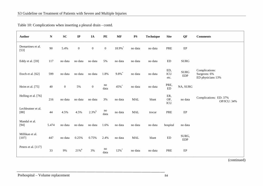
S3 Guideline on Treatment of Patients with Severe and Multiple Injuries
Prehospital – Volume replacement 84
Table 10: Complications when inserting a pleural drain - contd.
Author N SC IP IA PE MF PS Technique Site QF Comments
Demartines et al. [53]
90 5.4% 0 0 0 18.9%* no data no data PRE EP
Eddy et al. [59] 117 no data no data no data 5% no data no data no data ED SURG
Etoch et al. [62] 599 no data no data no data 1.8% 9.8%* no data no data ED, ICU etc.
SURG EDP
Complications: Surgeons: 6% ED physicians 13%
Heim et al. [75] 40 0 5% 0 no
data 45%* no data no data
PRE, ED
NA, SURG
Helling et al. [76] 216 no data no data no data 3% no data MAL blunt
ER, OP, ICU
no data Complications: ED: 37% OP/ICU: 34%
Lechleutner et al. [88] 44 4.5% 4.5% 2.3%§
no data
no data MAL trocar PRE EP
Mandal et al. [94] 5.474 no data no data no data 1.6% no data no data no data hospital no data
Millikan et al. [107] 447 no data 0.25% 0.75% 2.4% no data MAL blunt ED
SURG, EDP
Peters et al. [117] 33 9% 21%# 3%
no data
12%* no data no data PRE EP
(continued)
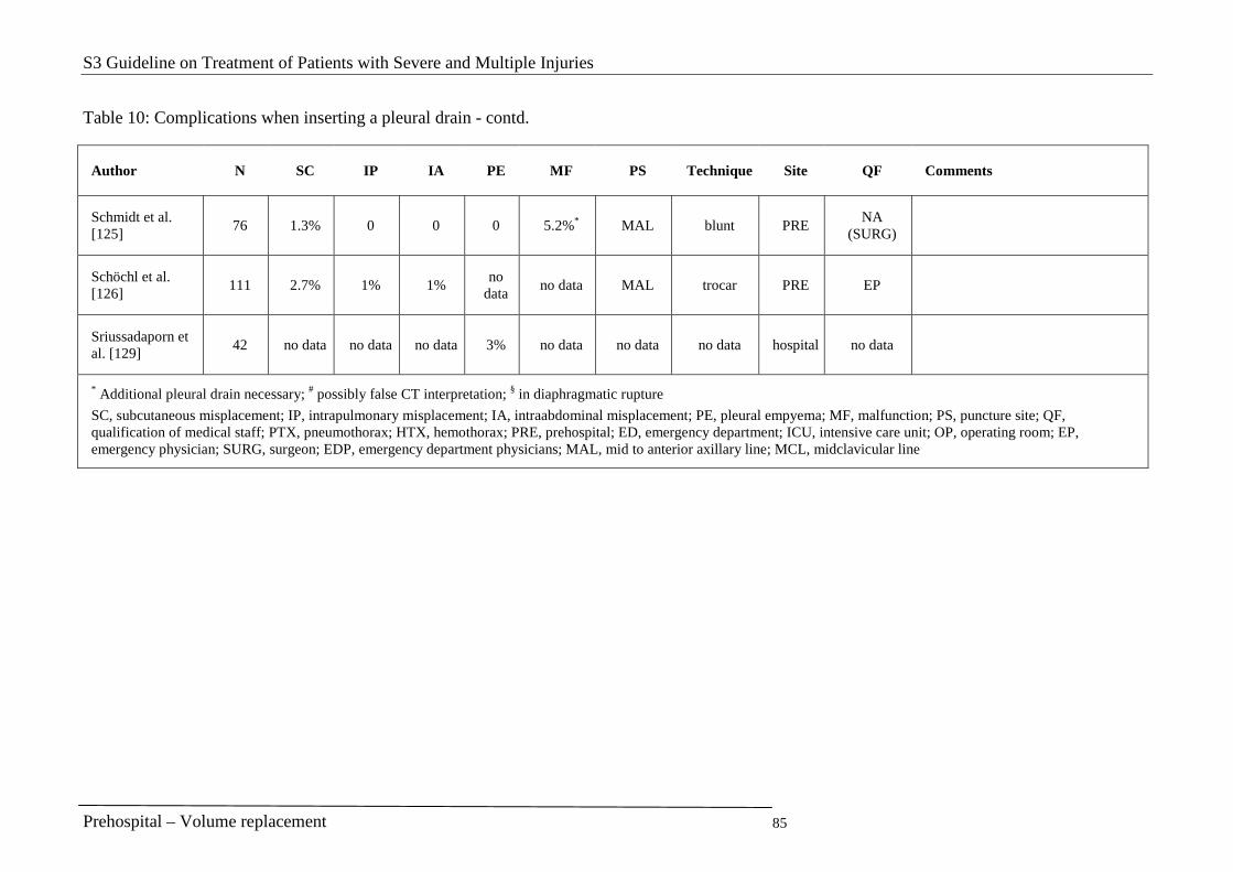
S3 Guideline on Treatment of Patients with Severe and Multiple Injuries
Prehospital – Volume replacement 85
Table 10: Complications when inserting a pleural drain - contd.
Author N SC IP IA PE MF PS Technique Site QF Comments
Schmidt et al. [125]
76 1.3% 0 0 0 5.2%* MAL blunt PRE NA
(SURG)
Schöchl et al. [126]
111 2.7% 1% 1% no
data no data MAL trocar PRE EP
Sriussadaporn et al. [129]
42 no data no data no data 3% no data no data no data hospital no data
* Additional pleural drain necessary; # possibly false CT interpretation; § in diaphragmatic rupture
SC, subcutaneous misplacement; IP, intrapulmonary misplacement; IA, intraabdominal misplacement; PE, pleural empyema; MF, malfunction; PS, puncture site; QF, qualification of medical staff; PTX, pneumothorax; HTX, hemothorax; PRE, prehospital; ED, emergency department; ICU, intensive care unit; OP, operating room; EP, emergency physician; SURG, surgeon; EDP, emergency department physicians; MAL, mid to anterior axillary line; MCL, midclavicular line
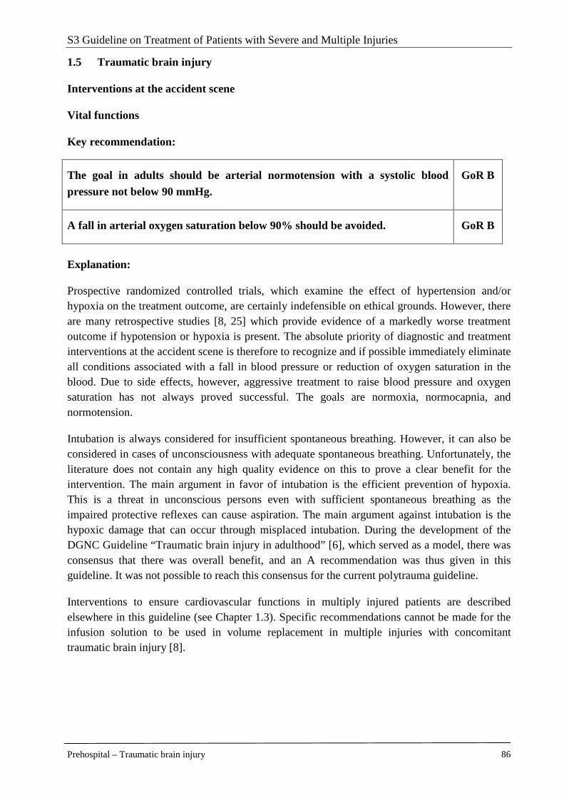
S3 Guideline on Treatment of Patients with Severe and Multiple Injuries
Prehospital – Traumatic brain injury 86
1.5 Traumatic brain injury
Interventions at the accident scene
Vital functions
Key recommendation:
The goal in adults should be arterial normotension with a systolic blood pressure not below 90 mmHg.
GoR B
A fall in arterial oxygen saturation below 90% should be avoided. GoR B
Explanation:
Prospective randomized controlled trials, which examine the effect of hypertension and/or hypoxia on the treatment outcome, are certainly indefensible on ethical grounds. However, there are many retrospective studies [8, 25] which provide evidence of a markedly worse treatment outcome if hypotension or hypoxia is present. The absolute priority of diagnostic and treatment interventions at the accident scene is therefore to recognize and if possible immediately eliminate all conditions associated with a fall in blood pressure or reduction of oxygen saturation in the blood. Due to side effects, however, aggressive treatment to raise blood pressure and oxygen saturation has not always proved successful. The goals are normoxia, normocapnia, and normotension.
Intubation is always considered for insufficient spontaneous breathing. However, it can also be considered in cases of unconsciousness with adequate spontaneous breathing. Unfortunately, the literature does not contain any high quality evidence on this to prove a clear benefit for the intervention. The main argument in favor of intubation is the efficient prevention of hypoxia. This is a threat in unconscious persons even with sufficient spontaneous breathing as the impaired protective reflexes can cause aspiration. The main argument against intubation is the hypoxic damage that can occur through misplaced intubation. During the development of the DGNC Guideline “Traumatic brain injury in adulthood” [6], which served as a model, there was consensus that there was overall benefit, and an A recommendation was thus given in this guideline. It was not possible to reach this consensus for the current polytrauma guideline.
Interventions to ensure cardiovascular functions in multiply injured patients are described elsewhere in this guideline (see Chapter 1.3). Specific recommendations cannot be made for the infusion solution to be used in volume replacement in multiple injuries with concomitant traumatic brain injury [8].
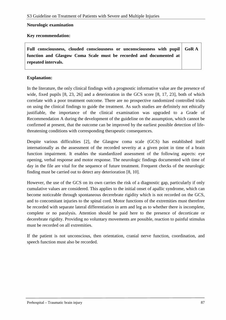
S3 Guideline on Treatment of Patients with Severe and Multiple Injuries
Prehospital – Traumatic brain injury 87
Neurologic examination
Key recommendation:
Full consciousness, clouded consciousness or unconsciousness with pupil function and Glasgow Coma Scale must be recorded and documented at repeated intervals.
GoR A
Explanation:
In the literature, the only clinical findings with a prognostic informative value are the presence of wide, fixed pupils [8, 23, 26] and a deterioration in the GCS score [8, 17, 23], both of which correlate with a poor treatment outcome. There are no prospective randomized controlled trials on using the clinical findings to guide the treatment. As such studies are definitely not ethically justifiable, the importance of the clinical examination was upgraded to a Grade of Recommendation A during the development of the guideline on the assumption, which cannot be confirmed at present, that the outcome can be improved by the earliest possible detection of life-threatening conditions with corresponding therapeutic consequences.
Despite various difficulties [2], the Glasgow coma scale (GCS) has established itself internationally as the assessment of the recorded severity at a given point in time of a brain function impairment. It enables the standardized assessment of the following aspects: eye opening, verbal response and motor response. The neurologic findings documented with time of day in the file are vital for the sequence of future treatment. Frequent checks of the neurologic finding must be carried out to detect any deterioration [8, 10].
However, the use of the GCS on its own carries the risk of a diagnostic gap, particularly if only cumulative values are considered. This applies to the initial onset of apallic syndrome, which can become noticeable through spontaneous decerebrate rigidity which is not recorded on the GCS, and to concomitant injuries to the spinal cord. Motor functions of the extremities must therefore be recorded with separate lateral differentiation in arm and leg as to whether there is incomplete, complete or no paralysis. Attention should be paid here to the presence of decorticate or decerebrate rigidity. Providing no voluntary movements are possible, reaction to painful stimulus must be recorded on all extremities.
If the patient is not unconscious, then orientation, cranial nerve function, coordination, and speech function must also be recorded.
S3 Guideline on Treatment of Patients with Severe and Multiple Injuries
Prehospital – Traumatic brain injury 88
Cerebral protection treatment
Key recommendation:
Glucocorticoids must not be administered. GoR A
Explanation:
According to the latest scientific knowledge, the goal of interventions to be taken at the accident scene is to achieve homeostasis (normoxia, normotension, prevention of hyperthermia) and prevention of threatening complications. The intention is to limit the extent of secondary brain damage and to provide those brain cells with functional impairment but which have not been destroyed with the best conditions for functional regeneration. This applies equally if multiple injuries are present.
Up till now, there is no evidence from the data in the scientific literature of benefit being derived from more extensive treatment regimens viewed as specifically cerebral-protective. At present, no recommendation can be given on the prehospital administration of 21-aminosteroids, calcium antagonists, glutamate receptor antagonists or tris-(tris[hydroxy methyl]aminomethane) buffer [8, 11, 18, 29].
Antiepileptic treatment prevents the incidence of epileptic seizures in the first week after trauma. However, the incidence of a seizure in the early phase does not lead to a worse clinical outcome [20, 25].
The administration of glucocorticoids is no longer indicated due to a significantly increased 14-day case fatality rate [1, 4] with no improvement in clinical outcome [5].
Treatment for suspected severely elevated intracranial pressure
Key recommendation:
If severely elevated intracranial pressure is suspected, particularly with signs of transtentorial herniation (pupil widening, decerebrate rigidity, extensor reaction to painful stimulus, progressive clouded consciousness), the following treatments can be given:
� Hyperventilation
� Mannitol
� Hypertonic saline solution
GoR 0

S3 Guideline on Treatment of Patients with Severe and Multiple Injuries
Prehospital – Traumatic brain injury 89
Explanation:
In cases of suspected transtentorial herniation and signs of apallic syndrome syndrome (pupil widening, decerebrate rigidity, extensor reaction to painful stimulus, progressive clouded consciousness), hyperventilation can be introduced as a treatment option in the early phase after trauma [8, 25]. The guide values are 20 breaths/min in adults. However, hyperventilation, which used to be used because of its often impressive effect in reducing intracranial pressure, also causes reduced cerebral perfusion because of the induced vasoconstriction. With aggressive hyperventilation, this involves the risk of cerebral ischemia and thus deterioration in clinical outcome [25].
The administration of mannitol can lower intracranial pressure [ICP] for a short time (up to 1 hour) [25]. It can also be given without measuring ICP if transtentorial herniation is suspected.
Up till now, there has been only scant evidence of the cerebral-protective effect of hypertonic saline solutions. Mortality appears to be somewhat less compared to mannitol. However, this conclusion is based on a small number of cases and is statistically not significant [28].
There is insufficient evidence [19] for the administration of barbiturates, which was recommended in previous guidelines for intracranial pressure crises not controllable by other means [23]. When administering barbiturates, attention must be paid to the negative inotropic effect, possible fall in blood pressure, and impaired neurologic assessment.
Transport
Key recommendation:
In the case of penetrating injuries, the penetrating object should be left in situ; in certain circumstances it must be detached.
GoR B
Explanation:
It is essential that multiply injured persons with symptoms of concomitant traumatic brain injury are admitted to a hospital with adequate treatment facilities. In the case of a traumatic brain injury with sustained unconsciousness (GCS ≤ 8), increasing cloudiness (deterioration in individual GCS scores), pupillary disorder, paralysis or seizures, the hospital should definitely have provision for neurosurgical management of intracranial injuries [8].
No clear recommendation can be given on analgesic sedation and relaxants for transportation as there is a lack of studies with evidence of a positive effect on traumatic brain injury. With these interventions, cardiopulmonary management is definitely easier to guarantee so that the decision on this must be left to the judgment of the treating emergency physician. The disadvantage of these interventions is a more or less severe limitation on the ability to make a neurologic assessment [23].

S3 Guideline on Treatment of Patients with Severe and Multiple Injuries
Prehospital – Traumatic brain injury 90
In the case of penetrating injuries, the penetrating object should be left in situ; in certain circumstances it must be detached. Injured intracranial vessels are often compressed by the foreign body so that removing it encourages the development of intracranial bleeding. Removal must therefore be carried out under surgical conditions with the possibility of hemostasis in the injured brain tissue. Even if there are no prospective randomized controlled trials on the optimum procedure for penetrating injuries, this procedure makes sense from a pathophysiologic viewpoint.
The possibility of a concomitant unstable spine fracture should be considered during transportation, and the patient should be appropriately positioned (see Chapter 1.6).
S3 Guideline on Treatment of Patients with Severe and Multiple Injuries
Prehospital – Traumatic brain injury 91
References
1. Alderson P, Roberts I. Corticosteroids for acute traumatic brain injury. The Cochrane Database of Systematic Reviews 2005, Issue 1.
2. Balestreri M, Czosnyka M, Chatfield DA, Steiner LA, Schmidt EA, Smielewski P, Matta B, Pickard JD: Predictive value of Glasgow Coma Scale after brain trauma: change in trend over the past ten years. J Neurol Neurosurg Psychiatry 75:161-162, 2004.
3. Brihaye J, Frowein RA, Lindgren S, Loew F, Stroobandt G. Report on the meeting of the WFNS Neuro-Traumatology Committee. Brussels. I. Coma scaling. Acta Neurochir (Wien) 40: 181-186, 1978
4. CRASH trial collaborators. Effect of intravenous corticosteroids on death within 14 days in 10008 adults with clinically significant head injury (MRC CRASH trial): randomised placebo-controlled trial. Lancet 364:1321 – 28, 2004.
5. CRASH trial collaborators. Final results of MRC CRASH, a randomised placebo-controlled trial of intravenous corticosteroid in adults with head injury - outcomes at 6 months. Lancet 365: 1957–59, 2005 [LoE 1b].
6. Firsching R, Messing-Jünger M, Rickels E, Gräber S und Schwerdtfeger K. Leitlinie Schädelhirntrauma im Erwachsenenalter der Deutschen Gesellschaft für Neurochirurgie. AWMF online 2007. http://www.uni-duesseldorf.de/AWMF/ll/008-001.htm.
7. Frowein RA. Classification of coma. Acta Neurochir 34: 5-10, 1976
8. Gabriel EJ, Ghajar J, Jagoda A, Pons PT, Scalea T, Walters BC; Brain Trauma Foundation. Guidelines for prehospital management of traumatic brain injury. J Neurotrauma. 19:111-74, 2002 [Evidenzbasierte Leitlinie]
9. Gurdjian ES, Brihaye J, Christensen JC, Frowein RA, Lindgren S, Luyendijk W, Norlen G, Ommaya AK, Prescu I, de Vasconcellos Marques A, Vigouroux RP. Glossary of Neurotraumatology. Acta Neurochir (Wien) Suppl. 25. Springer, Wien, New York, 1979
10. Karimi A, Burchardi H, Deutsche Interdisziplinäre Vereinigung für Intensiv- und Notfallmedizin (DIVI) Stellungnahmen, Empfehlungen zu Problemen der Intensiv- und Notfallmedizin, 5. Auflage. Köln, asmuth druck + crossmedia. 2004.
11. Langham J, Goldfrad C, Teasdale G, Shaw D, Rowan K. Calcium channel blockers for acute traumatic brain injury (Cochrane Review). In: The Cochrane Library, Issue 1, 2004. Chichester, UK: John Wiley & Sons, Ltd.
12. Lorenz, R. Neurotraumatologie. Standartisierte Nomenklatur. Berlin, Springer 1990
13. Kraus JF, Black MA, Hessol N, Ley P, Rokaw W, Sullivan C, Bowers S, Knowlton S, Marshall L. The incidence of acute brain injury and serious impairment in a defined population. Am J Epidemiol. 1984 Feb;119(2):186-201.
14. Kraus JF, Fife D and Conroy C. Incidence, Severity, and Outcomes of Brain Injuries Involving Bicycles Am J Public Health 1987; 77:76-78.
15. Maas, A. et al.: EBIC-Guidelines for mangement of severe head injury in adults. Acta Neurchir. (Wien) 139, 286-294, 1997 [Evidenzbasierte Leitlinie]
16. Marion DW and Carlier PM. Problems with initial Glasgow Coma Scale assessment caused by prehospital treatment of patients with head injuries: results of a national survey. J Trauma. 1994, 36(1):89-95.
17. Marmarou A, Lu J, Butcher I, McHugh GS, Murray GD, Steyerberg EW, Mushkudiani NA, Choi S, Maas AI. Prognostic value of the Glasgow Coma Scale and pupil reactivity in traumatic brain injury assessed
pre‐hospital and on enrollment: an IMPACT analysis.
J Neurotrauma. 2007; 24(2):270-80 [LoE 3a].
18. Roberts I Aminosteroids for acute traumatic brain injury (Cochrane Review). In: The Cochrane Library, Issue 1, Chichester, UK: John Wiley & Sons, Ltd. 2004
19. Roberts I. Barbiturates for acute traumatic brain injury (Cochrane Review). In: The Cochrane Library, Issue 1, 2004. Chichester, UK: John Wiley & Sons, Ltd.
20. Schierhout G, Roberts I. Anti-epileptic drugs for preventing seizures following acute traumatic brain injury (Cochrane Review). In: The Cochrane Library, Issue 1, 2004. Chichester, UK: John Wiley & Sons, Ltd.
21. Teasdale G, Jennett B. Assessment of coma and impaired consiousness. Lancet 2 81-84, 1974.
22. Teasdale G, Jennett B: Assessment and prognosis of coma after head injury. Acta Neurochir (Wien) 34: 45-55, 1976.
23. The Brain Trauma Foundation. The American Association of Neurological Surgeons. The Joint Section on Neurotrauma and Critical Care. Management and Prognosis of Severe Traumatic Brain Injury. 2000 http://www2.braintrauma.org/guidelines/downloads/btf_guidelines_management.pdf [Evidenzbasierte Leitlinie]
24. The Brain Trauma Foundation. The American Association of Neurological Surgeons. The Joint Section on Neurotrauma and Critical Care. Management and Prognosis of Severe Traumatic Brain Injury. Update 2003 http://www2.braintrauma.org/guidelines/downloads/btf_guidelines_cpp_u1.pdf [Evidenzbasierte Leitlinie]
25. The Brain Trauma Foundation. The American Association of Neurological Surgeons. The Joint Section on Neurotrauma and Critical Care. Guidelines for the Management of Severe Traumatic Brain Injury. 3rd Edition. http://braintrauma.org/guidelines/downloads/JON_24_Supp1.pdf [Evidenzbasierte Leitlinie]
26. Tien HC, Cunha JR, Wu SN, Chughtai T, Tremblay LN, Brenneman FD, Rizoli SB. Do trauma patients with a Glasgow Coma Scale score of 3 and bilateral

S3 Guideline on Treatment of Patients with Severe and Multiple Injuries
Prehospital – Traumatic brain injury 92
fixed and dilated pupils have any chance of survival? J Trauma. 2006; 60(2):274-8 [LoE 3b].
27. Tönnis W, Loew F. Einteilung der gedeckten Hirnschädigungen. Ärztliche Praxis 5: 13-14, 1953
28. Wakai A, Roberts IG, Schierhout G. Mannitol for acute traumatic brain injury. Cochrane Database of Systematic Reviews 2007, Issue 1 [LoE 3b].
29. Willis C, Lybrand S, Bellamy N. Excitatory amino acid inhibitors for traumatic brain injury (Cochrane Review). In: The Cochrane Library, Issue 1, 2004. Chichester, UK: John Wiley & Sons, Ltd.
S3 Guideline on Treatment of Patients with Severe and Multiple Injuries
Prehospital – Spine 93
1.6 Spine
When should a spinal injury be assumed?
What diagnostic procedures are required?
Key recommendation:
A thorough physical examination including the spine and the functions associated with it must be carried out.
GoR A
Explanation:
A physical examination of the patient is the basic requirement for making a diagnosis which, in turn, is the prerequisite for subsequent treatment interventions.
After the vital functions have been monitored and secured, the initial examination of a responsive patient’s spine in the emergency situation at the accident scene involves the exploratory neurologic assessment of sensitivity and motor functions. A segmental neurologic deficit indicates the presence of a spinal cord injury. The level and complete/incomplete lesions can be measured to a limited extent. An absence of back pain is not a definite sign that there can be no relevant injury to the thoracic or lumbar spine [28].
To complete the initial examination, the cervical spine and the entire back are inspected (for signs of injury, deformities) and felt (tenderness, percussion tenderness, steps, displacements, palpable gaps between spinous processes).
Assessing the mechanism of injury can provide clues on the probability of a spinal injury [20].
Even if there are no scientific studies on the importance and the necessary scope of the physical examination in the prehospital emergency examination, it is still an indispensable requirement for detecting symptoms and making (suspected) diagnoses. All the above-mentioned examinations are used to detect relevant, threatening or potentially threatening disorders and injuries, which altogether can make it necessary to administer immediate and specific treatment or make a logistic decision on the spot [2, 17].
The circulation parameters, blood pressure and pulse, should be measured more than once at least during the course (depending on finding, overall situation and timeframe). These are dynamic values, which are indicators for the occurrence of neurogenic shock.
Various scoring systems do not permit a clear statement but combining several scores increases the probability of success [63].
Which concomitant injuries make the presence of spinal injury likely?
Key recommendation:

S3 Guideline on Treatment of Patients with Severe and Multiple Injuries
Prehospital – Spine 94
The presence of a spinal injury must be assumed in unconscious patients until evidence to the contrary is found.
GoR A
Explanation:
The coincidence of spinal injuries and certain other injury patterns is increased. These are purely statistical probabilities.
How is the diagnosis for unstable spinal injury made and how definite is it?
Key recommendation:
If the following 5 criteria are absent, it can be assumed that no unstable spinal injury is present:
� impaired consciousness
� neurologic deficit
� spinal pain or myogelosis
� intoxication
� trauma in the extremities
GoR A
Explanation:
Several groups have developed clinical decision rules to simplify prehospital patient transportation and to set sensible limits to the radiologic primary diagnostic study after blunt trauma to the spine. Some of these decision rules relate to the prehospital situation [23, 24, 45] whereas others relate to the emergency department [9, 33, 35, 36, 56]. Whereas some studies examine the whole spine, others limit themselves to the cervical (C) or thoracic/lumbar spine (T/L).
The results of these studies correspond [9] to the maximum extent so that we can primarily rely hereinafter on the prospectively validated criteria of Domeier et al. and Muhr et al. [24, 25]. Smaller studies have concentrated solely on multiply injured patients [53] but find similar predictors so that it appears justified to generalize the results. On other hand, Muhr et al. and Holmes et al. regarded the presence of other relevant injuries as a criterion that made the definite exclusion of a spinal injury more difficult or impossible. A retrospective study of patients with thoracolumbar spinal injuries found that the presence of concomitant injuries lowered the frequency of (pressure) pain in the back from over 90% to 64% [43]. However, one can assume from this that multiply injured patients have either an extremity fracture or impaired consciousness so that, depending on the decision rules, a suspected spinal injury cannot be ruled out.
S3 Guideline on Treatment of Patients with Severe and Multiple Injuries
Prehospital – Spine 95
Taking into consideration the 5 criteria of impaired consciousness, neurologic deficit, spinal pain or myogelosis, intoxication, and trauma in the extremities, Domeier et al. missed only 2 relevant spinal injuries [24]. In addition, there were 13 stable spinal injuries that did not require osteosynthesis thus yielding a sensitivity of 95% with a negative predictor value of 99.5%. The study related to the whole spine and found approximately 100 fractures each in the cervical, thoracic, and lumbar spine.
Rotation injuries (type C according to AO) are relatively unstable and have an increased risk of further neurologic deterioration [32]. If a rotation injury is suspected on the basis of the mechanism of injury, immobilization should be carried out carefully and without delay on account of the instability.
How is the diagnosis for spinal injury without spinal cord involvement made and how definite is it?
Key recommendation:
Acute pain in the spinal region after trauma should be assessed as an indication of a spinal injury.
GoR B
Explanation:
The cited injury signs may be present both in a bony spinal injury and in a solely soft tissue injury surrounding the bone. There are no prehospital findings that can be collected which can prove or exclude a spinal injury with certainty. External injury signs - deformations, tenderness, percussion tenderness, steps, lateral displacements, palpable gaps between spinous processes - are indirect clues to the presence of an injury to the spine. An evaluation of the (positioning) stability of the injury cannot be made in the prehospital phase.
How is the diagnosis for spinal injury with spinal cord involvement made and how definite is it?
Explanation:
The neurologic deficit in sensitivity and/or motor functions is definitive in the diagnosis of damage to the spinal cord. It is highly probable that a bony injury to the spine is also present in adults. Neurologic deficits without bony involvement can occur more frequently in children (SCIWORA [Spinal Cord Injury Without Radiographic Abnormality] syndrome) [7].
The level and complete/incomplete lesions can only be measured to a limited extent. It is therefore not possible to make a conclusive statement on the prognosis of the injury at the accident scene.
On the other hand, a normal neurology finding does not exclude a spinal injury with spinal cord involvement.
S3 Guideline on Treatment of Patients with Severe and Multiple Injuries
Prehospital – Spine 96
How is a spinal injury treated in the prehospital phase?
What is the technical rescue procedure for a person with a spinal injury?
Key recommendations:
In the event of acute threat to life (e.g., fire/danger of explosion), which can only be eliminated by immediate rescue from the danger zone, immediate, direct rescue from the danger zone must be effected even if a spinal injury is suspected, if necessary even disregarding precautionary measures for the injured person.
GoR A
The cervical spine must be immobilized before technical rescue. GoR A
Explanation:
The first prehospital procedure for a casualty is to immobilize the cervical spine using a cervical collar. To date, however, we are not aware of any literature that confirms this procedure in preventing secondary damage during the technical rescue. No differences in the use of different immobilization collars have been found [18, 51].
During the rescue of an injured person, all non-physiologic spine movements, particularly flexion, segmental rotation and lateral inclination, must be avoided. The spine must be moved into its neutral position, i.e. flat supine position, in a coordinated way with enough assistants [6]. With due consideration of the time required, a more extended technical rescue - e.g., involving removal of a car roof - should be considered. Aids such as the scoop stretcher or spine boards make it easier to rescue a person with a spinal injury in the above-mentioned neutral position from a difficult accident scene.
How is a person with a spinal injury positioned/immobilized?
Explanation:
Up till now, the first prehospital procedure for a casualty is the immobilization of the cervical spine using a cervical collar, even if the evidence level for this is not high. The cervical spine is thereby put into the neutral position. If this causes pain or an increase in neurologic deficit, do not reposition in the neutral position.
In a prospective study, Bandiera and Stiell found evidence that clinically significant injuries could be detected with a sensitivity of 100% using the Canadian C-spine rule [56]. However, a proviso should be added here that this study was conducted in hospital on fully conscious patients [5]. Thus, a relatively long period of time has already elapsed since the accident and, at this later point in time, the symptoms of milder acceleration injuries to the cervical spine also manifest themselves for the first time; these may not occur at the accident scene due to the psychologic impairment.
S3 Guideline on Treatment of Patients with Severe and Multiple Injuries
Prehospital – Spine 97
When there is a traumatic brain injury and a suspected cervical spine injury, it should be weighed up whether to fit a rigid cervical collar or whether another type of immobilization (e.g., only a vacuum mattress) is possible in order to prevent a potential increase in ICP [21, 22, 37, 38, 39, 50]. In another clinical study, there was no evidence of an increase in ICP if the rigid cervical collar was fitted correctly [39]. So, when a rigid cervical collar is being fitted on a patient with a TBI, care should be taken that it is the correct size and not too tightly fastened so that the possibility of any venous outflow obstruction is excluded. In addition, the upper part of the body should be elevated if possible in this situation.
The above-mentioned position can also be immobilized on the vacuum mattress. This achieves the currently most effective immobilization of the whole spine as well. If the head is also enclosed with high cushions or belts, this further restricts possible residual movement of the cervical spine. To date, there is no randomized study that provides evidence of a positive effect from immobilizing the spine [40].
A patient carry sheet on the vacuum mattress makes subsequent re-positioning in hospital easier [8]. Other aids such as the scoop stretcher or spine boards can only immobilize the spine to a limited extent.
S3 Guideline on Treatment of Patients with Severe and Multiple Injuries
Prehospital – Spine 98
How is a person with a spinal injury transported?
Key recommendation:
Transport should be as gentle as possible and free of pain. GoR B
Explanation:
A patient with a spinal injury should be transported as gently as possible, i.e. without further external force to avoid pain and possible secondary damage. After positioning and strapping in, analgesics are administered during transportation. A helicopter offers the smoothest form of transport. In addition, it might offer a time advantage when a patient with a spinal injury and neurologic deficits has to be transported to a center.
Is there a specific treatment for spinal injury in the prehospital phase?
Explanation:
The benefit of a high dose of cortisone treatment being administered prehospital (or subsequently) for spinal injuries with neurologic deficit is controversial [65]. Following the successful administration of corticosteroids for spinal trauma in many animal experiments [1, 25, 26, 58, 59, 66], it has not been possible to confirm the results in all clinical studies. Criticism has been leveled at the NASCIS (National Acute Spinal Cord Injury Studies) several times [19], for instance, the lack of effect in the NASCIS I study (there was no control group here but low-dose cortisone was compared with too little high-dose cortisone) and the lack of placebo group in the NASCIS III study as well [19]. The positive effects in the NASCIS II study were only minor, of limited clinical relevance, and less marked after 1 year than after 6 months.
Here is a summary of the advantages and disadvantages of giving methylprednisolone according to current literature:
Reasons for cortisone treatment:
1. The NASCIS II study showed an improvement in motor outcome providing methyl prednisolone treatment was started within 8 hours [11, 12]. However, this outcome was only unilaterally verified and dependent on the researcher.
2. Other studies of worse methodological quality have also found a benefit from cortisone treatment but in this case the start of treatment was predominantly evaluated after admission to hospital.
3. The NASCIS III study showed a greater effect in treatment duration of 48 hours providing treatment commenced between 3 and 8 hours after trauma [14, 15].
4. Relevant side effects such as abdominal bleeding are not increased [27] and there has been no evidence of accumulation of femoral head necroses following high-dose cortisone treatment [64].
S3 Guideline on Treatment of Patients with Severe and Multiple Injuries
Prehospital – Spine 99
5. There is no other definitive pharmacologic treatment for spinal cord injury.
Reasons against cortisone treatment:
1. No relevant benefit could be found in the NASCIS I study [13].
2. The proven benefit was only found in patients who had received treatment within 8 hours.
3. The proven benefit was small, of unconfirmed clinical significance, and even smaller after 1 year than after 6 months [10, 12].
4. There was no placebo control group in the NASCIS III study [14, 15].
5. Further studies showed a higher complication rate in the patients treated with cortisone (increase in lung complications [29, 31], particularly in elderly patients [42], and gastrointestinal bleeding [48]).
6. Lack of neurologic benefit in other studies with frequently unclear injury pattern [30, 42, 48, 49].
Infusion treatment to stabilize the circulation is necessary in neurogenic shock with due consideration being paid to other possible sources of bleeding caused by injury. The infusion volume to be administered and the target mean arterial pressure is also disputed by expert opinion. Adequate analgesic treatment is necessary to prevent shock.
Extreme pulling forces on the cervical spine, e.g., when removing a motorbike helmet, and segmental torsions on unstable C injuries (cervical vertebrae) of the spine can lead to deterioriation in the neurologic deficit by directly affecting the spinal cord. It should be noted here that there are no references to secondary damage in the literature on this either.
S3 Guideline on Treatment of Patients with Severe and Multiple Injuries
Prehospital – Spine 100
Are there advantages for the patient with spinal injury in being transported primarily to a trauma center with a spine surgery facility?
Key recommendation:
Patients with neurologic deficits and suspected spinal injury should be transported primarily and as a minimum to a regional trauma center with a spine surgery facility.
GoR B
Explanation:
Early surgery on spinal injuries with spinal cord involvement can improve the neurologic outcome [44, 52].
Early surgery (within 72 hours) on cervical spine injuries with neurologic deficits does not conceal an increased risk of additional complications [44].
For this reason, particularly in the case of isolated spinal trauma and a non-acute threat to life, management should, if possible, be in a spinal center [60]. Patients with a spinal canal constriction, particularly in the cervical region, appear to gain from early surgery [4]. Even if there is only little evidence, it should still be assumed that patients with incomplete neurology and partial displacement of the spinal canal could gain from early reduction and, if necessary, surgical debridement.
Summary:
The vast majority of the screened literature relates mainly to the hospital situation, in other words, to studies which were conducted after admission to hospital. Provided they are relevant, these data must be extrapolated to the prehospital situation. There is a relatively large number of studies which were conducted in the USA and thus in the paramedic system. This initial management at the accident scene is only partially comparable with the German rescue and emergency physician system.
These two points must be taken into account as this means that conclusions (in terms of a guideline) have only restricted validity on applicability to the prehospital emergency physician situation in Germany.
S3 Guideline on Treatment of Patients with Severe and Multiple Injuries
Prehospital – Spine 101
References
1. Amar PA, Levy ML (1999) Pathogenesis and pharmacological strategies for mitigatin secondary damage in acute spinal cord injury. Neurosurgery 44:1027-1040
2. American, College, of, Surgeons (1997) Advanced Trauma Life Support (Chicago)
3. Asamoto S, Sugiyama H, Iida M, Doi H, Itoh T, Nagao T, Hayashi M, Matsumoto K, Morii M (2001) Trauma sites and clinical features associated with acute hyperextension spinal cord injury without bone damage--relationship between trauma site and severity. Neurol Med Chir (Tokyo) 41(1):1-6; discussion 6-7.
4. Asazuma T, Satomi K, Suzuki N, Fujimura Y, Hirabayashi K (1996) Management of patients with an incomplete cervical spinal cord injury. Spinal Cord 34(10):620-625.
5. Bandiera G, Stiell IG, Wells GA, Clement CM, De Maio VJ, Vandemheen KL (2003) The Canadian C-spine Rule Performs Better Than Unstructured Physician Judgment. Ann Emerg Med 42:395-402
6. Beck A, Krischak G, Bischoff M (2009) Wirbelsäulenverletzungen und spinales Trauma. Notfall & Rettungsmedizin 12(6):469-479
7. Beck A, Gebhard F, Kinzl L, Rüter A, Hartwig E (2000) Spinal cord injury without radiographic abnormalities in children and adolescents. Knee Surg, Sports Traumatol, Arthrosc 8(3):186-189
8. Beck A, Gebhard F, Kinzl L, Strecker W (2001) Prinzipien und Techniken der unfallchirurgischen Erstversorgung am Einsatzort. Unfallchirurg 104(11):1082-1099
9. Blackmore C, Emerson S, Mann F, Koepsell TD (1999) Cervical spine imaging in patients with trauma: Determination of fracture risk to optimize use. Radiology 211(3):759-766
10. Bracken MB, Holford TR (1993) Effects of timing of methylprednisolone or naloxone administration on recovery of segmental and long-tract neurological function in NASCIS 2. J Neurosurg 79(4):500-507.
11. Bracken MB, Shepard MJ, Collins WF, Holford TR, Young W, Baskin DS, Eisenberg HM, Flamm E, Leo-Summers L, Maroon J, et al. (1990) A randomized, controlled trial of methylprednisolone or naloxone in the treatment of acute spinal-cord injury. Results of the Second National Acute Spinal Cord Injury Study. N Engl J Med 322(20):1405-1411.
12. Bracken MB, Shepard MJ, Collins WF, Jr., Holford TR, Baskin DS, Eisenberg HM, Flamm E, Leo-Summers L, Maroon JC, Marshall LF, et al. (1992) Methylprednisolone or naloxone treatment after acute spinal cord injury: 1-year follow-up data. Results of the second National Acute Spinal Cord Injury Study. J Neurosurg 76(1):23-31.
13. Bracken MB, Shepard MJ, Hellenbrand KG, Collins WF, Leo LS, Freeman DF, Wagner FC, Flamm ES, Eisenberg HM, Goodman JH, et al. (1985) Methylprednisolone and neurological function 1 year after spinal cord injury. Results of the National Acute Spinal Cord Injury Study. J Neurosurg 63(5):704-713.
14. Bracken MB, Shepard MJ, Holford TR, Leo-Summers L, Aldrich EF, Fazl M, Fehlings M, Herr DL, Hitchon PW, Marshall LF, Nockels RP, Pascale V, Perot PL, Jr., Piepmeier J, Sonntag VK, Wagner F, Wilberger JE, Winn HR, Young W (1997) Administration of methylprednisolone for 24 or 48 hours or tirilazad mesylate for 48 hours in the treatment of acute spinal cord injury. Results of the Third National Acute Spinal Cord Injury Randomized Controlled Trial. National Acute Spinal Cord Injury Study. JAMA 277(20):1597-1604.
15. Bracken MB, Shepard MJ, Holford TR, Leo-Summers L, Aldrich EF, Fazl M, Fehlings MG, Herr DL, Hitchon PW, Marshall LF, Nockels RP, Pascale V, Perot PL, Jr., Piepmeier J, Sonntag VK, Wagner F, Wilberger JE, Winn HR, Young W (1998) Methylprednisolone or tirilazad mesylate administration after acute spinal cord injury: 1-year follow up. Results of the third National Acute Spinal Cord Injury randomized controlled trial. J Neurosurg 89(5):699-706.
16. Chandler DR, Nemejc C, Adkins RH, Waters RL (1992) Emergency cervical-spine immobilization. Ann Emerg Med 21(10):1185-1188.
17. Chen XY, Carp JS, Chen L, Wolpaw JR (2002) Corticospinal tract transection prevents operantly conditioned H-reflex increase in rats. Exp Brain Res 144(1):88-94
18. Cline JR, Scheidel E, Bigsby EF (1985) A comparison of methods of cervical immobilization used in patent extrication and transport. J Trauma 25(7):649-653
19. Coleman WP, Benzel D, Cahill DW, Ducker T, Geisler F, Green B, Gropper MR, Goffin J, Madsen PW, 3rd, Maiman DJ, Ondra SL, Rosner M, Sasso RC, Trost GR, Zeidman S (2000) A critical appraisal of the reporting of the National Acute Spinal Cord Injury Studies (II and III) of methylprednisolone in acute spinal cord injury. J Spinal Disord 13(3):185-199.
20. Cooper C, Dunham CM, Rodriguez A (1995) Falls and major injuries are risk factors for thoracolumbar fractures: cognitive impairment and multiple injuries impede the detection of back pain and tenderness. J Trauma 38:692-696
21. Craig G, Nielsen MS (1991) Rigid cervical collars and intracranial pressure. Intensive Care Med 17:504-505
22. Davies G, Deakin c, Wilson A (1996) The effects of a rigid collar on intracranial pressure. Injury 27:647-649
23. Domeier RM, Evans RW, Swor RA, Rivera-Rivera EJ, Frederiksen SM (1997) Prospective validation of out-of-hospital spinal clearance criteria: a preliminary report. Acad Emerg Med 4(6):643-646 [LoE 1a]
24. Domeier RM, Swor RA, Evans RW, Hancock JB, Fales W, Krohmer J, Frederiksen SM, Rivera-Rivera EJ, Schork MA (2002) Multicenter prospective validation of prehospital clinical spinal clearance criteria. J Trauma 53(4):744-750
25. Ducker TB (1990) Treatment of spinal-cord injury. N Engl J Med 322(20):1459-1461.
S3 Guideline on Treatment of Patients with Severe and Multiple Injuries
Prehospital – Spine 102
26. Ducker TB, Zeidman SM (1994) Spinal cord injury: Role of steroid therapy. Spine 19:2281-2287
27. Epstein N, Hood DC, Ransohoff J (1981) Gastrointestinal bleeding in patients with spinal cord trauma. Effects of steroids, cimetidine, and mini-dose heparin. J Neurosurg 54(1):16-20.
28. Frankel H, Rozycki G, Ochsner MG, Harviel JD, Champion HR (1994) Indication for obtaining surveillance thoracic and lumbar spine radiographs. J Trauma 37(4):673-676
29. Galandiuk S, Raque G, Appel S, Polk HCj (1993) The two-edged sword of large-dose steroids for spinal cord trauma. Ann Surg 218:419-427
30. George ER, Scholten DJ, Buechler CM, Jordan-Tibbs J, Mattice C, Albrecht RM (1995) Failure of methylprednisolone to improve the outcome of spinal cord injuries. Am Surg 61:659-664
31. Gerndt SJ, Rodriguez JL, Pawlik JW, Taheri PA, Wahl WL, Michaels AJ, Papadopoulos SM (1997) Consequences of high-dose steroid therapy for acute spinal cord injury. J Trauma 42:279-284
32. Gertzbein SD (1994) Neurologic deterioration in patients with thoracic and lumbar fractures after admission to the hospital. Spine 19(15):1723-1725.
33. Hanson JA, Blackmore CC, Mann FA, Wilson AJ (2000) Cervical spine injury: a clinical decision rule to identify high-risk patients for helical CT screening. AJR Am J Roentgenol 174(3):713-717
34. Ho AM-H, Fung KY, M. JG, K. KM, Peng Z (2002) Rigid Cervical Collar and Intracranial Pressure of Patients with Severe Head Injury. J Trauma 53(6):1185-1188
35. Hoffman JR, Mower WR, Wolfson AB, Todd KH, Zucker MI (2000) Validity of a set of clinical criteria to rule out injury to the cervical spine in patients with blunt trauma. National Emergency X-Radiography Utilization Study Group. New Engl J Med 343(2):94-99
36. Holmes JF, Panacek EA, Miller PQ, Lapidis AD, Mower WR (2003) Prospective evaluation of criteria for obtaining thoraxolumbar radiographs in trauma patients. J Emerg Med 24(1):1-7
37. Hunt K, Hallworth S, Smith M (2001) The effects of rigid collar placement on intracranial and cerebral perfussion pressures. Anaesthesia 56:511-513
38. Kolb JC, Summers RL, Galli RL (1999) Cervical collar-induced changes in intracranial pressure. Am J Emerg Med 17:135-137
39. Kuhnigk H, Bomke S, Sefrin P (1993) [Effect of external cervical spine immobilization on intracranial pressure]. Aktuelle Traumatol 23(8):350-353.
40. Kwan I, Bunn F, Roberts I, Committee obotWP-HTCs (2001) Spinal immobilisation for trauma patients (Cochrane Review). The Cochrane Library Issue 4):Update Software
41. Lam AM (1992) Spinal Cord Injury: Management. Curr Opin Anesth 5:632-639
42. Matsumoto T, Tamaki T, Kawakami M, Yoshida M, Ando M, Yamada H (2001) Early complications of high-dose methylprednisolone sodium succinate
treatment in the follow-up of acute cervical spinal cord injury. Spine 26(4):426-430.
43. Meldon SW, Moettus LN (1995) Thoracolumbar spine fractures: clinical presentation and the effect of altered sensorium and major injury. J Trauma 39(6):1110-1114 [LoE 4]
44. Mirza SK, Krengel WF, 3rd, Chapman JR, Anderson PA, Bailey JC, Grady MS, Yuan HA (1999) Early versus delayed surgery for acute cervical spinal cord injury. Clin Orthop 359:104-114 [LoE 4]
45. Muhr MD, Seabrook DL, Wittwer LK (1999) Paramedic use of a spinal injury clearance algorithm reduces spinal immobilization in the out-of-hospital setting. Prehosp Emerg Care 3(1):1-6 [LoE 1a]
46. Nechwatal E (1975) [Critical notes on the transport of patients with cervical spinal cord injuries]. Chirurg 46(11):521-523.
47. Nockels RP (2001) Nonoperative Management of Acute Spinal Cord Injury. Spine 26(24S):S31-"37
48. Pointillart V, Petitjean ME, Wiart L, Vital JM, Lassie P, Thicoipe M, Dabadie P (2000) Pharmacological therapy of spinal cord injury during the acute phase. Spinal Cord 38(2):71-76.
49. Prendergast MR, Saxe JM, Ledgerwood AM, Lucas CE, Lucas WF (1994) Massive steroids do not reduce the zone of injury after penetrating spinal cord injury. J Trauma 37(4):576-579; discussion 579-580.
50. Raphael J, Chotai R (1994) Effects of the cervical collar on cerebrospinal fluid pressure. Anaesthesia 49:437-439
51. Rosen PB, McSwain NE, Arata M, Stahl S, Mercer D (1992) Comparison of two new immobilization collars. Ann Emerg Med 21(10):1189-1195
52. Rosenfeld JF, Vaccaro AR, Albert TJ, Klein GR, Cotler JM (1998) The benefits of early decompression in cervical spinal cord injury. Am J Orthop 27(1):23-28.
53. Ross SE, O`Malley KF, DeLong WG, Born CT, Schwab CW (1992) Clinical predictors of unstable cervical spinal injury in multiply injured patients. Injury 23(5):317-319 [LoE 2b]
54. Sackett DL, Richardson WS, Rosenberg W, Haynes RB (1997) Evidence-based medicine: How to practice and teach EBM (London/UK, Churchill Livingstone)
55. Sneed RC, Stover SL (1988) Undiagnosed spinal cord injuries in brain-injured children. Am J Dis Child 142(9):965-967.
56. Stiell IG, Wells GA, Vandemheen KL, Clement CM, Lesiuk H, De Maio VJ, Laupacis A, Schull M, McKnight RD, Verbeek R, Brison R, Cass D, Dreyer J, Eisenhauer MA, Greenberg GH, MacPhail I, Morrison L, Reardon M, Worthington J (2001) The canadian c-spine rule for radiopraphy in alert and stable trauma patients. JAMA 286(15):1841-1848
57. Sung RD, Wang JC (2001) Correlation between a positive Hoffmann's reflex and cervical pathology in asymptomatic individuals. Spine 26(1):67-70.
58. Tator CH (1996) Experimental and clincal studies of the pathophysiology and managment of acute spinal cord injury. J Spinal Cord Med 19:206-214

S3 Guideline on Treatment of Patients with Severe and Multiple Injuries
Prehospital – Spine 103
59. Tator CH (1998) Biology of neurological recovery and functional restoration after spinal cord injury. Neurosurgery 42:696-708
60. Tator CH, Duncan EG, Edmonds VE, Lapczak LI, Andrews DF (1995) Neurological recovery, mortality and length of stay after acute spinal cord injury associated with changes in management. Paraplegia 33(5):254-262.
61. Vogel P (1992) [Neurologic disorders after injuries of the spine]. Langenbecks Arch Chir Suppl Kongressbd :271-273.
62. Weinstein DE, Ko HY, Graziani V, Ditunno JF, Jr. (1997) Prognostic significance of the delayed plantar reflex following spinal cord injury. J Spinal Cord Med 20(2):207-211.
63. Wells JD, Nicosia S (1995) Scoring acute spinal cord injury: a study of the utility and limitations of five different grading systems. J Spinal Cord Med 18(1):33-41.
64. Wing PC, Nance P, Connell DG, Gagnon F (1998) Risk of avascular necrosis following short term megadose methylprednisolone treatment. Spinal Cord 36(9):633-636.
65. Young W (1993) Secondary injury mechanisms in acute spinal cord injury. J Emerg Med 11 (Suppl 1):13-22.
66. Zeidman SM, Ling GS, Ducker TB, Ellenbogen RG (1996) Clinical applications of pharmacologic therapies for spinal cord injury. J Spinal Disord 9:367-380
67. Zhu Q, Ouyang J, Lu W, Lu H, Li Z, Guo X, Zhong S (1999) Traumatic instabilities of the cervical spine caused by high-speed axial compression in a human model. An in vitro biomechanical study. Spine 24(5):440-444.
S3 Guideline on Treatment of Patients with Severe and Multiple Injuries
Prehospital – Extremities 104
1.7 Extremities
Priority
Key recommendations:
Heavily bleeding extremity injuries, which can impair the vital function, must be given first priority.
GoR A
The management of extremity injuries must avoid further damage and not delay the total rescue time if there are additional threatening injuries present.
GoR A
Explanation:
Securing the vital functions and examining the head and trunk should precede the examination of the extremities. Specifics can occur in extremity injuries with severe blood loss [21, 27].
Severe and immediate life-threatening bleeding must be treated immediately even ignoring the ABCDE protocol (see page 133).
Confirmation of more major, external bleeding which is not directly life-threatening is important and is usually carried out under “C” (circulation) whereas more minor bleeding comes under the “secondary survey” [21].
The first rule is to avoid further damage, restore and maintain vital functions and transport to a suitable hospital [11, 25].
The management of extremity injuries (irrigation/wound management/splinting) should not delay the rescue time if there are additional threatening injuries present [23].
Diagnostic study
Medical history
A very detailed medical history (firsthand/third party) of the circumstances of the accident can be gathered to obtain sufficient information on the impacting force and, if applicable, the degree of contamination of open wounds [2, 27].
If possible, information (allergies, medication, previous diseases and fasting state) should be collected in addition to the accident history and the time of the accident. In addition, details of tetanus immunization status should be obtained [21, 34].

S3 Guideline on Treatment of Patients with Severe and Multiple Injuries
Prehospital – Extremities 105
Examination
Key recommendation:
All extremities of a casualty should undergo an exploratory assessment in the prehospital phase.
GoR B
Explanation:
Alert patients should be asked first whether they have any pain and where it is. If there is pain, adequate analgesics can be administered early on [21]. A prehospital examination should be carried out [11]. The examination at the accident scene should assess to an appropriate extent the severity of the injury without delaying the total rescue time too much [2]. The examination should be an exploratory survey from head to toe and not last longer than 5 minutes [34].
The examination should be carried in the following order: inspection (malposition/wounds/swelling/circulation), stability test (crepitation, abnormal mobility, stable and unstable fracture signs), assessment of circulation, motor functions and sensitivity. Soft tissue findings should also be assessed (closed versus open fracture, compartment syndrome) [11, 21, 27].
Leather clothing such as motorbike apparel, for example, should be left on if possible as this serves as a splint with compression effect particularly for the pelvis and the lower extremity [14, 21].
Capillary reperfusion can be tested by comparing with the uninjured limb [21].
Treatment
General
Key recommendation:
Even if an extremity injury is only suspected, it should be immobilized against rough movement and before transporting the patient.
GoR B
Explanation:
Immobilizing an injured extremity is an important procedure in prehospital management. An extremity injury should be immobilized against rough movement and before transporting the patient. Reasons for this are to alleviate pain, prevent further soft tissue damage/bleeding and reduce the risk of a fat embolism and neurologic damage [21, 34].
Even a suspected injury should be immobilized [10, 34].
S3 Guideline on Treatment of Patients with Severe and Multiple Injuries
Prehospital – Extremities 106
The joints proximal and distal to the injury should be included in the immobilization [10, 11, 24, 34]. The injured extremity should be supported flat [4]. Particularly in shortened femoral fractures, traction/immobilization under traction should be carried out to minimize bleeding [2, 21]. Vacuum splints are suitable for immobilizing an abnormal position. Vacuum splints are rigid and can adapt to the shape of the extremity [21]. Air chamber splints are suitable for splinting upper extremity injuries with the exclusion of injuries in the proximity of the shoulder joint. In the lower extremity, they are suitable for immobilizing knee, lower leg, and foot injuries. On their attachment, the pressure in the air chamber splints and the peripheral blood supply must be regularly checked [4]. The advantage of the air chamber splint is its low weight; the disadvantage is the compression of soft tissue which can cause secondary damage. Vacuum splints are therefore preferred. Air chamber and vacuum splints are unsuitable for immobilizing femoral fractures and those in the proximity of the shoulder joint [5]. Cooling can reduce swellings and help to alleviate pain [10]. Femur injuries can be adequately immobilized without complications with a spine board or rigid splinting. It is not absolutely necessary for traction splints to be carried in the emergency medical service.
In a retrospective study with 4,513 callouts made by emergency paramedics in an American emergency medical system (EMS), 16 patients (0.35%) with injuries to the mid-femur were singled out. While 11 of these patients had only minor injuries, 5 of these patients (0.11% of all patients) were treated under a femoral fracture diagnosis. Three of these 5 patients received a traction splint. In one of the cases, the traction splint had to be removed again due to severe pain and a rigid immobilization device was attached. One patient could not have a traction splint because of simultaneous hip trauma. Another patient who was free of pain was transported in a comfortable position. The authors conclude that femur injuries and/or a suspected fracture are rare and can be well managed with a backboard or rigid immobilization. For this reason, it is not absolutely necessary for traction splints to be carried in the emergency medical service [1]. Traction splints should not be used particularly on multiply injured patients as there are many contraindications for their use in these patients (pelvic fracture/knee/lower leg/ankle joint injury) [33]. They are only rarely used due to the existing contraindications on the use of a traction splint, particularly in critically injured patients. Dislocated proximal femoral fractures are also contraindications for the use of a traction splint [7].
Traction splints are useful and, depending on the model, easy to use for immobilizing femoral fractures, even dislocated proximal femoral fractures. Further studies are necessary [8]. Traction splints reduce muscle spasms and thus alleviate pain. Traction helps to restore the femur shape and by reducing volume leads to a decrease in bleeding [8, 30, 31]. Oxygen can be given via a non-rebreather mask (15 l/min) [2, 21]. Jewelry (rings/chains) must be removed from the injured extremity [2, 10].
Photos of wounds/open fractures can be taken for documentation (polaroid/digital). Photographic documentation of wounds, open fractures or discovered malpositions appears expedient as it can, under circumstances, avoid immobilized extremities or wounds already dressed in the prehospital phase from being exposed again in the hospital until they are definitively treated. Photographic documentation can assist the subsequent treating physician in assessing the injury. Photographic documentation must not extend the management/rescue time [2, 21].
S3 Guideline on Treatment of Patients with Severe and Multiple Injuries
Prehospital – Extremities 107
The severity and extent of the injuries must be documented in the emergency physician logbook and the local finding must be described to the subsequent treating surgeon, if possible in person [3].
Fractures
Key recommendation:
If possible, and particularly with concomitant ischemia in the extremity concerned/with a long rescue time, grossly dislocated fractures and dislocations should be approximately reduced in the prehospital phase.
GoR B
Explanation:
The primary goal is to secure the local and peripheral blood supply. The primary goal is not an exact anatomic reduction. What is more important is correct axial positioning and the restoration of an adequate local and peripheral blood supply [3, 5]. If the neurovascular supply to the extremity distal to the injury is not compromised, reduction can be ignored in principle [2]. If possible, and particularly with concomitant ischemia in the extremity concerned/with a long rescue time, grossly dislocated fractures and dislocations should be reduced in the prehospital phase by axial traction and manual correction into the neutral position or into a position that is nearest to the neutral position. It is important to check the peripheral blood supply and motor functions and sensitivity (where possible) before and after reduction [3, 4, 5, 11, 21, 25]. Too much longitudinal traction must be avoided as this increases compartmental pressure and worsens the blood supply in the soft tissue [3, 5].
A neurologic or vascular deficit distal to the fracture requires an immediate reduction attempt. The same applies if the soft tissue sheath/skin is compromised [21]. After successful immobilization, circulation, sensitivity, and peripheral motor functions should be checked again [2, 21]. If neurovascular circulation deteriorates after a reduction attempt, the extremity must be immediately placed back in the initial position and stabilized as well as possible [21].
Reduction of ankle fractures/ankle dislocation fractures should only be carried out by those experienced in this procedure. Otherwise, the goal is immobilization in the position found [21]. In the case of commonly dislocated ankle joint fractures with obvious malposition, reduction can be carried out at the accident scene. With adequate analgesia, an approximately correct axial position can be achieved by controlled, continuous longitudinal traction with both hands on the calcaneus and heel of the foot; this position can then be immobilized. After this, the blood supply and neurologic situation should be recorded again.
Obvious long bone fractures in the shaft area should also be treated in this way. Fractures in proximity to joints are difficult to assess in their extent and, after being immobilized in the pain-free position found, can be transferred as rapidly as possible for further hospital diagnosis [2, 34].

S3 Guideline on Treatment of Patients with Severe and Multiple Injuries
Prehospital – Extremities 108
Stronger longitudinal traction should be avoided in distal femoral fractures as this can compromise the popliteal vessels. The knee joint can be supported in a slightly bent position (30-50 degrees) [4].
Open fractures
Key recommendation:
Each open fracture should be cleaned of coarse contamination and covered with a sterile dressing.
GoR B
Explanation:
Each open fracture should be identified and coarse contamination immediately removed [21]. Open fractures should be irrigated with physiologic saline solution [2, 21, 23, 26]. All open wounds should be covered with a sterile dressing [3, 4, 11, 21, 26, 34]. Without further cleansing or disinfection measures, open wounds must be covered with a large sterile dressing. Coarse contamination is removed [3, 4, 5]. Thereafter, they should be immobilized as for closed injuries [26, 34]. It is best if the dressings are not removed until in the operating room [21, 26].
Antibiosis should be carried out at the earliest possible time. The risk of infection increases dramatically after 5 hours [26]. If available, intravenous antibiosis can be administered in the prehospital phase, usually with a 2nd generation cephalosporin which is easily distributed in the bone [4]. Prehospital antibiosis should be carried out if the rescue time is extended [23].

S3 Guideline on Treatment of Patients with Severe and Multiple Injuries
Prehospital – Extremities 109
Key recommendations:
Active bleeding should be treated according to the following stepwise approach:
� manual pressure/pressure dressing
� (elevation)
� tourniquet
GoR B
Indications for immediate use of a tourniquet/arrest of blood supply can be:
� life-threatening bleeding/multiple sources of bleeding in an extremity
� inability to reach the actual injury
� several injured persons with bleeding
GoR 0
Explanation:
The measures for arresting bleeding should follow a stepwise approach. A primary attempt should be made to arrest active bleeding by manual pressure and elevation of the extremity. Then a pressure dressing should be applied. If this is not adequate, a second pressure dressing should be applied over the first one. A sterile pad can be used to help to focus pressure. If bleeding persists, pressure should be applied to an artery proximal to the injury. In addition, if possible, a tourniquet should be applied. As an exception, the vessel can be clamped (amputation, longer transportation time, neck vessel, anatomic position makes the use of a tourniquet impossible) [3, 4, 11, 21, 32].
In regions where tourniquets cannot be applied (proximal extremities), hemostatic dressings can be used [13]. Applying a tourniquet requires appropriate analgesia [21]. A blood pressure cuff with 250 mmHg can be applied to the upper arm and one with 400 mmHg to the femur [3, 5]. The time at which the tourniquet was applied should be noted [21, 22, 28]. The tourniquet must interrupt the arterial blood flow completely. An incorrectly applied tourniquet can intensify bleeding (only compromises low pressure system) [22]. Effectiveness is monitored by an arrest in bleeding rather than the disappearance of the distal pulse. In the case of a fracture, bleeding can also come from the bone marrow [22].

S3 Guideline on Treatment of Patients with Severe and Multiple Injuries
Prehospital – Extremities 110
Indications for immediate use of a tourniquet can be [22]:
� Extreme bleeding/multiple sources of bleeding in an extremity necessitating parallel securing of vital functions
� Inability to reach the actual injury (e.g., trapped person)
� Mass casualty incident
The following points should be borne in mind when applying a tourniquet:
� Apply as far distally as possible, approx. 5 cm proximal to the injury
� Apply directly on the skin to prevent it slipping [22, 28].
If ineffective, re-apply with more pressure and only after that consider applying a second tourniquet directly proximal to the first [22]. Cooling an extremity that has a tourniquet applied can increase ischemic tolerance during long rescue times [15].
There is only insufficient data on the safe application time for a tourniquet. The general recommendation is 2 hours but this has emerged from data obtained from normovolemic patients with a pneumatic tourniquet [22]. If the transportation time until surgery is less than 1 hour, the tourniquet can remain in situ. For longer rescue times (> 1 hour), attempts should be made to release the tourniquet in a stabilized patient. If bleeding should start again, the newly applied tourniquet should then remain in situ until it is managed in the operating room [22]. After 30 minutes, the tourniquet should be checked to see if it is still necessary. This is not indicated if the patient is in shock or the attendant circumstances (personnel) are adverse [12].
In a retrospective case series on war injured from the database of the British military, tourniquets were applied to 70 patients out of 1,375 patients who had been treated during the period in English field hospitals (5.1%). A total of 107 tourniquets were applied (17 of the treated [24%] had 2 or more tourniquets applied). Of this number, 5 had a double tourniquet applied for the same injury and 12 of the injured had bilateral tourniquets (maximum number per injured 4 - 2 each on both lower extremities). A hundred and six tourniquets were applied prior to arrival at the field hospital. Sixty-one of these 70 patients (87.1%) survived. Mean value of the survivors: ISS = 16, mean value of fatalities (only 6 could be autopsied): ISS = 50.
Whereas prior to the introduction of tourniquets as standard (February 2003 to April 2006) only 9% (6 injured persons) were treated with a tourniquet, following introduction (April 2006 through February 2007) it was 64 (91%). Without details of the total number of injured persons during this period, the authors indicate a 20-fold increase in the use of tourniquets. Three complications directly caused by the tourniquets were observed. There were 2 cases of compartment syndromes (one each in the femur and lower leg, one of which was due to incorrect application of the tourniquet) and one case of damage to the ulnar nerve (with no further details on the course). The use of tourniquets was assessed as life-saving in 4 cases of patients with isolated extremity injuries, hypovolemic shock and massive transfusion (and factor VIIa administration) [9].

S3 Guideline on Treatment of Patients with Severe and Multiple Injuries
Prehospital – Extremities 111
In a retrospective study of 165 patients (inclusion criteria: traumatic amputation or severe vessel injury in extremities), Beekley et al. showed that the prehospital use of tourniquets led to improved control of bleeding; this relates particularly to multiply injured patients (ISS > 15). Forty percent of soldiers (n = 67) had a tourniquet. Reduced mortality could not be observed. The average tourniquet time was 70 minutes (min.: 5 minutes; max.: 210 minutes); damage due to use was not observed [6].
In a prospective cohort study of 232 patients who had 428 tourniquets applied, Kragh et al. showed that there was no link between tourniquet time (average 1.3 hours) and morbidity (thromboses, number of fasciotomies, pareses, amputations). With tourniquet times over 2 hours, there is a trend towards increased morbidity with respect to amputations and fasciotomies.
The tourniquet should be applied as early as possible. If a tourniquet does not lead to the disappearance of the distal pulse, a second should be applied directly proximal to the first one to increase effectiveness. There should be no materials underneath the tourniquet as they can lead to the tourniquet loosening. Tourniquets should be applied directly proximal to the wound. The effectiveness of tourniquets should be re-evaluated during the course [17]. The use of tourniquets is linked to a higher survival probability. The use of tourniquets before the occurrence of shock is linked to a higher survival probability, likewise when it is applied in the prehospital phase. No amputation has been require as a result of the use of a tourniquet.
In a study of the US army in Baghdad with 2,838 injured persons with severe extremity injury, 232 (8.2%) of those treated had 428 tourniquets applied (to 309 injured extremities). Of these, 13% died. In a matched pair analysis (Abbreviated Injury Scale [AIS], Injury Severity Score [ISS], all male, age) of 13 injured persons with tourniquet applied (survival rate 77% [10 out of 13]) and 5 (more were not identified in the time span) without tourniquet (but where there was an indication for tourniquet use and who all died in the prehospital phase [usually only 10-15 minutes!]), it was shown that early use of tourniquets significantly increased the survival probability in severe extremity injuries (p < 0007). Ten of the injured only received the tourniquet in a manifest state of shock, and 9 (90%) died. Two hundred and twenty-two received the tourniquet before the onset of shock and only 22 died (10%, p < 0001). Twenty-two of the 194 patients who already received the tourniquet in the prehospital phase (11%) and 9 of the 38 (24%) who only received the tourniquet in the hospital’s emergency department died (p = 0.05). Ten cases of transient nerve paralysis occurred without any correlation to the length of time the tourniquet was applied [18].
The use of tourniquets is an effective, simple (for medical and non-medical personnel) method to prevent exsanguination in the military prehospital setting [20]. The use of tourniquets is a safe, rapid and effective method to control bleeding from an open extremity injury and should be used routinely and not only as a last resort (civil study) [16]. Tourniquets can contribute towards a reduction in mortality of those injured in battle and show only low complication rates (nerve paralysis, compartment syndrome). The loss of an extremity due to the use of a tourniquet is a rarity [13].
Amputations
Key recommendation:

S3 Guideline on Treatment of Patients with Severe and Multiple Injuries
Prehospital – Extremities 112
The amputated part should be cleaned of coarse contamination and wrapped in sterile, damp compresses. It should be indirectly chilled while being transported.
GoR B
Explanation:
In addition to arresting bleeding, the amputation stump should be splinted and a sterile dressing applied. Only coarse contamination should be removed [3, 4]. The amputated part must be preserved. Bony parts or amputated digits should be taken from the accident scene or, if necessary, brought on afterwards.
Wrap the amputated part in sterile, damp compresses and transport chilled, if possible packed using the “double bag method”. Here, the amputated part is packed in an inner plastic bag with sterile, damp compresses. This bag is placed in a bag with iced water (1/3 ice cubes, 2/3 water) and sealed. This avoids secondary cold damage (no direct contact between ice or cool pack and the tissue) [2–4, 19].
Amputations influence the choice of designated hospital and advance warning should be given [2, 3].
S3 Guideline on Treatment of Patients with Severe and Multiple Injuries
Prehospital – Extremities 113
References
1. Abarbanell Nr (2001) Prehospital midthigh trauma and traction splint use: recommendations for treatment protocols. Am J Emerg Med 19:137-140
2. Anonymous (2006) Limb trauma. In: School WM (ed) Clinical Practice Guidelines. For use in U.K. Ambulance Services. Guidelines of the Joint Royal Colleges Ambulance Liaison Committee and The Ambulance Service Association. Warwick Medical School, London, p http://www2.warwick.ac.uk/fac/med/research/hsri/emergencycare/guidelines/limb_trauma_2006.pdf [Evidenzbasierte Leitlinie]
3. Beck A (2002) Notärztliche Versorgung des Traumapatienten. Notfall- und Rettungsmedizin 1:57-61
4. Beck A (2002) Wunde- Fraktur- Luxation. Notfall- und Rettungsmedizin 8:613-624
5. Beck A, Gebhard F, Kinzl L et al. (2001) [Principles and techniques of primary trauma surgery management at the site]]. Unfallchirurg 104:1082-1096; quiz 1097, 1099
6. Beekley Ac, Sebesta Ja, Blackbourne Lh et al. (2008) Prehospital tourniquet use in Operation Iraqi Freedom: effect on hemorrhage control and outcomes. J Trauma 64:S28-37; discussion S37
7. Bledsoe B, Barnes D (2004) Traction splint. An EMS relic? Jems 29:64-69
8. Borschneck Ag (2004) Traction splint: proper splint design & application are the keys. Jems 29:70, 72-75
9. Brodie S, Hodgetts Tj, Ollerton J et al. (2007) Tourniquet use in combat trauma: UK military experience. J R Army Med Corps 153:310-313
10. Cuske J (2008) The lost art of splinting. How to properly immobilize extremities & manage pain. JEMS 33:50-64; quiz 66
11. Dgu (2007) Leitlinie Polytrauma. In: Unfallchirurgie DGf (ed), p http://www.dgu-online.de/de/leitlinien/polytrauma.jsp
12. Doyle Gs, Taillac Pp (2008) Tourniquets: a review of current use with proposals for expanded prehospital use. Prehosp Emerg Care 12:241-256
13. Ficke Jr, Pollak An (2007) Extremity War Injuries: Development of Clinical Treatment Principles. J Am Acad Orthop Surg 15:590-595
14. Hinds Jd, Allen G, Morris Cg (2007) Trauma and motorcyclists: born to be wild, bound to be injured? Injury 38:1131-1138
15. Irving Ga, Noakes Td (1985) The protective role of local hypothermia in tourniquet-induced ischaemia of muscle. J Bone Joint Surg Br 67:297-301
16. Kalish J, Burke P, Feldman J et al. (2008) The return of tourniquets. Original research evaluates the effectiveness of prehospital tourniquets for civilian penetrating extremity injuries. JEMS 33:44-46, 49-50, 52, 54
17. Kragh Jf, Jr., Walters Tj, Baer Dg et al. (2008) Practical use of emergency tourniquets to stop bleeding in major limb trauma. J Trauma 64:S38-49; discussion S49-50
18. Kragh Jf, Jr., Walters Tj, Baer Dg et al. (2009) Survival with emergency tourniquet use to stop bleeding in major limb trauma. Ann Surg 249:1-7
19. Lackner Ck, Lewan U, Deiler S et al. (1999) Präklinische Akutversorgung von Amputationsverletzungen. Notfall- und Rettungsmedizin 2:188-192
20. Lakstein D, Blumenfeld A, Sokolov T et al. (2003) Tourniquets for hemorrhage control on the battlefield: a 4-year accumulated experience. J Trauma 54:S221-225
21. Lee C, Porter Km (2005) Prehospital management of lower limb fractures. Emerg Med J 22:660-663 [LoE 4]
22. Lee C, Porter Km, Hodgetts Tj (2007) Tourniquet use in the civilian prehospital setting. Emerg Med J 24:584-587
23. Melamed E, Blumenfeld A, Kalmovich B et al. (2007) Prehospital care of orthopedic injuries. Prehosp Disaster Med 22:22-25
24. Perkins Tj (2007) Fracture management. Effective prehospital splinting techniques. Emerg Med Serv 36:35-37, 39
25. Probst C, Hildebrand F, Frink M et al. (2007) [Prehospital treatment of severely injured patients in the field: an update]. Chirurg 78:875-884 [LoE 5]
26. Quinn Rh, Macias Dj (2006) The management of open fractures. Wilderness Environ Med 17:41-48
27. Regel G, Bayeff-Filloff M (2004) [Diagnosis and immediate therapeutic management of limb injuries. A systematic review of the literature]. Unfallchirurg 107:919-926 [LoE 3a]
28. Richey Sl (2007) Tourniquets for the control of traumatic hemorrhage: a review of the literature. World J Emerg Surg 2:28
29. Sackett Dl, Richardson Ws, Rosenberg W et al. (1997) Evidence-based medicine: How to practice and teach EBM. Churchill Livingstone, London
30. Scheinberg S (2004) Traction splint: questioning commended. Jems 29:78
31. Slishman S (2004) Traction splint: sins of commission vs. sins of omission. Jems 29:77-78
32. Strohm Pc, Bannasch H, Goos M et al. (2006) [Prehospital care of surgical emergencies]. MMW Fortschr Med 148:34, 36-38
33. Wood Sp, Vrahas M, Wedel Sk (2003) Femur fracture immobilization with traction splints in multisystem trauma patients. Prehosp Emerg Care 7:241-243
34. Worsing Ra, Jr. (1984) Principles of prehospital care of musculoskeletal injuries. Emerg Med Clin North Am 2:205-217
S3 Guideline on Treatment of Patients with Severe and Multiple Injuries
Prehospital – Genitourinary tract 114
1.8 Genitourinary tract
Key recommendation:
In the case of a suspected urethral injury, prehospital bladder catheterization should not be carried out.
GoR B
Explanation:
Genitourinary tract injuries can occur in approximately 5-10% of cases of multiply injured persons and are thus relatively frequent. In numbers, kidney injuries are at the forefront, followed by bladder and urethra. In contrast, about half of all urologic trauma are associated with further injuries consistent with multiple injuries [1, 7]. Relatively severe and combined genitourinary injuries typically occur only with multiple injuries [4, 7]. Due to their relative frequency and clinical importance, recommendations shall be given below for injuries to the kidney, ureters, bladder, and urethra. In contrast, injuries to the external genital organs are not discussed as they are relatively rare and are usually treated in a similar way in polytrauma as in monotrauma.
In contrast to other injuries, injuries to the ureter, bladder, and urethra do not represent a direct threat to life (evidence level [EL] 4 [2]). Although kidney ruptures are potentially life-threatening, they cannot be treated in the prehospital phase. Accordingly, there are scarcely any specific prehospital procedures for diagnosis and treatment of urological injuries. A diagnosis time advantage is assumed only for the transurethral catheterization of the bladder because the presence and severity grade of hematuria can be important both in the choice of designated hospital and for its management upon arrival in hospital. As time losses represent a relevant risk quoad vitam to multiply injured patients particularly in prehospital care, prehospital catheterization may be advantageous if longer rescue/transport times are predicted providing it in turn does not lead to delays. Internationally, the transurethral bladder catheter is a quite common procedure in the prehospital treatment of multiply injured patients.
There is a slight risk that an additional injury is caused through bladder catheterization (EL 4 [3]) by turning an incomplete urethral rupture into a complete rupture. In addition, the transurethral catheter can cause a via falsa in a complete urethral rupture (EL 5 [5, 6]). Based on these considerations, it seems advisable to dispense with transurethral catheterization in patients with clinical signs of a urethral injury until the diagnostic study has been completed. Hematuria and/or blood leakage from the meatus urethra are the main clinical criteria for a urethral injury. In addition, dysuria, suspected pelvic fracture, local hematoma development, and the general mechanism of injury can provide diagnostic clues.

S3 Guideline on Treatment of Patients with Severe and Multiple Injuries
Prehospital – Genitourinary tract 115
References
1 Cass AS, Cass BP. Immediate surgical management of severe renal injuries in multiple-injured patients. Urology 1983: 21(2):140-145.
2 Corriere JN, Jr., Sandler CM. Management of the ruptured bladder: seven years of experience with 111 cases. J Trauma 1986: 26(9):830-833 [LoE 4]
3 Glass RE, Flynn JT, King JB, Blandy JP. Urethral injury and fractured pelvis. Br J Urol 1978: 50(7):578-582 [LoE 4]
4 Monstrey SJ, vander WC, Debruyne FM, Goris RJ. Urological trauma and severe associated injuries. Br J Urol 1987: 60(5):393-398.
5 Morehouse DD, Mackinnon KJ. Posterior urethral injury: etiology, diagnosis, initial management. UrolClin North Am 1977: 4(1):69-73 [LoE 5]
6 Nagel R, Leistenschneider W. [Urologic injuries in patients with multiple injuries]. Chirurg 1978: 49(12):731-736 [LoE 5]
7 Zink RA, Muller-Mattheis V, Oberneder R. [Results of the West German multicenter study "Urological traumatology"]. Urologe A 1990: 29(5):243-250.
S3 Guideline on Treatment of Patients with Severe and Multiple Injuries
Prehospital – Transport and designated hospital 116
1.9 Transport and designated hospital
Key recommendation:
Primary air rescue can be used for the prehospital management of severely injured persons as it can result in a survival advantage particularly for medium to high injury severity.
GoR 0
Explanation:
For years, air rescue has been a permanent component in the care provided by the emergency services not only in Germany but also internationally. In most European countries, a comprehensive network of air rescue bases has been built up over recent decades covering the primary and secondary management sectors. Numerous studies to date have tried to prove the effectiveness of air rescue. Thus, a possibly shorter prehospital period (time of accident until hospital admission) and more aggressive prehospital treatment have been referred to as potential grounds for an improved outcome in multiply injured patients. For a long time, however, it remained controversial whether the use of air rescue actually led to a reduction in mortality. A lack of medical effectiveness together with high contingency costs has thus put a question mark over air rescue for primary use.
The necessity of the partly enormous logistic contingency costs in trauma centers has also come under question. In addition to expensive technology, staff resources in particular have been held in readiness, necessary for the optimum logistic management of multiply injured patients. Up till now, there has also been a lack of justifiable study conclusions on the rationale of high contingency costs.
The results of the prehospital management of multiply injured patients by air rescue were compared in 19 studies (evidence Level 2b [2–6, 8, 11, 14, 16, 17, 18, 20, 21, 28, 29, 32] with those of land-based rescue. The case fatality rate was the primary endpoint here in all cases. Nine studies were designed as prospective, 8 studies as retrospective, and 6 studies were multicenter. In 16 studies, the primary designated hospital was exclusively a Level 1 trauma center [1], and in one study [20] Level 2/3 hospitals were also involved.
Case fatality rate
In 11 studies there was evidence of a statistically significant reduction in case fatality rate (between -8.2 and -52%) through the use of air rescue. Six studies show no advantage in outcome for patients transported by air rescue but reveal the following abnormalities:
Phillips et al. 1999 [21]: With identical case fatality rates in both patient groups, the injury severity of the rescue helicopter (RTH) group was increased highly significantly (p < 0001); an adjusted case fatality rate comparison was not carried out. Schiller et al. 1988 [28]: The patients in the rescue helicopter group had both a significantly increased case fatality rate and a significantly higher injury severity; an adjusted case fatality rate comparison was not carried out. Nicholl et al. 1995 [20]: The patients in both treatment groups were also treated in Level 2 and 3

S3 Guideline on Treatment of Patients with Severe and Multiple Injuries
Prehospital – Transport and designated hospital 117
hospitals as well as trauma centers. Cunningham et al. 1997 [11]: Patients in the rescue helicopter group with mean injury severity (ISS = 21–30) had a significantly reduced case fatality rate; but this result was not confirmed in the logistic regression. Bartolomeo et al. 2001 [12]: Only patients with severe head injuries (AIS ≥ 4) were studied. The land-based emergency physician team also carried out invasive prehospital treatment interventions comparatively frequently so that the “gap” between their treatment level and that of the rescue helicopter group was only very slight. In Biewener et al. (2004) [5]: In their own paper it is also noticeable that a comparatively high level of invasive prehospital treatment is carried out by the land-based emergency physician team.
Comparability and transferability of study results
As a result of very different country-specific emergency service structures, the comparability of the studies must be questioned. For instance, a rescue system based on paramedics is found particularly in the North American region, whose structure cannot be compared with the German rescue service. The studies also differ noticeably in the injury pattern. For instance, blunt injuries in particular are predominant in European countries whereas penetrating trauma are predominant in North America. The studies also differ enormously in the transport distances to be covered and the aggressiveness of the prehospital management. The majority of the studies (11/17) show a statistically significant reduction in case fatality rate of multiply injured patients - particularly with average injury severity - through the use of air rescue. The 6 studies without evidence of a direct treatment advantage nevertheless reveal a trend towards better results with helicopter patients through increased injury severity with identical case fatality rate.
Furthermore, all studies show a marked extension to the prehospital period. This is firstly because of a partly markedly longer transport distance, and secondly because of a markedly more aggressive prehospital management strategy. However, further evidence on the effectiveness of aggressive prehospital treatment is incomplete.
In summary, these papers show a trend towards a fall in the case fatality rate of multiply injured patients through the use of air rescue compared with the land-based emergency service. This is particularly relevant to patients with average injury severity whose survival is particularly strongly dependent on treatment effects. The reasons are considered to be a better clinical diagnostic study and treatment due to the rescue helicopter team’s training and experience advantages. This conclusion is limited in its general validity and transferability by the listed systematic error sources of the cited papers and by the heterogeneity of the regional emergency service and hospital structures and of the types of injury.
Comparison of trauma center versus hospital level II and III
The importance of the duration of prehospital management of multiply injured patients has been proven in numerous studies and the term “golden hour” has been coined. The goal must be to transport the patient to a hospital which has at its disposal a 24-hour acute diagnostic and acute treatment unit in terms of prompt availability of all medical and surgical disciplines and the provision of corresponding capacities for acute treatment. Furthermore, it was shown that hospitals with a high footfall of critically injured patients had a clearly better outcome than facilities with markedly less annual revenue.
S3 Guideline on Treatment of Patients with Severe and Multiple Injuries
Prehospital – Transport and designated hospital 118
Key recommendation:
Severely injured patients should be primarily transferred to a trauma center. GoR B
Explanation:
Hospital level:
In the analysis of the studies, the term hospital levels 1-3 and partly also 1-4 are used. In this context, a Level 1 hospital equates to a maximum care hospital, which normally represents a trauma center, a term which does not have an internationally consistent definition.
A care Level 2 hospital equates to a specialist hospital, and a care level 3 hospital equates to a basic, general hospital.
Through the development of DGU trauma networks, 3 new categories of trauma care have been defined [24, 31]: “transboundary trauma center”, “regional trauma center”, and “basic care facilities”.
Each care level is clearly defined using a certification procedure and is obliged to maintain the required performance. In addition to the previous structures, these facilities are linked to each other via “network development”. This enables shared resources and integrated patient care. Based on those trauma networks which are being developed and the corresponding lack of studies, the existing hospital grades (Level 1-3) have to be used to define the designated hospital recommendations. However, it might be possible to assume from an interlinking of various care centers that even the care quality of regional trauma centers legitimizes polytrauma care.
The German Trauma Society (DGU) has developed the White Paper in association with the development of the trauma network [31]. It summarizes inter alia data of relevant international and national care studies, prospective data in the trauma registry of the German Trauma Society, and data and literature analyses of the interdisciplinary working group “S3 Guideline of the DGU on the treatment of seriously and multiply injured patients” in order to give recommendations on the structure, organization, and equipment for the care of the severely injured.
The authors of the White Paper recommend that a severely injured patient is transferred to the nearest regional or transboundary trauma center if there is an indication for emergency room management based on mechanism of injury, injury pattern, and vital parameters and if the trauma center can be reached within 30 minutes’ drive time. If it cannot be reached in that time, the patient must be transported to an adequately equipped smaller hospital (currently called a basic care facility). If a criterion exists for onward transfer, secondary transfer to a regional or transboundary trauma center is carried out from there after the vital parameters have been stabilized.

S3 Guideline on Treatment of Patients with Severe and Multiple Injuries
Prehospital – Transport and designated hospital 119
Comparison of Level 1-trauma center versus Level 2/3 hospitals
The research yielded 7 studies from the USA (n = 3), Canada (n = 2), Australia (n = 1), and Germany (n = 1), which directly compare the results of trauma centers (maximum care hospital) with Level 2/3 hospitals (specialist/basic and general care) [5, 9, 10, 15, 23, 26, 27].
All papers come to the conclusion that the case fatality rate is lowered if the primary treatment of patients with serious blunt and penetrating injuries is carried out in the trauma center. This result is statistically significant in 5 studies. The significance level falls just short (p = 0.055) in one study [27]. Differences in prehospital management (prehospital interval, amount of treatment) which could have contributed to the difference in the case fatality rate were documented only in their own paper. The interpretation of the study results is simplified in that all papers come to a comparable, statistically confirmed result: the case fatality rate of multiply injured patients is lowered through direct admission to a trauma center or a hospital with a comparable quality of care.
However, due to the considerable, not fully controlled sources of bias and the heterogeneity of the care systems studied, this conclusion cannot equate to definitive scientific evidence. Some authors pointed out that stabilization in a regional hospital followed by transfer to a trauma center, did not negatively influence the case fatality rate compared to patients directly admitted to the trauma center [7, 13, 19, 22, 30, 31, 33]. Patients who died before a possible transfer are not included in these papers. The “transfer” patient cohort is thus positively selected. This should be taken into consideration in the final analysis. It is thus not possible to conclude whether this care pathway actually does represent an equivalent alternative to direct admission to a trauma center or a hospital of comparable quality of care.
Furthermore, a fresh analysis of the treatment results at all care levels must be conducted after the trauma networks are implemented to provide scientific proof of potential positive effects on the outcome from networking.
Conclusion
The analyzed papers on comparing air rescue with the land-based emergency service reveal a trend towards a fall in the case fatality rate through the use of air rescue. If available, primary air rescue can be used for the prehospital care of severely injured persons as it can result in a survival advantage particularly for medium to high injury severity. Severely injured patients should undergo primary transfer to a trauma center as this procedure leads to a lowering of the case fatality rate. If a regional or transboundary trauma center cannot be reached within a reasonable time (White Paper recommendation: 30 minutes), then the patient should be taken to a closer hospital which is able to carry out primary stabilization and life-saving first aid measures. During the further course, if circulation is stable and specific criteria are present, secondary transfer to a regional or transboundary trauma center can be carried out if necessary.
S3 Guideline on Treatment of Patients with Severe and Multiple Injuries
Prehospital – Transport and designated hospital 120
References
1. Anonymous (1979) Hospital resources for optimal care of the injured patient. Prepared by a Task force of the Committee on Trauma of the American College of Surgeons. Bull Am Coll Surg 64:43-48
2. Bartolacci Ra, Munford Bj, Lee A et al. (1998) Air medical scene response to blunt trauma: effect on early survival. Med J Aust 169:612-616 [LoE 2b]
3. Baxt Wg, Moody P (1983) The impact of a rotorcraft aeromedical emergency care service on trauma mortality. JAMA 249:3047-3051 [LoE 2b]
4. Baxt Wg, Moody P (1987) The impact of advanced prehospital emergency care on the mortality of severely brain-injured patients. J Trauma 27:365-369 [LoE 2b]
5. Biewener A, Aschenbrenner U, Rammelt S et al. (2004) Impact of helicopter transport and hospital level on mortality of polytrauma patients. J Trauma 56:94-98 [LoE 2b]
6. Brathwaite Ce, Rosko M, Mcdowell R et al. (1998) A critical analysis of on-scene helicopter transport on survival in a statewide trauma system. J Trauma 45:140-144; discussion 144-146 [LoE 2b]
7. Bunn F, Kwan I, Roberts I et al. (2001) Effectiveness of pre-hospital trauma care. Report to the World Health Organisation Pre-Hospital Care Steering Committee. . In:
8. Buntman Aj, Yeomans Ka (2002) The effect of air medical transport on survival after trauma in Johannesburg, South Africa. S Afr Med J 92:807-811 [LoE 2b]
9. Clemmer Tp, Orme Jf, Jr., Thomas Fo et al. (1985) Outcome of critically injured patients treated at Level I trauma centers versus full-service community hospitals. Crit Care Med 13:861-863
10. Cooper Dj, Mcdermott Ft, Cordner Sm et al. (1998) Quality assessment of the management of road traffic fatalities at a level I trauma center compared with other hospitals in Victoria, Australia. Consultative Committee on Road Traffic Fatalities in Victoria. J Trauma 45:772-779
11. Cunningham P, Rutledge R, Baker Cc et al. (1997) A comparison of the association of helicopter and ground ambulance transport with the outcome of injury in trauma patients transported from the scene. J Trauma 43:940-946 [LoE 2b]
12. Di Bartolomeo S, Sanson G, Nardi G et al. (2001) Effects of 2 patterns of prehospital care on the outcome of patients with severe head injury. Arch Surg 136:1293-1300
13. Kearney Pa, Terry L, Burney Re (1991) Outcome of patients with blunt trauma transferred after diagnostic or treatment procedures or four-hour delay. Ann Emerg Med 20:882-886
14. Kerr Wa, Kerns Tj, Bissell Ra (1999) Differences in mortality rates among trauma patients transported by helicopter and ambulance in Maryland. Prehosp Disaster Med 14:159-164 [LoE 2b]
15. Mackenzie Ej, Rivara Fp, Jurkovich Gj et al. (2006) A national evaluation of the effect of trauma-center care on mortality. N Engl J Med 354:366-378
16. Moront Ml, Gotschall Cs, Eichelberger Mr (1996) Helicopter transport of injured children: system effectiveness and triage criteria. J Pediatr Surg 31:1183-1186; discussion 1187-1188 [LoE 2b]
17. Moylan Ja, Fitzpatrick Kt, Beyer Aj, 3rd et al. (1988) Factors improving survival in multisystem trauma patients. Ann Surg 207:679-685 [LoE 2b]
18. Nardi G, Massarutti D, Muzzi R et al. (1994) Impact of emergency medical helicopter service on mortality for trauma in north-east Italy. A regional prospective audit. Eur J Emerg Med 1:69-77 [LoE 2b]
19. Nathens Ab, Maier Rv, Brundage Si et al. (2003) The effect of interfacility transfer on outcome in an urban trauma system. J Trauma 55:444-449
20. Nicholl Jp, Brazier Je, Snooks Ha (1995) Effects of London helicopter emergency medical service on survival after trauma. BMJ 311:217-222 [LoE 2b]
21. Phillips Rt, Conaway C, Mullarkey D et al. (1999) One year's trauma mortality experience at Brooke Army Medical Center: is aeromedical transportation of trauma patients necessary? Mil Med 164:361-365 [LoE 2b]
22. Rogers Fb, Osler Tm, Shackford Sr et al. (1999) Study of the outcome of patients transferred to a level I hospital after stabilization at an outlying hospital in a rural setting. J Trauma 46:328-333
23. Rogers Fb, Osler Tm, Shackford Sr et al. (2001) Population-based study of hospital trauma care in a rural state without a formal trauma system. J Trauma 50:409-413; discussion 414
24. Ruchholtz S, Kuhne Ca, Siebert H (2007) [Trauma network of the German Association of Trauma Surgery (DGU). Establishment, organization, and quality assurance of a regional trauma network of the DGU]. Unfallchirurg 110:373-379
25. Sackett Dl, Richardson Ws, Rosenberg W et al. (1997) Evidence-based medicine: How to practice and teach EBM. Churchill Livingstone, London
26. Sampalis Js, Denis R, Lavoie A et al. (1999) Trauma care regionalization: a process-outcome evaluation. J Trauma 46:565-579; discussion 579-581
27. Sampalis Js, Lavoie A, Williams Ji et al. (1993) Impact of on-site care, prehospital time, and level of in-hospital care on survival in severely injured patients. J Trauma 34:252-261
28. Schiller Wr, Knox R, Zinnecker H et al. (1988) Effect of helicopter transport of trauma victims on survival in an urban trauma center. J Trauma 28:1127-1134 [LoE 2b]
29. Schwartz Rj, Jacobs Lm, Yaezel D (1989) Impact of pre-trauma center care on length of stay and hospital charges. J Trauma 29:1611-1615 [LoE 2b]
30. Sharar Sr, Luna Gk, Rice Cl et al. (1988) Air transport following surgical stabilization: an extension of regionalized trauma care. J Trauma 28:794-798

S3 Guideline on Treatment of Patients with Severe and Multiple Injuries
Prehospital – Transport and designated hospital 121
31. Siebert Hr, Ruchholtz S (2007) Projekt Traumanetzwerk DGU. Trauma Berufskrankheit 9:265-270
32. Thomas Sh, Harrison Th, Buras Wr et al. (2002) Helicopter transport and blunt trauma mortality: a multicenter trial. J Trauma 52:136-145 [LoE 2b]
33. Veenema Kr, Rodewald Le (1995) Stabilization of rural multiple-trauma patients at level III emergency departments before transfer to a level I regional trauma center. Ann Emerg Med 25:175-181

S3 Guideline on Treatment of Patients with Severe and Multiple Injuries
Prehospital – Mass casualty incident (MCI) 122
1.10 Mass casualty incident (MCI)
The mass casualty incident represents a big challenge for the medical team management. Screening and triage of emergency patients must utilize the available personnel and material resources as efficiently as possible in the prehospital individual management of the injured. Following the attacks inter alia in Madrid and London and the Football World Cup in Germany in 2006, for example, the extreme topicality of this problem should not be in dispute.
The major catastrophic event must be differentiated from a disaster. A strategy for controlling a major catastrophic event with a mass casualty incident was drawn up here according to methodological criteria; its transferability and application in a disaster scenario is only possible to a limited extent at this juncture.
Results
The literature screening revealed that there is no literature of Evidence Level 1 according to the current state of knowledge. The major literature citations for Level 2 and 3 are: [4, 6, 10, 11, 12, 15, 16, 20, 22, 23, 27, 37, 41, 43, 44, 50, 56, 57, 63, 64, 74]. Case histories are difficult to extract from literature citations for Evidence Level 3 as they are generally the only authentic and practically utilizable experience reports of major catastrophic events. For this reason, these publications are also classified in our guideline under Evidence Levels 4 and 5: [1, 3, 5, 8, 9, 13, 14, 17, 18, 21, 23, 30–33, 36, 38–40, 42, 45, 47, 49, 51–53, 55, 58–62, 67, 69–71, 73, 75, 76]. Computer simulations were also applied; their quality is also up for discussion [29, 65].
Status of discussion
Due to the present data status, the evidence-based development of a strategy for the mass casualty incident is currently not possible. As no evidence could be found for individual steps and individual research questions, the authors initially developed a proposal from the synopsis of literature and their own experiences to illustrate the process management in a mass casualty incident. While assessing available study results, the appointed experts group therefore carried out a formal consensus process to draw up the treatment strategy. Within the framework of the nominal group process, the individual opinions could be modified, thus enabling the requirements for a democratic consensus to be fulfilled with appropriate legitimization [35, 54]. The relevant decision criteria and intervention options were defined accordingly and assessed in order of priority with regard to presenting them using an algorithm. To present the results, a modified flow diagram was selected which gives sufficient clarity despite the complexity of the task [34]. The final approval was obtained in a Delphi conference [24]: the anonymized opinions of the experts were gathered by interview and listed. Several survey rounds followed and after each round the responses received were summarized and submitted again for appraisal by those surveyed. This led to systematic modification and criticism of the summarized responses. A group response was achieved by summarizing the individual opinions in a final round, after which it was possible to produce a convergence of opinions.
The algorithm developed for the prehospital processing of a major catastrophic event with a mass casualty incident defines as a treatment strategy both the entire process and the major
S3 Guideline on Treatment of Patients with Severe and Multiple Injuries
Prehospital – Mass casualty incident (MCI) 123
decision points and management steps. It consists of 2 parts: action instructions for the ambulance team management (SanEL, consisting of the lead emergency physician, LEP, and the organizational leader of the emergency service (OrgL) and part 2, the triage of the injured. The essential prerequisite for processing it is to ensure the accident scene is safe so that the personnel involved are not exposed to any unnecessary hazard.
Discussion
At a mass casualty incident, it is generally not possible for time reasons for the lead emergency physician to carry out prehospital individual triage of all patients. Although there is a good half dozen published triage instructions in the literature, these are neither applicable to the individual medical care systems in equal measure nor do they reveal any type of relatively high evidence [19]. Thus, our own triage algorithm, which can be well and easily applied to the German emergency services conditions, had to be developed from the data in the literature and from the experiences of the members of the consensus conference. The START (Simple Triage and Rapid Treatment) algorithm commonly used in North America, which enables a targeted sorting of the injured by the emergency services personnel first on the scene, serves as an important basis for the algorithm developed for prehospital triage. The START strategy was initially developed for the Californian fire department [10] and is superior to other triage algorithms in the recognition of critical injuries [28]. Besides the priorities according to the ATLS® specifications [2], the algorithm developed for prehospital triage also takes account of the specific requirements of the German emergency system [68], both with regard to the tasks and activity of the lead emergency physician or of the ambulance team management and to the appropriate screening categories.
Triage is generally made more difficult in that the severely injured must be rapidly and definitely identified from among a large number of casualties with minor injuries. Usually, the problem is less one of undertriage, in other words, of not recognizing critically at-risk patients, and far more one of overtriage, of the incorrect assessment of the non-critically injured. The rate of overtriage correlates linearly with the mortality of the critically injured [25] as prehospital and hospital resources are used up in the initially non-urgent treatment of those with minor injuries when these resources are urgently required for the treatment of the critically injured.
After triage has commenced, all the walking wounded are first of all sent to a collection point for those with minor injuries. Patients whose vital functions are acutely threatened are identified according to the ABCD priorities (Airway, Breathing, Circulation, Disability) and dispatched for treatment as rapidly as possible. If there is an acute surgical indication such as thoracotomy/laparotomy to arrest bleeding or decompression in the case of traumatic brain injury, the patient is transported without delay to the nearest suitable hospital after a second screening and approval given by the lead emergency physician (or the SanEL).
The algorithm particularly takes account of the problem that among the large number of injured only a small proportion of the patients have acutely threatened vital functions and require immediate treatment. Besides the life-saving interventions that can be carried out at the scene, it also includes the rapid, resource-dependent transportation to acute surgical care.
An additional fifth screening category for dead persons accommodates the requirement of the European Consensus Conference [68] to assign dead persons and still alive patients who
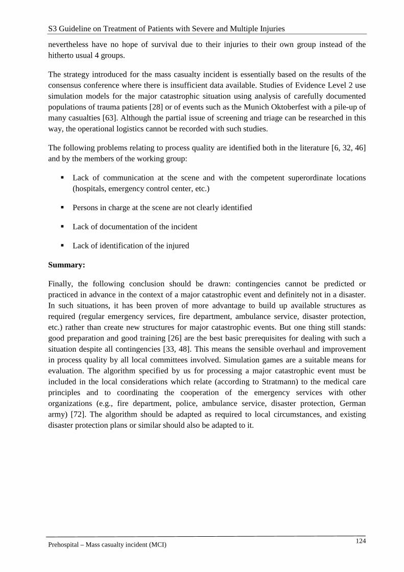
S3 Guideline on Treatment of Patients with Severe and Multiple Injuries
Prehospital – Mass casualty incident (MCI) 124
nevertheless have no hope of survival due to their injuries to their own group instead of the hitherto usual 4 groups.
The strategy introduced for the mass casualty incident is essentially based on the results of the consensus conference where there is insufficient data available. Studies of Evidence Level 2 use simulation models for the major catastrophic situation using analysis of carefully documented populations of trauma patients [28] or of events such as the Munich Oktoberfest with a pile-up of many casualties [63]. Although the partial issue of screening and triage can be researched in this way, the operational logistics cannot be recorded with such studies.
The following problems relating to process quality are identified both in the literature [6, 32, 46] and by the members of the working group:
� Lack of communication at the scene and with the competent superordinate locations (hospitals, emergency control center, etc.)
� Persons in charge at the scene are not clearly identified
� Lack of documentation of the incident
� Lack of identification of the injured
Summary:
Finally, the following conclusion should be drawn: contingencies cannot be predicted or practiced in advance in the context of a major catastrophic event and definitely not in a disaster. In such situations, it has been proven of more advantage to build up available structures as required (regular emergency services, fire department, ambulance service, disaster protection, etc.) rather than create new structures for major catastrophic events. But one thing still stands: good preparation and good training [26] are the best basic prerequisites for dealing with such a situation despite all contingencies [33, 48]. This means the sensible overhaul and improvement in process quality by all local committees involved. Simulation games are a suitable means for evaluation. The algorithm specified by us for processing a major catastrophic event must be included in the local considerations which relate (according to Stratmann) to the medical care principles and to coordinating the cooperation of the emergency services with other organizations (e.g., fire department, police, ambulance service, disaster protection, German army) [72]. The algorithm should be adapted as required to local circumstances, and existing disaster protection plans or similar should also be adapted to it.
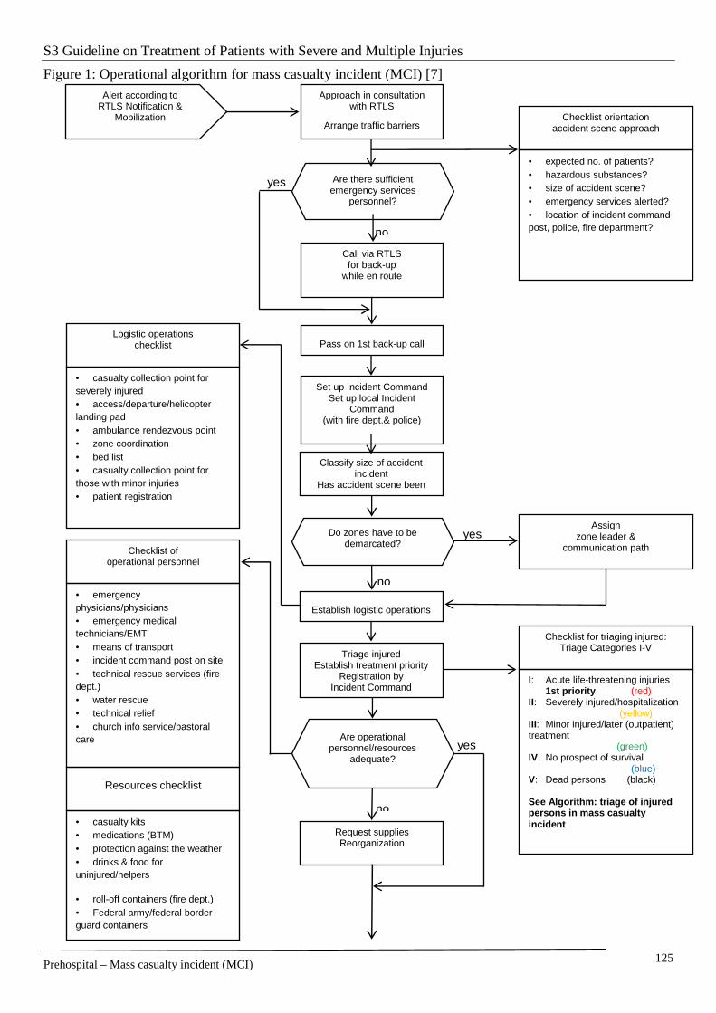
S3 Guideline on Treatment of Patients with Severe and Multiple Injuries
Prehospital – Mass casualty incident (MCI) 125
Figure 1: Operational algorithm for mass casualty incident (MCI) [7]
yes
no
Are operational personnel/resources
adequate?
Request supplies Reorganization
Logistic operations checklist
• casualty collection point for severely injured • access/departure/helicopter landing pad • ambulance rendezvous point • zone coordination • bed list • casualty collection point for those with minor injuries • patient registration
Checklist of operational personnel
• emergency physicians/physicians • emergency medical technicians/EMT • means of transport • incident command post on site • technical rescue services (fire dept.) • water rescue • technical relief • church info service/pastoral care
Resources checklist
• casualty kits • medications (BTM) • protection against the weather • drinks & food for uninjured/helpers • roll-off containers (fire dept.) • Federal army/federal border guard containers
yes
no
Classify size of accident incident
Has accident scene been made safe?
yes
no
Call via RTLS for back-up
while en route
Are there sufficient emergency services
personnel? X
Alert according to RTLS Notification &
Mobilization
Approach in consultation with RTLS
Arrange traffic barriers Checklist orientation
accident scene approach
• expected no. of patients? • hazardous substances? • size of accident scene? • emergency services alerted? • location of incident command post, police, fire department?
Pass on 1st back-up call
Set up Incident Command Set up local Incident
Command (with fire dept.& police)
Establish logistic operations
Do zones have to be demarcated?
X
Triage injured Establish treatment priority
Registration by Incident Command
Assign zone leader &
communication path
Checklist for triaging injured: Triage Categories I-V
I: Acute life-threatening injuries 1st priority (red) II: Severely injured/hospitalization (yellow) III: Minor injured/later (outpatient) treatment (green) IV: No prospect of survival (blue) V: Dead persons (black) See Algorithm: triage of injured persons in mass casualty incident
S3 Guideline on Treatment of Patients with Severe and Multiple Injuries
Prehospital – Mass casualty incident (MCI) 126
Sort patients according to available beds/suitable
hospital by Incident Command, if nec. discuss with zone
leader
no
yes Patients with acute
surgery indication from Triage Group I ?
Immediate transport with emergency physician/rescue physician
or form convoy
Complete triage
Allocate suitable means of transport
Release transport: Incident Command
Final registration Pass on to local police
incident command Media, tel. no., for relatives
Include feedback of accompanying emergency
physician/hospital in evaluation of available
beds
Final search of accident site with technical
incident command (police, fire dept.)
Operational documentation Quality control
Scoring
End of operation
Operational debriefing (at earliest opportunity)
Missing persons? Inform police incident
command

S3 Guideline on Treatment of Patients with Severe and Multiple Injuries
Prehospital – Mass casualty incident (MCI) 127
Figure 2: Triage of injured persons at mass casualty incident [7]
yes
no
yes
no
yes
no
no
no
yes
no
yes
yes
no
no
yes
no
Spontaneous respiration absent?
Respiratory rate above 30/min?
Palpable radial pulse?
Spurting haemorrhage?
Triage of injured persons at mass casualty incident
Do NOT approach
Commence triage
Able to walk?
Fatal injury
Central pulse absent?
no
No treatment V
Delayed treatment IV
yes
A
B
C
Treat as quickly as possible I
Unable to follow simple commands? D
no
yes Arrest bleeding (compression bandage)
Casualty collection point for minor injured
Urgent treatment II
Casualty collection point for severely injured
Re-evaluation – deterioration?
Re-evaluation/re-triage Transport acc. to urgency I/II
Delayed treatment III
Does the accident scene present a hazard?
Possible to release airways?
yes
Immediate transportation, if necessary form convoy
Incident Command to approve transport
Emergency physician/emergency medical
personnel to treat - NOT triage physician/local
ES/Incident Command
Acute surgical indication?
yes no
S3 Guideline on Treatment of Patients with Severe and Multiple Injuries
Prehospital – Mass casualty incident (MCI) 128
Literature:
1. Almogy G, Luria T, Richter ED, Pizov R, Bdolah-Abram T, Mintz Y, Zamir G, Rivkind AI (2005) Can external signs of trauma guide management? Lessons learned from suicide bombing attacks in Israel. Arch Surg 140: 390-393
2. American-College-of-Surgeons (1997) Advanced Trauma Life Support. Chicago
3. Anonymous (1990) Disaster epidemiology. Lancet 336: 845-846
4. Auf der Heide E (1996) Disaster planning, Part II. Disaster problems, issues, and challenges identified in research literature. Emerg Med Clin North Am 14: 453-480
5. Avitzour M, Libergal M, Assaf J, Adler J, Beyth S, Mosheiff R, Rubin A, Feigenberg Z, Slatnikovitz R, Gofin R, Shapira SC (2004) A Multicasualty Event: Out-of-hospital and In-Hospital Organizational Aspects. Acad Emerg Med 11: 1102-1104
6. Beck A, Bayeff-Filloff M, Bischoff M, Schneider BM, AG Notfall der DGU (2002) Analyse der Inzidenz und Ursachen von Großschadensereignissen in einem süddeutschen Rettungsdienstbereich. Unfallchirurg 105: 968-973
7. Beck A, Bayeff-Filloff M, Kanz K.-G, Sauerland S (2005): Algorithmus für den Massenanfall von Verletzten an der Unfallstelle: Ein systematisches Review. Notfall & Rettungsmedizin 8(7): 466-473
8. Beck A, Gebhard F, Kinzl L (2002) Notärztliche Versorgung des Traumapatienten. Notfall & Rettungsmedizin 5: 57-71
9. Beck A, Krischak G, Bischoff M (2009) Wirbelsäulenverletzungen und spinales Trauma. Notfall & Rettungsmedizin 12(6):469-479
10. Benson M, Koenig KL, Schultz CH (1996) Disaster triage: START, then SAVE - a new method of dynamic triage for victims of a catastrophic earthquake. Prehosp Disast Med 11: 117-124
11. Binder S, Sanderson LM (1987) The role of the epidemiologist in natural disasters. Ann Emerg Med 16: 1081-1084
12. Bissell RA, Becker BM, Burkle FMJ (1996) Health care personnel in disaster response. Emerg Med Clin North Am 14: 267-288
13. Buerk CA, Batdorf JW, Cammack KV, al. e (1982) The MGM Grand Hotel firs. Lessons learned from a major disaster. Arch Surg 117: 641
14. Burkle FM, Isaac-Renton J, Beck A, Belgica C-P, Blatherwick J, Brunet L-A, Hardy N-E, Kendall P, Kunii O, Lokey W, Sansom G, Stewart R (2001) Application of international standards to disasters: summary and action plan. Prehosp Disast Med 16: 36-38 [LoE 2b]
15. Burkle FMJ (1991) Triage of disaster related neuropsychiatric casualties. Emerg Med Clin North Am 9: 87-105
16. Butler FR, Hagemann J, Butler EG (1996) Tactical combat casualty in special combat operations. Military Med 161(suppl1): s3
17. Christen HT, Denney JP, Maniscalco PM, Rubin DL (1999) Terrorism. Part III - Response procedures for terrorist/tactical violence incidents. J Emerg Med Serv 24: 58-64, 66.
18. Cohen DL, Montalvo MA, Turnbull GP (1994) Medical support for a major military air show: the RAF Mildenhall Medical Emergency Support Plan. J Trauma 36: 237-244.
19. Cone DC, MacMillan DS (2005) Commentary: Mass-casualty Triage Systems: A Hint of Science. Acad Emerg Med 12: 739-741
20. Cowley RA, Myers RA, Gretes AJ (1984) EMS response to mass casualties. Emerg Med Clin North Am 2: 687-693
21. Dove DB, Del Guercio LR, Stahl WM, Star LD, Abelson LC (1982) A metropolitan airport disaster plan-coordination of a multihospital response to provide on-site resuscitation and stabilization before evacuation. J Trauma 22: 550-559.
22. Doyle CJ (1990) Mass casualty incident. Integration with prehospital care. Emerg Med Clin North Am 8: 163-175
23. Eisner ME, Waxman K, Mason GR (1985) Evaluation of possible patient survival in a mock airplane disaster. Am J Surg 150: 321-323.
24. Fink A, Kosecoff J, Chassin M, RH. B (1984) Consensus methods: characteristics and guidelines for use. Am J Public Health 74: 979-983 [Evidenzbasierte Leitlinie]
25. Frykberg ER (2002) Medical management of disasters and mass casualties from terrorist bombings: How we can cope? J Trauma 53: 201-212
26. Frykberg ER (2003) Disaster and Mass Casualty management: a commentary on the American College of Surgeons position statement. J Am Coll Surg 197: 857-859
27. Gans L, Kennedy T (1996) Management of unique clincal entities in disaster medicine. Emerg Med Clin North Am 14: 301-326
28. Garner A, Lee A, Harrison K, Schultz CH (2001) Comparative analysis of multiple-casualty incident triage algorithms. Ann Emerg Med 38: 541-548
29. Hirshberg A, Stein M, Walden R (1999) Surgical resource utilization in urban terrorist bombing: a computer simulation. J Trauma 47: 545-550
30. Hodgetts TJ (1993) Lessons from the Musgrave Park Hospital bombing. Injury 24: 219-221.
31. Hull D, Grindlinger GA, Hirsch EF, al. e (1985) The clinical consequences of an industrial aerosol plant explosion. J Trauma 25: 303
32. Hüls E, Oestern HJ (1999) Die ICE-Katastrophe von Eschede. Notfall & Rettungsmedizin 6: 327 – 336
S3 Guideline on Treatment of Patients with Severe and Multiple Injuries
Prehospital – Mass casualty incident (MCI) 129
33. Jakobson J (1990) What can we learn from the ferryboat accident in Zeebrugge? To be well prepared is the best way to meet disasters. Lakartidningen 87: 827-829.
34. Kanz KG, Eitel F, Waldner H, Schweiberer L (1994) Entwicklung von klinischen Algorithmen für die Qualitätssicherung in der Polytraumaversorgung. Unfallchirurg 97: 303-307
35. Kanz KG, Sturm J, Mutschler W, Arbeitsgemeinschaft Notfallmedizin der DGU (2002) Algorithmus für die präklinische Versorgung bei Polytrauma. Unfallchirurg 105: 1007-1014
36. Klein JS, Weigelt JA (1991) Disaster management. Lessons learned. Surg Clin North Am 71: 257-266.
37. Koenig KL, Dinerman N, Kuehl AE (1996) Disaster nomenclature - a functional impact approach: the PICE system. Acad Emerg Med 3: 723-727
38. Koscheyev VS, Leon GR, Greaves IA (1997) Lessons learned and unsolved public health problems after large-scale disasters. Prehospital Disaster Med 12: 120-131.
39. Leibovici D, Gofrit O, Stein M, al. e (1996) Blast injuries: bus vs open-air bombings - a comparative study of injuries in survivors of open-air and confined-space explosions. J Trauma 41: 1030
40. Leonard RB, Teitelman U (1991) Manmade disasters. Crit Care Clin 7: 293
41. Lewis CB, Aghababian RV (1996) Disaster planning part 1. Overview of hospital and emergency department planning for internal and external disasters. Emerg Med Clin North Am 14: 439
42. Lewis FR, Trunkey DD, Steele MR (1980) Autopsy of a disaster. J Trauma 20: 861
43. Mahoney LE, Reutershan TP (1987) Catastrophic disasters and the design of disaster medical care systems. Ann Emerg Med 16: 1085-1091
44. Mahoney LE, Whiteside DF, Belue HE, Belue HE, Mortisugu KP, Esch VH (1987) Disaster medical assistance teams. Ann Emerg Med 16: 354-358
45. Mallonee S, Shariat S, Stennies G, Waxweiler R, Hogan D, Jordan FB (1996) Physical injuries and fatalities from the Oklahoma City bombing. JAMA 276: 382-387
46. Martin TE (1990) The Ramstein airshow disaster. J R Army Med Corps 136: 19-26.
47. Matussek M, Beste R, Mascolo G (2005) Rekruten des Irrsinns. Der Spiegel 28: 20-27
48. Maxwell -CI, Strohl GR, Jr. (1985) The best way to prepare for the worst. Osteopath Hosp Leadersh 29: 15-16
49. Morris GP (1982) The Kenner airliner disaster. A 727 falls into a New Orleans suburb. J Emerg Med Serv JEMS 7: 58-65.
50. Nancekievil DG (1989) Disaster management. Practice makes perfect. BMJ 298: 477
51. Nania J, Bruva TE (1982) In the wake of Mount St Helens. Ann Emerg Med 11: 184
52. Nechaev EA, Malakhov SF, Dedushkin VS, Emelianov VA, Porembskii Ia O (1989) The characteristics of the medical evacuation measures in a railroad accident in Bashkiria. Voen Med Zh 12-17.
53. Nechaev EA, Reznik MI (1990) The methodological basis for a medical system in extreme situations. Voen Med Zh 6: 5-10.
54. Neugebauer E (1999) Development of a consensus protocol - a new approach to improve study design. Eur J Surg (Suppl) 584: 7-11
55. Nicholas RA, Oberheide JE (1988) EMS response to a ski lift disaster in the Colorado montains. J Trauma 28: 672
56. Noji EK (1991) Natural Disasters. Crit Care Clin 7: 271-292
57. Noji EK (1996) Disaster epidemiology. Emerg Med Clin North Am 14: 289-300
58. Noji EK, Kelen GD, Armenian HK, Oganessian A, Jones NP, Sivertson KT (1990) The 1988 earthquake in Soviet Armenia. A case study. Ann Emerg Med 19: 891-897
59. O'Hickey SP, Pickering CA, Jones PE, Evans JD (1987) Manchester air disaster. Br Med J (Clin Res Ed) 294: 1663-1667.
60. Rivkind A, Barach P, Israeli A, Berdugo M, Richter ED (1997) Emergency preparedness and response in Israel during the Gulf War. Ann Emerg Med 30: 513-521.
61. Rosemurgy AS, Norris PA, Olson SM, al. e (1993) Pre-hospital cardiac arrest: the cost of futility. J Trauma 35: 468
62. Roth PB, Vogel A, Key G, Hall D, Stockhoff CT (1991) The St Croix disaster and the National Disaster Medical System. Ann Emerg Med 20: 391-395
63. Ruppert M, Widmann JH, Schneider K, Schmidbauer S, Reith MW, Lackner CK, Schweiberer L (1998) Oktoberfest-Triage-Evaluationsstudie 1997 (OTES ´97): Prospektive Studie zur Sichtungsqualität von Notfallpatienten unter katastrophenmedizinischen Gesichtspunkten. Notfall & Rettungsmedizin 1: 268-278
64. Rutherford WH (1988) An analysis of civil aircrash statistics 1977-86 for the purposes of planning disaster exercises. Injury 19: 384-388.
65. Sacco WJ, Navin DM, Fiedler KE, K. WIR, Long WB, Buckman Jr RF (2005) Precise Formulation and Evidence-based Application of Resource-constrained Triage. Acad Emerg Med 12: 759-770
66. Sackett DL, Richardson WS, Rosenberg W, Haynes RB (1997) Evidence-based medicine: How to practice and teach EBM. Churchill Livingstone, London/UK
67. Sampalis JS, Tamim H, Nikolis A, Lavoie A, Williams JI (1996) Predictive validity and internal consistency of the pre-hospital index measured on-site by physicians. Accid Anal Prev 28: 675-684
68. Sefrin P, Weidringer JW, Weiss W (2003) Sichtungskategorien und deren Dokumentation. Einigung von Experten aus Deutschland sowie einigen europäischen Staaten. Dtsch Ärztebl 100 (Heft 2031-2032): A 2057-2058
69. Solieri KT (1990) Impressions: the crash of Avianca flight 052. J Emerg Nurs 16: 307-308.
70. Sopher LV, Petersen R, Talbott M (1990) The crash of Flight 232: an emergency care perspective. J Emerg Nurs 16: 61A-66A.
71. Steedman DJ, Gordon MW, Cusack S, White M, Robertson CE, Little K (1991) Lessons for mobile medical teams following the Lockerbie and Guthrie Street disasters. Injury 22: 215-218.

S3 Guideline on Treatment of Patients with Severe and Multiple Injuries
Prehospital – Mass casualty incident (MCI) 130
72. Stratmann D (2003) Strategien des Rettungsdienstes - Konsequenzen nach dem 11. September. Notfall & Rettungsmedizin 6: 102-106
73. Streger MR (1998) Prehospital triage. Emerg Med Serv 27: 21, 23-27
74. Waeckerle JF (1991) Disaster planning and response. N Engl J Med 324: 815-821
75. Washington B, Wilson RF, Steiger Z, al. e (1985) Emergency thoracotomy: a four-year review. Ann Thorac Surg 40: 188
76. Woltering HP, Schneider BM (2002) Das Unglück von Enschede. Unfallchirurg 105: 961-967
S3 Guideline on Treatment of Patients with Severe and Multiple Injuries
Emergency room - Introduction 131
2 Emergency room
2.1 Introduction
How would you treat?
You are called to the emergency room one winter’s night. As this is your first shift in trauma surgery, you’re feeling quite nervous when you enter the casualty department. You reach the preheated emergency room and shortly afterwards receive a handover from the emergency physician. He reports that a 42-year-old patient has had a motorbike accident. The initial GCS at the accident scene was 13, the right chest wall has marked crepitations, peripheral saturation is 85% during spontaneous breathing and, in addition, the patient is complaining of an intense tenderness in the right upper abdomen. The pelvis is stable and he has not noticed any extremity injuries. After prehospital anesthesia induction and oral intubation, peripheral saturation has risen to over 95%. Due to adequate oxygenation and normal capnometry, the insertion of a chest drain was dispensed with for the short transport journey. The patient is now intubated and ventilated and has stable circulation (110/80 mmHg, pulse 85). However, measurement of peripheral saturation yields a value of 90%. The patient is not fully undressed. A cervical collar has been applied. You look up at the wall in your emergency room and recognize an algorithm there which you are familiar with, which you recently learnt on a polytrauma course:
A Airway with immobilization of the cervical spine
B Breathing/Ventilation
C Circulation
D Disability/Neurology
E Expose – Environment/Undress - Keep Warm
Your confidence grows and you immediately start with the “primary survey and treatment” in the emergency room. You note that the cervical collar is sitting in the correct place. Under “B”, the on-duty anesthesiologist notices a weakened breath sound in the right chest, and peripheral saturation is now 83%. You have a brief discussion with him and decide to insert a chest drain via a mini-thoracotomy. Air is released and approximately 400 ml of blood. You check the success of your intervention and notice an increase in saturation to 99%. You are still nervous but know what you have to do next under “C”. Meanwhile, a central venous catheter and arterial access have been placed. Blood pressure is 110/80 mmHg, heart rate 85/min. Ultrasonography of the abdomen (FAST) detects free fluid in the Douglas cavity and around the liver, the on-duty radiologist estimates the volume to be approximately 500 ml. You do not detect any relevant bleeding to the outside. After alerting the visceral surgeon, the neurosurgeon has meanwhile checked the pupils under point “D”. After he has established that they are narrow but light-reactive, you start “E” and examine the now fully-undressed patient. A full-body CT scan is conducted because of the mechanism of injury. The laboratory values show a hemoglobin value of 11.5 g/dl, INR of 90%, and a base excess of -1.5 mmol/l. The patient has received a total of 1,500 ml fluid.
S3 Guideline on Treatment of Patients with Severe and Multiple Injuries
Emergency room - Introduction 132
The visceral surgeon who has meanwhile arrived confirms the ultrasonography finding. The full-body CT scan shows a severe pulmonary contusion on the right and a liver laceration without active bleeding. You come to a consensual decision to proceed as follows: conservative treatment of the abdominal injury and direct transfer of the patient to an intensive care unit.
The aim of this “emergency room” guideline section
The case presented at the start shows the importance of a logical and clear algorithm in an extreme situation. The sequence of actions in the emergency room must be checked against the evidence from as complete a literature base as possible. An expert committee examines the evidence levels, resulting in the grades of recommendation, which can strongly influence the sequence of actions. Thus, the aim of this guideline section is, on the one hand, to create clear, sustainable process sequences which, on the other hand, must contribute towards further improving care of the critically injured. For it is precisely the scientific reproducibility of the clinician’s actions in the emergency room that form the basis of reproducible, valid treatment and, in collaboration with the various medical disciplines, effectuate the parallelizing of processes and thus an improvement in the treatment.
Special notes:
A guideline does not claim to be able to treat every situation conclusively; this also applies to the emergency room. Not infrequently, the generation of clear recommendations is hampered by the lack of studies with a high evidence level. Clear views are expressed on this issue in the corresponding background texts to each chapter. In addition, various statements are subject to a time dynamic. This is accommodated by conducting a re-evaluation after 2 years. Nevertheless, there is a need to describe important correlations in detail.
Managing a multiply injured patient in the emergency room places a great demand on the treatment process because of the acuteness of the events and the large number of treating physicians from different specialties. As with all complex sequences of actions, errors do occur. But not every error need have a negative influence on the quality of treatment [1]. However, an accumulation of errors can occasionally have fatal consequences for the patient. For this reason, the basis of rational quality management lies in calmly working through the complications, and this approach should be firmly established within a quality circle in hospitals which are involved in the care of the critically injured [5]. Care in the emergency room should be quite rigorously characterized here by prescribed sequences and a common language with prehospital and emergency room care merging together seamlessly. Course strategies, such as depicted by Prehospital Trauma Life Support® (PHTLS®) for prehospital care and Advanced Trauma Life Support® (ATLS®) or European Trauma Course® (ETC®) for clinical care, can automate and thus improve this process through a clear hierarchy of treatment sequences and a common language [3, 4]. It is important that every hospital has an emergency room algorithm and that all those potentially involved are familiar with it.
Working groups and quality circles have been successfully introduced in many hospitals and they regularly evaluate and improve their own emergency room strategy based on actual cases. Both the leadership of such quality circles and the question of responsibility in the emergency room are issues that are the subject of heated debate in professional associations. Because severe
S3 Guideline on Treatment of Patients with Severe and Multiple Injuries
Emergency room - Introduction 133
injury is part of the core competence in trauma surgery, it may be legitimate for physicians qualified in this discipline to be in charge of both the quality circles and treatment in the emergency room [5]. However, it must not be forgotten that there are also other operational strategies in emergency room care [6, 7, 9]. For this reason, during the development of the guideline, this sensitive topic has been accommodated in various places in the background text as even strategies without a team leader are feasible involving just a multidisciplinary team working together. However, it should be discussed upfront who takes over responsibility in which situation in order to be prepared especially for medico-legal questions [8].

S3 Guideline on Treatment of Patients with Severe and Multiple Injuries
Emergency room - Introduction 134
References
1. Ruchholtz S, Nast-Kolb D, Waydhas C, Betz P, Schweiberer L (1994) Frühletalität beim Polytrauma – eine kritische Analyse vermeidbarer Fehler. Unfallchirurg 97: 285–291
2. Ertel W, Trentz O (1997) Neue diagnostische Strategien beim Polytrauma. Chirurg 68: 1071–1075
3. Sturm JA, Lackner CK, Bouillon B, Seekamp A, Mutschler WE (2002) Advanced Trauma Life Support (ATLS®) und Systematic Prehospital Life Support (SPLS). Unfallchirurg 105: 1027–1032
4. Advanced Trauma Life Support, 8th Edition, The Evidence for Change John B. Kortbeek, MD, FRCSC, FACS, Saud A. Al Turki, MD, FRCS, ODTS, FACA, FACS, Jameel Ali, MD, MMedEd, FRCS, FACS et. al. J Trauma. 2008;64:1638 –1650
5. Frink M, Probst C, Krettek C, Pape HC (2007) Klinisches Polytrauma-Management im Schockraum – Was muss und kann der Unfallchirurg leisten? Zentralbl Chir 132:49–53
6. Bergs EA, Rutten FL, Tadros T et al. (2005) Communication during trauma resuscitation: do we know what is happening? Injury 36:905–911
7. Cummings GE, Mayes DC (2007) A comparative study of designated Trauma Team Leaders on trauma patient survival and emergency department length-of-stay. CJEM 9:105–110
8. Bouillon B (2009) Brauchen wir wirklich keinen „trauma leader” im Schockraum? 112:400–401
9. Wurmb T, Balling H, Frühwald P, et al. Polytraumamanagement im Wandel. Zeitanalyse neuer Strategien für die Schockraumversorgung. Unfallchirurg 2009; 112: 390-399
S3 Guideline on Treatment of Patients with Severe and Multiple Injuries
Emergency room – Personnel and equipment resources 135
2.2 The emergency room - personnel and equipment resources
The annual number of multiply injured patients in Germany is approximately 32,000-38,000 [13, 30, 39, 53]. In the Federal Republic of Germany, there are currently approximately 700-800 hospitals available for the initial treatment of these patients (emergency room treatment). Although this figure would seem to indicate demand is adequately covered, it should be emphasized that a) not all hospitals have sufficient personnel or structural requirements or the specialist competence to care for these patients, b) there are still regions in Germany without sufficient provision of hospitals for polytrauma treatment, and c) fully equipped hospitals cannot always be reached within acceptable time periods, particularly at night if rescue helicopters are not allowed to take off due to the existing nighttime flying ban or because of capacity problems.
The introduction of regionalized trauma centers with defined standards in the management of trauma patients resulted in a reduction in the rate of avoidable deaths in the United States [8, 54].
To further improve polytrauma management in Germany, and make it more consistent across the whole country, it seems logical to define the requirements for the care of severely injured patients in terms of structure and personnel, and to standardize them to the maximum possible extent.
The DGU Trauma Network ProjectD has been initiated to implement this requirement. In the participating hospitals, which already number approximately 800 (as at April 2010), the DGU Trauma NetworkD is coordinating the implementation of the written contents of the DGU’s White Paper on the management of the severely injured [8, 54].
Emergency room team
Key recommendation:
In polytrauma management, fixed teams (known as emergency room teams) must work according to pre-structured plans and/or have completed special training.
GoR A
Explanation:
To achieve coordinated, balanced cooperation among various staff in polytrauma management, it is internationally commonplace to put together fixed teams for emergency room care, which work according to pre-structured plans and/or have completed special training (particularly ATLS®, ETC, Definitive Surgical Trauma Care [DSTC™]) [14a] [4, 47, 49, 51, 56]. Various studies have found that this emergency room strategy has clinical advantages [13, 30, 39, 53]. Ruchholtz et al. showed, for example, that an interdisciplinary team, integrated into a quality management (QM) system and acting on the basis of internal hospital guidelines and discussions, works very efficiently under joint surgical and anesthesiologic management [8, 54].
Key recommendation:
S3 Guideline on Treatment of Patients with Severe and Multiple Injuries
Emergency room – Personnel and equipment resources 136
The basic emergency room team must consist of at least 3 physicians (2 surgeons, 1 anesthesiologist), with at least 1 anesthesiologist and 1 surgeon having attained specialist grade.
GoR A
Explanation:
There are no validated studies on the composition of the emergency room team so that statements on team composition can only relate to how these are predominantly formed internationally. So, the question as to which specialties should be primarily represented in the emergency room team often depends on local conditions [6, 9, 11, 12, 14, 20–25, 27, 31–33, 38, 41, 45, 50]. On the other hand, individual studies from other countries show that a large proportion of multiply injured patients can be effectively managed even with only 2 physicians [3, 7, 12]. Depending on the injury pattern/severity, however, the team initially comprising at least 2-3 persons will then have to be supplemented with more colleagues [29, 40, 46]. In screening the international literature, it emerged that almost all teams worldwide had either special trauma surgeons of different training levels or general surgeons with (many years’) trauma experience, also with different training levels.
Accordingly, the cited team composition of at least 3 physicians can only serve as a minimum number and the team should be enlarged by one or two other 1-2 physicians (e.g., radiologist, neurosurgeon) depending on the size and care level of the hospital and the severely injured workload. In any event, the management of the severely injured must be undertaken by a qualified surgeon whose minimum qualification must be at the level of a specialist in general/visceral surgery or a specialist in orthopedics and trauma surgery (regional medical association (Landesärztekammer) [LÄK], Rules for Specialist Training for Physicians, as at 07/2009). The treating anesthesiologist must have the minimum qualification of specialist.
The function of and necessity for a “trauma leader” in the emergency room is a matter of much controversy in the literature. Even in the consensus conferences during the development of the S3 guideline, there was intense, heated debate about the necessity for a “team leader” and about what his duties should be and to which specific specialist area he should be assigned. A structured literature search was conducted on these issues during the guideline development. In terms of patient survival, no credible evidence was identified for the superiority of one particular management structure in the emergency room (“trauma leader” versus “interdisciplinary management group”) or for the assignment of a “trauma leader” to one particular specialist area (trauma surgery, surgery versus anesthesia).
Hoff et al. [21] showed that bringing in a team leader (known as a command physician) improved the care and treatment pathway [21]. Alberts et al. also found evidence of improved treatment pathways and treatment results after the strategy of “trauma leader” had been introduced [1]. Depending on the tasks - including patient handover, examining the patient, carrying out and monitoring therapeutic and diagnostic measures, consulting with other specialist disciplines, coordinating all medical and technical team members, preparing examinations following on from emergency room care, contacting relatives after completion - that the “trauma leader” needs to be able to take on in principle, this task must either be carried out on an

S3 Guideline on Treatment of Patients with Severe and Multiple Injuries
Emergency room – Personnel and equipment resources 137
interdisciplinary basis or by a “team leader” experienced in the management of multiply injured patients. In an interdisciplinary process even closer attention should be paid that the treatment pathways are agreed and consensual to avoid any time delays [18, 21, 37, 50].
According to the recommendations of the American College of Surgeons Committee on Trauma (ACS COT), a qualified surgeon must take over the team leadership [8, 54]. In a large comparative study of over 1,000 patients, there were almost identical case fatality rates and length-of-hospital-stays irrespective of whether 1 out of 4 trauma surgeons or 1 out of 12 general surgeons, albeit with trauma surgery knowledge, were responsible for the emergency room [41]. In a comparison between “trauma surgeons” and “emergency physicians”, Khetarpal showed that under traumatologic leadership the management times and the start of surgery were shorter, but without this apparently having had an effect on the treatment outcome [8, 54]. In a study by Sugrue et al. it is confirmed that no serious implications arise from who leads the ER team so long as he has adequate experience, expertise, and training [8, 54]. In many places, trauma teams have also been led very effectively, cooperatively, and successfully by anesthesiologists for many years.
High specialization in the individual specialist disciplines is particularly taken into account in interdisciplinary leadership models. Here, each specialist discipline has predefined tasks and is in charge of the devolved tasks at defined points in time within emergency room management. The leadership group, comprising anesthesiology, radiology, surgery, and trauma surgery (in alphabetical order), confers at fixed times and in addition to these if the situations in question demand it [57].
Notwithstanding, the experts argue for clear rules on responsibilities aligned to local conditions, agreements, and competences. Team leadership should be encouraged, irrespective of which specialist discipline it originates from or whether consisting of one person or a leadership group. The task of the team leadership is to collect and inquire about the findings of the individual specialized team members and to effectuate decision-making. The team leadership heads communication and establishes further diagnostic and treatment steps in agreement with the team. Within the quality circles of the establishment, the functions and qualifications of the “team leader” and of the “interdisciplinary leadership group” should be established in the emergency room. Ideally, after agreement, the “best” (one or more persons) must take on the task of “trauma leader” and “interdisciplinary leadership group”. In particular, rules must be made for the following points, which must stand up to a “best practice jurisdiction” inspection.
� Responsibility
� Leadership structure for coordination, communication and decision-making within
the context of emergency room management
S3 Guideline on Treatment of Patients with Severe and Multiple Injuries
Emergency room – Personnel and equipment resources 138
� Monitoring and ensuring quality (implementing quality circles; identifying quality
and patient safety indicators; continuous monitoring of structures, processes, and
results)
Key recommendation:
Trauma centers must have provision for enlarging emergency room teams GoR A
Explanation:
The size and composition of the enlarged emergency room team is determined by the care level of the hospital concerned and the corresponding injury severity to be expected there as well as by the maximum number of surgical interventions that can be performed there if necessary (White Paper). Accordingly, transboundary trauma centers, being the highest care level, should contain all specialist disciplines which perform emergency care. Table 11 gives an overview. A qualified specialist (specialist physician) from the department concerned must be able to get there within 20-30 minutes (see below).
S3 Guideline on Treatment of Patients with Severe and Multiple Injuries
Emergency room – Personnel and equipment resources 139
Key recommendation:
Attending physicians necessary for subsequent care must be present within 20-30 minutes of being alerted.
GoR A
Explanation:
A comparative study of hospitals found that it was not absolutely necessary for the trauma surgeon to be available in the hospital all hours provided the distance to the hospital was not greater than 15 minutes and a resident was already in the hospital [11]. Allen et al. and Helling et al. give 20 minutes as the limit here [3, 19]. In contrast, Luchette et al. and also Cornwell et al. found “in-house” readiness to be an advantage [9, 33] although Luchette showed that only the diagnosis and start of surgery was speeded up if an attending physician was initially present, both the period of intensive care and mortality of patients with severe thoraco-abdominal or head injuries remaining unaffected [13, 30, 39, 53].
In a comparison over several years, figures from the Trauma Audit and Research Network (TARN) (England & Wales) confirm that the increased presence of a qualified specialist/attending physician (60% versus 32%) contributes to significant reductions in mortality [28]. Wyatt et al. also found evidence that severely injured patients in Scotland (n = 1,427; ISS > 15) were treated faster and were more likely to survive if they were treated by an experienced attending physician/consultant instead of a resident [58]. In the ACS COT recommendations, the presence of an attending surgeon is not mandatory, provided a senior surgical resident is directly involved in the management of the severely injured [8, 54]. In a retrospective analysis over a period of 10 years, Helling et al. showed that no relevant improvement in treatment outcomes are achieved by the initial presence of an attending physician [35, 39, 52]. For patients with penetrating injuries, in shock, with a GCS < 9 or ISS ≥ 26, there was no difference in the care quality with regard to mortality, start of surgery, complications or length of treatment in the intensive care unit if the on-duty physician participated in the subsequent care within 20 minutes (“on call”). Only the initial care period and the total length of stay in hospital were less for blunt trauma if the attending physician could be in the emergency room (“in-house”) immediately. These results are confirmed to a large extent by Porter et al., Demarest et al., and Fulda et al. [11, 16, 43].
Overall, it can be concluded from these results that an attending physician does not have to be present immediately at the start of emergency room care if a surgeon qualified in the care of the severely injured (specialist grade and if applicable ATLS® and ETC certified) initially carries out the care of the injured. However, it should be ensured that an attending physician can be reached quickly.
A thoracic surgeon, ophthalmologist, maxillofacial surgeon and otolaryngologist (ENT) should be reachable within 20 minutes [18, 27, 34, 42]. According to Albrink et al. [2], the thoracic surgeon should be brought in as early as possible particularly in the case of aortic lesions.

S3 Guideline on Treatment of Patients with Severe and Multiple Injuries
Emergency room – Personnel and equipment resources 140
It seems that a pediatric surgeon is also not necessary in the basic ER team. The studies of Knudson et al. [26], Fortune et al. [15], Nakayama et al. [36], Rhodes et al. [44], Bensard et al. [5], D’Amelio et al. [10], Stauffer [48] and Hall et al. [17] were unable to find evidence of any improvement in treatment outcome through the involvement of special pediatric surgeons.
An anesthesiologist necessary for the subsequent care of the multiply injured patient must also be present within 20–30 minutes of being alerted.
Key recommendation:
The size of the emergency room should be 25-50 m2 (per patient to be treated). GoR B
Explanation:
The information given is based on a) the recommendations for initial management of the patient with traumatic brain injury and multiple injuries of the individual working group and circles of the German Society of Anesthesiology and Intensive Care Medicine (DGAI), the German Society for Neurosurgery (DGNC), and the German Interdisciplinary Association of Intensive Care and Emergency Medicine (DIVI). They recommend a minimum size per treatment unit of 25 m2 [55].
In addition, the room size can also be calculated b) using the specifications of the Technical Rules for Workplaces (Arbeitsstätten-Richtlinie) (ASR), the Workplaces Ordinance (Arbeitsstättenverordnung) (ArbStättV, 2nd section; room dimensions, air space), the German X-ray Ordinance (Röntgenverordnung) (RöV), and the Technical Rules for Hazardous Substances (Technische Regeln für Gefahrenstoffe) (TRGS). It specifies that 18 m3 of breathable air per person carrying out heavy physical activity and 15 m3 for average physical activity must be ensured in rooms with natural ventilation or air conditioning; 10 m3 is estimated for every additional person who is only temporarily there. Thus, a room volume of about 75-135 m3 would be required if there were 5-9 persons present (3-5 physicians, 1 medical radiologic technologist, 1-2 trauma surgery nurses, anesthesiology nurses) and the assumption of average physical work (lead aprons worn during care). With a ceiling height of 3.2 m, this corresponds to a room size of approximately 23-42 m2. Not included in the calculation is the loss of space through anesthesia and ultrasonography equipment, work surfaces, patient stretcher, cupboards and similar so that a total of 25-50 m2 per unit should be the starting point. If it is possible to treat a maximum of 2 severely injured patients simultaneously, the area is enlarged accordingly. Section 38 (2) of the German Workplaces Ordinance of 1986 specifies a clear door width of at least 1.2 m with a door height of 2 m for paramedic and first aid rooms.
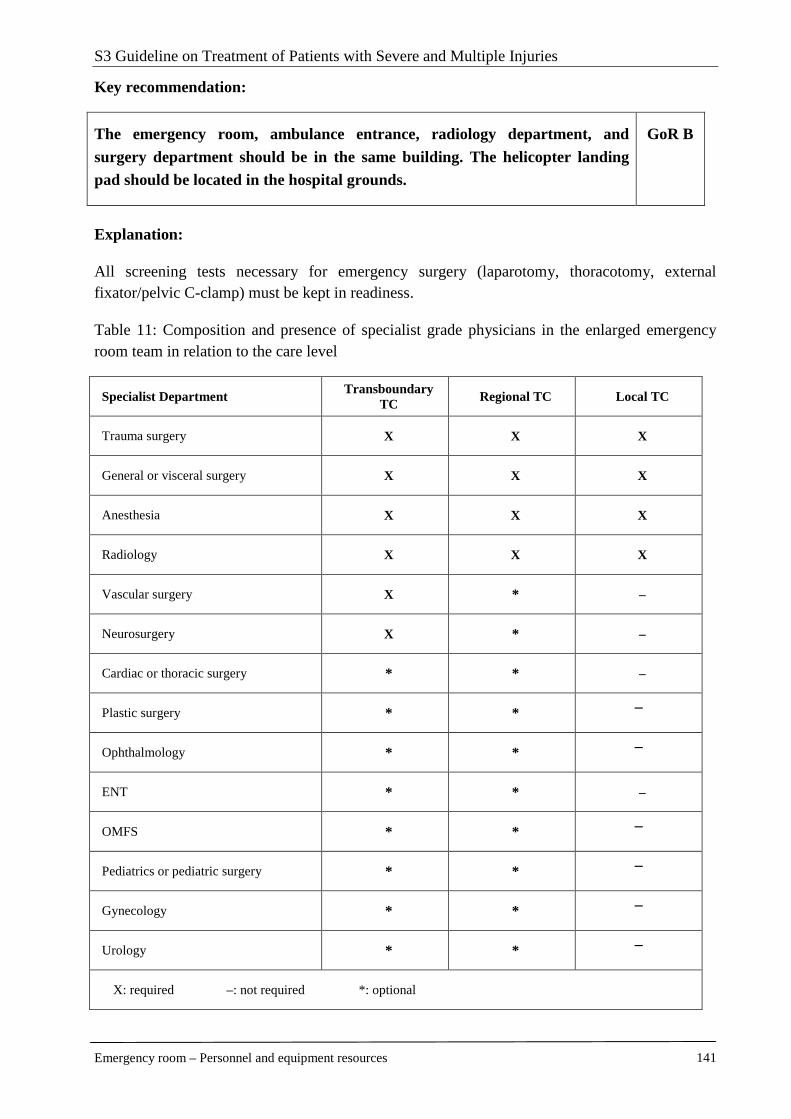
S3 Guideline on Treatment of Patients with Severe and Multiple Injuries
Emergency room – Personnel and equipment resources 141
Key recommendation:
The emergency room, ambulance entrance, radiology department, and surgery department should be in the same building. The helicopter landing pad should be located in the hospital grounds.
GoR B
Explanation:
All screening tests necessary for emergency surgery (laparotomy, thoracotomy, external fixator/pelvic C-clamp) must be kept in readiness.
Table 11: Composition and presence of specialist grade physicians in the enlarged emergency room team in relation to the care level
Specialist Department Transboundary
TC Regional TC Local TC
Trauma surgery X X X
General or visceral surgery X X X
Anesthesia X X X
Radiology X X X
Vascular surgery X * –
Neurosurgery X * –
Cardiac or thoracic surgery * * –
Plastic surgery * * –
Ophthalmology * * –
ENT * * –
OMFS * * –
Pediatrics or pediatric surgery * * –
Gynecology * * –
Urology * * –
X: required –: not required *: optional

S3 Guideline on Treatment of Patients with Severe and Multiple Injuries
Emergency room – Personnel and equipment resources 142
References
1. Alberts Ka, Bellander Bm, Modin G (1999) Improved trauma care after reorganisation: a retrospective analysis. Eur J Surg 165:426-430 [LoE 4]
2. Albrink Mh, Rodriguez E, England Gj et al. (1994) Importance of designated thoracic trauma surgeons in the management of traumatic aortic transection. South Med J 87:497-501 [LoE 2b]
3. Allen Dm, Hicks G, Bota Gw (1998) Outcomes after severe trauma at a northern Canadian regional trauma centre. Can J Surg 41:53-58 [LoE 2b]
4. Aprahamian C, Wallace Jr, Bergstein Jm et al. (1993) Characteristics of trauma centers and trauma surgeons. J Trauma 35:562-568
5. Bensard Dd, Mcintyre Rc, Jr., Moore Ee et al. (1994) A critical analysis of acutely injured children managed in an adult level I trauma center. J Pediatr Surg 29:11-18
5a. Bouillon B (2009) Brauchen wir wirklich keinen „trauma leader“ im Schockraum? Unfallchirurg 112: 400-401
6. Brennan R, Cohen Ss, Chambers Ja et al. (1994) The OR suite as a unique trauma resuscitation bay. AORN J 60:576-577, 580-574
7. Carmody Ic, Romero J, Velmahos Gc (2002) Day for night: should we staff a trauma center like a nightclub? Am Surg 68:1048-1051 [LoE 4]
8. Champion Hr, Sacco Wj, Copes Ws (1992) Improvement in outcome from trauma center care. Arch Surg 127:333-338
9. Cornwell Ee, 3rd, Chang Dc, Phillips J et al. (2003) Enhanced trauma program commitment at a level I trauma center: effect on the process and outcome of care. Arch Surg 138:838-843 [LoE 4]
10. D'amelio Lf, Hammond Js, Thomasseau J et al. (1995) "Adult" trauma surgeons with pediatric commitment: a logical solution to the pediatric trauma manpower problem. Am Surg 61:968-974
11. Demarest Gb, Scannell G, Sanchez K et al. (1999) In-house versus on-call attending trauma surgeons at comparable level I trauma centers: a prospective study. J Trauma 46:535-542 [LoE 4]
12. Deo Sd, Knottenbelt Jd, Peden Mm (1997) Evaluation of a small trauma team for major resuscitation. Injury 28:633-637 [LoE 4]
13. Dodek P, Herrick R, Phang Pt (2000) Initial management of trauma by a trauma team: effect on timeliness of care in a teaching hospital. Am J Med Qual 15:3-8 [LoE 2b]
14. Eastes Ls, Norton R, Brand D et al. (2001) Outcomes of patients using a tiered trauma response protocol. J Trauma 50:908-913
14a. Flohe S, Nast-Kolb D (2009) Chirurgisches Management vital bedrohlicher Verletzungen Unfallchirurg 112: 854-859.
15. Fortune Jb, Sanchez J, Graca L et al. (1992) A pediatric trauma center without a pediatric surgeon: a four-year outcome analysis. J Trauma 33:130-139
16. Fulda Gj, Tinkoff Gh, Giberson F et al. (2002) In-house trauma surgeons do not decrease mortality in a level I trauma center. J Trauma 53:494-502
17. Hall Jr, Reyes Hm, Meller Jl et al. (1996) The outcome for children with blunt trauma is best at a pediatric trauma center. J Pediatr Surg 31:72-76; discussion 76-77
18. Hartmann J, Gabram S, Jacobs L et al. (1996) A model for an integrated emergency medicine/trauma service. Acad Emerg Med 3:1136-1139 [LoE 5]
19. Helling Ts, Nelson Pw, Shook Jw et al. (2003) The presence of in-house attending trauma surgeons does not improve management or outcome of critically injured patients. J Trauma 55:20-25 [LoE 2b]
20. Highley Da (1994) Review of the composition and use of trauma teams within the Trent Region. J Accid Emerg Med 11:183-185
21. Hoff Ws, Reilly Pm, Rotondo Mf et al. (1997) The importance of the command-physician in trauma resuscitation. J Trauma 43:772-777 [LoE 2b]
22. Jacobs Ia, Kelly K, Valenziano C et al. (2000) Cost savings associated with changes in routine laboratory tests ordered for victims of trauma. Am Surg 66:579-584
23. Kaplan Lj, Santora Ta, Blank-Reid Ca et al. (1997) Improved emergency department efficiency with a three-tier trauma triage system. Injury 28:449-453
24. Kazemi Ar, Nayeem N (1997) The existence and composition of trauma teams in the UK. Injury 28:119-121
25. Khetarpal S, Steinbrunn Bs, Mcgonigal Md et al. (1999) Trauma faculty and trauma team activation: impact on trauma system function and patient outcome. J Trauma 47:576-581
26. Knudson Mm, Shagoury C, Lewis Fr (1992) Can adult trauma surgeons care for injured children? J Trauma 32:729-739
27. Krötz M, Bode Pj, Häuser H et al. (2002) Interdisziplinäre Schockraumversorgung: Personelle, apparative und räumlich-logistische Konzepte in 3 Traumakrankenhäusern in Europa. Radiologe 42:522-532 [LoE 5]
28. Lecky F, Woodford M, Yates Dw et al. (2000) Trends in trauma care in England and Wales 1989-97. Lancet 355:1771-1775 [LoE 4]
29. Lloyd Da, Patterson M, Robson J et al. (2001) A stratified response system for the emergency management of the severely injured. Ann R Coll Surg Engl 83:15-20
30. Lomas Ga, Goodall O (1994) Trauma teams vs non-trauma teams. Accid Emerg Nurs 2:205-210 [LoE 2b]
31. Lossius Hm, Langhelle A, Pillgram-Larsen J et al. (2000) Efficiency of activation of the trauma team in a Norwegian trauma referral centre. Eur J Surg 166:760-764
32. Lu Wh, Kolkman K, Seger M et al. (2000) An evaluation of trauma team response in a major trauma
S3 Guideline on Treatment of Patients with Severe and Multiple Injuries
Emergency room – Personnel and equipment resources 143
hospital in 100 patients with predominantly minor injuries. Aust N Z J Surg 70:329-332
33. Luchette F, Kelly B, Davis K et al. (1997) Impact of the in-house trauma surgeon on initial patient care, outcome, and cost. J Trauma 42:490-497 [LoE 2b]
34. Mathiasen Ra, Eby Jb, Jarrahy R et al. (2001) A dedicated craniofacial trauma team improves efficiency and reduces cost. J Surg Res 97:138-143 [LoE 2b]
35. Mclauchlan Ca, Jones K, Guly Hr (1997) Interpretation of trauma radiographs by junior doctors in accident and emergency departments: a cause for concern? J Accid Emerg Med 14:295-298 [LoE 2b]
36. Nakayama Dk, Copes Ws, Sacco W (1992) Differences in trauma care among pediatric and nonpediatric trauma centers. J Pediatr Surg 27:427-431
37. Nerlich M, Maghsudi M (1996) Polytrauma-Management: Präklinische und Schockraumversorgung. Unfallchirurg 99:595-606
38. Ochsner Mg, Schmidt Ja, Rozycki Gs et al. (1995) The evaluation of a two-tier trauma response system at a major trauma center: is it cost effective and safe? J Trauma 39:971-977
39. Palmer Sh, Maheson M (1995) A radiological review of cervical spine injuries from an accident and emergency department: has the ATLS® made a difference? J Accid Emerg Med 12:189-190 [LoE 2b]
40. Plaisier Br, Meldon Sw, Super Dm et al. (1998) Effectiveness of a 2-specialty, 2-tiered triage and trauma team activation protocol. Ann Emerg Med 32:436-441
41. Podnos Yd, Wilson Se, Williams Ra (1998) Effect of surgical panel composition on patient outcome at a level I trauma center. Arch Surg 133:847-854 [LoE 2b]
42. Poon A, Mccluskey Pj, Hill Da (1999) Eye injuries in patients with major trauma. J Trauma 46:494-499 [LoE 4]
43. Porter Jm, Ursic C (2001) Trauma attending in the resuscitation room: does it affect outcome? Am Surg 67:611-614
44. Rhodes M, Smith S, Boorse D (1993) Pediatric trauma patients in an 'adult' trauma center. J Trauma 35:384-393
45. Ruchholtz S, Waydhas C, Lewan U et al. (2002) A multidisciplinary quality management system for the early treatment of severely injured patients: implementation and results in two trauma centers. Intensive Care Med 28:1395-1404
46. Ryan Jm, Gaudry Pl, Mcdougall Pa et al. (1998) Implementation of a two-tier trauma response. Injury 29:677-683
47. Sakellariou A, Mcdonald Pj, Lane Rh (1995) The trauma team concept and its implementation in a district general hospital. Ann R Coll Surg Engl 77:45-52
48. Stauffer Ug (1995) Surgical and critical care management of children with life-threatening injuries: the Swiss experience. J Pediatr Surg 30:903-910
49. Stoneham J, Riley B, Brooks A et al. (2001) Recent advances in trauma management. Trauma 3:143-150
50. Sugrue M, Seger M, Kerridge R et al. (1995) A prospective study of the performance of the trauma team leader. J Trauma 38:79-82
51. Tscherne H, Regel G, Sturm Ja et al. (1987) Schweregrad und Prioritäten bei Mehrfachverletzungen. Chirurg 58:631-640
52. Velmahos Gc, Fili C, Vassiliu P et al. (2001) Around-the-clock attending radiology coverage is essential to avoid mistakes in the care of trauma patients. Am Surg 67:1175-1177 [LoE 2b]
53. Vernon Dd, Furnival Ra, Hansen Kw et al. (1999) Effect of a pediatric trauma response team on emergency department treatment time and mortality of pediatric trauma victims. Pediatrics 103:20-24 [LoE 2b]
54. Wenneker Ww, Murray Dh, Jr., Ledwich T (1990) Improved trauma care in a rural hospital after establishing a level II trauma center. Am J Surg 160:655-658
55. Wissenschaftlicher Arbeitskreis Neuroanästhesie Der Deutschen Gesellschaft Für Anästhesiologie Und Intensivmedizin, Arbeitsgemeinschaft Intensivmedizin Und Neurotraumatologie Der Deutschen Gesellschaft Der Deutschen Gesellschaft Für Neurochirurgie, Sektion Rettungswesen Und Katastrophenmedizin Der Deutschen Interdisziplinären Vereinigung Für Intensiv- Und Notfallmedizin (2000) Empfehlungen zur Erstversorgung des Patienten mit Schädel-Hirn-Trauma bei Mehrfachverletzung. In: Opderbecke HW, Weißbauer W (eds) [LoE 5]
56. Wong K, Petchell J (2003) Trauma teams in Australia: a national survey. ANZ J Surg 73:819-825
57. Wurmb et al. Unfallchirurg 2009; 112: 390-399
58. Wyatt Jp, Henry J, Beard D (1999) The association between seniority of accident and emergency doctor and outcome following trauma. Injury 30:165-168 [LoE 2b]

S3 Guideline on Treatment of Patients with Severe and Multiple Injuries
Emergency room – Criteria for emergency room activation 144
2.3 Criteria for emergency room activation
Efficiently working trauma score systems or parameters should select and identify patients so precisely that the necessary treatment is allotted to each casualty according to his injury severity. The difficulty lies in being able to assess injury severity adequately. Ideally, trauma/emergency room activation criteria should minimize as much as possible the rate of undertriage as well as that of overtriage of severely injured patients. Undertriage describes the proportion of patients who, despite a relevant severe injury requiring emergency room treatment, for example, are not identified as such. In contrast, overtriage describes the proportion of patients with a minor injury or none at all who nevertheless are classified as severely injured and, for example, are delivered to an emergency room. An advantage of overtriage - besides the optimum treatment of each patient - can be team training as the interplay and sequences can be practiced in “quasi-serious situations” even with non-severely injured persons. However, overtriage is associated with considerable costs and an often considerable disruption to routine sequences. The effectiveness of individual, different trauma score systems/trauma criteria can be described by parameters such as sensitivity, specificity, positive predictive value, and the calculation of overtriage and undertriage. The American College of Surgeons Committee on Trauma [25] gives an undertriage rate of 5-10% with simultaneous 30-50% overtriage as necessary in order to carry out efficient emergency room care. In a paper by Kane et al., the authors describe how, in order to attain a sensitivity of more than 80%, the rate of overtriage could not be brought below 70%.
The primary goal of trauma/emergency room activation criteria is therefore to keep undertriage low and at the same time not increase overtriage to an unacceptable level.

S3 Guideline on Treatment of Patients with Severe and Multiple Injuries
Emergency room – Criteria for emergency room activation 145
Activation criteria
Key recommendation:
The trauma/emergency room team should be activated for the following injuries:
� Systolic blood pressure below 90 mmHg after trauma
� Penetrating injuries to the neck and torso regions
� Gunshot wounds to the neck and torso regions
� GCS below 9 after trauma
� Respiratory impairment/requirement for intubation a fter trauma
� Fracture of more than 2 proximal bones
� Unstable chest
� Pelvic fractures
� Amputation injury proximal to hands/feet
� Spinal cord injury
� Open head wounds
� Burns > 20% and degree ≥2b
GoR A
Explanation:
Blood pressure/respiratory rate
Individual studies have shown that hypotension following trauma with a systolic blood pressure below 90 mmHg is a good predictor/good criterion for activating the emergency room team. Thus, Franklin et al. [1] showed that 50% of trauma patients with prehospital hypotension or hypotension at admission were sent for immediate surgery or transferred to an intensive care unit. A total of 75% of patients with hypotension were operated on during the course of the hospital stay.
Tinkoff et al. [2] found a 24-fold increased mortality, a 7-fold increased admission to the intensive care unit, and a 1.6-fold increased emergency surgery rate where hypotension was present after trauma as an expression of existing shock. In the recommendations of the American College of Surgeons Committee of Trauma [25], hypotension is found to be an important criterion for admission to a trauma center. Smith et al. [3] cite hypotension as a consistently used criterion for trauma team activation in all hospitals in the state of New South Wales in Australia. In a review by Henry et al. [4] of the New York State Trauma Registry, there were mortality

S3 Guideline on Treatment of Patients with Severe and Multiple Injuries
Emergency room – Criteria for emergency room activation 146
rates of 32.9% in trauma patients with an SBP (systolic blood pressure) of < 90 mmHg and 28.8% for trauma patients with a respiratory rate of < 10 or > 29/min.
Gunshot wounds
In a study by Sava et al. [5], the authors ascertained that gunshot wounds to the torso as a single activation criterion had a high informative value similar to the TTAC (Trauma Team Activation Criteria) used up till then. In a subgroup with gunshot wounds to the abdominal/pelvic region, the frequency of severe injuries was equal in the group with and without TTAC (74.1% and 70.8%, p = 0.61). A proviso must be made here that the overwhelming proportion of patients (94.4%) with gunshot wounds had already been identified using the TTAC. Tinkoff et al. [2] also found a significant correlation between gunshot wounds to the neck or torso and the need to admit to an intensive care unit (see below). Furthermore, this criterion was predictive for the existence of severe or fatal injuries and for emergency surgery. In a retrospective analysis, Velmahos et al. [6] report an overall survival rate following penetrating gunshot and stab wounds of over 5.1% in patients without vital signs in the emergency room. In a literature review (25 years, 24 studies), Rhee et al. [7] found a survival rate of 8.8% following emergency thoracotomy due to penetrating trauma.
The American College of Surgeons Committee on Trauma [25] listed various, differently-weighted triage criteria in its last edition (2006). The Step One and Step Two criteria, which necessitate admission to a Level 1 or Level 2 trauma center include: a) GCS below 15 or b) systolic blood pressure (BP) below 90 mmHg or c) respiratory rate below 10/min or above 29/min (Step One). Step Two criteria are a) penetrating injuries to the head, neck, torso, and proximal long bones, b) unstable thorax, c) fracture of 2 or more proximal long bones, d) amputation(s) proximal to the hands/feet, e) unstable pelvic fractures, f) open head fractures, and g) spinal cord injuries. Overall, there is currently rather a lack of evidence for the cited criteria. In a study by Knopp [8] on 1,473 trauma patients, the authors found a positive predictive value (PPV) of 100% for an ISS > 15 for spinal cord injuries and amputation injuries; however, fractures of the long bones only had a PPV of 19.5%.
In their study, Tinkoff et al. [2] examined several of these criteria for their accuracy in identifying severely injured and high-risk patients. Trauma patients who fulfilled the criteria of the ACS COT had more severe injuries, higher mortality, and longer stay in intensive care than patients in the control group. Systolic blood pressure below 90 mmHg, endotracheal intubation, and a gunshot wound to the neck/torso were predictive in the study for the necessity of emergency surgery or admission to intensive care. Mortality was markedly increased with systolic blood pressure below 90 mmHg, endotracheal intubation or GCS of less than 9. In their study, Kohn et al. [9] analyzed various trauma team activation criteria (see Table 1a), which are similar to those of the ACS COT. Kohn et al. found the parameter “respiratory rate below 10 or above 29/min” as the most predictive in terms of informative value for the presence of a severe injury. Additional parameters with high prediction were: a) burns with more than 20% body surface (BS), b) spinal cord injury, c) systolic blood pressure below 90 mmHg, d) tachycardia, and e) gunshot wounds to the head, neck or torso.

S3 Guideline on Treatment of Patients with Severe and Multiple Injuries
Emergency room – Criteria for emergency room activation 147
Open head injuries
With a lack of studies on the relevance of open head injuries, this criterion is regarded by the ACS COT rather as a significant indicator of severe injuries which require a high level of specialist medical competence and should thus be assigned to the Step One criteria.
GCS
Kohn et al. [9] regard a GCS of less than 10 as an important predictor of severe trauma. 44.2% of patients, for whom the ER team was activated because they had a low GCS, had confirmed severe injuries. The value of the GCS as a predictor of a severe injury and as an activation criterion for the emergency room team was also confirmed in studies by Tinkoff et al., Norwood et al., and Kühne et al. [2, 10, 11]. In these patients, Norwood as well as Kühne found that even GCS scores of less than 14 indicated pathologic intracerebral findings and the necessity for inpatient admission. However, according to this, the activation of the trauma/emergency room team does not appear to be absolutely necessary with these patients (GCS ≤ 14 and ≥ 11). For a GCS of less than 10, Engum [12] found a sensitivity of 70% for the endpoint OP, intensive care unit (ICU) or death. The odds ratio (OR) was 3.5 (95% CI: 1.6–7.5). The authors found a PPV of 78% for the presence of a severe injury in children with a GCS < 12.

S3 Guideline on Treatment of Patients with Severe and Multiple Injuries
Emergency room – Criteria for emergency room activation 148
Key recommendation:
The trauma/emergency room team should be activated for the following additional criteria:
� fall from more than 3 meters height
� road traffic accident (RTA) with
- frontal collision with intrusion by more than 50-75 cm
- a change in speed of delta >30 km/h
- collision involving a pedestrian or two-wheeler
- death of a driver or passenger
- ejection of a driver or passenger
GoR B
Explanation:
Accident-related/-dependent criteria
Accident-related/-dependent criteria are evaluated very differently in the literature with regard to their informative value for the presence of severe trauma.
Norcross et al., Bond et al., and Santaniello et al. [13, 14, 15] report on rates of overtriage of up to 92%, sensitivities of 70-50%, and PPV of 16.1% if accident-related mechanisms have been included as the sole criterion for describing the injury severity. If physiologic criteria were also used, a sensitivity of 80% was attained with a specificity of 90% [14].
Knopp et al. found only poor positive predictive values for the parameters road traffic accident (RTA) with ejection or death of a driver or passenger and road traffic accident involving a pedestrian [8]. Engum et al. also found the lowest predictive power for the road traffic accident involving a pedestrian at 20 mph (miles per hour) and the road traffic accident with death of a driver or passenger and trauma from being run over [12]. In the ACS COT recommendations, the trauma from being run over was removed from the criteria in the current version. Frontal collision with intrusion by more than 20-30 inches, death of a driver or passenger, road traffic accident involving pedestrian/two-wheeler collision at ≥ 20 mph, and ejection of a driver or passenger are cited as Step Three criteria, i.e. there is no necessity to transport these patients to centers of the maximum care level. Kohn et al. [9] also regard the rollover trauma as lacking in suitability. According to Kohn et al., the same also applies to the criteria/parameters road traffic accident (RTA) with ejection or death of a driver or passenger and road traffic accident involving a pedestrian [9].
Champion et al. [16] regard a vehicle rollover as an important indication of severe injury. The average probability of suffering a fatal injury is markedly greater after a rollover than not after one.

S3 Guideline on Treatment of Patients with Severe and Multiple Injuries
Emergency room – Criteria for emergency room activation 149
Nevertheless, the ACS COT removed the rollover mechanism from its triage criteria because relevant injuries after such an accident incident would already be included in Step One or Step Two.
Deformation to vehicle bodywork
In a multivariate analysis of 621 patients, Palanca et al. [17] found no significant relationship between vehicle deformation (intrusion of > 30 cm or > 11.8 inches) and the presence of a relevant severe injury (OR: 1.5; 95% CI: 1.0–2.3; p = 0.05). Henry came to comparable results in the multivariate analysis in his study [4]. Using the data of the National Automotive Sampling System Crashworthiness Data System (NASS CDS), Wang found a PPV of 20% for an ISS > 15 [18].
Death of a driver or passenger
Knopp et al. found an increased risk of surgery or death if a driver or passenger was fatally injured (OR: 39.0; 95% CI: 2.7–569; PPV 21.4%) [8]. Palanca et al. [17] could not confirm any statistically significant relationship between the death of a driver or passenger and the existence of a severe injury even if the simultaneous frequency of a severe injury was 7%.
Fall from a great height
In a prospective study by Kohn et al. [9], 9.4% of patients who had suffered a fall from more than 6 meters height had severe injuries - defined as intensive care admission or immediate surgery. Yagmur et al. [19] found 9 meters to be the average height for patients who died from the consequences of a fall.
Burns
It is essential to distinguish whether a thermal injury is present without additional injuries. In the case of a combination injury where the non-thermal component is predominant, the patient should be brought to a trauma center [25].
Age
Kohn et al. [9] analyzed various trauma team activation criteria which are similar to those of the ACS COT. Of the criteria examined, “age over 65” had the least informative value. The authors therefore recommended that this criterion be removed from the “first-tier activations”. Demetriades et al. [20] found a markedly higher mortality (16%), increased admission to intensive care, and an increased necessity for surgical intervention (19%) in patients over 70 years of age compared to younger patients. However, all patients who could remain outpatients were excluded from the study beforehand so that the cited percentages are probably an overestimation. Kühne et al. [21] found an increase in mortality - irrespective of ISS - with increasing age in a retrospective study of over 5,000 trauma patients in the DGU Trauma Registry. The cut-off value of mortality increase was 56 years. MacKenzie et al. [22] also found a marked increase in (fatal) injuries from > 55 years of age upwards. In a 13-year review, Grossmann et al. [23] found that mortality increased by 6.8% per additional year over 65 years

S3 Guideline on Treatment of Patients with Severe and Multiple Injuries
Emergency room – Criteria for emergency room activation 150
of age. In a study by Morris et al. [24], patients who died from the consequences of an accident had a lower ISS than younger patients in the control group.
Overall, there is variation and controversy over the assessment of the influence of age on the outcome of trauma. The American College of Surgeons COT has classified age as a criterion for triage in a Level 1 or Level 2 trauma center as rather low (Step Four criterion).
S3 Guideline on Treatment of Patients with Severe and Multiple Injuries
Emergency room – Criteria for emergency room activation 151
References
1 Franklin GA, Boaz PW, Spain DA, Lukan JK, Carrillo EH, Richardson JD. Prehospital hypotension as a valid indicator of trauma team activation. J Trauma. 2000; 48: 1034-7; discussion 1037-9.
2 Tinkoff GH,O'Connor RE. Validation of new trauma triage rules for trauma attending response to the emergency department. J Trauma. 2002; 52: 1153-8; discussion 1158-9.
3 Smith J, Caldwell E, Sugrue M. Difference in trauma team activation criteria between hospitals within the same region. Emerg Med Australas. 2005; 17: 480-7.
4 Henry MC. Trauma triage: New York experience. Prehosp Emerg Care. 2006; 10: 295-302.
5 Sava J, Alo K, Velmahos GC, Demetriades D. All patients with truncal gunshot wounds deserve trauma team activation. J Trauma. 2002; 52: 276-9.
6 Velmahos GC, Degiannis E, Souter I, Allwood AC, Saadia R. Outcome of a strict policy on emergency department thoracotomies. Arch Surg. 1995; 130: 774-7.
7 Rhee PM, Acosta J, Bridgeman A, Wang D, Jordan M, Rich N. Survival after emergency department thoracotomy: review of published data from the past 25 years. J Am Coll Surg. 2000; 190: 288-98.
8 Knopp R, Yanagi A, Kallsen G, Geide A, Doehring L. Mechanism of injury and anatomic injury as criteria for prehospital trauma triage. Ann Emerg Med. 1988; 17: 895-902.
9 Kohn MA, Hammel JM, Bretz SW, Stangby A. Trauma team activation criteria as predictors of patient disposition from the emergency department. Acad Emerg Med. 2004; 11: 1-9.
10 Kuhne CA, Homann M, Ose C, Waydhas C, Nast-Kolb D, Ruchholtz S. [Emergency room patients]. Unfallchirurg. 2003; 106: 380-6.
11 Norwood SH, McAuley CE, Berne JD, Vallina VL, Creath RG, McLarty J. A prehospital glasgow coma scale score < or = 14 accurately predicts the need for full trauma team activation and patient hospitalization after motor vehicle collisions. J Trauma. 2002; 53: 503-7.
12 Engum SA, Mitchell MK, Scherer LR, Gomez G, Jacobson L, Solotkin K, Grosfeld JL. Prehospital triage in the injured pediatric patient. J Pediatr Surg. 2000; 35: 82-7.
13 Norcross ED, Ford DW, Cooper ME, Zone-Smith L, Byrne TK, Yarbrough DR, 3rd. Application of American College of Surgeons' field triage guidelines by pre-hospital personnel. J Am Coll Surg. 1995; 181: 539-44 [Evidenzbasierte Leitlinie]
14 Bond RJ, Kortbeek JB, Preshaw RM. Field trauma triage: combining mechanism of injury with the prehospital index for an improved trauma triage tool. J Trauma. 1997; 43: 283-7.
15 Santaniello JM, Esposito TJ, Luchette FA, Atkian DK, Davis KA, Gamelli RL. Mechanism of injury does not predict acuity or level of service need: field triage criteria revisited. Surgery. 2003; 134: 698-703; discussion 703-4.
16 Champion HR, Lombardo LV, Shair EK. The importance of vehicle rollover as a field triage criterion. J Trauma. 2009; 67: 350-7.
17 Palanca S, Taylor DM, Bailey M, Cameron PA. Mechanisms of motor vehicle accidents that predict major injury. Emerg Med (Fremantle). 2003; 15: 423-8.
18 Wang SW, Review of NASS CDS and CIREN data for mechanism criteria for field triage. Presented at the National Expert Panel on Field Triage meeting. 2005: Atlanta, Georgia.
19 Yagmur Y, Guloglu C, Aldemir M, Orak M. Falls from flat-roofed houses: a surgical experience of 1643 patients. Injury. 2004; 35: 425-8.
20 Demetriades D, Sava J, Alo K, Newton E, Velmahos GC, Murray JA, Belzberg H, Asensio JA, Berne TV. Old age as a criterion for trauma team activation. J Trauma. 2001; 51: 754-6; discussion 756-7.
21 Kuhne CA, Ruchholtz S, Kaiser GM, Nast-Kolb D. Mortality in severely injured elderly trauma patients--when does age become a risk factor? World J Surg. 2005; 29: 1476-82.
22 MacKenzie EJ, Rivara FP, Jurkovich GJ, Nathens AB, Frey KP, Egleston BL, Salkever DS, Scharfstein DO. A national evaluation of the effect of trauma-center care on mortality. N Engl J Med. 2006; 354: 366-78.
23 Grossman MD, Miller D, Scaff DW, Arcona S. When is an elder old? Effect of preexisting conditions on mortality in geriatric trauma. J Trauma. 2002; 52: 242-6.
24 Morris JA, Jr., MacKenzie EJ, Edelstein SL. The effect of preexisting conditions on mortality in trauma patients. JAMA. 1990; 263: 1942-6.
25 American College of Surgeons Committee on Trauma (2006) Resources for optimal care of the injured patient. American College of Surgeons, Chicago

S3 Guideline on Treatment of Patients with Severe and Multiple Injuries
Emergency room – Abdomen 152
2.4 Thorax
What importance does the previous medical history have?
Key recommendations:
A detailed previous medical history (from third party if necessary) should be taken.
GoR B
High energy trauma and road traffic accidents with lateral collision should be interpreted as indications of a chest injury/aortic rupture.
GoR B
Explanation:
Even if there are only a few studies on taking the medical history with regard to chest injury, it is still an essential requirement for assessing the injury severity and the injury pattern and is used to establish whether an accident has in fact occurred. Collecting exact details of the circumstances of the accident is important in taking the medical history. The speed of the vehicle at the moment of impact and the direction of the impacting force are particularly important questions in road traffic accidents involving passenger vehicles. For instance, there are marked differences in the occurrence and severity of the chest injury and the overall injury severity depending on whether the impact is lateral or frontal.
Horton et al. [1] demonstrated a sensitivity of 100% and a specificity of 34% for aortic rupture in a lateral collision of the vehicle and/or with a change in velocity (delta V) ≥ 30 km/h. In another study [2], high velocity injuries at speeds of > 100 km/h were graded as suspicious for aortic rupture. Richter et al [3] also found an increased risk of chest injury in lateral collisions. In this study, delta V correlated with the AIS (thorax), ISS, and clinical course. In the study by Ruchholtz et al. [4], chest injury was diagnosed in 8 out of 10 cases of passenger vehicle accidents involving lateral collision. In this study, 72% of patients who had an accidental fall also sustained a chest injury.
In a study of 286 passenger vehicle occupants with an ISS ≥ 16, the probability of an aortic injury after a lateral collision was twice as high as after a frontal collision [5]. An impact in the region of the superior thoracic aperture appears to be particularly important and there appears to be increased incidence of fractures to ribs 1-4 [6].
Children also have a 5-fold higher risk of a severe chest injury (AIS ≥ 3) and a significantly higher overall injury severity when they are passenger vehicle occupants in a lateral collision compared to a frontal collision [7].
The effect of a seatbelt on the presence of a chest injury appears uncertain. Thus, in a retrospective study of 1,124 patients with relatively minor overall injury severity (ISS 11.6), Porter and Zaho [8] did find cumulative incidence of sternum fractures (4% versus 0.7%) in
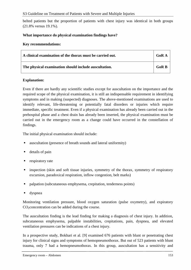
S3 Guideline on Treatment of Patients with Severe and Multiple Injuries
Emergency room – Abdomen 153
belted patients but the proportion of patients with chest injury was identical in both groups (21.8% versus 19.1%).
What importance do physical examination findings have?
Key recommendations:
A clinical examination of the thorax must be carried out. GoR A
The physical examination should include auscultation. GoR B
Explanation:
Even if there are hardly any scientific studies except for auscultation on the importance and the required scope of the physical examination, it is still an indispensable requirement in identifying symptoms and in making (suspected) diagnoses. The above-mentioned examinations are used to identify relevant, life-threatening or potentially fatal disorders or injuries which require immediate, specific treatment. Even if a physical examination has already been carried out in the prehospital phase and a chest drain has already been inserted, the physical examination must be carried out in the emergency room as a change could have occurred in the constellation of findings.
The initial physical examination should include:
� auscultation (presence of breath sounds and lateral uniformity)
� details of pain
� respiratory rate
� inspection (skin and soft tissue injuries, symmetry of the thorax, symmetry of respiratory excursion, paradoxical respiration, inflow congestion, belt marks)
� palpation (subcutaneous emphysema, crepitation, tenderness points)
� dyspnea
Monitoring ventilation pressure, blood oxygen saturation (pulse oxymetry), and expiratory CO2concentration can be added during the course.
The auscultation finding is the lead finding for making a diagnosis of chest injury. In addition, subcutaneous emphysema, palpable instabilities, crepitations, pain, dyspnea, and elevated ventilation pressures can be indications of a chest injury.
In a prospective study, Bokhari et al. [9] examined 676 patients with blunt or penetrating chest injury for clinical signs and symptoms of hemopneumothorax. But out of 523 patients with blunt trauma, only 7 had a hemopneumothorax. In this group, auscultation has a sensitivity and
S3 Guideline on Treatment of Patients with Severe and Multiple Injuries
Emergency room – Abdomen 154
negative predictive value of 100%. The specificity was 99.8% and positive predictive value was 87.5%. In penetration injuries, the sensitivity of auscultation is 50%, specificity and positive predictive value 100%, and negative predictive value 91.4%. In both mechanisms of injury, pain and tachypnea are inadequate indications of the presence of a hemopneumothorax.
In a retrospective study of 118 patients with penetrating trauma, Chen et al. [10] also found for auscultation only a sensitivity of 58%, specificity and positive predictive value of 98%, and negative predictive value of 61%. In a prospective study of 51 patients with penetrating trauma, the combination of percussion and auscultation exhibited a sensitivity of 96%, specificity of 93%, and positive predictive value of 83% [11].
These studies show that in penetrating trauma a weakened breath sound generally has an underlying pneumothorax and a chest drain can then be inserted before a radiograph is taken.
In their search for a clinical decision aid to identify children with chest injury, Holmes et al. [12] studied 986 patients, 80 of whom had a chest injury. This yielded an odds ratio of 8.6 for a positive auscultation finding, an odds ratio of 3.6 for an abnormal physical examination (reddening, skin lesions, crepitation, tenderness), and an odds ratio of 2.9 for an elevated respiratory rate.
What importance is attached to the diagnostic equipment (chest radiograph, ultrasound, CT, angiography, ECG, laboratory tests) and when is it indicated?
Key recommendations:
If a chest injury cannot be clinically excluded, a radiologic diagnostic study must be carried out in the emergency room.
GoR A
Every patient with clinical and anamnestic indications of a severe chest injury should undergo a helical CT scan of the thorax with contrast agent.
GoR B
Explanation:
As given under points 1 and 2, both the mechanism of injury and the findings from the physical examination provide important information on the presence or absence of a chest injury. For this reason, a chest radiograph can be dispensed with if, with respect to the circumstances of the accident, a chest injury can be excluded and at the same time there are no findings from the physical examination that make an intrathoracic injury probable.
On the other hand, a chest radiograph should be taken of all patients with confirmed chest injury. This serves to confirm diagnoses already made and to confirm or exclude further possible diagnoses. The initially taken radiograph is used to diagnose a pneumothorax and/or hemothorax, rib fractures, tracheobronchial injuries, pneumomediastinum, mediastinal hematoma, and pulmonary contusion [13]. The chest radiograph is widely used as the primary diagnostic tool due to its low costs and availability. Nevertheless, there is little evidence on

S3 Guideline on Treatment of Patients with Severe and Multiple Injuries
Emergency room – Abdomen 155
sensitivity and specificity in the diagnosis of pulmonary or thoracic injuries. There are only a few studies that report on a series of major injuries missed in the radiographs.
In a prospective study of 100 patients, there was evidence that the most important chest injuries can be detected by a chest radiograph examination. The sensitivity of images taken upright was 78.7% and that of supine images 58.3% [14]. On the other hand, McLellan et al. [15] found autopsy evidence in a series of 37 patients who died within 24 hours of admission that the chest radiograph did not detect important injuries in 11 cases. Among these were 11 cases of multiple rib fractures, 3 sternum fractures, 2 diaphragmatic ruptures, and 1 aortic intimal tear.
The chest radiograph offers sufficient accuracy for indicating a chest drain, for example. In a prospective study of 400 multiply injured patients, Peytel et al. [16] thus showed that insertion of chest drains (n = 77) based on the radiographic findings was correct in all cases.
Yet, numerous studies have shown that intrathoracic injuries can be revealed with significantly higher frequency by CT scan than by chest radiograph alone. In particular, there is a marked superiority in the detection of pneumothoraces and hemothoraces, pulmonary contusion, and aortic injuries. Here, preference should be given to the helical CT with administration of intravenous (i.v.) contrast agent [17]. By using multi-slice helical CTs, the examination time for a full-body scan can be reduced from an average of 28 to 16 minutes compared to the single-slice helical CT, and initial diagnostic information can even be taken from the realtime images on the monitor [18].
In a series of 103 severely injured patients, Trupka et al. [19] obtained additional information from 65% of patients on the underlying chest injury (pulmonary contusion n = 33, pneumothorax n = 34, hemothorax n = 21) compared to the radiographic examination. In 63% of these patients, direct therapeutic consequences resulted from the additional information, which in the majority of cases consisted of the chest drain being re-inserted or corrected.
In patients with relevant trauma (road traffic accidents with crash speed > 15 km/h, fall from a height of > 1.5 m), Exadaktylos et al. [20] were unable to detect chest injuries in 25 out of 93 patients using the conventional radiograph. In 13 of these 25 patients, however, the CT showed in part substantial chest injuries, including 2 aortic lacerations. In a prospective study of 112 patients with deceleration trauma, Demetriades et al. [21] performed a helical CT scan of the chest, which produced the diagnosis of aortic rupture in 9 patients. Four of these patients exhibited a normal chest radiograph. The aortic rupture was confirmed by CT in 8 patients. In one patient with an injury to the brachiocephalic artery, the CT revealed a local hematoma but the vessel was not visible in the CT slices. Even in patients without clinical signs of chest injury and with a negative radiographic finding, chest injuries showed up in the CT in 39% of patients, and in 5% of cases led to a change in treatment [22].
Blostein et al. [23] come to the conclusion that a routine CT is not to be recommended generally in blunt chest injury as, out of 40 prospectively studied patients with defined chest injuries, 6 patients had a change in treatment (5x chest drains, 1x aortography with negative result). However, the authors also state that, in patients who require intubation and ventilation, the CT produces findings that are not visible on the conventional radiograph. In patients with an oxygenation index (PaO2/FiO2) < 300, the CT can help to estimate the extent of the pulmonary

S3 Guideline on Treatment of Patients with Severe and Multiple Injuries
Emergency room – Abdomen 156
contusion and to identify patients at risk of pulmonary failure. Moreover, patients can be identified in whom an incompletely decompressed hemo- and/or pneumothorax could lead to further decompensation. In a retrospective study with 45 children [24] with 1) pathologic radiographic finding (n = 27), 2) abnormal physical examination finding (n = 8), and 3) substantial impact on the chest wall (n = 33), additional injuries were found in the CT in 40%, of which 18% of cases led to a change in treatment.
Although for blunt chest trauma the supplementary diagnostic information from the chest CT is generally accepted in more recent literature [25], there is controversy surrounding the benefit of the effect on the clinical outcome and it is not yet confirmed. In a prospective study by Guerrero-Lopez et al. [26], the chest CT proved to be more sensitive in detecting hemo/pneumothorax, pulmonary contusion, spinal fractures, and chest drain misplacements and led to treatment changes in 29% of cases. In the multivariate analysis, no therapeutic relationship could be ascertained between the CT and ventilation time, intensive care stay or mortality. The authors therefore conclude that a chest CT should only be performed if there are suspected severe injuries that can be confirmed or excluded by the CT.
Current studies showed a clear benefit from multi-slice CTs of the chest if there was a defined indication. Brink et al. [122] studied its routine, selective use in 464 and 164 patients. The indications for a routine CT were: high-energy trauma, vital parameters under threat, and severe injuries such as pelvic or spinal fractures, for example. The indications for a selective CT were: abnormal mediastinum, more than 3 rib fractures, pulmonary shadowing, emphysema, and fractures in the thoracolumbar spine. Injuries which were not visible in the conventional radiograph were found in 43% of patients who underwent routine CT. This led to changes in treatment for 17% of patients. Among the 7.9% of patients with a normal chest radiograph, Salim et al. [121] found pneumothoraces in 3.3%, a suspected aortic rupture in 0.2%, pulmonary contusions in 3.3%, and rib fractures in 3.7%.
If the literature results are summarized, this produces an indication for chest CT in the presence of the following indication criteria:
Indication criteria for chest CT (summarized according to [121, 122]):
� road traffic accident Vmax > 50 km/h
� fall from > 3 m height
� patient ejected from vehicle
� rollover trauma
� substantial vehicle deformation
� pedestrian knocked down at > 10 km/h
� biker knocked down at > 30 km/h
� crush

S3 Guideline on Treatment of Patients with Severe and Multiple Injuries
Emergency room – Abdomen 157
� pedestrian hit by vehicle and flung > 3 m
� GCS < 12
� Cardio-circulatory abnormalities (respiratory rate > 30/min, pulse > 120/min, systolic blood pressure < 100 mmHg, blood loss > 500 ml; capillary refill > 4 seconds)
� Severe concomitant injuries (pelvic ring fracture, unstable spinal fracture or spinal cord compression)
A retrospective multicenter analysis using the database of the DGU Trauma Registry found evidence of an improvement in survival probability for patients who had initially undergone a full-body CT scan [128]. The use of full-body CT leads to a relative reduction in mortality of 25% in TRISS and of 13% in the RISC score. The CT proved to be an independent predictor for survival in the multivariate analysis.

S3 Guideline on Treatment of Patients with Severe and Multiple Injuries
Emergency room – Abdomen 158
Key recommendation:
Every patient with clinical signs of chest injury should undergo an initial ultrasound examination (as part of the ultrasound examination of the torso) unless an initial chest helical CT with contrast agent has been carried out.
GoR B
Explanation:
In a prospective study of 27 patients, chest X-rays, ultrasound examinations and CT were compared for accuracy in diagnosing a pneumothorax. The ultrasound examination of the thorax showed a sensitivity and a negative predictive value of 100% and a specificity of 94% [27]. In another study, the ultrasound examination compared with the X-ray examination showed a sensitivity and a positive predictive value of 95% and a negative predictive value of 100% for diagnosing a pneumothorax [28]. However, emphysema bullae, pleural adhesions or extensive subcutaneous emphysema can falsify the results of ultrasonography.
As a retrospective study of 240 patients showed, the ultrasound examination ranks equally with the X-ray in diagnosing hemothorax. In 26 of these patients, the hemothorax was confirmed either by a chest drain or by chest CT. Ultrasound and chest X-ray each showed a sensitivity of 96%, a specificity and a negative predictive value of 100%, and a positive predictive value of 99.5% [29].
In a prospective study of 261 patients with penetrating injuries, chest ultrasound had a sensitivity of 100% and specificity of 96.9% for detecting pericardial tamponade [30]. However, false-negative ultrasound results can occur especially in patients with larger hemothoraces which can conceal smaller hematomas in the pericardium [31]. For this reason, sensitivity of the ultrasound was only 56% in this study.
In a retrospective study of 37 patients with a pulmonary contusion confirmed in a CT, ultrasonography revealed a sensitivity of 94.6%, specificity of 96.1%, and a positive and negative predictive value of 94.6% and 96.1%, respectively [127].
A chest helical CT scan with contrast agent excluded aortic injuries in patients without detected mediastinal hematoma, resulting in angiography not being necessary. Due to inadequate sensitivity, conventional CT examinations are less suited for the exclusion of an aortic injury [32, 33, 34].
In the prospective study by Gavant et al. [35], 1,518 patients with blunt trauma underwent helical CT scans with contrast agent. Of this group, 127 patients with abnormalities in the mediastinum or aorta received aortography. An aortic injury was detected in 21 of these patients. Sensitivity for the CT and the aortography was 100 and 94.4%, respectively, whereas specificity was 81.7 and 96.3%, respectively. It was concluded from this that in the absence of a mediastinal hematoma or if the aorta presented normally despite mediastinal hematoma, CT was sufficient for diagnosis and aortography was not necessary.

S3 Guideline on Treatment of Patients with Severe and Multiple Injuries
Emergency room – Abdomen 159
In a prospective study, Dyer et al. [2] studied 1,346 patients after blunt chest trauma using contrast CT and 19 of the patients exhibited an aortic injury in the CT. All patients with positive CT findings had additional angiography. On the assumption of a periaortal hematoma as indication of an aortic injury, the CT has a sensitivity and a negative predictive value of 100%, a specificity of 95%, and a positive predictive value of 22%. The authors conclude that aortography should only be carried out in patients who have undergone a CT scan that cannot be interpreted or have a periaortal hematoma without direct signs of an aortic injury. Aortography can also prove necessary if the proximal extent of the aortic injury cannot be reliably assessed from the CT scan.
In another prospective study of 494 patients with blunt chest trauma and mediastinal hematoma, the sensitivity for helical CT with contrast agent was 100% compared to 92% for aortography [36]. The specificity for the CT was 83% compared to 99% for aortography. The positive predictive value for the CT was 50% compared to 97% for aortography and the negative predictive value was 100% compared to 97%. In contrast to the above-mentioned study by Dyer et al. [2], Fabian et al. [36] conclude that patients with a mediastinal hematoma but no direct indication of an aortic injury also require no further diagnostic workup.
The prospective study by Parker et al. [37] of 142 patients with radiographically abnormal mediastinum showed that both the helical CT and the aortography produced a sensitivity and a negative predictive value of 100% for aortic injury. In a retrospective study of 74 patients, Tello et al. [38] found normal CT findings in 39 patients. Of these 39 patients, 5 received an angiography which showed all findings normal and 34 patients were asymptomatic at a clinical follow-up examination 12 months later.
There is general consensus now that helical CT with contrast agent is suitable for the exclusion of an aortic rupture [123, 124, 126]. There is a high probability that patients without detectable mediastinal hematoma have no aortic injury. Through the use of computed tomography, a large number of unnecessary aortographs can thus be avoided. However, if a brain CT scan is required, it should be carried out before the chest CT scan as the administration of contrast agent hampers the traumatic brain injury diagnosis.
As comparative studies on angiography have shown, a CT without evidence of a mediastinal hematoma has a negative predictive value of 100% for the injury of large intrathoracic vessels [39]. However, the specificity in the study by Parker et al. [37] is only 89% due to 14 false-positive findings. It is therefore recommended that angiography is performed on patients with a para-aortic hematoma detected by CT or with peribranch vessel hematomas and abnormal aortic contours. A negative contrast agent CT scan definitively excludes an aortic rupture [34, 40, 41].
In an analysis of 54 patients with surgically detected aortic ruptures, Downing et al. [42] showed a sensitivity of 100% and specificity of 96% for helical CT. In a prospective study of 1,104 patients with blunt chest trauma, Mirvis et al. [43] found mediastinal bleeding in 118 cases, of which 25 patients had an aortic rupture. For the aortic rupture, the helical CT showed a sensitivity and a negative predictive value of 100%, a specificity of 99.7%, and a positive predictive value of 89%. In a retrospective study on chest CT, Bruckner et al. found a negative

S3 Guideline on Treatment of Patients with Severe and Multiple Injuries
Emergency room – Abdomen 160
predictive value of 99%, a positive predictive value of 15%, a sensitivity of 95%, and a specificity of 40%.
In another prospective study of 1,009 patients, 10 patients had an aortic injury [44]. For the detection of direct signs of an aortic injury, the helical CT showed a sensitivity and a negative predictive value of 100%, a specificity of 96%, and a positive predictive value of 40%.
In contrast to the above-mentioned prospective studies, Collier et al. [45] found only a sensitivity of 90% and a negative predictive value of 99% in a retrospective study of 242 patients; an aortic injury was found during the autopsy of one patient with a normal CT finding who had subsequently died from the consequences of a traumatic brain injury. In another retrospective study, angiography did not detect any aortic injury in 72 patients with an intrathoracic hematoma detected in a CT scan but no evidence of a direct aortic or other intrathoracic vessel injury [125].
Transesophageal echocardiography (TEE) is a sensitive screening test [46, 47, 48] but angiography was often additionally carried out afterwards [49, 50]. TEE requires an experienced examiner [51] and is generally not so rapidly available as CT or angiography. The benefit of TEE may lie in imaging small intimal tears [47] which might not be visible in angiography or helical CT. However, TEE cannot provide good images of the ascending aorta and the branches of the aorta, which thereby elude diagnosis [52]. To date, there is only one prospective study in which helical CT has been compared to TEE in the diagnosis of aortic injury. CT and TEE showed a sensitivity of 73 and 93%, respectively, and a negative predictive value of 95%.

S3 Guideline on Treatment of Patients with Severe and Multiple Injuries
Emergency room – Abdomen 161
Key recommendations:
A 3-lead ECG must be carried out to monitor vital functions. GoR A
A 12-lead ECG should be carried out if there is a suspected blunt myocardial injury.
GoR B
Explanation:
The initial ECG is essential for every severely injured patient. The ECG is necessary particularly in the absence of palpable pulses in order to differentiate in cardiac arrest between rhythms that can be defibrillated and those that cannot be defibrillated. The ECG can also be used as a screening test for potential cardiac complications from a blunt cardiac injury.
Patients with a normal ECG, normal hemodynamics and no other additional relevant injuries do not require any further diagnostic tests or treatment. Cardiac enzymes are irrelevant in predicting complications from a blunt cardiac injury although raised troponin I levels can predict abnormalities in echocardiography. The echocardiogram should not be used in the emergency room for the diagnosis of blunt cardiac injury as it does not correlate with the occurrence of clinical complications. Echocardiography should be carried out on hemodynamically unstable patients in order to diagnose pericardial tamponade or pericardial rupture. Transthoracic echocardiography should be the method of choice here as to date there have been no clear evidence that transesophageal echocardiography is superior in diagnosing blunt cardiac injury.
The ECG is a rapid, cost-effective, non-invasive examination which is always available in the emergency room. In a meta-analysis of 41 studies, it was shown that the ECG and the creatine kinase MB (CK-MB) levels have a higher importance than radionuclide examinations and the echocardiogram in diagnosing clinically relevant blunt cardiac injury (defined as a complication requiring treatment) [53].
Fides et al. [54] report prospectively on 74 hemodynamically stable patients with normal initial ECG with no existing heart disease or other injuries. None of these patients developed cardiac complications. Another retrospective study of 184 children with blunt cardiac injury showed that patients with a normal ECG in the emergency room did not develop complications [55]. In a meta-analysis of 41 studies, an abnormal admission ECG correlated with the development of complications requiring treatment [53]. In contrast, in a prospective study by Biffl et al. [56], 17 out of 107 patients with a contusion developed complications. Only 2 out of 17 patients initially had an abnormal ECG and 3 had sinus tachycardia. In another retrospective study of 133 patients in 2 establishments with clinical suspicion of a blunt cardiac injury, 13 patients (9.7%) developed complications but no patient with a normal initial ECG showed other abnormalities [57]. In the study by Miller et al. [58], 4 out of 172 patients developed arrhythmias requiring treatment with all 4 patients having an abnormal initial ECG. Wisner et al. [59] studied 95 patients with suspected blunt cardiac injury and discovered that 4 patients developed clinically significant arrhythmias, only 1 of which had a normal admission ECG. In summary, the majority

S3 Guideline on Treatment of Patients with Severe and Multiple Injuries
Emergency room – Abdomen 162
of authors recommend that asymptomatic, patients with stable circulation with a normal ECG do not require any further diagnostic tests or treatment.
Key recommendation:
The measurement of troponin I levels can be undertaken as an additional laboratory test in the diagnosis of blunt myocardial injuries.
GoR 0
Explanation:
The studies on creatine kinase MB (CK-MB) in the diagnosis of blunt cardiac injury reveal a major limitation in the lack of a clear definition of blunt cardiac injury and the lack of a gold standard. In a retrospective study of 359 patients, 217 of whom were included to exclude blunt cardiac injury, 107 were diagnosed either because of an abnormal ECG finding or elevated CK-MB level. 16% of patients developed complications requiring treatment (arrhythmias or cardiogenic shock). All of these patients had an abnormal ECG but only 41% of them had elevated CK-MB levels. The course was without complication in patients with normal ECG and elevated CK-MB [56]. In a prospective study of 92 patients who all received an ECG, a CK-MB analysis, and continuous monitoring, 23 patients developed arrhythmias which, however, did not require any specific treatment. This shows that the number of arrhythmias requiring clinical treatment is small. 52% of patients with arrhythmias revealed elevated CK-MB levels whereas 19% of patients without arrhythmias also had elevated CK-MK levels [60]. In addition, other studies showed no correlation between elevated CK-MB levels and cardiac complications [58, 59, 61-65].
Troponin I and T are sensitive markers in the diagnosis of myocardial infarction and considerably more specific than CK-MB as they are not present in skeletal muscle. In a study of 44 patients, the 6 patients with blunt cardiac injury confirmed by echocardiography showed simultaneously elevated CK-MB and troponin I. Of the 37 patients without cardiac injury, 26 had elevated CK-MB levels but no patient had elevated troponin I [66]. In another study of 28 patients, 5 of whom had a blunt cardiac injury detected by echocardiography, troponin I had a specificity and sensitivity of 100% for the contusion. In a study of 29 patients, troponin T showed higher sensitivity (31%) than CK-MB (9%) in diagnosing blunt cardiac injury. Troponin T showed a sensitivity of 27% and a specificity of 91% in 71 patients for predicting clinically significant ECG changes [67].
In a more current prospective study of 94 patients, 26 patients were diagnosed with blunt cardiac injury either by ECG or echocardiography. Troponin I and T showed a sensitivity of 23 and 12%, respectively, sensitivity of 97 and 100%, respectively, and a negative predictive value of 76.5 and 74%, respectively. The authors describe an unsatisfactory correlation between the two enzymes and the occurrence of complications [68]. In another prospective study, sensitivity, specificity, and the positive and negative predictive values of troponin I are given as 63, 98, 40, and 98%, respectively, for detecting blunt cardiac injury [69]. Velmahos et al. carried out ECG tests and troponin I measurements prospectively in 333 patients with blunt chest trauma [70]. In 44 diagnosed cardiac injuries, the ECG and troponin I showed a sensitivity of 89 and 73%,

S3 Guideline on Treatment of Patients with Severe and Multiple Injuries
Emergency room – Abdomen 163
respectively, and a negative predictive value of 98% and 94%, respectively. The combination of ECG and troponin I produced a sensitivity and a negative predictive value of 100% each. Rajan et al. [71] showed that a cTnI level below 1.05 µg/l at admission and after 6 hours excludes myocardial injury.
The results available to date show that troponin I in particular is a more specific indicator than CK-MB for the presence of a blunt cardiac injury. However, the importance of troponin in predicting complications is still the subject of current discussion.
A transthoracic echocardiography (TTE) is often carried out in the diagnosis of blunt cardiac injury but has hardly any importance in patients with stable circulation. In a prospective study, Beggs et al. carried out TTE in 40 patients with suspected blunt chest injury. Half of the patients had at least one pathologic finding either in the ECG, in the cardiac enzymes or in TTE. There was no correlation between TTE, the enzyme or ECG findings, and TTE could not predict the development of complications [72]. In another prospective study of 73 patients who all underwent TTE, CK-MB measurements, and cardiac monitoring, 14 patients presented abnormalities in the echocardiography. However, only 1 patient who initially had a pathologic ECG developed a complication in the form of a ventricular arrhythmia [73]. A prospective study of 172 patients came to the conclusion that only an abnormal ECG or shock has a predictive value with reference to monitoring or to a specific treatment. Patients with abnormalities in TTE or elevated CK-MB levels without a simultaneous pathologic ECG developed no complications requiring treatment [58]. Although there are a number of studies which show the benefit of TTE in the diagnosis of pericardial effusion or of pericardial tamponade in penetrating trauma, the benefit of this study on blunt trauma is debatable [30, 58, 74].
There are a number of studies which show that the accuracy of transesophageal echocardiography (TEE) is greater than that of TTE in the diagnosis of cardiac injuries [75-79]. In addition, other cardiovascular changes such as aortic injuries, for example, can be diagnosed by TEE. Vignon et al. [80] prospectively carried out helical CT and, in the intensive care unit, TEE on 95 patients with risk factors for an aortic injury. The sensitivity of TEE and CT was 93 and 73%, respectively, the negative predictive value was 99% and 95%, respectively, and the specificity and the positive predictive value were 100% for both examination methods. TEE proved to be superior in identifying intimal tears whereas an aortic branch lesion was missed.
In summary, echocardiography should be carried out if a pericardial tamponade or pericardial rupture is suspected.
What additional diagnostic tests exist for emergency room patients?
Fabian et al. [36] state that patients with a mediastinal hematoma and no direct evidence of an aortic injury require no further assessment. This also applies to intimal tears and pseudo-aneurysms. However, patients with changes that cannot be classified in more detail should undergo angiography for further assessment. Gavant et al. [35] also stated that, in the absence of a mediastinal hematoma or if the aorta presented normally despite mediastinal hematoma, helical CT with contrast agent was sufficient for diagnosis and aortography was not necessary.

S3 Guideline on Treatment of Patients with Severe and Multiple Injuries
Emergency room – Abdomen 164
Mirvis et al. [43] and Dyer et al [44] suggest that an aortic injury detected in a CT or an injury to the main lateral branches and a mediastinal hematoma require either an angiography or direct thoracotomy depending on the experience of the establishment concerned. Angiography is also necessary for a mediastinal hematoma in direct contact with the aorta or with the proximal great vessels without direct evidence of a vessel injury or for abnormal aortic contours [37].
Downing et al. [42] conclude from the results of their study that surgical treatment can be carried out without further diagnostic tests if a helical CT clearly detects an aortic rupture. In contrast to the above-mentioned study by Dyer et al. [2], Fabian et al. [36] conclude that patients with a mediastinal hematoma but no direct evidence of an aortic injury require no further work-up.
To date, there are no comparative studies which investigate the necessity of additional angiography prior to a planned intervention for an aortic injury detected in a CT scan. For this reason, the recommendations are based, on the one hand, on conclusions from studies which evaluated angiography and CT in the diagnosis of aortic injury and, on the other hand, on data from diagnostic tests carried out prior to endovascular treatment.
Thus, Gavant et al. [35], recommend that aortography is carried out prior to surgical or endovascular treatment in order to confirm the injury and define the extent of the damage. Parker et al. [37] also consider angiography necessary for confirming positive CT findings.
In patients with direct indication of an aortic injury and a mediastinal hematoma, Mirvis et al. [43] and Dyer et al [44] suggest either angiography or direct thoracotomy depending on the experience of the establishment concerned.
Downing et al. [42] and Fabian et al. [36] hold the view that a thoracotomy can also be carried out without additional angiography if the CT finding is clear.
In a series of 5 patients with acute traumatic rupture of the thoracic aorta, a CT scan and angiography were carried out on all patients prior to stent implantation [81].
What importance is attached to emergency procedures (chest drain, intubation, pericardiocentesis, thoracotomy)?
Key recommendations:
A clinically relevant or progressive pneumothorax must first be decompressed in the ventilated patient.
GoR A
A progressive pneumothorax should be decompressed in the non-ventilated patient.
GoR B
A chest drain must be inserted for this purpose. GoR A
Preference should be given to wide lumen chest drains. GoR B
S3 Guideline on Treatment of Patients with Severe and Multiple Injuries
Emergency room – Abdomen 165
Explanation:
A pneumothorax detected in the radiographic image represents an indication to insert a chest drain particularly if mechanical ventilation is necessary. This represents general clinical practice although there are no comparative studies on this in the literature [12, 82-85]. Due to the underlying pathophysiology, it is upgraded to Grade of Recommendation A. Westaby and Brayley [86] recommend that a chest drain should always be inserted for a pneumothorax which exceeds 1.5 cm in size and is at the level of the 3rd intercostal space. If the size is less than 1.5 cm, a chest drain should only be inserted if ventilation is necessary or if there is bilateral occurrence. The insertion of a chest drain can be omitted only in small ventral pneumothoraces detected by CT although close clinical monitoring is required.
The insertion of a chest drain should be carried out in the emergency room as the risk of a progressive pneumothorax can lead to a tension pneumothorax and the timespan of such a development cannot be estimated. The risk of a tension pneumothorax occurring should be rated markedly higher in ventilated patients than in non-ventilated patients. In non-ventilated patients, small pneumothoraces less than 1-1.5 cm in width can initially be treated conservatively by close clinical monitoring. If this is not possible for logistic reasons, the pneumothorax should also be decompressed in this situation.
The increasing use of abdominal and chest CT in the diagnosis of blunt trauma has led to pneumothoraces being detected in a CT scan which had not been detected previously by conventional supine radiographic images. These so-called occult pneumothoraces, usually lying ventrally, are found in 2-25% of patients after severe multiple injuries [19, 22, 23, 87-89]. Based on the available literature, the initial insertion of a Bülau drain should be omitted in an occult pneumothorax diagnosed by CT if:
� the patients are hemodynamically stable and have a largely normal lung function,
� there are frequent clinical checks with the possibility of radiography in between
and
� a chest drain can be inserted at any time by a qualified physician.
Also in a prospective randomized study, Brasel et al. [91] studied the necessity of inserting a chest drain for an occult traumatic pneumothorax. Chest drains were inserted in 18 patients while 21 patients were clinically observed only. Ventilation was necessary in 9 patients in each group. In the group with chest drains, the pneumothorax increased in 4 patients; in the group without chest drain, a Bülau drain was inserted in 3 patients, of whom 2 patients were then also ventilated.
In a prospective study of 36 patients with 44 occult pneumothoraces, the subdivision was made into minimal (< 1 cm visible on a maximum of 4 CT slices), anterior (> 1 cm but not extending laterally into the dorsal half of the chest), and anterolateral pneumothoraces [92]. Fifteen minimal pneumothoraces were closely clinically monitored irrespective of the necessity for ventilation. The secondary insertion of a chest drain was then required in 2 cases. A drain was
S3 Guideline on Treatment of Patients with Severe and Multiple Injuries
Emergency room – Abdomen 166
always inserted for anterior and anterolateral pneumothoraces if ventilation was required. In a prospective study of children, Holmes et al. identified 11 patients with occult pneumothoraces which were also subdivided according to the above-mentioned plan [93]. In the case of minimal pneumothoraces, the patients were also conservatively treated irrespective of the necessity for ventilation.
In a retrospective study, patients with pneumothorax were treated with (13) and without (13) a chest drain [94]. Out of 10 patients who required mechanical ventilation, 2 patients had to have a secondary chest drain inserted. However, there are no data on the size of the initial pneumothorax. In another retrospective study, the size of the occult pneumothorax was compared against the requirement to insert a chest drain and it was suggested that pneumothoraces less than 5 x 80 mm could be observed irrespective of the necessity for mechanical ventilation [95]. Weißberg et al. [96] stated in their retrospective study of 1,199 patients (of whom 403 patients had traumatic pneumothorax) that management by clinical observation is possible for a pneumothorax volume less than 20% of the pleural space. However, there are no details on the effect of possible mechanical ventilation.
A score was proposed by De Moya for the improved definition of the “small” pneumothorax, which is composed of 2 parts: 1) the largest diameter of the pneumothorax and 2) its relationship to the pulmonary hilus. If the pneumothorax does not exceed the pulmonary hilus, 10 is added to the millimeter figure of the pneumothorax; if the hilus is exceeded, 20 is added. The sum of the individual values for each side gives the score value. The positive predictive value for a chest drain was 78% for a score > 30 and the negative predictive value was 70% for a score < 20 [136]. In a randomized study of 21 ventilated patients, observation of the occult pneumothorax proved to be reliable. In 13 patients initially treated without a chest drain, there was no need for emergency decompression in any case even though pleural effusion had to be decompressed during the course in 2 patients and an increasing pneumothorax had to be decompressed after insertion of a central venous catheter in one patient. It seems justifiable to take a “wait and see” attitude towards inserting a chest drain for an occult pneumothorax both in spontaneously breathing and ventilated patients [129, 130].
Key recommendation:
Pericardial decompression should be carried out if there is evidence of pericardial tamponade and acute deterioration in vital parameters.
GoR B
Explanation:
Irrespective of the patient’s condition, the diagnosis of pericardial tamponade should be made rapidly and reliably so that surgery can be performed quickly if required. Although the diagnosis of tamponade can be confirmed by the insertion of a pericardial window, this is an invasive procedure, particularly if there is only slight suspicion of a cardiac injury. Ultrasound examination has been proven to be a sensitive procedure in the diagnosis of pericardial effusion and thus represents the current method of choice. In a prospective multicenter study of 261 patients with penetrating pericardial chest injuries, there was a sensitivity of 100%, a specificity

S3 Guideline on Treatment of Patients with Severe and Multiple Injuries
Emergency room – Abdomen 167
of 96.7%, and an accuracy of 97% [30]. There were no false-negative study results. In another study, fluid was detected by ultrasound scan in the pericardium in 3 cases out of 34 patients. One patient, who was hemodynamically unstable, underwent a thoracotomy and the other two patients had a negative pericardial window [105].
Pericardiocentesis is now of lesser importance in the diagnosis of pericardial tamponade, having been replaced by ultrasound examination [30, 106].

S3 Guideline on Treatment of Patients with Severe and Multiple Injuries
Emergency room – Abdomen 168
Key recommendation:
A thoracotomy can be performed if there is an initial blood loss of > 1,500 ml from the chest drain or if there is persistent blood loss of > 250 ml/h over more than 4 hours.
GoR 0
Explanation:
The indication for thoracotomy depending on the volume of initial or continuous blood loss from the chest drain has been intensely discussed by the guideline group, not least because of the inconsistent volumes described in the literature. These are almost exclusively cohort studies on penetrating trauma; randomized studies are not available on this research question. The available data is considerably less clear for blunt trauma; a thoracotomy is indicated rather less frequently and usually later than for penetrating trauma. Under certain circumstances, with a certain volume of blood loss, the thoracotomy can also be useful in hemodynamically stable patients. There are no data on coagulation status as a decision criterion but body temperature can be taken into account.
In the 1970s, based on the experiences of penetrating injuries in the Vietnam War, McNamara et al. [107] described a reduction in mortality after early thoracotomy. Indication criteria for thoracotomy were given as an initial blood loss after chest drainage of 1,000-1,500 ml and a blood loss of 500 ml during the first hour after insertion of the drain.
Kish et al. [108] analyzed 59 patients in whom one thoracotomy was necessary. A thoracotomy was performed in 4 out of 44 patients with penetrating injuries and in 2 out of 15 patients with blunt trauma 6-36 hours after the accident where there was continuous bleeding of 150 ml/hour over more than 10 hours or 1,500 ml over a shorter time span. The strategy of performing a thoracotomy where there is an initial blood loss of > 1,500 ml after insertion of a chest drain or a continuous hourly blood loss of > 250 ml over 4 hours is accepted for penetrating injuries [109].
In a multicenter study of 157 patients who had a thoracotomy because of chest bleeding, there was a correlation between mortality and the level of thoracic blood loss [110]. With a blood loss of 1,500 ml compared to 500 ml, the mortality risk was increased by the factor 3.2. The authors thus conclude that a thoracotomy should be considered in patients with penetrating and blunt trauma with a thoracic blood loss of 1,500 ml in the first 24 hours after admission even if there are no signs of hemorrhagic shock.
In the current version of the NATO Handbook [111], an initial blood loss of 1,500 ml and drainage of 250 ml over more than 4 hours are given as indication for a thoracotomy. The different volumes given as threshold values for indicating a thoracotomy were checked by the guideline group. Agreement was reached on the volume laid down in the recommendation of > 1,500 ml initially or > 250 ml/h over more than 4 hours.
Key recommendation:

S3 Guideline on Treatment of Patients with Severe and Multiple Injuries
Emergency room – Abdomen 169
An emergency thoracotomy should not be performed in the emergency room on patients with blunt trauma and absence of vital signs at the accident scene.
GoR B
Explanation:
If vital signs are absent at the accident scene, an emergency thoracotomy is not indicated in the emergency room for patients with blunt trauma. Vital signs include pupillary reaction to light, any type of spontaneous breathing, movement caused by painful stimulus or supraventricular activity in the ECG [112]. However, if cardiac arrest only develops on admission to hospital, an immediate thoracotomy should be performed particularly in the case of penetrating trauma.
Boyd et al. carried out a retrospective study of 28 patients who underwent a thoracotomy in the emergency room for the purpose of resuscitation. A meta-analysis was also carried out [112]. The survival rate was 2 out of 11 patients with penetrating trauma and 0 out of 17 patients with blunt trauma, with the survival rate (2 out of 3 patients) being highest if vital signs were present both at the accident scene and in the emergency room. A meta-analysis of 2,294 patients yielded a survival rate of 11% with the survival rate being significantly better after penetrating trauma compared to blunt trauma (14% versus 2%). There were no survivors in the patient group with absent vital signs at the accident scene and there were no survivors of blunt trauma without neurologic deficit among the patients with absent vital signs in the emergency room.
Velhamos et al. [113] retrospectively analyzed 846 patients, who underwent an emergency thoracotomy in the emergency room. All patients presented a loss of vital signs at the time of admission or cardiac arrest in the emergency room. Out of 162 patients who were successfully resuscitated, it was possible to discharge 43 (5.1%) from hospital with 38 of these patients having no neurologic deficit. Out of 176 patients with blunt trauma, only 1 patient (0.2%) survived with serious neurologic deficits.
Branney et al. [114] found an overall survival rate of 4.4% in 868 patients who underwent emergency thoracotomy. Eight out of 385 patients with blunt trauma survived (2%). Of these, 4 patients had no neurologic deficit. Out of patients with blunt trauma and absent vital signs at the accident scene, 2 patients survived with serious neurologic deficit. In contrast, the outcome for absent vital signs at the accident scene and penetrating trauma was markedly better with 12 neurologically-intact surviving patients out of 355. This result differs markedly from the above-mentioned meta-analysis by Boyd et al. [112] and later studies by Esposito et al. [115], Mazzorana et al. [116], Brown et al. [117], and Lorenz et al. [118], which described no surviving patients among those with penetrating trauma and absent vital signs.
Another retrospective study of 273 thoracotomies performed in the emergency room yielded 10 surviving patients without neurologic deficit [119]. These all had penetrating injuries and presented vital signs either at the accident scene or in the emergency room. Out of 21 patients with blunt trauma, no patient survived. The authors thus conclude that an emergency room thoracotomy should only be performed on patients with penetrating trauma who show vital signs either at the accident scene or in the emergency room. Out of 19 patients with blunt trauma, Grove et al. [120] were also unable to list any surviving patients after an emergency

S3 Guideline on Treatment of Patients with Severe and Multiple Injuries
Emergency room – Abdomen 170
thoracotomy. On admission, 5 of these patients showed no vital signs and 14 patients showed vital signs. All patients died within 4 days. The survival rate for penetrating trauma was 3 out of 10 patients.
Based on a meta-analysis of 42 outcome studies with a total of 7,035 documented “Emergency Department Thoracotomies”, the American College of Surgeons has published a guideline on the indication and performance of an emergency room thoracotomy [131]. The resulting statements are based chiefly on the finding that, with an overall survival rate of 7.8%, only 1.6% of patients survived after blunt trauma but 11.2% after penetrating trauma. More recent studies have also confirmed that an emergency thoracotomy during cardiopulmonary resuscitation (CPR) can improve the prognosis particularly in the case of penetrating trauma and appears to be particularly expedient if vital signs are initially present [132, 133, 134, 135].
S3 Guideline on Treatment of Patients with Severe and Multiple Injuries
Emergency room – Abdomen 171
Literature:
1. Horton T, Cohn S, Heid M, Augenstein J, Bowen J, McKenney M, et al. Identification of trauma patients at risk of thoracic aortic tear by mechanism of injury. J Trauma 2000;48:1008-1013 [LoE 2b].
2. Dyer D, Moore E, Ilke D, McIntyre R, Bernstein S, Durham J, et al. Thoracic aortic injury: how predictive is mechanism and is chest computed tomography a reliable screening tool? A prospective study of 1,561 patients. J Trauma 2000;48:673-682 [LoE 2b].
3. Richter M, Krettek C, Otte D, Wiese B, Stalp M, Ernst S, et al. Correlation between crash severity, injury severity , and clinical course in car occupants with thoracic trauma: a technical and medical study. J Trauma 2001;50:10-16 [LoE 2b].
4. Ruchholtz S, Nast-Kolb D, Waydhas C, Schweiberer L. Das Verletzungsmuster beim Polytrauma. Unfallchirurg 1996;99:633-641 [LoE 4].
5. Pattimore D, Thomas P, Dave S. Torso injury patterns and mechanisms in car crashes: an additional diagnostic tool. Injury 1992;23:123-126 [LoE 2b].
6. Siegel J, Smith J, Siddiqi S. Change in Velocity and Energy Dissipation on Impact in Motor Vehicle Crashes as a Function of the Direction of Crash: Key Factors in the Production of Thoracic Aortic Injuries, Their Pattern of Associated Injuries and Patient Survival A Crash Injury Research Engineering Network (CIREN) Study. J Trauma 2004;57:760-778 [LoE 2b].
7. Orzechowski K, Edgerton E, Bulas D, McLaughlin P, Eichelberger M. Patterns of injury to restrained children in side impact motor vehicle crashes: the side impact syndrome. J Trauma 2003;54:1094-1101 [LoE 2b].
8. Porter R, Zhao N. Patterns of injury in belted and unbelted individuals presenting to trauma center after motor vehicle crash: seat belt syndrome revisted. Ann Emerg Med 1998;32:418-424 [LoE 2b].
9. Bokhari F, Brakenridge S, Nagy K, Roberts R, Smith R, Joseph K, et al. Prospective evaluation of the sensitivity of physical examination in chest trauma. J Trauma 2002;53:1135-1138 [LoE 2b].
10. Chen S, Markmann J, Kauder D, William S. Hemopneumothorax missed by auscultation in penetrating chest injury. J Trauma 1997;42:86-89 [LoE 2b].
11. Hirshberg A, Thomson S, Huinzinga K. Reliability of physical examination in penetrating chest injuries. Injury 1988;19(6):407-409 [LoE 2b].
12. Holmes J, Sokolove P, Brant W, Kuppermann N. A clinical decision rule for identifying children with thoracic injuries after blunt torso trauma. Ann Emerg Med 2002;39:492-499 [LoE 2b].
13. Greenberg M, Rosen C. Evaluation of the patient with blunt chest trauma: an evidence based approach. Emerg Med Clin North Am 1999;17:41-62.
14. Hehir M, Hollands M, Deane S. The accuracy of the first chest x-ray in the trauma patient. Aust N Z J Surg 1990;60:529-532 [LoE 2b].
15. McLellan B, Ali J, Towers M, Sharkey W. Role of the trauma-room chest x-ray film in assessing the patient with severe blunt traumatic injury. Can J Surg 1996;39:36-41 [LoE 2b].
16. Peytel E, Menegaux F, Cluzel P, Langeron O, Coriat P, Riou B. Initial imaging assessment of severe blunt trauma. Intensive Care Med 2001;27:1756-1761 [LoE 2b].
17. Okamoto K, Norio H, Kaneko N, Sakamoto T, Kaji T, Okada Y. Use of early-phase dynamic spiral computed tomography for the primary screening of multiple trauma. Am J Emerg Med 2002;20:528-534 [LoE 2b].
18. Klöppel R, Schreiter D, Dietrich J, Josten C, Kahn T. Frühes klinisches Management nach Polytrauma mit 1- und 4-Schicht-Spiral-CT. Radiologe 2002;42:541-546 [LoE 2b].
19. Trupka A, Kierse R, Waydhas C, Nast-Kolb D, Blahs U, Schweiberer L, et al. Schockraumdiagnostik beim Polytrauma - Wertigkeit der Thorax CT. Unfallchirurg 1997;100:469-476 [LoE 2b].
20. Exadaktylos AK, Sclabas G, Schmid SW, Schaller B, Zimmermann H. Do we really need routine computed tomographic scanning in the primary evaluation of blunt chest trauma in patients with "normal" chest radiograph? J Trauma 2001;51:1173-1176 [LoE 2b].
21. Demetriades D, Gomez H, Velmahos G, Asensio J, Murray J, Cornwell E, et al. Routine helical computed tomographic evaluation of the mediastinum in high-risk blunt trauma patients. Arch Surg 1998;133:1084-1088 [LoE 2b].
22. Omert L, Yeaney WW, Protetch J. Efficacy of thoracic computerized tomography in blunt chest trauma. Am Surg 2001;67:660-664 [LoE 2b].
23. Blostein P, Hodgman C. Computed tomography of the chest in blunt thoracic trauma: results of a prospective study. J Trauma 1997;43:13-18 [LoE 2b].
24. Renton J, Kincaid S, Ehrlich P. Should helical CT scanning of the thoracic cavity replace the conventional chest x-ray as a primary assessment tool in pediatric trauma? An efficacy and cost analysis. J Pediatr Surg 2003;38:793-797 [LoE 2b].
25. Grieser T, Bühne KH, Häuser H, Bohndorf K. Relevanz der Befunde von Thoraxröntgen und Thorax-CT im routinemäßigen Schockraumeinsatz bei 102 polytraumatisierten Patienten. Eine prospektive Studie. Fortschr Röntgenstr 2001;173:44-51.
26. Guerrero-Lopez F, Vazquez-Mata G, Alcazar-Romero P, Fernandez-Mondejar E, Aguayo-Hoyos E, Linde-Valverde C. Evaluation of the utility of computed tomography in the initial assessment of the critical care patient with chest trauma. Crit Care Med 2000;28:1370-1375 [LoE 2b].
27. Rowan K, Kirkpatrick A, Liu D, Forkheim K, Mayo J, Nicolaou S. Traumatic pneumothorax detection with thoracic US: correlation with chest radiography and CT - initial experience. Radiology 2002;225:210-214 [LoE 2b].

S3 Guideline on Treatment of Patients with Severe and Multiple Injuries
Emergency room – Abdomen 172
28. Dulchavsky S, Schwarz K, Kirkpatrick A, Billica R, Williams D, Diebel L, et al. Prospective evaluation of thoracic ultrasound in the detection of pneumothorax. J Trauma 2001;50:201-205 [LoE 2b].
29. Ma J, Mateer J. Trauma ultrasound examination versus chest radiography in the detection of hemothorax. Ann Em Med 1997;29:312-316 [LoE 2b]
30. Rozycki G, Feliciano D, Ochsner M, Knudson M, Hoyt D, Davis F, et al. The role of ultrasound in patients with possible penetrating cardiac wounds: a prospective multicenter study. J Trauma 1999;46:543-552 [LoE 2b].
31. Meyer D, Jessen M, Grayburn P. Use of echocardiography to detect occult cardiac injury after penetrating thoracic trauma: a prospective study. J Trauma 1995;39:907-909 [LoE 2b].
32. Durham R, Zuckerman D, Wolverson M, Heiberg E, Luchtefeld W, Herr D, et al. Computed tomography as a screening exam in patients with suspected blunt aortic injury. Ann Surg 1994; 220: 699-704.
33. Miller F, Richardson J, Thomas H, Cryer H, Willing S. Role of CT in diagnosis of major arterial injury after blunt thoracic trauma. Surgery 1989; 106: 596-602.
34. Fisher R, Chasen M, Lamki N. Diagnosis of injuries of the aorta and brachiocephalic arteries caused by blunt chest trauma: CT vs aortography. Am J Roentgenol 1994;162:1047-1052.
35. Gavant M, Menke P, fabian T, Flick P, Graney M, Gold R. Blunt traumatic aortic rupture: detection with helical CT of the chest. Radiology 1995;197:125-133 [LoE 2b].
36. Fabian T, Davis K, Gavant M, Croce M, Melton S, Patton J, et al. Prospective study of blunt aortic injury: helical CT is diagnostic and antihypertensive therapy reduces rupture. Ann Surg 1998;227:666-677 [LoE 2b].
37. Parker M, Matheson T, Rao A, Sherbourne C, Jordan K, Landay M, et al. Making the transition: the role of helical CT in the evaluation of potentially acute thoracic aortic injuries. Am J Roentgenol 2001;176:1267-1272 [LoE 2b].
38. Tello R, Munden R, Hooton S, Kandapra K, Pugatch R. Value of spiral CT in hemodynamically stable patients following blunt chest trauma. Comput Med Imaging Graph 1998;22:557-452 [LoE 3b].
39. Raptopoulos V, Sheiman R, Philipps D, Davidoff A, Silva W. Traumatic aortic tear: screening with chest CT. Radiology 1992;182:667-673.
40. Mirvis S, Shanmuganathan K, Miller B, White C, Turney S. Traumatic aortic injury: diagnosis with contrast enhanced thoracic CT - five year experiance at a major trauma center. Radiology 1996;200:413-422 [LoE 2b].
41. von Segesser L, Fischer A, Vogt P, Turina M. Diagnosis and management of blunt great vessel trauma. J Card Surg 1997;12:181-186.
42. Downing S, Sperling J, Mirvis S, Cardarelli M, Gilbert T, Scalea T, et al. Experience with spiral computed tomography as the sole diagnostic method for traumatic aortic rupture. Ann Thorac Surg 2001;72:495-502 [LoE 2b].
43. Mirvis S, Shanmuganathan K, Buell J, Aurelio R. Use of spiral computed tomography for the assessment of blunt trauma patients with potential aortic injury. J Trauma 1998;45:922-930 [LoE 2b].
44. Dyer D, Moore E, Mestek M, Bernstein S, Ikle D, Durham J, et al. Can chest CT be used to exclude aortic injury? Radiology 1999;213:195-202 [LoE 2b]
45. Collier B, Hughes K, Mishok K, Kraner G, Rodriguez A. Is helical computed tomography effective for diagnosis of blunt aortic injury? Am J Emerg Med 2002;20:558-561 [LoE 2b].
46. Vignon P, Lagrange P, Boncoeur M, Francois B, Gastinne H, Lang R. Routine transesophageal echocardiography for the diagnosis of aortic disruption in trauma patients without enlarged mediastinum. J Trauma 1996;40:422-427.
47. Brooks S, Young J, Cmolik B, Schina M, Dianzumba S, Townsend R, et al. The use of transesophageal echocardiography in the evaluation of chest trauma. J Trauma 1992;32:761-768.
48. Cohn S, Burns G, Jaffe C, Milner K. Exclusion of aortic tear in the unstable trauma patient: the utility of transesophageal echocardiography. J Trauma 1995;39:1087-1090.
49. Buckmaster M, Kearney P, Johnson S, Smith M, Sapin P. Further experience with transesophageal echocardiography in the evaluation of thoracic aortic injury. J Trauma 1994;37:989-995.
50. Minard G, Schurr M, Croce M, Gavant M, Kudsk K, Taylor M, et al. A prospective analysis of transesophageal echocardiography in the diagnosis of traumatic disruption of the aorta. J Trauma 1996;40: 225-230.
51. Goarin J, Catoire P, Jacquens Y, Saada M, Riou B, Bonnet F, et al. Use of transesophageal echocardiography for diagnosis of traumatic aortic injury. Chest 1997;112:71-80.
52. Mollod M, Felner J. Transesophageal echocardiography in the evaluation of cardiothoracic trauma. Am Heart J 1996;132:841-849.
53. Maenza R, Seaberg D, DiAmico F. A meta-analysis of blunt cardiac trauma: Ending myocardial confusion. Am J Emerg Med 1996;14:237-241 [LoE 1b].
54. Fildes F, Betlej T, Mangliano R, al e. Limiting cardiac evaluations in patients with suspected myocardial contusion. Am Surg 1995;61:832-835 [LoE 2b].
55. Dowd M, Krug S. Pediatric blunt cardiac injury: Epidemiology, clinical features, and diagnosis. Pediatric emergency medicine collaborative research committee: Working group on blunt cardiac injury. J Trauma 1996;40:61-67 [LoE 2b].
56. Biffl W, Moore F, Moore E. Cardiac enzymes are irrelevant in the patient with suspected myocardial contusion. Am J Surg 1994;168:523-527 [LoE 2b].
57. Faller JP, Feissel M, Kara A, Camelot R, Simon G. [Ventilation in prone position in acute respiratory distress syndrome of severe course. 3 cases (letter)]. Presse Med 1988;17(22): 1.154 [LoE 4].
58. Miller F, Shumate C, Richardson J. Myocardial contusion: When can the diagnosis be eliminated? Arch Surg 1989;124:805-807 [LoE 2b].
S3 Guideline on Treatment of Patients with Severe and Multiple Injuries
Emergency room – Abdomen 173
59. Wisner D, Reed W, Riddick R. Suspected myocardial contusion: Triage and indications for monitoring. Ann Surg 1990;212:82-86 [LoE 2b]
60. Fabian T, Cicala R, Croce M. A prospective evaluation of myocardial contusion: Correlation of significant arrhythmias and cardiac output with CPK-MB measurements. J Trauma 1991;31:653-659 [LoE 2b]
61. Gunnar W, Martin M, Smith R. The utility of cardiac evaluation in the hemodynamically stable patient with suspected myocardial contusion. Am Surg 1991;57:373-374.
62. Helling T, Duke P, Beggs C. A prospective evaluation of 68 patients suffering blunt chest trauma for evidence of cardiac injury. J Trauma 1989;29:961-965.
63. Keller K, Shatney C. Creatin-phosphokinase-MB assays in patients with suspected myocardial contusion: Diagnostic test or test of diagnosis. J Trauma 1988;28:58-63.
64. Soliman M, Waxman K. Value of a conventional approach to the diagnosis of traumatic cardiac contusion after chest injury. Crit Care Med 1987;15:218-220.
65. Frazee R, Mucha P, Farnell M. Objective evaluation of blunt cardiac trauma. J Trauma 1986;26:510.
66. Adams J, Davila-Roman V, Bessey P. Improved detection of cardiac contusion with cardiac troponin I. Am Heart J 1996;131:308-312.
67. Fulda G, Gilberson F, Hailstone D. An evaluation of serum troponin T and signal-averaged electrocardiography in predicting electrocardiographic abnormalities after blunt chest trauma. J Trauma 1997;43:304310.
68. Bertinchant J, Polge A, Mohty D, Nguyen-Ngoc-Lam R, Estorc J, Cohendy R, et al. Evaluation of incidence, clinical significance, and prognostic value of circulating cardiac troponin I and T elevation in hemodynamically stable patients with suspected myocardial contusion after blunt chest trauma. J Trauma 2000;48:924-931 [LoE 2b]
69. Edouard A, Felten M, Hebert J, Cosson C, Martin L, Benhamou D. Incidence and significance of cardiac troponin I release in severe trauma patients. Anesthesiology 2004;101:1262-1268 [LoE 2b]
70. Velmahos G, Karaiskakis M, Salim A, Toutouzas K, Murray J, Asensio J, et al. Normal electrocardiography and serum troponin I levels preclude the presence of clinically significant blunt cardiac injury. J Trauma 2003;54:45-51 [LoE 2b]
71. Rajan GP ZR. Cardiac Troponin I as a Predictor of Arrhythmia and Ventricular Dysfunction in Trauma Patients With Myocardial Contusion. J Trauma 2004;57:801-808 [LoE 2b]
72. Beggs C, Helling T, Evans L. Early evaluation of cardiac injury by two-dimensional echocardiography in patients suffering blunt chest trauma. Ann Em Med 1987;16:542-545 [LoE 2b]
73. Hiatt J, Yeatman L, Child,JS. The value of echocardiography in blunt chest trauma. J Trauma 1988;28:914-922 [LoE 2b]
74. Nagy K, Lohmann C, Kim D. Role of echocardiography in the diagnosis of occult penetrating cardiac injury. J Trauma 1995;38:859-862.
75. Chirillo F, Totis O, Cavarzerani A. Usefulness of transthoracic and transesophageal echocardiography in recognition and management of cardiovascular injuries after blunt chest trauma. Heart 1996;75:301-306.
76. Catoire P, Orliaguet G, Liu N. Systematic transesophageal echocardiography for detection of mediastinal lesions in patients with multiple injuries. J Trauma 1995;38:96-102.
77. Brooks S, Young J, Cmolik B. The use of transesophageal echocardiography in the evaluation of chest trauma. J Trauma 1992;32:761-765.
78. Goldberg S, Karalis D, Ross J. Severe right ventricular contusion mimicking cardiac tamponade: the value of transesophageal echocardiography in blunt chest trauma. Ann Emerg Med 1993;22:745-747.
79. Weiss R, Brier J, O´Connor W. The usefulness of transesophageal echocardiography in diagnosing cardiac contusions. Chest 1996;109:73-77.
80. Vignon P, Boncoeur M, Francois B, Rambaud G, Maubon A, Gastinne H. Comparison of multiplane transesophageal echocardiography and contrast-enhanced helical CT in the diagnosis of blunt traumatic cardiovascular injuries. Anesthesiology 2001;94:615-622 [LoE 2b]
81. Thompson C, Rodriguez J, Ramaiah V, DiMugno L, Shafique S, Olsen D, et al. Acute Traumatic rupture of the thoracic aorta treated with endoluminal stent grafts. J Trauma 2002;52:1173-1177 [LoE 3]
82. Adrales G, Huynh T, Broering B, Sing R, Miles W, Thomason M, et al. A thoracostomy guideline improves management efficiency in trauma patients. J Trauma 2002;52:210-216
83. Gambazzi F, Schirren J. Thoraxdrainagen-Was ist "evidence based" Chirurg 2003;74:99-107.
84. Waydhas C. Thoraxtrauma. Unfallchirurg 2000;103:871-890.
85. Gilbert T, McGrath B, Soberman M. Chest tubes: indications, placement, management and complications. J Intensive Care 1993;8:73-86.
86. Westaby S, Brayley N. Thoracic trauma - I. BMJ 1990;300:1639-1643.
87. Tocino I, Miller M, Frederick P, Bahr A, Thomas F. CT detection of occult pneumothorax in head trauma. AJR 1984;143:987-990.
88. Rhea JT, Novelline RA, Lawrason J, Sacknoff R, Oser A. The frequency and significance of thoracic injuries detected on abdominal CT scans of multiple trauma patients. J Trauma 1989; 29:502-505.
89. Wall S, Federle M, Jeffrey R, Brett C. CT diagnosis of unsuspected pneumothorax after blunt abdominal trauma. AJR 1983;141:919-921.
90. Enderson B, Abdalla R, Frame S, Casey M, Gould H, Maull K. Tube thoracostomy for occult pneumothorax: a prospective randomized study of its use. J Trauma 1993;35:729-730.
S3 Guideline on Treatment of Patients with Severe and Multiple Injuries
Emergency room – Abdomen 174
91. Brasel K, Stafford R, Weigelt J, Tenquist J, Borgstrom D. Treatment of occult pneumothoraces from blunt trauma. J Trauma 1999;46:987-991 [LoE 1b]
92. Wolfman N, Myers W, Glauser S, Meredith J, Chen M. Validity of CT classification on management of occult pneumothorax: a prospective study. AJR 1998;171:1317-1320 [LoE 2b]
93. Holmes J, Brant W, Bogren H, London K, Kuppermann N. Prevalence and importance of pneumothoraces visualized an abdominal computed tomographic scan in children with blunt trauma. J Trauma 2001;50:516-520 [LoE 2b]
94. Collins J, Levine G, Waxman K. Occult traumatic pneumothorax: immediate tube thoracostomy versus expectant management. Am Surg 1992;58:743-746 [LoE 2b]
95. Garramone R, Jacobs L, Sahdev P. An objective method to measure and manage occult pneumothorax. Surg Gynecol Obstet 1991(173):257-261 [LoE 2b]
96. Weissberg D, Refaely Y. Pneumothorax - Experience with 1199 patients. Chest 2000;117:1279-1285 [LoE 4]
97. Richardson J, Adams L, Flint L. Selective management of flail chest and pulmonary contusion. Ann Surg 1982;196:481-486.
98. Trupka A, Nast-Kolb D, Schweiberer L. Das Thoraxtrauma. Unfallchirurg 1998;101:244-258.
99. Trupka A, Waydhas C, Nast-Kolb D, Schweiberer L. Der Einfluß der Frühintubation auf die Reduktion des posttraumatischen Organversagens. Unfallchirurg 1995;98:111-117.
100. Kalbe P, Kant C. Erstmaßnahmen am Unfallort aus der Sicht des Unfallchirurgen. Orthopäde 1988;17:2-10.
101. Moylan J, Fitzpatrick K, Beyer A, Georgiade G. Factors improving survival in multisystem trauma patients. Ann Surg 1988;207:679-685.
102. de Pay A, Hohlbach G, Pursche R. Zum Einfluß der präklinischen Beatmung auf die Prognose des Polytraumatisierten. Heft Unfallheilkd 1983;156:209-216.
103. Prien T, Meyer J, Lawin P. Wertigkeit der Frühbeatmung beim posttraumatischen Schock. Hefte Unfallheilkd 1990;212:86-90.
104. Ruchholtz S, Waydhas C, Ose C, Lewan U, Nast-Kolb D. Prehospital intubation in severe thoracic trauma without respiratory insufficiency: a matched-pair analysis based on the trauma registry of the german trauma society. J Trauma 2002;52:879-886.
105. Boulanger B, Kearney P, Tsuei B, Ochoa J. The routine use of sonography in penetrating torso injury is beneficial. J Trauma 2001;51:320-325.
106. Thourani V, Feliciano D, Cooper W, Brady K, Adams A, Rozycki G, et al. Penetrating cardiac trauma at an urban trauma center: a 22-year perspective. Am Surg 1999;65:811-816.
107. McNamara J, Messersmith J, Dunn R, Molot M, Stremple J. Thoracic injuries in combat casualties in Vietnam. Ann Thorac Surg 1970;10:389-401 [LoE 2b]
108. Kish G, Kozloff L, Joseph W, Adkins P. Indications for early thoracotomy in the management of chest trauma. Ann Thorac Surg 1976;22:23-28 [LoE 2b]
109. Mansour M, Moore E, Moore F, Read R. Exigent post injury thoracotomy analysis of blunt vs. penetrating trauma. Surg Gynecol Obst 1992;175:97-101.
110. Karmy-Jones R, Jurkovich G, Nathens A, Shatz D, Brundage S, Wall M, et al. Timing of urgent thoracotomy for hemorrhage after trauma: a multicenter study. Arch Surg 2001;136:513-518 [LoE 2b]
111. Bowen T, Bellamy R. Emergency war surgery: second United States revision of the emergency war surgery NATO handbook: US Department of Defense; 1988.
112. Boyd M, Vanek V, Bourguet C. Emergency room resuscitative thoracotomy: when is it indicated? J Trauma 1992;33:714-721 [LoE 1b]
113. Velmahos G, Degiannis E, Souter I, Allwood A, Saadia R. Outcome of a strict policy on emergency department thoracotomies. Arch Surg 1995;130:774-777 [LoE 2b]
114. Branney S, Moore E, Feldhaus K, Wolfe R. Critical analysis of two decades of experience with postinjury emergency department thoracotomy in a regional trauma center. J Trauma 1998;45:87-94 [LoE 2b]
115. Esposito T, Jurkovich G, Rice C, Maier R, Copass M, Ashbaugh D. Reappraisal of emergency room thoracotomy in a changing environment. J Trauma 1991;31:881-885 [LoE 2b]
116. Mazzorana V, Smith R, Morabito D, Brar H. Limited utility of emergency department thoracotomy. Am Surg 1994;60:516-520 [LoE 2b]
117. Brown S, Gomez G, Jacobson L, Scherer T, McMillan R. Penetrating chest trauma: should indications for emergency room thoracotomy be limited? Am Surg 1996;62:530-533 [LoE 2b]
118. Lorenz H, Steinmetz B, Lieberman J, Schecoter W, Macho J. Emergency thoracotomy: survival correlates with physiologic status. J Trauma 1992;32:780-785 [LoE 2b]
119. Jahangiri M, Hyde J, Griffin S, Magee P, Youhana A, Lewis T, et al. Emergency thoracotomy for thoracic trauma in the accident and emergency department: indications and outcome. Ann R Coll Surg Engl 1996;78:221-224 [LoE 2b]
120. Grove C, Lemmon G, Anderson G, McCarthy M. Emergency thoracotomy: appropriate use in the resuscitation of trauma patients. Am Surg 2002;68:316-317 [LoE 2b]
121. Salim A, Sangthong B, Martin M et al. Whole Body Imaging in Blunt multisystem trauma patients without obvious signs of injury. Arch Surg 2006; 141: 468- 475 [LoE 2b]
122. Brink M, Deunk J, Dekker HM et al. Added value of routine chest mdct after blunt trauma: evaluation of additional findings and impact on patient management. AJR 2008; 190: 1591-1598 [LoE 2b]
123. Bruckner BA, Di Bardino DJ, Cumbie TC et al. Critical evaluation of chest computed tomography scans for blunt descending thoracic aortic injury. Ann Thorac Surg 2006; 81: 1339-1347 [LoE 2b]

S3 Guideline on Treatment of Patients with Severe and Multiple Injuries
Emergency room – Abdomen 175
124. Ellis JD, Mayo JR. Computed tomography evaluation of traumatic rupture of the thoracic aorta: an outcome study. Can J Assoc Radial 2007; 58: 22-26
125. Sammer M, Wang E, Blackmore CC et al. Indeterminate ct angiography in blunt thoracic trauma: is ct angiography enough: AJR 2007; 189: 603-608 [LoE 2b]
126. Melton SM, Kerby JD, McGriffin D et al. The evolution of chest computed tomography for the definitive diagnosis of blunt aortic injury: a single-center experience. J Trauma 2004; 56: 243-250 [LoE 2b].
127. Soldati G, Testa A, Silva FR et al. Chest ultrasonography in lung contusion. Chest 2006; 130: 533-538 [LoE 2b]
128. Huber-Wagner S, Lefering R, Quick LM et al. Effect of whole-body ct during trauma resuscitation on survival: a retrospective, multicentre study. Lancet 2009; 373: 1455-1461 [LoE 2b]
129. Barrios C, Tran T, Malinoski D et al. Successful management of occult pneumothorax without tube thoracostomy despite positive pressure ventilation. Am Surg 2008; 74: 958-961 [LoE 2b]
130. Wilson H, Ellsmere J, Tallon J, Kirkpatrick A. Occult pneumothorax in the blunt trauma patient: tube thoracostomy or observation Injury 2009; June 16 (Epub ahead of print) [LoE 2b]
131. Working Group, Ad Hoc Subcommittee on Outcomes, American College of Surgeons-Committee on Trauma. Practice management guidelines for emergency department thoracotomy. J Am Coll Surg 2001;193:303—9 [Evidenzbasierte Leitlinie]
132. Karmy-Jones R, Nathens A, et al. Urgent and emergent thoracotomy for penetrating chest trauma. J T Trauma 2004; 56(3): 664-8
133. Powell, D. W., E. E. Moore, et al. Is emergency department resuscitative thoracotomy futile care for the critically injured patient requiring prehospital cardiopulmonary resuscitation? J Am Coll Surg 2004; 199(2): 211-5.
134. Seamon MJ, Fisher CA et al. Emergency department thoracotomy: survival of the least expected. World J Surg 2008; 32(4): 604-12.
135. Fialka C, Sebok C, et al. Open-chest cardiopulmonary resuscitation after cardiac arrest in cases of blunt chest or abdominal trauma: a consecutive series of 38 cases. J Trauma 2004; 57: 809-14.
136. De Moya MA, Seaver C et al. Occult pneumothorax in trauma patients: development of an objective scoring system. J Trauma2007; 63: 13-7 [LoE 2b].
S3 Guideline on Treatment of Patients with Severe and Multiple Injuries
Emergency room – Abdomen 176
2.5 Abdomen
Key recommendation:
The abdomen must be examined although a normal finding does not exclude a relevant intraabdominal injury even in the alert patient.
GoR A
Explanation:
In a prospective study of hemodynamically stable patients after blunt abdominal trauma, Miller et al. describe how, out of 372 patients examined, an intraabdominal injury could be detected by CT in only 25.5% of 157 with a painful abdomen or pelvis. The CT detected an injury in only 20% of patients with “seatbelt sign” [24].
Livingston et al. [18] report in a multicenter prospective study of 2,299 patients with blunt abdominal trauma (exclusion criteria: GCS ≤ 14, children ≤ 16, patients having undergone emergency laparotomy) that 1,406 (61%) of patients had a positive clinical examination with regard to external signs of injury or stomach pain. Of these, an abdominal injury could only be detected by CT in 26 % whilst the clinical examination was recorded as normal in 11% of patients with an injury detected in the CT. Out of 265 patients with free intraabdominal fluid detected in the CT, 212 (80%) had an abnormal finding in the clinical examination. In the study, the sensitivity of the clinical examination for free fluid detected in CT is 85%, specificity 28%, the positive predictive value 63%, and the negative predictive value 57%.
In a prospective study of 350 patients, Ferrara et al. studied the informative value of abdominal painfulness for the presence of an intraabdominal injury which had been verified either by CT or diagnostic peritoneal lavage (DPL) [6]. They calculated a sensitivity of 82%, a specificity of 45%, and a positive predictive value of 21% with a negative predictive value of 93%.
In a prospective study of 162 patients (2001-2003, Level 1 trauma center) after blunt trauma with a state of clear consciousness (GCS ≥ 14) and normal clinical examination of the abdomen (but with the necessity of an emergency extraabdominal surgical intervention [88% trauma surgery] and a CT scan of the abdomen), Gonzalez et al. [7] showed that these patients do not need to receive any CT diagnostic test prior to the emergency intervention being carried out as the clinical examination offers sufficient reliability in this patient population. The CT diagnostic study produced pathologic intraperitoneal findings in only 2 cases (1.2%), which did not require further intervention (spleen injury, mesenteric hematoma).
Concomitant injuries
In a study of 1,096 patients with blunt abdominal trauma, Grieshop et al. [9] attempted to discriminate clinical options by which patients who do not require a further diagnostic test such as CT or DPL could be filtered out. Patients in a state of shock with a GCS value < 11 or who had suffered spinal trauma were analyzed but due to the limited possibility of clinical examination were not included in the statistics (n = 140). The authors came to the conclusion that
S3 Guideline on Treatment of Patients with Severe and Multiple Injuries
Emergency room – Abdomen 177
besides an abnormal clinical examination (abdominal tenderness, guarding or other signs of peritonism) the presence of gross hematuria or chest trauma (fractures in ribs 1 or 2, multiple rib fractures, sternum fracture, scapula fracture, mediastinal widening, hemo- or pneumothorax) must also be viewed as risk factors. According to them, the risk of an intraabdominal injury with concomitant chest trauma increases by a factor of 7.6 and in the case of a concomitant gross hematuria by a factor of 16.4. All patients with relevant intraabdominal injuries (n = 44) belonged to the group with either an abnormal clinical examination or the presence of either or both cited risk factors (n = 253) corresponding to a sensitivity of 100%. To exclude an injury to an organ, the authors further claim that additional diagnostic tests, e.g., performing a computed tomography scan of the abdomen, must be carried out in such cases. No intraabdominal injuries were found in the remaining 703 patients who had neither an abnormal clinical examination nor a risk factor. The calculated negative predictive value was 100% so that further diagnostic tests could be dispensed with in these cases. A concomitant bony pelvic injury, a closed traumatic brain injury, spinal injuries, and fractures of the long bones in the lower extremity are not significant independent risk factors according to this study.
In contrast, Ballard et al. and Mackersie et al. found in prospective studies that pelvic fractures are also linked to an increased risk of intraabdominal organ injury so that a computed tomography diagnostic test is thus required for several reasons [2, 20].
Schurink et al. [39] studied the importance of the clinical examination in a retrospective study of 204 patients with further subdivision of the collective into 4 groups: patients with isolated abdominal trauma (n = 23), patients with lower rib fractures (ribs 7-12) (n = 30), patients with isolated head injury (n = 56), and multiply injured patients (ISS ≥ 18) (n = 95). All patients received an abdominal ultrasound examination. With reference to the group with isolated abdominal trauma, the researchers found in the clinical examination of 20 patients a sensitivity of 95%, and a negative predictive value of 71% with a positive predictive value of 84% for the presence of an intraabdominal injury. In the patients with rib fractures, there was a sensitivity and a negative predictive value of 100% in 4 abnormal clinical findings.
S3 Guideline on Treatment of Patients with Severe and Multiple Injuries
Emergency room – Abdomen 178
Ultrasonography
Key recommendations:
Initial focused abdominal ultrasonography should be performed to screen for free fluid, “focused assessment with ultrasonography for trauma” (FAST).
GoR B
Ultrasound examinations should be repeated at intervals if a computed tomography scan cannot be performed promptly.
GoR B
If computed tomography cannot be performed, a focused ultrasonographic search for parenchymal injuries can represent an alternative to FAST.
GoR 0
Explanation:
In a systematic review of 4 randomized controlled trials on the value of ultrasound-based algorithms in diagnosing patients after blunt abdominal trauma, Stengel et al. showed that there is no evidence at present to recommend ultrasound-based algorithms [45]. The same author carried out an earlier meta-analysis/systematic review on the topic of the diagnostic value of ultrasonography as the primary test tool for detecting free intraabdominal fluid (FAST) (19 studies) or an intraabdominal organ injury (11 studies) after blunt abdominal trauma. The 30 analyzed studies included studies up to July 2000 with a total of 9,047 patients and evidence levels from IIb-IIIb [44]. One reported result of the analysis is that abdominal ultrasonography has only low sensitivity in diagnosing free fluid and intraabdominal organ injuries. It is stated, for example, that 1 in 10 organ lesions are not identified in primary ultrasonography. For this reason, ultrasonography is considered inadequate in the primary diagnostic study after abdominal trauma, and additional diagnostic tests (e.g., helical CT) are recommended both in the case of a negative and a positive finding [44, 45].
FAST
In a prospective study of 359 hemodynamically stable patients, Miller et al. studied the importance of FAST under the hypothesis that confidence in the reliability of a FAST examination leads to intraabdominal injuries after abdominal trauma being missed [24]. As the gold standard, an abdominal CT scan was performed on all patients within 1 hour of the ultrasound examination. FAST was carried out in 4 views and positively assessed if there was evidence of free fluid. The FAST examination yielded 313 true-negative, 16 true-positive, 22 false-negative, and 8 false-positive findings. This led to a sensitivity of 42%, a specificity of 98%, a positive predictive value of 67%, and a negative predictive value of 93%. Of the 22 patients with a false-negative diagnosis, 16 had parenchymal damage of the liver or spleen, one each had a mesenteric injury and a gallbladder rupture, 2 had a retroperitoneal injury, and 2 further patients had free fluid without any injury detectable by CT. Six patients in this group required surgery and one underwent vascular embolization by means of angiography. Among the 313 patients with a true-negative FAST finding, a further 19 hepatic and splenic injuries, and 11 retroperitoneal injuries (inter alia hematoma in the aortic wall, bleeding from pancreas head,
S3 Guideline on Treatment of Patients with Severe and Multiple Injuries
Emergency room – Abdomen 179
renal contusion) were diagnosed by the CT scan. None of these patients had to undergo surgery. Consequently, irrespective of the FAST examination finding, the authors call for further assessment of the adequately hemodynamically stable patient by a CT scan of the abdomen and pelvis [24].
In a systematic review of studies by McGahan et al. on the importance of FAST in the diagnostic study after abdominal trauma, the sensitivity of the examination for detecting free fluid ranged widely from 63 to 100%. McGahan et al. are critical of the fact that, in the studies which gave high sensitivities and which cited FAST as a suitable initial screening method, significant weaknesses can be found in the study design (no standard reference, no consecutive inclusion) [22].
Various other authors also report on organ injuries which could not be diagnosed by FAST and led to subsequent surgical intervention. In a retrospective study of 2,576 patients, Dolich et al. found that there were false-negative FAST findings in 1.7% (43 patients) [5]. Ten of these patients had to undergo a laparotomy as a result. The lack of hemoperitoneum in detected intraabdominal injuries is described as a limitation of the FAST examination, which is intended to be used for the primary rapid screening of free fluid. In a retrospective study, it was demonstrated that 34% of patients (157 out of 467 patients) with an intraabdominal injury had no hemoperitoneum and thus eluded diagnosis. Twenty-six of these patients had to undergo surgery [40].
Soyuncu et al. describe a prospective case series with 442 included patients who sustained a blunt abdominal trauma. They were able to show that a FAST examination carried out by an operator experienced in abdominal ultrasonography (minimum 1 year’s experience) has a sensitivity of 86% and a specificity of 99% with 0.95% false-positive and 1.1% false-negative results (verified by laparotomy, CT, autopsy) [43].
Ultrasonography with organ diagnosis
In a prospective study, Liu et al. [17] compared among 55 hemodynamically stable patients the diagnostic evidential value of ultrasound (with screening for free fluid and organ lesion), computed tomography, and DPL each on the same patients. The DPL was carried out after the imaging procedures so that they did not falsify the diagnosis of free fluid. In the diagnosis of an intraabdominal injury (without differentiating between the detection of free fluid and the direct detection of an organ lesion), the authors found a sensitivity of 91.7% and a specificity of 94.7% for ultrasound, which lay below the results of DPL and CT. The disadvantages of ultrasound are: (1) the technical difficulty of ultrasound in subcutaneous emphysema, (2) in pre-operated patients free fluid possibly may not flow into the Douglas space and thus elude diagnosis, (3) pancreas and hollow organ injuries might not be well assessed and (4) the poor assessability of the retroperitoneal space. In conclusion, the authors recommended ultrasound because of its practicability as a primary diagnostic tool in the examination of hemodynamically unstable patients. However, due to the limitations cited, they warned against overestimating its informative value.
In a study of 3,264 patients, Richards et al. [34] examined the quality of the abdominal ultrasound examination in the diagnosis of free fluid and parenchymal organ lesions after
S3 Guideline on Treatment of Patients with Severe and Multiple Injuries
Emergency room – Abdomen 180
abdominal trauma. Diverging from the FAST examination, ultrasound was thus used explicitly in this study to detect parenchymal organ lesions in the liver and spleen or kidneys. The results were verified by CT, laparotomy, DPL or clinical observation. Free fluid was detected by ultrasonography in 288 patients and checked by CT and laparotomy. This yielded a sensitivity of 60%, a specificity of 98%, a negative predictive value of 95%, and a positive predictive value of 82% for the diagnosis of free fluid alone. Specific organ lesions were found in 76 cases, 45 with simultaneously occurring free fluid. The simultaneous focused ultrasound for a parenchymal organ lesion increased the sensitivity of the diagnosis of an intraabdominal injury to 67%.
Like Richards and Liu et al., Brown et al. [4] examined 2,693 patients after abdominal trauma for free fluid and also focused for parenchymal injuries. Of these, 172 had an intraabdominal injury which had been verified by laparotomy, DPL, CT, clinical course or autopsy. In 44 patients (26%) no hemoperitoneum could be detected in the ultrasound but in 19 of these patients (43%) an organ lesion could be diagnosed in the ultrasound. The authors conclude that organ injuries are missed by limiting to short ultrasonography focused (FAST) on the diagnosis of free fluid. As part of the emergency diagnostic study, therefore, an ultrasound examination must be carried out to look for free fluid and injuries to parenchymal organs.
In a prospective study of 800 patients, higher sensitivities (88%) for the detection of an intraabdominal injury were found by Healey et al. [10]. This study also included screening for free fluid and organ lesions, which were compared to CT, DPL, laparotomy, repeated ultrasonography or clinical course.
In a comparative study design, Poletti et al. [33] also reported higher sensitivities. They examined 439 patients after abdominal trauma. Of these patients, 222 were not further examined after the primary normal finding and were discharged with the proviso that they should return if they thought there was deterioration. Only the remaining 217 patients were analyzed: For the ultrasonography, a sensitivity of 93% (77 out of 83 patients) was demonstrated for detecting free fluid and a sensitivity of 41% (39 out of 99 patients) for the direct detection of a parenchymal organ injury, injuries to the liver being well diagnosed compared to other organ lesions. In a repeat examination in the case of a primary negative finding, these values could be increased further but a pathologic finding had previously been found in a CT examination and was also known to the examiner. A total of 205 patients underwent a follow-up CT examination.
Likewise, in these two studies and in another by Yoshii et al. [52], the high sensitivities for detecting free fluid are debatable as not all patients received a baseline examination and/or different baseline examinations (DPL, CT, laparotomy, repeat ultrasonography, clinical observation) had been used. In addition, patients with primary normal findings were discharged and did not receive a follow-up examination either [31].
McElveen et al. [21] studied 82 consecutive patients (for free fluid and organ lesion) and verified all these patients with a baseline examination (71 by CT, 6 by repeat examination, 3 by DPL, and 2 by laparotomy) and a follow-up for a period of 1 week after trauma, either as an inpatient or an outpatient. With a sensitivity of 88% and a specificity of 98%, accompanied by a negative predictive value of 97% for the diagnosis of an intraabdominal injury, they recommended the ultrasound examination as the initial examination method after abdominal trauma.
S3 Guideline on Treatment of Patients with Severe and Multiple Injuries
Emergency room – Abdomen 181
In a prospective examination of 210 consecutively included, hemodynamically stable patients after blunt abdominal trauma, Poletti et al. compared the diagnostic quality of ultrasonography (with and without intravenous contrast agent) with CT. The patients first received conventional ultrasonography (including organ diagnostic workup) and then a CT scan. Patients with false-negative findings in the primary ultrasonography then received conventional repeat ultrasonography and, if that yielded a negative finding as well, contrast agent enhanced ultrasound examination. Poletti et al. [32] showed that neither conventional repeat ultrasonography nor contrast agent enhanced ultrasonography achieved the quality of computed tomography in detecting organ injuries. In the computed tomography, 88 organ injuries (solid organs) were detected in 71 patients. Out of 142 patients in whom no free fluid (intra- or retroperitoneal) could be detected in the CT, 33 (23%) organ lesions (all organs) were found. Four of these patients (12%) required an intervention (laparotomy/interventional angiography). Primary ultrasonography identified 40% (35 out of 88), monitoring ultrasonography 57% (50 out of 88), and contrast agent enhanced ultrasonography 80% (70 out of 88) of injuries to solid organs. They concluded that even contrast agent enhanced ultrasonography cannot replace computed tomography in hemodynamically stable patients.
Repeat examinations
With regard to the importance of repeat sonographic monitoring of the patient after abdominal trauma, Hoffmann et al. [13] showed that in 19 (18%) of 105 patients with a primary unclear finding it was only possible to definitely detect free fluid intraabdominally with a repeat ultrasound examination in the emergency room (after circulation-stabilizing procedures). The authors pointed out that, if possible, the examination should be carried out by the same examiner to achieve optimum monitoring. The monitoring examination should be carried out about 10-15 minutes after the primary examination in patients with initially minimal evidence of fluid (1-2 mm border) or unclear findings. Compared to a DPL, a possible increase in free fluid can by documented by repeat ultrasonography and the ultrasonography can also be used to diagnose retroperitoneal and intrathoracic injuries.
In the above-mentioned study, Richards et al. [34] also report an increase in sensitivity of the ultrasound examination through a repeat examination.
In a prospective study of 156 patients after blunt or penetrating abdominal trauma, Numes et al. [29] showed that, through repeat ultrasound examinations during the course, false-negative results for the detection of free intraabdominal fluid could be reduced by 50% and thus the sensitivity of 69% (with a single scan) was increased to 85%.
Practitioners
With regard to the question as to who must carry out the examination, Hoffmann et al. [13] are of the opinion that stand-alone screening for free fluid by ultrasound is easily learnt and can then be reliably carried out by a member of the emergency room team. However, the extent to which specific questions can be reliably answered depending on the type and length of training remains unclear.

S3 Guideline on Treatment of Patients with Severe and Multiple Injuries
Emergency room – Abdomen 182
A prospective study by Ma et al. [19] showed that a 10-hour theory introduction coupled with carrying out 10-15 examinations on healthy subjects is sufficient to achieve diagnostic certainty in emergency ultrasonography of the abdomen provided this is restricted to detection/exclusion of free fluid.
McElveen et al. [21] make the same recommendation although it is not based on a study. They stipulate 15 examinations on normal patients and 50 monitored examinations on trauma patients.
A retrospective study by Smith et al. [42] on the quality of the ultrasound by trained, experienced surgeons showed that previous extensive ultrasound experience is not required and there is no learning curve.
Although also without a comparator study, Brown et al. [4] call for screening for specific organ lesions to be also included in terms of increasing the sensitivity of the ultrasound examination, and recommend that this is carried out by an experienced practitioner.

S3 Guideline on Treatment of Patients with Severe and Multiple Injuries
Emergency room – Abdomen 183
Diagnostic peritoneal lavage (DPL)
Key recommendation:
Diagnostic peritoneal lavage (DPL) must only be used in exceptional cases. GoR A
Explanation:
With a sensitivity of 100% and a specificity of 84.2%, DPL was the most sensitive method for detecting an intraabdominal injury in the comparative study with CT and ultrasonography by Liu et al. [17]. As the authors argue, however, the high sensitivity (e.g., by detection of blood from catheter insertion) leads to a relevant number of non-therapeutic laparotomies. Lui et al. are critical of DPL also when a retroperitoneal hematoma is present as even small tears in the peritoneum were reported to yield a positive result, which led to an unnecessary laparotomy in half of the 6 patients with a retroperitoneal hematoma.
DPL is rapid and, like ultrasonography, can be carried out in parallel with stabilization of the patient. Its interpretation is not as practitioner-dependent as ultrasonography, it is easy to learn and can also be repeated. The complication rate is generally given as approximately 1% [8, 23, 47]. Limitations of DPL are its invasiveness and lack of ability to confirm precisely the underlying injury type and location of the injury and thus the assessment of its clinical relevance. Using a study of 167 patients with stable circulation with suspected intraabdominal lesion, Mele et al. showed that, firstly, the number of missed injuries could be reduced by combining a positive DPL with a subsequent specific examination such as CT compared to a single diagnostic test by CT and, secondly, as with stand-alone DPL, the number of unnecessary laparotomies could be reduced [23].
Gonzalez et al. [8] came to the same results in a study of 252 hemodynamically stable patients.
Due to the lower complication rate, with identical diagnostic accuracy, preference should be given to the open technique over the closed technique for carrying out DPL [12].
Hoffmann [13] sees the indication for DPL only in exceptional cases where patients cannot be examined with ultrasound (e.g., extreme obesity or abdominal wall emphysema) as DPL permits no conclusion on retroperitoneal injuries compared to ultrasound and to CT. Waydas cites prior laparotomies in the lower abdomen in particular as a contraindication of DPL. However, in a prospective study of 106 multiply injured patients, the authors found a marked lower sensitivity for ultrasonography (88%) compared to DPL (95%). Despite the lower sensitivity, they recommended ultrasound as the initial screening method because it is non-invasive, never contraindicated, and as a diagnostic tool is also able to detect specific organ lesions. In the case of hemodynamic instability with unclear or negative ultrasound finding, this method can be supplemented by DPL to increase sensitivity [47].
Indications for primary use of DPL theoretically exist for hemodynamically unstable patients and if other diagnostic tools (ultrasonography) have failed.
S3 Guideline on Treatment of Patients with Severe and Multiple Injuries
Emergency room – Abdomen 184
Computed tomography
Key recommendation:
Multi-slice helical CT (MSCT) has a high sensitivity and the highest specificity for identifying intraabdominal injuries and must th erefore be carried out after abdominal trauma.
GoR A
Explanation:
In the prospective study by Liu et al. [17], the authors compared among 55 hemodynamically stable patients the diagnostic evidential value of ultrasound (with screening for free fluid and organ lesion), computed tomography, and DPL each on the same patients. They found a sensitivity of 97.2% with a specificity of 94.7% for CT. Correspondingly good results are also separately described in more recent studies [15, 30] for the detection of a hollow organ injury by computed tomography (after administration of an oral, intravenous contrast agent) but in other studies this was identified as a diagnostic weak point of CT [41]. In addition, Liu et al. describe the advantages of computed tomography of the abdomen compared to ultrasonography and DPL because of the option of reliably imaging the retroperitoneum as well. CT can differentiate well between hemoperitoneum and fluid retention and can localize fresh bleeding by means of contrast agent. In addition, computed tomography of the abdomen (through the bone window) could simultaneously provide a diagnostic study of the spine and the pelvis (or a full-body helical scan depending on the injury pattern) [28]. Due to the results likewise already reported, Miller et al. and other authors recommend computed tomography of the abdomen in patients with stable circulation irrespective of the ultrasound result from a FAST examination as CT appears, in comparison, to be more sensitive in diagnosing an intraabdominal lesion [24].
With regard to the technicalities of the examination, Linsenmaier recommends a multi-slice helical CT (MSCT) with regular venous administration of contrast agent for abdominal trauma. At a pitch of 1.5, the layer thickness should be a minimum of 5-8 mm in the craniocaudal scanning direction. If there is a suspected injury to the genitourinary system, a delayed scan (3-5 minutes after bolus dose) should be carried out [16]. If it is feasible, an oral contrast agent can also be administered in principle to improve the diagnosis of intestinal injuries [16, 28]. Novelline describes the administration of Gastrografin via nasogastric tube first in the emergency room directly after insertion, then shortly before transfer, and lastly in the gantry. Normally, the stomach, duodenum, and jejunum could be visualized in this way. It is also possible to contrast the rectum/sigmoid via administration of a contrast agent through a rectal drain [28].
In a retrospective case-control study of 96 patients (54 consecutively included with intestinal/mesenteric injury and 42 matched pairs without injury) with laparotomy after abdominal trauma and with pre-operative CT (standardized with administration of an oral contrast agent via the nasogastric tube while still in the emergency room), Atri et al. [1] showed that the multi-slice CT reliably detects relevant injuries in the intestine/mesentery and has a high negative predictive value. Three radiologists at different stages of training evaluated the CTs

S3 Guideline on Treatment of Patients with Severe and Multiple Injuries
Emergency room – Abdomen 185
without knowing the outcome. Thirty-eight (40%) of those examined had surgically relevant injuries, 58 (60%) had either no or negligible injuries in the intestine or mesentery. Sensitivity was between 87-95% for the 3 examiners. Only 10 CTs were carried out without oral contrast agent as the patients were transferred immediately to CT.
In contrast, Stuhlfaut et al. came to the conclusion in a retrospective study of 1,082 patients (2001-2003) who underwent a multi-slice CT of the abdomen and pelvis without oral contrast agent that this procedure is sufficient to detect intestinal and mesenteric injuries that require surgical treatment. Fourteen patients had a suspected intestinal or mesenteric injury after the CT. Four CTs of these patients showed a pneumoperitoneum, 2 a mesenteric hematoma and intestinal wall changes, and 4 each showed only a mesenteric hematoma or intestinal wall thickening. In 11 of these patients, an intestinal/mesenteric injury was surgically confirmed. There were 1,066 true-negative, 9 true-positive, 2 false-negative, and 5 false-positive results. Sensitivity was 82% and specificity 99%. The negative predictive value of the multi-slice helical CT (MSCT) examination without contrast agent was 99% [46].
In cases of unclarity (only unspecific radiologic findings), Brofman et al. [3] recommend a clinical re-evaluation and a repeat examination for the possible presence of intestinal/mesenteric injuries.
The introduction of multi-slice helical CTs is evaluated unanimously by expert opinion as progress in helical CT technology because, in addition to better resolution, the scanning period can be considerably shortened and motion artifacts have less effect [16, 28, 33, 35]. The same authors point out the importance of using pre-programmed protocols for CT diagnosis of recently injured persons (positioning, layer thickness, table advance, time and type of administration of contrast agent, bone/soft tissue window, reconstructions) as the examination can thereby be considerably shortened. In considering the concomitant injuries, some authors recommend the use of a full-body MSCT after stabilization, if necessary (during which ultrasonography of the abdomen must be carried out to detect free fluid). Besides the examination of the abdomen, the full-body MSCT also allows the diagnostic study of head, thorax, skeletal trunk, and the extremities in one examination round [35].
Computed tomography is the only diagnostic method for which injury scores [25] exist, on the basis of which treatment decisions can be derived [38].
Carrying out an MSCT can be restricted by the hemodynamic status of the patient (see section “Influence of the hemodynamic status of the patient on diagnostic study”).
Influence of the hemodynamic status of the patient on the diagnostic study
Key recommendation:
An emergency laparotomy should be introduced without delay in patients who cannot be hemodynamically stabilized because of an intraabdominal lesion (free fluid). The possibility of shock of non-abdominal origin should be considered here.
GoR B
S3 Guideline on Treatment of Patients with Severe and Multiple Injuries
Emergency room – Abdomen 186
Explanation:
The diagnostic algorithm of a patient with blunt abdominal trauma is fundamentally influenced by his vital parameters.
The immediate evaluation and stabilization of the vital parameters thus have top priority in the early phase of treatment. In this section of the early hospital treatment phase, a systolic blood pressure of > 90 mmHg after infusion of 2,000 ml of crystalloid solution (or > 100 mmHg in elderly patients) is considered hemodynamically stable with regard to circulatory stability. If, despite immediate volume replacement and massive transfusion, adequate circulatory function cannot be restored, Nast-Kolb et al. call for an immediate emergency laparotomy to be performed in the event of a positive accident anamnesis and existing suspicion of an intraabdominal injury [27]. It is imperative that, even with unstable vital parameters, the indication for emergency laparotomy should be supported by ultrasonography of the abdomen in parallel with polytrauma management. This basic diagnostic work-up is possible without an additional delay in time [17, 33]. Nast-Kolb’s working group calls for early laparotomy when a state of shock exists and in multiply injured patients (ISS ≥ 29) even if the detection of fluid is only small. The authors justify this with the fact that a retrospective, non-therapeutic laparotomy, compared to the necessary secondary operation in the case of primary missed organ injury, represents considerably less traumatization and danger [27].
A CT examination of the abdomen should not be carried out until there is adequate circulatory stability [26, 27, 35, 36, 48] as therapeutic interventions which can sometimes be necessary for stabilizing the patient are only possible to a limited extent in the CT gantry [27, 35, 36, 48]. According to some authors, this recommendation maintains its validity despite the integration of the CT in the emergency room (in terms of a priority-oriented use of the emergency room CT after ABC with basic diagnostic work-up) [14, 49, 50], while Hilbert et al. [11] already discuss the primary use of CT even in unstable patients.

S3 Guideline on Treatment of Patients with Severe and Multiple Injuries
Emergency room – Abdomen 187
References
1. Atri M, Hanson Jm, Grinblat L et al. (2008) Surgically important bowel and/or mesenteric injury in blunt trauma: accuracy of multidetector CT for evaluation. Radiology 249:524-533 [LoE 3b]
2. Ballard Rb, Rozycki Gs, Newman Pg et al. (1999) An algorithm to reduce the incidence of false-negative FAST examinations in patients at high risk for occult injury. Focused Assessment for the Sonographic Examination of the Trauma patient. J Am Coll Surg 189:145-150; discussion 150-141 [LoE 2b]
3. Brofman N, Atri M, Hanson Jm et al. (2006) Evaluation of bowel and mesenteric blunt trauma with multidetector CT. Radiographics 26:1119-1131 [LoE 5]
4. Brown Ma, Casola G, Sirlin Cb et al. (2001) Importance of evaluating organ parenchyma during screening abdominal ultrasonography after blunt trauma. J Ultrasound Med 20:577-583; quiz 585 [LoE 3b]
5. Dolich Mo, Mckenney Mg, Varela Je et al. (2001) 2,576 ultrasounds for blunt abdominal trauma. J Trauma 50:108-112 [LoE 4]
6. Ferrera Pc, Verdile Vp, Bartfield Jm et al. (1998) Injuries distracting from intraabdominal injuries after blunt trauma. Am J Emerg Med 16:145-149 [LoE 3b]
7. Gonzalez Rp, Han M, Turk B et al. (2004) Screening for abdominal injury prior to emergent extra-abdominal trauma surgery: a prospective study. J Trauma 57:739-741 [LoE 4]
8. Gonzalez Rp, Ickler J, Gachassin P (2001) Complementary roles of diagnostic peritoneal lavage and computed tomography in the evaluation of blunt abdominal trauma. J Trauma 51:1128-1134; discussion 1134-1126 [LoE 1b]
9. Grieshop Na, Jacobson Le, Gomez Ga et al. (1995) Selective use of computed tomography and diagnostic peritoneal lavage in blunt abdominal trauma. J Trauma 38:727-731 [LoE 2b]
10. Healey Ma, Simons Rk, Winchell Rj et al. (1996) A prospective evaluation of abdominal ultrasound in blunt trauma: is it useful? J Trauma 40:875-883; discussion 883-875 [LoE 2b]
11. Hilbert P, Zur Nieden K, Hofmann Go et al. (2007) New aspects in the emergency room management of critically injured patients: a multi-slice CT-oriented care algorithm. Injury 38:552-558 [LoE 4]
12. Hodgson Nf, Stewart Tc, Girotti Mj (2000) Open or closed diagnostic peritoneal lavage for abdominal trauma? A meta-analysis. J Trauma 48:1091-1095 [LoE 1a]
13. Hoffmann R, Nerlich M, Muggia-Sullam M et al. (1992) Blunt abdominal trauma in cases of multiple trauma evaluated by ultrasonography: a prospective analysis of 291 patients. J Trauma 32:452-458 [LoE 2b]
14. Kanz Kg, Korner M, Linsenmaier U et al. (2004) [Priority-oriented shock trauma room management with the integration of multiple-view spiral computed tomography]. Unfallchirurg 107:937-944 [LoE 4]
15. Killeen Kl, Shanmuganathan K, Poletti Pa et al. (2001) Helical computed tomography of bowel and mesenteric injuries. J Trauma 51:26-36 [LoE 3b]
16. Linsenmaier U, Kanz Kg, Rieger J et al. (2002) [Structured radiologic diagnosis in polytrauma]. Radiologe 42:533-540 [LoE 4]
17. Liu M, Lee Ch, P'eng F K (1993) Prospective comparison of diagnostic peritoneal lavage, computed tomographic scanning, and ultrasonography for the diagnosis of blunt abdominal trauma. J Trauma 35:267-270 [LoE 2b]
18. Livingston Dh, Lavery Rf, Passannante Mr et al. (2001) Free fluid on abdominal computed tomography without solid organ injury after blunt abdominal injury does not mandate celiotomy. Am J Surg 182:6-9 [LoE 2b]
19. Ma Oj, Kefer Mp, Stevison Kf et al. (2001) Operative versus nonoperative management of blunt abdominal trauma: Role of ultrasound-measured intraperitoneal fluid levels. Am J Emerg Med 19:284-286 [LoE 2b]
20. Mackersie Rc, Tiwary Ad, Shackford Sr et al. (1989) Intra-abdominal injury following blunt trauma. Identifying the high-risk patient using objective risk factors. Arch Surg 124:809-813 [LoE 2b]
21. Mcelveen Ts, Collin Gr (1997) The role of ultrasonography in blunt abdominal trauma: a prospective study. Am Surg 63:184-188 [LoE 3b]
22. Mcgahan Jp, Richards J, Gillen M (2002) The focused abdominal sonography for trauma scan: pearls and pitfalls. J Ultrasound Med 21:789-800 [LoE 2a]
23. Mele Ts, Stewart K, Marokus B et al. (1999) Evaluation of a diagnostic protocol using screening diagnostic peritoneal lavage with selective use of abdominal computed tomography in blunt abdominal trauma. J Trauma 46:847-852 [LoE 2b]
24. Miller Mt, Pasquale Md, Bromberg Wj et al. (2003) Not so FAST. J Trauma 54:52-59; discussion 59-60 [LoE 2b]
25. Moore Ee, Cogbill Th, Jurkovich Gj et al. (1995) Organ injury scaling: spleen and liver (1994 revision). J Trauma 38:323-324
26. Nast-Kolb D, Bail Hj, Taeger G (2005) [Current diagnostics for intra-abdominal trauma]. Chirurg 76:919-926 [LoE 5]
27. Nast-Kolb D, Trupka A, Ruchholtz S et al. (1998) [Abdominal trauma]. Unfallchirurg 101:82-91 [LoE 5]
28. Novelline Ra, Rhea Jt, Rao Pm et al. (1999) Helical CT in emergency radiology. Radiology 213:321-339 [LoE 5]
29. Nunes Lw, Simmons S, Hallowell Mj et al. (2001) Diagnostic performance of trauma US in identifying abdominal or pelvic free fluid and serious abdominal or pelvic injury. Acad Radiol 8:128-136 [LoE 3b]
30. Pal Jd, Victorino Gp (2002) Defining the role of computed tomography in blunt abdominal trauma: use in the hemodynamically stable patient with a depressed level of consciousness. Arch Surg 137:1029-1032; discussion 1032-1023 [LoE 3b]

S3 Guideline on Treatment of Patients with Severe and Multiple Injuries
Emergency room – Abdomen 188
31. Poletti Pa, Kinkel K, Vermeulen B et al. (2003) Blunt abdominal trauma: should US be used to detect both free fluid and organ injuries? Radiology 227:95-103 [LoE 4]
32. Poletti Pa, Platon A, Becker Cd et al. (2004) Blunt abdominal trauma: does the use of a second-generation sonographic contrast agent help to detect solid organ injuries? AJR Am J Roentgenol 183:1293-1301 [LoE 4]
33. Poletti Pa, Wintermark M, Schnyder P et al. (2002) Traumatic injuries: role of imaging in the management of the polytrauma victim (conservative expectation). Eur Radiol 12:969-978 [LoE 4]
34. Richards Jr, Schleper Nh, Woo Bd et al. (2002) Sonographic assessment of blunt abdominal trauma: a 4-year prospective study. J Clin Ultrasound 30:59-67 [LoE 3b]
35. Rieger M, Sparr H, Esterhammer R et al. (2002) [Modern CT diagnosis of acute thoracic and abdominal trauma]. Anaesthesist 51:835-842 [LoE 4]
36. Ruchholtz S, Waydhas C, Schroeder T et al. (2002) [The value of computed tomography in the early treatment of seriously injured patients]. Chirurg 73:1005-1012 [LoE 4]
37. Sackett Dl, Richardson Ws, Rosenberg W et al. (1997) Evidence-based medicine: How to practice and teach EBM. Churchill Livingstone, London
38. Schueller G (2008) [Evidence-based diagnosis of abdominal trauma]. Radiologe 48:474-479 [LoE 5]
39. Schurink Gw, Bode Pj, Van Luijt Pa et al. (1997) The value of physical examination in the diagnosis of patients with blunt abdominal trauma: a retrospective study. Injury 28:261-265 [LoE 4]
40. Shanmuganathan K, Mirvis Se, Sherbourne Cd et al. (1999) Hemoperitoneum as the sole indicator of abdominal visceral injuries: a potential limitation of screening abdominal US for trauma. Radiology 212:423-430 [LoE 4]
41. Sherck Jp, Oakes Dd (1990) Intestinal injuries missed by computed tomography. J Trauma 30:1-5; discussion 5-7 [LoE 3b]
42. Smith Rs, Kern Sj, Fry Wr et al. (1998) Institutional learning curve of surgeon-performed trauma ultrasound. Arch Surg 133:530-535; discussion 535-536 [LoE 4]
43. Soyuncu S, Cete Y, Bozan H et al. (2007) Accuracy of physical and ultrasonographic examinations by emergency physicians for the early diagnosis of intraabdominal haemorrhage in blunt abdominal trauma. Injury 38:564-569 [LoE 4]
44. Stengel D, Bauwens K, Sehouli J et al. (2001) Systematic review and meta-analysis of emergency ultrasonography for blunt abdominal trauma. Br J Surg 88:901-912 [LoE 2a]
45. Stengel D, Bauwens K, Sehouli J et al. (2005) Emergency ultrasound-based algorithms for diagnosing blunt abdominal trauma. Cochrane Database Syst Rev:CD004446 [LoE 1a]
46. Stuhlfaut Jw, Soto Ja, Lucey Bc et al. (2004) Blunt abdominal trauma: performance of CT without oral contrast material. Radiology 233:689-694 [LoE 3b]
47. Waydhas C, Nast-Kolb D, Blahs U et al. (1991) [Abdominal sonography versus peritoneal lavage in shock site diagnosis in polytrauma]. Chirurg 62:789-792; discussion 792-783 [LoE 3b]
48. Wintermark M, Poletti Pa, Becker Cd et al. (2002) Traumatic injuries: organization and ergonomics of imaging in the emergency environment. Eur Radiol 12:959-968 [LoE 5]
49. Wurmb T, Balling H, Fruhwald P et al. (2009) [Polytrauma management in a period of change: time analysis of new strategies for emergency room treatment]. Unfallchirurg 112:390-399
50. Wurmb T, Fruhwald P, Brederlau J et al. (2005) [The Wurzburg polytrauma algorithm. Concept and first results of a sliding-gantry-based computer tomography diagnostic system]. Anaesthesist 54:763-768; 770-762 [LoE 5]
51. Wurmb Te, Fruhwald P, Hopfner W et al. (2009) Whole-body multislice computed tomography as the first line diagnostic tool in patients with multiple injuries: the focus on time. J Trauma 66:658-665 [LoE 4]
52. Yoshii H, Sato M, Yamamoto S et al. (1998) Usefulness and limitations of ultrasonography in the initial evaluation of blunt abdominal trauma. J Trauma 45:45-50; discussion 50-41 [LoE 4]

S3 Guideline on Treatment of Patients with Severe and Multiple Injuries
Emergency room – Traumatic brain injury 189
2.6 Traumatic brain injury
Acute management in the emergency room
Once the clinical finding has been checked and the vital functions secured, an imaging diagnostic study that includes the brain is generally required for multiply injured patients with traumatic brain injury. As the immediate elimination of intracranial bleeding can be life-saving, there is no reason for delay if both respiratory and circulatory functions are stable. This requirement also applies to the responsive injured person at the accident scene who is sedated for intubation and transport because only a CT examination can differentiate between intracranial bleeding that is developing and a drug cause for unconsciousness.
Monitoring the clinical finding
Key recommendation:
State of consciousness with pupil function and Glasgow Coma Scale (bilateral motor function) must be recorded and documented at repeated intervals.
GoR A
Explanation:
In the literature, the only clinical findings with a prognostic informative value are the presence of wide, fixed pupils [11, 23, 26] and a deterioration in the GCS score [11, 15, 23], both of which correlate with a poor outcome. There are no prospective randomized controlled trials on using the clinical findings to guide the treatment. As such studies are definitely not ethically justifiable, the importance of the clinical examination was upgraded to a Grade of Recommendation A during the development of the guideline on the assumption, which cannot be confirmed at present, that the outcome can be improved by the earliest possible detection of life-threatening conditions with corresponding therapeutic consequences (see the following recommendations).
Despite various difficulties [3], the Glasgow coma scale (GCS) has established itself internationally as the assessment of the recorded severity at a given point in time of a brain function impairment. It enables the standardized assessment of the following aspects: eye opening, verbal response and motor response. The neurologic findings documented with time of day in the file are vital for the sequence of future treatment. Frequent checks of the neurologic finding must be carried out to detect any deterioration [11, 13].
However, the use of the GCS on its own carries the risk of a diagnostic gap, particularly if only cumulative values are considered. This applies to the initial onset of apallic syndrome, which can become noticeable through spontaneous decerebrate rigidity which is not recorded on the GCS, and to concomitant injuries to the spinal cord. Motor functions of the extremities must therefore be recorded with separate lateral differentiation in arm and leg as to whether there is incomplete, complete or no paralysis. Attention should be paid here to the presence of decorticate or decerebrate rigidity. Providing no voluntary movements are possible, reaction to painful stimulus must be recorded on all extremities.

S3 Guideline on Treatment of Patients with Severe and Multiple Injuries
Emergency room – Traumatic brain injury 190
If the patient is not unconscious, then orientation, cranial nerve function, coordination, and speech function must also be recorded.
2.7.3.2 Vital functions
Key recommendations:
The goals are normoxia, normocapnia, and normotension. A fall in arterial oxygen saturation below 90% must be avoided.
GoR A
Intubation with adequate ventilation (with capnometry and blood gas analysis) must be carried out in unconscious patients (reference value GCS ≤ 8).
GoR A
The goal in adults should be arterial normotension with a systolic blood pressure not below 90 mmHg.
GoR B
Explanation:
Prospective randomized controlled trials which study the effect of hypotension and/or hypoxia on the outcome are certainly indefensible on ethical grounds. However, there are many retrospective studies [11, 25] which provide evidence of a markedly worse outcome if hypotension or hypoxia is present. The first priority is to avoid all conditions associated with a fall in blood pressure or reduction of oxygen saturation in the blood. Due to side effects, however, aggressive treatment to raise blood pressure and oxygen saturation has not always proved successful. The goals are normoxia, normocapnia, and normotension.
Intubation is necessary in the case of inadequate spontaneous breathing but definitely also in the case of unconscious persons with adequate spontaneous breathing. Unfortunately, the literature does not contain any high quality evidence on this to prove a clear benefit for the intervention. The main argument in favor of intubation is the efficient prevention of hypoxia. This is a threat in unconscious persons even with adequate spontaneous breathing as the impaired protective reflexes can cause aspiration. The main argument against intubation is the hypoxic damage that can occur through misplaced intubation. In the conditions of the emergency room, however, it can be assumed that misplaced intubation can be recognized and corrected immediately. After intubating, it is frequently necessary to ventilate, the effectiveness of which must be monitored by capnometry and blood gas analyses.
Procedures to secure cardiac circulatory functions are arresting obvious bleeding (provided this has not already been done), monitoring blood pressure and pulse, and replacing fluid losses, as described in this guideline. Specific recommendations cannot be made for the infusion solution to be used in the case of concomitant traumatic brain injury [11].

S3 Guideline on Treatment of Patients with Severe and Multiple Injuries
Emergency room – Traumatic brain injury 191
Imaging diagnostic tests
Key recommendations:
A CCT scan must be performed in the case of polytrauma with suspected traumatic brain injury.
GoR A
A (monitoring) CT scan must be performed in the case of neurologic deterioration.
GoR A
A monitoring CCT should be performed within 8 hours on unconscious patients and/or if there are signs of injury in the initial CCT.
GoR B
Explanation:
The literature does not disclose any high quality evidence on which situations require cranial imaging when there is suspected traumatic brain injury. In TBI on its own, the following findings are associated with an increased risk of intracranial bleeding (absolute indication [16]).
� coma
� clouded consciousness
� amnesia
� other neurologic disorders
� vomiting if there is a close time relationship to the impact of force
� cramp seizure
� clinical signs or roentgenologic evidence of a brain fracture
� suspected impression fracture and/or penetrating injuries
� suspected cerebrospinal fluid fistula
� evidence of a coagulation disorder (third party medical history, “marcumar pass”, non-arresting bleeding from superficial injuries, etc.)

S3 Guideline on Treatment of Patients with Severe and Multiple Injuries
Emergency room – Traumatic brain injury 192
Optional indications that require close monitoring as an alternative to imaging comprise:
� unclear information about the accident history
� severe headache
� intoxication through alcohol or drugs
� evidence of high-energy trauma. These are [1] a vehicle speed > 60 km/h, a large deformation of the vehicle, penetration of > 30 cm into the passenger cabin, time required to rescue from vehicle > 20 min, a fall > 6 m, a rollover trauma, a pedestrian or motorbike collision at > 30 km/h or the rider being thrown from his motorbike.
As a bigger impact force can always be assumed in a multiply injured patient, there was consensus in the development of the guideline that cranial imaging must be performed in the event of symptoms of brain damage. If symptoms first occur during the course of treatment or increase in severity during the course, the imaging must be monitored as intracranial bleeding can have a delayed onset or can increase in size. The finding of compressive intracranial bleeding (see Chapter 3.5) requires surgical intervention without delay.
This recommendation is based on the clinical observation that in patients with initially apparently normal cranial computed tomogram (CCT), intracranial bleeding causing compression can develop or smaller findings not requiring surgery increase markedly in size and thus represent a surgery indication. The occurrence of neurologic symptoms can take several hours and is concealed by the intensive care treatment of unconscious patients. For this reason, there was agreement that monitoring of CCT should be carried out regularly in these cases.
Computed tomography is the gold standard of cranial imaging because of its generally rapid availability and easier examination procedure compared to magnetic resonance imaging [28]. Magnetic resonance imaging has a higher sensitivity for localized tissue injuries [10]. For this reason, it is recommended particularly in patients with neurologic disorders without pathologic CT finding.

S3 Guideline on Treatment of Patients with Severe and Multiple Injuries
Emergency room – Traumatic brain injury 193
Cerebral protection treatment
Key recommendation:
Glucocorticoids must not be administered in the treatment of TBI. GoR A
Explanation:
Replacement of failed functions (respiration, nutrient intake [17, 25] etc.) is necessary in brain-injured patients. In the current state of scientific knowledge, the most important goal is to achieve homeostasis (normoxia, normotension, prevention of hyperthermia) and avert threatening (e.g., infectious) complications. Sepsis, pneumonia, and blood coagulation disorders are independent predictors of a poor clinical outcome [18]. The goal of these measures is to limit the extent of secondary brain damage and to provide those brain cells which have functional impairment but which have not been destroyed with the best conditions for functional regeneration. This applies equally if a traumatic brain injury is present in multiple injuries.
Controversy has surrounded the necessity of antibiotic prophylaxis in frontobasal fractures with liquorrhea. However, there is no evidence for administering antibiotics [5, 27].
Thrombosis prophylaxis by means of physical measures (e.g., compression stockings) is an undisputed measure for preventing secondary complications. When administering heparin or heparin derivatives, the benefit must be weighed up against the risk of an increase in the scale of intracranial bleeding as these drugs are not approved for brain injuries and thus their off-label use must be approved by the patient or his legal representative.
Antiepileptic treatment prevents the incidence of epileptic seizures in the first week after trauma. However, the incidence of a seizure in the early phase does not lead to a worse clinical outcome [22, 25]. Administration of antiepileptics extending over 1-2 weeks is not associated with a reduction in late traumatic seizures [6, 22, 25].
Up till now, the available data in the scientific literature has been unable to prove the benefit of other treatment regimens regarded as specifically cerebral-protective. At present, no recommendation can be given on hyperbaric oxygen treatment [4], therapeutic hypothermia [12, 21], administration of 21-aminosteroids, calcium antagonists, glutamate receptor antagonists or TRIS buffer [11, 14, 20, 30].
Administering glucocorticoids is no longer indicated due to a significantly increased 14-day case fatality rate [2, 7] with no improvement in clinical outcome [8].

S3 Guideline on Treatment of Patients with Severe and Multiple Injuries
Emergency room – Traumatic brain injury 194
Treatment of increased intracranial pressure
Key recommendation:
If severely elevated intracranial pressure is suspected, particularly with signs of transtentorial herniation (pupil widening, decerebrate rigidity, extensor reaction to painful stimulus, progressive clouded consciousness), the following treatments can be given:
� Hyperventilation
� Mannitol
� Hypertonic saline solution
GoR 0
Explanation:
In cases of suspected transtentorial herniation and signs of apallic syndrome (pupil widening, decerebrate rigidity, extensor reaction to painful stimulus, progressive clouded consciousness), hyperventilation can be introduced as a treatment option in the early phase after trauma [11, 25]. The guide values are 20 breaths/min in adults. However, hyperventilation, which used to be used because of its often impressive effect in reducing intracranial pressure, also causes reduced cerebral perfusion because of the induced vasoconstriction. With aggressive hyperventilation, this involves the risk of cerebral ischemia and thus deterioration in clinical outcome [25].
The administration of mannitol can lower intracranial pressure [ICP] for a short time (up to 1 hour) [25]. Preference should be given to managing treatment through ICP measurement [29]. Mannitol can also be given without measuring ICP if transtentorial herniation is suspected. Attention must be paid to serum osmolarity and renal function.
Up till now, there has been a paucity of evidence on the cerebral-protective effect of hypertonic saline solutions. Mortality appears to be somewhat less compared to mannitol. However, this conclusion is based on a small number of cases and is statistically not significant [29].
The raised position of the upper body to 30 ° is often recommended although CPP is not affected by this. However, extremely high ICP values are reduced.
(Analgesic) sedation per se has no ICP reducing effect. However, restless states with abnormal independent breathing can lead to an increase in ICP but can be favorably influenced. In addition, improved oxygenation can be achieved through improved breathing. There is insufficient evidence [19] for the administration of barbiturates, which was recommended in previous guidelines for intracranial pressure crises not controllable by other means [23]. When administering barbiturates, attention must be paid to the negative inotropic effect, possible fall in blood pressure, and impaired neurologic assessment.

S3 Guideline on Treatment of Patients with Severe and Multiple Injuries
Emergency room – Traumatic brain injury 195
References
1. Advanced Trauma Life Support (ATLS®) for Doctors. American College of Surgeons Committee on Trauma, 7th edn. Chicago/IL, 2004
2. Alderson P, Roberts I. Corticosteroids for acute traumatic brain injury. The Cochrane Database of Systematic Reviews 2005, Issue 1. Art. No.: CD000196.pub2. DOI: 10.1002/14651858.CD000196.pub2
3. Balestreri M, Czosnyka M, Chatfield DA, Steiner LA, Schmidt EA, Smielewski P, Matta B, Pickard JD: Predictive value of Glasgow Coma Scale after brain trauma: change in trend over the past ten years. J Neurol Neurosurg Psychiatry 75:161-162, 2004.
4. Bennett M, Heard R. Hyperbaric oxygen therapy for multiple sclerosis. The Cochrane Database of Systematic Reviews 2004, Issue 1. Art. No.: CD003057.pub2. DOI: 10.1002/14651858.CD003057.pub2.
5. Brodie HA. Prophylactic antibiotics for posttraumatic cerebrospinal fluid fistulae. A meta-analysis. Arch Otolaryngol Head Neck Surg. 123:749-52, 1997.
6. Chang BS, Lowenstein DH. Practice parameter: Antiepileptic drug prophylaxis in traumatic brain injury. Neurology 60: 10 – 16, 2003.
7. CRASH trial collaborators. Effect of intravenous corticosteroids on death within 14 days in 10008 adults with clinically significant head injury (MRC CRASH trial): randomised placebo-controlled trial. Lancet 364:1321 – 28, 2004.
8. CRASH trial collaborators. Final results of MRC CRASH, a randomised placebo-controlled trial of intravenous corticosteroid in adults with head injury - outcomes at 6 months. Lancet 365: 1957–59, 2005 [LoE 1b].
9. Firsching R, Messing-Jünger M, Rickels E, Gräber S und Schwerdtfeger K. Leitlinie Schädelhirntrauma im Erwachsenenalter der Deutschen Gesellschaft für Neurochirurgie. AWMF online 2007. http://www.uni-duesseldorf.de/AWMF/ll/008-001.htm.
10. Firsching R, Woischneck D, Klein S, Reissberg S, Döhring W, Peters B. Classification of severe head injury based on magnetic resonance imaging. Acta Neurochir (Wien) 143: 263-71, 2001
11. Gabriel EJ, Ghajar J, Jagoda A, Pons PT, Scalea T, Walters BC; Brain Trauma Foundation. Guidelines for prehospital management of traumatic brain injury. J Neurotrauma. 19:111-74, 2002 [Evidenzbasierte Leitlinie]
12. Harris OA, Colford JM Jr, Good MC, Matz PG. The role of hypothermia in the management of severe brain injury: a meta-analysis. Arch Neurol 59:1077-83, 2002.
13. Karimi A, Burchardi H, Deutsche Interdisziplinäre Vereinigung für Intensiv- und Notfallmedizin (DIVI) Stellungnahmen, Empfehlungen zu Problemen der Intensiv- und Notfallmedizin, 5. Auflage. Köln, asmuth druck + crossmedia. 2004.
14. Langham J, Goldfrad C, Teasdale G, Shaw D, Rowan K. Calcium channel blockers for acute traumatic brain
injury (Cochrane Review). In: The Cochrane Library, Issue 1, 2004. Chichester, UK: John Wiley & Sons, Ltd.
15. Marmarou A, Lu J, Butcher I, McHugh GS, Murray GD, Steyerberg EW, Mushkudiani NA, Choi S, Maas AI. Prognostic value of the Glasgow Coma Scale and pupil reactivity in traumatic brain injury assessed pre‐hospital and on enrollment: an IMPACT analysis. J Neurotrauma. 2007; 24(2):270-80 [LoE 3a].
16. Mendelow AD, Teasdale G, Jennett B, Bryden J, Hessett C, Murray G. Risks of intracranial haematoma in head injured adults. Br Med J (Clin Res Ed) 287, 1173-1176, 1983.
17. Perel P, Yanagawa T, Bunn F, Roberts IG, Wentz R. Nutritional support for head-injured patients. Cochrane Database of Systematic Reviews 2006, Issue 4.
18. Piek J, Chesnut RM, Marshall LF, van Berkum-Clark M, Klauber MR, Blunt BA, Eisenberg HM, Jane JA, Marmarou A, Foulkes MA. Extracranial complications of severe head injury. J Neurosurg 77:901-7, 1992
19. Roberts I. Barbiturates for acute traumatic brain injury (Cochrane Review). In: The Cochrane Library, Issue 1, 2004. Chichester, UK: John Wiley & Sons, Ltd.
20. Roberts I Aminosteroids for acute traumatic brain injury (Cochrane Review). In: The Cochrane Library, Issue 1. Chichester, UK: John Wiley & Sons, Ltd. 2004
21. Saxena M, Andrews PJD, Cheng A .Modest cooling therapies (35ºC to 37.5ºC) for traumatic brain injury. CochraneDatabase of Systematic Reviews 2008, Issue 3.
22. Schierhout G, Roberts I. Anti-epileptic drugs for preventing seizures following acute traumatic brain injury (Cochrane Review). In: The Cochrane Library, Issue 1, 2004. Chichester, UK: John Wiley & Sons, Ltd.
23. The Brain Trauma Foundation. The American Association of Neurological Surgeons. The Joint Section on Neurotrauma and Critical Care. Management and Prognosis of Severe Traumatic Brain Injury. 2000 http://www2.braintrauma.org/guidelines/downloads/btf_guidelines_management.pdf [Evidenzbasierte Leitlinie]
24. The Brain Trauma Foundation. The American Association of Neurological Surgeons. The Joint Section on Neurotrauma and Critical Care. Management and Prognosis of Severe Traumatic Brain Injury. Update 2003 http://www2.braintrauma.org/guidelines/downloads/btf_guidelines_cpp_u1.pdf [Evidenzbasierte Leitlinie]
25. The Brain Trauma Foundation. The American Association of Neurological Surgeons. The Joint Section on Neurotrauma and Critical Care. Guidelines for the Management of Severe Traumatic Brain Injury. 3rd Edition.

S3 Guideline on Treatment of Patients with Severe and Multiple Injuries
Emergency room – Traumatic brain injury 196
http://braintrauma.org/guidelines/downloads/JON_24_Supp1.pdf [Evidenzbasierte Leitlinie]
26. Tien HC, Cunha JR, Wu SN, Chughtai T, Tremblay LN, Brenneman FD, Rizoli SB. Do trauma patients with a Glasgow Coma Scale score of 3 and bilateral fixed and dilated pupils have any chance of survival? J Trauma. 2006;60(2):274-8 [LoE 3b].
27. Villalobos T, Arango C, Kubilis P, Rathore M. Antibiotic prophylaxis after basilar skull fractures: a meta-analysis. Clin Infect Dis. 27:364-69, 1998.
28. Vos PE, Alekseenko Y, Battistin L, Birbamer G, Gerstenbrand F, Potapov A, Prevec T, Stepan Ch A, Traubner P, Twijnstra A, Vecsei L, von Wild K. Ch 16 Mild Traumatic Brain Injury. In: Hughes RA, Brainin M, Gilhus NE, eds. European Handbook of Neurological Management, 1ed. Blackwell Publishing, 2006.
29. Wakai A, Roberts IG, Schierhout G. Mannitol for acute traumatic brain injury. Cochrane Database of Systematic Reviews 2007, Issue 1. Art. No.: CD001049. DOI: 10.1002/14651858.CD001049.pub4 [LoE 3b].
30. Willis C, Lybrand S, Bellamy N. Excitatory amino acid inhibitors for traumatic brain injury (Cochrane Review). In: The Cochrane Library, Issue 1, 2004. Chichester, UK: John Wiley & Sons, Ltd.
S3 Guideline on Treatment of Patients with Severe and Multiple Injuries
Emergency room – Pelvis 197
2.7 Pelvis
What importance does the initial clinical evaluation of the pelvis have?
Key recommendations:
An acute life-threatening pelvic injury must be excluded when the patient is admitted to the hospital.
GoR A
The stability of the patient’s pelvis must be clinically examined. GoR A
Explanation:
Circulatory unstable polytrauma with external pelvic massive bleeding represents an acute life-threatening situation. There is no alternative to immediate surgery to arrest bleeding and to accelerated blood replacement (expert opinion with strong evidence from medical experience in general). A life-threatening pelvic injury must therefore be excluded at the earliest opportunity within the first minutes after arrival in the emergency room [1].
Prerequisites for making the diagnosis are the pelvic examination for stability and external injury signs and inspection of the abdomen by ultrasonography.
As a rollover trauma is associated with a pelvic fracture in approximately 80% of cases, the detailed circumstances of the accident event should be ascertained.
The following definitions are commonly used for the most serious type of pelvic fracture with threatened vitals:
“in extremis” pelvic injury: external pelvic massive bleeding such as, for example, in traumatic hemipelvectomy or crush injuries after a severe rollover trauma
complex trauma of the pelvis and acetabulum: pelvic and acetabular fractures/ dislocations with additional peripelvic injuries to the cutaneous muscle sheath, the genitourinary system, the intestine, the great vessels and/or the major neural pathways
complex pelvic trauma, modified according to Pohlemann [43, 45]: similar see above including pelvic bleeding from torn pelvic veins and venous plexus
traumatic hemipelvectomy: unilateral or bilateral tearing of the bony hemipelvis combined with the tearing of the major intrapelvic neural and vessel pathways
pelvic-induced circulatory instability (importance of initial blood loss, e.g., > 2,000 ml according to Bone [6] and > 150 ml/min according to Trunkey [62])
If, based on the clinical assessment, a complex pelvic trauma in terms of an “in extremis” situation is probable (complex trauma with circulatory instability), the pelvic ring must be closed immediately, if possible while the patient is still in the emergency room.
S3 Guideline on Treatment of Patients with Severe and Multiple Injuries
Emergency room – Pelvis 198
The priorities of individual injuries should be weighed up against each other if several injuries are present. If one or several injuries per se are also life-threatening, only emergency pelvic stabilization is initially undertaken.
What procedures should be performed during the primary diagnostic study if pelvic injuries are suspected?
Key recommendation:
During the diagnostic study a pelvic survey radiograph should be taken and/or computed tomography (CT) be performed.
GoR A
Explanation:
Clinical examination
If the patient does not have acute life-threatening injuries, the physical examination can be carried out in more detail. It consists of an external inspection and palpation of the pelvic region ventrally and dorsally. The examination comprises the external search for bruising or hematomas, checking pelvic stability, and inspection of body orifices with vaginal and rectal examination. Shlamovitz et al. attest only a low sensitivity of the clinical examination of the pelvis for detecting a, by definition, mechanically unstable pelvic ring fracture [52]. In a study from Essen, the sensitivity and specificity of the clinical examination of the pelvis for instability was 44% and 99%, respectively. However, approximately 1/5 of the unstable pelvic injuries were first diagnosed using the survey radiograph of the pelvis [38]. In contrast to Kessel et al. [33] and Their et al. [57], who questioned the necessity of an emergency pelvic survey radiograph if there is provision for an emergency CT, the pelvic survey should continue to remain part of the emergency room diagnostic study of polytrauma, according to Pehle [38]. This also corresponds to the current recommendation of the ATLS® algorithm. The circulatory situation must be given priority in decision-making: according to the data from Miller et al. [35], if blood pressure does not respond to volume replacement, a 30% specificity can be concluded for relevant intrapelvic bleeding. Conversely, relevant bleeding can be excluded with a high degree of certainty if blood pressure exceeds 90 mmHg (negative predictive value 100%).
Imaging diagnostic tests:
The radiograph diagnosis should consist of a minimum of an a.p. (anteroposterior) view, which is then supplemented if necessary by inlet/outlet or oblique views according to Judet. Young et al. [66] describe that 94% of all pelvic fractures are correctly classified with only an a.p. view of the pelvis. Edeiken-Monroe [19] found a success rate of 88% for the a.p. view of the pelvis. Petrisor [41] found that the Judet views usually provide no relevant information.
There are several studies available on the comparison of CT and radiography diagnostic tests with respect to pelvic fractures: In a retrospective study by Berg [4], 66% of all pelvic fractures were detected in the a.p. radiograph whereas this rate was 86% in the CT scan with 10-mm axial slices. The inlet/outlet views also only achieved a success rate of 56%. The study by Harley [30]
S3 Guideline on Treatment of Patients with Severe and Multiple Injuries
Emergency room – Pelvis 199
also found a higher sensitivity in the CT scan especially for identifying fractures in the sacrum and acetabulum. Resnik [46] also described how the plain radiograph misses 9% of fractures but noted that these missed fractures were not clinically relevant. In contrast, Stewart [55] recommends that plain radiography should be omitted if computed tomography is already planned. Kessel et al. [33], Their et al. [57], and Duane et al [18] also question the necessity of an emergency pelvic survey radiograph if there is provision for an emergency CT.
There is also a series of studies on the different modalities of CT diagnostic tests, which suggest the conclusion that a 3D reconstruction and particularly the multi-plane reconstruction provide clear information and simplify the presentation of the extent of the injury.
Naturally, the plain radiograph is of little help in diagnosing bleeding from the pelvic vessels. Such bleeding can be excluded with high probability only in cases in which no fracture can be detected in the radiograph. Individual studies have examined to what extent a classification of fractures can be deduced using conventional diagnostic radiology of vessel lesions. Thus, Dalal et al [15] found a significantly higher volume requirement particularly in critical anteroposterior pelvic fractures but which can also be explained by the intraabdominal injuries.
In addition, there are figures on the comparison of CT and angiography in the diagnostic study of relevant pelvic bleeding: In the study by Pereira [39], an accuracy exceeding 90% was demonstrated for the dynamic helical CT in identifying pelvic bleeding which required embolization. Similarly, Miller [35] also reports a sensitivity and specificity of 60% and 92%, respectively. For Kamaoui, the CT scan of the pelvis with or without contrast agent extravasation also assists in selecting patients who should undergo angiography [32].
In a study by Brown et al [9], 73% of patients with pelvic fracture and contrast blush in the CT showed relevant bleeding in the subsequent angiography. Conversely, a source of bleeding was found in the angiography in almost 70% of patients with a negative CT so that the relevance of the bleeding must be questioned here. Brasel et al. also describe contrast agent extravasation in the CT as a marker for the injury severity of pelvic injuries which, however, do not make angiography compulsory. Similarly to Brown, even though the CT was negative, they found bleeding in the pelvic region in 33% of cases, which benefit from angiography and embolization [8].
Blackmore [5] suggested inferring intrapelvic bleeding from contrast agent extravasation in the CT of 500 ml or more. The analysis of 759 patients produced a highly significant association for this correlation with a relative risk of 4.8 (95% CI, 3.0-7.8). With an extravasate exceeding 500 ml, bleeding is thus present in almost half of cases. However, provided that less than 200 ml extravasate is visible, it can be assumed with 95% certainty that there is no bleeding. Sheridan [51] reports that the bleeding can also be estimated in the plain CT as a correlation exists in the CT between hematoma formation and bleeding area exceeding 10 cm3.
A current study from 2007 [24] studied as an alternative to CT the sensitivity and specificity of FAST in patients with pelvic fracture as a decision aid between emergency laparotomy and emergency angiography. The sensitivity and specificity for FAST yielded 26% and 96% but the emergency ultrasonography with negative result did not assist in deciding between the need for a laparotomy and angiography in patients with pelvic fracture [24]. A CT scan of the abdomen is
S3 Guideline on Treatment of Patients with Severe and Multiple Injuries
Emergency room – Pelvis 200
stipulated for this decision and an ultrasound examination in terms of FAST is not classified as adequate [8].
Classification of injuries
The injuries of the bony pelvis should be classified using imaging diagnostic tests. A precise classification of the pelvic fracture forms the basis of prioritized treatment [19]. This classification should also be undertaken as soon as possible in patients with life-threatening vitals.
Generally, the classification of the Working Group on Osteosynthesis is used here, which distinguishes according to Tile between 3 groups:
� stable A injuries with osteoligamentous integrity in the posterior pelvis ring, intact pelvic floor; the pelvis can resist physiologic forces without dislocation
� rotationally unstable B injuries with partially retained stability in the posterior pelvic ring
� translationally unstable C injuries with disruption in all posterior osteoligamentous structures and also in the pelvic floor. The dislocation direction (vertical, posterior, distraction, excess rotation) plays a subordinate role. Thus, the pelvic ring is disrupted anterior and posterior and the pelvic halves are unstable.
The concept of a complex pelvic fracture applies to all bony injuries of the pelvis with an injury to the hollow visceral pelvic organs being simultaneously present or injuries to nerves and vessels or to the efferent urinary tract.
In addition, it is helpful to differentiate between open and closed pelvic injuries. A pelvic injury is described as open in the following situations:
� primary open pelvic fractures: according to the definition, direct link between bone fracture and skin or membrane of vagina or of anorectum
� closed pelvic fracture with enclosed tamponades for hemostasis
� closed pelvic lesion with documented contamination of the retroperitoneum due to an intraabdominal injury [31]
� In contrast, pelvic fractures only with an injury to the bladder or urethra should not be described as open but instead as complex. Due to the concomitant intraabdominal injuries with the risk of acute exsanguination and late onset sepsis, open pelvic injuries also have a high mortality rate of approximately 45% [17].
How is an unstable pelvic fracture detected?
Instability, particularly in the posterior pelvic ring, is accompanied by a strong bleeding tendency from the presacral venous plexus. Detection of instability should lead to increased attention being paid to the circulatory situation. Instabilities are described, depending on the rotational ability of the iliac wing, inwards or outwards, as internal and external rotational instability. In the
S3 Guideline on Treatment of Patients with Severe and Multiple Injuries
Emergency room – Pelvis 201
case of translatory instability, this can be present in the horizontal plane as craniocaudal instability or in the sagittal direction as anteroposterior instability. Besides increased risk of bleeding, the instability can lead to further complications such as thrombosis and secondary nerve, vessel and organ injuries. The last-cited injuries can also be primary and have to be excluded during the primary diagnostic study of unstable pelvic injuries. The pelvic instability should be managed by early surgery which, depending on the condition of the patient, can only be done as an emergency procedure initially or can be definitive straightaway, which often takes more time.
Signs of pelvic instability can be identified in imaging diagnostic tests. These include, for example, a widening in the symphysis or in the SI joints. A displacement of the iliac wings in a horizontal or vertical direction should likewise be interpreted as instability. It must always be borne in mind that the dislocation at the time of the accident is often more drastic than at the time of the diagnostic study. Thus, the fracture in the transverse process of the 5th lumbar vertebra should also be classed as a sign of instability if there is simultaneously a pelvic injury but no displacement of the iliac wing can be detected in the imaging diagnostic study.
The direction of the pelvic instability is important for classification. If there is only rotational instability of the pelvis around the vertical axis of the posterior pelvic ring, this is a group B injury. If there is translational instability in the vertical or horizontal direction, this is a group C injury.

S3 Guideline on Treatment of Patients with Severe and Multiple Injuries
Emergency room – Pelvis 202
How is emergency stabilization of the pelvis carried out?
Key recommendation:
Emergency mechanical stabilization should be carried out if the pelvic ring is unstable and there is hemodynamic instability.
GoR B
Explanation:
Only simple and rapidly applicable procedures are suitable for emergency stabilization of the pelvis. With regard to the mechanical stability achieved, wrapping a sheet round the pelvis or using a pneumatic or other type of industrial pelvic girdle is clearly inferior to the ventral external fixator and the pelvic C-clamp. Nevertheless, both procedures represent an effective emergency procedure at least temporarily in the emergency situation [16]. On the other hand, the Ganz pelvic C-clamp or an external fixator differs in the achievable mechanical stability depending on the fracture type.
Controversy remains around the question of whether to use the ventral external fixator (supraacetabular) or the pelvic C-clamp. In unstable pelvic injuries of type C according to Tile et al., preference should be given to the pelvic C-clamp over the external fixator as evidenced by biomechanical studies [44]. In unstable pelvic injuries of type B, no notable differences could be found between the external fixator and pelvic C-clamp. There have also been no studies to date on the question of which method of emergency stabilization has the best effect on arresting bleeding [10, 14].
Overall, the pelvic C-clamp is used less often than the fixator as it is of a preliminary nature with regard to pelvic stabilization and is not without risk in its use compared to the external fixator. Trans-iliac pelvic fractures represent a contraindication because, in the event of dislocation, the pins can lead to an organ injury in the lesser pelvis. On the other hand, reliable stabilization with an external fixator is not always possible in dorsal instabilities. Siegmeth et al. [53] hypothesize that an external fixator is sufficient for instabilities in the anterior pelvic ring but that an injury to the posterior pelvic ring also requires additional compression in an emergency. As early as the 1980s, Trafton et al. [61] also stipulated the same. The most recent studies on a commercial emergency pelvic girdle produce contradictory results regarding the reduction in mortality and the reduction in transfusion of packed red blood cells and the length of hospital stay due to the accident. While Croce [13] found advantages in applying the pelvic girdle studied by him, this assumption was not confirmed in the results by Ghaemmaghami et al. [25].

S3 Guideline on Treatment of Patients with Severe and Multiple Injuries
Emergency room – Pelvis 203
What procedures should be applied in pelvic fractures with regard to concomitant hemodynamic instability?
Key recommendation:
In the case of persistent bleeding, surgical hemostasis or selective angiography with subsequent angioembolization should be performed.
GoR B
Explanation:
Depending on the degree of dislocation of the posterior pelvic ring, an unstable pelvis fracture often leads to a strong bleeding tendency. If an unstable pelvic fracture is diagnosed in combination with circulatory instability, the pelvic fracture should be considered as the possible cause of the circulatory instability. Except in the case of severe pelvic rollover trauma, emergency stabilization of the pelvis can effectuate sustained circulatory stabilization with the methods already illustrated in combination with the infusion treatment so that the indication for surgical hemostasis should be re-considered.
If circulatory instability continues despite the previous procedures, further measures should be taken. There are principally 2 options available: surgical packing and embolization. In selecting the procedure, it should be considered that only arterial bleeding can be embolized and that it is estimated that it is the cause of bleeding in severe pelvic injuries in only 10-20% of cases. The remaining 80% of bleeding is of venous origin [36].
In view of these circumstances, arrest of bleeding through surgically undertaken packing of the lesser pelvis appears expedient and, at least in the German-speaking world, is considered to be the first line choice in such a case ([20], prospective study with 20 patients). Likewise in a prospective study with 150 patients, Cook [11] showed the advantage of rapid mechanical stabilization and subsequent surgical arrest of bleeding or packing. Pohlemann [43] also came to similar recommendations based on a prospective study with 19 patients, as did Bosch [7] after a retrospective analysis of 132 patients.
But embolization can also be considered. Miller [35] values angiography and embolization over mechanical stabilization. He considers surgical stabilization as simply constituting a delay in effective hemostasis and moreover as an avoidable surgical trauma for the patient. According to Hagiwara as well, patients with hypotension and partial responders after 2 l fluid with blunt abdominal trauma and injuries to the pelvis and/or liver and/or spleen, etc. benefit from angiography and subsequent embolization. Volume requirement fell significantly after embolization and the shock index normalized [28, 29].
Agolini [2]states that only a small percentage of patients with pelvic fractures require embolization. However, if applied, it can then be almost 100% effective. The age of the patient, the time of embolization, and the extent of the initial circulatory instability influence the survival rate; e.g., angiography performed 3 hours after the accident showed a mortality of 75% compared to 14% at less than 3 hours after the accident. In their article from 2004, Pieri et al. also report 100% effectiveness in emergency angiography with embolization in pelvic-induced

S3 Guideline on Treatment of Patients with Severe and Multiple Injuries
Emergency room – Pelvis 204
circulatory instability and bleeding from the obturator artery and the gluteal arteries [42]. In a more recent study by Tottermann, 2.5% of patients with pelvic injury showed significant arterial bleeding from the internal iliac artery. With an all-cause mortality of 16% in the patient population, he found an inverse proportionality between age and survival probability [59].
Panetta [37] postulated early embolization with his own time of 1-5.5 hours (mean: 2.5 hours) but sees no correlation between the time of the procedure and mortality. No advantages from embolization were found in outcome reports with a success rate of approximately 50% with a time of procedure of less than 6 hours after the accident [39]. The group from Kimbrell [34] und Velmahos [63] confirms the liberal use of embolization in abdominal and pelvic injuries with detected arterial bleeding even in patients without initial signs of hemodynamic instability.
Gourlay et al. [26] describe angiography as the gold standard in arterial bleeding with pelvic fractures. A special subpopulation of approximately 7-8% even needed follow-up angiography due to persistent circulatory instability. In a study by Shapiro [50], indicators for re-angiography were persistent shock symptoms (BP < 90 mmHg), absence of any other intraabdominal injury, and persistent base excess of < -10 for more than 6 hours after admission. In the subsequent re-angiography, there was pelvic-induced bleeding in 97% of cases.
In a study by Fangio, approximately 10% of patients with pelvic injury were circulatory unstable. Subsequent angiography was successful in 96% of cases. Angiography enabled pelvic-independent bleeding to be diagnosed and treated in 15% of cases. This led to the rate of false-positive emergency laparotomies falling in the stated patient population [23]. Sadri et al [47] also discovered that a specific subgroup of pelvic injuries (approximately 9%) with persistent volume requirement benefited from emergency mechanical stabilization of the pelvic ring with the pelvic C-clamp and subsequent angiography/embolization.
On the other hand, Perez [40] basically considers embolization a reliable procedure as well but sees a need for clarification of the parameters that define the indication and the effectiveness. In a study by Salim et al., the following parameters were found to have significant predictive values in identifying the patient population which benefits from angioembolization: SI joint disruption, female gender, and persistent hypotension [48].
According to Euler [21], interventional-radiologic procedures such as embolization or balloon catheter occlusion only have importance in the later, post-primary treatment phase and not during the management of polytrauma. Only 3-5% of patients with unstable circulation with pelvic injury require or benefit from embolization [3, 22, 36].
As illustrated, there are differing opinions in the literature. To some extent, these differences can be explained through considerable differences in the patient collectives and their injury severity.
Ultimately, no exclusive recommendation can be given due to the lack of high quality evidence both for packing and for embolization. Which procedure is given preference in each case certainly also depends on the local conditions. Besides the availability of embolization, it should be particularly taken into account that no other procedures can be carried out in parallel on the patient during this procedure. Finally, reference is made to strict time management, which should be adhered to in each case.

S3 Guideline on Treatment of Patients with Severe and Multiple Injuries
Emergency room – Pelvis 205
It is an interesting fact that 2 studies with favored pelvic packing turn up for the first time in the North American studies of 2007 which originally emphasized angiography: this corresponds to a paradigm shift. In his study, Tottermann found a significant BP increase after surgical packing was carried out. In the subsequent angiography, evidence of arterial bleeding was still demonstrated in 80% of cases in the patient population studied so that a graduated scheme with surgical packing and subsequent embolization has been proposed by him [60]. In the study by Cothren, a significant reduction in packed red blood cells requirement within 24 hours after hospital admission (approximately 6 versus 12 packed red blood cells (ECs);[12]) was demonstrated in the pelvic packing-only group compared to the angiography group.
In contrast to this, the latest but not yet published data of the Working Group Pelvis III of the DGU indicate an increase in emergency angiographies carried out in Germany from approximately 2% to 4%. In 2008, Westhoff recommended the early clinical integration of interventional emergency embolization for pelvic fractures if the appropriate infrastructure was available [65].
Verbeek also discussed the necessity of adapting current treatment protocols in the management of seriously injured patients with pelvic fractures. The goal is to arrest the pelvic-induced bleeding, and non-therapeutic and false-positive laparotomies in particular must be avoided in the future [64].
Are there abnormalities present in children and elderly persons with pelvic fractures which must be noted?
A severe pelvic injury is much more life-threatening to a child and also to the elderly than to a middle-aged adult, thus requiring even more rapid action. The physiologic compensation options for circulatory regulation and homeostasis are markedly fewer. The time pressure for decision-making is increased. The challenge with regard to the child is firstly to identify the threat to vital function. Circulatory decompensation does not emerge but appears suddenly as the physiology of the child scarcely offers compensation options. Emergency stabilization of the pelvis can be carried out through simple, lateral compression on both sides, if necessary using the hands. There are no large series of pediatric pelvic fractures in the literature. The papers of Torode [58], Silber [54], and Tarman [56] can be cited, which all report that the treatment guidelines essentially do not differ from those for adults. There are no reports of the use of a pelvic C-clamp in a child. The requirements of infusion treatment and surgical arrest of bleeding apply as for adults. Regarding the imaging diagnostic tests, magnetic resonance imaging has the advantage over computed tomography in the young growing skeleton in representing structures that are not yet ossified, thus enabling a multi-planar presentation of a pelvic injury as well. Compared to computed tomography, plain radiographs have a markedly weaker informative value in diagnosing bony pelvic structures and, according to Guillamondegui et al. [27], can be subordinate to CT or completely omitted. As part of screening for injury, the conventional pelvic survey radiograph is only definitely indicated in patients with unstable circulation, according to the authors. The elasticity of the pediatric pelvis should be particularly taken into account; it can lead to a complete restoration of the pelvic skeleton despite severe rollover trauma. In 20% of pediatric complex pelvic injuries, a normal pelvic skeleton is visualized in the plain radiograph and in the computed tomography.
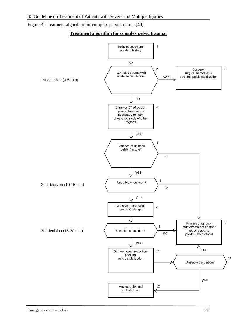
S3 Guideline on Treatment of Patients with Severe and Multiple Injuries
Emergency room – Pelvis 206
Figure 3: Treatment algorithm for complex pelvic trauma [49]
Treatment algorithm for complex pelvic trauma:
8 9
3
no
no 3rd decision (15-30 min)
6 Unstable circulation?
yes
no
yes
5
4
1st decision (3-5 min)
2
1 Initial assessment, accident history
Surgery: surgical hemostasis,
packing, pelvic stabilization Complex trauma with unstable circulation? yes
X-ray or CT of pelvis, general treatment, if necessary primary
diagnostic study of other regions
no
yes
Evidence of unstable pelvic fracture?
no
Primary diagnostic study/treatment of other
regions acc. to polytrauma protocol
11
Unstable circulation?
2nd decision (10-15 min)
Massive transfusion, pelvic C-clamp 7
yes
10
yes
12 Angiography and embolization
Surgery: open reduction, packing,
pelvic stabilization Unstable circulation?

S3 Guideline on Treatment of Patients with Severe and Multiple Injuries
Emergency room – Pelvis 207
Despite the varying and in part quite weak level of evidence, a treatment algorithm can be derived from the current state of knowledge which can, however, be modified depending on local logistic conditions.
S3 Guideline on Treatment of Patients with Severe and Multiple Injuries
Emergency room – Pelvis 208
References
1. Adams Je, Davis Gg, Alexander Cb et al. (2003) Pelvic trauma in rapidly fatal motor vehicle accidents. J Orthop Trauma 17:406-410 [LoE 4]
2. Agolini Sf, Shah K, Jaffe J et al. (1997) Arterial embolization is a rapid and effective technique for controlling pelvic fracture hemorrhage. J Trauma 43:395-399 [LoE 4]
3. Ben-Menachem Y, Coldwell Dm, Young Jw et al. (1991) Hemorrhage associated with pelvic fractures: causes, diagnosis, and emergent management. AJR Am J Roentgenol 157:1005-1014 [LoE 5]
4. Berg Ee, Chebuhar C, Bell Rm (1996) Pelvic trauma imaging: a blinded comparison of computed tomography and roentgenograms. J Trauma 41:994-998 [LoE 2]
5. Blackmore Cc, Jurkovich Gj, Linnau Kf et al. (2003) Assessment of volume of hemorrhage and outcome from pelvic fracture. Arch Surg 138:504-508; discussion 508-509 [LoE 3]
6. Bone L (1992) Emergency treatment of the injured patient. Saunders, Philadelphia, London, Toronto [LoE 5]
7. Bosch U, Pohlemann T, Tscherne H (1992) [Primary management of pelvic injuries]. Orthopade 21:385-392 [LoE 4]
8. Brasel Kj, Pham K, Yang H et al. (2007) Significance of contrast extravasation in patients with pelvic fracture. J Trauma 62:1149-1152 [LoE 4]
9. Brown Cv, Kasotakis G, Wilcox A et al. (2005) Does pelvic hematoma on admission computed tomography predict active bleeding at angiography for pelvic fracture? Am Surg 71:759-762 [LoE 4]
10. Burkhardt M, Culemann U, Seekamp A et al. (2005) [Strategies for surgical treatment of multiple trauma including pelvic fracture. Review of the literature]. Unfallchirurg 108:812, 814-820 [LoE 3]
11. Cook Re, Keating Jf, Gillespie I (2002) The role of angiography in the management of haemorrhage from major fractures of the pelvis. J Bone Joint Surg Br 84:178-182 [LoE 3]
12. Cothren Cc, Osborn Pm, Moore Ee et al. (2007) Preperitonal pelvic packing for hemodynamically unstable pelvic fractures: a paradigm shift. J Trauma 62:834-839; discussion 839-842 [LoE 3]
13. Croce Ma, Magnotti Lj, Savage Sa et al. (2007) Emergent pelvic fixation in patients with exsanguinating pelvic fractures. J Am Coll Surg 204:935-939; discussion 940-932 [LoE 4]
14. Culemann U, Tosounidis G, Reilmann H et al. (2003) [Pelvic fracture. Diagnostics and current treatment options]. Chirurg 74:687-698; quiz 699-700 [LoE 4]
15. Dalal Sa, Burgess Ar, Siegel Jh et al. (1989) Pelvic fracture in multiple trauma: classification by mechanism is key to pattern of organ injury, resuscitative requirements, and outcome. J Trauma 29:981-1000; discussion 1000-1002 [LoE 4]
16. Deangelis Na, Wixted Jj, Drew J et al. (2008) Use of the trauma pelvic orthotic device (T-POD) for provisional stabilisation of anterior-posterior
compression type pelvic fractures: a cadaveric study. Injury 39:903-906 [LoE 3]
17. Dente Cj, Feliciano Dv, Rozycki Gs et al. (2005) The outcome of open pelvic fractures in the modern era. Am J Surg 190:830-835 [LoE 4]
18. Duane Tm, Dechert T, Wolfe Lg et al. (2008) Clinical examination is superior to plain films to diagnose pelvic fractures compared to CT. Am Surg 74:476-479; discussion 479-480 [LoE 3]
19. Edeiken-Monroe Bs, Browner Bd, Jackson H (1989) The role of standard roentgenograms in the evaluation of instability of pelvic ring disruption. Clin Orthop Relat Res:63-76 [LoE 4]
20. Ertel W, Keel M, Eid K et al. (2001) Control of severe hemorrhage using C-clamp and pelvic packing in multiply injured patients with pelvic ring disruption. J Orthop Trauma 15:468-474 [LoE 3]
21. Euler E, Nast-Kolb D, Schweiberer L (1997) [Acetabular and pelvic fractures in multiple trauma]. Orthopade 26:354-359 [LoE 4]
22. Failinger Ms, Mcganity Pl (1992) Unstable fractures of the pelvic ring. J Bone Joint Surg Am 74:781-791 [LoE 5]
23. Fangio P, Asehnoune K, Edouard A et al. (2005) Early embolization and vasopressor administration for management of life-threatening hemorrhage from pelvic fracture. J Trauma 58:978-984; discussion 984 [LoE 4]
24. Friese Rs, Malekzadeh S, Shafi S et al. (2007) Abdominal ultrasound is an unreliable modality for the detection of hemoperitoneum in patients with pelvic fracture. J Trauma 63:97-102 [LoE 4]
25. Ghaemmaghami V, Sperry J, Gunst M et al. (2007) Effects of early use of external pelvic compression on transfusion requirements and mortality in pelvic fractures. Am J Surg 194:720-723; discussion 723 [LoE 4]
26. Gourlay D, Hoffer E, Routt M et al. (2005) Pelvic angiography for recurrent traumatic pelvic arterial hemorrhage. J Trauma 59:1168-1173; discussion 1173-1164 [LoE 4]
27. Guillamondegui Od, Mahboubi S, Stafford Pw et al. (2003) The utility of the pelvic radiograph in the assessment of pediatric pelvic fractures. J Trauma 55:236-239; discussion 239-240 [LoE 4]
28. Hagiwara A, Minakawa K, Fukushima H et al. (2003) Predictors of death in patients with life-threatening pelvic hemorrhage after successful transcatheter arterial embolization. J Trauma 55:696-703 [LoE 4]
29. Hagiwara A, Murata A, Matsuda T et al. (2004) The usefulness of transcatheter arterial embolization for patients with blunt polytrauma showing transient response to fluid resuscitation. J Trauma 57:271-276; discussion 276-277 [LoE 3]
30. Harley Jd, Mack La, Winquist Ra (1982) CT of acetabular fractures: comparison with conventional radiography. AJR Am J Roentgenol 138:413-417 [LoE 3]
S3 Guideline on Treatment of Patients with Severe and Multiple Injuries
Emergency room – Pelvis 209
31. Holting T, Buhr Hj, Richter Gm et al. (1992) Diagnosis and treatment of retroperitoneal hematoma in multiple trauma patients. Arch Orthop Trauma Surg 111:323-326 [LoE 4]
32. Kamaoui I, Courbiere M, Floccard B et al. (2008) [Pelvic trauma: impact of iodinated contrast material extravasation at MDCT on patient management]. J Radiol 89:1729-1734 [LoE 4]
33. Kessel B, Sevi R, Jeroukhimov I et al. (2007) Is routine portable pelvic X-ray in stable multiple trauma patients always justified in a high technology era? Injury 38:559-563 [LoE 4]
34. Kimbrell Bj, Velmahos Gc, Chan Ls et al. (2004) Angiographic embolization for pelvic fractures in older patients. Arch Surg 139:728-732; discussion 732-723 [LoE 4]
35. Miller Pr, Moore Ps, Mansell E et al. (2003) External fixation or arteriogram in bleeding pelvic fracture: initial therapy guided by markers of arterial hemorrhage. J Trauma 54:437-443 [LoE 4]
36. Mucha P, Jr., Welch Tj (1988) Hemorrhage in major pelvic fractures. Surg Clin North Am 68:757-773 [LoE 4]
37. Panetta T, Sclafani Sj, Goldstein As et al. (1985) Percutaneous transcatheter embolization for massive bleeding from pelvic fractures. J Trauma 25:1021-1029 [LoE 3]
38. Pehle B, Nast-Kolb D, Oberbeck R et al. (2003) [Significance of physical examination and radiography of the pelvis during treatment in the shock emergency room]. Unfallchirurg 106:642-648 [LoE 3]
39. Pereira Sj, O'brien Dp, Luchette Fa et al. (2000) Dynamic helical computed tomography scan accurately detects hemorrhage in patients with pelvic fracture. Surgery 128:678-685 [LoE 4]
40. Perez Jv, Hughes Tm, Bowers K (1998) Angiographic embolisation in pelvic fracture. Injury 29:187-191 [LoE 4]
41. Petrisor Ba, Bhandari M, Orr Rd et al. (2003) Improving reliability in the classification of fractures of the acetabulum. Arch Orthop Trauma Surg 123:228-233 [LoE 2]
42. Pieri S, Agresti P, Morucci M et al. (2004) [Percutaneous management of hemorrhages in pelvic fractures]. Radiol Med 107:241-251 [LoE 4]
43. Pohlemann T, Culemann U, Gansslen A et al. (1996) [Severe pelvic injury with pelvic mass hemorrhage: determining severity of hemorrhage and clinical experience with emergency stabilization]. Unfallchirurg 99:734-743 [LoE 2]
44. Pohlemann T, Krettek C, Hoffmann R et al. (1994) [Biomechanical comparison of various emergency stabilization measures of the pelvic ring]. Unfallchirurg 97:503-510 [LoE 4]
45. Pohlemann T, Paul C, Gansslen A et al. (1996) [Traumatic hemipelvectomy. Experiences with 11 cases]. Unfallchirurg 99:304-312 [LoE 3]
46. Resnik Cs, Stackhouse Dj, Shanmuganathan K et al. (1992) Diagnosis of pelvic fractures in patients with acute pelvic trauma: efficacy of plain radiographs. AJR Am J Roentgenol 158:109-112 [LoE 3]
47. Sadri H, Nguyen-Tang T, Stern R et al. (2005) Control of severe hemorrhage using C-clamp and arterial embolization in hemodynamically unstable patients with pelvic ring disruption. Arch Orthop Trauma Surg 125:443-447 [LoE 4]
48. Salim A, Teixeira Pg, Dubose J et al. (2008) Predictors of positive angiography in pelvic fractures: a prospective study. J Am Coll Surg 207:656-662 [LoE 3]
49. Seekamp A, Burkhardt M, Pohlemann T. (2004) Schockraummanagement bei Verletzungen des Beckens: Eine systematische Literaturübersicht. Unfallchirurg 107(10): 903-10
50. Shapiro M, Mcdonald Aa, Knight D et al. (2005) The role of repeat angiography in the management of pelvic fractures. J Trauma 58:227-231 [LoE 4]
51. Sheridan Mk, Blackmore Cc, Linnau Kf et al. (2002) Can CT predict the source of arterial hemorrhage in patients with pelvic fractures? Emerg Radiol 9:188-194 [LoE 3]
52. Shlamovitz Gz, Mower Wr, Bergman J et al. (2009) How (un)useful is the pelvic ring stability examination in diagnosing mechanically unstable pelvic fractures in blunt trauma patients? J Trauma 66:815-820 [LoE 3]
53. Siegmeth A, Mullner T, Kukla C et al. (2000) [Associated injuries in severe pelvic trauma]. Unfallchirurg 103:572-581 [LoE 4]
54. Silber Js, Flynn Jm, Koffler Km et al. (2001) Analysis of the cause, classification, and associated injuries of 166 consecutive pediatric pelvic fractures. J Pediatr Orthop 21:446-450 [LoE 4]
55. Stewart Bg, Rhea Jt, Sheridan Rl et al. (2002) Is the screening portable pelvis film clinically useful in multiple trauma patients who will be examined by abdominopelvic CT? Experience with 397 patients. Emerg Radiol 9:266-271 [LoE 4]
56. Tarman Gj, Kaplan Gw, Lerman Sl et al. (2002) Lower genitourinary injury and pelvic fractures in pediatric patients. Urology 59:123-126; discussion 126 [LoE 4]
57. Their Me, Bensch Fv, Koskinen Sk et al. (2005) Diagnostic value of pelvic radiography in the initial trauma series in blunt trauma. Eur Radiol 15:1533-1537 [LoE 4]
58. Torode I, Zieg D (1985) Pelvic fractures in children. J Pediatr Orthop 5:76-84 [LoE 4]
59. Totterman A, Dormagen Jb, Madsen Je et al. (2006) A protocol for angiographic embolization in exsanguinating pelvic trauma: a report on 31 patients. Acta Orthop 77:462-468 [LoE 3]
60. Totterman A, Madsen Je, Skaga No et al. (2007) Extraperitoneal pelvic packing: a salvage procedure to control massive traumatic pelvic hemorrhage. J Trauma 62:843-852 [LoE 4]
61. Trafton Pg (1990) Pelvic ring injuries. Surg Clin North Am 70:655-669 [LoE 5]
62. Trunkey Dd (1983) Trauma. Accidental and intentional injuries account for more years of life lost in the U.S. than cancer and heart disease. Among the prescribed remedies are improved preventive efforts,

S3 Guideline on Treatment of Patients with Severe and Multiple Injuries
Emergency room – Pelvis 210
speedier surgery and further research. Sci Am 249:28-35 [LoE 5]
63. Velmahos Gc, Toutouzas Kg, Vassiliu P et al. (2002) A prospective study on the safety and efficacy of angiographic embolization for pelvic and visceral injuries. J Trauma 53:303-308; discussion 308 [LoE 4]
64. Verbeek D, Sugrue M, Balogh Z et al. (2008) Acute management of hemodynamically unstable pelvic trauma patients: time for a change? Multicenter review of recent practice. World J Surg 32:1874-1882 [LoE 3]
65. Westhoff J, Laurer H, Wutzler S et al. (2008) [Interventional emergency embolization for severe pelvic ring fractures with arterial bleeding. Integration into the early clinical treatment algorithm]. Unfallchirurg 111:821-828 [LoE 4]
66. Young Jw, Burgess Ar, Brumback Rj et al. (1986) Pelvic fractures: value of plain radiography in early assessment and management. Radiology 160:445-451 [LoE 4]

S3 Guideline on Treatment of Patients with Severe and Multiple Injuries
Emergency room – Genitourinary tract 211
2.8 Genitourinary tract
Primary clinical diagnostic study
Key recommendation:
During the initial exploratory survey, the external urethral meatus and the transurethral bladder catheter (if the latter is already inserted) should be examined for blood.
GoR B
Explanation:
Gross hematuria is the cardinal symptom for injuries to the kidney, bladder and/or urethra whereas in this primary survey ureter injuries are clinically normal in about half of cases [15]. For this reason, the urinary catheter or the meatus should be inspected for blood during the primary survey of the undressed patient. Blood at the urethral meatus and hematuria must be differentiated in the clinical examination because they have different diagnostic meanings.
Key recommendation:
The region of flank, abdomen, perineum, and external genitals should be inspected for hematomas, ecchymoses, and external injuries.
GoR B
Explanation:
As the external physical examination can be carried out rapidly and easily, it should be carried out in full on all multiply injured patients even if it has only low diagnostic informative value [16]. The examination includes the search for external injury signs (hematomas, abrasions, swellings, etc.) in the region of the flanks, perineum, groins, and external genitals. Cotton et al. and Allen et al. showed that ecchymoses and abrasions in the abdominal region have a close correlation with the risk of an intraabdominal injury [17, 18]. However, a hematoma on the penile shaft or a perineal butterfly hematoma indicates an anterior urethral injury.
The value of the digital rectal examination is very critically evaluated in the current literature [19, 20] as abnormalities are generally found only rarely. In addition to assessing sphincter tone in the patient with spinal cord injury, the rectal examination should also be carried out if blood on the meatus or the presence of a relatively severe pelvic fracture indicates a urethral injury. The finding of a non-palpable, dislocated or hematoma-surrounded prostate represents additional clinically valuable information which in turn indicates a prostato-membranous tear.
The responsive patient can be questioned about possible details of the accident and pain from an injury to the genitourinary organs. Abdominal pain can give nonspecific clues to the presence of intraabdominal lesions [17, 21, 22]. In addition, a bladder rupture is specifically indicated if a patient experiences the urge to urinate before the trauma but no longer experiences this urge after

S3 Guideline on Treatment of Patients with Severe and Multiple Injuries
Emergency room – Genitourinary tract 212
the trauma (without evidence of neurologic lesions) [23] or if the patient tries to urinate without success [24].
Further information is provided by the circumstances of the accident, the mechanism of injury [25, 26], and the general condition of the patient [27]. In the injury pattern, particular attention should be paid to the close relationship between a pelvic fracture and efferent urinary tract lesions; this will be differentiated below according to organ. From a general view, injuries to the bladder and/or urethra are present in 6% of all pelvic fractures but on an Abbreviated Injury Scale (AIS) ≥ 4 injuries are markedly more frequent at 15% than on an AIS ≤ 3 injuries at 5% [25]. With the same severity of pelvic injury, men have almost double the risk for urologic injuries due to their anatomy, in particular the urethra [25, 28]. Rib fractures and injuries to intraabdominal organs increase the probability of injuries being present in the kidneys, ureters, and bladder [29]. If hypotension cannot be explained by blood losses of other origin, this can indicate a relatively severe injury to the kidney.
If there is complete urethral rupture, this can cause the transurethral catheter to go off-course [13, 14]. Likewise, an already existing urinary tract injury can be aggravated by the insertion of a transurethral catheter [30]. Based on these considerations, the patient with clinical signs of a urethral injury can have a transurethral catheter inserted during the diagnostic examination in the emergency room in order to better monitor the patient’s urination. Contraindications for catheterization only exist in very unstable patients for whom catheter insertion would represent an unnecessary time delay and in unclear conditions even during the diagnostic test (e.g., retrograde urethrogram). This also applies to the possibility that transurethral catheterization is impossible, e.g., due to a complete urethral tear. A more detailed diagnostic work-up follows below.

S3 Guideline on Treatment of Patients with Severe and Multiple Injuries
Emergency room – Genitourinary tract 213
Key recommendation:
In the case of circulatory instability that does not permit initial continuing diagnostic tests and if it is impossible to insert a transurethral bladder catheter, a suprapubic urinary diversion should be performed percutaneously or by laparotomy (with simultaneous exploration).
GoR B
Explanation:
If circulatory instability is present and if the patient cannot be diagnosed further due to the time delay and for these reasons a laparotomy should be performed, a suprapubic catheter should be inserted during this intervention [31] as this can then also be used subsequently for diagnostic purposes [30]. A rapid urine test and measurement of serum creatinine should be carried out for laboratory tests.
A rapid urine test (e.g., strip test) of the urine should be carried out to detect hematuria. Compared to the microscopic examination, the rapid urine test (e.g., strip test) has over 95% sensitivity and specificity [32-36]. The advantage of the rapid test lies in the results being available in less than 10 minutes. It is also helpful for the further course of action to have verification of bacteriuria; this occurs more frequently in elderly patients and can then be particularly problematic when combined with a urinary tract injury.
Measurement of serum creatinine can assist the ongoing course assessment and the detection of pre-existing kidney diseases. Hematologic parameters which permit the detection of bleeding, e.g., from the kidney, are also measured.
Calling on a qualified urologist is considered advisable for all patients with evidence of genitourinary injuries [37-39], even if this naturally depends on the qualifications of the physicians involved and the physical and organizational conditions.
S3 Guideline on Treatment of Patients with Severe and Multiple Injuries
Emergency room – Genitourinary tract 214
The necessity of imaging diagnostic tests
Key recommendation:
All patients with hematuria, blood discharge from the urethral meatus, dysuria, impossibility of catheterization or any other medical history information (local hematoma, concomitant injuries, mechanism of injury) have an increased risk of genitourinary injuries and should be given a focused diagnostic work-up of the kidney and/or the efferent urinary tract.
GoR B
Explanation:
Even if lesions in the upper and lower urinary tract occur simultaneously in only approximately 0.6% of patients with urologic injuries [40], a complete urological diagnostic study is still usually carried out in all patients with corresponding indications as this normally records the complete urinary system in the form of computed tomography with confirmed microscopic or gross hematuria [41, 42].
Whereas gross hematuria is pathognomonic for genitourinary injuries, microscopic hematuria represents a borderline situation. In general, however, it is accepted nowadays that microscopic hematuria should only entail further diagnostic study if other diagnostic injury evidence is simultaneously present [43-47].
In a large series of 1,588 patients with microscopic hematuria after sharp trauma, only 3 patients were found with relevant kidney injury [48]. In a similar study of 605 patients with blunt trauma, none of the patients with only microscopic hematuria had an injury requiring surgery [49]. This rate was 1 out of 77 in Fallon et al. [50]. Prospective series have confirmed these results [51]. In a pseudo-randomized study [52], in which the patients received different care depending on the admission team, Fuhrmann et al. compared 2 different indications for a cystogram: They were either examined for pelvic fracture, gross or microscopic hematuria (n = 134 patients) or the examination was limited to patients with gross hematuria only (> 200 erythrocytes per field of view). All urological injuries in the two groups were correctly identified. Thus, further acute diagnostic study of the kidneys can be dispensed with in patients who only have microscopic hematuria without additional injury signs. There are similar results for pediatric trauma and multiple injuries [53-56].
An important exception is the fact that vertical deceleration trauma in particular contains an increased risk for kidney injuries [57], which show up as normal in the primary clinical examination. Biomechanical studies support this argument so that further diagnostic study is recommended in strong deceleration trauma even without other criteria being present.
S3 Guideline on Treatment of Patients with Severe and Multiple Injuries
Emergency room – Genitourinary tract 215
Key recommendation:
Further imaging diagnostic tests should be carried out on the efferent urinary tract if one or more of the following criteria apply: hematuria, bleeding from the urethral meatus or vagina, dysuria, and local hematoma.
GoR B
Explanation:
Numerous studies have shown that bladder ruptures are associated with a pelvic fracture in 80-90% of cases [24, 25, 58, 59]. This correlation varies slightly depending on what severity grade the injuries have [11]. Hochberg and Stone [60] found a direct correlation between the number of fractured pubic rami (1, 2 or 3, 4) and the frequency of bladder ruptures (4%, 12%, 40%). Aihara et al. [61] also found that the symphysis or the sacroiliac joint had separated in 75% of bladder ruptures after blunt trauma. Nevertheless, a bladder rupture cannot be deduced from the presence of a complex pelvic fracture because only 20% (positive predictive value) of patients with symphysis and sacroiliac joint separation had a bladder rupture.
The close correlation between pelvic fracture and urethral injury is also well documented. However, the severity grade of injuries again plays a major role [25, 62, 63]. Koraitim et al, Morgan et al. and Aihara et al. showed consistently that fractures to the pubic rami increase the risk of a urethral injury, but that this risk rises hugely particularly in more complex pelvic fractures (type C) [61, 64, 65]. Aihara et al. emphasize that fractures of the lower pubic rami in particular indicate a urethral injury [61]. Palmer et al. noticed in a series of 200 patients with pelvic fracture that 26 out of the 27 patients with urologic lesions had a fracture in the anterior and posterior pelvic ring [66]. This association is less marked in women due to the shorter length and less connective tissue fusing in the female urethra [67]. Urethra injuries in women are usually accompanied by bleeding vaginal injuries [68-70].
The classic combination of pelvic fracture and gross hematuria allows the conclusion of a bladder and/or urethral injury to be made with great certainty [71]. Rehm et al. found that of 719 patients with blunt pelvic/abdominal injury all 21 cases with bladder injury were indicated by the presence of hematuria, which showed up in 17 cases also as gross hematuria [72]. Morey et al. [71] also reported that all their 85 patients with pelvic fracture had gross hematuria with simultaneous bladder rupture. In Palmer et al. this rate was in 10 out of 11 patients [66], in Hsieh et al. in 48 out of 51 [73]. A gap in the symphysis and separation in the sacroiliac joint doubled the risk for a bladder injury in the study by Aihara et al [61]. But even without a pelvic fracture being detectable, patients with gross hematuria or blood discharge from the urethral meatus must be assumed to have an injury to the efferent urinary tract [74].
The difference between hematuria and blood at the urethral meatus can be helpful in differentiating between bladder and urethral injuries. Thus, Morey et al. describe how all 53 patients with bladder rupture had a hematuria but that the simultaneous presence of blood at the urethral meatus correctly indicated in all 6 cases a concomitant urethral injury [71].
S3 Guideline on Treatment of Patients with Severe and Multiple Injuries
Emergency room – Genitourinary tract 216
Studies available internationally show clearly that the absence of hematuria and the simultaneous exclusion of a pelvic fracture definitely exclude a relevant injury to the bladder or urethra. This assessment is somewhat more difficult if there is positive evidence of a pelvic fracture. Hochberg and Stone found that a urologic injury is very unlikely here as well provided the pelvic fracture does not affect the pubic rami [60].
Imaging diagnostic test of kidneys and ureters
Key recommendations:
Computed tomography with contrast agent should be performed in the case of suspected kidney injury.
GoR B
Explanation:
The importance of computed tomography (CT) in the primary assessment of blunt abdominal trauma is not the subject of this text as the diagnostic test focuses on all intraabdominal trauma. Thus, only the degree of accuracy with which injuries to the kidney and efferent urinary tract can be detected in the CT will be discussed below. In the literature review, the CT diagnostic test appears to be the most reliable, comfortable method in assessing blunt abdominal trauma [9, 41, 47, 75-82].
Intravenous pyelography is inferior to CT with respect to diagnostic accuracy [44, 76] yet represents an important option if CT cannot be performed. This may be the case if the admitting hospital does not have the necessary equipment available or, more usually, if the patient is hemodynamically unstable and requires immediate emergency surgery [83]. In such cases, i.v. pyelography makes it possible to carry out the urologic diagnostic study directly during surgery [9, 41, 44]. The images are available approximately 10 minutes after administration of the contrast agent (2 ml/kg). For 284 patients with blunt kidney trauma, Nicolaisen et al. report perfect sensitivity of i.v. pyelography for identifying blunt injuries and state that in 87% of cases it was also possible to classify the injury severity grade correctly [29]. Although in 5 cases of 60 patients with normal excretory urography Halsell et al. found smaller renal lesions in the computed tomography [84], these lesions were clinically less important and could be conservatively treated.
Various studies report a sensitivity of over 90% in detecting renal injuries by ultrasonography [85, 86] but the sensitivity is obviously less if the injury has not resulted in free fluid in the abdomen [85]. This can occur in about 10-20% of cases [87]. Overall, however, ultrasonography is not sufficiently reliable. On average, intraabdominal lesions will be present in 10-20% of cases despite negative ultrasonography [87, 88]. Ultrasonography is therefore only suitable as an additional diagnostic test. However, a randomized study showed that primary ultrasonography could reduce the necessity of a CT diagnostic test [89] as negative ultrasonography was assessed as adequately reliable in individual patients without evidence of abdominal injuries.
Angiographic techniques have much more of a therapeutic role than a diagnostic one as angiography does not really provide any additional diagnostic information compared to CT [77,
S3 Guideline on Treatment of Patients with Severe and Multiple Injuries
Emergency room – Genitourinary tract 217
90]. In cases where an injury to the renal artery or its lateral branches can be assumed or active bleeding is detected by computed tomography, it is expedient to use angiography as preparation for an embolization [91-95]. In vascular injuries to the renal pedicle (e.g., intimal tear), this enables the patency of the renal artery to be restored using an endovascular stent. Moreover, in relatively severe renal injuries with massive bleeding, selective embolization of the bleeding vessel should be carried out [9, 41, 42] provided the patient has stable circulation. The number of primary operated patients can be minimized through this radiologic intervention option, which leads to a reduction in the nephrectomy rate. The success rate of radiologic intervention is about 70-80% [42]. Angiography can also be necessary if CT equipment is not locally available and the i.v. pyelography does not show the kidney.
In addition to the methods cited, magnetic resonance imaging has been tested by Leppaniemi et al. [96-98] and it can image some details better than CT [96, 99]. Due to the increased time involved, however, it is seldom advisable to use this procedure in multiply injured patients during the acute phase. Magnetic resonance imaging could be beneficial in rare cases if CT is unavailable or cannot be used because of an allergy to the contrast agent or the CT finding is unclear.
Detecting a ureter lesion is much more difficult [100-102]. Medina et al. described a sensitivity of 20% although a very wide range of diagnostic modalities (CT, intravenous pyelography [IVP], and retrograde pyelogram) was used [15]. In 81 patients with non-iatrogenic blunt ureter injury, Dobrowolski et al. [103] found i.v. and retrograde pyelography helpful. Ghali et al. [102] considered only pyelography to be diagnostically reliable even when compared to intraoperative inspection.
S3 Guideline on Treatment of Patients with Severe and Multiple Injuries
Emergency room – Genitourinary tract 218
Imaging diagnostic tests of the lower urinary tract
Key recommendation:
If prioritizing permits, retrograde urethrography a nd a cystogram should be performed in patients with clinical reference points for a urethral lesion.
GoR B
If prioritizing permits, a retrograde cystogram should be performed in patients with clinical reference points for a bladder injury.
GoR B
Explanation:
If there is a suspected urethral and/or bladder lesion, retrograde urethrography and a cystogram should be performed [30]. Retrograde urethrography consists of the transurethral administration of approximately 400 ml of contrast agent. Provided the urethra is uninjured, urethrography enables the bladder to be adequately filled with contrast agent. Thereafter, a radiograph is taken, ideally on 2 planes but often limited for practical reasons to the anteroposterior plane in multiply injured patients [72]. However, both planes should be represented so that retrovesicular extravasates are not missed [23]. The cystogram consists of an image after drainage in addition to the voided image and the filling image as otherwise there will be approximately 10% false-negative results [104]. In cases where no retrograde bladder filling can be achieved, the bladder must be filled via a suprapubic catheter as combined injuries to the bladder and urethra make up 10-20% of all bladder or urethral injuries [61].
In multiply injured patients, due to concomitant injuries, it is not possible in about 20% of cases to carry out the cystogram within the initial emergency room phase [73]. This may be unavoidable in individual cases but the diagnostic test must be carried out as soon as possible thereafter so that no injuries are missed. On the other hand, Hsieh et al. [73] saw no serious disadvantages in the cases where the diagnosis of a bladder rupture had been delayed until later.
Ultrasound does not play a big role in assessing bladder or urethral injuries but is very helpful in localizing the bladder for inserting the suprapubic bladder catheter. I.v. pyelography is also unreliable in assessing uncertain bladder injuries as the bladder resting pressure is too low and dilutes the contrast agent too much. Several clinical studies have shown that i.v. pyelography does not detect 64%-84% of bladder injuries [105-108].
Although computed tomography cannot make definite statements on urethral injuries, it is still very valuable in diagnosing bladder ruptures [109]. However, without separate filling of the bladder with contrast agent, the CT diagnostic test can only supply indirect evidence. Although the absence of pelvic fluid collections makes bladder ruptures less likely [110], it can never definitely exclude a relevant injury. Only the CT cystogram is suitable for this, and it can also be performed as an alternative to the normal cystogram. In a series of 316 patients, Deck et al. found evidence of a sensitivity and specificity of 95% and 100%, respectively, for the CT cystogram in identifying bladder ruptures [111, 112]. Even if these values were somewhat worse for intraperitoneal ruptures (78% and 99%), the authors still consider that the CT cystogram

S3 Guideline on Treatment of Patients with Severe and Multiple Injuries
Emergency room – Genitourinary tract 219
ranks at least equally with the conventional cystogram. Other groups have reported similar results [113, 114]. For this reason, the CT cystogram can offer time and organizational advantages particularly in multiply injured patients as the CT diagnostic test is often indicated here because of other injuries. However, the prerequisite for a definite diagnosis is the sufficient administration of contrast agent (> 350 ml) to be able to produce and detect an extravasate at all in the presence of a rupture through sufficient fill pressure [104, 109, 115].

S3 Guideline on Treatment of Patients with Severe and Multiple Injuries
Emergency room – Genitourinary tract 220
References
1 Ahmed S, Neel KF. Urethral injury in girls with fractured pelvis following blunt abdominal trauma. Br J Urol 1996: 78(3):450-453.
2 Aihara R, Blansfield JS, Millham FH, LaMorte WW, Hirsch EF. Fracture locations influence the likelihood of rectal and lower urinary tract injuries in patients sustaining pelvic fractures. J Trauma 2002: 52(2):205-208.
3 Allen GS, Moore FA, Cox CS, Jr., Wilson JT, Cohn JM, Duke JH. Hollow visceral injury and blunt trauma. J Trauma 1998: 45(1):69-75.
4 Altman AL, Haas C, Dinchman KH, Spirnak JP. Selective nonoperative management of blunt grade 5 renal injury. J Urol 2000: 164(1):27-30.
5 Ambiavagar R, Nambiar R. Traumatic closed avulsion of the upper ureter. Injury 1979: 11(1):71-76.
6 Anselmo G, Fandella A, Faggiano L, Merlo F, Maccatrozzo L. Fractures of the penis: therapeutic approach and long-term results. Br J Urol 1991: 67(5):509-511.
7 Armenakas NA. Current methods of diagnosis and management of ureteral injuries. World J Urol 1999: 17(2):78-83.
8 Asci R, Sarikaya S, Buyukalpelli R, Saylik A, Yilmaz AF, Yildiz S. Voiding and sexual dysfunctions after pelvic fracture urethral injuries treated with either initial cystostomy and delayed urethroplasty or immediate primary urethral realignment. Scand J Urol Nephrol 1999: 33(4):228-233.
9 Batislam E, Ates Y, Germiyanoglu C, Karabulut A, Gulerkaya B, Erol D. Role of Tile classification in predicting urethral injuries in pediatric pelvic fractures. J Trauma 1997: 42(2):285-287 [LoE 5].
10 Baverstock R, Simons R, McLoughlin M. Severe blunt renal trauma: a 7-year retrospective review from a provincial trauma centre. Can J Urol 2001: 8(5):1372-1376.
11 Beaujeux R, Saussine C, al-Fakir A, Boudjema K, Roy C, Jacqmin D et al. Superselective endo-vascular treatment of renal vascular lesions. J Urol 1995: 153(1):14-17 [LoE 4].
12 Beck D, Marley R, Salvator A, Muakkassa F. Prospective study of the clinical predictors of a positive abdominal computed tomography in blunt trauma patients. J Trauma 2004: 57(2):296-300.
13 Becker CD, Mentha G, Schmidlin F, Terrier F. Blunt abdominal trauma in adults: role of CT in the diagnosis and management of visceral injuries. Part 2: Gastrointestinal tract and retroperitoneal organs. Eur Radiol 1998: 8(5):772-780 [LoE 5].
14 Black PC, Miller EA, Porter JR, Wessells H. Urethral and bladder neck injury associated with pelvic fracture in 25 female patients. J Urol 2006: 175(6):2140-2144 [LoE 5].
15 Bozeman C, Carver B, Zabari G, Caldito G, Venable D. Selective operative management of major blunt renal trauma. J Trauma 2004: 57(2):305-309 [LoE 4].
16 Brandes S, Borrelli J, Jr. Pelvic fracture and associated urologic injuries. World J Surg 2001: 25(12):1578-1587 [LoE 2].
17 Brandes S, Coburn M, Armenakas N, McAninch J. Diagnosis and management of ureteric injury: an evidence-based analysis. BJU Int 2004: 94(3):277-289 [LoE 2b].
18 Brandes SB, McAninch JW. Reconstructive surgery for trauma of the upper urinary tract. Urol Clin North Am 1999: 26(1):183-99, [LoE 4].
19 Brandes SB, McAninch JW. Urban free falls and patterns of renal injury: a 20-year experience with 396 cases. J Trauma 1999: 47(4):643-649 [LoE 4].
20 Bretan PN, Jr., McAninch JW, Federle MP, Jeffrey RB, Jr. Computerized tomographic staging of renal trauma: 85 consecutive cases. J Urol 1986: 136(3):561-565 [LoE 4].
21 Brosman SA, Paul JG. Trauma of the bladder. Surg Gynecol Obstet 1976: 143(4):605-608 [LoE 2b].
22 Buchberger W, Penz T, Wicke K, Eberle J. [Diagnosis and staging of blunt kidney trauma. A comparison of urinalysis, i.v. urography, sonography and computed tomography]. Rofo 1993: 158(6):507-512 [LoE 2b].
23 Buckley JC, McAninch JW. Selective management of isolated and nonisolated grade IV renal injuries. J Urol 2006: 176(6 Pt 1):2498-2502 [LoE 4].
24 Carroll PR, McAninch JW. Major bladder trauma: the accuracy of cystography. J Urol 1983: 130(5):887-888 [LoE 5].
25 Carroll PR, McAninch JW. Major bladder trauma: mechanisms of injury and a unified method of diagnosis and repair. J Urol 1984: 132(2):254-257 [LoE 4].
26 Carter CT, Schafer N. Incidence of urethral disruption in females with traumatic pelvic fractures. Am J Emerg Med 1993: 11(3):218-220 [LoE 2].
27 Cass AS, Cass BP. Immediate surgical management of severe renal injuries in multiple-injured patients. Urology 1983: 21(2):140-145 [LoE 2].
28 Cass AS, Luxenberg M. Features of 164 bladder ruptures. J Urol 1987: 138(4):743-745 [LoE 4].
29 Cass AS, Vieira J. Comparison of IVP and CT findings in patients with suspected severe renal injury. Urology 1987: 29(5):484-487 [LoE 4].
30 Chandhoke PS, McAninch JW. Detection and significance of microscopic hematuria in patients with blunt renal trauma. J Urol 1988: 140(1):16-18 [LoE 5].
31 Chapple C, Barbagli G, Jordan G, Mundy AR, Rodrigues-Netto N, Pansadoro V et al. Consensus statement on urethral trauma. BJU Int 2004: 93(9):1195-1202 [LoE 4].
32 Chatziioannou A, Brountzos E, Primetis E, Malagari K, Sofocleous C, Mourikis D et al. Effects of superselective embolization for renal vascular injuries on renal parenchyma and function. Eur J Vasc Endovasc Surg 2004: 28(2):201-206 [LoE 2b].

S3 Guideline on Treatment of Patients with Severe and Multiple Injuries
Emergency room – Genitourinary tract 221
33 Cheng DL, Lazan D, Stone N. Conservative treatment of type III renal trauma. J Trauma 1994: 36(4):491-494 [LoE 2b].
34 Corr P, Hacking G. Embolization in traumatic intrarenal vascular injuries. Clin Radiol 1991: 43(4):262-264 [LoE 2b].
35 Corriere JN, Jr., Sandler CM. Management of the ruptured bladder: seven years of experience with 111 cases. J Trauma 1986: 26(9):830-833 [LoE 2b].
36 Corriere JN, Jr., Sandler CM. Management of extraperitoneal bladder rupture. Urol Clin North Am 1989: 16(2):275-277 [LoE 2b].
37 Cotton BA, Beckert BW, Smith MK, Burd RS. The utility of clinical and laboratory data for predicting intraabdominal injury among children. J Trauma 2004: 56(5):1068-1074 [LoE 4].
38 Culty T, Boccon-Gibod L. Anastomotic urethroplasty for posttraumatic urethral stricture: previous urethral manipulation has a negative impact on the final outcome. J Urol 2007: 177(4):1374-1377 [LoE 4].
39 Daum GS, Krolikowski FJ, Reuter KL, Colby JM, Silva WM. Dipstick evaluation of hematuria in abdominal trauma. Am J Clin Pathol 1988: 89(4):538-542 [LoE 5].
40 Deck AJ, Shaves S, Talner L, Porter JR. Computerized tomography cystography for the diagnosis of traumatic bladder rupture. J Urol 2000: 164(1):43-46 [LoE 4].
41 Deck AJ, Shaves S, Talner L, Porter JR. Current experience with computed tomographic cystography and blunt trauma. World J Surg 2001: 25(12):1592-1596 [LoE 3a].
42 Demetriades D, Karaiskakis M, Toutouzas K, Alo K, Velmahos G, Chan L. Pelvic fractures: epidemiology and predictors of associated abdominal injuries and outcomes. J Am Coll Surg 2002: 195(1):1-10 [LoE 3].
43 Dinkel HP, Danuser H, Triller J. Blunt renal trauma: minimally invasive management with microcatheter embolization experience in nine patients. Radiology 2002: 223(3):723-730 [LoE 4].
44 Djakovic N, Plas E, Martinez-Pineiro L, Lynch TH, Mor Y, Santucci RA et al. Guidelines on urological trauma. http://uroweb.org/files/uploaded files/guidelines/urotrauma.pdf: Ezropean Association of Urology , 1-84. 2009 [Evidenzbasierte Leitlinie]
45 Dobrowolski Z, Kusionowicz J, Drewniak T, Habrat W, Lipczynski W, Jakubik P et al. Renal and ureteric trauma: diagnosis and management in Poland. BJU Int 2002: 89(7):748-751 [LoE 4].
46 Dokucu AI, Ozdemir E, Ozturk H, Otcu S, Onen A, Cigdem K et al. Urogenital injuries in childhood: a strong association of bladder trauma to bowel injuries. Int Urol Nephrol 2000: 32(1):3-8 [LoE 4].
47 Eastham JA, Wilson TG, Larsen DW, Ahlering TE. Angiographic embolization of renal stab wounds. J Urol 1992: 148(2 Pt 1):268-270 [LoE 3a].
48 El-Sherbiny MT, Aboul-Ghar ME, Hafez AT, Hammad AA, Bazeed MA. Late renal functional and morphological evaluation after non-operative treatment of high-grade renal injuries in children. BJU Int 2004: 93(7):1053-1056 [LoE 4].
49 Elliott DS, Barrett DM. Long-term followup and evaluation of primary realignment of posterior urethral disruptions. J Urol 1997: 157(3):814-816 [LoE 4].
50 Elliott SP, McAninch JW. Ureteral injuries from external violence: the 25-year experience at San Francisco General Hospital. J Urol 2003: 170(4 Pt 1):1213-1216 [LoE 4].
51 Fallon B, Wendt JC, Hawtrey CE. Urological injury and assessment in patients with fractured pelvis. J Urol 1984: 131(4):712-714 [LoE 4].
52 Fanney DR, Casillas J, Murphy BJ. CT in the diagnosis of renal trauma. Radiographics 1990: 10(1):29-40 [LoE 2b].
53 Festini G, Gregorutti S, Reina G, Bellis GB. Isolated intraperitoneal bladder rupture in patients with alcohol intoxication and minor abdominal trauma. Ann Emerg Med 1991: 20(12):1371-1372 [LoE 4].
54 Fisher RG, Ben-Menachem Y, Whigham C. Stab wounds of the renal artery branches: angiographic diagnosis and treatment by embolization. AJR Am J Roentgenol 1989: 152(6):1231-1235.
55 Flancbaum L, Morgan AS, Fleisher M, Cox EF. Blunt bladder trauma: manifestation of severe injury. Urology 1988: 31(3):220-222.
56 Follis HW, Koch MO, McDougal WS. Immediate management of prostatomembranous urethral disruptions. J Urol 1992: 147(5):1259-1262 [LoE 4].
57 Fortune JB, Brahme J, Mulligan M, Wachtel TL. Emergency intravenous pyelography in the trauma patient. A reexamination of the indications. Arch Surg 1985: 120(9):1056-1059 [LoE 4].
58 Frohmüller H, Theiß M. Blutungen bei Verletzungen des Harntrakts. Langenbecks Arch Chir 1993: 110(Suppl):352-357 [LoE 4].
59 Fuhrman GM, Simmons GT, Davidson BS, Buerk CA. The single indication for cystography in blunt trauma. Am Surg 1993: 59(6):335-337 [LoE 4].
60 Ghali AM, El Malik EM, Ibrahim AI, Ismail G, Rashid M. Ureteric injuries: diagnosis, management, and outcome. J Trauma 1999: 46(1):150-158 [LoE 4].
61 Gheiler EL, Frontera JR. Immediate primary realignment of prostatomembranous urethral disruptions using endourologic techniques. Urology 1997: 49(4):596-599 [LoE 2b].
62 Glass RE, Flynn JT, King JB, Blandy JP. Urethral injury and fractured pelvis. Br J Urol 1978: 50(7):578-582 [LoE 4].
63 Goldin AR, Funston MR. Percutaneous embolization for post-traumatic renal haemorrhage. S Afr Med J 1978: 53(26):1061-1063 [LoE 4].
64 Goldner AP, Mayron R, Ruiz E. Are urine dipsticks reliable indicators of hematuria in blunt trauma patients? Ann Emerg Med 1985: 14(6):580-582 [LoE 2b].
65 Guldner G, Babbitt J, Boulton M, O'Callaghan T, Feleke R, Hargrove J. Deferral of the rectal examination in blunt trauma patients: a clinical decision rule. Acad Emerg Med 2004: 11(6):635-641 [LoE 2b].

S3 Guideline on Treatment of Patients with Severe and Multiple Injuries
Emergency room – Genitourinary tract 222
66 Hagiwara A, Murata A, Matsuda T, Matsuda H, Shimazaki S. The usefulness of transcatheter arterial embolization for patients with blunt polytrauma showing transient response to fluid resuscitation. J Trauma 2004: 57(2):271-276 [LoE 4].
67 Halsell RD, Vines FS, Shatney CH, Slepin MJ, Northup HM, Avara WT, III et al. The reliability of excretory urography as a screening examination for blunt renal trauma. Ann Emerg Med 1987: 16(11):1236-1239 [LoE 4].
68 Hammer CC, Santucci RA. Effect of an institutional policy of nonoperative treatment of grades I to IV renal injuries. J Urol 2003: 169(5):1751-1753 [LoE 4].
69 Herschorn S, Thijssen A, Radomski SB. The value of immediate or early catheterization of the traumatized posterior urethra. J Urol 1992: 148(5):1428-1431 [LoE 4].
70 Heyns CF, van VP. Increasing role of angiography and segmental artery embolization in the management of renal stab wounds. J Urol 1992: 147(5):1231-1234 [LoE 4].
71 Hochberg E, Stone NN. Bladder rupture associated with pelvic fracture due to blunt trauma. Urology 1993: 41(6):531-533 [LoE 4].
72 Holevar MEJLFNKSRSJPYC. Practice management guidelines for the management of genitourinary trauma. http://www.east.org/tpg.html: Eastern Association for the Surgery of Trauma (EAST) , 1-101. 2004 [Evidenzbasierte Leitlinie]
73 Holmes JF, Sokolove PE, Brant WE, Palchak MJ, Vance CW, Owings JT et al. Identification of children with intra-abdominal injuries after blunt trauma. Ann Emerg Med 2002: 39(5):500-509 [LoE 4].
74 Horstman WG, McClennan BL, Heiken JP. Comparison of computed tomography and conventional cystography for detection of traumatic bladder rupture. Urol Radiol 1991: 12(4):188-193 [LoE 4].
75 Hsieh CH, Chen RJ, Fang JF, Lin BC, Hsu YP, Kao JL et al. Diagnosis and management of bladder injury by trauma surgeons. Am J Surg 2002: 184(2):143-147 [LoE 3].
76 Husmann DA, Gilling PJ, Perry MO, Morris JS, Boone TB. Major renal lacerations with a devitalized fragment following blunt abdominal trauma: a comparison between nonoperative (expectant) versus surgical management. J Urol 1993: 150(6):1774-1777 [LoE 3].
77 Husmann DA, Morris JS. Attempted nonoperative management of blunt renal lacerations extending through the corticomedullary junction: the short-term and long-term sequelae. J Urol 1990: 143(4):682-684 [LoE 4].
78 Husmann DA, Wilson WT, Boone TB, Allen TD. Prostatomembranous urethral disruptions: management by suprapubic cystostomy and delayed urethroplasty. J Urol 1990: 144(1):76-78 [LoE 4].
79 Ichigi Y, Takaki N, Nakamura K, Sato S, Kato A, Matsuo Y et al. Significance of hematoma size for evaluating the grade of blunt renal trauma. Int J Urol 1999: 6(10):502-508 [LoE 4].
80 Ingram MD, Watson SG, Skippage PL, Patel U. Urethral injuries after pelvic trauma: evaluation with urethrography. Radiographics 2008: 28(6):1631-1643 [LoE 5].
81 Iverson AJ, Morey AF. Radiographic evaluation of suspected bladder rupture following blunt trauma: critical review. World J Surg 2001: 25(12):1588-1591 [LoE 5].
82 Keller MSCCEGJJSKHGMCWTR. Functional outcome of nonoperatively managed renal injuries in children. J Trauma 2004: 57:108-110 [LoE 5].
83 Kennedy TJ, McConnell JD, Thal ER. Urine dipstick vs. microscopic urinalysis in the evaluation of abdominal trauma. J Trauma 1988: 28(5):615-617 [LoE 4].
84 Kinnunen J, Kivioja A, Poussa K, Laasonen EM. Emergency CT in blunt abdominal trauma of multiple injury patients. Acta Radiol 1994: 35(4):319-322 [LoE 4].
85 Klein S, Johs S, Fujitani R, State D. Hematuria following blunt abdominal trauma. The utility of intravenous pyelography. Arch Surg 1988: 123(9):1173-1177 [LoE 5].
86 Knudson MM, McAninch JW, Gomez R, Lee P, Stubbs HA. Hematuria as a predictor of abdominal injury after blunt trauma. Am J Surg 1992: 164(5):482-485 [LoE 5].
87 Koraitim MM. Pelvic fracture urethral injuries: evaluation of various methods of management. J Urol 1996: 156(4):1288-1291 [LoE 4].
88 Koraitim MM. Pelvic fracture urethral injuries: the unresolved controversy. J Urol 1999: 161(5):1433-1441 [LoE 2a].
89 Koraitim MM, Marzouk ME, Atta MA, Orabi SS. Risk factors and mechanism of urethral injury in pelvic fractures. Br J Urol 1996: 77(6):876-880 [LoE 1b].
90 Kotkin L, Koch MO. Impotence and incontinence after immediate realignment of posterior urethral trauma: result of injury or management? J Urol 1996: 155(5):1600-1603 [LoE 4].
91 Ku JH, Jeon YS, Kim ME, Lee NK, Park YH. Is there a role for magnetic resonance imaging in renal trauma? Int J Urol 2001: 8(6):261-267 [LoE 4].
92 Ku JH, Jeon YS, Kim ME, Lee NK, Park YH. Comparison of long-term results according to the primary mode of management and type of injury for posterior urethral injuries. Urol Int 2002: 69(3):227-232 [LoE 4].
93 Leppaniemi A, Lamminen A, Tervahartiala P, Haapiainen R, Lehtonen T. Comparison of high-field magnetic resonance imaging with computed tomography in the evaluation of blunt renal trauma. J Trauma 1995: 38(3):420-427 [LoE 4].
94 Leppaniemi A, Lamminen A, Tervahartiala P, Salo J, Haapiainen R, Lehtonen T. MRI and CT in blunt renal trauma: an update. Semin Ultrasound CT MR 1997: 18(2):129-135 [LoE 4].
95 Leppaniemi AK, Kivisaari AO, Haapiainen RK, Lehtonen TA. Role of magnetic resonance imaging in blunt renal parenchymal trauma. Br J Urol 1991: 68(4):355-360 [LoE 4].

S3 Guideline on Treatment of Patients with Severe and Multiple Injuries
Emergency room – Genitourinary tract 223
96 Levitt MA, Criss E, Kobernick M. Should the emergency IVP be used more selectively in blunt renal trauma? Ann Emerg Med 1985: 14(10):959-965 [LoE 3].
97 Levy JB, Baskin LS, Ewalt DH, Zderic SA, Bellah R, Snyder HM, III et al. Nonoperative management of blunt pediatric major renal trauma. Urology 1993: 42(4):418-424 [LoE 4].
98 Lis LE, Cohen AJ. CT cystography in the evaluation of bladder trauma. J Comput Assist Tomogr 1990: 14(3):386-389 [LoE 4].
99 Livingston DH, Lavery RF, Passannante MR, Skurnick JH, Fabian TC, Fry DE et al. Admission or observation is not necessary after a negative abdominal computed tomographic scan in patients with suspected blunt abdominal trauma: results of a prospective, multi-institutional trial. J Trauma 1998: 44(2):273-280 [LoE 3].
100 Lupetin AR, Mainwaring BL, Daffner RH. CT diagnosis of renal artery injury caused by blunt abdominal trauma. AJR Am J Roentgenol 1989: 153(5):1065-1068 [LoE 4].
101 Lynch D, Martinez-Pineiro L, las E, erafetinides E, urkeri L, ohenfellner M. Guidelines on urological trauma. http://uroweb.org/files/uploaded files/guidelines/urotrauma.pdf: Ezropean Association of Urology , 1-70. 2003 [Evidenzbasierte Leitlinie]
102 Lynch TH, Martinez-Pineiro L, Plas E, Serafetinides E, Turkeri L, Santucci RA et al. EAU guidelines on urological trauma. Eur Urol 2005: 47(1):1-15 [Evidenzbasierte Leitlinie]
103 MacMahon R, Hosking D, Ramsey EW. Management of blunt injury to the lower urinary tract. Can J Surg 1983: 26(5):415-418 [LoE 4].
104 Mariani AJ, Luangphinith S, Loo S, Scottolini A, Hodges CV. Dipstick chemical urinalysis: an accurate cost-effective screening test. J Urol 1984: 132(1):64-66 [LoE 5].
105 Matthews LA, Smith EM, Spirnak JP. Nonoperative treatment of major blunt renal lacerations with urinary extravasation. J Urol 1997: 157(6):2056-2058 [LoE 4].
106 Mayher BE, Guyton JL, Gingrich JR. Impact of urethral injury management on the treatment and outcome of concurrent pelvic fractures. Urology 2001: 57(3):439-442 [LoE 4].
107 Mayor B, Gudinchet F, Wicky S, Reinberg O, Schnyder P. Imaging evaluation of blunt renal trauma in children: diagnostic accuracy of intravenous pyelography and ultrasonography. Pediatr Radiol 1995: 25(3):214-218 [LoE 4].
108 McAndrew JD, Corriere JN, Jr. Radiographic evaluation of renal trauma: evaluation of 1103 consecutive patients. Br J Urol 1994: 73(4):352-354 [LoE 4].
109 Medina D, Lavery R, Ross SE, Livingston DH. Ureteral trauma: preoperative studies neither predict injury nor prevent missed injuries. J Am Coll Surg 1998: 186(6):641-644 [LoE 5].
110 Mee SL, McAninch JW, Robinson AL, Auerbach PS, Carroll PR. Radiographic assessment of renal trauma:
a 10-year prospective study of patient selection. J Urol 1989: 141(5):1095-1098 [LoE 1].
111 Middlebrook PF, Schillinger JF. Hematuria and intravenous pyelography in pediatric blunt renal trauma. Can J Surg 1993: 36(1):59-62 [LoE 2].
112 Miller DC, Forauer A, Faerber GJ. Successful angioembolization of renal artery pseudoaneurysms after blunt abdominal trauma. Urology 2002: 59(3):444 [LoE 2].
113 Miller KS, McAninch JW. Radiographic assessment of renal trauma: our 15-year experience. J Urol 1995: 154(2 Pt 1):352-355 [LoE 2].
114 Mohr AM, Pham AM, Lavery RF, Sifri Z, Bargman V, Livingston DH. Management of trauma to the male external genitalia: the usefulness of American Association for the Surgery of Trauma organ injury scales. J Urol 2003: 170(6 Pt 1):2311-2315 [LoE 4].
115 Monstrey SJ, vander WC, Debruyne FM, Goris RJ. Urological trauma and severe associated injuries. Br J Urol 1987: 60(5):393-398 [LoE 4].
116 Moore EE, Shackford SR, Pachter HL, McAninch JW, Browner BD, Champion HR et al. Organ injury scaling: spleen, liver, and kidney. J Trauma 1989: 29(12):1664-1666.
117 Morehouse DD, Mackinnon KJ. Posterior urethral injury: etiology, diagnosis, initial management. Urol Clin North Am 1977: 4(1):69-73.
118 Morey AF, Hernandez J, McAninch JW. Reconstructive surgery for trauma of the lower urinary tract. Urol Clin North Am 1999: 26(1):49-60, viii.
119 Morey AF, Iverson AJ, Swan A, Harmon WJ, Spore SS, Bhayani S et al. Bladder rupture after blunt trauma: guidelines for diagnostic imaging. J Trauma 2001: 51(4):683-686.
120 Morey AF, McAninch JW, Tiller BK, Duckett CP, Carroll PR. Single shot intraoperative excretory urography for the immediate evaluation of renal trauma. J Urol 1999: 161(4):1088-1092.
121 Morgan DE, Nallamala LK, Kenney PJ, Mayo MS, Rue LW, III. CT cystography: radiographic and clinical predictors of bladder rupture. AJR Am J Roentgenol 2000: 174(1):89-95.
122 Moudouni SM, Hadj SM, Manunta A, Patard JJ, Guiraud PH, Guille F et al. Management of major blunt renal lacerations: is a nonoperative approach indicated? Eur Urol 2001: 40(4):409-414.
123 Moudouni SM, Patard JJ, Manunta A, Guiraud P, Guille F, Lobel B. A conservative approach to major blunt renal lacerations with urinary extravasation and devitalized renal segments. BJU Int 2001: 87(4):290-294.
124 Mouraviev VB, Coburn M, Santucci RA. The treatment of posterior urethral disruption associated with pelvic fractures: comparative experience of early realignment versus delayed urethroplasty. J Urol 2005: 173(3):873-876.
125 Nagel R, Leistenschneider W. [Urologic injuries in patients with multiple injuries]. Chirurg 1978: 49(12):731-736.

S3 Guideline on Treatment of Patients with Severe and Multiple Injuries
Emergency room – Genitourinary tract 224
126 Nane I, Esen T, Tellaloglu S, Selhanoglu M, Akinci M. Penile fracture: emergency surgery for preservation of penile functions. Andrologia 1991: 23(4):309-311.
127 Netto Junior NR, Ikari O, Zuppo VP. Traumatic rupture of female urethra. Urology 1983: 22(6):601-603.
128 Nicolaisen GS, McAninch JW, Marshall GA, Bluth RF, Jr., Carroll PR. Renal trauma: re-evaluation of the indications for radiographic assessment. J Urol 1985: 133(2):183-187.
129 Palmer JK, Benson GS, Corriere JN, Jr. Diagnosis and initial management of urological injuries associated with 200 consecutive pelvic fractures. J Urol 1983: 130(4):712-714.
130 Pao DM, Ellis JH, Cohan RH, Korobkin M. Utility of routine trauma CT in the detection of bladder rupture. Acad Radiol 2000: 7(5):317-324.
131 Peng MY, Parisky YR, Cornwell EE, III, Radin R, Bragin S. CT cystography versus conventional cystography in evaluation of bladder injury. AJR Am J Roentgenol 1999: 173(5):1269-1272.
132 Perez-Brayfield MR, Gatti JM, Smith EA, Broecker B, Massad C, Scherz H et al. Blunt traumatic hematuria in children. Is a simplified algorithm justified? J Urol 2002: 167(6):2543-2546.
133 Peterson NE, Schulze KA. Selective diagnostic uroradiography for trauma. J Urol 1987: 137(3):449-451.
134 Pfitzenmaier J, Buse S, Haferkamp A, Pahernik S, Djakovic N, Hohenfellner M. [Kidney trauma]. Urologe A 2008: 47(6):759-767.
135 Pfitzenmaier J, Buse S, Haferkamp A, Pahernik S, Hohenfellner M. Nierentrauma. Der Unfalllchirurg 2009: 112:317-326.
136 Podesta ML, Medel R, Castera R, Ruarte A. Immediate management of posterior urethral disruptions due to pelvic fracture: therapeutic alternatives. J Urol 1997: 157(4):1444-1448.
137 Poletti PA, Kinkel K, Vermeulen B, Irmay F, Unger PF, Terrier F. Blunt abdominal trauma: should US be used to detect both free fluid and organ injuries? Radiology 2003: 227(1):95-103.
138 Porter JR, Takayama TK, Defalco AJ. Traumatic posterior urethral injury and early realignment using magnetic urethral catheters. J Urol 1997: 158(2):425-430.
139 Rehm CG, Mure AJ, O'Malley KF, Ross SE. Blunt traumatic bladder rupture: the role of retrograde cystogram. Ann Emerg Med 1991: 20(8):845-847.
140 Rhea JT, Garza DH, Novelline RA. Controversies in emergency radiology. CT versus ultrasound in the evaluation of blunt abdominal trauma. Emerg Radiol 2004: 10(6):289-295.
141 Richman SD, Green WM, Kroll R, Casarella WJ. Superselective transcatheter embolization of traumatic renal hemorrhage. AJR Am J Roentgenol 1977: 128(5):843-846.
142 Robert M, Drianno N, Muir G, Delbos O, Guiter J. Management of major blunt renal lacerations: surgical
or nonoperative approach? Eur Urol 1996: 30(3):335-339.
143 Rose JS, Levitt MA, Porter J, Hutson A, Greenholtz J, Nobay F et al. Does the presence of ultrasound really affect computed tomographic scan use? A prospective randomized trial of ultrasound in trauma. J Trauma 2001: 51(3):545-550.
144 Routt ML, Simonian PT, Defalco AJ, Miller J, Clarke T. Internal fixation in pelvic fractures and primary repairs of associated genitourinary disruptions: a team approach. J Trauma 1996: 40(5):784-790.
145 Russell RS, Gomelsky A, McMahon DR, Andrews D, Nasrallah PF. Management of grade IV renal injury in children. J Urol 2001: 166(3):1049-1050.
146 Sandler CM, Goldman SM, Kawashima A. Lower urinary tract trauma. World J Urol 1998: 16(1):69-75.
147 Santucci RA, McAninch JM. Grade IV renal injuries: evaluation, treatment, and outcome. World J Surg 2001: 25(12):1565-1572.
148 Santucci RA, McAninch JW, Safir M, Mario LA, Service S, Segal MR. Validation of the American Association for the Surgery of Trauma organ injury severity scale for the kidney. J Trauma 2001: 50(2):195-200.
149 Santucci RA, Wessells H, Bartsch G, Descotes J, Heyns CF, McAninch JW et al. Evaluation and management of renal injuries: consensus statement of the renal trauma subcommittee. BJU Int 2004: 93(7):937-954.
150 Schmidlin F. [Renal trauma. Treatment strategies and indications for surgical exploration]. Urologe A 2005: 44(8):863-869.
151 Shekarriz B, Stoller ML. The use of fibrin sealant in urology. J Urol 2002: 167(3):1218-1225.
152 Singh PB, Karmakar D, Gupta RC, Dwivedi US, Tripathi VN. Result of suprapubic cystostomy only as primary management of posterior urethral rupture following pelvic fracture. Int Surg 1988: 73(1):59-62.
153 Smith EM, Elder JS, Spirnak JP. Major blunt renal trauma in the pediatric population: is a nonoperative approach indicated? J Urol 1993: 149(3):546-548.
154 Stein JP, Kaji DM, Eastham J, Freeman JA, Esrig D, Hardy BE. Blunt renal trauma in the pediatric population: indications for radiographic evaluation. Urology 1994: 44(3):406-410.
155 Stengel D, Bauwens K, Porzsolt F, Rademacher G, Mutze S, Ekkernkamp A. [Emergency ultrasound for blunt abdominal trauma--meta-analysis update 2003]. Zentralbl Chir 2003: 128(12):1027-1037.
156 Stengel D, Bauwens K, Sehouli J, Porzsolt F, Rademacher G, Mutze S et al. Systematic review and meta-analysis of emergency ultrasonography for blunt abdominal trauma. Br J Surg 2001: 88(7):901-912.
157 Taylor GA, Eichelberger MR, O'Donnell R, Bowman L. Indications for computed tomography in children with blunt abdominal trauma. Ann Surg 1991: 213(3):212-218.
158 Thall EH, Stone NN, Cheng DL, Cohen EL, Fine EM, Leventhal I et al. Conservative management of penetrating and blunt Type III renal injuries. Br J Urol 1996: 77(4):512-517.

S3 Guideline on Treatment of Patients with Severe and Multiple Injuries
Emergency room – Genitourinary tract 225
159 Thambi Dorai CR, Boucaut HA, Dewan PA. Urethral injuries in girls with pelvic trauma. Eur Urol 1993: 24(3):371-374.
160 Thomae KR, Kilambi NK, Poole GV. Method of urinary diversion in nonurethral traumatic bladder injuries: retrospective analysis of 70 cases. Am Surg 1998: 64(1):77-80.
161 Thomason RB, Julian JS, Mostellar HC, Pennell TC, Meredith JW. Microscopic hematuria after blunt trauma. Is pyelography necessary? Am Surg 1989: 55(3):145-150.
162 Toutouzas KG, Karaiskakis M, Kaminski A, Velmahos GC. Nonoperative management of blunt renal trauma: a prospective study. Am Surg 2002: 68(12):1097-1103.
163 Uflacker R, Paolini RM, Lima S. Management of traumatic hematuria by selective renal artery embolization. J Urol 1984: 132(4):662-667.
164 Vaccaro JP, Brody JM. CT cystography in the evaluation of major bladder trauma. Radiographics 2000: 20(5):1373-1381.
165 Venn SN, Greenwell TJ, Mundy AR. Pelvic fracture injuries of the female urethra. BJU Int 1999: 83(6):626-630.
166 Wah TM, Spencer JA. The role of CT in the management of adult urinary tract trauma. Clin Radiol 2001: 56(4):268-277.
167 Werkman HA, Jansen C, Klein JP, Ten Duis HJ. Urinary tract injuries in multiply-injured patients: a rational guideline for the initial assessment. Injury 1991: 22(6):471-474.
168 Wessells H, McAninch JW, Meyer A, Bruce J. Criteria for nonoperative treatment of significant penetrating renal lacerations. J Urol 1997: 157(1):24-27.
169 Wolk DJ, Sandler CM, Corriere JN, Jr. Extraperitoneal bladder rupture without pelvic fracture. J Urol 1985: 134(6):1199-1201.
170 Yoshii H, Sato M, Yamamoto S, Motegi M, Okusawa S, Kitano M et al. Usefulness and limitations of ultrasonography in the initial evaluation of blunt abdominal trauma. J Trauma 1998: 45(1):45-50.
171 Zink RA, Muller-Mattheis V, Oberneder R. [Results of the West German multicenter study "Urological traumatology"]. Urologe A 1990: 29(5):243-250.
172 Zwergel T, op den WR, Zwergel U, Schwaiger R, Muhr G, Ziegler M. [Concept of interdisciplinary procedures within the scope of traumatology--the status of urology]. Unfallchirurgie 1983: 9(5):244-248.
S3 Guideline on Treatment of Patients with Severe and Multiple Injuries
Emergency room – Spine 226
2.9 Spine
A suspected spinal injury exists in principle in patients who are transferred to hospital with suspected multiple injuries. In our own hospital population between the years 2000-2002, 34% of the multiply injured patients (245 out of 720) had a spinal injury. Other studies have found a rate of 20% [29]. Conversely, about 1/3 of all spinal injuries are associated with concomitant injuries [34, 91]. Overall, the figure for Germany is approximately 10,000 serious spinal injuries per year, of which 1/5 involve the cervical spine and 4/5 the thoracic/lumbar spine [31]. Approximately 10% of multiply injured patients will have a cervical spine injury [33]. At 1-27 injuries/million children/year in Western Europe/North America, pediatric spinal injury is relatively rare [9].
The presence of a spinal injury as part of multiple injuries has considerable consequences for the diagnostic study and therapeutic course of action. Typical concomitant injuries, e.g., thoracic or abdominal, must first be excluded. If surgical stabilization is necessary, a comprehensive pre-operative CT diagnostic work-up is required of the injured region. Intensive care positioning options depend on the stability of a detected spinal injury. For this reason, it is desirable to assess the stability of a spinal injury if the general condition of the patient permits this (circulation, temperature, coagulation, intracranial pressure, etc.) and before the trauma patient is transferred from the emergency room or from the operating room to the intensive care unit.
Medical history
Key recommendation:
The medical history has high importance and should be taken. GoR B
Explanation:
In the case of multiply injured patients, the medical history is usually taken from a third party. The mechanism of injury is an important piece of information here and should be passed on from the prehospital to the hospital care. Multiple injuries as such [4, 39, 49], high energy road traffic accidents [4, 16, 34, 101, 115, 149], road traffic accidents involving persons not restrained by belt or airbag [81, 101], pedestrians who have been run over [16], falls from a great height [14, 39, 49, 128, 132], alcohol or drug influence [138], and advanced age [16, 100, 134] represent predispositions for a spinal injury. In the unconscious patient, the medical history should also include active movement of the extremities and information about pain before loss of consciousness or intubation.
Traumatic brain injury and facial injuries are considered risk factors for the presence of a cervical spine injury. According to the multivariate analysis by Blackmore et al. [16], patients with a head fracture or continued unconsciousness have a markedly higher risk of having a cervical spine injury (odds ratio 8.5) whereas with milder injuries such as facial/jaw fracture or temporary unconsciousness, for example, this is less common (OR 2.6). Similarly, Hills and Deane [73] found that the risk of a cervical injury in patients with TBI is about 4 times higher than in patients without TBI. With a GCS below 8, the risk is actually 7 times higher. Other
S3 Guideline on Treatment of Patients with Severe and Multiple Injuries
Emergency room – Spine 227
studies on the importance of traumatic brain injury [73, 83], loss of consciousness [46, 77, 79, 131, 149], and craniofacial fractures [63, 73, 103, 122] confirm the association with spinal injuries. Only one study with a large number of patients described a tendentially reduced risk of cervical injuries in patients with facial or head injuries [165] but where the GCS was significant as a predictor. It is debatable whether clavicular fractures can also be considered as a predictor [165].
Clinical examination
Key recommendation:
The clinical examination for spinal injuries has a high importance in the emergency room and should be carried out.
GoR B
Explanation:
Due to its simplicity and speed, the clinical examination of the spine is a valuable diagnostic aid in the emergency room [49]. It comprises the inspection and palpation of the spine where contusions and hematomas are seen and displacement or malposition of the spinal process and indentations in the segments concerned can be felt. Information about pain in the head and torso can indicate a spinal injury. Tenderness, distraction or movement and involuntary malpositions are additional features of spinal injury [25, 128]. Provided the patient is conscious, motor functions and sensitivity should be tested. If there are existing deficits, the neurologic examination should document a precise, standardized finding, if possible according to the ASIA-IMSOP (American Spinal Injury Association - International Medical Society of Paraplegia) classification sheet [32, 33].
Although there are well-validated clinical decision rules for monotrauma [11, 74, 150, 151] that enable a spinal injury to be definitely excluded, in turn saving on unnecessary diagnostic radiology, these decision rules cannot be transferred to polytrauma because prehospital interventions (particularly intubation) and concomitant injuries (particularly to the head) generally make it impossible to obtain a reliable medical history and carry out an examination [36, 159]. Thus, Cooper et al. [39] found that pain from a spinal fracture could only be found in 63% of severely injured patients compared to 91% of the minor injured, patients with a TBI not being included. Meldon and Moettus reported quite similar figures (58% versus 93%) [105] so that a clinical examination was only considered reliable if there was a GCS of 15. Mirvis et al. and Barba et al. observed that about 10-20% of all apparently severely injured patients were actually less severely injured and thus adequately evaluable to be able to exclude a spinal injury clinically [12, 108]. This shows that the clinical examination of multiply injured patients is heavily dependent on the overall injury severity. The radiologic work-up of the spine can perhaps only be dispensed with in those cases where a patient is admitted with suspected multiple injuries but the injury severity then turns out to be less (ISS < 16). This is, however, outside the focus of this guideline. The clinical diagnostic study is not sufficiently reliable in polytrauma for clearing with adequate certainty a suspected spinal injury.
S3 Guideline on Treatment of Patients with Severe and Multiple Injuries
Emergency room – Spine 228
On the other hand, if specific signs of a spinal injury are present, the clinical examination can affirm a suspected diagnosis [16, 59, 149]. Despite low sensitivity but due to its high positive predictive value (> 66%), the following signs permit the suspected diagnosis of spinal injury in polytrauma [80]: palpable step formation in the median-sagittal plane, pain on palpation, peripheral neurologic deficits or blood effusion around the spine. The papers by Holmes et al. [79], Gonzalez et al. [59], and Ross et al. 1992 [131] support the valency of the clinical finding. For the clinical examination, Gonzalez et al. and Holmes et al. report overall a sensitivity exceeding 90% in the cervical spine and up to 100% in the thoracic/lumbar spine but patients with a medical history of risk factors (painful or concomitant injuries affecting level of consciousness) were separately distinguished as a clinical risk group. These studies are thus not transferable to polytrauma.
In unconscious trauma patients, slack muscle tone, particularly also the anal sphincter, lack of pain resistance, solely abdominal breathing, and priapism indicate a transverse lesion. Thus, the overall data status is somewhat better than the medical history for rating the clinical examination even if some of the studies have been conducted on monotrauma or mixed patient populations. In essence, a spinal injury can be predicted by the presence of clinical symptoms. Their absence, however, does not definitely exclude a spinal injury.
Imaging diagnostic tests
Key recommendation:
After circulatory stabilization and before transfer to the intensive care unit, a spinal injury should be cleared by imaging diagnostic tests.
GoR B
Explanation:
In principle, the diagnostic study of the spine should be concluded as early as possible because otherwise the continuing immobilization makes medical and nursing procedures more difficult (e.g., positioning, central venous access, intubation) and immobilization itself can lead to side effects (e.g., pressure sores, infection) [110, 161].
The diagnostic work-up of the multiply injured patient with unstable circulation presents a challenge. Prioritization is applied here, giving priority to treatment and also surgery of life-threatening injuries (e.g., epidural hematoma, pneumothorax). If this goal is achieved and there are no other contraindications (e.g., hypothermia), the spine is cleared by imaging technology as described above before transfer to the intensive care unit. If this is not advisable because of the situation, e.g., there is no current consequence, then the spine is usually cleared by imaging technology the next day, after stabilization of the overall condition [160].
In individual cases, other injuries can make it necessary to dispense with the primary imaging diagnostic study of the spine [160]. If such be the case, the usual safety precautions must be applied until further notice: cervical collar, positioning and turning en bloc, re-positioning using a rollboard, vacuum mattress, etc. [56, 136]. “Excluded by imaging” means no dislocation or unstable spinal fracture in evaluable X-ray images or in a CT scan of the spine. The
S3 Guideline on Treatment of Patients with Severe and Multiple Injuries
Emergency room – Spine 229
immobilization of the spine can only be terminated when the imaging diagnostic study has been completed or the patient has recovered sufficiently that a spinal injury can be excluded by the clinical finding. However, a few authors deliberately dispense with the primary diagnostic radiology in patients with minor injuries if it is foreseeable that the patient can be clinically evaluated again within 24 hours so that diagnostic radiology can definitely be circumvented [25]. However, this is rarely the case in polytrauma so that this course of action is not recommended here.
Key recommendation:
Depending on the facilities of the admitting hospital, the spine should be cleared if circulation is stable during the emergency room diagnosis: preferably by multi-slice helical CT from head to pelvis or alternatively by conventional diagnostic radiology of the entire spine (a.p. and lateral, odontoid view).
GoR B
Explanation:
Plain diagnostic radiology with focused CT work-up is clinically common in many cases [50, 109]. The radiologic cervical work-up has the highest priority over the rest of the spine. This work-up is possible by means of CT and conventional diagnostic radiology (a.p., lateral, and odontoid view). A lateral-only view of the cervical spine has proved to be inadequate to enable bony injuries to be adequately excluded [37, 143, 152, 162, 169]. The following requirements must be met: all 7 cervical vertebrae should be viewed in the lateral plane [55, 111]. An a.p. projection should be taken of the C2-T1 spinous process; the C1 and C2 lateral masses should be easy to evaluate in the odontoid view [48]. The 45 ° oblique views for the C7/T1 alignment, swimmer’s and similar projections are of subordinate priority as they provide less informative value, waste time, and have a higher radiation dose [52, 102, 125]. If necessary, oblique views should take priority over swimmer’s views [84]. On the other hand, other authors have found that patients with inadequate visualization of the C7-T1 junction in the primary imaging were then better cleared using oblique views than using computed tomography [88] because the CT diagnostic study could be avoided in over 10% of all cases.
Functional views of the cervical spine of unconscious patients should be held under image converters by the physician to exclude ligamentous injuries if there is justified suspicion [3, 45, 97, 142]. Their sensitivity is 92%, their specificity 99% in patients with maintained consciousness [25]. However, as morbid findings are overall only seldom revealed in the functional views, the routine and also selective use of functional views in the primary diagnostic study is of questionable effectiveness [6, 62, 97, 121]. Computed tomography or particularly magnetic resonance imaging provides an alternative (see below).
Missed musculo-skeletal injuries comprise approximately 12% in polytrauma [51]. The cervical spine is the first priority [5, 30, 133, 155]. The causes are radiology examinations that are inappropriate or not carried out, or a required diagnostic test not followed consistently [55, 104, 106, 133], which is why CT should be used for clearance in unconscious patients with lack of
S3 Guideline on Treatment of Patients with Severe and Multiple Injuries
Emergency room – Spine 230
visualization in the C0-C3 and C6/7 regions [25, 157]. Twenty per cent of spinal injuries are missed because the diagnostic study is incomplete [19, 42]. This is confirmed by data on 39 multiply injured patients, 9 of whom had a cervical spine injury which could be diagnosed using conventional radiography in only 6 of these patients. In contrast, supplementary examinations (1x functional views and 2x CT) were necessary in the remaining 3 patients [141].
The diagnosis of polytrauma contains per se a considerable risk that important injuries will be missed in the primary survey [129]. Fifty percent of missed injuries in polytrauma affect the whole spine. The result is an extended length of hospital stay and additional follow-up operations [133]. It is therefore recommended in polytrauma to clear the whole spine as a matter of routine [44, 116]. Particularly in the case of blunt, high energy traumas and falls from a great height, injuries with second fractures at other levels are seen with a frequency of 10%. For this reason, thoracic and lumbar spine must also be X-rayed in 2 planes [32, 166].
Computed tomography
Key recommendation:
Pathologic, suspect and non-evaluable regions in conventional radiography should be further cleared with CT.
GoR B
Explanation:
Due to greater diagnostic accuracy in detecting spinal injuries, preference should be given to the CT diagnostic test, if available [7]. Another practical advantage of the CT diagnostic test is the markedly faster clearing of the spine compared to conventional diagnostic radiology [68, 71, 72] because non-evaluable views virtually no longer occur. The CT diagnostic test is usually performed with administration of i.v. contrast agent. The CT diagnostic test is also considered to be advantageous in children even though the radiation dose at approximately 400 mrem (millirem) is higher than in conventional diagnostic radiology (150-300 mrem), as shown by a pseudo-randomized study [2]. Despite the above-mentioned problems, it is recommended that the options for clinical findings are exhausted fully in children [65]. Essentially, the procedure for children in the emergency room is no different from that for adults.
Detected spinal injuries should not be operated on without CT [75] as fracture evaluation and classification is often changed decisively through CT compared to the plain radiograph [68, 70]. The preceding CT visualization and analysis is necessary particularly for rotationally unstable fractures [144]. The helical CT examination from head to pelvis without conventional diagnostic radiology is particularly suitable for clearing the spine in polytrauma because it saves time, has greater reliability compared to conventional diagnostic radiology, is associated with less discomfort, and costs less [112]. If the spine is visualized normally in the CT, additional conventional radiology is superfluous [26, 28, 35, 123, 135] as the negative predictive value reaches almost 100%. With today’s permanent availability, the CT diagnostic study appears at present to be the tool of choice for detecting spinal injuries in polytrauma in the emergency room phase [86].
S3 Guideline on Treatment of Patients with Severe and Multiple Injuries
Emergency room – Spine 231
Cervical spine (C)
Harris et al. (2000) [66] describe the conventional diagnostic radiology in cervical spine injuries as not satisfactory so that CT or, if applicable, magnetic resonance imaging (MRI) is recommended particularly in polytrauma. CT is markedly more accurate than conventional diagnostic radiology for cervical spine injuries: The cervical spine injury was detected in 38 out of 70 patients using the conventional X-ray image and 67 out of the same 70 patients using CT [139]. Similar results are provided by a current meta-analysis [78] and the reviews by Crim et al. (2001) [41] and Link et al. (1994) [99]: using conventional lateral radiographs, 60-80% of cervical spine injuries were identified, and 97-100% with CT [119] (Table 2). Further studies show that the layer thickness in computed tomography affects the diagnostic accuracy [70], which must also be taken into account when assessing older studies with CT equipment which is obsolete by today’s standards.
Based on the figures in the literature, Blackmore et al. also come to the conclusion that the primary CT diagnostic study has better clinical and economic results compared to conventional radiography in patients with average and high risk of a spinal injury [17].
Thoracic/lumbar spine (T/L)
Table 3 gives a summary of important studies on the CT diagnostic test in the emergency room for thoracic/lumbar spine injuries as part of polytrauma. This also shows a clearly greater sensitivity of the CT diagnostic test compared to the conventional diagnostic test. It must be noted that not all additional findings such as transverse process avulsions were clinically relevant in the CT but could easily refer to other relevant injuries (abdominal injuries). In addition, there are advantages with regard to time and planning of surgery. According to Hauser et al. (2003) [68], the time for sufficient clearing of the spine was 3 hours for conventional diagnostic radiology, and one hour for CT. Moreover, the rate of false fracture classifications in CT was 1.4% and in radiography 12.6%.
Concomitant injuries in head/thorax/abdomen
To clear the spine and concomitant injuries in polytrauma, a standard CT from head to pelvis is recommended initially in polytrauma, which takes approximately 20 minutes [96]. Computed tomography is indicated on the day of admission particularly for cervical spine injuries combined with TBI [141]. For thoracic spine fractures, the emergency CT examination of the thorax is indicated because of the high risk of complex thoracic-pulmonary injuries [58]. The constellation of lumbar spine injuries and abdominal trauma in the form of bleeding into the abdominal wall after a belt injury also supports the clearing of the spinal injury by CT in order to enable a simultaneous evaluation of the abdomen [13]. Miller et al. (2000) [107] and Patten et al. (2000) [117] also refer to the importance of transverse process fractures in the lumbar spine as important indications of a concomitant abdominal injury, which is why CT is recommended. Moreover, clearing the thoracic-lumbar spine by CT is also recommended for acetabular and pelvic fractures [8, 70]. In conventional diagnostic radiology, significant spinal fractures are missed in 11% of cases of transverse process fractures. These are only picked up by CT, which is why it is stated that CT is necessary for clearing these fractures [92].
S3 Guideline on Treatment of Patients with Severe and Multiple Injuries
Emergency room – Spine 232
Magnetic resonance imaging (MRI)
Magnetic resonance imaging examinations play a quantitatively subordinate role overall in polytrauma during the emergency room phase [148]. For logistic reasons (access, metal objects, time, availability), an MRI examination in the acute phase is usually not expedient for polytrauma. The main indication for MRI is in clearing unclear neurologic deficits. In particular, lesions on the spinal cord, the invertebral disc, and ligaments can be visualized [41, 57, 89]. However, in view of the rarity of this injury, Patton et al. [118] considered a search for ligamentous injuries using MRI to be superfluous. There are no studies on the direct comparison between conventional functional views and MRI imaging so that both options appear to be worthy of recommendation. With a sensitivity of only 12% and a specificity of 97%, MRI is little suited to the detection of fractures [90].
MRI examinations are indicated for neurologic symptoms during the further course and have partially replaced the functional views for defined research questions such as in the case of the hangman fracture, for example [87]. In general, there is no need to worry about false-negative results but specificity is low [25]. If a neurologic deficit without morphologic correlation is present in the CT, the corresponding spinal segment must be examined by MRI as a matter of urgency. Additional indications arise occasionally in the early post-operative or post-traumatic course to be able to evaluate, e.g., intraspinal epidural hematomas, prevertebral bleeding or invertebral disc injuries [43, 147, 163].
Emergency procedures such as reduction and cortisone treatment
Key recommendations:
In the exceptional case of a closed emergency reduction of the spine, this should only be carried out after sufficient CT diagnostic study of the injury.
GoR B
Administration of methyl prednisolone (“NASCIS scheme”) is no longer standard practice but can be introduced within 8 hours after the accident if there is neurologic deficit and evidence of injury.
GoR 0
Explanation:
A precise analysis of the spinal injury must be made before each reduction, i.e. preceded by a careful analysis of the imaging (CT). Despite the poor quality of evidence, the recommendation has been upgraded because of the risk of complication. Generally, reduction is directly carried out preoperatively in the operating room or open during surgery, followed by surgical stabilization of the reduced injury. Care must be taken in closed reduction without surgical stabilization or invertebral disc removal as it can herniate dorsally during reduction and have a detrimental effect on the neurology [60].
A Cochrane Review [21] found on the basis of 3 randomized studies [22, 114, 120] that, compared to a placebo, methyl prednisolone improves the neurologic outcome one year after the
S3 Guideline on Treatment of Patients with Severe and Multiple Injuries
Emergency room – Spine 233
accident if it is given within 8 hours after the accident. The recommended dose (“NASCIS scheme”) is methyl prednisolone 30 mg/kg body weight i.v. over 15 minutes in the first 8 hours after the accident, thereafter 5.4 mg/kg BW each hour for 23 hours. In the NASCIS-3 study, administration of methyl prednisolone over 48 hours proved at best to have a trend towards improvement [23] and was recommended only for patients who could be started on the treatment after 3 or more hours.
If there is evidence of neurologic symptoms or they can be assumed, with corresponding CT morphologic evidence of narrowing of the spinal canal, a NASCIS (National Acute Spinal Cord Injury Study) scheme can be started early [10]. The rehabilitation time can thus be shortened. However, other analyses show no effect from cortisone treatment [145, 146] or do not recommend cortisone treatment because the positive effect was not seen [82]. In addition, the validity of the NASCIS-2 study has been questioned [38]. The more recent results on administering corticosteroids for TBI [40] also cast a shadow on the efficacy of steroids for spinal cord trauma.
Although, overall, the high-dose steroid administration to surgical/traumatologic patients can be seen as safe and to some extent even as advantageous [130, 137, 154], the possible side effects are an important argument against administration of steroids according to the NASCIS protocol [94, 153]. Known complications of steroid treatment in patients with spinal cord injury are: infections [53, 54], pancreatitis [69], myopathies [124], psychologic problems [158], and severe lactic acidosis when combined with the high dose of methyl prednisolone with i.v. adrenaline supply [67].

S3 Guideline on Treatment of Patients with Severe and Multiple Injuries
Emergency room – Spine 234
References
1. Acheson Mb, Livingston Rr, Richardson Ml et al. (1987) High-resolution CT scanning in the evaluation of cervical spine fractures: comparison with plain film examinations. AJR Am J Roentgenol 148:1179-1185
2. Adelgais Km, Grossman Dc, Langer Sg et al. (2004) Use of helical computed tomography for imaging the pediatric cervical spine. Acad Emerg Med 11:228-236 [LoE 2b]
3. Ajani Ae, Cooper Dj, Scheinkestel Cd et al. (1998) Optimal assessment of cervical spine trauma in critically ill patients: a prospective evaluation. Anaesth Intensive Care 26:487-491 [LoE 4]
4. Albrecht Rm, Malik S, Kingsley Dd et al. (2003) Severity of cervical spine ligamentous injury correlates with mechanism of injury, not with severity of blunt head trauma. Am Surg 69:261-265 [LoE 4]
5. Alker Gj, Oh Ys, Leslie Ev et al. (1975) Postmortem radiology of head neck injuries in fatal traffic accidents. Radiology 114:611-617 [LoE 4]
6. Anglen J, Metzler M, Bunn P et al. (2002) Flexion and extension views are not cost-effective in a cervical spine clearance protocol for obtunded trauma patients. J Trauma 52:54-59 [LoE 4]
7. Antevil Jl, Sise Mj, Sack Di et al. (2006) Spiral computed tomography for the initial evaluation of spine trauma: A new standard of care? J Trauma 61:382-387
8. Archdeacon Mt, Anderson R, Harris Am et al. (2006) Concomitant fractures of the acetabulum and spine: a retrospective review of over 300 patients. J Trauma 60:609-612
9. Augutis M, Levi R (2003) Pediatric spinal cord injury in Sweden: incidence, etiology and outcome. Spinal Cord 41:328-336
10. Bagnall Am, Jones L, Richardson G et al. (2003) Effectiveness and cost-effectiveness of acute hospital-based spinal cord injuries services: systematic review. Health Technol Assess 7: 1-92 [LoE 1a]
11. Bandiera G, Stiell Ig, Wells Ga et al. (2003) The Canadian C-spine rule performs better than unstructured physician judgment. Ann Emerg Med 42:395-402 [LoE 1b]
12. Barba Ca, Taggert J, Morgan As et al. (2001) A new cervical spine clearance protocol using computed tomography. J Trauma 51:652-657 [LoE 4]
13. Beaunoyer M, St-Vil D, Lallier M et al. (2001) Abdominal injuries associated with thoraco-lumbar fractures after motor vehicle collision. J Pediatr Surg 36:760-762 [LoE 4]
14. Bensch Fv, Kiuru Mj, Koivikko Mp et al. (2004) Spine fractures in falling accidents: analysis of multidetector CT findings. Eur Radiol 14:618-624 [LoE 4]
15. Berne Jd, Velmahos Gc, El-Tawil Q et al. (1999) Value of complete cervical helical computed tomographic scanning in identifying cervical spine injury in the unevaluable blunt trauma patient with multiple injuries: a prospective study. J Trauma 47:896-903
16. Blackmore Cc, Emerson Ss, Mann Fa et al. (1999) Cervical spine imaging in patients with trauma: determination of fracture risk to optimize use. Radiology 211:759-765 [LoE 4]
17. Blackmore Cc, Ramsey Sd, Mann Fa et al. (1999) Cervical spine screening with CT in trauma patients: a cost-effectiveness analysis. Radiology 212:117-125 [LoE 5]
18. Blacksin Mf, Lee Hj (1995) Frequency and significance of fractures of the upper cervical spine detected by CT in patients with severe neck trauma. AJR Am J Roentgenol 165:1201-1204
19. Blauth M, Knop C, Bastian L et al. (1998) Komplexe Verletzungen der Wirbelsäule. Orthopäde 27:17-31 [LoE 5]
20. Borock Ec, Gabram Sg, Jacobs Lm et al. (1991) A prospective analysis of a two-year experience using computed tomography as an adjunct for cervical spine clearance. J Trauma 31:1001-1006
21. Bracken Mb (2002) Steroids for acute spinal cord injury. Cochrane Database Syst Rev:CD001046 [LoE 1a]
22. Bracken Mb, Shepard Mj, Collins Wf et al. (1990) A randomized, controlled trial of methylprednisolone or naloxone in the treatment of acute spinal-cord injury. Results of the Second National Acute Spinal Cord Injury Study. N Engl J Med 322:1405-1411 [LoE 1b]
23. Bracken Mb, Shepard Mj, Holford Tr et al. (1997) Administration of methylprednisolone for 24 or 48 hours or tirilazad mesylate for 48 hours in the treatment of acute spinal cord injury. Results of the Third National Acute Spinal Cord Injury Randomized Controlled Trial. JAMA 277:1597-1604 [LoE 1b]
24. Brandt Mm, Wahl Wl, Yeom K et al. (2004) Computed tomographic scanning reduces cost and time of complete spine evaluation. J Trauma 56:1022-1028
25. British Trauma Society (2003) Guidelines for the initial management and assessment of spinal injury. Injury 34:405-425 [Evidenzbasierte Leitlinie]
26. Brohi K, Healy M, Fotheringham T et al. (2005) Helical computed tomographic scanning for the evaluation of the cervical spine in the unconscious, intubated trauma patient. J Trauma 58:897-901 [LoE 4]
27. Brooks Ra, Willett Km (2001) Evaluation of the Oxford protocol for total spinal clearance in the unconscious trauma patient. J Trauma 50:862-867
28. Brown Cvr, Antevil Jl, Sise Mj et al. (2005) Spiral computed tomography for the diagnosis of cervical, thoracic, and lumbar spine fractures: its time has come. J Trauma 58:890-896 [LoE 4]
29. Buchinger W (1999) [Vertebromedullary injuries in polytrauma]. Anasthesiol Intensivmed Notfallmed Schmerzther 34 Suppl 1:S28-31
30. Bucholz Rw, Burkhead Wz, Graham W et al. (1979) Occult cervical spine injuries in fatal traffic accidents. J Trauma 19:768-771 [LoE 4]

S3 Guideline on Treatment of Patients with Severe and Multiple Injuries
Emergency room – Spine 235
31. Buhren V (2003) [Injuries to the thoracic and lumbar spine]. Unfallchirurg 106:55-68; quiz 68-59
32. Bühren V (2003) Frakturen und Instabilitäten der Brust- und Lendenwirbelsäule. Unfallchirurg 106:55-69 [LoE 5]
33. Bühren V (2002) Frakturen und Instabilitäten der Halswirbelsäule. Unfallchirurg 105:1049-1066
34. Burney Re, Maio Rf, Maynard F et al. (1993) Incidence, characteristics, and outcome of spinal cord injury at trauma centers in North America. Arch Surg 128:596-599 [LoE 4]
35. Calendine Cl, Fajman Wa, Hanna Sl et al. (2002) Is there need for thoracic spine radiographs following a negative chest CT in trauma patients? Emerg Radiol 9:254-256 [LoE 4]
36. Chang Ch, Holmes Jf, Mower Wr et al. (2005) Distracting injuries in patients with vertebral injuries. J Emerg Med 28:147-152 [LoE 4]
37. Cohn Sm, Lyle Wg, Linden Ch et al. (1991) Exclusion of cervical spine injury: a prospective study. J Trauma 31:570-574 [LoE 4]
38. Coleman Wp, Benzel D, Cahill Dw et al. (2000) A critical appraisal of the reporting of the National Acute Spinal Cord Injury Studies (II and III) of methylprednisolone in acute spinal cord injury. J Spinal Disord 13:185-199 [LoE 5]
39. Cooper C, Dunham Cm, Rodriguez A (1995) Falls and major injuries are risk factors for thoracolumbar fractures: cognitive impairment and multiple injuries impede the detection of back pain and tenderness. J Trauma 38:692-696 [LoE 4]
40. Crash Trial Collaborators (2004) Effect of intravenous corticosteroids on death within 14 days in 10.008 adults with clinically significant head injury (MRC CRASH trial): randomised placebo-controlled trial. Lancet 364:1321-1328 [LoE 1b]
1. Crim Jr, Moore K, Brodke D (2001) Clearance of the cervical spine in multitrauma patients: the role of advanced imaging. Semin Ultrasound CT MR 22:283-305 [LoE 5]
42. Cusmano F, Ferrozzi F, Uccelli M et al. (1999) Fratture dei primi due metameri cervicali. Cause di errore diagnostico. Radiol Med (Torino) 98:230-235 [LoE 4]
43. D'alise Md, Benzel Ec, Hart Bl (1999) Magnetic resonance imaging evaluation of the cervical spine in the comatose or obtunded trauma patient. J Neurosurg 91:54-59 [LoE 4]
44. Dai Ly, Yao Wf, Cui Ym et al. (2004) Thoracolumbar fractures in patients with multiple injuries: diagnosis and treatment-a review of 147 cases. J Trauma 56:348-355 [LoE 4]
45. Davis Jw, Parks Sn, Detlefs Cl et al. (1995) Clearing the cervical spine in obtunded patients: the use of dynamic fluoroscopy. J Trauma 39:435-438 [LoE 4]
46. Demetriades D, Charalambides K, Chahwan S et al. (2000) Nonskeletal cervical spine injuries: epidemiology and diagnostic pitfalls. J Trauma 48:724-727 [LoE 4]
47. Diaz Jj, Jr., Gillman C, Morris Ja, Jr. et al. (2003) Are five-view plain films of the cervical spine unreliable?
A prospective evaluation in blunt trauma patients with altered mental status. J Trauma 55:658-664
48. Digiacomo Jc, Frankel Hl, Rotondo Mf (2002) Clearing the cervical spine in victims of blunt trauma. Mil Med 167:398-401 [LoE 4]
49. Durham Rm, Luchtefeld Wb, Wibbenmeyer L et al. (1995) Evaluation of the thoracic and lumbar spine after blunt trauma. Am J Surg 170:681-685 [LoE 4]
50. Eastern Association for Trauma (East) Practice Management Guidelines Work Group (2000) Practice management guidelines for identifying cervical spine injuries following trauma.[at http://www.east.org/tpg.html] [Evidenzbasierte Leitlinie]
51. Enderson Bl, Reath Db, Meadors J et al. (1990) The tertiary trauma survey: a prospective study of missed injury. J Trauma 30:666-670 [LoE 4]
52. Freemyer B, Knopp R, Piche J et al. (1989) Comparison of five-view and three-view cervical spine series in the evaluation of patients with cervical trauma. Ann Emerg Med 18:818-821 [LoE 2b]
53. Galandiuk S, Raque G, Appel S et al. (1993) The two-edged sword of large-dose steroids for spinal cord trauma. Ann Surg 218:419-427 [LoE 2b]
54. Gerndt Sj, Rodriguez Jl, Pawlik Jw et al. (1997) Consequences of high-dose steroid therapy for acute spinal cord injury. J Trauma 42:279-284 [LoE 2b]
55. Gerrelts Bd, Petersen Eu, Mabry J et al. (1991) Delayed diagnosis of cervical spine injuries. J Trauma 31:1622-1626 [LoE 4]
56. Gertzbein Sd (1994) Neurologic deterioration in patients with thoracic and lumbar fractures after admission to the hospital. Spine 19:1723-1725 [LoE 4]
57. Ghanta Mk, Smith Lm, Polin Rs et al. (2002) An analysis of Eastern Association for the Surgery of Trauma practice guidelines for cervical spine evaluation in a series of patients with multiple imaging techniques. Am Surg 68:563-568 [Evidenzbasierte Leitlinie]
58. Glaesener Jj, Hasse W, Exner G et al. (1992) Thorakopulmonale Komplikationen bei frischen Frakturen der Brustwirbelsäule mit neurologischem Schaden. Unfallchirurgie 18:274-279 [LoE 4]
59. Gonzalez Rp, Fried Po, Bukhalo M et al. (1999) Role of clinical examination in screening for blunt cervical spine injury. J Am Coll Surg 189:152-157 [LoE 4]
60. Grant Ga, Mirza Sk, Chapman Jr et al. (1999) Risk of early closed reduction in cervical spine subluxation injuries. J Neurosurg Spine 90:13-18 [LoE 4]
61. Griffen Mm, Frykberg Er, Kerwin Aj et al. (2003) Radiographic clearance of blunt cervical spine injury: plain radiograph or computed tomography scan? J Trauma 55:222-227
62. Griffiths Hj, Wagner J, Anglen J et al. (2002) The use of forced flexion/extension views in the obtunded trauma patient. Skeletal Radiol 31:587-591 [LoE 4]
63. Hackl W, Fink C, Hausberger K et al. (2001) The incidence of combined facial and cervical spine injuries. J Trauma 50:41-45 [LoE 4]

S3 Guideline on Treatment of Patients with Severe and Multiple Injuries
Emergency room – Spine 236
64. Hadley Mn, Walters Bc, Grabb Pa et al. (2002) Guidelines for the management of acute cervical spine and spinal cord injuries. Clin Neurosurg 49:407-498 [Evidenzbasierte Leitlinie]
65. Hanson Ja, Blackmore Cc, Mann Fa et al. (2000) Cervical spine injury: a clinical decision rule to identify high-risk patients for helical CT screening. AJR Am J Roentgenol 174:713-717 [LoE 1b]
66. Harris Mb, Kronlage Sc, Carboni Pa et al. (2000) Evaluation of the cervical spine in the polytrauma patient. Spine 25:2884-2892
67. Hasse W, Weidtmann A, Voeltz P (2000) Laktatazidose: ein Komplikation beim querschnittgelähmten Polytrauma. Unfallchirurg 103:495-498 [LoE 4]
68. Hauser Cj, Visvikis G, Hinrichs C et al. (2003) Prospective validation of computed tomographic screening of the thoracolumbar spine in trauma. J Trauma 55:228-235 [LoE 3b]
69. Heary Rf, Vaccaro Ar, Mesa Jj et al. (1997) Steroids and gunshot wounds to the spine. Neurosurgery 41:576-584 [LoE 2b]
70. Herzog C, Ahle H, Mack Mg et al. (2004) Traumatic injuries of the pelvis and thoracic and lumbar spine: does thin-slice multidetector-row CT increase diagnostic accuracy? Eur Radiol 14:1751-1760 [LoE 2b]
71. Heuchemer T, Waidelich H, Haberle Hj et al. (1992) Diagnostik des Wirbelsäulentraumas: Indikation zur CT und Myelo-CT am Unfalltag. Röfo Fortschr Röntgenstr 156:156-159 [LoE 4]
72. Heyer Cm, Rduch Gj, Wick M et al. (2005) [Evaluation of multiple trauma victims with 16-row multidetector CT (MDCT): a time analysis]. Rofo 177:1677-1682 [LoE 4]
73. Hills Mw, Deane Sa (1993) Head injury and facial injury: is there an increased risk of cervical spine injury? J Trauma 34:549-554 [LoE 4]
74. Hoffman Jr, Mower Wr, Wolfson Ab et al. (2000) Validity of a set of clinical criteria to rule out injury to the cervical spine in patients with blunt trauma. N Engl J Med 343:94-99 [LoE 1b]
75. Hofmann D (1992) Polytraumamanagement: Möglichkeiten und Grenzen eines Allgemeinkrankenhauses. Unfallchirurgie 18:105-110 [LoE 4]
76. Holley J, Jorden R (1989) Airway management in patients with unstable cervical spine fractures. Ann Emerg Med 18:1237-1239 [LoE 4]
77. Holly Lt, Kelly Df, Counelis Gj et al. (2002) Cervical spine trauma associated with moderate and severe head injury: incidence, risk factors, and injury characteristics. J Neurosurg Spine 96:285-291 [LoE 4]
78. Holmes Jf, Akkinepalli R (2005) Computed tomography versus plain radiography to screen for cervical spine injury: a meta-analysis. J Trauma 58:902-905 [LoE 1a]
79. Holmes Jf, Panacek Ea, Miller Pq et al. (2003) Prospective evaluation of criteria for obtaining thoracolumbar radiographs in trauma patients. J Emerg Med 24:1-7 [LoE 4]
80. Hsu Jm, Joseph T, Ellis Am (2003) Thoracolumbar fracture in blunt trauma patients: guidelines for diagnosis and imaging. Injury 34:426-433 [Evidenzbasierte Leitlinie]
81. Huelke Df, Mackay Gm, Morris A (1995) Vertebral column injuries and lap-shoulder belts. J Trauma 38:547-556 [LoE 4]
82. Hurlbert Rj (2000) Methylprednisolone for acute spinal cord injury: an inappropriate standard of care. J Neurosurg Spine 93:1-7
83. Iida H, Tachibana S, Kitahara T et al. (1999) Association of head trauma with cervical spine injury, spinal cord injury, or both. J Trauma 46:450-452 [LoE 4]
84. Ireland Aj, Britton I, Forrester Aw (1998) Do supine oblique views provide better imaging of the cervicothoracic junction than swimmer's views? J Accid Emerg Med 15:151-154 [LoE 4]
85. Jelly Lme, Evans Dr, Easty Mj et al. (2000) Radiography versus spiral CT in the evaluation of cervicothoracic junction injuries in polytrauma patients who have undergone intubation. Radiographics 20 Spec No:S251-S262
86. Josten C, Katscher S (2003) Radiologische Diagnostik bei Wirbelsäulenverletzungen. Aktuelle Traumatol 33:157-164 [LoE 5]
87. Junge A, El-Sheik M, Celik I et al. (2002) Pathomorphologische Diagnostik und Behandlung der Hangman-Fraktur. Unfallchirurg 105:775-782 [LoE 4]
88. Kaneriya Pp, Schweitzer Me, Spettell C et al. (1998) The cost-effectiveness of oblique radiography in the exclusion of C7-T1 injury in trauma patients. AJR Am J Roentgenol 171:959-962 [LoE 4]
89. Katzberg Rw, Benedetti Pf, Drake Cm et al. (1999) Acute cervical spine injuries: prospective MR imaging assessment at a level 1 trauma center. Radiology 213:203-212 [LoE 4]
90. Klein Gr, Vaccaro Ar, Albert Tj et al. (1999) Efficacy of magnetic resonance imaging in the evaluation of posterior cervical spine fractures. Spine 24:771-774 [LoE 2b]
91. Knop C, Blauth M, Bühren V et al. (1999) Operative Behandlung von Verletzungen des thorakolumbalen übergangs. Teil 1: Epidemiologie. Unfallchirurg 102:924-935
92. Krueger Ma, Green Da, Hoyt D et al. (1996) Overlooked spine injuries associated with lumbar transverse process fractures. Clin Orthop Relat Res:191-195
93. Lawrason Jn, Novelline Ra, Rhea Jt et al. (2001) Can CT eliminate the initial portable lateral cervical spine radiograph in the multiple trauma patient? A review of 200 cases. Emerg Radiol 8:272-275
94. Lee Hc, Cho Dy, Lee Wy et al. (2007) Pitfalls in treatment of acute cervical spinal cord injury using high-dose methylprednisolone: a retrospect audit of 111 patients. Surg Neurol 68 Suppl 1:S37-41; discussion S41-32
95. Lee Hj, Sharma V, Shah K et al. (2001) The role of spiral CT vs plain films in acute cervical spine trauma: a comparative study. Emerg Radiol 8:311-314

S3 Guideline on Treatment of Patients with Severe and Multiple Injuries
Emergency room – Spine 237
96. Leidner B, Adiels M, Aspelin P et al. (1998) Standardized CT examination of the multitraumatized patient. Eur Radiol 8:1630-1638
97. Lewis Lm, Docherty M, Ruoff Be et al. (1991) Flexion-extension views in the evaluation of cervical-spine injuries. Ann Emerg Med 20:117-121 [LoE 4]
98. Link Tm, Schuierer G, Hufendiek A et al. (1995) Substantial head trauma: value of routine CT examination of the cervicocranium. Radiology 196:741-745
99. Link Tm, Schuierer G, Hufendiek A et al. (1994) HWS-Frakturen. Diagnostik bei polytraumatisierten Patienten. Radiologe 34:721-727 [LoE 5]
100. Lomoschitz Fm, Blackmore Cc, Mirza Sk et al. (2002) Cervical spine injuries in patients 65 years old and older: epidemiologic analysis regarding the effects of age and injury mechanism on distribution, type, and stability of injuries. AJR Am J Roentgenol 178:573-577 [LoE 4]
101. Lund Pj, Ruth Jt, Dzioba R et al. (1997) Traumatic thoracolumbar facet instability: characteristic imaging findings. Skeletal Radiol 26:360-365 [LoE 4]
102. Macdonald Rl, Schwartz Ml, Mirich D et al. (1990) Diagnosis of cervical spine injury in motor vehicle crash victims: how many X-rays are enough? J Trauma 30:392-397 [LoE 4]
103. Martin Rc, 2nd, Spain Da, Richardson Jd (2002) Do facial fractures protect the brain or are they a marker for severe head injury? Am Surg 68:477-481 [LoE 4]
104. Mclain Rf, Benson Dr (1998) Missed cervical dissociation--recognizing and avoiding potential disaster. J Emerg Med 16:179-183 [LoE 4]
105. Meldon Sw, Moettus Ln (1995) Thoracolumbar spine fractures: clinical presentation and the effect of altered sensorium and major injury. J Trauma 39:1110-1114 [LoE 4]
106. Metak G, Scherer Ma, Dannöhl C (1994) Übersehene Verletzungen des Stütz- und Bewegungsapparats beim Polytrauma - eine retrospektive Studie. Zentralbl Chir 119:88-94 [LoE 4]
107. Miller Cd, Blyth P, Civil Id (2000) Lumbar transverse process fractures--a sentinel marker of abdominal organ injuries. Injury 31:773-776
108. Mirvis Se, Diaconis Jn, Chirico Pa et al. (1989) Protocol-driven radiologic evaluation of suspected cervical spine injury: efficacy study. Radiology 170:831-834 [LoE 4]
109. Morris Cg, Mccoy É (2004) Clearing the cervical spine in unconscious polytrauma victims, balancing risks and effective screening. Anaesthesia 59:464-482 [LoE 5]
110. Morris Cg, Mccoy Ep, Lavery Gg (2004) Spinal immobilisation for unconscious patients with multiple injuries. BMJ 329:495-499 [LoE 5]
111. Mower Wr, Hoffman Jr, Pollack Cv, Jr. et al. (2001) Use of plain radiography to screen for cervical spine injuries. Ann Emerg Med 38:1-7 [LoE 4]
112. Novelline Ra, Rhea Jt, Rao Pm et al. (1999) Helical CT in emergency radiology. Radiology 213:321-339 [LoE 5]
113. Nuñez Db, Jr., Zuluaga A, Fuentes-Bernardo Da et al. (1996) Cervical spine trauma: how much more do we learn by routinely using helical CT? Radiographics 16:1307-1321
114. Otani K, Abe H, Kadoya S et al. (1994) Beneficial effect of methylprednisolone sodium succinate in the treatment of acute spinal cord injury [In Japanese]. Spine and Spinal Cord 7:633-647 [LoE 1b]
115. Otte D, Sandor L, Zwipp H (1990) Bedeutung und Mechanismen von Brust- und Lendenwirbelsäulenverletzungen bei Verkehrsunfällen. Unfallchirurg 93:418-425 [LoE 4]
116. Pal Jm, Mulder Ds, Brown Ra et al. (1988) Assessing multiple trauma: is the cervical spine enough? J Trauma 28:1282-1284 [LoE 4]
117. Patten Rm, Gunberg Sr, Brandenburger Dk (2000) Frequency and importance of transverse process fractures in the lumbar vertebrae at helical abdominal CT in patients with trauma. Radiology 215:831-834
118. Patton Jh, Kralovich Ka, Cuschieri J et al. (2000) Clearing the cervical spine in victims of blunt assault to the head and neck: what is necessary? Am Surg 66:326-331 [LoE 4]
119. Platzer P, Jaindl M, Thalhammer G et al. (2006) Clearing the cervical spine in critically injured patients: a comprehensive C-spine protocol to avoid unnecessary delays in diagnosis. Eur Spine J 15:1801-1810
120. Pointillart V, Petitjean Me, Wiart L et al. (2000) Pharmacological therapy of spinal cord injury during the acute phase. Spinal Cord 38:71-76 [LoE 1b]
121. Pollack Cv, Jr., Hendey Gw, Martin Dr et al. (2001) Use of flexion-extension radiographs of the cervical spine in blunt trauma. Ann Emerg Med 38:8-11 [LoE 4]
122. Prasad Vssv, Schwartz A, Bhutani R et al. (1999) Characteristics of injuries to the cervical spine and spinal cord in polytrauma patient population: experience from a regional trauma unit. Spinal Cord 37:560-568 [LoE 4]
123. Ptak T, Kihiczak D, Lawrason Jn et al. (2001) Screening for cervical spine trauma with helical CT: experience with 676 cases. Emerg Radiol 8:315-319 [LoE 4]
124. Qian T, Guo X, Levi Ad et al. (2004) High-dose methylprednisolone may cause myopathy in acute spinal cord injury patients. Spinal Cord [LoE 2b]
125. Ralston Me, Ecklund K, Emans Jb et al. (2003) Role of oblique radiographs in blunt pediatric cervical spine injury. Pediatr Emerg Care 19:68-72 [LoE 4]
126. Rhea Jt, Sheridan Rl, Mullins Me et al. (2001) Can chest and abdominal trauma CT eliminate the need for plain films of the spine? Experience with 329 multiple trauma patients. Emerg Radiol 8:99-104
127. Rhee Kj, Green W, Holcroft Jw et al. (1990) Oral intubation in the multiply injured patient: the risk of exacerbating spinal cord damage. Ann Emerg Med 19:511-514 [LoE 4]
128. Roberge Rj, Wears Rc, Kelly M et al. (1988) Selective application of cervical spine radiography in alert victims of blunt trauma: a prospective study. J Trauma 28:784-788 [LoE 4]

S3 Guideline on Treatment of Patients with Severe and Multiple Injuries
Emergency room – Spine 238
129. Rogers Lf, Hendrix Rw (1990) Evaluating the multiply injured patient radiographically. Orthop Clin North Am 21:437-447 [LoE 4]
130. Rokkanen P, Alho A, Avikainen V et al. (1974) The efficacy of corticosteroids in severe trauma. Surg Gynecol Obstet 138:69-73 [LoE 1b]
131. Ross Se, O'malley Kf, Delong Wg et al. (1992) Clinical predictors of unstable cervical spinal injury in multiply injured patients. Injury 23:317-319 [LoE 4]
132. Ruchholtz S, Nast-Kolb D, Waydhas C et al. (1996) Das Verletzungsmuster beim Polytrauma: Stellenwert der Information über den Unfallhergang zum Zeitpunkt der klinischen Akutversorgung. Unfallchirurg 99:633-641 [LoE 4]
133. Ryan M, Klein S, Bongard F (1993) Missed injuries associated with spinal cord trauma. Am Surg 59:371-374 [LoE 4]
134. Ryan Md, Henderson Jj (1992) The epidemiology of fractures and fracture-dislocations of the cervical spine. Injury 23:38-40 [LoE 4]
135. Rybicki Fj, Knoll B, Mckenney K et al. (2000) Imaging of the cervical spine trauma: are the anterioposterior and odontoid radiographs needed when CT of the entire cervical spine is routine? Emerg Radiol 7:352-355 [LoE 4]
136. Santora Ta, Kaplan Lj, Trooskin Sz (2001) Evaluation of the cervical spine. In: Sing RF, Reilly PM, Messick WJ (eds) Initial mangament of injuries: an evidence based approach. BMJ Books, London, p 20-30 [LoE 5]
137. Sauerland S, Nagelschmidt M, Mallmann P et al. (2000) Risks and benefits of preoperative high dose methylprednisolone in surgical patients: a systematic review. Drug Saf 23:449-461 [LoE 1a]
138. Schandler Sl, Cohen Mj, Vulpe M et al. (1995) Incidence and characteristics of spinal cord injured patients with a family history of alcoholism. J Stud Alcohol 56:522-527 [LoE 3]
139. Schenarts Pj, Diaz J, Kaiser C et al. (2001) Prospective comparison of admission computed tomographic scan and plain films of the upper cervical spine in trauma patients with altered mental status. J Trauma 51:663-669 [LoE 3b]
140. Schleehauf K, Ross Se, Civil Id et al. (1989) Computed tomography in the initial evaluation of the cervical spine. Ann Emerg Med 18:815-817
141. Schnarkowski P, Friedrich Jm, Arand M et al. (1991) Halswirbelsäulenverletzungen bei Schädel-Hirn-Trauma: Röntgendiagnostik am Unfalltag. Röfo Fortschr Röntgenstr 154:605-609
142. Sees Dw, Rodriguez Cruz Lr, Flaherty Sf et al. (1998) The use of bedside fluoroscopy to evaluate the cervical spine in obtunded trauma patients. J Trauma 45:768-771
143. Shaffer Ma, Doris Pe (1981) Limitation of the cross table lateral view in detecting cervical spine injuries: a retrospective analysis. Ann Emerg Med 10:508-513 [LoE 4]
144. Shanmuganathan K, Mirvis Se, Levine Am (1994) Rotational injury of cervical facets: CT analysis of fracture patterns with implications for management
and neurologic outcome. AJR Am J Roentgenol 163:1165-1169 [LoE 4]
145. Short D (2001) Is the role of steroids in acute spinal cord injury now resolved? Curr Opin Neurol 14:759-763 [LoE 5]
146. Short Dj, El Masry Ws, Jones Pw (2000) High dose methylprednisolone in the management of acute spinal cord injury - a systematic review from a clinical perspective. Spinal Cord 38:273-286 [LoE 1a]
147. Stabler A, Eck J, Penning R et al. (2001) Cervical spine: postmortem assessment of accident injuries--comparison of radiographic, MR imaging, anatomic, and pathologic findings. Radiology 221:340-346 [LoE 4]
148. Staedele Hg, Gross T, Jacob Al et al. (2003) Bildgebende Diagnostik bei der primären Abklärung polytraumatisierter Patienten. Aktuelle Traumatol 33:148-156 [LoE 5]
149. Stanislas Mj, Latham Jm, Porter Km et al. (1998) A high risk group for thoracolumbar fractures. Injury 29:15-18 [LoE 4]
150. Stiell Ig, Clement Cm, Mcknight Rd et al. (2003) The Canadian C-spine rule versus the NEXUS low-risk criteria in patients with trauma. N Engl J Med 349:2510-2518 [LoE 1b]
151. Stiell Ig, Wells Ga, Vandemheen Kl et al. (2001) The Canadian C-spine rule for radiography in alert and stable trauma patients. JAMA 286:1841-1848 [LoE 1b]
152. Streitwieser Dr, Knopp R, Wales Lr et al. (1983) Accuracy of standard radiographic views in detecting cervical spine fractures. Ann Emerg Med 12:538-542 [LoE 3b]
153. Suberviola B, Gonzalez-Castro A, Llorca J et al. (2008) Early complications of high-dose methylprednisolone in acute spinal cord injury patients. Injury 39:748-752
154. Svennevig Jl, Bugge-Asperheim B, Vaage J et al. (1984) Corticosteroids in the treatment of blunt injury of the chest. Injury 16:80-84 [LoE 1b]
155. Sweeney Jf, Rosemurgy As, Gill S et al. (1992) Is the cervical spine clear? Undetected cervical fractures diagnosed only at autopsy. Ann Emerg Med 21:1288-1290 [LoE 4]
156. Tan E, Schweitzer Me, Vaccaro L et al. (1999) Is computed tomography of nonvisualized C7-T1 cost-effective? J Spinal Disord 12:472-476
157. Tehranzadeh J, Bonk Rt, Ansari A et al. (1994) Efficacy of limited CT for nonvisualized lower cervical spine in patients with blunt trauma. Skeletal Radiol 23:349-352 [LoE 4]
158. Travlos A, Hirsch G (1993) Steroid psychosis: a cause of confusion on the acute spinal cord injury unit. Arch Phys Med Rehabil 74:312-315 [LoE 4]
159. Ullrich A, Hendey Gw, Geiderman J et al. (2001) Distracting painful injuries associated with cervical spinal injuries in blunt trauma. Acad Emerg Med 8:25-29 [LoE 4]
160. Van Beek Ej, Been Hd, Ponsen Kk et al. (2000) Upper thoracic spinal fractures in trauma patients - a diagnostic pitfall. Injury 31:219-223 [LoE 4]

S3 Guideline on Treatment of Patients with Severe and Multiple Injuries
Emergency room – Spine 239
161. Vickery D (2001) The use of the spinal board after the pre-hospital phase of trauma management. Emerg Med J 18:51-54 [LoE 4]
162. West Oc, Anbari Mm, Pilgram Tk et al. (1997) Acute cervical spine trauma: diagnostic performance of single-view versus three-view radiographic screening. Radiology 204:819-823 [LoE 4]
163. White P, Seymour R, Powell N (1999) MRI assessment of the pre-vertebral soft tissues in acute cervical spine trauma. Br J Radiol 72:818-823 [LoE 4]
164. Widder S, Doig C, Burrowes P et al. (2004) Prospective evaluation of computed tomographic scanning for the spinal clearance of obtunded trauma patients: preliminary results. J Trauma 56:1179-1184
165. Williams J, Jehle D, Cottington E et al. (1992) Head, facial, and clavicular trauma as a predictor of cervical-spine injury. Ann Emerg Med 21:719-722 [LoE 4]
166. Winslow Je, 3rd, Hensberry R, Bozeman Wp et al. (2006) Risk of thoracolumbar fractures doubled in victims of motor vehicle collisions with cervical spine fractures. J Trauma 61:686-687 [LoE 5]
167. Wintermark M, Mouhsine E, Theumann N et al. (2003) Thoracolumbar spine fractures in patients who have sustained severe trauma: depiction with multi-detector row CT. Radiology 227:681-689
168. Woodring Jh, Lee C (1993) Limitations of cervical radiography in the evaluation of acute cervical trauma. J Trauma 34:32-39
169. Zabel Dd, Tinkoff G, Wittenborn W et al. (1997) Adequacy and efficacy of lateral cervical spine radiography in alert, high-risk blunt trauma patient. J Trauma 43:952-958 [LoE 4]
S3 Guideline on Treatment of Patients with Severe and Multiple Injuries
Emergency room – Extremities 240
2.10 Extremities
The importance of evaluation and examination
Even if there are no scientific studies on the importance and the necessary scope of the physical examination in the emergency room examination, it is still an indispensable requirement in identifying symptoms and in making (suspected) diagnoses. The systematic examination of the extremities of the undressed patient “in craniocaudal sequence” serves primarily to detect relevant, partially threatening injuries which can lead to a radiologic diagnostic study, immediate specific treatment and, in many cases, also a logistic decision taken while still in the emergency room [2, 14]. Its intended use is to estimate the overall injury severity.
The examination in the region of the extremities consists of the detailed inspection and manual examination of the extremities for any type of external injury signs such as swelling, hematoma or wounds. Any closed or open soft tissue damage present is also classified. Definite fracture signs should be noted down. The systematic examination of the extremities allows fractures, dislocations, and dislocation fractures to be clinically detected or at least delimited. The stability test should be carried out on the large and small joints.
The purport of the primary survey is also to distinguish a disorder in circulation, motor functions, and sensitivity. The possibility of compartment syndrome should be excluded. The neurologic finding for all extremities can only be collected from alert patients; otherwise, the reflex status must be checked as a minimum. It is essential for the treatment of extremity injuries to distinguish again between neurologic disorders in the central nervous system and those from peripheral causes.
Missed injuries are also found retrospectively in the extremities region, particularly in unconscious and multiply injured patients. These injuries often require surgical management [3]. The incidence of missed injuries is independent from any interruption in emergency room diagnosis due to emergency surgery.
The examination of the extremities is sometimes neglected in an unstable patient and injuries are missed [4, 5]. Another source of error is the examiner-dependent evaluation of radiographs, which can be subject to a false interpretation [6, 7, 8].
In this context, process optimization and monitored training [9], and the introduction of guidelines lead to an improvement in patient care [10]. However, missed injuries to the extremities are rarely life-threatening and, after the multiply injured have been stabilized, can often be diagnosed in the secondary survey and surgically managed [32].

S3 Guideline on Treatment of Patients with Severe and Multiple Injuries
Emergency room – Extremities 241
Diagnostic equipment
Key recommendations:
If there are confirmed or unconfirmed fracture signs, extremity findings should be assessed depending on the patient’s condition using a suitable radiologic procedure (plain radiograph in 2 planes or CT).
GoR B
The radiologic diagnostic study should be performed at the earliest possible opportunity.
GoR B
Explanation:
The length of stay in the emergency room affects the treatment results and the morbidity/case fatality rate of a severely injured patient [10]. There is no absolute value to adhere to such as the “golden hour”, for example [11].
In certain regions, the scope of diagnostic radiology can be limited by the confirmed clinical examination. For example, without weight-bearing pain, effusion or hematoma, a fracture has been excluded in knee injuries (as monotrauma) [12].
A lateral radiograph is sufficient for specific screening of a knee fracture. It is 100% sensitive [13].
If a bony extremity injury is clinically suspected in stable patients, a radiograph should be taken in at least 2 planes. Deliberately dispensing with the radiologic visualization is only justifiable if the emergency room diagnostic tests are interrupted due to emergency surgery [14].
Studies on length of stay in the emergency room and on treatment results specifically on extremity injuries are not known. There are also no studies on the issue of whether a deliberate postponement of diagnostic radiology on extremity injuries to shorten the emergency room phase affects the treatment results of the injured.
There are several scientific studies with a poor outcome on the delayed management of injuries to the extremities. However, they are not considered in conjunction with a postponement in the emergency room diagnostic tests.

S3 Guideline on Treatment of Patients with Severe and Multiple Injuries
Emergency room – Extremities 242
Diagnostic study/Treatment
Should obvious malpositions in the extremities be reduced? Key recommendations:
Malpositions and dislocations in the extremities should be reduced and stabilized.
GoR B
The reduction outcome should not be altered through other interventions. GoR B
Explanation:
An injured extremity that has been correctly immobilized by the emergency services should be left alone in the emergency room until definitive care. Any alteration in immobilization in the actual injury area can potentially lead to a worsening in soft tissue damage and pain reactions, particularly in bony unstable conditions [15]. A reliable interface with the emergency services avoids unnecessary repositioning. To date, there have been no scientific studies on whether repositioning measures in the emergency room affect the extremity injury.
With the prehospital care of the injured by an emergency services system, it can be assumed that extremity injuries are immobilized in the neutral position. If this immobilization is correctly performed, repositioning measures of the whole patient have virtually no effect on the individual injury to the extremities. If immobilization is correctly performed, removing/altering the immobilization of an extremity is unnecessary until in the operating room.
Lack of pulse after a prehospital fracture reduction has not been described in the literature up till now.
Open fractures
Key recommendations:
If sufficiently reliable information has been provided by the emergency services, a sterile emergency dressing should be left in place until entry to the operating room.
GoR B
Explanation:
In the emergency room, open fractures should be managed according to the basic principles of aseptic wound management. In principle, open fractures are a surgical emergency, requiring immediate surgery. The decisive factors for a possible infection lie outside the emergency room: for infectiologic reasons, do not repeatedly open. This is because resistant hospital germs are more dangerous than the germs collected at the accident scene. A direct correlation between frequency of infection and exposure could not be proven by Merritt [35, 36].
S3 Guideline on Treatment of Patients with Severe and Multiple Injuries
Emergency room – Extremities 243
Pulseless extremity
Key recommendations:
If there is no peripheral pulse (Doppler/palpation) in an extremity, further diagnostic tests should be carried out.
GoR B
Depending on the finding and condition of the patient, conventional arterial digital subtraction angiography (DSA), duplex ultrasonography or angio-CT (CTA) should be performed.
GoR B
Intraoperative angiography should be given priority in vascular injuries to the extremities that were not diagnosed in the emergency room in order to shorten the period of ischemia.
GoR B
Explanation:
Compared to the sensitivity of the other diagnostic equipment, the duplex ultrasonography examination is at least equivalent to invasive arteriography [19]. Good results from ultrasonography are to a large extent dependent on the examiner [20. 21].
The period of ischemia is crucial for the prognosis of the extremity as well as the whole body. A quick diagnosis with localization of potential injuries is essential to then enable rapid surgical management.
The diagnosis of a vascular lesion cannot be made solely on the basis of the clinical examination. Vascular injuries require a rapid, definite emergency room diagnostic study. Depending on the examiner, the duplex ultrasound examination best fulfills the above requirements. If there is already a clear clinical indication for surgery, preference should be given to intraoperative angiography over the emergency room diagnostic tests. Here, as in the above-mentioned studies on ultrasonography, the hospital structure plays a considerable role so that a generally valid recommendation can only be made with reservations.
More recent papers show that preference should be given to CT angiography over conventional arterial digital subtraction angiography (DSA) in appropriately stable patients. The procedure of computed tomography angiography (CTA) takes up markedly less time and is also cheaper [31]. It is less invasive than DSA and the rapid development in technology now permits visualization of all arteries in a short time. However, its value is limited by the large quantity of iodinated contrast agent and the high radiation exposure. Calcified plaques also compromise the detailed visualization of medium and small arteries (33, 34). The extent of ischemia in peripheral extremities depends on the localization and length of the vascular obstruction as well as on the possible presence of developed collaterals. In a healthy vascular system, even a short length of obstruction or an isolated break in an extremity artery can lead to necrosis of the dependent musculature.

S3 Guideline on Treatment of Patients with Severe and Multiple Injuries
Emergency room – Extremities 244
The tolerated ischemia period is shorter the healthier the vascular system.
In polytrauma, there is the added difficulty that the injury to the extremity triggers arterial vascular spasms, which themselves entail a marked decrease in blood flow to the extremity [22].
If there is insufficient blood flow to the peripheral muscle tissue after 3 hours, the risk of compartment syndrome following revascularization must be taken in to account. Very pronounced direct soft tissue trauma can worsen the prognosis of revascularization.
Compartment syndrome
Key recommendation:
If there is suspected compartment syndrome, the invasive compartment pressure measurement can be used in the emergency room.
GoR 0
Explanation:
Compartment syndrome is a time-dependent noxious agent and can develop dynamically. It arises from an increase in intrafascial pressure in the compartments. It can affect all regions of the extremities, primarily the ankle. Burns and positioning damage as well as injuries are also part of the etiology. In the clinical examination, there are many compartment signs which are nevertheless not all evidentiary: pain, intensified through passive exertion of the muscle part involved, swelling of the muscle part involved, sensitivity disorders in the muscle dermatome.
In a suspected diagnosis, based on the above-mentioned clinical signs, the intrafascial pressure is measured objectively without delay, if applicable as the baseline value in the emergency room. It is advantageous to carry out continuous pressure measurements. A diastolic blood pressure in mmHg minus the compartment measurement value in mmHg less than 30 mmHg is given as the pathologic value [23, 24].
Particularly in polytrauma, the onset of compartment syndrome must be taken into account in massive infusion and massive transfusion. The possibility of clinically assessing a threatening or manifest compartment syndrome is often inadequate in anesthetized patients so that only the blood measurement of the intrafascial pressure permits an indicatory statement. It must be noted here that the accuracy of the compartment pressure measurement depends on the examiner and can be false-positive/negative.
Amputation injuries
In the multiply injured, the meaningfulness of attempting to salvage the extremity needs to be discussed for soft tissue damage grade 3 of closed and grade 4 of open fractures. Particularly in the multiply injured patient, it must be taken into account that a protracted salvage attempt with long surgery times can endanger the patient’s vital functions.
S3 Guideline on Treatment of Patients with Severe and Multiple Injuries
Emergency room – Extremities 245
The decision to attempt to salvage an injured extremity is advisable only after the primary survey according to ATLS® and ETC has been completed. Only then can the complete injury pattern be evaluated with regard to a stable patient for extended surgical management.
On the other hand, from experience, the indication for attempting to salvage an extremity should only be made by a competent surgeon after a detailed inspection of the injured soft tissue. This can only be done in the operating room.
Thus, emergency completion of a subtotal amputation on an unstable patient in the emergency room remains an unresolved issue. These are case-by-case decisions, which depend more on the remainder of the injury pattern and less on the extremity finding. There are many case histories to be found on this topic in the literature, such as successful reconstructions or replantation of extremities. It is not possible to conclude recommendations. It appears unrealistic to conduct a study.
In the case of open extremity injuries, a decision should be taken in the emergency room on the operability in relation to the expected operating time to salvage the extremity in the multiply injured patient.
An emergency completion of an amputation in the emergency room remains subject to the unstable patient and requires an individual decision from the trauma surgery team leader.
CT diagnostic test
The use of computed tomography (CT) in emergency room diagnostic tests primarily concerns torso injuries including pelvic fractures. Ultimately, the CT diagnostic test in emergency room management is being increasingly preferred over conventional diagnostic radiology of the extremities because of structural measures and ongoing software development.
Whether this allows conventional diagnostic radiology to be dispensed with can only be decided on a case-by-case basis at present. A generally valid recommendation is not possible. In a retrospective study, Wurmb et al showed that, in a comparative patient collective of a first group of 82 patients who received a complete CT work-up of their injuries and a second group of 79 patients who first received conventional conservative diagnostic radiology and then a focused CT scan, there was a time saving of 23 minutes versus 70 minutes in the second group [27]. However, Ruchholtz et al. in their study highlight missed injuries in the CT as well and also cite the increased radiation exposure [28].
A CT diagnostic test can be performed after conventional diagnostic radiology in the emergency room on a stable patient with a suspected talus or scaphoid fracture in order to plan surgery and so as not to miss fractures in this region [29, 30].

S3 Guideline on Treatment of Patients with Severe and Multiple Injuries
Emergency room – Extremities 246
References
1. Sackett, D.L., et al., Evidence-based medicine: How to practice and teach EBM. 1997, London: Churchill Livingstone.
2. Tscherne H, Regel G. Care of the polytraumatised patient. J Bone Joint Surg Br. 1996.78(5):840-5 [LoE 3a]
3. Enderson BL, Reath DB, Meadors J, Dallas W, DeBoo JM, Maull KI. The tertiary trauma survey: a prospective study of missed injury. J Trauma. 1990 30(6):666-9 [LoE 3b]
4. McLaren CA, Robertson C, Little K. Missed orthopaedic injuries in the resuscitation room. J R Coll Surg Edinb. 1983 28(6):399-401
5. Born CT, Ross SE, Iannacone WM, Schwab CW, DeLong WG. Delayed identification of skeletal injury in multisystem trauma: the 'missed' fracture. J Trauma. 1989 29(12):1643-6 [LoE 3b]
6. Laasonen EM, Kivioja A. Delayed diagnosis of extremity injuries in patients with multiple injuries. J Trauma. 1991 31(2):257-60 [LoE 3b]
7. Metak G, Scherer MA, Dannöhl C. Missed injuries of the musculoskeletal system in multiple trauma--a retrospective study Zentralbl Chir. 1994 119(2):88-94 [LoE 3b]
8. Kremli MK Missed musculoskeletal injuries in a University Hospital in Riyadh: types of missed injuries and responsible factors.Injury. 1996 27(7):503-6 [LoE 3b]
9. Hoyt DB, Shackford SR, Fridland PH, Mackersie RC, Hansbrough JF, Wachtel TL, Fortune JB Video recording trauma resuscitations: an effective teaching technique. J Trauma. 1988 28(4):435-40 [LoE 3b]
10. Ruchholtz S, Zintl B, Nast-Kolb D, Waydhas C, Schwender D, Pfeifer KJ, Schweiberer L. Quality management in early clinical polytrauma management. II. Optimizing therapy by treatment guidelines Unfallchirurg. 1997 100(11):859-66 [Evidenzbasierte Leitlinie]
11. Lerner EB, Moscati RM The golden hour: scientific fact or medical "urban legend"? Acad Emerg Med 2001; 8(7): 758-60 [LoE 3a]
12. Bauer SJ, Hollander JE, Fuchs SH, Thode HC A clinical decision rule in the evaluation of acute knee injuries. The Journal of Emerg. Med. 1995 13/5:611-615 [LoE 1b]
13. Verma A, Su A, Golin AM, O`Marrah B, Amorosa JK (2001) The Lateral View. Acad Radiol 8:392-397 [LoE 3b]
14. American College of Surgeons Advanced Trauma Life Support (Chicago) 1997
15. Beck A, Gebhard F, Kinzl L, Strecker W. Principles and techniques of primary trauma surgery management at the site Unfallchirurg. 2001 104(11):1082-96 [LoE 3a]
16. Willett KM, Dorrell H, Kelly P ABC of major trauma. Management of limb injuries BMJ. 1990 28;301(6745):229-33 [LoE 3b]
17. Schlickewei W, Kuner EH, Mullaji AB, Gotze B Upper and lower limb fractures with concomitant
arterial injury. J Bone Joint Surg Br 1992 74(2): 181-8 [LoE 3b]
18. Vollmar J Bone fracture and vascular lesion (author's transl) Langenbecks Arch Chir. 1975 339:473-7 [LoE 5]
19. Ruppert, V., M. Sadeghi-Azandaryani, et al..Vascular injuries in extremities. Chirurg 2004 75(12): 1229-38 [LoE 3a]
20. Panetta TF, Hunt JP, Buechter KJ, Pottmeyer A, Batti JS (1992) Duplex Ultrasonography versus arteriography in the diagnosis of arterial injury: an experimental study. J Trauma 33:627-636 [LoE 1b]
21. Kuzniec S, Kauffmann P, Molnár LJ, Aun R, Puech-Leão P Diagnosis of limbs and neck arterial trauma using duplex ultrasonography. Cardiovascular Surgery 1998 6/4:358-366 [LoE 3b]
22. Glass GE, Pearse MF, Nanchahal J. Improving lower limb salvage following fractures with vascular injury: a systematic review and new management algorithm. J Plast Reconstr Aesthet Surg. 2009 62(5):571-9 [LoE 3a]
23. Elliott KGB, Johnstone AJ Diagnosting acute compartment syndrome. J Bone Joint Surg 2003 85-B:625-32 [LoE 3b]
24. Kosir R, Moore FA, Selby JH, Cocanour CS, Kozar RA, Gonzalez EA, Todd SR Acute lower extremity compartment syndrome (ALECS) screening protocol in critically ill trauma patients. J Trauma. 2007 63(2):268-75 [LoE 3b]
25. Aufmkolk M, Dominguez E, Letsch R, Neudeck F, Niebel W. Results of peripheral arterial vascular injury in polytraumatized patients Unfallchirurg 1996. 99(8):555-60 [LoE 4]
26. Leidner B, Adiels M, Aspelin P, Gullstrand P, Wallen S Standardized CT examination of the multitraumatized patient. Eur Radiol 1998; 8(9): 1630-8 [LoE 3b]
27. Wurmb, T. E., P. Fruhwald, et al.Whole-body multislice computed tomography as the first line diagnostic tool in patients with multiple injuries: the focus on time. J Trauma 2009 66(3): 658-65 [LoE 3b]
28. Ruchholtz S, Waydhas C, Schroeder T, Piepenbrink K, Kuhl H, Nast-Kolb D The value of computed tomography in the early treatment of seriously injured patients. Chirurg 2002; 73(10): 1005-12 [LoE 3b]
29. Blum, A., B. Sauer, et al. The diagnosis of recent scaphoid fractures: review of the literature. J Radiol 2007 88(5 Pt 2): 741-59 [LoE 3a]
30. Boack, D. H., S. Manegold, et al.Treatment strategy for talus fractures. Unfallchirurg 2004 107(6): 499-514 [LoE 3a]
31. Seamon MJ, Smoger D, et al. A prospective validation of a current practice: the detection of extremity vascular injury with CT angiography. J Trauma. 2009
67(2):238-43 [LoE 2b] 32. Pehle B, Kuehne CA, et. al. The significance of
delayed diagnosis of lesions in multiply traumatised patients. A study of 1,187 shock room patients. Unfallchirurg. 2006 109(11):964-74

S3 Guideline on Treatment of Patients with Severe and Multiple Injuries
Emergency room – Extremities 247
33. Jakobs TE, Wintersperger BJ, Becker CR: MDCT-imaging of peripheral arterial disease. Semin Ultrasound CT MR 2004 25(2):145-155
34. Ota H, Takase K, Igrashi K, Chiba Y, Haga K, Saito H et al: MDCT compared with digital subtraction angiography for assessment of lower extremity arterial occlusive disease: importaance of reviewing cross-sectional images. AJR Am J Roentgenol 2004 182 (1):201-209
35. Merritt K: Factors increasing the Risk of infection in patients with open fractures. J Trauma 1988 28:823-827
36. Rojczyk M: Keimbesiedlung und Keimverhalten bei offenen Frakturen. Unfallheilkunde 1981 84: 458-462
37. Barnes CJ, Pietrobon R, Higgins LD: Does the pulse examination in patients with traumatic knee dislocation predict a surgical aterial injury? A meta-analysis. J Trauma 2002 53: 1109-1114
S3 Guideline on Treatment of Patients with Severe and Multiple Injuries
Emergency room – Hand 248
2.11 Hand
There are no studies above a Level of Evidence 4 on the diagnostic tests and surgical treatment for hand injuries, particularly in polytrauma. The available literature describes only injury frequencies and combinations. Recommendations on diagnosis and treatment methods exist only in the form of expert opinions. The following evidence-based recommendations must therefore be based on studies in which monotrauma in the hand have been studied.
Hand injuries, especially fractures, can occur in up to 25% of cases of multiply injured patients [1, 12, 15, 18]. The most common injury here involves fractures of the hand skeleton including the distal radius; the latter occurs in 2-16% of all multiply injured patients [1, 4, 10, 13, 19]. Tendon and nerve injuries are less common at 2-11% and 1.5%, respectively [15]. Amputations to the hand occur in only 0.2-3% of polytrauma cases [3, 11]. Severe combination hand injuries are also seldom found in polytrauma [17].
Primary diagnosis
Key recommendation:
The clinical evaluation of the hands should be carried out during the basic diagnostic work-up as it is crucial for establishing the indication for carrying out further examinations requiring the use of equipment.
GoR B
Explanation:
The probability of the occurrence of a hand injury does not depend on the severity of the polytrauma. In addition, it cannot be assumed that the probability for missing a hand injury increases with the injury severity [15]. However, primary missed and untreated hand injuries can later lead to considerable function impairments [8]. During the emergency diagnostic study, closed tendon injuries (tractus intermedius, distal extensor tendon, avulsion of deep flexor tendon), carpal fractures, and dislocations are frequently missed [6, 9, 16]. The clinical basic diagnostic work-up should comprise the examination for skin damage, swelling, hematoma, abnormal position and mobility, and monitoring perfusion (radial and ulnar arteries, capillary refill in finger pads) [17].
S3 Guideline on Treatment of Patients with Severe and Multiple Injuries
Emergency room – Hand 249
Key recommendation:
If there is clinical suspicion of a hand injury, basic radiologic work-up should consist of a radiographic examination of the hand and wrist on 2 standard planes for each.
GoR B
Explanation:
Radiographs of the hand and wrist should be taken on 2 planes in unconscious patients with clinical evidence of a hand injury (see above). Special attention should be paid to the possible presence of carpal fractures and dislocations. If there is clinical evidence of phalangeal fractures and if radiographs of the full hand cannot definitely exclude these or define them in a clear morphologic way, particularly in the case of a series fracture of several digits, it is advisable to radiograph the injured digit in isolation on 2 planes at the earliest possible opportunity [16, 17].
Key recommendation:
If there is clinical suspicion of an arterial vascular injury, Doppler or duplex ultrasonography should be performed.
GoR B
Explanation:
If there is clinical suspicion of an arterial vascular injury, a rapid, accurate diagnosis can be made by Doppler or duplex examination [5, 7, 14]. In the remaining unclear cases with urgent clinical suspicion of an arterial injury, angiography is only indicated if the general condition of the patient forbids surgical exploration [7] or the localization of the lesion is uncertain [2]. The Allen test permits definite confirmation of the patency of the arterial radio-ulnar link and the two forearm arteries [5].
S3 Guideline on Treatment of Patients with Severe and Multiple Injuries
Emergency room – Hand 250
References
1. Aldrian, S., T. Nau, et al. (2005) [Hand injury in polytrauma] Wien Med Wochenschr 155(9-10): 227-32 [LoE 4]
2. Bongard FS, White GH, Klein SR. (1989) Management strategy of complex extremity injuries. Am J Surg. Aug;158(2):151-5 [LoE 4]
3. Brenner P, Reichert B, Berger A. (1995) Replantation bei Mehrfachverletzungen ? Handchir Mikrochir Plast Chir. Jan;27(1):12-6 [LoE 4]
4. Dittel KK, Weller S. (1981) Zur Problematik des polytraumatisierten Patienten. Akt Traumatol 11: 35-42 [LoE 4]
5. Gelberman RH, Menon J, Fronek A. (1980) The peripheral pulse following arterial injury. J Trauma. Nov;20(11):948-51 [LoE 4]
6. Herzberg G, Comtet JJ, Linscheid RL, Amadio PC, Cooney WP, Stalder J. (1993) Perilunate dislocations and fracture-dislocations: a multicenter study. J Hand Surg [Am]. Sep;18(5):768-79 [LoE 2b]
7. Koman LA, Ruch DS, Smith BP, Smith TL (1999) Vascular disorders. In Green DP, Hotchkiss RN, Pederson WC (Hrsg.): Operative Hand Surgery. Churchill Livingstone, New York, Edinburgh, London, Melbourne, Tokyo [LoE 2a]
8. Mark G. (1989) Das Schicksal des polytraumatisierten Patienten mit einer "Bagatellverletzung" an der Hand. Handchir Mikrochir Plast Chir. Jan;21(1):51-4 [LoE 5]
9. Moore MN (1988) Orthopedic pitfalls in emergency medicine. South Med J; 81(3): 371-8 [LoE 5]
10. Nast-Kolb D, Keßler S, Duswald KH, Betz A, Schweiberer L (1986) Extremtitätenverletzungen polytraumatisierter Patienten: stufengerechte Behandlung. Unfallchirurg 89 (1986), 149-154 [LoE 4]
11. Partington MT, Lineaweaver WC, O'Hara M, Kitzmiller J, Valauri FA, Oliva A, Buncke GM, Alpert BS, Siko PP, Buncke HJ (1993) Unrecognized injuries in patients referred for emergency microsurgery. J Trauma 34 238-241 [LoE 4]
12. Regel G, Seekamp A, Takacs J, Bauch S, Sturm JA, Tscherne H. (1993) Rehabilitation und Reintregration polytraumatisierter Patienten. Unfallchirurg 96 341-349 [LoE 4]
13. Reynolds BM, Balsano NA, Reynolds FX. Fall from heights: A surgical experience of 200 consecutive cases. Ann Surg 174 (1971), 304-308 [LoE 4]
14. Rothkopf DM, Chu B, Gonzalez F, Borah G, Ashmead D 4th, Dunn R. (1993) Radial and ulnar artery repairs: assessing patency rates with color Doppler ultrasonographic imaging. J Hand Surg. 18(4):626-8 [LoE 4]
15. Schaller P, Geldmacher J (1994) Die Handverletzung beim Polytrauma. Eine retrospektive Studie an 728 Fällen. Handchir Mikrochir Plast Chir 26 307-312 [LoE 4]
16. Skroudies B, Wening VJ, Jungbluth KH (1989) Perilunäre Luxationen und Luxationsfrakturen beim Polytraumatisierten – Diagnostik und Therapie. Unfallchirurgie 15 236-242 [LoE 4]
17. Spier W. (1971) Die Handverletzung bei Mehrfachverletzten. Med Welt 22, 169-172 [LoE 5]
18. Vossoughi, F., B. Krantz, et al. (2007). Hand injuries as an indicator of other associated severe injuries. Am Surg 73(7): 706-8 [LoE 4]
19. Welkerling H, Wening JV, Langendorff HU, Jungbluth KH. (1991) Computergestützte Datenanalyse von Verletzten des knöchernen Bewegungsapparates beim polytraumatisierten Patienten. Zbl Chir 116 1263-1272 [LoE 4]
S3 Guideline on Treatment of Patients with Severe and Multiple Injuries
Emergency room – Foot 251
2.12 Foot
Diagnostic study of foot injuries
In the unconscious multiply injured patient, foot injuries can be excluded by repeated clinical examinations. Foot injuries are initially missed with an above average frequency in multiply injured patients. The reasons for this are more eye-catching and life-threatening injuries, deficient radiography technique in the emergency situation, extremely variable clinical standards, lack of experience on the part of the examiner with to some extent low case numbers of different foot injuries, and breakdown in communication in the treatment of the multiply injured by several teams [4–6, 9, 11, 16]. In the unconscious patient, repeated clinical examinations are thus necessary in the case of partially subtle injury signs in order not to miss foot injuries with potentially serious late complications [6, 17]. In a retrospective analysis, Metak et al. [9] found that 50% of all missed injuries to the lower extremities related to the foot and recommended a thorough clinical examination every 24 hours. If foot injuries are clinically suspected, radiography follow-up in the standardized settings (see below) is initially indicated and, if this does not provide adequate clarification, then stress views and a foot CT.
Standard projections on the foot (review in [16, 17]):
� Pilon, ankle joint ankle joint ┴
� Talus ankle joint ┴, foot dorsoplantar (beam tilted 30 ° in craniocaudal direction)
� Calcaneus calcaneus lateral, axial, foot dorsoplantar (beam tilted 30 ° in craniocaudal direction)
� Chopart/Lisfranc foot true lateral, foot dorsoplantar (beams for Chopart tilted 30 °,
for Lisfranc tilted 20 ° in caudocranial direction), 45 ° oblique view midfoot
� Midfoot/toes mid/forefoot a. p., 45 ° oblique views, true lateral
The occurrence of tension blisters on the foot must also be taken as an indicator for ischemic damage to the skin [10]. Besides clinical criteria, Doppler ultrasonography is recommended for the initial assessment of the vascular status of the foot [2, 12]. Controversy surrounds routine angiography where there is no Doppler signal [13] but it is indicated if the goal is for more complex reconstructions [7]. An important indicator for skin nutrition is the ankle brachial index. If the Doppler flow detects at least 50% of the brachial artery value, the wound healing rate is 90% [14]. The same was confirmed for transcutaneously measured oxygen tension exceeding 30 mmHg [15]. Poorer healing rates after surgical interventions can be expected in elderly persons (peripheral arterial obstructive disease [pAOD]), in diabetics, and in smokers [1, 3, 8].

S3 Guideline on Treatment of Patients with Severe and Multiple Injuries
Emergency room – Foot 252
References
1. Abidi, NA et al. (1998) Wound-healing risk factors after open reduction and internal fixation of calcaneal fractures. Foot Ankle Int, 19: 856-61 [LoE 4]
2. Attinger C (1995) The use of skin grafts in the foot. J Am Podiatr Med Assoc 85: 49-56 [LoE 4]
3. Folk JW, Starr AJ, Early JS (1999) Early wound complications of operative treatment of calcaneus fractures: analysis of 190 fractures. J Orthop Trauma 13: 369-372 [LoE 4]
4. Haapamaki V, Kiuru M, Koskinen S (2004) Lisfranc fracture-dislocation in patients with multiple trauma: diagnosis with multidetector computed tomography. Foot Ankle Int 25: 614-619 [LoE 4]
5. Kremli MK (1996) Missed musculoskeletal injuries in a University Hospital in Riyadh: types of missed injuries and responsible factors. Injury 27: 503-506 [LoE 4]
6. Kotter A, Wieberneit J, Braun W, Ruter A (1997) Die Chopart-Luxation. Eine häufig unterschätzte Verletzung und ihre Folgen. Eine klinische Studie. Unfallchirurg 100: 737-741 [LoE 4]
7. Levin LS, Nunley JA (1993) The management of soft tissue problems associated with calcaneal fractures. Clin Orthop 290: 151-160 [LoE 4]
8. McCormack RG, Leith JM (1998) Ankle fractures in diabetics. Complications of surgical management. J Bone Joint Surg Br 80: 689-692 [LoE 4]
9. Metak G, Scherer MA, Dannohl C (1994) Übersehene Verletzungen des Stütz- und Bewegungsapparates beim Polytrauma – eine retrospective Studie. Zentralbl Chir 119: 88-94 [LoE 4]
10. Peterson WC, Sanders WE (1996) Principles of fractures and dislocations. In: Rockwood CA, Green DP, Bucholz RW eds) Fractures in adults. J B Lippincott, Philadelphia, 365-368 [LoE 5]
11. Rammelt S, Biewener A, Grass R, Zwipp H (2005) Verletzungen des Fußes beim polytraumatisierten Patienten. Unfallchirurg 108: 858-865 [LoE 5]
12. Sanders LJ (1987) Amputations in the diabetic foot. Clin Podiatr Med Surg 4: 481-501 [LoE 4]
13. Shah DM, Corson JD, Karmody AM, Fortune JB, Leather RP (1988) Optimal management of tibial arterial trauma. J Trauma 28: 228-234 [LoE 4]
14. Wagner FW (1979) Transcutaneous Doppler ultrasound in the prediction of healing and the
selection of surgical level for dysvascular lesions of the toes and forefoot. Clin Orthop: 110-114 [LoE 3]
15. Wyss CR, Harrington RM, Burgess EM, Matsen FA, 3rd (1988) Transcutaneous oxygen tension as a predictor of success after an amputation. J Bone Joint Surg Am 70: 203-207 [LoE 3]
16. Zwipp H (1994) Chirurgie des Fußes. Springer-Verlag, Wien - New York [LoE 5]
17. Zwipp H, Rammelt S (2002) Frakturen und Luxationen. In: Wirth CJ, Zichner, L. (Hrsg.): Orthopädie und Orthopädische Chirurgie. Vol. 8. Georg Thieme Verlag, Stuttgart, New York, 531-618 [LoE 5]
S3 Guideline on Treatment of Patients with Severe and Multiple Injuries
Emergency room – Mandible and midface 253
2.13 Mandible and midface
The frequency of injuries to the mandible and midface in multiply injured patients is about 18% [2, 19].
The most common concomitant injuries in craniofacial fractures are cerebral hematomas at over 40% followed by pulmonary contusions at over 30% [1].
Examination
Key recommendation:
Functional and esthetic injuries should be excluded in the clinical examination of the head-neck region in multiply injured patients.
GoR B
Explanation:
Calling on qualified specialists (maxillofacial specialists/otorhinolaryngologists, depending on availability or in-house arrangement) is considered advisable for all patients with evidence of mandible and maxillofacial injuries, even if this naturally depends on the qualifications of the physicians involved and the physical and organizational conditions [12, 15, 22].
The examination should comprise a thorough inspection and palpation [3, 7]. It serves inter alia to confirm external and internal injuries (e.g., bruising, hematomas, abrasions, soft tissue injuries, bleeding, tooth injuries, eye injuries, cerebrospinal fluid leak, intracranial leak, and mandible and maxillofacial fractures).
Diagnostic study
Key recommendation:
If there is clinical evidence of mandible and maxillofacial injuries, further diagnostic interventions should be carried out to provide a complete evaluation of the situation.
GoR B
Explanation:
Conventional radiography and/or computed tomography are used for the diagnostic tests [13]. In order to visualize corresponding regions, a panoramic slice view (orthopantomogram), paranasal sinuses view, specific dental X-rays, and a Clementschitsch p.a. view of the skull or a lateral view of the skull are taken. Using computed tomography, progressive intracranial pressure signs, asymmetries, fractures, and larger maxillofacial defects as well as the degree of dislocation can be visualized [9, 11, 23, 24]. Axial, sagittal, and coronal slices can be calculated [9, 18] (EL 3, EL 4). Preoperative planning can be carried out in more detail using computed tomography [4, 9]. This entails a reduction in operating time and higher quality [9, 21].

S3 Guideline on Treatment of Patients with Severe and Multiple Injuries
Emergency room – Mandible and midface 254
For small deformities, preference can be given to radiographic visualization on 2 planes with lower radiation exposure [17]. Pages et al. point out that, especially in children who are more sensitive than adults to the effects of ionizing rays by a factor of 3, particular attention should be paid because of the danger to eyes [14].
The methods of the imaging diagnostic test (radiography or CT) are usually determined by the type of concomitant injuries and the local availability of equipment.
In the case of orbita involvement, some authors recommend visually evoked potentials (VEP) or electroretinograms (ERG) to evaluate the optic nerves [5, 6, 8]. Particularly in the cases where the clinical function diagnosis of the optic path is not possible or uncertain (as a result of unconsciousness, morphine doses, massive swelling), this can serve to objectify the optic path function and thus enable an early intervention.

S3 Guideline on Treatment of Patients with Severe and Multiple Injuries
Emergency room – Mandible and midface 255
Literature
1. Alvi A, Doherty T, Lewen G (2003) Facial fractures and concomitant injuries in trauma patients. Laryngoscope 113:102-106
2. Cannell H, Silvester Kc, O'regan Mb (1993) Early management of multiply injured patients with maxillofacial injuries transferred to hospital by helicopter. Br J Oral Maxillofac Surg 31:207-212
3. Cantore Gp, Delfini R, Gambacorta D et al. (1979) Cranio-orbito-facial injuries: technical suggestions. J Trauma 19:370-375 [LoE 4]
4. Carls Fr, Schuknecht B, Sailer Hf (1994) Value of three-dimensional computed tomography in craniomaxillofacial surgery. J Craniofac Surg 5:282-288 [LoE 3]
5. Coutin-Churchman P, Padron De Freytez A (2003) Vector analysis of visual evoked potentials in migraineurs with visual aura. Clin Neurophysiol 114:2132-2137 [LoE 4]
6. Dempf R, Hausamen Je (2000) [Fractures of the facial skull]. Unfallchirurg 103:301-313 [LoE 5]
7. Ellis E, 3rd, Scott K (2000) Assessment of patients with facial fractures. Emerg Med Clin North Am 18:411-448, vi [LoE 4]
8. Gellrich Nc, Gellrich Mm, Zerfowski M et al. (1997) [Clinical and experimental study of traumatic optic nerve damage]. Ophthalmologe 94:807-814 [LoE 5]
9. Holmgren Ep, Dierks Ej, Homer Ld et al. (2004) Facial computed tomography use in trauma patients who require a head computed tomogram. J Oral Maxillofac Surg 62:913-918 [LoE 3]
10. Larcan A, Bollaert Pe, Audibert G et al. (1990) [Polytraumatized patient. First aid care, transport and resuscitation]. Chirurgie 116:615-621; discussion 622
11. Lewandowski Rj, Rhodes Ca, Mccarroll K et al. (2004) Role of routine nonenhanced head computed tomography scan in excluding orbital, maxillary, or zygomatic fractures secondary to blunt head trauma. Emerg Radiol 10:173-175 [LoE 3]
12. Mathiasen Ra, Eby Jb, Jarrahy R et al. (2001) A dedicated craniofacial trauma team improves efficiency and reduces cost. J Surg Res 97:138-143 [LoE 2]
13. Merville Lc, Diner Pa, Blomgren I (1989) Craniofacial trauma. World J Surg 13:419-439 [LoE 5]
14. Pages J, Buls N, Osteaux M (2003) CT doses in children: a multicentre study. Br J Radiol 76:803-811 [LoE 2]
15. Perry M (2008) Advanced Trauma Life Support (ATLS®) and facial trauma: can one size fit all? Part 1: dilemmas in the management of the multiply injured patient with coexisting facial injuries. Int J Oral Maxillofac Surg 37:209-214 [LoE 3]
16. Perry M, Morris C (2008) Advanced trauma life support (ATLS®) and facial trauma: can one size fit all? Part 2: ATLS®, maxillofacial injuries and airway management dilemmas. Int J Oral Maxillofac Surg 37:309-320
17. Ploder O, Klug C, Backfrieder W et al. (2002) 2D- and 3D-based measurements of orbital floor fractures from CT scans. J Craniomaxillofac Surg 30:153-159 [LoE 4]
18. Pozzi Mucelli Rs, Stacul F, Smathers Rl et al. (1986) [3-dimensional craniofacial computerized tomography]. Radiol Med 72:399-404
19. Regel G, Tscherne H (1997) [Fractures of the facial bones--second most frequent concomitant injury in polytrauma]. Unfallchirurg 100:329
20. Sackett Dl, Richardson Ws, Rosenberg W et al. (1997) Evidence-based medicine: How to practice and teach EBM. Churchill Livingstone, London
21. Santler G, Karcher H, Ruda C (1998) Indications and limitations of three-dimensional models in cranio-maxillofacial surgery. J Craniomaxillofac Surg 26:11-16 [LoE 3]
22. Schierle Hp, Hausamen Je (1997) [Modern principles in treatment of complex injuries of the facial bones]. Unfallchirurg 100:330-337 [LoE 5]
23. Thai Kn, Hummel Rp, 3rd, Kitzmiller Wj et al. (1997) The role of computed tomographic scanning in the management of facial trauma. J Trauma 43:214-217; discussion 217-218 [LoE 4]
24. Treil J, Faure J, Braga J et al. (2002) [Three-dimensional imaging and cephalometry of cranio-facial asymmetry]. Orthod Fr 73:179-197 [LoE 4]

S3 Guideline on Treatment of Patients with Severe and Multiple Injuries
Emergency room – Neck 256
2.14 Neck
Key recommendations:
Securing the airway must take priority when treating injuries to the neck. GoR A
In the case of tracheal tears, avulsions or open tracheal injuries, surgical exploration with insertion of a tracheostoma or direct reconstruction should be carried out.
GoR B
In the case of all neck injuries, intubation or, if not possible, insertion of a tracheostoma should be given early consideration.
GoR B
Explanation:
Depending on the injury pattern, intubation must be given early consideration. This can be done transorally, transnasally, transvulnar or via tracheostomy. Even in the case of complete rupture of the trachea, distal sections can be intubated with defect bridging via endoscopic intubation. If oral or transnasal intubation is not possible, a tracheotomy must be considered [2].
A tracheostomy is always an elective operation; in the acute emergency, access should be made via a coniotomy as an emergency tracheotomy [13]. In the case of tracheal tears, avulsions or open tracheal injuries, surgical exploration with insertion of a tracheostoma or direct reconstruction is recommended. In the case of injuries of short length not involving all layers, conservative treatment can be attempted [6]. The same applies to trauma in the region of the larynx [2, 3, 14].

S3 Guideline on Treatment of Patients with Severe and Multiple Injuries
Emergency room – Neck 257
Diagnostic study
Key recommendations:
To confirm the type and severity of the injury, computed tomography of the neck soft tissues should be performed on hemodynamically stable patients.
GoR B
In the case of clinically or CT suspected neck injury, an endoscopic examination should be carried out on the traumatized region.
GoR B
Explanation:
In order not to generate any additional trauma through diagnostic or therapeutic measures, a search should first be made for injuries to the cervical spine or these should be minimized as much as possible through immobilization techniques [10, 12, 14]. Although the resulting injury sequelae from tracheal tears or avulsions can be visualized by means of imaging diagnostic tests (CT/MRI/conventional radiography), part of the actual lesion is often difficult to see. Skin emphysema after tracheal injury is given as an example, whereby the actual lesion can often not be identified in the imaging or cannot be visualized in conservative imaging if there is a pronounced hematoma on the neck without detectable source of bleeding. In addition, endoscopic examinations are recommended in the diagnostic evaluation of cervical injuries [1]. If there are suspected vascular injuries, an alternative diagnostic procedure is duplex ultrasonography, which is a non-invasive examination procedure and equivalent to angiography [7, 8]; both thus represent the gold standard for injuries to the neck vessels. This applies especially to the neck zones I and III according to Roon and Christensen [12]. Surgical exploration is additionally recommended for zone II. Although this is hotly debated in the literature, it is not in dispute that 100% of defects can be identified and if necessary treated in this way [7, 12].

S3 Guideline on Treatment of Patients with Severe and Multiple Injuries
Emergency room – Neck 258
Treatment
Key recommendations:
Open neck trauma with acute bleeding should be compressed initially and then managed by surgical exploration thereafter.
GoR B
In the case of closed neck trauma, an assessment of the vascular status should be carried out.
GoR B
Explanation:
The angiography or alternatively duplex ultrasonography represent the gold standard for injuries to the neck vessels, especially in zones I and III according to Roon and Christensen [12]. Surgical exploration is additionally recommended for zone II.
The first-line choice for function and trauma diagnostic tests is Doppler ultrasonography, being the least invasive, rapid, and not cost-intensive examination method. This is at least equal in diagnostic evidence to angiography and computed tomography, and even superior due to its lesser invasiveness and lower costs and higher speed [7, 8, 12].
S3 Guideline on Treatment of Patients with Severe and Multiple Injuries
Emergency room – Neck 259
References
1. Demetriades D, Velmahos GG, Asensio JA. Cervical pharyngoesophageal and laryngotracheal injuries. World J Surg. 2001 Aug;25(8):1044-8 [LoE 2b]
2. Dienemann H, Hoffmann H (2001). Tracheobronchial injuries and fistulas. Chirurg 72(10):1131-6 [LoE 3a]
3 Donald, Paul J. Trachealchirurgie: Kopf – und Hals Chirurgie (H.H. Naumann et al) 1998 Georg Thieme Verlag (S. 243 - 57)
4. Erhart J, Mousavi M, Vecsei V. Penetrating injuries of the neck, injury pattern and diagnostic algorithm. Chirurg. 2000 Sep;71(9):1138-43
5. Etl S, Hafer G, Mundinger A. Cervical vascular penetrating trauma. Unfallchirurg. 2000 Jan;103(1):64-7. German
6. Gabor S, Renner H, Pinter H, Sankin O, Maier A, Tomaselli F, Smolle Juttner FM (2001). Indications for surgery in tracheobronchial ruptures. Eur J Cardiothorac Surg 20(2):399-404 [LoE 4]
7. Ginzburg E, Montalvo B, LeBlang S, Nunez D, Martin L. The use of duplex ultrasonography in penetrating neck trauma. Arch Surg. 1996 Jul;131(7):691-3 [LoE 1a]
8. LeBlang SD, Nunez DB Jr. Noninvasive imaging of cervical vascular injuries. AJR Am J Roentgenol. 2000 May;174(5):1269-78 [LoE 4]
9. Munera F, Soto JA, Palacio D, Velez SM, Medina E. Diagnosis of arterial injuries caused by penetrating trauma to the neck: comparison of helical CT angiography and conventional angiography. Radiology. 2000 Aug;216(2):356-62
10. Oestreicher E, Koch O, Brucher B. Impalement injury of the neck. HNO 2003 Oct;51(10):829-32 [LoE 4]
11. Pitcock, J. Traumatologie der Halsweichteile:Kopf – und Hals Chirurgie (H.H. Naumann et al) 1998 Georg Thieme Verlag (S. 459 - 75)
12. Roon AJ, Christensen N. Evaluation and treatment of penetrating cervical injuries. J Trauma. 1979 Jun;19(6):391-7 [LoE 2a]
13. Walz MK. Tracheostomy: indications, methods, risks.Chirurg. 2001 Oct;72(10):1101-10 [LoE 2a]
14. Welkoborsky, H. – J. Verletzungen der Halsregion und der Halswirbelsäule: Praxis der HNO – Heilkunde, Kopf- und Halschirurgie (J. Strutz, W. Mann) 2001 Georg Thieme Verlag (S. 843-6) [LoE 4]

S3 Guideline on Treatment of Patients with Severe and Multiple Injuries
Emergency room - Resuscitation 260
2.15 Resuscitation
Criteria for cardiac arrest
Key recommendations:
In the case of definitive cardiac arrest, uncertainties in detecting a pulse or other clinical signs that make cardiac arrest likely, resuscitation must be started immediately.
GoR A
Explanation:
A cardiac cause accounts for around 70-90% of patients affected by cardiac arrest. A post-traumatic cause accounts for only 3.1% of cases [1] and these patients have a markedly worse prognosis. Based on retrospective analyses of patient collectives, mainly from the 1980s to 1990s, an average survival rate of about 2% and, in the absence of serious neurologic deficits, only 0.8% is given in the “ERC Guidelines for Resuscitation 2005”, with slightly better survival for penetrating injuries than for blunt trauma [2]. Somewhat better prognoses have been published in more recent studies [3-5]. In an analysis of the DGU Trauma Registry of 10,359 patients from the period 1993-2004, 17.2% of the multiply injured patients were successfully resuscitated after traumatic cardiac arrest, 9.7% of whom with a moderate to good neurologic outcome (Glasgow Outcome Scale [GOS] ≥ 4, Table 12). Seventy-seven (10%) of the resuscitated patients received an emergency thoracotomy with a survival rate of 13% [6]. In some studies, survival after traumatic and non-traumatic cardiac arrest was even comparable [7]. Table 12: Glasgow Outcome Scale (GOS) [8]:
Classification of course after traumatic brain injury with intracranial lesions and neuronal damage (point scale 1-5):
1. Died as a result of acute brain damage 2. Apallic, permanent vegetative condition 3. Severely disabled (mentally and/or physically), requiring permanent care, no earning
capacity 4. Moderately disabled, mostly independent but marked neurologic and/or psychiatric
disorders, considerable restriction in earning capacity 5. Not/mildly disabled, normal life despite possible minor deficits, only slight or no
restriction in earning capacity The criteria for detecting cardiac arrest in trauma patients do not differ from the criteria in non-traumatic cardiac arrest. The diagnosis to resuscitate the trauma patient must be made according to the guidelines of the European Resuscitation Council and, when indicated, must be started or continued [37]. The main criterion in the diagnosis of cardiac arrest in a traumatized patient in the emergency room is also apnea in combination with absence of pulse with/without electrical activity of the heart. In earlier resuscitation guidelines, the conscious state, breathing, and circulation were checked first [8, 9]. However, studies have meanwhile shown that both
S3 Guideline on Treatment of Patients with Severe and Multiple Injuries
Emergency room - Resuscitation 261
checking for the absence of spontaneous breathing [10, 11] and particularly checking for absence of pulse [11, 12] is beset with a high error rate even by trained personnel. Low specificity in particular could lead to a delay in resuscitation. Medical personnel in the emergency room should search for a maximum of 10 seconds for the presence or absence of a central pulse. In case of doubt or if there are other clinical signs that make cardiac arrest likely, resuscitation should be started immediately [13].
An allegedly normal ECG does not exclude cardiac arrest in terms of an electromechanical decoupling (pulseless electrical activity) any more than a pathologic or even possibly artificially altered ECG provides evidence of blood circulation [13]. Nevertheless, an immediate ECG recording is an essential component in monitoring in the emergency room and is always included in the evaluation of the cardiovascular situation.
If there is suspicion or evidence of cardiac arrest, the ECG and its changes also determine the use and timing of defibrillation treatment [13]. Pulse oxymetry and particularly also capnography are essential components in monitoring a multiply injured patient. Both procedures are able to indicate cardiac arrest (absent pulse wave in pulse oxymetry, rapidly falling etCO2 in capnography). However, the limitations of pulse oxymetry in shock, centralization, and hypothermia should be noted.
S3 Guideline on Treatment of Patients with Severe and Multiple Injuries
Emergency room - Resuscitation 262
Are there special features to note when resuscitating trauma patients?
Key recommendation:
During resuscitation, trauma-specific reversible causes of cardiac arrest (e.g., airway obstruction, esophageal intubation, hypovolemia, tension pneumothorax or pericardial tamponade) should be diagnosed and treated.
GoR A
An intraarterial catheter should be inserted for invasive continuous blood pressure measurement.
GoR B
Explanation:
In principle, establishing the indication for resuscitation even in the trauma patient must be done according to the guidelines of the ERC [2, 13]. More recent analyses on trauma-associated cardiac arrest show much better survival [3-6]. This is chiefly due to more advanced emergency systems and more consistent application of resuscitation guidelines (see Figure 4) as well as to adherence to emergency room algorithms. The survival rates correlate above all with the prehospital rescue time and the period of cardiopulmonary resuscitation [2, 14–17].
The main causes of cardiac arrest after a trauma are severe traumatic brain injury and massive bleeding [2, 18, 19]. The success of cardiopulmonary resuscitation is only ensured if the cause of cardiac arrest can be treated. To ascertain the trauma-specific reversible causes of cardiac arrest, the tube placement should be monitored by auscultation and capnometry/capnography, ultrasonography of the abdomen, pleural space, cardiac ventricles, and pericardium (standardized procedure if possible, e.g., FAST), and the hemoglobin value in the BGA measured during ongoing cardiopulmonary resuscitation.
Current studies in the Federal Republic of Germany have detected a frequency of esophageal intubations in up to 7% of cases, and so immediate monitoring of the correct placement of the tracheal tube by auscultation and capnometry/capnography is indispensable immediately after intubation both by the emergency physician and after the patient has arrived in the emergency room [20].
With emergency ultrasonography (e.g., FAST) [21, 22] and with recording the hemoglobin value in the (arterial or venous) blood gas analysis, etiologic abdominal or thoracic massive bleeding should be recorded during resuscitation and appropriate aggressive volume replacement and specific surgical treatment carried out. With pronounced hypovolemia, the use of hyperosmolar solutions and dispensing with PEEP ventilation can be expedient to improve preload and cardiac fill rapidly. If hypovolemia is the cause of cardiac arrest, the success rate of cardiopulmonary resuscitation can be increased with these measures [19, 23].
Retrospective analyses showed that the insertion of chest drains could be valued as a positive predictor for survival after post-traumatic cardiac arrest, which could be explained by the removal or prevention of a tension pneumothorax [6, 24–26].

S3 Guideline on Treatment of Patients with Severe and Multiple Injuries
Emergency room - Resuscitation 263
According to expert opinion of the guideline project group, the early insertion of an intraarterial catheter into the femoral artery for invasive continuous blood pressure measurement can objectify the diagnosis of cardiac arrest and the efficiency of resuscitation efforts in the emergency room. In so doing, there must be no interruption or delay in cardiopulmonary resuscitation due to the insertion of the intraarterial catheter.

S3 Guideline on Treatment of Patients with Severe and Multiple Injuries
Emergency room - Resuscitation 264
Figure 4: CPR algorithm according to the ERC Guidelines [36]
Advance Life Support for Adults (ALS):
© by European Resuscitation Council (ERC) 2010
Immediately resume: CPR for 2 min
Minimize interruptions
Immediate treatment • use ABCDE algorithm • give oxygen + ventilation • 12-lead ECG • treat trigger factors • temperature control/
therapeutic hypothermia
X
Shock not indicated (PEA/asystole)
Circulation re-starts
spontaneously
Unresponsive?
Respiratory arrest or only gasping breaths
Cardiopulmonary resuscitation
(CPR) 30 : 2
Attach defibrillator/ECG monitor
Minimize interruptions
Notify resuscitation team/
emergency services
Assess ECG rhythm
Shock indicated Ventricular fibrillation/
pulseless ventricular tachycardia
1 shock
Immediately resume: CPR for 2 min
Minimize interruptions
During CPR • ensure highly qualified CPR: rate, depth, decompression • plan actions prior to CPR interruption • give oxygen • airway management; consider capnography • cardiac massage without interruption if airway secured • vascular access: intravenous, intraossary • inject adrenalin every 3-5 min • treat reversible causes
Reversible causes • Hypoxia • Hypovolemia • Hypo-/hyperkalemia/metabolic • Hypothermia • Pericardial tamponade • Intoxication • Thrombosis (AMI, LAE) • Tension pneumothorax
S3 Guideline on Treatment of Patients with Severe and Multiple Injuries
Emergency room - Resuscitation 265
Failure and discontinuation criteria
Key recommendation:
If resuscitation is unsuccessful after eliminating possible trauma-specific causes of cardiac arrest, cardiopulmonary resuscitation must be stopped.
GoR A
If there are definite signs of death or injuries that are not compatible with life, cardiopulmonary resuscitation must not be started.
GoR A
Explanation:
Most multiply injured patients die in the early phase from the consequences of severe traumatic brain injuries and massive bleeding [2, 18, 19]. Injuries not compatible with life may be present (e.g., injuries to the great vessels). The success of cardiopulmonary resuscitation firstly depends on the time since cardiac arrest occurred and secondly on the possibility of eliminating trauma-specific causes of cardiac arrest during resuscitation. Despite implementation of the afore-mentioned therapeutic measures (e.g., insertion of a chest drain) to eliminate trauma-specific causes of cardiac arrest, cardiopulmonary resuscitation can be unsuccessful. If no causes can be established during cardiopulmonary resuscitation or if the elimination of possible trauma-specific causes does not lead to a return of spontaneous circulation [ROSC]), resuscitation must be discontinued. The recognized definite signs of death signal an irreversible cell death of organs essential for life and are therefore indicators for failure of cardiopulmonary resuscitation. If there are definite signs of death or injuries that are not compatible with life, cardiopulmonary resuscitation must not be started. The decision to continue or discontinue cardiopulmonary resuscitation is the responsibility of the treating physicians and must be made in consensus. A time limit for unsuccessful resuscitation cannot be given.
S3 Guideline on Treatment of Patients with Severe and Multiple Injuries
Emergency room - Resuscitation 266
What importance does the emergency thoracotomy have in post-traumatic cardiac arrest in the emergency room?
Key recommendation:
Emergency thoracotomy should be performed in the case of penetrating injuries, particularly if the onset of cardiac arrest is recent and vital signs are initially present.
GoR B
Explanation:
Performing an emergency thoracotomy is described as being relatively straightforward [27, 28] but requires, according to the ATLS® criteria of the American College of Surgeons [29], a trained, experienced surgeon and, according to the DGU guidelines [30], the availability of a thoracotomy set in the emergency room.
Based on a meta-analysis of 42 studies with a total of 7,035 documented “emergency department thoracotomies”, the American College of Surgeons has published a guideline on the indication for and performance of an emergency room thoracotomy under cardiopulmonary resuscitation [31]. The resulting statements are based chiefly on the finding that, with an overall survival rate of 7.8%, only 1.6% of patients survived after blunt trauma but 11.2% after penetrating trauma. An emergency room resuscitative thoracotomy can improve the prognosis particularly in the case of penetrating trauma and appears to be particularly expedient if vital signs are initially present [31-33]. Appropriate logistic equipment is mandatory [34]. In blunt trauma, on the other hand, an emergency thoracotomy should be carried out more reluctantly. If an emergency thoracotomy is performed and there is simultaneously intraabdominal massive bleeding, a laparotomy to arrest the bleeding should be performed parallel to the thoracotomy. Appropriate logistic equipment is mandatory [35].
S3 Guideline on Treatment of Patients with Severe and Multiple Injuries
Emergency room - Resuscitation 267
References
1. Pell JP, Sirel JM, Marsden AK, et al. Presentation, management, and outcome of out of hospital cardiopulmonary arrest: comparison by underlying aetiology. Heart 2003;89:839-42
2. Soar J, Deakin CD, Nolan JP, et al. European Resuscitation Council Guidelines for Resusitation 2005. Section 7. Cardiac arrest in special circumstaandes. Resusitation 2005 ;67S1:S135-S170 [Evidenzbasierte Leitlinie]
3. Hess EP, Campell RL, et al. Epidemiology, trends, and outcome of out6-of hospital cardiac arrest of non-cardiac origin. Resusitation 2007;72:200-206 [LoE 2c]
4. Pickens K, Copass JMK, et al. Trauma patients receiving CPR: predictors of survival. J Trauma 2005;58:951-958 [LoE 2c]
5. Willis CD, Cameron PA, Bernard SA, Fitzgerald M. Cardiopulmonary resuscitation after traumatic cardiac arrest is not always futile. Injury 2006;37:448-454 [LoE 2c]
6. Huber-Wagner S, Lefering R, Qvick M, et al. Outcome in 757 severely injured patients with traumatic cardiorespiratory arrest. Resuscitation 2007;75:276-285 [LoE 2c]
7. David, JS, Gueugniaud PY, Riou B, et al. Does the prognosis of cardiac arrest differ in trauma patients? Crit Care Med 2007;35:2251-2255
8. Handley AJ, Becker LB, Allen M, et al. Single rescuer adult basic life support. An advisory statement from the Basic Life Support Working Group of the International Liaison Committee on Resuscitation (ILCOR). Resuscitation 1997;34:101-108 [LoE 1c]
9. Basic Life Support Working Group of the European Resuscitation Council European Resuscitation Council guidelines for adult single rescuer basic life support. Resuscitation 1998;37:67-80 [Evidenzbasierte Leitlinie]
10. Ruppert M, Reith MW, Widmann JH, et al. Checking for breathing: Evaluation of the diagnostic capability of emergency medical services personnel, physicians, medical students, and medical laypersons. Ann Emerg Med 1999;34:720-729 [LoE 1b]
11. Cummins RO, Hazinsk MF. Cardiopulmonary resuscitation techniques and instruction: When does evidence justify revision? Ann Emerg Med 1999;34:780-784 [LoE 1b]
12. Eberle B, Dick WF, Schneider T, et al. Checking the carotid pulse check: diagnostic accuracy of first responers in patients with and without a pulse. Resuscitation 1996;33:107-116 [LoE 1b]
13. International Liaison Committee on Resuscitation. 2005 International consensus on cardiopulmonary resuscitation and emergency cardiovascular care science with treatment recommendations. Resuscitation 2005;67:181–341 [LoE 1c]
14. Fulton RL, Voigt WJ, Hilakos AS. Confusion surrounding the treatment of traumatic cardiac arrest. J Am Coll Surg 1995;181:209-214 [LoE 2c]
15. Gervin AS, Fischer RP. The importance of prompt transport of salvage of patients with penetrating heart wounds. J Trauma 1982;22:443-448 [LoE 2c]
16. Durham III LA, Richardson RJ, Wall Jr MJ, Pepe PE, Mattox KL. Emergency center thoracotomy: impact of prehospital resuscitation. J Trauma 1992;32:775-779 [LoE 2c]
17. Powell DW, Moore EE, Cothren CC, et al. Is emergency department resuscitative thoracotomy futile care for the critically injured patient requiring prehospital cardiopulmonary resuscitation? J Am Coll Surg 2004;199:211-215 [LoE 2c]
18. Søreide K, Krüger AJ, Vårdal AL, et al. Epidemiology and Contemporary Patterns of Trauma Deaths: Changing Place, Similar Pace, Older Face. World J Surg. 2007;31:2092-2103
19. Bansal V, Fortlage D, Lee JG, et al. Hemorrhage is More Prevalent than Brain Injury in Early Trauma Deaths: The Golden Six Hours. Eur J Trauma Emerg Surg 2009;35:26–30
20. Timmermann A, Russo SG, Eich C, et al. The out-of-hospital esophageal and endobronchial intubations performed by emergency physicians. Anesth Analg 2007;104:619-623
21. Geeraedts LM Jr, Kaasjager HA, van Vugt AB, Frölke JP. Exsanguination in trauma: A review of diagnostics and treatment options. Injury 2009 40:11-20 [LoE 1b]
22. Ollerton JE, Sugrue M, Balogh Z, et al. Prospective study to evaluate the influence of FAST on trauma patient management. J Trauma 2006;60:785-791 [LoE 1b]
23. Herff H, Paal P, von Goedecke A, et. al. Ventilation strategies in the obstructed airway in a bench model simulating a nonintubated respiratory arrest patient. Anesth Analg 2009 ;108:1585-1588
24. Deakin CD, Davies G, Wilson A. Simple thoracostomy avoids chest drain insertion in prehospital trauma. J Trauma 1995;39:373-374 [LoE 2a]
25. Mistry N, Bleetman A, Roberts KJ. Chest decompression during the resuscitation of patients in prehospital traumatic cardiac arrest. Emerg Med J 2009;26:738-740 [LoE 2a]
26. Bushby N, Fitzgerald M, Cameron P et al. Prehospital intubation and chest decompression is associated with unexpected survival in major thoracic blunt trauma. Emerg Med Australas 2005;17:443-449 [LoE 2a]
27. Wise D, Davies G, Coats T, et al. Emergency thoracotomy: ‘‘how to do it’’. Emerg Med J 2005;22:22-24
28. Voiglio EJ, Coats TJ, Baudoin YP, Davies GD, Wilson AW. Resuscitative transverse thoracotomy. Ann Chir 2003;128:728-733
29. American College of Surgeons – Committee on Trauma (2008) ATLS®: Advanced Trauma Life Support for Doctors, 8th ed. American College of Surgeons, Chicago [LoE 5]
30. Arbeitsgruppe Unfallchirurgische Leitlinien der Deutschen Gesellschaft für Unfallchirurgie (DGU) in
S3 Guideline on Treatment of Patients with Severe and Multiple Injuries
Emergency room - Resuscitation 268
Zusammenarbeit mit der Arbeitsgemeinschaft Polytrauma der Deutschen Gesellschaft für Unfallchirurgie e.V. (2002) Polytrauma: http://www.uniduesseldorf.de/WWW/AWMF/awmf-frs.htm [LoE 5]
31. Working Group, Ad Hoc Subcommittee on Outcomes, American College of Surgeons-Committee on Trauma. Practice management guidelines for emergency department thoracotomy. J Am Coll Surg 2001;193:303-309 [Evidenzbasierte Leitlinie]
32. Karmy-Jones R, Nathens A, Jurkovich GJ, et al. Urgent and emergent thoracotomy for penetrating chest trauma. J Trauma 2004;56:664-668; discussion 668-9 [LoE 2c]
33. Powell DW, Moore EE, Cothren CC, et al. Is emergency department resuscitative thoracotomy futile care for the critically injured patient requiring prehospital cardiopulmonary resuscitation? J Am Coll Surg 2004;199:211-215 [LoE 2c]
34. Seamon MJ, Fisher CA, Gaughan JP, et al. Emergency department thoracotomy: survival of the least expected. World J Surg 2008;32: 604-612
35. Fialka C, Sebok C, Kernetzhofer P, et al. Open-chest cardiopulmonary resuscitation after cardiac arrest in cases of blunt chest or abdominal trauma: a consecutive series of 38 cases. J Trauma 2004;57:809-814
36. Deakin CD, Nolan JP et al. Erweiterte Reanimations-maßnahmen für Erwachsene („advanced life support“). Notfall&Rettungsmedizin 2010; 13(7): p. 559-620
37. Nolan JP, Deakin CD, Soar J et al. (2005) European Resuscitation Council Guidelines for Resuscitation 2005. Section 4. Adult advanced life support. Resuscitation 67(1): S39-S86
S3 Guideline on Treatment of Patients with Severe and Multiple Injuries
Emergency room – Coagulation system 269
2.16 Coagulation system
The term “polytrauma” refers to a very heterogeneous patient group. Perioperative coagulation therapy, particularly in this patient population, was carried out for decades according to gut instinct. The attempt to find a broader basis for medical management led to expert recommendations, which were based on physiologic concepts as well as pharmacologic and pharmacodynamic considerations. Increasingly, there are experimental and clinical studies on individual research questions. Even if the pathophysiologic correlations between the therapy choices described and the impaired coagulation can be presented conclusively and coherently, confirmation through randomized controlled trials is still awaited for virtually all recommendations. As with the 4th edition of the “Cross-Sectional Guideline on Therapy using Blood Components and Plasma Derivatives” by the German Medical Association (BÄK), the following recommendations are largely based on case observations and not randomized studies. Thus, the majority of conclusions can only be awarded a grade of recommendation [GoR] 0. For this reason, and also because of the partly considerable costs of the replacements described, it must be emphasized that the listed threshold values should in no way be seen as a cue for simply improving laboratory measurements. Rather, the indication for replacement using the cited drugs is only given in the event of massive, life-threatening bleeding [70].
Trauma-induced coagulopathy
Key recommendations:
Trauma-induced coagulopathy is an autonomous clinical picture with clear influences on survival. For this reason, coagulation diagnostic tests and therapy must be started immediately in the emergency room.
GoR A
Thrombelastography and thrombelastometry can be carried out to guide the coagulation diagnostic test and coagulation replacement.
GoR 0
Explanation:
The early trauma-induced mortality is usually a consequence of traumatic brain injury (40-50% of deaths) or of massive bleeding (20-40%). Bleeding is greatly increased by additional coagulopathy [102]. A coagulation disorder in multiply injured patients (trauma-induced coagulopathy [TIC]) has been known for over 20 years [61]. This coagulopathy was originally seen as a secondary occurrence, i.e. caused by loss/dilution and intensified by acidosis and hypothermia (“lethal triad”, “bloody vicious circle”) [55]. However, evidence of the existence of an autonomous, multifactorial, primary disease, which is intensified by the secondary factors (consumption, loss, dilutional coagulopathy), has been found in the present literature [9, 95], even in the Federal Republic of Germany [76]. TIC significantly influences the survival of multiply injured patients [72, 74]. It is independently correlated with a 4 to 8 times increased all-cause case fatality rate [72] and 8 times increased case fatality rate within 24 hours [76]. Patients who are coagulopathic on admission to the emergency room remain longer in the intensive care

S3 Guideline on Treatment of Patients with Severe and Multiple Injuries
Emergency room – Coagulation system 270
unit and in hospital, have a higher risk of renal insufficiency and multi-organ failure, have to be ventilated for longer, and exhibit a trend towards increased lung failure [8, 76]. An internationally valid, uniform term for this clinical picture is still lacking; suggestions are “acute traumatic coagulopathy” [10], “coagulopathy of trauma” [47], and “acute coagulopathy of trauma shock” [46]. The hypoperfusion and hyperfibrinolysis induced by the tissue injury and the shock are the triggers of TIC [9, 46]. The extent of hyperfibrinolysis appears to be correlated to the injury severity [67]. In a retrospective analysis, Schöchl et al. [102] found evidence of hyperfibrinolysis with an ISS > 25 in almost 15% of cases. TIC is already present on arrival in the emergency room in about 30% of the multiply injured and leads to an increased case fatality rate [10, 72, 74, 76, 95]. As there is no intravasal coagulation and thrombosis in the early phase of a trauma, this clinical picture does not involve disseminated intravasal coagulopathy (DIC) [34, 47].
The definition of massive bleeding consists of a blood loss of ≥ 100% of blood volume within 24 hours, of ≥ 50% within 3 hours, and 150 ml/min or 1.5 ml/kg BW/min over 20 minutes [114].
Diagnostic test: While the clinical picture of TIC is characterized by non-surgical, diffuse bleeding from mucous membrane, serous membrane, and wound areas, occurrence of bleeding from puncture sites of intravasal catheters and bleeding from indwelling bladder catheters or abdominal tubes, there is a gross lack of suitable laboratory parameters [70]. The prothrombin time/Quick test/INR and the partial thromboplastin time are poor determinants of a reduced level of coagulation factors and weak predictors for bleeding tendency in critically ill patients [15, 66]. In addition, these parameters only measure the time to the start of clot formation. A conclusion on the clotting strength and quality, its fibrinolytic activity or platelet function is not possible [7]. A prompt diagnostic test on the patient is also not possible.
The “classic” laboratory parameters are measured at 37 °C, buffered, with excess of calcium in serum and plasma. Thus, acidosis, hypothermia, hypocalcemia, and anemia are not included [36, 71] although these factors can have a considerable effect [71]. However, due to the epidemiologically increasing number of elderly patients, anticoagulation must be assumed in the case of trauma in this clientele. An INR > 1.5 in the over-50s is thus correlated with a significantly increased case fatality rate; this applies particularly to traumatic brain injuries [134]. Data from trauma registries showed that a prolonged prothrombin time in traumatized patients is a predictor for mortality [10, 72, 94]. Hess et al. [48] were able to confirm that pathologic “classic” laboratory parameters occurred with increasing frequency with increasing injury severity. These pathologic values, particularly the Quick test/INR, were associated with an increased case fatality rate.
Thrombelastography: (Rotational) thrombelastography (TEG) and (rotational) thrombelastometry (RoTEM) are being increasingly studied for the monitoring of multiply injured patients. In contrast to the standard coagulation test, this allows not only the time until onset of coagulation but also the speed of clot formation and the maximum clotting firmness to be recorded. This test procedure can be carried out without delay in the emergency room. Thus, treatment decisions can be made sooner [92, 95]. Several TEG/RoTEM-based algorithms for trauma management have already been published (e.g., [36, 59, 64]).
S3 Guideline on Treatment of Patients with Severe and Multiple Injuries
Emergency room – Coagulation system 271
In the pig model for massive bleeding, Martini et al. [80] found a better correlation of thrombelastographic parameters compared to the Quick test, to PTT (partial thromboplastin time) or to INR. In a prospective observation study, Rugeri et al. [95] showed in 88 patients that different RoTEM parameters with a sensitivity and specificity between 74 and 100% are suitable for visualizing in vivo the changes in coagulation. The results from Levrat et al. [67] on 87 trauma patients lay in the same range for sensitivity and specificity. The trauma-induced hyperfibrinolysis in particular could be effectively confirmed. In 44 soldiers with penetrating injuries, Plotkin et al. [92] noted the TEG as a more precise indicator than the Quick test, PTT or INR for the need for blood products. Schöchl et al. [101] measured mortality-predicting hyperfibrinolysis using ROTEM in 33 critically injured patients: The time of fibrinolysis within 30 minutes after the start of coagulation, after 30-60 minutes, and after more than 60 minutes correlated with the mortality rate (100%, 91%, 73%), late fibrinolysis allowing the best prognosis (p = 0.0031). The results were available within 10-20 minutes and showed an increasing number of hyperfibrinolyses with increasing injury severity.
The use of thrombelastography and thrombelastometry in traumatized patients is very promising for guiding the coagulation diagnostic tests and replacement, particularly in the case of hyperfibrinolyses [11], but requires further prospective evaluation [70, 111].

S3 Guideline on Treatment of Patients with Severe and Multiple Injuries
Emergency room – Coagulation system 272
Damage control resuscitation
Key recommendations:
In patients who are actively bleeding, the goal can be set at permissive hypotension (mean arterial pressure ~ 65 mmHg, systolic arterial pressure ~ 90 mmHg) until surgical hemostasis. This strategy is contraindicated in injuries of the central nervous system.
GoR 0
Suitable measures should be taken and treatment given to avoid the patient cooling down.
GoR B
Acidemia should be avoided and treated by suitable shock treatment. GoR B
Hypocalcemia < 0.9 mmol/l should be avoided and can be treated. GoR 0
Explanation:
Similar to damage control surgery, where definitive anatomic management is temporarily deferred in favor of stabilizing the vital physiologic signs, the concept of damage control resuscitation was developed to prevent trauma-induced coagulopathy [2]. Permissive hypotension, the prevention of acidemia, hypocalcemia and hypothermia, and the administration of coagulation-promoting products are part of this procedure [111]. The prerequisite for determining these parameters is consistent, invasive hemodynamic monitoring and the possibility for prompt, repetitive blood gas analyses.
Permissive hypotension: The term describes 2 approaches: firstly, to tolerate a lower than normal blood pressure and even to aim towards that in order to support thrombus formation and, secondly, to infuse only a small amount of fluid in order to prevent iatrogenic dilution while still ensuring adequate perfusion of the end organs. The correlation between “normal” blood pressure and bleeding tendency in trauma was already known by the end of the First World War [12]. The idea evolved in the military environment of tolerating low blood pressure values with a radial pulse that could still be felt as long as no surgical hemostasis is warranted [2].
The basis for the clinical application is a study by Bickell et al. [3] from 1994 of penetrating injuries in which patients with prehospital replacement therapy had an increased case fatality rate. The accompanying editorial [57] and a large number of readers’ letters mentioned the deficiencies in study design, conduct, and interpretation. Using the data from the German trauma registry, Maegele et al. [76] were able to show that an increasing frequency in coagulopathy occurred with increasing prehospital fluid therapy. In a randomized controlled trial with paramedics, Turner et al. [127] found no evidence in trauma patients of either advantage or disadvantage in prehospital fluid therapy (odds ratio [OR] for death from volume administration: 1.07 with 95% CI: 0.73-1.54; with exclusion of ambiguous patient data: OR 1.04; 95% CI: 0.70–1.53). Dutton et al. [28] noted no change in the duration of active bleeding in each of 55

S3 Guideline on Treatment of Patients with Severe and Multiple Injuries
Emergency room – Coagulation system 273
traumatized patients (51% penetrating injury), whose volume replacement was titrated to a systolic blood pressure of > 100 mmHg or 70 mmHg. However, the authors showed that a sudden reduction in fluid requirement with increasing blood pressure is a sign of arrested bleeding [26]. A Cochrane Review from 2003 [65] also found no evidence for or against early, greater volume therapy for uncontrolled bleeding.
Due to pathophysiologic considerations, a target blood pressure of 65 mmHg as mean pressure or 90 mmHg as systolic value is recommended in current review papers for patients with massive bleeding despite a lack of evidence-based proofs. As adequate perfusion pressure is necessary if there is damage to the central nervous system, this recommendation does not apply to patients with a traumatic brain injury [53, 74, 106, 123].
Warming up: Hypothermia ≤ 34 °C has a major effect on platelet function and the activity of coagulation factors [71]. For the cooling down of the patient to be kept to a minimum, the initial fluid therapy must be provided exclusively by warmed infusions [2, 124] and, from the emergency room onwards, any volume therapy must be administered only via infusion warming devices with an infusion temperature of 40–42 °C [97, 124]. Both passive (e.g., foil space blankets, blankets, removal of wet clothing) and active measures (e.g., replacing infusions brought in with warmed infusions, constant use of heating pads, radiant heaters, hot air fans) are helpful. Both during the diagnostic study and later in the operating room, the room temperature should be as high as possible - in the thermoneutral zone if possible, i.e. 28–29 °C [97, 106, 126].
In the pig model, hypothermia reduces thrombin formation in the initial phase and impairs fibrinogen formation [79]. The hypothermia-induced platelet dysfunction can be partially corrected in vitro with an infusion of desmopressin (DDAVP) in the typical dose of 0.3 µg/kg [135]. In a thrombelastography study, Rundgren et al. [96] found evidence of an increasing effect of coagulation with decreasing temperature in a temperature range of 25 to 40 °C. There are no randomized controlled trials on trauma patients.
Acidosis balance: Acidosis ≤ 7.2 has a marked negative effect on coagulation [11, 71]. As the main cause of acidemia is hypoperfusion, acidosis will persist until adequate tissue perfusion is restored. Interventions that can intensify acidosis such as hypoventilation or NaCl infusion, for example, should be avoided [2]. The base excess (BE) also impairs coagulation [71] and can be used as prognostic evidence of complications and death [106]. Using data from Meng et al. [84], Zander et al. [137] showed that with a BE of - 15 the activity of various coagulation factors is halved. Critical values for the BE start in a range from - 6 to - 10 [106, 136].
In the pig model, Martini et al. [81] could not achieve any improvement in coagulopathy through buffering. As a single measure, buffering to pH values of ≥ 7.2 thus apparently leads to no improvement in coagulopathy [7] and is only meaningful from a hemostaseologic viewpoint in combination with the administration of coagulation products. Even a massive transfusion of stored PRBC can strongly increase acidosis [107]. The BE of fresh PRBC is - 20 mmol/l but after 6 weeks - 50 mmol/l [71]. Acidosis reduces thrombin formation in the propagation phase and accelerates fibrinogenolysis [79]. In the pig model, Martini et al. [82] proved that sodium bicarbonate balances pH and BE but cannot normalize either the fibrinogen level or the impaired

S3 Guideline on Treatment of Patients with Severe and Multiple Injuries
Emergency room – Coagulation system 274
thrombin formation. Tris(hydroxymethyl)aminomethane (TRIS, THAM) also did not impair the reduction in fibrinogen but stabilized the kinetics of thrombin generation in pigs. At present, it cannot be concluded which of the two substances is better suited for buffering for hemostaseologic reasons.
Calcium replacement: The reduction in ionized calcium (Ca++) after transfusions depends on the citrate used as anticoagulant in the banked blood and is particularly marked in fresh frozen plasma (FFP). The reduction is more marked the quicker the banked blood is transfused, particularly at a transfusion speed > 50 ml/min [11]. The calcium monoproducts available in Germany for intravenous use contain very variable amounts of calcium ions [69]. This must be taken into account during replacement. A marked impairment of coagulation must be assumed below an ionized Ca++ concentration of 0.9 mmol/l [11].
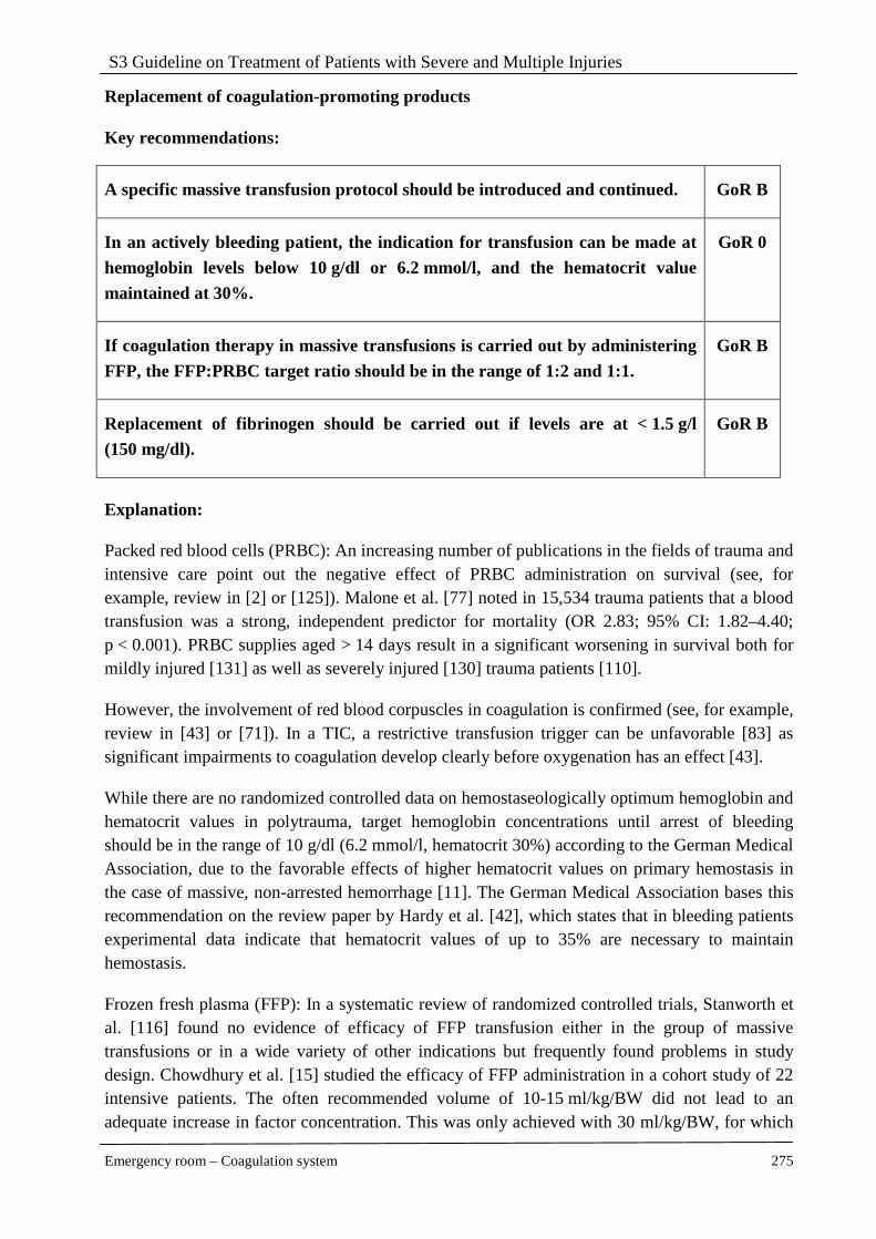
S3 Guideline on Treatment of Patients with Severe and Multiple Injuries
Emergency room – Coagulation system 275
Replacement of coagulation-promoting products
Key recommendations:
A specific massive transfusion protocol should be introduced and continued. GoR B
In an actively bleeding patient, the indication for transfusion can be made at hemoglobin levels below 10 g/dl or 6.2 mmol/l, and the hematocrit value maintained at 30%.
GoR 0
If coagulation therapy in massive transfusions is carried out by administering FFP, the FFP:PRBC target ratio should be in the range of 1:2 and 1:1.
GoR B
Replacement of fibrinogen should be carried out if levels are at < 1.5 g/l (150 mg/dl).
GoR B
Explanation:
Packed red blood cells (PRBC): An increasing number of publications in the fields of trauma and intensive care point out the negative effect of PRBC administration on survival (see, for example, review in [2] or [125]). Malone et al. [77] noted in 15,534 trauma patients that a blood transfusion was a strong, independent predictor for mortality (OR 2.83; 95% CI: 1.82–4.40; p < 0.001). PRBC supplies aged > 14 days result in a significant worsening in survival both for mildly injured [131] as well as severely injured [130] trauma patients [110].
However, the involvement of red blood corpuscles in coagulation is confirmed (see, for example, review in [43] or [71]). In a TIC, a restrictive transfusion trigger can be unfavorable [83] as significant impairments to coagulation develop clearly before oxygenation has an effect [43].
While there are no randomized controlled data on hemostaseologically optimum hemoglobin and hematocrit values in polytrauma, target hemoglobin concentrations until arrest of bleeding should be in the range of 10 g/dl (6.2 mmol/l, hematocrit 30%) according to the German Medical Association, due to the favorable effects of higher hematocrit values on primary hemostasis in the case of massive, non-arrested hemorrhage [11]. The German Medical Association bases this recommendation on the review paper by Hardy et al. [42], which states that in bleeding patients experimental data indicate that hematocrit values of up to 35% are necessary to maintain hemostasis.
Frozen fresh plasma (FFP): In a systematic review of randomized controlled trials, Stanworth et al. [116] found no evidence of efficacy of FFP transfusion either in the group of massive transfusions or in a wide variety of other indications but frequently found problems in study design. Chowdhury et al. [15] studied the efficacy of FFP administration in a cohort study of 22 intensive patients. The often recommended volume of 10-15 ml/kg/BW did not lead to an adequate increase in factor concentration. This was only achieved with 30 ml/kg/BW, for which
S3 Guideline on Treatment of Patients with Severe and Multiple Injuries
Emergency room – Coagulation system 276
the patients required a median volume of 2.5 l FFP. A randomized double-blind trial of 90 patients with severe, closed traumatic brain injury (GCS ≤ 8) was carried out by Etemadrezaie et al. [29]. One group received a slow transfusion 10–15 ml/kg BW FFP while the control group received the same amount of common salt. In the FFP group, a new intracerebral hematoma developed more frequently (p = 0.012), and the patients had a significantly elevated mortality rate (63% versus 35%, p = 0.006). Hedim et al. [44] achieved a plasma dilution of 21% for 2.9 hours with an infusion of only 10 ml/kg BW FFP.
The transfusion of FFP also contains a series of risks: Dara et al. [20] noted a more frequent occurrence of acute lung injuries (ALI; 18% versus 4%, p = 0.021) after FFP transfusion in patients in the medical intensive care unit. Sperry et al. [109] found prospectively in 415 patients approximately double the TRALI (transfusion-associated acute lung insufficiency) risk with a ratio of FFP:PRBC > 1:1.5 (47.1% versus 24.0%, p = 0.001). Sarani et al. [99] showed after FFP transfusion in non-traumatized patients in the surgical intensive care unit a relative risk for the occurrence of an infection of 2.99, severe, ventilator-associated pneumonia of 5.42, severe bacteriemia of 3.35, and sepsis of 3.2 (each p < 001). There was a cumulative risk of infection of ~ 4% per FFP. Chaiwat et al. [14] identified the transfusion of FFP in trauma as an independent predictor for ARDS in 14,070 trauma patients: relative risk after transfusion of 1-5 FFP of 1.66 (95% CI: 0.88–3.15) and at > 5 FFP of 2.55 (95% CI: 1.17–5.55).
The German Medical Association regards the prevention and treatment of microvascular bleeding as an indication for FFP but described the treatment of coagulopathy with FFP alone as of little efficacy (increased rate, volume loading) [11]. It recommends rapid transfusion of initially 20 ml/kg BW but emphasizes that higher volumes may be necessary. There are no controlled studies to determine effective plasma doses [11].
FFP ratio to PRBC: The military field provided the first evidence that the replacement of lost blood volume by “full blood” can provide a survival advantage in critically injured patients with massive transfusions (> 10 PRBC/24 h) [93]. However, full blood is not available in the Federal Republic of Germany.
Hirshberg et al. [51] showed the necessity of early FFP replacement on a computer model and they recommended an FFP:PRBC of 1:1.5 or the transfusion of 2 FFP with the first PRBC. Ho et al. [52] calculated on a mathematical model that a bleeding-induced heavy loss of coagulation factors can only be remedied with a transfusion of 1-1.5 FFP per PRBC; if the FFP administration starts before the factor concentration has dropped below 50%, then a 1:1 ratio is sufficient. Borgmann et al. [6] showed the advantage of a 1:1 replacement of FFP and PRBC in military personnel. A retrospective study was carried out on the survival of 246 soldiers who had received replacement in a ratio of 1:8, 1:2.5 or 1:1.4. The hemorrhage-induced case fatality rates were 92.5%, 78%, and 37% (p < 0001). In the regression analysis, the FFP:PRBC ratio was linked independently with survival and discharge from hospital (OR 8.6; 95% CI: 2.1–35.2). Dente et al. [21] prospectively compared 73 civil patients after introducing a massive transfusion protocol containing PRBC, FPP, and platelets in the ratio 1:1:1 with 84 patients before introducing the protocol. The results showed a drastic reduction in early coagulopathy (p = 0.023), 24-hour case fatality rate (17% versus 36%, p = 0.008), and 30-day case fatality rate for blunt trauma (34% versus 55%, p = 0.04). In 135 patients, Duchesne et al. [24] proved a
S3 Guideline on Treatment of Patients with Severe and Multiple Injuries
Emergency room – Coagulation system 277
significant survival advantage with an FFP-PRBC ratio of 1:1 compared to 1:4 (26% versus 87.5%, p = 0.0001). Gonzalez et al. [39] compared the results of their massive transfusion protocol on 97 patients for the emergency room (PRBC:FFP > 2:1) with those of the ICU protocol (PRBC:FFP = 1:1). A TIC present on arrival in the emergency room could not be remedied until in the ICU, and the TIC at ICU admission correlated with the mortality rate (p = 0.02). A significantly reduced 30-day mortality was noted by Gunter et al. [41] for patients who received FFP and PRBC in a ratio ≥ 2:3 (41% versus 62%, p = 0.008). In a multicenter study of 466 patients, Holcomb et al. [54] showed that an FFP-PRBC ratio ≥ 1:2 compared to < 1:2 permitted better 30-day survival (40.4% versus 59.9%, p < 001). The combination of a high FFP-PRBC and a high platelet-PRBC ratio increased the 6-hour, 24-hour, and 30-day survival (p < 005). Kashuk et al. [60] showed the advantage of a 1:2 FFP-PRBC ratio (survivors: median 1:2, non-survivors: median 1:4, p < 0001) in 133 civil patients who received a massive transfusion within 6 hours. The authors nevertheless found a U-shaped curve: patients who received FFP and PRBC in the ratio 1:2 to 1:3 had the highest survival rate whereas the predicted mortality probability increased again with a ratio of 1:1. The authors thus recommended a ratio of 1:2 to 1:3. In 713 patients in the German trauma registry who required ≥ 10 PRBC until ICU admission, Maegele et al. [75] detected a survival advantage in relation to the PRBC-FFP ratio of > 1:1, 1:1 or < 1:1 (6 hours: 24.6% versus 9.6% versus 3.5%, p < 00001; 24 hours: 32.6% versus 16.7% versus 11.3%, p < 00001; 30 days: 45.5% versus 35.1% versus 24.3%, p < 0001). The ratio < 1:1 led to a longer ventilation dependency and a longer stay in ICU (p < 00005). In 415 patients with an FFP-PRBC ratio of > 1:1.5, Sperry et al. [109] found prospectively a significant 24-hour survival advantage (3.9% versus 12.8%, p = 0.012) with increased TRALI risk (47.1% versus 24.0%, p = 0.001). Scalea et al. [100] prospectively studied 250 patients. No survival advantage with a PRBC-FFP ratio of 1:1 was registered either in the total population or in the 81 massively transfused patients. With a total mortality within 24 hours of only 4% (6% with massive transfusions), the authors concluded that other (less severely injured) patients had been studied than in the majority of other studies. In 383 trauma patients (exclusion: severe TBI), Teixeira et al. [121] showed a linear increase in survival with increasing FFP-PRBC ratio up to a ratio of 1:3. After the admission GCS, the FFP-PRBC ratio was the second most important predictor for survival. Snyder et al. [105] retrospectively carried out an outcome study on 134 patients who required transfusion ≥ 10 PRBC/24 h. They noted a significantly reduced 24-hour mortality of 40% when FFP and PRBC were administered in a ratio ≥ 1:2 (median 1:1.3) compared to 58% at < 1:2 (median 1:3.7) (relative risk [RR] 0.37, 95% CI: 0.22–0.64). However, the significance could no longer be confirmed if the exact time of FFP transfusion within the first 24 hours was taken into account (RR 0.84, 95% CI: 0.47–1.50). The authors justified this with a potential “survival bias”: the patients did not die because they received less FFP but received less FFP because they died. In a randomized multicenter study, by means of a high FFP-PRBC ratio, Zink et al. [138] significantly improved survival in massive transfusions within 6 hours (37.3% at < 1:4 versus 15.2% at 1:4 to 1:1 versus 2.0% at ≥ 1:1; p < 0001) and lowered the total number of required PRBC (18 PRBC in the first 24 hours at < 1:1 versus 13 PRBC at > 1:1, p < 0001).
The majority of studies available indicate that patients who (will) require massive transfusions or have a life-threatening shock gain from a high FFP-PRBC ratio [111].
S3 Guideline on Treatment of Patients with Severe and Multiple Injuries
Emergency room – Coagulation system 278
Massive transfusion protocol: The term “massive transfusion” mostly implies the transfusion of ≥ 10 PRBC per 24 hours [111]; but as the highest mortality of multiply injured patients occurs within the first 6 hours, some authors also recommend ≥ 10 PRBC per 6 hours [60]. In Cotton et al. [19], the introduction of a massive transfusion protocol with an FFP-PRBC ratio of 1:1.5 led to a 74% fall in mortality probability (p = 0.001) and to a higher 30-day survival rate (56.8% versus 37.6%, p < 0001) [18] with shorter stay (12 days versus 16 days, p = 0.049) and fewer ventilation days (5.7 days versus 8.2 days, p = 0.017). The authors attributed the improved survival to the earlier and faster infusion of the increased FFP-PRBC ratio. After introducing a massive transfusion protocol, Dente et al. [21] found evidence of an improved 24-hour survival (previously 36% versus 14%, p = 0.008) and 30-day survival (previously 55% versus 34%, p = 0.04) and a lower early mortality due to the coagulopathy (previously 21/31 versus 4/13, p = 0.023).
The Trauma Associated Severe Hemorrhage (TASH) Score of the German DGU Trauma Registry [136] can be used in the civil arena for predicting a massive transfusion. It comprises the factors systolic blood pressure, hemoglobin (Hb), BE, heart rate, free intraabdominal fluid, pelvic and femoral fracture, and male sex (0-28 points). An increasing TASH score could be attributed with good accuracy and discrimination to an increasing probability for a massive transfusion (area under the curve [AUC] 0.89). Nunez et al. [88] developed a prediction system for massive transfusions [assessment of blood consumption (ABC)] with the parameters penetrating injury, positive finding from focused trauma sonography (FAST), systolic blood pressure on arrival ≤ 90 mmHg and heart rate ≥ 120/min. For a value ≥ 2, they could attribute a sensitivity of 75% and a specificity of 86% to this score.
Platelet concentrates (PC): In the case of acute loss, platelets are initially increasingly released from bone marrow and spleen, which is why there is quite a delay before the platelet count falls after bleeding starts. After transfusion, the transferred vital platelets are distributed in the blood and the spleen so that the recovery rate in the peripheral blood is only about 60-70% and even lower with DIC [11].
In their retrospective multicenter study of civil trauma patients, Holcomb et al. [54] showed an improved 30-day survival with a PC-PRBC ratio of ≥ 1:2 (40.1% versus 59.9%, p < 001). The PC:PRBC ratio was an independent predictor for mortality. Perkins et al. [90] studied the effect on 694 soldiers who received massive transfusions of apharesis platelet concentrate (aPC) in relation to PRBC in 3 groups (aPC:PRBC 1:16, 1:18 to < 1:8 and > 1:8). The transfusion of aPC and PRBC in a ratio > 1:8 was characterized by a significantly higher 24-hour (64% versus 87% versus 95%, p < 0001) and 30-day survival (43% versus 60% versus 75%, p < 0001). In the multivariate analysis, the aPC-PRBC ratio was independently linked with the 24-hour and 30-day survival. In a PRBC-FFP platelet ratio of 1:1:1, Dente et al. [21] noted a drastic reduction in early coagulopathy (p = 0.023), 24-hour case fatality rate (17% versus 36%, p = 0.008) and 30-day case fatality rate in blunt trauma (34% versus 55%, p = 0.04). The randomized multicenter study by Zink et al. [138] with massive transfusion within 6 hours yielded significantly better values for the high ratio (6-hour survival: 22.8% versus 19% versus 3.2%, p < 0002; hospital survival: 43.7% versus 46.8% versus 27.4%, p < 003) for the 3 groups with a PC-PRBC ratio of < 1:4, 1:4 to 1:1 and ≥ 1:1.

S3 Guideline on Treatment of Patients with Severe and Multiple Injuries
Emergency room – Coagulation system 279
If the patient is in acute danger due to a massive blood loss or due to the location (intracerebral bleeding), the German Medical Association recommends platelet replacement if the value falls below 100,000/µl [11]. If there are resulting platelet dysfunctions and tendency to bleed, a concomitant therapy with antifibrinolytics or desmopressin can be indicated [11].
Fibrinogen: As the substrate of coagulation, factor I (= fibrinogen) is essential not only for the formation of the fibrin network but is also a ligand for the GPIIb-IIIa receptor at the platelet surface and thus responsible for platelet aggregation. During dilution or severe bleeding, fibrinogen appears to be the most vulnerable of all coagulation factors and is the first to reach its critical concentration [11, 50]. As early as 1995, Hiippala [50] had confirmed in patients who received a colloid transfusion that the measurement of derived fibrinogen (measured using the Quick test) as well as fibrinogen measured using the method according to Clauss produces significantly higher values than correspond to the actual fibrinogen levels. According to the German Medical Association, the administration of 3 g fibrinogen in a volume of 3 liters of plasma elevates the measured fibrinogen concentration by approximately 1 g/l; initial doses of (2–) 4 (–6) g are thus necessary in adults [11].
Singbartl et al. [104] illustrated in a mathematical model that the maximum permissible blood loss until the critical fibrinogen value has been reached is dependent on the baseline value. However, this is generally not known in the initial management during emergency admission. Fries et al. [35] showed in an in vitro study that dilution occurs through infusion of crystalloid or colloid solutions, inter alia also through 6% HES 130/0.4. Giving fibrinogen in a concentration which when converted was roughly equivalent to 3 g/70 kg BW was sufficient to achieve an improvement in the RoTEM parameters. The cause of this effect is regarded as an effect of the interaction between thrombin, factor XIII, and fibrinogen; this particularly applies for a dilution through HES [86]. Madjdpour et al. [73] proved in the pig that, with varying molecular weight (900, 500, 130) and the same degree of replacement (0.42), HES impairs coagulation in a similar way. In thrombocytopenia induced in the pig model, Velik-Salchner et al. [128] normalized the impaired coagulation parameters in the TEG by administering fibrinogen. Mittermayr et al. [85] proved in 61 randomized patients with major spinal interventions that colloids reduce the speed and quality of clot formation through impaired fibrin polymerization. Stinger et al. [119] showed a highly significant correlation between the amount of fibrinogen administered and survival in 252 patients who had been injured during the fighting in the Iraq war. Injured patients who received less than 1 g of fibrinogen per 5 PRBC showed a case fatality rate of 52% compared to 24% if more than 1 g/5 PRBC was given. In establishing a concentration of < 2 g/l as indication for fibrinogen replacement (average 2 g, range 1-5 g), Fenger-Eriksen et al. [31] found evidence in 35 severely bleeding patients of a significant reduction in the required PRBC (p < 00001), FFP (p < 00001) and PC (p < 0001) and blood loss (p < 005). In 30 massively bleeding patients with hypofibrinogenemia of varying genesis, Weinkove et al. [132] raised the level of 0.65 to 2.01 g/l by giving 4 g of fibrinogen (median, range 2-14 g) (measurement according to Clauss). Farriols Danes et al. [30] showed that patients with acute, bleeding-induced hypofibrinogenemia (compared to chronic deficiency) reacted to fibrinogen replacement with a more marked increase and a significantly better 7-day survival rate (p = 0.014).
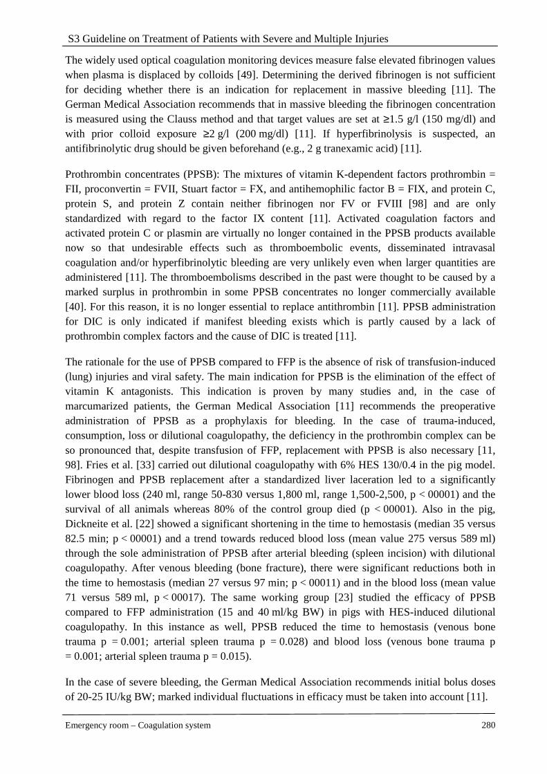
S3 Guideline on Treatment of Patients with Severe and Multiple Injuries
Emergency room – Coagulation system 280
The widely used optical coagulation monitoring devices measure false elevated fibrinogen values when plasma is displaced by colloids [49]. Determining the derived fibrinogen is not sufficient for deciding whether there is an indication for replacement in massive bleeding [11]. The German Medical Association recommends that in massive bleeding the fibrinogen concentration is measured using the Clauss method and that target values are set at ≥1.5 g/l (150 mg/dl) and with prior colloid exposure ≥2 g/l (200 mg/dl) [11]. If hyperfibrinolysis is suspected, an antifibrinolytic drug should be given beforehand (e.g., 2 g tranexamic acid) [11].
Prothrombin concentrates (PPSB): The mixtures of vitamin K-dependent factors prothrombin = FII, proconvertin = FVII, Stuart factor = FX, and antihemophilic factor B = FIX, and protein C, protein S, and protein Z contain neither fibrinogen nor FV or FVIII [98] and are only standardized with regard to the factor IX content [11]. Activated coagulation factors and activated protein C or plasmin are virtually no longer contained in the PPSB products available now so that undesirable effects such as thromboembolic events, disseminated intravasal coagulation and/or hyperfibrinolytic bleeding are very unlikely even when larger quantities are administered [11]. The thromboembolisms described in the past were thought to be caused by a marked surplus in prothrombin in some PPSB concentrates no longer commercially available [40]. For this reason, it is no longer essential to replace antithrombin [11]. PPSB administration for DIC is only indicated if manifest bleeding exists which is partly caused by a lack of prothrombin complex factors and the cause of DIC is treated [11].
The rationale for the use of PPSB compared to FFP is the absence of risk of transfusion-induced (lung) injuries and viral safety. The main indication for PPSB is the elimination of the effect of vitamin K antagonists. This indication is proven by many studies and, in the case of marcumarized patients, the German Medical Association [11] recommends the preoperative administration of PPSB as a prophylaxis for bleeding. In the case of trauma-induced, consumption, loss or dilutional coagulopathy, the deficiency in the prothrombin complex can be so pronounced that, despite transfusion of FFP, replacement with PPSB is also necessary [11, 98]. Fries et al. [33] carried out dilutional coagulopathy with 6% HES 130/0.4 in the pig model. Fibrinogen and PPSB replacement after a standardized liver laceration led to a significantly lower blood loss (240 ml, range 50-830 versus 1,800 ml, range 1,500-2,500, p < 00001) and the survival of all animals whereas 80% of the control group died (p < 00001). Also in the pig, Dickneite et al. [22] showed a significant shortening in the time to hemostasis (median 35 versus 82.5 min; p < 00001) and a trend towards reduced blood loss (mean value 275 versus 589 ml) through the sole administration of PPSB after arterial bleeding (spleen incision) with dilutional coagulopathy. After venous bleeding (bone fracture), there were significant reductions both in the time to hemostasis (median 27 versus 97 min; p < 00011) and in the blood loss (mean value 71 versus 589 ml, p < 00017). The same working group [23] studied the efficacy of PPSB compared to FFP administration (15 and 40 ml/kg BW) in pigs with HES-induced dilutional coagulopathy. In this instance as well, PPSB reduced the time to hemostasis (venous bone trauma p = 0.001; arterial spleen trauma p = 0.028) and blood loss (venous bone trauma p = 0.001; arterial spleen trauma p = 0.015).
In the case of severe bleeding, the German Medical Association recommends initial bolus doses of 20-25 IU/kg BW; marked individual fluctuations in efficacy must be taken into account [11].

S3 Guideline on Treatment of Patients with Severe and Multiple Injuries
Emergency room – Coagulation system 281
Antifibrinolytic drugs: Hyperfibrinolysis now appears to be more frequently assumed than before in multiply injured patients (~ 15% in [102]) and the extent correlates with the injury severity [67]. Hyperfibrinolysis is most common in patients with chest trauma, blunt abdominal trauma, pelvic trauma, and traumatic brain injury [58]. A prompt diagnosis of hyperfibrinolysis and also of the effectiveness of antifibrinolytic treatment is only possible by means of thrombelastography [67, 101]. The administration of the antifibrinolytic drug must be part of an overall therapeutic plan for the treatment of coagulopathies because hyperfibrinolysis can often involve heavy consumption of fibrinogen or even complete defibrination of the patient [58]. This fibrinogen deficiency must then be appropriately balanced after the manifestation of hyperfibrinolysis [58], i.e. the antifibrinolytic drug should be administered before the fibrinogen if hyperfibrinolysis is suspected [11]. Tranexamic acid is a synthetic lysine analogue, which inhibits the conversion of plasminogen into plasmin by blocking the plasminogen from binding to the fibrin molecule. The onset of effect of tranexamic acid is delayed compared to aprotinin as free plasmin continues to be active [102]. With a lack of evidence for multiply injured patients, it is recommended that initially 2–4 g (10–30 mg/kg BW [36, 58] is administered or a bolus dose of 10–15 mg/kg BW followed by 1–5 mg/kg BW/h [106].
Due to a lack of evidence-based data, a Cochrane Review from 2004 [17] could neither confirm nor refute the administration of antifibrinolytics in trauma patients. Another Cochrane Review from 2007 on the question of reducing perioperative blood transfusions through antifibrinolytic drugs [45] established for tranexamic acid a relative risk for PRBC administration of 0.61 (95% CI: 0.54-0.70) and a trend towards fewer re-operations. Tranexamic acid reduced the necessity of a blood transfusion relatively by 43% (RR 0.57; 95% CI: 0.49–0.66). A cumulative occurrence of serious side effects was not noted. A study published in The Lancet in 2010 (CRASH [Clinical Randomization of Antifibrinolytics in Significant Hemorrhage]-2 Study) [16] supports this conclusion: 1 g of tranexamic acid in 10 minutes + 1 g over 8 hours led to a significant reduction in all-cause mortality and in bleeding-induced mortality without an increased rate in thromboembolisms.
Desmopressin (DDAVP): Desmopressin (1-deamino-8-D-arginine vasopressin) is a synthetic vasopressin analogue. Desmopressin (e.g., Minirin) effectuates nonspecific platelet activation (increased expression of the platelet GpIb receptor [89]), releases the von Willebrand factor and FVIII from the endothelium of the hepatic sinusoids, and thus improves primary hemostasis [32]. The main indication lies in perioperative treatment of von Willebrand syndrome. DDAVP also shows good efficacy in patients after heparin administration and with restricted platelet function due to taking aspirin (acetyl salicylic acid) or ADP receptor antagonists/thienopyridine derivatives with uremia and with liver disease or thrombocytopenia [32]. The maximum effect after i.v. administration occurs only after about 90 minutes [68]. With repeated administration, the tissue plasminogen activator (tPA) can be released, thereby possibly leading to hyperfibrinolysis. Thus, with repeated administration, some authors recommend simultaneous administration of tranexamic acid [68]. In cardio-surgical patients, a not significantly increased rate in heart attacks was confirmed after DDAVP (OR 2.07; 95% CI: 0.74–5.85; p = 0.19) [68]. Ying et al. [135] corrected in vitro the hypothermia-induced dysfunctions in hemostasis at least partially through DDAVP. Whereas the Cochrane Review of 18 studies with 1,295 patients by Carless et al. [13] could not confirm any efficacy for the prophylactic administration of
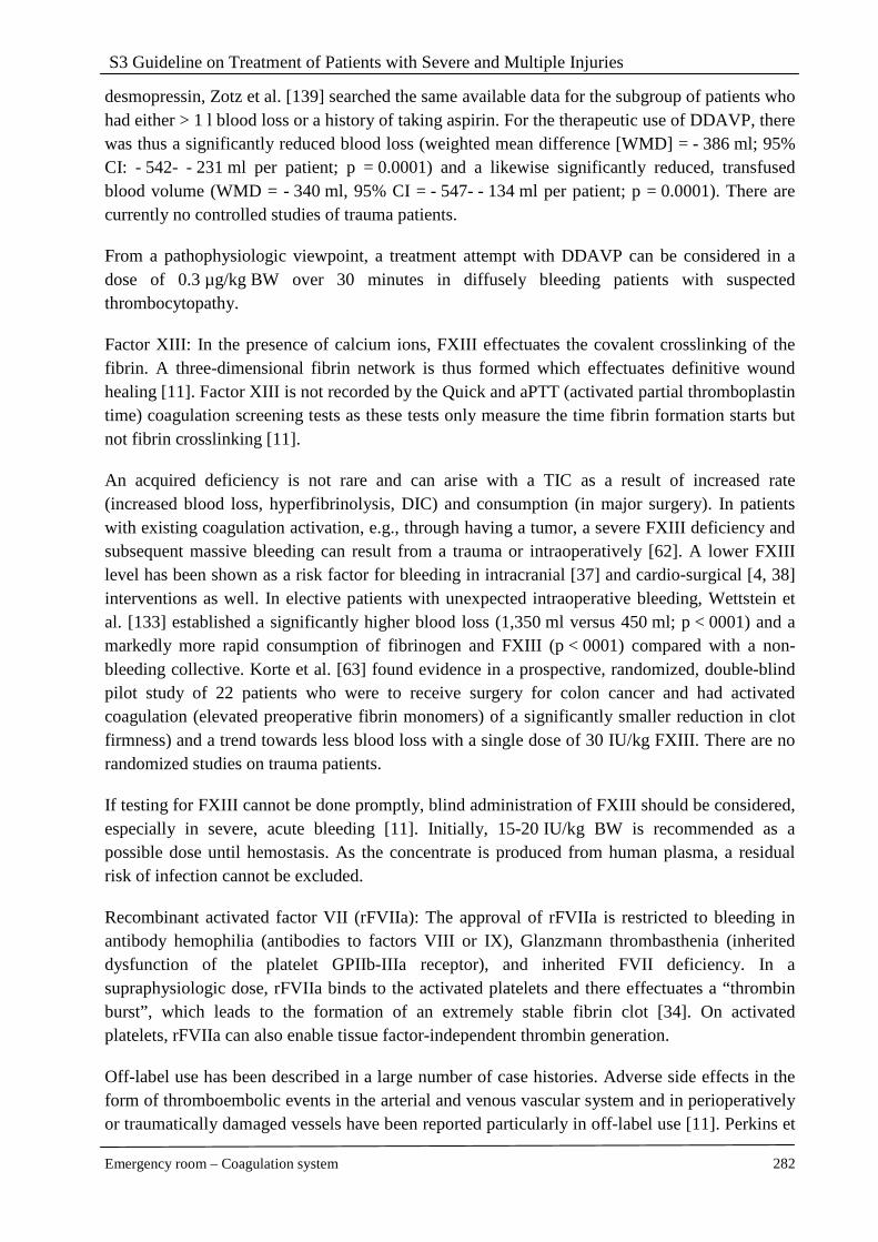
S3 Guideline on Treatment of Patients with Severe and Multiple Injuries
Emergency room – Coagulation system 282
desmopressin, Zotz et al. [139] searched the same available data for the subgroup of patients who had either > 1 l blood loss or a history of taking aspirin. For the therapeutic use of DDAVP, there was thus a significantly reduced blood loss (weighted mean difference [WMD] = - 386 ml; 95% CI: - 542- - 231 ml per patient; p = 0.0001) and a likewise significantly reduced, transfused blood volume (WMD = - 340 ml, 95% CI = - 547- - 134 ml per patient; p = 0.0001). There are currently no controlled studies of trauma patients.
From a pathophysiologic viewpoint, a treatment attempt with DDAVP can be considered in a dose of 0.3 µg/kg BW over 30 minutes in diffusely bleeding patients with suspected thrombocytopathy.
Factor XIII: In the presence of calcium ions, FXIII effectuates the covalent crosslinking of the fibrin. A three-dimensional fibrin network is thus formed which effectuates definitive wound healing [11]. Factor XIII is not recorded by the Quick and aPTT (activated partial thromboplastin time) coagulation screening tests as these tests only measure the time fibrin formation starts but not fibrin crosslinking [11].
An acquired deficiency is not rare and can arise with a TIC as a result of increased rate (increased blood loss, hyperfibrinolysis, DIC) and consumption (in major surgery). In patients with existing coagulation activation, e.g., through having a tumor, a severe FXIII deficiency and subsequent massive bleeding can result from a trauma or intraoperatively [62]. A lower FXIII level has been shown as a risk factor for bleeding in intracranial [37] and cardio-surgical [4, 38] interventions as well. In elective patients with unexpected intraoperative bleeding, Wettstein et al. [133] established a significantly higher blood loss (1,350 ml versus 450 ml; p < 0001) and a markedly more rapid consumption of fibrinogen and FXIII (p < 0001) compared with a non-bleeding collective. Korte et al. [63] found evidence in a prospective, randomized, double-blind pilot study of 22 patients who were to receive surgery for colon cancer and had activated coagulation (elevated preoperative fibrin monomers) of a significantly smaller reduction in clot firmness) and a trend towards less blood loss with a single dose of 30 IU/kg FXIII. There are no randomized studies on trauma patients.
If testing for FXIII cannot be done promptly, blind administration of FXIII should be considered, especially in severe, acute bleeding [11]. Initially, 15-20 IU/kg BW is recommended as a possible dose until hemostasis. As the concentrate is produced from human plasma, a residual risk of infection cannot be excluded.
Recombinant activated factor VII (rFVIIa): The approval of rFVIIa is restricted to bleeding in antibody hemophilia (antibodies to factors VIII or IX), Glanzmann thrombasthenia (inherited dysfunction of the platelet GPIIb-IIIa receptor), and inherited FVII deficiency. In a supraphysiologic dose, rFVIIa binds to the activated platelets and there effectuates a “thrombin burst”, which leads to the formation of an extremely stable fibrin clot [34]. On activated platelets, rFVIIa can also enable tissue factor-independent thrombin generation.
Off-label use has been described in a large number of case histories. Adverse side effects in the form of thromboembolic events in the arterial and venous vascular system and in perioperatively or traumatically damaged vessels have been reported particularly in off-label use [11]. Perkins et

S3 Guideline on Treatment of Patients with Severe and Multiple Injuries
Emergency room – Coagulation system 283
al. [91] showed in soldiers from the Iraq war that early administration of rFVIIa could lower PRBC consumption by 20%. Of 365 massive transfused patients, 117 received rFVIIa. Likewise, in patients in the military who received massive transfusions, Spinella et al. [113] noted a reduced 24-hour (14% versus 35%, p = 0.01) and 30-day mortality rate (31% versus 51%, p = 0.03) in early (median 2 hours after admission and 2.5 hours after trauma) administration of rFVIIa. However, the logistic options for preparation of, for example, blood components under war conditions cannot be compared with those of a European hospital. In the study in 2005 by Boffard et al. [5], the efficacy of 400 µg/kg BW rFVIIa (after the 8th PRBC initially 200 µg/kg BW, then 100 µg/kg BW each after 1 and 3 hours) as adjunctive therapy was tested compared to placebo in 143 blunt and 134 penetrating trauma injuries. In blunt trauma, there was a significant reduction in the number of transfused PRBCs (calculated reduction by 2.6 PRBC, p = 0.02) and in the necessity for a transfusion of ≥ 20 PRBC (14% versus 33%; p = 0.03). In penetrating injuries, there was a trend in this direction for both parameters. Neither a lowering in the mortality rate nor an accumulation of thromboembolic side effects was observed. In 2009, Stein et al. [117] posed the question of costs. With the same rate of mortality and side effects, the authors could establish no significant difference in the costs (mean value US$63,403 conventionally versus US$66,086 in rFVIIa) in 179 patients with traumatic brain injury. For the 110 patients who were placed in the intensive care unit, there was even a significant cost reduction through rFVIIa (mean value US$108,900 conventionally versus US$77,907 in rFVIIa). However, really low doses were used in this study (5.9–115 µg/kg BW; mean value 41.9 ± 35.5 µg/kg BW, median 25.1 µg/kg BW). Several meta-analyses of RCTs have studied the efficacy of off-label use: Stanworth et al. [115] found 13 placebo-controlled, double-blind RCTs on the use of rFVIIa in patients who were not hemophiliac. In prophylactic use (n = 724, 379 received rFVIIa), there was a trend towards reduced transfusion frequency (pooled RR 0.85; 95% CI: 0.72-1.01), which contradicts a trend towards increased thromboembolisms (pooled RR 1.25; 95% CI: 0.76–2.07). In therapeutic use (n = 1,214; 687 received rFVIIa), there was a trend towards reduced mortality (pooled RR 0.82; 95% CI: 0.64-1.04) and again a trend towards increased thromboembolisms (RR 1.50; 95% CI: 0.86–2.62). In 2008, Hsia et al. [56] published the results of 22 RCTs on bleeding of different genesis in 3,184 non-hemophiliac patients (only the study by Boffard et al. [5] contains 301 trauma patients). The result was a reduced need for transfusion (OR 0.54; 95% CI: 0.34–0.86), a trend towards reduced case fatality rate (OR 0.88; 95% CI: 0.71–1.09) and no change in venous but a trend towards cumulative arterial thromboembolisms (OR 1.50; 95% CI: 0.93–2.41). In 2008, Duchesne et al. [25] examined 19 studies of trauma patients: Based on Boffard et al. [5], the authors gave a Level 1 recommendation for the use of rFVIIa in blunt trauma. A Level 2 recommendation was given for trauma-associated hemorrhaging with 400 µg/kg BW (lower dose could also be effective) if other treatment options failed. Early use was assessed as Level 3. In 2009, Nishijima et al. [87] only found the above-mentioned study by Boffard [5] on the question of rFVIIa use in the emergency room. A large international phase III study on the use of rFVIIa in trauma patients was recently discontinued as the planned lowering in mortality could not be achieved [111].
The effectiveness of rFVIIa and of the coagulation sequences thus indicated is linked to a series of “framework conditions”, which should be attained before administration: fibrinogen value ≥ 1 g/dl, Hb ≥ 7 g/dl, platelet count ≥ 50,000 (preferably ≥ 100,000)/µl), ionized calcium ≥ 0.9 mmol/l, core temperature ≥ 34 °C, pH value ≥ 7.2, and the exclusion of hyperfibrinolysis
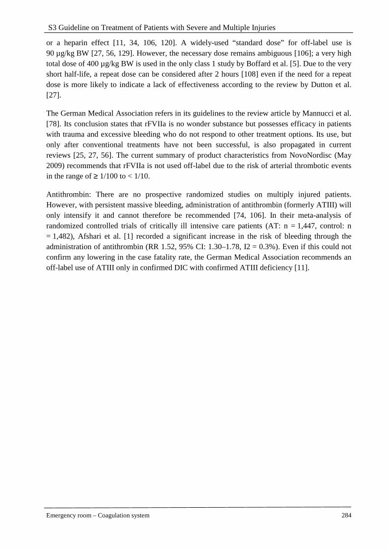
S3 Guideline on Treatment of Patients with Severe and Multiple Injuries
Emergency room – Coagulation system 284
or a heparin effect [11, 34, 106, 120]. A widely-used “standard dose” for off-label use is 90 µg/kg BW [27, 56, 129]. However, the necessary dose remains ambiguous [106]; a very high total dose of 400 µg/kg BW is used in the only class 1 study by Boffard et al. [5]. Due to the very short half-life, a repeat dose can be considered after 2 hours [108] even if the need for a repeat dose is more likely to indicate a lack of effectiveness according to the review by Dutton et al. [27].
The German Medical Association refers in its guidelines to the review article by Mannucci et al. [78]. Its conclusion states that rFVIIa is no wonder substance but possesses efficacy in patients with trauma and excessive bleeding who do not respond to other treatment options. Its use, but only after conventional treatments have not been successful, is also propagated in current reviews [25, 27, 56]. The current summary of product characteristics from NovoNordisc (May 2009) recommends that rFVIIa is not used off-label due to the risk of arterial thrombotic events in the range of ≥ 1/100 to < 1/10.
Antithrombin: There are no prospective randomized studies on multiply injured patients. However, with persistent massive bleeding, administration of antithrombin (formerly ATIII) will only intensify it and cannot therefore be recommended [74, 106]. In their meta-analysis of randomized controlled trials of critically ill intensive care patients (AT: n = 1,447, control: n = 1,482), Afshari et al. [1] recorded a significant increase in the risk of bleeding through the administration of antithrombin (RR 1.52, 95% CI: 1.30–1.78, I2 = 0.3%). Even if this could not confirm any lowering in the case fatality rate, the German Medical Association recommends an off-label use of ATIII only in confirmed DIC with confirmed ATIII deficiency [11].
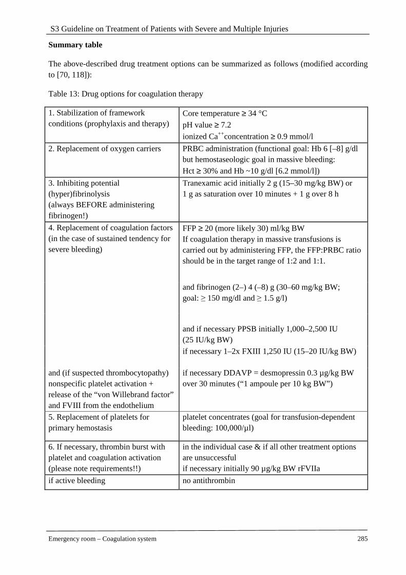
S3 Guideline on Treatment of Patients with Severe and Multiple Injuries
Emergency room – Coagulation system 285
Summary table
The above-described drug treatment options can be summarized as follows (modified according to [70, 118]):
Table 13: Drug options for coagulation therapy
1. Stabilization of framework conditions (prophylaxis and therapy)
Core temperature ≥ 34 °C
pH value ≥ 7.2
ionized Ca++concentration ≥ 0.9 mmol/l
2. Replacement of oxygen carriers PRBC administration (functional goal: Hb 6 [–8] g/dl but hemostaseologic goal in massive bleeding:
Hct ≥ 30% and Hb ~10 g/dl [6.2 mmol/l])
3. Inhibiting potential (hyper)fibrinolysis (always BEFORE administering fibrinogen!)
Tranexamic acid initially 2 g (15–30 mg/kg BW) or 1 g as saturation over 10 minutes + 1 g over 8 h
FFP ≥ 20 (more likely 30) ml/kg BW If coagulation therapy in massive transfusions is carried out by administering FFP, the FFP:PRBC ratio should be in the target range of 1:2 and 1:1.
and fibrinogen (2–) 4 (–8) g (30–60 mg/kg BW; goal: ≥ 150 mg/dl and ≥ 1.5 g/l)
and if necessary PPSB initially 1,000–2,500 IU (25 IU/kg BW)
4. Replacement of coagulation factors (in the case of sustained tendency for severe bleeding)
if necessary 1–2x FXIII 1,250 IU (15–20 IU/kg BW)
and (if suspected thrombocytopathy) nonspecific platelet activation + release of the “von Willebrand factor” and FVIII from the endothelium
if necessary DDAVP = desmopressin 0.3 µg/kg BW over 30 minutes (“1 ampoule per 10 kg BW”)
5. Replacement of platelets for primary hemostasis
platelet concentrates (goal for transfusion-dependent bleeding: 100,000/µl)
6. If necessary, thrombin burst with platelet and coagulation activation (please note requirements!!)
in the individual case & if all other treatment options are unsuccessful if necessary initially 90 µg/kg BW rFVIIa
if active bleeding no antithrombin
S3 Guideline on Treatment of Patients with Severe and Multiple Injuries
Emergency room – Coagulation system 286
References
1. Afshari A, Wetterslev J, Brok J et al. (2007) Antithrombin III in critically ill patients: systematic review with meta-analysis and trial sequential analysis. BMJ 335: 1248-1251 [LoE 1a]
2. Beekley AC (2008) Damage control resuscitation: a sensible approach to the exsanguinating surgical patient. Crit Care Med 36: S267-S274
3. Bickell WH, Wall MJ, Jr., Pepe PE et al. (1994) Immediate versus delayed fluid resuscitation for hypotensive patients with penetrating torso injuries. N Engl J Med 331: 1105-1109
4. Blome M, Isgro F, Kiessling AH et al. (2005) Relationship between factor XIII activity, fibrinogen, haemostasis screening tests and postoperative bleeding in cardiopulmonary bypass surgery. Thromb Haemost 93: 1101-1107
5. Boffard KD, Riou B, Warren B et al. (2005) Recombinant factor VIIa as adjunctive therapy for bleeding control in severely injured trauma patients: two parallel randomized, placebo-controlled, double-blind clinical trials. J Trauma 59: 8-15 [LoE 1b]
6. Borgman MA, Spinella PC, Perkins JG et al. (2007) The ratio of blood products transfused affects mortality in patients receiving massive transfusions at a combat support hospital. J Trauma 63: 805-813 [LoE 2c]
7. Brohi K, Cohen MJ, Davenport RA (2007) Acute coagulopathy of trauma: mechanism, identification and effect. Curr Opin Crit Care 13: 680-685
8. Brohi K, Cohen MJ, Ganter MT et al. (2007) Acute traumatic coagulopathy: initiated by hypoperfusion: modulated through the protein C pathway? Ann Surg 245: 812-818
9. Brohi K, Cohen MJ, Ganter MT et al. (2008) Acute coagulopathy of trauma: hypoperfusion induces systemic anticoagulation and hyperfibrinolysis. J Trauma 64: 1211-1217 [LoE 2b]
10. Brohi K, Singh J, Heron M et al. (2003) Acute traumatic coagulopathy. J Trauma 54: 1127-1130 [LoE 3b]
11. Bundesärztekammer (BÄK). Querschnitts-Leitlinien (BÄK) zur Therapie mit Blutkomponenten und Plasmaderivaten 2009 - 4.Auflage. Erreichbar unter: http://www.bundesaerztekammer.de/downloads/LeitQuerBlutkomponenten4Aufl.pdf
12. Cannon W, Frawer J, Cowell E (1918) The preventive treatment of wound shock. JAMA 70: 618-621
13. Carless PA, Henry DA, Moxey AJ et al. (2004) Desmopressin for minimising perioperative allogeneic blood transfusion. Cochrane Database Syst Rev CD001884
14. Chaiwat O, Lang JD, Vavilala MS et al. (2009) Early packed red blood cell transfusion and acute respiratory distress syndrome after trauma. Anesthesiology 110: 351-360 [LoE 2b]
15. Chowdhury P, Saayman AG, Paulus U et al. (2004) Efficacy of standard dose and 30 ml/kg fresh frozen plasma in correcting laboratory parameters of haemostasis in critically ill patients. Br J Haematol 125: 69-73 [LoE 4]
16. CRASH-2 trial collaborators, Shakur H, Roberts I, Bautista R, et al. Effects of tranexamic acid on death, vascular occlusive events, and blood transfusion in trauma patients with significant haemorrhage (CRASH-2): a randomised, placebo-controlled trial. Lancet 2010; 376:23-32.
17. Coats T, Roberts I, Shakur H (2004) Antifibrinolytic drugs for acute traumatic injury. Cochrane Database Syst Rev CD004896 [LoE 1a]
18. Cotton BA, Au BK, Nunez TC et al. (2009) Predefined massive transfusion protocols are associated with a reduction in organ failure and postinjury complications. J Trauma 66: 41-48 [LoE 3b]
19. Cotton BA, Gunter OL, Isbell J et al. (2008) Damage control hematology: the impact of a trauma exsanguination protocol on survival and blood product utilization. J Trauma 64: 1177-1182
20. Dara SI, Rana R, Afessa B et al. (2005) Fresh frozen plasma transfusion in critically ill medical patients with coagulopathy. Crit Care Med 33: 2667-2671 [LoE 2b]
21. Dente CJ, Shaz BH, Nicholas JM et al. (2009) Improvements in early mortality and coagulopathy are sustained better in patients with blunt trauma after institution of a massive transfusion protocol in a civilian level I trauma center. J Trauma 66: 1616-1624 [LoE 2c]
22. Dickneite G, Doerr B, Kaspereit F (2008) Characterization of the coagulation deficit in porcine dilutional coagulopathy and substitution with a prothrombin complex concentrate. Anesth Analg 106: 1070-1077 [LoE 5]
23. Dickneite G, Pragst I (2009) Prothrombin complex concentrate vs fresh frozen plasma for reversal of dilutional coagulopathy in a porcine trauma model. Br J Anaesth 102: 345-354 [LoE 5]
24. Duchesne JC, Hunt JP, Wahl G et al. (2008) Review of current blood transfusions strategies in a mature level I trauma center: were we wrong for the last 60 years? J Trauma 65: 272-276 [LoE 3b]
25. Duchesne JC, Mathew KA, Marr AB et al. (2008) Current evidence based guidelines for factor VIIa use in trauma: the good, the bad, and the ugly. Am Surg 74: 1159-1165 [Evidenzbasierte Leitlinie]
26. Dutton RP (2002) Hypotensive resuscitation during active hemorrhage: impact on in-hospital mortality - Leserbrief. J Trauma 52: 1196-1197
27. Dutton RP, Conti BM (2009) The role of recombinant-activated factor VII in bleeding trauma patients. Curr Opin Anaesthesiol 22: 299-304
28. Dutton RP, Mackenzie CF, Scalea TM (2002) Hypotensive resuscitation during active hemorrhage: impact on in-hospital mortality. J Trauma 52: 1141-1146 [LoE 2b]
29. Etemadrezaie H, Baharvahdat H, Shariati Z et al. (2007) The effect of fresh frozen plasma in severe closed head injury. Clin Neurol Neurosurg 109: 166-171 [LoE 1b]

S3 Guideline on Treatment of Patients with Severe and Multiple Injuries
Emergency room – Coagulation system 287
30. Farriols Danés A, Gallur Cuenca L, Rodríguez Bueno S et al. (2008) Efficacy and tolerability of human fibrinogen concentrate administration to patients with acquired fibrinogen deficiency and active or in high-risk severe bleeding. Vox Sang 94: 221-226 [LoE 3b]
31. Fenger-Eriksen C, Lindberg-Larsen M, Christensen AQ et al. (2008) Fibrinogen concentrate substitution therapy in patients with massive haemorrhage and low plasma fibrinogen concentrations. Br J Anaesth 101: 769-773 [LoE 3b]
32. Franchini M (2007) The use of desmopressin as a hemostatic agent: a concise review. Am J Hematol 82: 731-735
33. Fries D, Haas T, Klingler A et al. (2006) Efficacy of fibrinogen and prothrombin complex concentrate used to reverse dilutional coagulopathy--a porcine model. Br J Anaesth 97: 460-467 [LoE 5]
34. Fries D, Haas T, Salchner V et al. (2005) Gerinnungsmanagement beim Polytrauma. Anaesthesist 54: 137-144
35. Fries D, Innerhofer P, Reif C et al. (2006) The effect of fibrinogen substitution on reversal of dilutional coagulopathy: an in vitro model. Anesth Analg 102: 347-351 [LoE 5]
36. Fries D, Innerhofer P, Schobersberger W (2009) Time for changing coagulation management in trauma-related massive bleeding. Curr Opin Anaesthesiol 22: 267-274
37. Gerlach R, Tolle F, Raabe A et al. (2002) Increased risk for postoperative hemorrhage after intracranial surgery in patients with decreased factor XIII activity: implications of a prospective study. Stroke 33: 1618-1623
38. Godje O, Gallmeier U, Schelian M et al. (2006) Coagulation factor XIII reduces postoperative bleeding after coronary surgery with extracorporeal circulation. Thorac Cardiovasc Surg 54: 26-33
39. Gonzalez EA, Moore FA, Holcomb JB et al. (2007) Fresh frozen plasma should be given earlier to patients requiring massive transfusion. J Trauma 62: 112-119 [LoE 2c]
40. Grundman C, Plesker R, Kusch M et al. (2005) Prothrombin overload causes thromboembolic complications in prothrombin complex concentrates: in vitro and in vivo evidence. Thromb Haemost 94: 1338-1339
41. Gunter OL, Jr., Au BK, Isbell JM et al. (2008) Optimizing outcomes in Damage Control resuscitation: identifying blood product ratios associated with improved survival. J Trauma 65: 527-534 [LoE 3b]
42. Hardy JF (2004) Current status of transfusion triggers for red blood cell concentrates. Transfus Apher Sci 31: 55-66
43. Hardy JF, De MP, Samama M (2004) Massive transfusion and coagulopathy: pathophysiology and implications for clinical management. Can J Anaesth 51: 293-310
44. Hedin A, Hahn RG (2005) Volume expansion and plasma protein clearance during intravenous infusion
of 5 % albumin and autologous plasma. Clin Sci (Lond) 108: 217-224 [LoE 4]
45. Henry DA, Carless PA, Moxey AJ et al. (2007) Anti-fibrinolytic use for minimising perioperative allogeneic blood transfusion. Cochrane Database Syst Rev CD001886 [LoE 1a]
46. Hess JR, Brohi K, Dutton RP et al. (2008) The coagulopathy of trauma: a review of mechanisms. J Trauma 65: 748-754
47. Hess JR, Lawson JH (2006) The coagulopathy of trauma versus disseminated intravascular coagulation. J Trauma 60: S12-S19
48. Hess JR, Lindell AL, Stansbury LG et al. (2009) The prevalence of abnormal results of conventional coagulation tests on admission to a trauma center. Transfusion 49: 34-39 [LoE 2b]
49. Hiippala ST (1995) Dextran and hydroxyethyl starch interfere with fibrinogen assays. Blood Coagul Fibrinolysis 6: 743-746
50. Hiippala ST, Myllyla GJ, Vahtera EM (1995) Hemostatic factors and replacement of major blood loss with plasma-poor red cell concentrates. Anesth Analg 81: 360-365
51. Hirshberg A, Dugas M, Banez EI et al. (2003) Minimizing dilutional coagulopathy in exsanguinating hemorrhage: a computer simulation. J Trauma 54: 454-463 [LoE 5]
52. Ho AM, Dion PW, Cheng CA et al. (2005) A mathematical model for fresh frozen plasma transfusion strategies during major trauma resuscitation with ongoing hemorrhage. Can J Surg 48: 470-478 [LoE 5]
53. Holcomb JB (2007) Damage control resuscitation. J Trauma 62: S36-S37
54. Holcomb JB, Wade CE, Michalek JE et al. (2008) Increased plasma and platelet to red blood cell ratios improves outcome in 466 massively transfused civilian trauma patients. Ann Surg 248: 447-458 [LoE 2b]
55. Hoyt DB, Bulger EM, Knudson MM et al. (1994) Death in the operating room: an analysis of a multi-center experience. J Trauma 37: 426-432
56. Hsia CC, Chin-Yee IH, McAlister VC (2008) Use of recombinant activated factor VII in patients without hemophilia: a meta-analysis of randomized control trials. Ann Surg 248: 61-68 [LoE 1a]
57. Jacobs LM (1994) Timing of fluid resuscitation in trauma. N Engl J Med 331: 1153-1154
58. Jambor C, Görlinger K (2007) Einsatz von Antifibrinolytika bei Massivtransfusionen. Anästh Intensivmed 48: S167-S173
59. Jambor C, Heindl B, Spannagl M et al. (2009) Hämostaseologisches Management beim Polytrauma - Stellenwert der patientennahen diagnostischen Methoden. Anästhesiol Intensivmed Notfallmed Schmerzther 44: 200-210
60. Kashuk JL, Moore EE, Johnson JL et al. (2008) Postinjury life threatening coagulopathy: is 1:1 fresh frozen plasma:packed red blood cells the answer? J Trauma 65: 261-270 [LoE 2c]

S3 Guideline on Treatment of Patients with Severe and Multiple Injuries
Emergency room – Coagulation system 288
61. Kashuk JL, Moore EE, Millikan JS et al. (1982) Major abdominal vascular trauma--a unified approach. J Trauma 22: 672-679
62. Korte W (2006) Fibrinmonomer und Faktor XIII. Neues Konzept bei ungeklärter intraoperativer Blutungsneigung. Hamostaseologie 26: S30-S35
63. Korte WC, Szadkowski C, Gahler A et al. (2009) Factor XIII substitution in surgical cancer patients at high risk for intraoperative bleeding. Anesthesiology 110: 239-245 [LoE 2b]
64. Kozek-Langenecker S (2007) Monitoring hemostasis in emergency medicine. Yearb Intensive Care Emerg Med 848-859
65. Kwan I, Bunn F, Roberts I (2003) Timing and volume of fluid administration for patients with bleeding. Cochrane Database Syst Rev CD002245 [LoE 1a]
66. Lauzier F, Cook D, Griffith L et al. (2007) Fresh frozen plasma transfusion in critically ill patients. Crit Care Med 35: 1655-1659
67. Levrat A, Gros A, Rugeri L et al. (2008) Evaluation of rotation thrombelastography for the diagnosis of hyperfibrinolysis in trauma patients. Br J Anaesth 100: 792-797 [LoE 3b]
68. Levy JH (2008) Pharmacologic methods to reduce perioperative bleeding. Transfusion 48: 31S-38S
69. Lier H, Kampe S, Schroeder S (2007) Rahmenbedingungen für eine intakte Hämostase. Anaesthesist 56: 239-251
70. Lier H, Krep H, Schöchl H (2009) Gerinnungsmanagement bei der Polytraumaversorgung. Anaesthesist 58: 1010-1026
71. Lier H, Krep H, Schroeder S et al. (2008) Preconditions of hemostasis in trauma: a review. The influence of acidosis, hypocalcemia, anemia, and hypothermia on functional hemostasis in trauma. J Trauma 65: 951-960
72. MacLeod JB, Lynn M, McKenney MG et al. (2003) Early coagulopathy predicts mortality in trauma. J Trauma 55: 39-44 [LoE 2b]
73. Madjdpour C, Dettori N, Frascarolo P et al. (2005) Molecular weight of hydroxyethyl starch: is there an effect on blood coagulation and pharmacokinetics? Br J Anaesth 94: 569-576 [LoE 5]
74. Maegele M (2009) Frequency, risk stratification and therapeutic management of acute post-traumatic coagulopathy. Vox Sang 97: 39-49
75. Maegele M, Lefering R, Paffrath T et al. (2008) Red-blood-cell to plasma ratios transfused during massive transfusion are associated with mortality in severe multiple injury: a retrospective analysis from the Trauma Registry of the Deutsche Gesellschaft fur Unfallchirurgie. Vox Sang 95: 112-119 [LoE 2c]
76. Maegele M, Lefering R, Yucel N et al. (2007) Early coagulopathy in multiple injury: an analysis from the German Trauma Registry on 8724 patients. Injury 38: 298-304 [LoE 3b]
77. Malone DL, Dunne J, Tracy JK et al. (2003) Blood transfusion, independent of shock severity, is associated with worse outcome in trauma. J Trauma 54: 898-905 [LoE 2c]
78. Mannucci PM, Levi M (2007) Prevention and treatment of major blood loss. N Engl J Med 356: 2301-2311
79. Martini WZ (2009) Coagulopathy by hypothermia and acidosis: mechanisms of thrombin generation and fibrinogen availability. J Trauma 67: 202-208 [LoE 5]
80. Martini WZ, Cortez DS, Dubick MA et al. (2008) Thrombelastography is better than PT, aPTT, and activated clotting time in detecting clinically relevant clotting abnormalities after hypothermia, hemorrhagic shock and resuscitation in pigs. J Trauma 65: 535-543 [LoE 5]
81. Martini WZ, Dubick MA, Pusateri AE et al. (2006) Does bicarbonate correct coagulation function impaired by acidosis in swine? J Trauma 61: 99-106 [LoE 5]
82. Martini WZ, Dubick MA, Wade CE et al. (2007) Evaluation of tris-hydroxymethylaminomethane on reversing coagulation abnormalities caused by acidosis in pigs. Crit Care Med 35: 1568-1574 [LoE 5]
83. McDonald V, Ryland K (2008) Coagulopathy in trauma: optimising haematological status. Trauma 10: 109-123
84. Meng ZH, Wolberg AS, Monroe DMI et al. (2003) The effect of temperature and pH on the activity of factor VIIa: implications for the efficacy of high-dose factor VIIa in hypothermic and acidotic patients. J Trauma 55: 886-891
85. Mittermayr M, Streif W, Haas T et al. (2007) Hemostatic changes after crystalloid or colloid fluid administration during major orthopedic surgery: the role of fibrinogen administration. Anesth Analg 105: 905-17 [LoE 2b]
86. Nielsen VG (2005) Colloids decrease clot propagation and strength: role of factor XIII-fibrin polymer and thrombin-fibrinogen interactions. Acta Anaesthesiol Scand 49: 1163-1171
87. Nishijima DK, Zehtabchi S (2009) The Efficacy of Recombinant Activated Factor VII in Severe Trauma. Ann Emerg Med 54: 737-744
88. Nunez TC, Voskresensky IV, Dossett LA et al. (2009) Early prediction of massive transfusion in trauma: simple as ABC (assessment of blood consumption)? J Trauma 66: 346-352 [LoE 2b]
89. Pereira A, del Valle OM, Sanz C (2003) DDAVP enhances the ability of blood monocytes to form rosettes with activated platelets by increasing the expression of P-selectin sialylated ligands on the monocyte surface. Br J Haematol 120: 814-820
90. Perkins JG, Andrew CP, Spinella PC et al. (2009) An evaluation of the impact of apheresis platelets used in the setting of massively transfused trauma patients. J Trauma 66: S77-S84 [LoE 3b]
91. Perkins JG, Schreiber MA, Wade CE et al. (2007) Early versus late recombinant factor VIIa in combat trauma patients requiring massive transfusion. J Trauma 62: 1095-1099 [LoE 3b]
92. Plotkin AJ, Wade CE, Jenkins DH et al. (2008) A reduction in clot formation rate and strength assessed by thrombelastography is indicative of transfusion
S3 Guideline on Treatment of Patients with Severe and Multiple Injuries
Emergency room – Coagulation system 289
requirements in patients with penetrating injuries. J Trauma 64: S64-S68 [LoE 3b]
93. Repine TB, Perkins JG, Kauvar DS et al. (2006) The use of fresh whole blood in massive transfusion. J Trauma 60: S59-S69
94. Rixen D, Raum M, Bouillon B et al. (2001) Prognoseabschätzung des Schwerverletzten – Eine Analyse von 2069 Patienten des Traumaregisters der DGU. Unfallchirurg 104: 230-239
95. Rugeri L, Levrat A, David JS et al. (2007) Diagnosis of early coagulation abnormalities in trauma patients by rotation thrombelastography. J Thromb Haemost 5: 289-295 [LoE 3b]
96. Rundgren M, Engstrom M (2008) A thromboelastometric evaluation of the effects of hypothermia on the coagulation system. Anesth Analg 107: 1465-1468 [LoE 5]
97. Sagraves SG, Toschlog EA, Rotondo MF (2006) Damage control surgery--the intensivist's role. J Intensive Care Med 21: 5-16
98. Samama CM (2008) Prothrombin complex concentrates: a brief review. Eur J Anaesthesiol 25: 784-789
99. Sarani B, Dunkman WJ, Dean L et al. (2008) Transfusion of fresh frozen plasma in critically ill surgical patients is associated with an increased risk of infection. Crit Care Med 36: 1114-1118 [LoE 3b]
100. Scalea TM, Bochicchio KM, Lumpkins K et al. (2008) Early aggressive use of fresh frozen plasma does not improve outcome in critically injured trauma patients. Ann Surg 248: 578-584 [LoE 2b]
101. Schochl H, Frietsch T, Pavelka M et al. (2009) Hyperfibrinolysis after major trauma: differential diagnosis of lysis patterns and prognostic value of thrombelastometry. J Trauma 67: 125-131 [LoE 3b]
102. Schöchl H (2006) Gerinnungsmanagement bei Polytrauma. Hamostaseologie 26: S52-S55
103. Shaz BH, Dente CJ, Harris RS et al. (2009) Transfusion management of trauma patients. Anesth Analg 108: 1760-1768
104. Singbartl K, Innerhofer P, Radvan J et al. (2003) Hemostasis and hemodilution: a quantitative mathematical guide for clinical practice. Anesth Analg 96: 929-35 [LoE 5]
105. Snyder CW, Weinberg JA, McGwin G, Jr. et al. (2009) The relationship of blood product ratio to mortality: survival benefit or survival bias? J Trauma 66: 358-362 [LoE 2c]
106. Spahn DR, Cerny V, Coats TJ et al. (2007) Management of bleeding following major trauma: a European guideline. Crit Care 11: R17 [Evidenzbasierte Leitlinie]
107. Spahn DR, Rossaint R (2005) Coagulopathy and blood component transfusion in trauma. Br J Anaesth 95: 130-139
108. Spahn DR, Tucci MA, Makris M (2005) Is recombinant FVIIa the magic bullet in the treatment of major bleeding? Br J Anaesth 94: 553-555
109. Sperry JL, Ochoa JB, Gunn SR et al. (2008) An FFP:PRBC transfusion ratio >/=1:1.5 is associated
with a lower risk of mortality after massive transfusion. J Trauma 65: 986-993 [LoE 2b]
110. Spinella PC (2008) Warm fresh whole blood transfusion for severe hemorrhage: U.S. military and potential civilian applications. Crit Care Med 36: S340-S345
111. Spinella PC, Holcomb JB (2009) Resuscitation and transfusion principles for traumatic hemorrhagic shock. Blood Rev 23: 231-240
112. Spinella PC, Perkins JG, Grathwohl KW et al. (2008) Effect of plasma and red blood cell transfusions on survival in patients with combat related traumatic injuries. J Trauma 64: S69-S77 [LoE 3b]
113. Spinella PC, Perkins JG, McLaughlin DF et al. (2008) The effect of recombinant activated factor VII on mortality in combat-related casualties with severe trauma and massive transfusion. J Trauma 64: 286-293 [LoE 3b]
114. Stainsby D, MacLennan S, Hamilton PJ (2000) Management of massive blood loss: a template guideline. Br J Anaesth 85: 487-491
115. Stanworth SJ, Birchall J, Doree CJ et al. (2007) Recombinant factor VIIa for the prevention and treatment of bleeding in patients without haemophilia. Cochrane Database Syst Rev CD005011 [LoE 1a]
116. Stanworth SJ, Brunskill SJ, Hyde CJ et al. (2004) Is fresh frozen plasma clinically effective? A systematic review of randomized controlled trials. Br J Anaesth 126: 139-152 [LoE 1a]
117. Stein DM, Dutton RP, Kramer ME et al. (2009) Reversal of coagulopathy in critically ill patients with traumatic brain injury: recombinant factor VIIa is more cost-effective than plasma. J Trauma 66: 63-72 [LoE 3b]
118. Steuernagel,C. Bleeding Card. Erreichbar unter: http://www.card.haemostase.info/
119. Stinger HK, Spinella PC, Perkins JG et al. (2008) The ratio of fibrinogen to red cells transfused affects survival in casualties receiving massive transfusions at an army combat support hospital. J Trauma 64: S79-S85 [LoE 3b]
120. Tanaka KA, Taketomi T, Szlam F et al. (2008) Improved clot formation by combined administration of activated factor VII (NovoSeven) and fibrinogen (Haemocomplettan P). Anesth Analg 106: 732-738 [LoE 5]
121. Teixeira PG, Inaba K, Shulman I et al. (2009) Impact of plasma transfusion in massively transfused trauma patients. J Trauma 66: 693-697 [LoE 2c]
122. The CRASH-2 Trial Collaborators (2006) Improving the evidence base for trauma care: Progress in the international CRASH-2 trial. PLoS Clin Trials 1: e30
123. Theusinger OM, Spahn DR, Ganter MT (2009) Transfusion in trauma: why and how should we change our current practice? Curr Opin Anaesthesiol 22: 305-312
124. Tieu BH, Holcomb JB, Schreiber MA (2007) Coagulopathy: its pathophysiology and treatment in the injured patient. World J Surg. 31: 1055-1064
S3 Guideline on Treatment of Patients with Severe and Multiple Injuries
Emergency room – Coagulation system 290
125. Tinmouth A, Fergusson D, Yee IC et al. (2006) Clinical consequences of red cell storage in the critically ill. Transfusion 46: 2014-2027
126. Tsuei BJ, Kearney PA (2004) Hypothermia in the trauma patient. Injury 35: 7-15
127. Turner J, Nicholl J, Webber L et al. (2000) A randomised controlled trial of prehospital intravenous fluid replacement therapy in serious trauma. Health Technol Assess 4: 1-57 [LoE 2b]
128. Velik-Salchner C, Haas T, Innerhofer P et al. (2007) The effect of fibrinogen concentrate on thrombocytopenia. J Thromb Haemost 5: 1019-1025 [LoE 5]
129. Weeterings C, de Groot PG, Adelmeijer J et al. (2008) The glycoprotein Ib-IX-V complex contributes to tissue factor-independent thrombin generation by recombinant factor VIIa on the activated platelet surface. Blood 112: 3227-3233
130. Weinberg JA, McGwin G, Jr., Griffin RL et al. (2008) Age of transfused blood: an independent predictor of mortality despite universal leukoreduction. J Trauma 65: 279-282
131. Weinberg JA, McGwin G, Jr., Marques MB et al. (2008) Transfusions in the less severely injured: does age of transfused blood affect outcomes? J Trauma 65: 794-798
132. Weinkove R, Rangarajan S (2008) Fibrinogen concentrate for acquired hypofibrinogenaemic states. Transfus Med 18: 151-157 [LoE 2b]
133. Wettstein P, Haeberli A, Stutz M et al. (2004) Decreased factor XIII availability for thrombin and early loss of clot firmness in patients with unexplained intraoperative bleeding. Anesth Analg 99: 1564-1569 [LoE 3b]
134. Williams TM, Sadjadi J, Harken AH et al. (2008) The necessity to assess anticoagulation status in elderly injured patients. J Trauma 65: 772-776
135. Ying CL, Tsang SF, Ng KF (2008) The potential use of desmopressin to correct hypothermia-induced impairment of primary haemostasis-An in vitro study using PFA-100((R)). Resuscitation 76: 129-133 [LoE 5]
136. Yucel N, Lefering R, Maegele M et al. (2006) Trauma Associated Severe Hemorrhage (TASH)-Score: probability of mass transfusion as surrogate for life threatening hemorrhage after multiple trauma. J Trauma 60: 1228-1236 [LoE 2b]
137. Zander R. Base Excess und Gerinnung. Erreichbar unter: http://www.physioklin.de/content/view/77/
138. Zink KA, Sambasivan CN, Holcomb JB et al. (2009) A high ratio of plasma and platelets to packed red blood cells in the first 6 hours of massive transfusion improves outcomes in a large multicenter study. Am J Surg 197: 565-570 [LoE 2b]
139. Zotz RB, Araba F, Bux I (2009) Desmopressin (DDAVP) for minimising perioperative allogenic blood transfusion: a stratified metaanalysis. Hamostaseologie 29: A53 [LoE 1a]
S3 Guideline on Treatment of Patients with Severe and Multiple Injuries
Emergency room – Interventional control of bleeding 291
2.17 Interventional control of bleeding
Key recommendations:
If possible, embolization should be carried out on patients whose hemodynamics can be stabilized.
GoR B
A stent/a stent graft must be used if an intimal dissection, vessel tear, AV fistula, pseudoaneurysm or a traumatic aortic rupture is present.
GoR A
If a patient with unstable circulation has a ruptured iliac artery or distal abdominal aortic hernia, a balloon occlusion can be temporarily carried out for up to 60 minutes.
GoR 0
If there is renewed bleeding after successful embolization, further treatment should also be interventional.
GoR B
Explanation:
Indication for interventional treatment and decision algorithm
The basic requirement for carrying out interventional radiology to monitor bleeding should be the multi-slice CT scan (MSCT) with contrast agent. Generally, the source of bleeding can be identified using this examination. Embolization should only be considered if there is evidence of an active contrast agent extravasation in the MSCT as only then is there adequate prospect of a successful visualization of the source of bleeding and subsequent treatment. In particular, interventional treatment may be considered in the following injury patterns:
� pelvic fractures with evidence of contrast agent extravasation in the MSCT
� spinal fractures with clear contrast agent extravasation
� injuries to the great vessels
� parallel or supplementary to surgical control of bleeding
S3 Guideline on Treatment of Patients with Severe and Multiple Injuries
Emergency room – Interventional control of bleeding 292
Technical and personnel requirements
Carrying out interventional treatment to control bleeding is only advisable if the following requirements are met:
� A conditional stabilization of the patient must be possible under massive transfusion and intensive medical treatment.
� The personnel requirements must be met in the form of physicians on site who are experienced in angiography.
� Supplies of the required embolization materials and stents must be available.
Before carrying out a radiologic intervention, which generally lasts between 30 and 60 minutes including transport to the angiography unit, it is essential to clarify whether the patient can be stabilized with transfusions up to the time of the intervention and for the period of the intervention, whether the bleeding is located in an area that is typically accessible for embolization, and whether other sources of bleeding (e.g., extensive craniofacial injuries with massive diffuse bleeding), which are responsible for the blood loss, have been excluded.
Materials and techniques for interventional control of bleeding
In interventional radiology, the following materials are available for interventional control of bleeding:
� non-covered and covered stents
� metal coils
� detachable balloons
� solid particles
− polyvinyl alcohol (Contour®)
− gelatin foam (gel type)
− microspheres (Embospheres®)
� liquid embolization materials
− ethanol
− tissue adhesives (Bucrylat®)
− occlusion gel (Ethiblock®)
S3 Guideline on Treatment of Patients with Severe and Multiple Injuries
Emergency room – Interventional control of bleeding 293
The coils are available in different diameters, shapes, and lengths. They are particularly suitable for the precise embolization of severely bleeding vessels of larger diameter. The positioning of the coils can be very precise and dislocations are very rare.
Contour particles are also available in different sizes between 100 and 500 µm and are particularly suitable for the treatment of diffuse bleeding from fracture zones. Which of the above-mentioned materials are to be used depends on the bleeding and the experience of the interventional radiologist and requires an individual decision adapted to the situation.
The goal of every embolization must be to carry out treatment without damaging the tissue if possible. In so doing, attention should be given to maintaining residual perfusion in the downstream organs and keeping damage to downstream tissue to the minimum.
The main indication for implanting a non-covered stent in trauma management is the presence of an intimal dissection. There is an indication to implant a stent coated with polytetrafluroethylene (PTFE), dacron or polyester in vessel tears, AV fistulas, pseudoaneurysms or traumatic aortic ruptures in order to cover the vessel leak.
Temporary balloon occlusion is available as a last resort. This can take the form of an occlusion of the infrarenal abdominal aorta for 30-60 minutes either under DSA or CT monitoring or a more selective occlusion in the internal iliac artery. However, if there is severe bleeding from the proximal internal iliac artery, preference should be given to coils for primary embolization. The goal of temporary balloon occlusion is to permit restoration of central circulation in patients with maximum circulation instability and thus to extend the timeframe until surgical or interventional care.
Planning interventional control of bleeding
A full-body MSCT scan is routinely performed before carrying out a radiologic intervention for controlling bleeding. There is a proven standardized examination protocol for this. Besides the plain examination of head and spine, the MSCT examination consists of a contrast-enhanced examination of thorax, abdomen, and pelvis. The primary contrast-enhanced examination after intravenous administration of 120 ml contrast agent at an injection rate of 2 ml/sec has been proven for the examination of thorax, abdomen, and pelvis. The examination should be carried out 85 seconds after contrast agent administration has started. This process ensures that, firstly, there is good, homogeneous contrasting of the parenchymatous organs but, secondly, sufficiently good contrasting of the great vessels is still ensured. In children and in a body weight less than 60 kg, the quantity of contrast agent and the flow rate should be adjusted appropriately (e.g., child weighing 30 kg: 60 ml contrast agent, flow rate 1 ml/s, child weighing 15 kg: 30 ml contrast agent, flow rate 0.5 ml/s). The start delay for the scan should remain at 80-85 seconds. The MSCT is able to visualize minimal density gradients reliably. Thus, the MSCT allows differentiation between already coagulated blood (density values between 40 and 70 HU) and active bleeding (density values between 25 and 370 HU, mean value 132 HU) [7].
S3 Guideline on Treatment of Patients with Severe and Multiple Injuries
Emergency room – Interventional control of bleeding 294
Discussion
Embolization of the pelvis
Overall, the embolization of pelvic fractures is only seldom indicated as most patients with pelvic fractures are hemodynamically stable. According to a study by Agolini et al. [8], only 15 patients (1.9%) required embolization out of 806 patients with pelvic fractures. Other authors give the rate of necessary embolizations at 3% [9].
The management of patients with significant bleeding from pelvic fractures is very challenging. In addition to arterial bleeding, venous bleeding also represents a big problem. Arterial bleeding can be stopped by arterial embolization. The resulting hematoma then acts as a tamponade and also contributes towards arresting the venous bleeding. It is surgical hemostasis in arterial bleeding that frequently fails [10, 11] as the tamponade effect of the hematoma is removed during access to the iliac arteries, and massive, uncontrollable venous bleeding can occur. Even if definite evidence is still lacking, there are clear signs from clinical experience that arterial embolization can help even in diffuse venous bleeding as the arterial forward flow is cut off. According to our experience, the evidence here of active contrast agent extravasation in the MSCT greatly assists the decision-making process. If the personnel and logistic requirements are in place, embolization should be considered if there is evidence of active, relevant contrast agent extravasation in the presence of a critical circulation situation. If the examination technique is appropriate, the quantity of contrast agent sufficient, and the start delay 80-85 seconds after contrast agent administration has started, the lack of evidence of contrast agent extravasation in the MSCT is generally a reliable indication that arterial embolization could not promise success.
The studies by Agolini et al. [8] also showed that early embolization leads to a more favorable result with regard to mortality. Thus, in this study with an admittedly relatively small patient collective which was embolized, there was an advantage for mortality in the group that was embolized within 3 hours (mortality 14%) compared to the group embolized later (mortality 75%).
Embolization of the spleen
Arterial embolizations in splenic injuries are carried out only in isolated cases, usually as an alternative to surgical interventions to preserve the spleen [12]. Selective embolization without subsequent surgery is successful in 87-95% of cases [3, 13]. Proximal embolizations of the lienal artery should be avoided due to the risk of massive abdominal wall or pancreatic infarction. In addition, proximal embolization of the lienal artery is often not suited to achieving permanent hemostasis due to a reduction in pressure in the intrasplenic vessels.
Embolization of the liver
Embolization of the hepatic artery can be successfully used in the management of post-traumatic bleeding [14, 15, 16], involving overall relatively small series. Patients with sustained bleeding after primary surgical hemostasis to the liver in particular should undergo angiography and, if necessary, be embolized to avoid another operation [17].
S3 Guideline on Treatment of Patients with Severe and Multiple Injuries
Emergency room – Interventional control of bleeding 295
If there is renewed bleeding after successful embolization, further treatment should also be carried out by angiography. In addition to the improved surgical results, arterial embolization has contributed particularly to improving the outcome after traumatic hepatic injuries [18].
Embolization of the kidneys
Many kidney injuries can be treated conservatively. Avulsions to the vascular pedicle must be surgically managed within the first few hours in order to preserve renal function. Angiography is indicated if, during the MSCT, contrast agent extravasation could be visualized in the kidney or around the kidney. Hemorrhagic-induced extravasation of contrast agent must not be confused with a dense contrast agent collection, e.g., in a urinoma. The success of renal embolization depends on this being carried out rapidly and as selectively as possible. A proximal occlusion of the renal artery is only indicated when a nephrectomy is indicated due to the organ being damaged but this must only be done later when the patient is more stable. In any case, the search for pole vessels is important for the angiographic work-up of the traumatic kidney injury as these might also require embolization. Studies have shown that kidney embolization is successful as primary treatment in 82-100% of cases.
Endovascular treatment of traumatic aortic rupture
Numerous studies have been carried out during recent years on the value of endovascular treatment of traumatic aortic rupture [19–26]. Practically all come to the conclusion that in an acute situation preference should be given to endovascular treatment as opposed to the open-surgical procedure. Thus, Ott et al. [24] found evidence that the mortality and paraplegia rates in endovascular treatment are markedly better at 0% than the results of open-surgical treatment with a mortality of 17% and a paraplegia rate of 16%.
S3 Guideline on Treatment of Patients with Severe and Multiple Injuries
Emergency room – Interventional control of bleeding 296
References
1. Gruen G, Leit M, Gruen R, Peitzman A. The acute management of hemodynamically unstable multiple trauma patients with pelvic ring fractures. J Trauma 1994; 36:706-711; discussion 711-703.
2. Nix J, Costanza M, Daley B, Powell M, Enderson B. Outcome of the current management of splenic injuries. J Trauma 2001; 50:835-842.
3. Hagiwara A, Yukioka T, Ohta S, Nitatori T, Matsuda H, Shimazaki S. Nonsurgical management of patients with blunt splenic injury: efficacy of transcatheter arterial embolization. AJR Am J Roentgenol 1996; 167:159-166.
4. Saidi A, Bocqueraz F, Descotes J, et al. Blunt kidney trauma: a ten-year experience. Prog Urol 2004; 14:1125-1131.
5. Gourlay D, Hoffer E, Routt M, Bulger E. Pelvic angiography for recurrent traumatic pelvic arterial hemorrhage. J Trauma 2005; 59:1168-1173; discussion 1173-1164.
6. Siritongtaworn P. Management of life threatening hemorrhage from facial fracture. J Med Assoc Thai 2005; 88:382-385.
7. Shanmuganathan K, Mirvis S, Sover E. Value of contrast-enhanced CT in detecting active hemorrhage in patients with blunt abdominal or pelvic trauma. AJR Am J Roentgenol 1993; 161:65-69.
8. Agolini S, Shah K, Jaffe J, Newcomb J, Rhodes M, Reed J. Arterial embolization is a rapid and effective technique for controlling pelvic fracture hemorrhage. J Trauma 1997; 43:395-399.
9. Mucha P, Welch T. Hemorrhage in major pelvic fractures. Surg Clin North Am 1988; 68:757-773.
10. Ben-Menachem Y, Coldwell D, Young J, Burgess A. Hemorrhage associated with pelvic fractures: causes, diagnosis, and emergent management. AJR Am J Roentgenol 1991; 157:1005-1014.
11. Panetta T, Sclafani S, Goldstein A, Phillips T, Shaftan G. Percutaneous transcatheter embolization for massive bleeding from pelvic fractures. J Trauma 1985; 25:1021-1029.
12. Chuang V, Reuter S. Selective arterial embolization for the control of traumatic splenic bleeding. Invest Radiol 1975; 10:18-24.
13. Sclafani S, Weisberg A, Scalea T, Phillips T, Duncan A. Blunt splenic injuries: nonsurgical treatment with CT, arteriography, and transcatheter arterial embolization of the splenic artery. Radiology 1991; 181:189-196.
14. Yao D, Jeffrey R, Mirvis S, et al. Using contrast-enhanced helical CT to visualize arterial extravasation after blunt abdominal trauma: incidence and organ distribution. AJR Am J Roentgenol 2002; 178:17-20.
15. Kos X, Fanchamps J, Trotteur G, Dondelinger R. Radiologic Damage Control: evaluation of a combined CT and angiography suite with a pivoting table. Cardiovasc Intervent Radiol 1999; 22:124-129.
16. Inoguchi H, Mii S, Sakata H, Orita H, Yamashita S. Intrahepatic pseudoaneurysm after surgical hemostasis for a delayed hemorrhage due to blunt liver injury: report of a case. Surg Today 2001; 31:367-370.
17. De Toma G, Mingoli A, Modini C, Cavallaro A, Stipa S. The value of angiography and selective hepatic artery embolization for continuous bleeding after surgery in liver trauma: case reports. J Trauma 1994; 37:508-511.
18. Richardson D, Franklin G, Lukan J, et al. Evolution in the management of hepatic trauma: a 25-year perspective. Ann Surg 2000; 232:324-330.
19. Duncan I, Wright N, Fingleson L, Coetzee J. Immediate endovascular stent-graft repair of an acute traumatic rupture of the thoracic aorta: case report and subject review. S Afr J Surg 2004; 42:47-50.
20. Lawlor D, Ott M, Forbes T, Kribs S, Harris K, De RG. Endovascular management of traumatic thoracic aortic injuries. Can J Surg 2005; 48:293-297.
21. Verdant A. Endovascular management of traumatic aortic injuries. Can J Surg 2006; 49:217; author reply 217-218.
22. Orend K, Pamler R, Kapfer X, Liewald F, Gorich J, Sunder-Plassmann L. Endovascular repair of traumatic descending aortic transection. J Endovasc Ther 2002; 9:573-578.
23. Amabile P, Collart F, Gariboldi V, Rollet G, Bartoli J, Piquet P. Surgical versus endovascular treatment of traumatic thoracic aortic rupture. J Vasc Surg 2004; 40:873-879.
24. Ott M, Stewart T, Lawlor D, Gray D, Forbes T. Management of blunt thoracic aortic injuries: endovascular stents versus open repair. J Trauma 2004; 56:565-570.
25. Lin P, Bush R, Zhou W, Peden E, Lumsden A. Endovascular treatment of traumatic thoracic aortic injury--should this be the new standard of treatment? J Vasc Surg 2006; 43 Suppl A:22A-29A.
26. Dunham M, Zygun D, Petrasek P, Kortbeek J, Karmy-Jones R, Moore R. Endovascular stent grafts for acute blunt aortic injury. J Trauma 2004; 56:1173-1178.
S3 Guideline on Treatment of Patients with Severe and Multiple Injuries
Emergency surgery phase - Introduction 297
3 Emergency surgery phase
3.1 Introduction
How would you decide?
A 35-year-old cyclist has an accident. The patient is intubated and ventilated at the accident scene by the emergency physician. Volume replacement to support circulation is introduced. The patient is brought to you for primary management. After exclusion of relevant intraabdominal or intrathoracic bleeding and after a thorough diagnostic study, the following injury pattern manifests itself: traumatic brain injury II °, chest trauma with multiple rib fracture and pronounced left pulmonary contusion, I ° left open femoral shaft fracture, right distal lower leg fracture. The laboratory tests initially carried out show a hemoglobin value of 9.3 g/dl, INR of 77%, and a base excess of - 4.5 mmol/l.
You consider what care options exist in the first surgical phase for this patient and weigh up their advantages and disadvantages. The longer you think about it, the more questions arise: What is the first-line choice of surgery strategy for the femoral shaft fracture? Which care strategy is best for the distal lower leg fracture? Does the fibula have to be managed at the same time? Is primary definitive osteosynthesis sensible or is temporary osteosynthesis better? What role does the traumatic brain injury or the chest trauma play in the decision-making? You remember the management of similar cases in your department, the dogmatically repeated ideas of your “teacher” or other colleagues, the economic “restraints” of your hospital administration, and the perennial lack of time to deal properly for once with the almost limitless complex literature on polytrauma management. In the end, you opt once more to carry out the care based on your own experiences.
How would other providers in Germany decide?
By way of example, we will focus on the question of femoral shaft management. According to the available data in the trauma registry of the German Trauma Society, more than 65% of all multiple injuries involve injuries to the extremities and/or the pelvis (AIS > 2). It is, therefore, all the more astonishing that contradictory surgical management strategies for femoral shaft fractures in polytrauma are practiced and published [1]. According to analyses of the trauma registry, the primary management of femoral shaft fractures in multiply injured patients in Germany is, almost dogmatically, always with an external fixator in some hospitals, always with a medullary nail in other hospitals, and finally in many hospitals, in every conceivable combination, sometimes with fixators and sometimes with nails [1].
The aim of this “emergency surgery phase” guideline section
Such depictions of “reality” refer to an alternative, often even contradictory range of decisions from different hospitals. They support the need for an overview of the evidence levels and grades of recommendation of differing management strategies. Thus, the aim of this section of the guideline is to gain an overview of the evidence levels of different management strategies in the emergency surgery phase after multiple injury, and from this either to derive clinical treatment
S3 Guideline on Treatment of Patients with Severe and Multiple Injuries
Emergency surgery phase - Introduction 298
algorithms (if there is sufficient evidence) or to document the need for scientific verification of the evidence (grade of recommendation).
Special notes:
In this guideline section, the assessment of core questions is often hampered by the lack of “hard”, scientifically based data or by only results on mono-injuries being available. In this regard, the corresponding locations are explicitly referred to and attempts are made, despite the partly contradictory information from the literature, to provide the clearest possible recommendations for clinical routine in individual key recommendations.
Moreover, in terms of fracture discussions, the initial assumption, if not explicitly mentioned elsewhere, is a closed fracture without vascular involvement and with no compartment syndrome. The open fracture, vascular involvement, and compartment syndrome are regarded as an indication for emergency surgery and require, if necessary, a different management strategy.
In addition, in many surgically demanding fractures (e.g., distal complex femur or humerus condyle fracture), particularly in polytrauma, it should be taken into account that primary definitive care can only be considered if: a) careful planning has been carried out (if appropriate, on the basis of 3D CT); b) the expected duration of surgery is not too long; c) an experienced surgeon is present; d) a suitable implant is in stock in the hospital. For this reason, in many German trauma centers, such surgically demanding fractures in the multiply injured patient ought first to receive primary temporary stabilization before subsequently undergoing secondary definitive reconstruction.
Finally, it is assumed hereafter that the patient has otherwise stable circulation with additional injuries of the extremities. The management strategy for a patient with multiple injuries and cardiopulmonary, metabolic, or coagulatory “instability” may be very different from this, depending on different priorities. Please refer to the relevant literature [1–7] for a risk assessment of the multiply injured patient as a decision aid in the management strategy. Damage control is a strategy for management of severely injured patients with the goal of minimizing secondary damage and maximizing the outcome for the patient. In the area of fracture treatment, for example, this would mean not carrying out primary definitive osteosynthesis but instead stabilizing the fracture temporarily with an external fixator. The smaller intervention and the shorter surgery time are intended to make it possible to limit the additional trauma burden to the maximum possible extent in terms of secondary damage. In precisely this respect, it must therefore be emphasized that individual biologic requirements (e.g., age), overall injury severity, but also additional severe injuries (e.g., severe traumatic brain injury), required surgery time, compensated dysfunctions in vital parameters (borderline patients), and the physiologic status of the patient (metabolism, coagulation, temperature, etc.) should also be included in the decision-making.
S3 Guideline on Treatment of Patients with Severe and Multiple Injuries
Emergency surgery phase - Introduction 299
References
1. Rixen D, Grass G, Sauerland S, Lefering R, Raum MR, Yücel N, Bouillon B, Neugebauer EAM, and the „Polytrauma Study Group“ of the German Trauma Society (2005) Evaluation of criteria for temporary external fixation in risk-adapted Damage Control orthopaedic surgery of femur shaft fractures in multiple trauma patients: “evidence based medicine” versus “reality” in the trauma registry of the German Trauma Society. J Trauma 59:1375-1395
2. Giannoudis PV (2003) Surgical priorities in Damage Control in polytrauma. J Bone Joint Surg (Br) 85: 478-483
3. Pape H, Stalp M, Dahlweid M, Regel G, Tscherne H, Arbeitsgemeinschaft „Polytrauma“ der Deutschen Gesellschaft für Unfallchirurgie (1999) Welche primäre Operationsdauer ist hinsichtlich eines „Borderline-Zustandes“ polytraumatisierter Patienten vertretbar? Unfallchirurg 102: 861-869
4. Pape HC, van Griensven M, Sott AH, Giannoudis P, Morley J, Roise O, Ellingsen E, Hildebrand F, Wiese B, Krettek C, EPOFF study group (2003) Impact of intramedullary instrumentation versus Damage Control for femoral fractures on immunoinflammatory parameters: prospective randomized analysis by the EPOFF study group. J Trauma 55: 7-13
5. Scalea TM, Boswell SA, Scott JD, Mitchell KA, Kramer ME, Pollak AN (2000) External fixation as a bridge to intramedullary nailing for patients with multiple injuries and with femur fractures: Damage Control orthopedics. J Trauma 48: 613-623
6. Bouillon B, Rixen D, Maegele M, Steinhausen E, Tjardes T, Paffrath T (2009) Damage control orthopedics – was ist der aktuelle Stand? Unfallchirurg 112:860–869
7. Pape HC, Rixen D, Morley J, Husebye EE, Mueller M, Dumont C, Gruner A,Oestern HJ, Bayeff-Filoff M, Garving C, Pardini D, van Griensven M, Krettek C, Giannoudis P and the EPOFF study group (2007) Impact of the method of initial stabilization for femoral shaft fractures in patients with multiple injuries at risk for complications (borderline patients). Ann Surg 246:491-501

S3 Guideline on Treatment of Patients with Severe and Multiple Injuries
Emergency surgery phase - Thorax 300
3.2 Thorax
Surgical approach route
Key recommendation:
Depending on the injury location, an anterolateral thoracotomy, a posterolateral thoracotomy or a sternotomy can be selected. If the injury location is unclear, the clamshell approach may be selected.
GoR 0
Explanation:
The standard approach is the anterolateral or posterolateral thoracotomy on the injury side at the level of the 4th-6th intercostal space. If a bilateral chest injury is suspected, a bilateral anterolateral thoracotomy or a clamshell thoracotomy can be performed. If the injury can be located precisely, the appropriate approaches are used, e.g., posterolateral approach for interventions to the thoracic aorta or a higher intercostal approach for injuries to the subclavian vessels or to the intrathoracic trachea [8, 17, 51, 52].
The anterolateral thoracotomy appears to provide insufficient exposure of the injured organs in up to 20% of cases [25]. If required, this approach can thus be enlarged in the posterior direction or into a flap approach [51].
The median sternotomy is preferred for injuries to the heart, the ascending aorta, and the aortic arch as well as injuries to the great vessels [25, 50, 51].
In trauma surgery practice, the thoracoscopy is unsuitable in life-threatening emergencies. The video-based thoracoscopy can be used for the diagnostic work-up of diaphragm injuries or in the search for sources of bleeding but also for performing smaller interventions such as draining a hemothorax, etc. [17, 33, 51].

S3 Guideline on Treatment of Patients with Severe and Multiple Injuries
Emergency surgery phase - Thorax 301
Penetrating chest injuries
Key recommendation:
If there are perforating chest injuries, embedded foreign bodies should only be removed during surgery under controlled conditions after opening up the chest.
GoR B
Explanation:
If it can be assumed that the chest has been perforated, foreign bodies penetrating the chest must not be removed due to a possible tamponade effect. Removal is always performed during surgery via an exploratory thoracotomy. The airtight closure or bandaging of puncture openings is also contraindicated because it prevents the pleural space from being decompressed. In complicated injuries, the goal should be a two-step closure of the chest wall after thorough lavage and generous wound debridement to avoid septic complications [51].
Indication for thoracotomy
Key recommendations:
A penetrating chest injury, which is the cause of hemodynamic instability in the patient, must undergo an immediate exploratory thoracotomy.
GoR A
A thoracotomy can be performed if there is an initial blood loss of > 1,500 ml from the chest drain or if there is persistent blood loss of > 250 ml/h over more than 4 hours.
GoR 0
Explanation:
The indication for immediate thoracotomy in penetrating injuries arises if the following are already present on admission to the emergency room: severe hemodynamic shock states, signs of pericardial tamponade, diffuse bleeding, absence of peripheral pulses, and cardiac arrest [2–4, 17, 25, 32, 51]. Hemodynamically stable patients can be monitored after insertion of a chest drain or can undergo further diagnostic tests such as helical CT.
Studies during the Vietnam War showed a reduction in mortality and the complication rate in predominantly penetrating injuries with a thoracotomy performed after a blood loss of initially > 1,500 ml or exceeding 500 ml in the first hour after drain insertion [32].
In a multicenter study, there was evidence also of the dependence of mortality on thoracic blood loss irrespective of the mechanism of injury (blunt versus penetrating). Mortality rose here by a factor of 3.2 in the group with a blood loss of more than 1,500 ml in the first 24 hours compared with a blood loss from the chest drain of 500 ml/24 h. The mean time for performing the
S3 Guideline on Treatment of Patients with Severe and Multiple Injuries
Emergency surgery phase - Thorax 302
thoracotomy was 2.4 ± 5.4 hours after admission [24]. Other authors agree with the strategy of performing a thoracotomy for blunt or penetrating injuries after an initial blood loss of 1,500 ml or with continuous bleeding of 250 ml/h over 4 hours [12, 24, 29, 32, 50]. If the drainage volume per time unit is used as an indication criterion for thoracotomy, this requires the drains to be correctly positioned and have reliable patency [51].
In the case of a combination thoracic injury with high blood loss and marked metabolic derangement, a temporary chest closure consistent with damage control surgery can be carried out after acute management with controlling of bleeding. After stabilization of the patient in intensive care, the definitive surgical management and chest closure is carried out later [12, 19, 22, 50].
Lung injuries
Key recommendation:
If an indication for surgery exists for lung injuri es (persistent bleeding and/or air leak), the intervention should be parenchymal-sparing.
GoR B
Explanation:
Lung parenchymal injuries in a penetrating or blunt chest trauma with persistent bleeding and/or air leak require surgical management [16, 17, 51]. One of the main indicators for an exploratory thoracotomy is marked or persistent bleeding (1,500 ml initially or 500 ml/h) [24]. If necessary, appropriate surgical management of possible lung parenchymal injuries is then indicated for hemostasis. Compared to parenchymal-sparing surgical procedures such as oversewing, tractotomy, atypical resection or segment resection, the lobectomy and pneumonectomy carry a higher complication and mortality rate [12, 19, 22, 30, 50]. At the same time, blunt injuries appear to be associated with a worse prognosis with regard to number of days in situ, complications, and mortality [30].
S3 Guideline on Treatment of Patients with Severe and Multiple Injuries
Emergency surgery phase - Thorax 303
Great vessel injuries
Key recommendations:
In the case of aortic ruptures, preference should be given over open revascularization procedures to implantation of an endostent graft if technically and anatomically possible.
GoR B
A systolic blood pressure of 90-120 mmHg should be set until reconstruction of the aorta or if under conservative management.
GoR B
Explanation:
The treatment for an aortic rupture traditionally consists of aortic reconstruction by direct suture with clamping of the aorta and using different bypass procedures to perfuse the lower body half and spinal cord during the clamping phase (left heart bypass, Gott shunt, heart-lung machine) [1, 11, 18, 28, 31, 35, 42, 50].
Current studies identify acute stenting for aortic ruptures as a minimally invasive, time-saving treatment option with minimal access damage [1, 36]. Complications such as cerebral or spinal hypoperfusion with corresponding late complications such as paraplegia occur less often. In the long term, anticoagulation as required in most bypass procedures can be dispensed with [6, 9, 14, 37, 47]. Also in a current meta-analysis which compares open aortic reconstruction with endovascular stenting, evidence was found of a significantly lower mortality rate and a significantly lower rate of post-operative neurologic deficits (paraplegia, strokes) with the same technical success rate for endovascular stenting [37]. However, there are to date no data on long-term survival after endovascular aortic reconstruction [21, 41]. Overall, according to the literature currently available, the implantation of an endostent graft appears to be preferable to the conventional procedure [15].
Complications such as paraplegia and acute kidney failure due to the open procedure are the result of operative-induced ischemia. The complication rate correlates with the aortic clamping time [23, 45].
If perfusion is maintained during bypass procedure surgery instead of clamping the aorta, the complication rate is reduced (paraplegia, kidney failure) [10, 11, 16, 35].
The hemodynamic status of the patient at the time of admission determines the timing for management of the aortic rupture. Patients in a hemodynamically unstable condition or in extremis must undergo surgery immediately [10]. In patients with concomitant traumatic brain injury, severe abdominal or skeletal injuries which require immediate surgery and in elderly patients with extensive cardiac and pulmonary comorbidities, the aortic injury can be managed with delayed urgency after treatment of additional life-threatening injuries and/or after stabilization [28, 50, 51]. In a series of 395 patients, Camp et al. showed that in hemodynamically stable patients the mortality was not significantly increased in non-urgent (> 4
S3 Guideline on Treatment of Patients with Severe and Multiple Injuries
Emergency surgery phase - Thorax 304
hours) or delayed surgery (> 24 hours) compared to emergency surgery (< 4 hours) [10]. Other authors agree with this opinion [10, 15, 16, 45]. Delays of up to 2 months are tolerated in some cases [39].
If surgery is not carried out as an emergency, strict pharmacologic control of blood pressure (systolic blood pressure between 90 and 120 mmHg and heart rate < 100/min) is required with beta blockers and vasodilators [15, 16].
Cardiac injuries
Life-threatening cardiac injuries occur primarily due to penetrating trauma. Injuries to several chambers are particularly associated with high mortality [2, 3, 17, 51]. An intrathoracic injury to the inferior vena cava often causes a life-threatening pericardial tamponade. The surgical management of the vein is carried out after pericardial decompression via the right atrium by means of a direct suture or with a patch closure using extracorporeal circulation [49–52].
The approach is via a median sternotomy or, in the case of absolute urgency, by a left anterolateral thoracotomy. After decompression of the cardiac tamponade, which is present in more than 50% of cases, via a longitudinal incision of the pericardium, bleeding must be quickly controlled by staple or suture. After removal of the clamp from the bleeding atrial wall, this can be closed with a direct suture [34]. Ventricle lesions are closed by means of a pericardial patch or Teflon felt augmentation. Finally, the pericardial incision is adjusted by loosening to avoid a retamponade [2, 3, 17, 51]. Injuries that do not require an immediate thoracotomy are isolated septal defects, valve injuries or ventricle aneurysms [34].
Proximal lesions of the coronary vessels must be reconstructed or in an emergency managed with a coronary artery bypass using a heart-lung machine. Distal lesions of the coronaries can be ligated [17, 51].
The patient’s cardiac rhythm and cardiorespiratory function on arrival in the emergency room are important factors in prognosis [2, 3]. At all times, attempts must therefore be made to maintain cardiac pump function and treat cardiac arrhythmias as this lowers mortality [2, 3].

S3 Guideline on Treatment of Patients with Severe and Multiple Injuries
Emergency surgery phase - Thorax 305
Injuries of the tracheobronchial system
Key recommendations:
If there is clinical suspicion of an injury to the tracheobronchial system, a bronchoscopy should be carried out to confirm the diagnosis.
GoR B
Traumatic injuries to the tracheobronchial system should be surgically managed early following the diagnosis.
GoR B
In the case of localized injuries to the tracheobronchial system, conservative treatment can be attempted.
GoR 0
Explanation:
Injuries to the tracheobronchial system are rare and there is often a delay in making the diagnosis [5, 7, 26, 40, 44]. Occasionally, tracheobronchial injuries also occur as a complication in orotracheal intubation [43]. Penetrating injuries predominantly affect the cervical trachea whereas blunt injuries usually give rise to intrathoracic injuries. The right main bronchus in the immediate vicinity of the carina is affected more often [7, 26, 40]. If there are persistent pneumothoraces despite a functioning chest drain and despite the presence of soft tissue emphysema or atelectases, a tracheobronchoscopy should be performed to confirm the suspected diagnosis of a tracheobronchial injury [5, 7, 26, 27, 40]. Fiberoptic intubation with placement of the cuff distal to the defect can be directly connected to secure the airway. In a retrospective study, Kummer et al. established that a large number of patients require a definitive airway (tracheostomy) [27]. The emphasis here was on penetrating injuries. Surgical management of the tracheobronchial system should be carried out as soon as possible after making the diagnosis as delayed management is associated with an increased complication rate [7, 13, 26, 34, 40]. Surgical management of airway injuries is associated with a markedly lower mortality compared to conservative treatment [7, 26, 40]. Conservative treatment should be considered after bronchoscopic inspection only in patients with small bronchial tissue defects (defect smaller than 1/3 of the bronchial circumference) and well adapted bronchial margins [7, 13, 26, 34, 40]. In a retrospective study, Schneider and colleagues found no difference between the conservative and the surgical method in iatrogenic tracheal injuries without ventilation disorders and superficial or covered tracheal tears [43].
Cervical injuries are managed by a collar incision. A right-sided posterolateral thoracotomy should be performed in the 4th-5th ICS as an approach to intrathoracic tracheal injuries [5, 7, 26, 34, 40]. In simple transverse tears, tension-free end-to-end anastomosis of the bronchus is performed after its immobilization and, if necessary, resection of the cartilaginous bridge. If a direct suture is not possible, longitudinal tears with a defective formation of the membrane wall are closed with a patch to avoid bronchial stenoses developing [7, 26, 34, 40]. Managing the tracheobronchial injuries with a stent appears to have no role to play according to current literature.

S3 Guideline on Treatment of Patients with Severe and Multiple Injuries
Emergency surgery phase - Thorax 306
Injuries to the bony thorax (excluding spine)
Key recommendation:
The majority of injuries to the bony thorax including flail chest should be conservatively treated.
GoR B
Explanation:
The vast majority of multiple rib fractures with an unstable thorax can be non-surgically treated by internal pneumatic splinting, CPAP (continuous positive airway pressure) ventilation, sensible bronchial toilet, and adequate pain therapy [38, 48]. Surgical treatment should be considered in patients with persistent respiratory insufficiency due to chest instability despite existing ventilation, in patients with extensive chest wall defects, and flail chest with threatening intrathoracic injury [38, 46, 48]. Voggenreiter et al. showed that primary surgical stabilization of multiple rib fractures with flail chest and respiratory insufficiency without pulmonary contusion has better results with a shorter ventilation period or a lower complication rate than conservative treatment. However, patients with a marked pulmonary contusion do not gain from surgical stabilization of the bony thorax [50].
In a prospective randomized study of surgically managed multiple rib fractures in patients with flail chest and respiratory insufficiency, Tanaka et al. found evidence of a shorter ventilation time, a shorter stay in the intensive care unit, and a lower complication rate in the group surgically stabilized with Judet clamps compared to the control group who received internal pneumatic splints [46].
S3 Guideline on Treatment of Patients with Severe and Multiple Injuries
Emergency surgery phase - Thorax 307
References
1. Amabile P, Collart F, Gariboldi V et al. (2004) Surgical versus endovascular treatment of traumatic thoracic aortic rupture. J Vasc Surg 40:873-879 [LoE 4]
2. Asensio Ja, Berne Jd, Demetriades D et al. (1998) One hundred five penetrating cardiac injuries: a 2-year prospective evaluation. J Trauma 44:1073-1082 [LoE 2b]
3. Asensio Ja, Murray J, Demetriades D et al. (1998) Penetrating cardiac injuries: a prospective study of variables predicting outcomes. J Am Coll Surg 186:24-34 [LoE 2b]
4. Athanasiou T, Krasopoulos G, Nambiar P et al. (2004) Emergency thoracotomy in the pre-hospital setting: a procedure requiring clarification. Eur J Cardiothorac Surg 26:377-386 [LoE 2b]
5. Ayed Ak, Al-Shawaf E (2004) Diagnosis and treatment of traumatic intrathoracic major bronchial disruption. Injury 35:494-499 [LoE 2b]
6. Baguley Cj, Sibal Ak, Alison Pm (2005) Repair of injuries to the thoracic aorta and great vessels: Auckland, New Zealand 1995-2004. ANZ J Surg 75:383-387 [LoE 2b]
7. Balci Ae, Eren N, Eren S et al. (2002) Surgical treatment of post-traumatic tracheobronchial injuries: 14-year experience. Eur J Cardiothorac Surg 22:984-989 [LoE 4]
8. Branney Sw, Moore Ee, Feldhaus Km et al. (1998) Critical analysis of two decades of experience with postinjury emergency department thoracotomy in a regional trauma center. J Trauma 45:87-94; discussion 94-85 [LoE 4]
9. Buz S, Zipfel B, Mulahasanovic S et al. (2008) Conventional surgical repair and endovascular treatment of acute traumatic aortic rupture. Eur J Cardiothorac Surg 33:143-149 [LoE 4]
10. Camp Pc, Shackford Sr (1997) Outcome after blunt traumatic thoracic aortic laceration: identification of a high-risk cohort. Western Trauma Association Multicenter Study Group. J Trauma 43:413-422 [LoE 3b]
11. Cardarelli Mg, Mclaughlin Js, Downing Sw et al. (2002) Management of traumatic aortic rupture: a 30-year experience. Ann Surg 236:465-469; discussion 469-470 [LoE 2b]
12. Cothren C, Moore Ee, Biffl Wl et al. (2002) Lung-sparing techniques are associated with improved outcome compared with anatomic resection for severe lung injuries. J Trauma 53:483-487 [LoE 4]
13. Dienemann H, Hoffmann H (2001) [Tracheobronchial injuries and fistulas]. Chirurg 72:1131-1136 [LoE 4]
14. Dunham Mb, Zygun D, Petrasek P et al. (2004) Endovascular stent grafts for acute blunt aortic injury. J Trauma 56:1173-1178 [LoE 4]
15. Fabian Tc, Davis Ka, Gavant Ml et al. (1998) Prospective study of blunt aortic injury: helical CT is diagnostic and antihypertensive therapy reduces rupture. Ann Surg 227:666-676; discussion 676-667 [LoE 2b]
16. Fabian Tc, Richardson Jd, Croce Ma et al. (1997) Prospective study of blunt aortic injury: Multicenter Trial of the American Association for the Surgery of Trauma. J Trauma 42:374-380; discussion 380-373 [LoE 4]
17. Feliciano Dv, Rozycki Gs (1999) Advances in the diagnosis and treatment of thoracic trauma. Surg Clin North Am 79:1417-1429 [LoE 4]
18. Fujikawa T, Yukioka T, Ishimaru S et al. (2001) Endovascular stent grafting for the treatment of blunt thoracic aortic injury. J Trauma 50:223-229 [LoE 4]
19. Gasparri M, Karmy-Jones R, Kralovich Ka et al. (2001) Pulmonary tractotomy versus lung resection: viable options in penetrating lung injury. J Trauma 51:1092-1095; discussion 1096-1097 [LoE 4]
20. Go Mr, Barbato Je, Dillavou Ed et al. (2007) Thoracic endovascular aortic repair for traumatic aortic transection. J Vasc Surg 46:928-933
21. Hoornweg Ll, Dinkelman Mk, Goslings Jc et al. (2006) Endovascular management of traumatic ruptures of the thoracic aorta: a retrospective multicenter analysis of 28 cases in The Netherlands. J Vasc Surg 43:1096-1102; discussion 1102 [LoE 4]
22. Huh J, Wall Mj, Jr., Estrera Al et al. (2003) Surgical management of traumatic pulmonary injury. Am J Surg 186:620-624 [LoE 4]
23. Jahromi As, Kazemi K, Safar Ha et al. (2001) Traumatic rupture of the thoracic aorta: cohort study and systematic review. J Vasc Surg 34:1029-1034 [LoE 3b]
24. Karmy-Jones R, Jurkovich Gj, Nathens Ab et al. (2001) Timing of urgent thoracotomy for hemorrhage after trauma: a multicenter study. Arch Surg 136:513-518 [LoE 3b]
25. Karmy-Jones R, Nathens A, Jurkovich Gj et al. (2004) Urgent and emergent thoracotomy for penetrating chest trauma. J Trauma 56:664-668; discussion 668-669 [LoE 4]
26. Kiser Ac, O'brien Sm, Detterbeck Fc (2001) Blunt tracheobronchial injuries: treatment and outcomes. Ann Thorac Surg 71:2059-2065 [LoE 4]
27. Kummer C, Netto Fs, Rizoli S et al. (2007) A review of traumatic airway injuries: potential implications for airway assessment and management. Injury 38:27-33 [LoE 2b]
28. Maggisano R, Nathens A, Alexandrova Na et al. (1995) Traumatic rupture of the thoracic aorta: should one always operate immediately? Ann Vasc Surg 9:44-52 [LoE 3b]
29. Mansour Ma, Moore Ee, Moore Fa et al. (1992) Exigent postinjury thoracotomy analysis of blunt versus penetrating trauma. Surg Gynecol Obstet 175:97-101 [LoE 3b]
30. Martin Mj, Mcdonald Jm, Mullenix Ps et al. (2006) Operative management and outcomes of traumatic lung resection. J Am Coll Surg 203:336-344 [LoE 2b]
31. Marty-Ane Ch, Berthet Jp, Branchereau P et al. (2003) Endovascular repair for acute traumatic rupture
S3 Guideline on Treatment of Patients with Severe and Multiple Injuries
Emergency surgery phase - Thorax 308
of the thoracic aorta. Ann Thorac Surg 75:1803-1807 [LoE 4]
32. Mcnamara Jj, Messersmith Jk, Dunn Ra et al. (1970) Thoracic injuries in combat casualties in Vietnam. Ann Thorac Surg 10:389-401 [LoE 3b]
33. Mcswain Ne, Jr. (1992) Blunt and penetrating chest injuries. World J Surg 16:924-929 [LoE 4]
34. Meredith Jw, Hoth Jj (2007) Thoracic trauma: when and how to intervene. Surg Clin North Am 87:95-118, vii [LoE 2a]
35. Miller Pr, Kortesis Bg, Mclaughlin Ca, 3rd et al. (2003) Complex blunt aortic injury or repair: beneficial effects of cardiopulmonary bypass use. Ann Surg 237:877-883; discussion 883-874 [LoE 2b]
36. Ott Mc, Stewart Tc, Lawlor Dk et al. (2004) Management of blunt thoracic aortic injuries: endovascular stents versus open repair. J Trauma 56:565-570 [LoE 4]
37. Peterson Bg, Matsumura Js, Morasch Md et al. (2005) Percutaneous endovascular repair of blunt thoracic aortic transection. J Trauma 59:1062-1065 [LoE 1]
38. Pettiford Bl, Luketich Jd, Landreneau Rj (2007) The management of flail chest. Thorac Surg Clin 17:25-33 [LoE 4]
39. Reed Ab, Thompson Jk, Crafton Cj et al. (2006) Timing of endovascular repair of blunt traumatic thoracic aortic transections. J Vasc Surg 43:684-688 [LoE 4]
40. Rossbach Mm, Johnson Sb, Gomez Ma et al. (1998) Management of major tracheobronchial injuries: a 28-year experience. Ann Thorac Surg 65:182-186 [LoE 4]
41. Rousseau H, Dambrin C, Marcheix B et al. (2005) Acute traumatic aortic rupture: a comparison of surgical and stent-graft repair. J Thorac Cardiovasc Surg 129:1050-1055 [LoE 3b]
42. Rousseau H, Soula P, Perreault P et al. (1999) Delayed treatment of traumatic rupture of the thoracic aorta with endoluminal covered stent. Circulation 99:498-504 [LoE 4]
43. Schneider T, Storz K, Dienemann H et al. (2007) Management of iatrogenic tracheobronchial injuries: a retrospective analysis of 29 cases. Ann Thorac Surg 83:1960-1964 [LoE 2b]
44. Schneider T, Volz K, Dienemann H et al. (2009) Incidence and treatment modalities of tracheobronchial injuries in Germany. Interact Cardiovasc Thorac Surg 8:571-576 [LoE 2b]
45. Symbas Pn, Sherman Aj, Silver Jm et al. (2002) Traumatic rupture of the aorta: immediate or delayed repair? Ann Surg 235:796-802 [LoE 2b]
46. Tanaka H, Yukioka T, Yamaguti Y et al. (2002) Surgical stabilization of internal pneumatic stabilization? A prospective randomized study of management of severe flail chest patients. J Trauma 52:727-732; discussion 732 [LoE 4]
47. Tang Gl, Tehrani Hy, Usman A et al. (2008) Reduced mortality, paraplegia, and stroke with stent graft repair of blunt aortic transections: a modern meta-analysis. J Vasc Surg 47:671-675 [LoE 2a]
48. Vodicka J, Spidlen V, Safranek J et al. (2007) [Severe injury to the chest wall--experience with surgical therapy]. Zentralbl Chir 132:542-546 [LoE 4]
49. Voggenreiter G, Neudeck F, Aufmkolk M et al. (1998) Operative chest wall stabilization in flail chest--outcomes of patients with or without pulmonary contusion. J Am Coll Surg 187:130-138 [LoE 4]
50. Wall Mj, Jr., Hirshberg A, Lemaire Sa et al. (2001) Thoracic aortic and thoracic vascular injuries. Surg Clin North Am 81:1375-1393 [LoE 4]
51. Wall Mj, Jr., Soltero E (1997) Damage control for thoracic injuries. Surg Clin North Am 77:863-878 [LoE 4]
52. Xenos Es, Freeman M, Stevens S et al. (2003) Covered stents for injuries of subclavian and axillary arteries. J Vasc Surg 38:451-454[LoE 4]

S3 Guideline on Treatment of Patients with Severe and Multiple Injuries
Emergency surgery phase - Diaphragm 309
3.3 Diaphragm
Key recommendation:
When detected during the primary diagnostic study and/or intraoperative diagnosis, a traumatic diaphragmatic rupture should be quickly closed.
GoR B
Explanation:
A diaphragmatic rupture in up to 1.6% of cases due to blunt injuries is mainly caused by a lateral collision in road traffic accidents and predominantly affects the left diaphragm side [1–7].
There are no valid data available on the ideal time for surgery in the multiply injured patient. Only a pragmatic recommendation can be made that the rupture should be quickly closed if there is intrathoracic displacement of abdominal organs. This also applies to the intraoperative identification of a diaphragmatic rupture in the case of a cavity opening due to other injuries.
There is currently no clear evidence that a deferred closure increases the case fatality rate. With an all-cause mortality of 17%, the random effects meta-regression of 22 studies (n = 980) from 1976-1992 [7] showed no correlation between the frequency of deferred management and the case fatality rate (beta -0.013, 95% CI: - 0.67– - 0.240). In a current analysis of 4,153 patients on the National Trauma Database, pleural empyema was also not associated with the timing of the surgical intervention [8].
In the acute situation in patients with unstable circulation and if there are no thoracic lesions, surgical access is ideally via a transabdominal approach [9]. A thoraco-abdominal approach is used in confirmed combination injuries or if the suture is technically difficult to carry out. If management is delayed for 7-10 days, a thoracotomy is recommended due to intrathoracic adhesions [7, 10].
The diaphragm defect can usually be closed using a direct suture; defect grafting is only rarely necessary [1, 6, 10]. On the basis of the available data, no conclusions can be drawn on the success rates of specific suturing techniques (continuous versus single knot) or suturing materials (monofilament versus braided, absorbable versus non-absorbable). There are numerous reports in the literature on endoscopic techniques for closing post-traumatic diaphragmatic hernias [11, 12]; at present, however, no importance can be ascribed to these in the emergency surgery phase.
S3 Guideline on Treatment of Patients with Severe and Multiple Injuries
Emergency surgery phase - Diaphragm 310
References
1. Athanassiadi K, Kalavrouziotis G, Athanassiou M et al. Blunt diaphragmatic rupture. Eur J Cardiothorac Surg 1999;15(4):469-474 [LoE 4].
2. Bergeron E, Clas D, Ratte S et al. Impact of deferred treatment of blunt diaphragmatic rupture: a 15-year experience in six trauma centers in Quebec. J Trauma 2002;52(4):633-640 [LoE 4].
3. Brasel KJ, Borgstrom DC, Meyer P, Weigelt JA. Predictors of outcome in blunt diaphragm rupture. J Trauma 1996;41(3):484-487 [LoE 4].
4. Chughtai T, Ali S, Sharkey P, Lins M, Rizoli S. Update on managing diaphragmatic rupture in blunt trauma: a review of 208 consecutive cases. Can J Surg 2009;52(3):177-181 [LoE 4].
5. Kearney PA, Rouhana SW, Burney RE. Blunt rupture of the diaphragm: mechanism, diagnosis, and treatment. Ann Emerg Med 1989;18(12):1326-1330 [LoE 4].
6. Mihos P, Potaris K, Gakidis J et al. Traumatic rupture of the diaphragm: experience with 65 patients. Injury 2003;34(3):169-172 [LoE 4].
7. Shah R, Sabanathan S, Mearns AJ, Choudhury AK. Traumatic rupture of diaphragm. Ann Thorac Surg 1995;60(5):1444-1449 [LoE 5].
8. Barmparas G, Dubose J, Teixeira PG et al. Risk factors for empyema after diaphragmatic injury: results of a National Trauma Databank analysis. J Trauma 2009;66(6):1672-1676 [LoE 2b].
9. Waldschmidt ML, Laws HL. Injuries of the diaphragm. J Trauma 1980;20(7):587-592 [LoE 4].
10. Matsevych OY. Blunt diaphragmatic rupture: four year's experience. Hernia 2008;12(1):73-78 [LoE 5].
11. Lomanto D, Poon PL, So JB et al. Thoracolaparoscopic repair of traumatic diaphragmatic rupture. Surg Endosc 2001;15(3):323.
12. Ouazzani A, Guerin E, Capelluto E et al. A laparoscopic approach to left diaphragmatic rupture after blunt trauma. Acta Chir Belg 2009;109(2):228-231.
13. Waldschmidt ML, Laws HL. Injuries of the diaphragm. J Trauma 1980;20(7):587-592

S3 Guideline on Treatment of Patients with Severe and Multiple Injuries
Emergency surgery phase - Abdomen 311
3.4 Abdomen
Surgical approach path
Key recommendation:
In the trauma situation, preference should be given to the midline laparotomy over other approach paths.
GoR B
Explanation:
The midline laparotomy represents an anatomically justified universal surgical approach path to the traumatized abdomen. It can be performed quickly causing little bleeding and permits a good overview of all 4 quadrants [9, 10].
There is only one quasi-randomized study, which is over 25 years old (treatment group allocation according to even or odd admission number), in which the midline laparotomy was compared with a transverse upper abdominal laparotomy in patients with abdominal trauma [11]. The wound infection rates in patients with negative and positive laparotomy were 2% and 11% irrespective of the selected approach path. The mean period under anesthesia was 25 minutes shorter after positive midline laparotomy than after the transverse upper abdominal laparotomy (Table 14). This difference was statistically significant according to the published data (p = 0.02). However, there were no standard deviations reported nor was a further breakdown of surgery times undertaken. The study cannot serve as proof in favor of a specific type of incision but supports the possibility of surgical preferences (“Adequacy of organ exposure is still a matter of personal preference”).
Indirect evidence comes from randomized studies of elective abdominal interventions. A Cochrane Review suggests an advantage of the transverse incision with regard to the postoperative requirement for morphine equivalents, lung function, and the rate of incisional hernias [12]. A difference in the rate of pulmonary complications or in wound infections could not be detected. The multicenter randomized POVATI (Postsurgical Pain Outcome of Vertical and Transverse Abdominal Incision) Study published in 2009 showed an equivalence in the primary endpoint of postoperative analgesia requirement and lack of differences in secondary endpoints such as pulmonary complications, mortality, and incisional hernias after 1 year [13]. The authors also stress here the possibility of a situation-dependent approach to the abdomen (“The decision about the incision should be driven by surgeon preference with respect to the patient’s disease and anatomy”).

S3 Guideline on Treatment of Patients with Severe and Multiple Injuries
Emergency surgery phase - Abdomen 312
Table 14: Midline laparotomy versus transverse upper abdominal laparotomy in abdominal trauma
Study LoE Patients Results
Midline laparotomy (n = 177)
Transverse upper abdominal laparotomy (n = 162)
Stone et al. 1983 [11]
2b 339 patients with blunt or penetrating abdominal trauma
Mean period under anesthesia: positive laparotomy (n = 66) 215 min, negative laparotomy (n = 111) 126 min
Mean period under anesthesia: positive laparotomy (n = 61) 240 min, negative laparotomy (n = 101) 132 min
Indications for a diagnostic laparoscopy are dealt with in the subsection “Emergency room: diagnostic study of the abdomen”. The recommendations updated in 2007 of the Society of American Gastrointestinal and Endoscopic Surgeons (SAGES) also apply [14]. Reference is made to the evidence-based guideline of the European Association for Endoscopic Surgery (EAES) for therapeutic laparoscopy in abdominal trauma [15]. Numerous authors report on laparoscopic and hand-assisted laparoscopic abdominal surgery interventions performed on blunt and penetrating abdominal trauma (e.g., hemostasis, oversewing, and resection of hollow organs) [16–20]. There are no clinical studies in which laparoscopy was compared with a laparotomy or used in the particular case of polytrauma. The consensus of the EAES should be followed, namely that the currently available data prohibits a clear recommendation in favor of therapeutic laparoscopic interventions for abdominal trauma (“Nevertheless, the scarceness of clinical data prohibits a clear recommendation in favor of therapeutic laparoscopy for trauma”).
Damage control: General principles
Key recommendation:
In patients with unstable circulation and complex intraabdominal damage, priority should be given to the damage control principle (hemostasis, packing/wrapping, temporary abdominal wall closure) over attempted definitive treatment.
GoR B
Explanation:
The term “damage control” (DC) was coined by the US navy and originally referred to the capacity of a ship to absorb damage yet maintain mission integrity [21]. The basis and indication for DC or an abbreviated/truncated laparotomy is the AHC triad consisting of acidosis (pH < 7.2), hypothermia (< 34 °C), and coagulopathy (International Normalized Ratio [INR] > 1.6 or transfusion requirement during surgery > 4 l) [22]. There is currently no standardized or uniform DC algorithm. Major accepted elements are 1) rapid hemostasis in injuries to the parenchymatous upper abdominal organs and avoiding peritoneal contamination through the
S3 Guideline on Treatment of Patients with Severe and Multiple Injuries
Emergency surgery phase - Abdomen 313
simple repair of hollow organ injuries, if necessary also discontinuance of resection, 2) temporary closure of the abdomen, 3) intensive medical stabilization of body temperature, hemodynamics, and coagulation, 4) planned re-surgery to repair and reconstruct organ injuries, and 5) definitive abdominal wall closure [22–25].
An important element in bleeding from the liver is the perihepatic packing. The liver should be completely mobilized from its suspensory ligaments and the packing inserted around the posterior paracaval surface and subhepatic between liver and hepatic flexure in order to achieve compression against the diaphragm without hindering the venous outflow from the hepatic veins [26–30].
Despite the existence of an AHC triad, a survival advantage for patients after DC compared to one-step, definitive surgical treatment (definitive laparotomy, DL) was confirmed in 3 small retrospective cohort studies [31–33]. On the other hand, another retrospective cohort study showed a survival advantage in the DL group [34] (Table 15). The pooled relative risk (random effects) is 0.79 (95% CI: 0.48–1.33) in favor of DC. If only the maximum injured in the study by Rotondo are considered [32], the pooled relative risk is 0.60 (95% CI: 0.30–1.19). There was no multivariate adjustment in any of these studies for differences in injury severity or other confounders; the results are thus subject to bias.
In a current Cochrane Review, the authors could not identify any randomized studies despite a comprehensive search strategy in 9 databases (including congress abstracts and “gray” literature) and a hand search [35].
Individual reports suggest survival rates of 90% after DC even in a prognostically unfavorable baseline situation [36]. In the majority of larger case series, on the other hand, the case fatality rate of the injured who required a DC laparotomy is 25-50% [37–39].

S3 Guideline on Treatment of Patients with Severe and Multiple Injuries
Emergency surgery phase - Abdomen 314
Table 15: Damage Control versus definitive management
Study LoE Patients Result
Definitive management (n = 14)
Damage control (n = 17)a
Overall survival rate: 1/14 (7%)
Overall survival rate: 11/17 (65%)
Stone et al. 1983 [31]
2b 31 patients with penetrating or blunt abdominal injuries and intra-operative development of a coagulopathy
RR 0.11 (95% confidence interval: 0.02–0.75)
Definitive management (n = 22)
Damage Control (n = 24)b
Overall survival rate: 12/22 (55%)
Overall survival rate: 14/24 (58%)
RR 0.94 (95% confidence interval: 0.56–1.56)
Survival rate for max. injury: 1/9 (11%)c
Survival rate for max. injury: 10/13 (77%)c
Rotondo et al. 1993 [32]
2b 46 patients with penetrating abdominal injuries
RR 0.14 (95% confidence interval: 0.02–0.94)
Definitive management (n = 30)
Damage control (n = 7)¶
Overall survival rate: 19/30 (63%)
Overall survival rate: 7 /7 (100%)
MacKenzie et al. 2007 [33]
2b 37 patients with penetrating or blunt hepatic injuries, grade 4/5
RR 0.63 (95% confidence interval: 0.48–0.83)
Definitive management (n = 205)
Damage control (n = 45)
Overall survival rate: 184/205 (90%)
Overall survival rate: 33/45 (73%)
Nicholas et al. 2003 [34]
2b 250 patients with penetrating abdominal injuries
RR 1.22 (95% confidence interval: 1.02–1.47, p = 0.0032)
a: Immediate arrest, packing, abdominal closure under tension, mean time until second look: 27 h
b: four-quadrant packing, hemostasis, ligature or simple (clamp) suture for hollow organ injuries, temporary abdominal wall closure, mean time until second look: 32h
c: Injury to great vessels + ≥ 2 visceral injuries; packing + angioembolization
The Pringle maneuver with clamping of the portal vein and common hepatic artery is possibly one of the oldest DC techniques for the temporary hemostasis of severe hepatic injuries [40]. Although an ischemia time of 45-60 minutes through the hepatic parenchyma is tolerated in patients with no preoperative shock event without serious postoperative function deficit, the full utilization of this ischemia period would seem to increase noticeably the risk of postoperative liver failure in the multiply injured patient [41]. In a Chinese case series, 5 out of 7 patients who had undergone a Pringle maneuver died because of a retrohepatic caval tear [42].
S3 Guideline on Treatment of Patients with Severe and Multiple Injuries
Emergency surgery phase - Abdomen 315
Damage control: Temporary abdominal wall closure
Key recommendations:
After DC laparotomy, the abdomen should be closed only temporarily and not using a fascial suture.
GoR B
The temporary abdominal wall closure in DC laparotomy should be performed using synthetic material which enables a stepwise convergence of the fascial edges.
GoR B
Explanation:
Primary fascial closure after DC laparotomy increases the risk of abdominal compartment syndrome (ACS). After primary fascial suture, a 6-fold increased risk for ACS was reported compared to only skin closure and insertion of a 3-liter irrigation bag for cystoscopies (Bogotá bag) [43]. Against the reduced risk for ACS by using a temporary closure, there is fluid loss and disturbed temperature regulation due to the large exchange surface and the difficulty of reconstructing the abdominal wall. Bogotá bag equivalents or commercial products with zip or hook-and-loop closure (Wittmann patch or Artificial Burr) have established themselves as temporary materials [44]. In addition, there is widespread use of vacuum sealing. The results of case series were summarized in a current systematic review paper [45]. According to this, the Wittmann patch is associated with the highest success rate for an abdominal wall closure. A retrospective cohort study comes to similar results [46]. In a small randomized study, no difference between a temporary closure using a vacuum dressing and polyglactin-910 mesh could be detected [47].
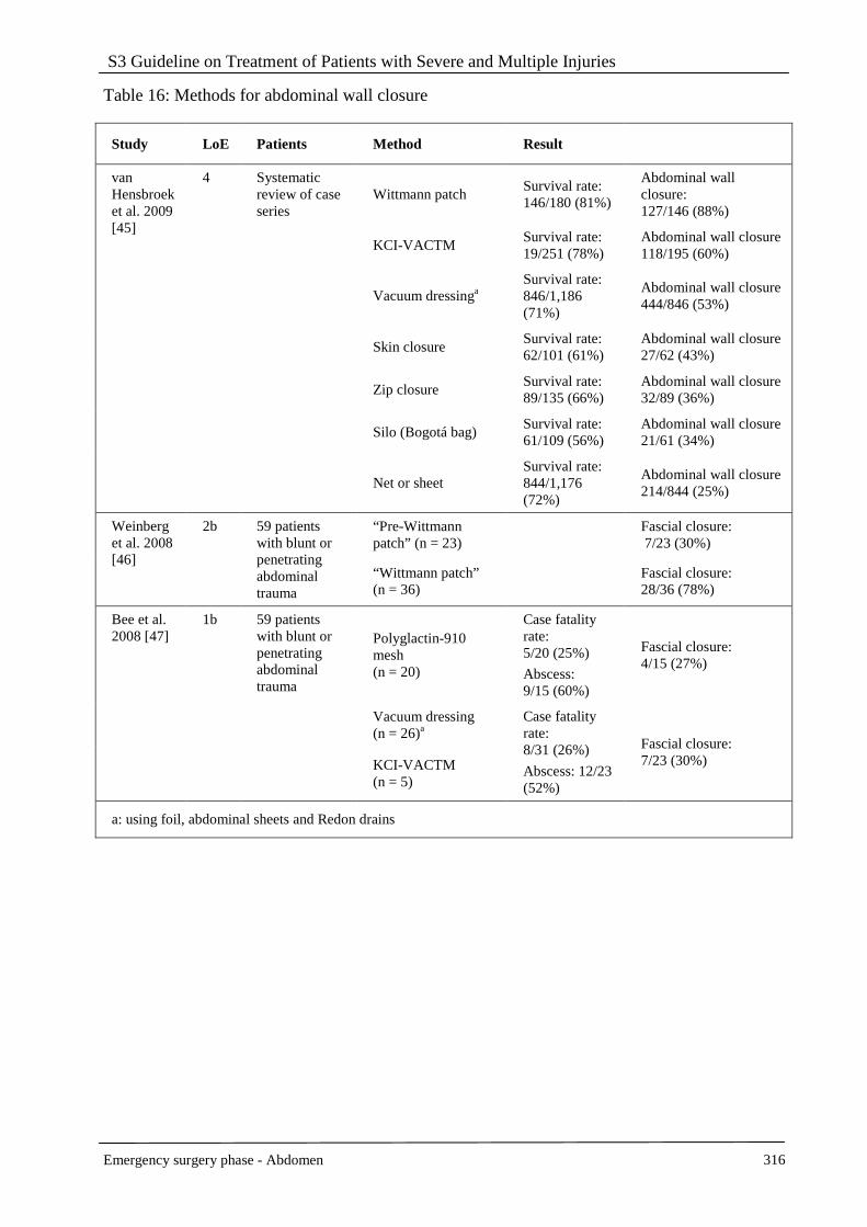
S3 Guideline on Treatment of Patients with Severe and Multiple Injuries
Emergency surgery phase - Abdomen 316
Table 16: Methods for abdominal wall closure
Study LoE Patients Method Result
Wittmann patch Survival rate: 146/180 (81%)
Abdominal wall closure: 127/146 (88%)
KCI-VACTM Survival rate: 19/251 (78%)
Abdominal wall closure 118/195 (60%)
Vacuum dressinga Survival rate: 846/1,186 (71%)
Abdominal wall closure 444/846 (53%)
Skin closure Survival rate: 62/101 (61%)
Abdominal wall closure 27/62 (43%)
Zip closure Survival rate: 89/135 (66%)
Abdominal wall closure 32/89 (36%)
Silo (Bogotá bag) Survival rate: 61/109 (56%)
Abdominal wall closure 21/61 (34%)
van Hensbroek et al. 2009 [45]
4 Systematic review of case series
Net or sheet Survival rate: 844/1,176 (72%)
Abdominal wall closure 214/844 (25%)
“Pre-Wittmann patch” (n = 23)
Fascial closure: 7/23 (30%)
Weinberg et al. 2008 [46]
2b 59 patients with blunt or penetrating abdominal trauma
“Wittmann patch” (n = 36)
Fascial closure: 28/36 (78%)
Polyglactin-910 mesh (n = 20)
Case fatality rate: 5/20 (25%)
Abscess: 9/15 (60%)
Fascial closure: 4/15 (27%)
Vacuum dressing (n = 26)a
Bee et al. 2008 [47]
1b 59 patients with blunt or penetrating abdominal trauma
KCI-VACTM (n = 5)
Case fatality rate: 8/31 (26%)
Abscess: 12/23 (52%)
Fascial closure: 7/23 (30%)
a: using foil, abdominal sheets and Redon drains
S3 Guideline on Treatment of Patients with Severe and Multiple Injuries
Emergency surgery phase - Abdomen 317
Damage control: Second look after packing
Key recommendation:
After packing intraabdominal bleeding, a second look should be undertaken and the tamponade replaced between 24 and 48 hours after the first intervention.
GoR B
Explanation:
After packing and intensive medical stabilization as part of the damage control sequence, a re-laparotomy is necessary to replace the abdominal sheets and also for definitive injury management, if applicable. A balance must be maintained here between the risk of fresh bleeding and the possible complications (infections, fistula, restricted pulmonary function, abdominal compartment syndrome) from the foreign material.
The available data from retrospective cohort studies show that unpacking after 24-36 hours is associated with an increased risk of bleeding (pooled relative risk, fixed effects: 3.51, 95% confidence interval: 1.39–8.90) [48, 49]. There is no clear evidence that leaving the abdominal sheets for a period of 48 hours increases the risk of septic complications (pooled relative risk, fixed effects: 1.01; 95% CI: 0.59–1.70) [48–51]. In the study by Abikhaled, however, leaving the tamponades > 72 hours was associated with an almost 7-fold increase in the relative risk for intraabdominal abscesses (6.77; 95% CI: 0.84–54.25) [50]. From a pragmatic viewpoint, therefore, the re-laparotomy should be planned for not sooner than 24 hours and not later than 48 hours after the first intervention.

S3 Guideline on Treatment of Patients with Severe and Multiple Injuries
Emergency surgery phase - Abdomen 318
Table 17: Second look after packing
Study LoE Patients Result
Second look 24h: (n = 25):
Subsequent bleeding: 8/25 (32%)
Second look 48h: (n = 44):
Subsequent bleeding 5/44 (11%)
Second look 72 h (n = 3):
Subsequent bleeding: 0/3
Nicol et al. 2007 [48]
2b 93 patients with penetrating or blunt hepatic trauma
Packing in situ 24 h (n = 8):
Complications: 5/8 (63%)
Packing in situ 48 h (n = 44):
Complications: 6/44 (14%)
Packing in situ 72 h (n = 20):
Complications: 3/20 (15%)
Cué et al. 1990 [51]
2b 21 patients with penetrating or blunt hepatic trauma
Packing in situ 24 h (n = 7):
Abscess: 2/7 (29%)
Packing in situ 48 h (n = 6):
Abscess: 2/6 (33%)
Packing in situ 72 h (n = 8):
Abscess: 3/8 (38%)
Caruso et al. 1999 [49]
2b 93 patients with penetrating or blunt hepatic trauma
Second look < 36 h (n = 39):
Subsequent bleeding: 8/39 (21%)
Complications: 13/39 (33%)
Case fatality rate: 7/39 (18%)
Second look 36-72 h (n = 24):
Subsequent bleeding: 1/24 (4%)
Complications: 7/29 (29%)
Case fatality rate: 7/24 (29%)
Sharp et al. 1992 [52]
2b 22 patients with penetrating or blunt hepatic trauma
6 patients with septic complications:
Packing in situ 2.2 ± 0.4 (2–3) days
6 patients without septic complications:
Packing in situ 2.0 ± 1.0 (1–7) days
Abikhaled et al. 1997 [50]
2b 35 patients with penetrating or blunt abdominal trauma
Packing in ≤ 72 h (n = 22):
Abscess 1/22 (5%)
Sepsis 11/22 (50%)
Case fatality rate 1/22 (5%)
Packing in situ > 72 h (n = 13):
Abscess 4/13 (31%)
Sepsis 10/13 (77%)
Case fatality rate 6/13 (46%)
Definitive abdominal wall closure
Key recommendation:
Definitive fascial closure should be continuous using slow absorbable or non-absorbable suture material.
GoR B
Explanation:
The technique of fascial closure after a laparotomy is well-known to be controversial and is often determined by the surgeon’s preference. The best available evidence on decision-making is obtained from randomized studies of elective abdominal interventions. It appears pragmatic and expedient to transfer any clear trends in favor of a specific method to the trauma scenario.
S3 Guideline on Treatment of Patients with Severe and Multiple Injuries
Emergency surgery phase - Abdomen 319
There are 2 meta-analyses of randomized studies for which the data pool only partially overlaps. Both show a significant reduction in risk for incisional hernias through non-absorbable suture material and continuous sutures [53, 54]. The results of the multicenter INSECT (Interrupted or Continuous Slowly Absorbable Sutures – Evaluation of Abdominal Closure Techniques) trial published in 2009 show a similar though not significantly statistical trend [55].
The updated common Peto odds ratio from all available randomized studies on the comparison of continuous slowly absorbable and rapidly absorbable single knot sutures is 0.79 for incisional hernias (95% CI: 0.61–1.01) and for wound infections 1.49 (95% CI: 1.15–1.94).
Angioembolization
Key recommendations:
If, in the case of a patient with hepatic injury who can be hemodynamically stabilized, there is evidence of arterial bleeding in a contrast agent CT, selective angioembolization or a laparotomy should be performed.
GoR B
In the case of splenic injuries grade 1-3 which require intervention, selective angioembolization can be performed instead of surgical hemostasis.
GoR 0
In the case of retroperitoneal bleeding which requires intervention, selective angioembolization can be performed instead of or in addition to surgical hemostasis.
GoR 0
Explanation:
Interventional radiology has an established value in polytrauma management and is used both in primary non-surgical treatment of organ injuries and as a neo-adjuvant and adjuvant intervention [56, 57]. If there is evidence of active bleeding from the contrast agent enhanced CT scan which cannot or must not be addressed operatively and if there is a good response to fluid and blood replacement in the emergency room, angioembolization can contribute towards sustained stabilization of the circulation [58, 59].
There are no randomized studies. The currently best available evidence is on blunt and penetrating hepatic injuries and suggests a reduction in case fatality rate through additional angioembolization during DC management compared to operative treatment only (common RR [fixed effects] 0.47, 95% CI: 0.28–0.78) [60–65]. The bias due to lack of multivariate adjustment must be taken into consideration. Currently, there is no answer to the question as to whether angioembolization in hepatic injuries should be performed before or after the DC laparotomy. Two studies support early neoadjuvant angioembolization based on the lower complication rates [63, 65]. In 2 other studies, on the other hand, mortality was lowered if angioembolization was performed after a DC laparotomy [64, 66].

S3 Guideline on Treatment of Patients with Severe and Multiple Injuries
Emergency surgery phase - Abdomen 320
Decision-making must be on an individual case basis on the availability and presence of an experienced interventional radiologist, the success of circulation-stabilizing measures in the emergency room, the intraoperative finding, and the postoperative hemodynamics.
The same applies to angioembolization in the case of bleeding from the spleen, where more up-to-date data now seems to call for caution [67–70]. Compared to nonoperative treatment, angioembolization did not lead to a reduction in either the treatment failure rate (common RR [random effects] 1.13; 95% CI: 0.86-1.48) or mortality (common RR [fixed effects] 1.19; 95% CI: 0.66–1.15).

S3 Guideline on Treatment of Patients with Severe and Multiple Injuries
Emergency surgery phase - Abdomen 321
Table 18: Angioembolization
Study LoE Patients Result
Angioembolization directly after DC laparotomy (n = 17) DC laparotomy without angioembolization (n = 58) Asensio et al.
2007 [61]
2b 75 patients with penetrating or blunt hepatic trauma grade 4/5 Case fatality rate 2/17 (12%) Case fatality rate 21/58 (36%)
Angioembolization directly after DC laparotomy (n = 8) DC laparotomy without angioembolization (n = 11) Johnson et al.
2002 [62]
2b 19 patients with penetrating or blunt hepatic trauma grade 1–5 Case fatality rate 1/8 (13%) Case fatality rate 4/11 (36%)
Angioembolization directly after DC laparotomy (n = 23) DC laparotomy without angioembolization (n = 80)
Case fatality rate 7/23 (30%)
(grade 4: 4/14 [28%], grade 5: 3/9 [33%])
Case fatality rate 52/80 (65%)
(grade 4: 15/37 [39%], grade 5: 37/43 [86%])
RR 0.51 (95% confidence interval 0.27-0.98)
Asensio et al.
2003 [60]
2b 103 patients with penetrating or blunt hepatic trauma grade 4/5
OR (multivariate adjusted for RTS, direct surgical access to hepatic veins and packing): 0.20 (95% confidence interval 0.05-0.72)
Early AE before/instead of DC laparotomy (n = 6)
Late AE after DC laparotomy (n = 6)
DC laparotomy (n = 20) Nonoperative treatment (n = 94) Wahl et al.
2002 [65]
2b 126 patients with blunt hepatic trauma grade 1–6
Case fatality rate 0/6 (0%), complications 3/6 (50%)
Case fatality rate 3/6 (50%), complications 6/6 (100%)
Case fatality rate 7/20 (35%), complications 9/20 (45%)
Case fatality rate 2/94 (2%), complications 2/94 (2%)
(continued)
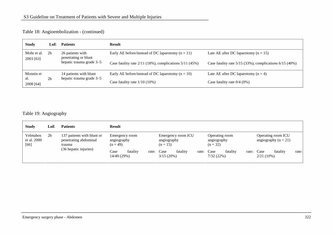
S3 Guideline on Treatment of Patients with Severe and Multiple Injuries
Emergency surgery phase - Abdomen 322
Table 18: Angioembolization - (continued)
Study LoE Patients Result
Early AE before/instead of DC laparotomy (n = 11) Late AE after DC laparotomy (n = 15) Mohr et al.
2003 [63]
2b 26 patients with penetrating or blunt hepatic trauma grade 3–5 Case fatality rate 2/11 (18%), complications 5/11 (45%) Case fatality rate 5/15 (33%), complications 6/15 (40%)
Early AE before/instead of DC laparotomy (n = 10) Late AE after DC laparotomy (n = 4) Monnin et al.
2008 [64] 2b
14 patients with blunt hepatic trauma grade 3–5
Case fatality rate 1/10 (10%) Case fatality rate 0/4 (0%)
Table 19: Angiography
Study LoE Patients Result
Emergency room angiography (n = 49)
Emergency room ICU angiography (n = 15)
Operating room angiography (n = 32)
Operating room ICU angiography (n = 21)
Velmahos et al. 2000 [66]
2b 137 patients with blunt or penetrating abdominal trauma (36 hepatic injuries)
Case fatality rate: 14/49 (29%)
Case fatality rate: 3/15 (20%)
Case fatality rate: 7/32 (22%)
Case fatality rate: 2/21 (10%)
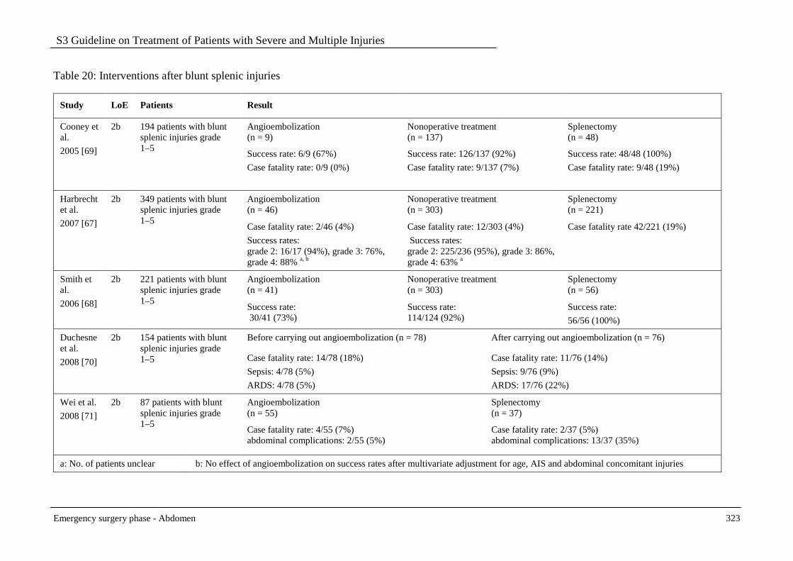
S3 Guideline on Treatment of Patients with Severe and Multiple Injuries
Emergency surgery phase - Abdomen 323
Table 20: Interventions after blunt splenic injuries
Study LoE Patients Result
Angioembolization (n = 9)
Nonoperative treatment (n = 137)
Splenectomy (n = 48)
Cooney et al.
2005 [69]
2b 194 patients with blunt splenic injuries grade 1–5
Success rate: 6/9 (67%)
Case fatality rate: 0/9 (0%)
Success rate: 126/137 (92%)
Case fatality rate: 9/137 (7%)
Success rate: 48/48 (100%)
Case fatality rate: 9/48 (19%)
Angioembolization (n = 46)
Nonoperative treatment (n = 303)
Splenectomy (n = 221)
Harbrecht et al.
2007 [67]
2b 349 patients with blunt splenic injuries grade 1–5
Case fatality rate: 2/46 (4%)
Success rates: grade 2: 16/17 (94%), grade 3: 76%, grade 4: 88% a, b
Case fatality rate: 12/303 (4%)
Success rates: grade 2: 225/236 (95%), grade 3: 86%, grade 4: 63% a
Case fatality rate 42/221 (19%)
Angioembolization (n = 41)
Nonoperative treatment (n = 303)
Splenectomy (n = 56)
Smith et al.
2006 [68]
2b 221 patients with blunt splenic injuries grade 1–5
Success rate: 30/41 (73%)
Success rate: 114/124 (92%)
Success rate:
56/56 (100%)
Before carrying out angioembolization (n = 78) After carrying out angioembolization (n = 76) Duchesne et al.
2008 [70]
2b 154 patients with blunt splenic injuries grade 1–5 Case fatality rate: 14/78 (18%)
Sepsis: 4/78 (5%)
ARDS: 4/78 (5%)
Case fatality rate: 11/76 (14%)
Sepsis: 9/76 (9%)
ARDS: 17/76 (22%)
Angioembolization (n = 55)
Splenectomy (n = 37)
Wei et al.
2008 [71]
2b 87 patients with blunt splenic injuries grade 1–5
Case fatality rate: 4/55 (7%) abdominal complications: 2/55 (5%)
Case fatality rate: 2/37 (5%) abdominal complications: 13/37 (35%)
a: No. of patients unclear b: No effect of angioembolization on success rates after multivariate adjustment for age, AIS and abdominal concomitant injuries
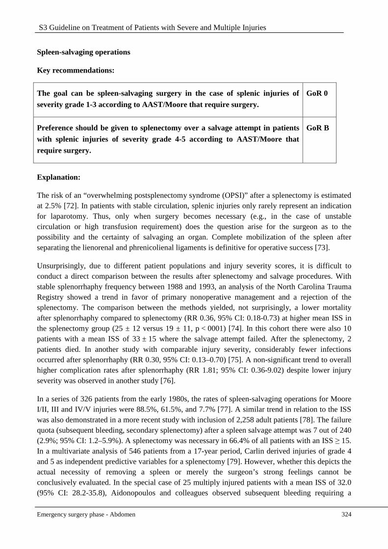
S3 Guideline on Treatment of Patients with Severe and Multiple Injuries
Emergency surgery phase - Abdomen 324
Spleen-salvaging operations
Key recommendations:
The goal can be spleen-salvaging surgery in the case of splenic injuries of severity grade 1-3 according to AAST/Moore that require surgery.
GoR 0
Preference should be given to splenectomy over a salvage attempt in patients with splenic injuries of severity grade 4-5 according to AAST/Moore that require surgery.
GoR B
Explanation:
The risk of an “overwhelming postsplenectomy syndrome (OPSI)” after a splenectomy is estimated at 2.5% [72]. In patients with stable circulation, splenic injuries only rarely represent an indication for laparotomy. Thus, only when surgery becomes necessary (e.g., in the case of unstable circulation or high transfusion requirement) does the question arise for the surgeon as to the possibility and the certainty of salvaging an organ. Complete mobilization of the spleen after separating the lienorenal and phrenicolienal ligaments is definitive for operative success [73].
Unsurprisingly, due to different patient populations and injury severity scores, it is difficult to conduct a direct comparison between the results after splenectomy and salvage procedures. With stable splenorrhaphy frequency between 1988 and 1993, an analysis of the North Carolina Trauma Registry showed a trend in favor of primary nonoperative management and a rejection of the splenectomy. The comparison between the methods yielded, not surprisingly, a lower mortality after splenorrhaphy compared to splenectomy (RR 0.36, 95% CI: 0.18-0.73) at higher mean ISS in the splenectomy group (25 ± 12 versus 19 ± 11, p < 0001) [74]. In this cohort there were also 10 patients with a mean ISS of 33 ± 15 where the salvage attempt failed. After the splenectomy, 2 patients died. In another study with comparable injury severity, considerably fewer infections occurred after splenorrhaphy (RR 0.30, 95% CI: 0.13–0.70) [75]. A non-significant trend to overall higher complication rates after splenorrhaphy (RR 1.81; 95% CI: 0.36-9.02) despite lower injury severity was observed in another study [76].
In a series of 326 patients from the early 1980s, the rates of spleen-salvaging operations for Moore I/II, III and IV/V injuries were 88.5%, 61.5%, and 7.7% [77]. A similar trend in relation to the ISS was also demonstrated in a more recent study with inclusion of 2,258 adult patients [78]. The failure quota (subsequent bleeding, secondary splenectomy) after a spleen salvage attempt was 7 out of 240 (2.9%; 95% CI: 1.2–5.9%). A splenectomy was necessary in 66.4% of all patients with an ISS ≥ 15. In a multivariate analysis of 546 patients from a 17-year period, Carlin derived injuries of grade 4 and 5 as independent predictive variables for a splenectomy [79]. However, whether this depicts the actual necessity of removing a spleen or merely the surgeon’s strong feelings cannot be conclusively evaluated. In the special case of 25 multiply injured patients with a mean ISS of 32.0 (95% CI: 28.2-35.8), Aidonopoulos and colleagues observed subsequent bleeding requiring a

S3 Guideline on Treatment of Patients with Severe and Multiple Injuries
Emergency surgery phase - Abdomen 325
splenectomy in 2 patients with injury grade 3 after suturing with a ‘figure of eight’ (0-0 chromic cat gut) [80].
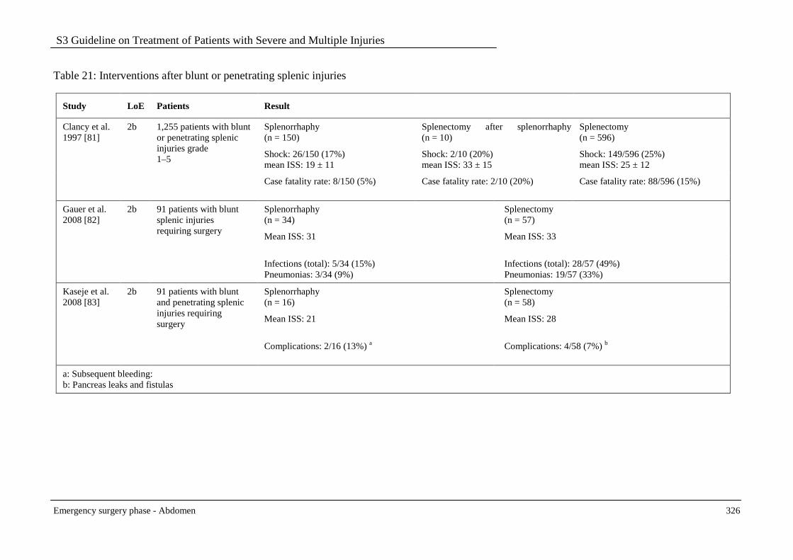
S3 Guideline on Treatment of Patients with Severe and Multiple Injuries
Emergency surgery phase - Abdomen 326
Table 21: Interventions after blunt or penetrating splenic injuries
Study LoE Patients Result
Splenorrhaphy (n = 150)
Splenectomy after splenorrhaphy (n = 10)
Splenectomy (n = 596)
Shock: 26/150 (17%) mean ISS: 19 ± 11
Shock: 2/10 (20%) mean ISS: 33 ± 15
Shock: 149/596 (25%) mean ISS: 25 ± 12
Clancy et al. 1997 [81]
2b 1,255 patients with blunt or penetrating splenic injuries grade 1–5
Case fatality rate: 8/150 (5%) Case fatality rate: 2/10 (20%) Case fatality rate: 88/596 (15%)
Splenorrhaphy (n = 34)
Splenectomy (n = 57)
Mean ISS: 31 Mean ISS: 33
Gauer et al. 2008 [82]
2b 91 patients with blunt splenic injuries requiring surgery
Infections (total): 5/34 (15%) Pneumonias: 3/34 (9%)
Infections (total): 28/57 (49%) Pneumonias: 19/57 (33%)
Splenorrhaphy (n = 16)
Splenectomy (n = 58)
Mean ISS: 21 Mean ISS: 28
Kaseje et al. 2008 [83]
2b 91 patients with blunt and penetrating splenic injuries requiring surgery
Complications: 2/16 (13%) a Complications: 4/58 (7%) b
a: Subsequent bleeding: b: Pancreas leaks and fistulas

S3 Guideline on Treatment of Patients with Severe and Multiple Injuries
Emergency surgery phase - Abdomen 327
Hollow organ injuries
Key recommendation:
In the case of penetrating colon injuries, if technically possible, preference must be given over a two-step procedure with temporary stoma to oversewing only or to primary anastomosis in order to reduce the risk of intraabdominal infections.
GoR A
Explanation:
Due to the contamination of the sterile abdominal cavity with mixed anaerobic flora, penetrating colon injuries represent a potentially life-threatening clinical picture. Thus, patients with abdominal gunshot wounds who must undergo immediate surgical treatment have a 100-fold higher relative risk of dying compared to patients with injuries that can be treated non-surgically or during secondary surgery [84].
Since 1979, 6 randomized trials (RCTs) have been published in which the results after primary operative management to maintain continuity were compared with those after temporary insertion of an ileostomy [85–90]. These studies were summarized in a Cochrane Review updated in 2009 [91]. The observed trends were also reproduced in the multicenter study of the American Association for the Surgery of Trauma (AAST) [92].
Based on the best available evidence, there is a non-significant trend in mortality in favor of primary anastomosis (RR 0.67; 95% CI: 0.31-1.45) with a marked reduction in complication rates (RR 0.73; 95% CI: 0.52-1.02). The risk of intraabdominal infections could possibly be reduced by 23% through primary anastomosis (RR 0.77; 95% CI: 0.55-1.06) even though clear scientific proof from an appropriately designed randomized study is unavailable. Current data from the US Iraq operations support the trends in favor of primary anastomosis [93]. It is unclear whether the available data can be transferred to blunt injuries. In this situation, however, biologic and clinical considerations argue more in favor of maintaining continuity.
Stapling instruments represent a major valuable addition to the equipment for elective gastrointestinal interventions. Deep colorectal anastomoses were first made possible through the availability of circular staplers; laparoscopic intestinal surgery also gained from the option of stapled anastomoses.
In a meta-analysis of 9 randomized studies (1,233 patients), however, there was no evidence of advantage from staplers compared to a hand suture in the endpoints mortality, anastomosis failure, wound infection, re-operation, and length of stay in hospital [94]. On the other hand, there was a significantly increased risk of strictures after stapled anastomosis (Peto OR 3.59; 95% CI: 2.02–6.35). The multicenter studies of the Western Trauma Association and AAST have produced evidence of a possible disadvantage from stapled colon anastomoses in the trauma situation [95, 96]. The weighted relative risk for all complications after hand suture compared to stapled anastomosis from both studies is 0.72 (95% CI: 0.45-1.15). For anastomosis

S3 Guideline on Treatment of Patients with Severe and Multiple Injuries
Emergency surgery phase - Abdomen 328
failures and intraabdominal abscesses, the common RR can be estimated at 0.90 (95% CI: 0.36-2.28) and 0.74 (95% CI: 0.42-1.28).
Two multicenter studies yielded similar, again non-significant trends for small intestine anastomoses [95, 97]. The hand suture is possibly associated with a reduction in all complications (RR 0.75; 95% CI: 0.31-1.82). Anastomosis failures and intraabdominal abscesses were also observed less frequently after hand suturing (RR 0.43; 95% CI: 0.08–2.42 and RR 0.54; 95% CI: 0.18–1.64).
The results of a randomized trial conducted under elective conditions suggest that a single-layer, continuous hand suture can be carried out without risk. In this study [98], no difference could be detected in the failure rates between a single-layer (2/65) and a two-layer/Lembert suture (1/67). The observed frequency of abscesses was also identical between the two treatment arms (2/65 and 2/67). Nineteen and 12 trauma patients were also included in the study.
S3 Guideline on Treatment of Patients with Severe and Multiple Injuries
Emergency surgery phase - Abdomen 329
Table 22: Primary anastomosis versus ileostomy after penetrating colon injury
Study LoE Patients Result
Primary anastomosis (n = 361)
Ileostomy (n = 344)
Case fatality rate: 7/361 (2%) Case fatality rate: 6/344 (2%)
All complications: 135/361 (37%) All complications: 173/346 (50%)
Nelson et al. 2009 [91]
1a Meta-analysis of 6 RCTs (n = 707)
Infections: 120/361 (33%) Infections: 144/346 (42%)
Primary anastomosis (n = 197)
Ileostomy (n = 100)
Case fatality rate: 8/197 (4%) Case fatality rate: 10/100 (10%)
All complications: 44/197 (22%) All complications: 27/100 (27%)
Demetriades et al. 2001 [92]
2b 297 patients with penetrating colon injuries
Infections: 33/197 (17%) Infections: 21/100 (21%)
Primary anastomosis (n = 38)
Ileostomy (n = 27)
Case fatality rate: 1/38 (2%) Case fatality rate: 0/27 (0%)
all colon-associated complications: 11/38 (29%) all colon-associated complications: 10/27 (37%)
Vertrees et al. 2009 [93]
2b 65 wounded (Enduring Freedom/ Iraqi Freedom) with penetrating colon injuries
Infections: 5/38 (13%) Infections: 9/27 (33%)
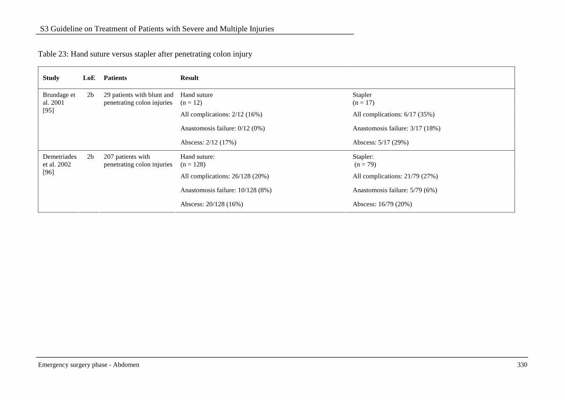
S3 Guideline on Treatment of Patients with Severe and Multiple Injuries
Emergency surgery phase - Abdomen 330
Table 23: Hand suture versus stapler after penetrating colon injury
Study LoE Patients Result
Hand suture (n = 12)
Stapler (n = 17)
All complications: 2/12 (16%) All complications: 6/17 (35%)
Anastomosis failure: 0/12 (0%) Anastomosis failure: 3/17 (18%)
Brundage et al. 2001 [95]
2b 29 patients with blunt and penetrating colon injuries
Abscess: 2/12 (17%) Abscess: 5/17 (29%)
Hand suture: (n = 128)
Stapler: (n = 79)
All complications: 26/128 (20%) All complications: 21/79 (27%)
Anastomosis failure: 10/128 (8%) Anastomosis failure: 5/79 (6%)
Demetriades et al. 2002 [96]
2b 207 patients with penetrating colon injuries
Abscess: 20/128 (16%) Abscess: 16/79 (20%)
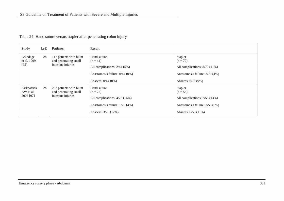
S3 Guideline on Treatment of Patients with Severe and Multiple Injuries
Emergency surgery phase - Abdomen 331
Table 24: Hand suture versus stapler after penetrating colon injury
Study LoE Patients Result
Hand suture (n = 44)
Stapler (n = 70)
All complications: 2/44 (5%) All complications: 8/70 (11%)
Anastomosis failure: 0/44 (0%) Anastomosis failure: 3/70 (4%)
Brundage et al. 1999 [95]
2b 117 patients with blunt and penetrating small intestine injuries
Abscess: 0/44 (0%) Abscess: 6/70 (9%)
Hand suture (n = 25)
Stapler (n = 55)
All complications: 4/25 (16%) All complications: 7/55 (13%)
Anastomosis failure: 1/25 (4%) Anastomosis failure: 3/55 (6%)
Kirkpatrick AW et al. 2003 [97]
2b 232 patients with blunt and penetrating small intestine injuries
Abscess: 3/25 (12%) Abscess: 6/55 (11%)

S3 Guideline on Treatment of Patients with Severe and Multiple Injuries
Emergency surgery phase - Abdomen 332
References
1. Cothren CC, Moore EE, Hoyt DB. The U.S. trauma surgeon's current scope of practice: can we deliver acute care surgery? J Trauma 2008;64(4):955-965.
2. Hoyt DB, Kim HD, Barrios C. Acute care surgery: a new training and practice model in the United States. World J Surg 2008;32(8):1630-1635.
3. Hess JR, Holcomb JB, Hoyt DB. Damage control resuscitation: the need for specific blood products to treat the coagulopathy of trauma. Transfusion 2006;46(5):685-686.
4. Hodgetts TJ, Mahoney PF, Kirkman E. Damage control resuscitation. J R Army Med Corps 2007;153(4):299-300.
5. Holcomb JB, Jenkins D, Rhee P et al. Damage control resuscitation: directly addressing the early coagulopathy of trauma. J Trauma 2007;62(2):307-310.
6. Jansen JO, Thomas R, Loudon MA, Brooks A. Damage control resuscitation for patients with major trauma. BMJ 2009;338b1778.
7. Tieu BH, Holcomb JB, Schreiber MA. Coagulopathy: its pathophysiology and treatment in the injured patient. World J Surg 2007;31(5):1055-1064.
8. Edwards PS, Lipp A, Holmes A. Preoperative skin antiseptics for preventing surgical wound infections after clean surgery. Cochrane Database Syst Rev 2004(3):CD003949.
9. Clarke JM. Case for midline incisions. Lancet 1989;1(8638):622 [LoE 5].
10. Guillou PJ, Hall TJ, Donaldson DR, Broughton AC, Brennan TG. Vertical abdominal incisions--a choice? Br J Surg 1980;67(6):395-399 [LoE 5].
11. Stone HH, Hoefling SJ, Strom PR, Dunlop WE, Fabian TC. Abdominal incisions: transverse vs vertical placement and continuous vs interrupted closure. South Med J 1983;76(9):1106-1108 [LoE 2b].
12. Brown SR, Goodfellow PB. Transverse verses midline incisions for abdominal surgery. Cochrane Database Syst Rev 2005(4):CD005199 [LoE 1a].
13. Seiler CM, Deckert A, Diener MK et al. Midline versus transverse incision in major abdominal surgery: a randomized, double-blind equivalence trial (POVATI: ISRCTN60734227). Ann Surg 2009;249(6):913-920 [LoE 1b].
14. Hori Y. Diagnostic laparoscopy guidelines : This guideline was prepared by the SAGES Guidelines Committee and reviewed and approved by the Board of Governors of the Society of American Gastrointestinal and Endoscopic Surgeons (SAGES), November 2007. Surg Endosc 2008;22(5):1353-1383 [Evidenzbasierte Leitlinie]
15. Sauerland S, Agresta F, Bergamaschi R et al. Laparoscopy for abdominal emergencies: evidence-based guidelines of the European Association for Endoscopic Surgery. Surg Endosc 2006;20(1):14-29 [Evidenzbasierte Leitlinie]
16. Chol YB, Lim KS. Therapeutic laparoscopy for abdominal trauma. Surg Endosc 2003;17(3):421-427 [LoE 4].
17. Kaban GK, Novitsky YW, Perugini RA et al. Use of laparoscopy in evaluation and treatment of penetrating and blunt abdominal injuries. Surg Innov 2008;15(1):26-31 [LoE 4].
18. Mallat AF, Mancini ML, Daley BJ, Enderson BL. The role of laparoscopy in trauma: a ten-year review of diagnosis and therapeutics. Am Surg 2008;74(12):1166-1170 [LoE 4].
19. Smith RS, Fry WR, Morabito DJ, Koehler RH, Organ CH, Jr. Therapeutic laparoscopy in trauma. Am J Surg 1995;170(6):632-636 [LoE 4].
20. Zantut LF, Ivatury RR, Smith RS et al. Diagnostic and therapeutic laparoscopy for penetrating abdominal trauma: a multicenter experience. J Trauma 1997;42(5):825-829 [LoE 4].
21. Committee on Technology for Future Naval Forces. Technology for the United States Navy and Marine Corps, 2000-2035. Becoming a 21st-Century Force. 1997.
22. Rotondo MF, Zonies DH. The Damage Control sequence and underlying logic. Surg Clin North Am 1997;77(4):761-777.
23. Germanos S, Gourgiotis S, Villias C et al. Damage control surgery in the abdomen: an approach for the management of severe injured patients. Int J Surg 2008;6(3):246-252.
24. Lee JC, Peitzman AB. Damage-control laparotomy. Curr Opin Crit Care 2006;12(4):346-350.
25. Loveland JA, Boffard KD. Damage control in the abdomen and beyond. Br J Surg 2004;91(9):1095-1101.
26. Kremer B, Henne-Bruns D. [Value of various techniques in liver rupture]. Chirurg 1993;64(11):852-859 [LoE 4].
27. Feliciano DV, Mattox KL, Burch JM, Bitondo CG, Jordan GL, Jr. Packing for control of hepatic hemorrhage. J Trauma 1986;26(8):738-743 [LoE 4].
28. Feliciano DV, Mattox KL, Jordan GL, Jr. Intra-abdominal packing for control of hepatic hemorrhage: a reappraisal. J Trauma 1981;21(4):285-290 [LoE 4].
29. Baracco-Gandolfo V, Vidarte O, Baracco-Miller V, del Castillo M. Prolonged closed liver packing in severe hepatic trauma: experience with 36 patients. J Trauma 1986;26(8):754-756 [LoE 4].
30. Reed RL, Merrell RC, Meyers WC, Fischer RP. Continuing evolution in the approach to severe liver trauma. Ann Surg 1992;216(5):524-538 [LoE 4].
31. Stone HH, Strom PR, Mullins RJ. Management of the major coagulopathy with onset during laparotomy. Ann Surg 1983;197(5):532-535 [LoE 2b].
32. Rotondo MF, Schwab CW, McGonigal MD et al. 'Damage control': an approach for improved survival in exsanguinating penetrating abdominal injury. J Trauma 1993;35(3):375-382 [LoE 2b].
33. MacKenzie S, Kortbeek JB, Mulloy R, Hameed SM. Recent experiences with a multidisciplinary approach to complex hepatic trauma. Injury 2004;35(9):869-877 [LoE 2b].
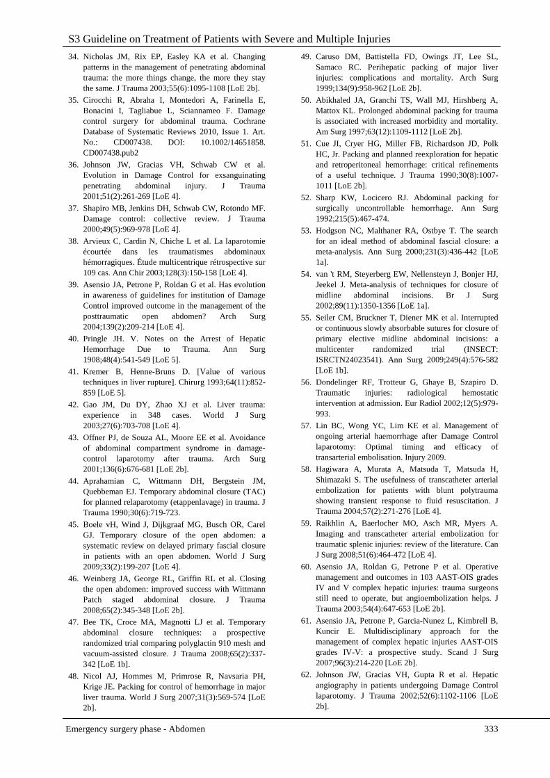
S3 Guideline on Treatment of Patients with Severe and Multiple Injuries
Emergency surgery phase - Abdomen 333
34. Nicholas JM, Rix EP, Easley KA et al. Changing patterns in the management of penetrating abdominal trauma: the more things change, the more they stay the same. J Trauma 2003;55(6):1095-1108 [LoE 2b].
35. Cirocchi R, Abraha I, Montedori A, Farinella E, Bonacini I, Tagliabue L, Sciannameo F. Damage control surgery for abdominal trauma. Cochrane Database of Systematic Reviews 2010, Issue 1. Art. No.: CD007438. DOI: 10.1002/14651858. CD007438.pub2
36. Johnson JW, Gracias VH, Schwab CW et al. Evolution in Damage Control for exsanguinating penetrating abdominal injury. J Trauma 2001;51(2):261-269 [LoE 4].
37. Shapiro MB, Jenkins DH, Schwab CW, Rotondo MF. Damage control: collective review. J Trauma 2000;49(5):969-978 [LoE 4].
38. Arvieux C, Cardin N, Chiche L et al. La laparotomie écourtée dans les traumatismes abdominaux hémorragiques. Étude multicentrique rétrospective sur 109 cas. Ann Chir 2003;128(3):150-158 [LoE 4].
39. Asensio JA, Petrone P, Roldan G et al. Has evolution in awareness of guidelines for institution of Damage Control improved outcome in the management of the posttraumatic open abdomen? Arch Surg 2004;139(2):209-214 [LoE 4].
40. Pringle JH. V. Notes on the Arrest of Hepatic Hemorrhage Due to Trauma. Ann Surg 1908;48(4):541-549 [LoE 5].
41. Kremer B, Henne-Bruns D. [Value of various techniques in liver rupture]. Chirurg 1993;64(11):852-859 [LoE 5].
42. Gao JM, Du DY, Zhao XJ et al. Liver trauma: experience in 348 cases. World J Surg 2003;27(6):703-708 [LoE 4].
43. Offner PJ, de Souza AL, Moore EE et al. Avoidance of abdominal compartment syndrome in damage-control laparotomy after trauma. Arch Surg 2001;136(6):676-681 [LoE 2b].
44. Aprahamian C, Wittmann DH, Bergstein JM, Quebbeman EJ. Temporary abdominal closure (TAC) for planned relaparotomy (etappenlavage) in trauma. J Trauma 1990;30(6):719-723.
45. Boele vH, Wind J, Dijkgraaf MG, Busch OR, Carel GJ. Temporary closure of the open abdomen: a systematic review on delayed primary fascial closure in patients with an open abdomen. World J Surg 2009;33(2):199-207 [LoE 4].
46. Weinberg JA, George RL, Griffin RL et al. Closing the open abdomen: improved success with Wittmann Patch staged abdominal closure. J Trauma 2008;65(2):345-348 [LoE 2b].
47. Bee TK, Croce MA, Magnotti LJ et al. Temporary abdominal closure techniques: a prospective randomized trial comparing polyglactin 910 mesh and vacuum-assisted closure. J Trauma 2008;65(2):337-342 [LoE 1b].
48. Nicol AJ, Hommes M, Primrose R, Navsaria PH, Krige JE. Packing for control of hemorrhage in major liver trauma. World J Surg 2007;31(3):569-574 [LoE 2b].
49. Caruso DM, Battistella FD, Owings JT, Lee SL, Samaco RC. Perihepatic packing of major liver injuries: complications and mortality. Arch Surg 1999;134(9):958-962 [LoE 2b].
50. Abikhaled JA, Granchi TS, Wall MJ, Hirshberg A, Mattox KL. Prolonged abdominal packing for trauma is associated with increased morbidity and mortality. Am Surg 1997;63(12):1109-1112 [LoE 2b].
51. Cue JI, Cryer HG, Miller FB, Richardson JD, Polk HC, Jr. Packing and planned reexploration for hepatic and retroperitoneal hemorrhage: critical refinements of a useful technique. J Trauma 1990;30(8):1007-1011 [LoE 2b].
52. Sharp KW, Locicero RJ. Abdominal packing for surgically uncontrollable hemorrhage. Ann Surg 1992;215(5):467-474.
53. Hodgson NC, Malthaner RA, Ostbye T. The search for an ideal method of abdominal fascial closure: a meta-analysis. Ann Surg 2000;231(3):436-442 [LoE 1a].
54. van 't RM, Steyerberg EW, Nellensteyn J, Bonjer HJ, Jeekel J. Meta-analysis of techniques for closure of midline abdominal incisions. Br J Surg 2002;89(11):1350-1356 [LoE 1a].
55. Seiler CM, Bruckner T, Diener MK et al. Interrupted or continuous slowly absorbable sutures for closure of primary elective midline abdominal incisions: a multicenter randomized trial (INSECT: ISRCTN24023541). Ann Surg 2009;249(4):576-582 [LoE 1b].
56. Dondelinger RF, Trotteur G, Ghaye B, Szapiro D. Traumatic injuries: radiological hemostatic intervention at admission. Eur Radiol 2002;12(5):979-993.
57. Lin BC, Wong YC, Lim KE et al. Management of ongoing arterial haemorrhage after Damage Control laparotomy: Optimal timing and efficacy of transarterial embolisation. Injury 2009.
58. Hagiwara A, Murata A, Matsuda T, Matsuda H, Shimazaki S. The usefulness of transcatheter arterial embolization for patients with blunt polytrauma showing transient response to fluid resuscitation. J Trauma 2004;57(2):271-276 [LoE 4].
59. Raikhlin A, Baerlocher MO, Asch MR, Myers A. Imaging and transcatheter arterial embolization for traumatic splenic injuries: review of the literature. Can J Surg 2008;51(6):464-472 [LoE 4].
60. Asensio JA, Roldan G, Petrone P et al. Operative management and outcomes in 103 AAST-OIS grades IV and V complex hepatic injuries: trauma surgeons still need to operate, but angioembolization helps. J Trauma 2003;54(4):647-653 [LoE 2b].
61. Asensio JA, Petrone P, Garcia-Nunez L, Kimbrell B, Kuncir E. Multidisciplinary approach for the management of complex hepatic injuries AAST-OIS grades IV-V: a prospective study. Scand J Surg 2007;96(3):214-220 [LoE 2b].
62. Johnson JW, Gracias VH, Gupta R et al. Hepatic angiography in patients undergoing Damage Control laparotomy. J Trauma 2002;52(6):1102-1106 [LoE 2b].

S3 Guideline on Treatment of Patients with Severe and Multiple Injuries
Emergency surgery phase - Abdomen 334
63. Mohr AM, Lavery RF, Barone A et al. Angiographic embolization for liver injuries: low mortality, high morbidity. J Trauma 2003;55(6):1077-1081 [LoE 2b].
64. Monnin V, Sengel C, Thony F et al. Place of arterial embolization in severe blunt hepatic trauma: a multidisciplinary approach. Cardiovasc Intervent Radiol 2008;31(5):875-882 [LoE 2b].
65. Wahl WL, Ahrns KS, Brandt MM, Franklin GA, Taheri PA. The need for early angiographic embolization in blunt liver injuries. J Trauma 2002;52(6):1097-1101 [LoE 2b].
66. Velmahos GC, Chahwan S, Falabella A, Hanks SE, Demetriades D. Angiographic embolization for intraperitoneal and retroperitoneal injuries. World J Surg 2000;24(5):539-545 [LoE 2b].
67. Harbrecht BG, Ko SH, Watson GA et al. Angiography for blunt splenic trauma does not improve the success rate of nonoperative management. J Trauma 2007;63(1):44-49 [LoE 2b].
68. Smith HE, Biffl WL, Majercik SD et al. Splenic artery embolization: Have we gone too far? J Trauma 2006;61(3):541-544 [LoE 2b].
69. Cooney R, Ku J, Cherry R et al. Limitations of splenic angioembolization in treating blunt splenic injury. J Trauma 2005;59(4):926-932 [LoE 2b].
70. Duchesne JC, Simmons JD, Schmieg RE, Jr., McSwain NE, Jr., Bellows CF. Proximal splenic angioembolization does not improve outcomes in treating blunt splenic injuries compared with splenectomy: a cohort analysis. J Trauma 2008;65(6):1346-1351 [LoE 2b].
71. Wei B, Hemmila MR, Arbabi S, Taheri PA, Wahl WL. Angioembolization reduces operative intervention for blunt splenic injury. J Trauma 2008;64(6):1472-1477.
72. Velanovich V. Blunt splenic injury in adults: a decision analysis comparing options for treatment. Eur J Surg 1995;161(7):463-470.
73. Peitzman AB, Ford HR, Harbrecht BG, Potoka DA, Townsend RN. Injury to the spleen. Curr Probl Surg 2001;38(12):932-1008 [LoE 5].
74. Clancy TV, Ramshaw DG, Maxwell JG et al. Management outcomes in splenic injury: a statewide trauma center review. Ann Surg 1997;226(1):17-24 [LoE 2b].
75. Gauer JM, Gerber-Paulet S, Seiler C, Schweizer WP. Twenty years of splenic preservation in trauma: lower early infection rate than in splenectomy. World J Surg 2008;32(12):2730-2735 [LoE 2b].
76. Kaseje N, Agarwal S, Burch M et al. Short-term outcomes of splenectomy avoidance in trauma patients. Am J Surg 2008;196(2):213-217 [LoE 2b].
77. Feliciano DV, Bitondo CG, Mattox KL et al. A four-year experience with splenectomy versus splenorrhaphy. Ann Surg 1985;201(5):568-575 [LoE 4].
78. Hunt JP, Lentz CW, Cairns BA et al. Management and outcome of splenic injury: the results of a five-year statewide population-based study. Am Surg 1996;62(11):911-917 [LoE 2b].
79. Carlin AM, Tyburski JG, Wilson RF, Steffes C. Factors affecting the outcome of patients with splenic trauma. Am Surg 2002;68(3):232-239 [LoE 2b].
80. Aidonopoulos AP, Papavramidis ST, Goutzamanis GD et al. Splenorrhaphy for splenic damage in patients with multiple injuries. Eur J Surg 1995;161(4):247-251 [LoE 4].
81. Clancy TV, Ramshaw DG, Maxwell JG et al. Management outcomes in splenic injury: a statewide trauma center review. Ann Surg 1997;226(1):17-24.
82. Gauer JM, Gerber-Paulet S, Seiler C, Schweizer WP. Twenty years of splenic preservation in trauma: lower early infection rate than in splenectomy. World J Surg 2008;32(12):2730-2735.
83. Kaseje N, Agarwal S, Burch M et al. Short-term outcomes of splenectomy avoidance in trauma patients. Am J Surg 2008;196(2):213-217.
84. Velmahos GC, Demetriades D, Toutouzas KG et al. Selective nonoperative management in 1,856 patients with abdominal gunshot wounds: should routine laparotomy still be the standard of care? Ann Surg 2001;234(3):395-402 [LoE 2b].
85. Chappuis CW, Frey DJ, Dietzen CD et al. Management of penetrating colon injuries. A prospective randomized trial. Ann Surg 1991;213(5):492-497 [LoE 1b].
86. Falcone RE, Wanamaker SR, Santanello SA, Carey LC. Colorectal trauma: primary repair or anastomosis with intracolonic bypass vs. ostomy. Dis Colon Rectum 1992;35(10):957-963 [LoE 1b].
87. Gonzalez RP, Falimirski ME, Holevar MR. Further evaluation of colostomy in penetrating colon injury. Am Surg 2000;66(4):342-346 [LoE 1b].
88. Kamwendo NY, Modiba MC, Matlala NS, Becker PJ. Randomized clinical trial to determine if delay from time of penetrating colonic injury precludes primary repair. Br J Surg 2002;89(8):993-998 [LoE 1b].
89. Sasaki LS, Allaben RD, Golwala R, Mittal VK. Primary repair of colon injuries: a prospective randomized study. J Trauma 1995;39(5):895-901 [LoE 1b].
90. Stone HH, Fabian TC. Management of perforating colon trauma: randomization between primary closure and exteriorization. Ann Surg 1979;190(4):430-436 [LoE 1b].
91. Nelson R, Singer M. Primary repair for penetrating colon injuries. Cochrane Database Syst Rev 2002(3):CD002247 [LoE 1a].
92. Demetriades D, Murray JA, Chan L et al. Penetrating colon injuries requiring resection: diversion or primary anastomosis? An AAST prospective multicenter study. J Trauma 2001;50(5):765-775 [LoE 2b].
93. Vertrees A, Wakefield M, Pickett C et al. Outcomes of primary repair and primary anastomosis in war-related colon injuries. J Trauma 2009;66(5):1286-1291 [LoE 2b].
94. Lustosa SA, Matos D, Atallah AN, Castro AA. Stapled versus handsewn methods for colorectal anastomosis surgery. Cochrane Database Syst Rev 2001(3):CD003144 [LoE 1a].

S3 Guideline on Treatment of Patients with Severe and Multiple Injuries
Emergency surgery phase - Abdomen 335
95. Brundage SI, Jurkovich GJ, Hoyt DB et al. Stapled versus sutured gastrointestinal anastomoses in the trauma patient: a multicenter trial. J Trauma 2001;51(6):1054-1061 [LoE 2b].
96. Demetriades D, Murray JA, Chan LS et al. Handsewn versus stapled anastomosis in penetrating colon injuries requiring resection: a multicenter study. J Trauma 2002;52(1):117-121 [LoE 2b].
97. Kirkpatrick AW, Baxter KA, Simons RK et al. Intra-abdominal complications after surgical repair of small bowel injuries: an international review. J Trauma 2003;55(3):399-406 [LoE 2b].
98. Burch JM, Franciose RJ, Moore EE, Biffl WL, Offner PJ. Single-layer continuous versus two-layer interrupted intestinal anastomosis: a prospective randomized trial. Ann Surg 2000;231(6):832-837 [LoE 1b]

S3 Guideline on Treatment of Patients with Severe and Multiple Injuries
Emergency surgery phase – Traumatic brain injury 336
3.5 Traumatic brain injury
Surgical management
Emergency surgical management
Key recommendation:
Compressive intracranial injuries must be surgically managed as an emergency.
GoR A
Explanation:
The goal of the treatment after a TBI is to limit the extent of secondary brain damage and to provide the brain cells whose function is impaired but not destroyed with the best conditions for functional regeneration. Injury sequelae requiring surgery must be treated in a timely manner.
The indication for surgical decompression of traumatic intracranial compression has never been tested in prospective randomized controlled trials. There are several retrospective analyses [3-9, 13] from which the benefit of surgical decompression can be derived. Due to the decades of consensual experience, the need for a surgical procedure can be regarded as a basic incontrovertible assumption of good clinical practice.
Compressive intracranial injuries represent an absolute urgent indication for surgery. This applies both to traumatic intracranial bleeding (epidural hematoma, subdural hematoma, intracerebral hematoma/contusion) and to compressive impression fractures. The definition of compression refers to the shift of cerebral structures, particularly the 3rd ventricle normally located at the midline. In addition to the finding in the computed tomography (layer thickness, volume, and location of hematoma, extent of midline shift), the clinical finding is key to establishing the indication and the speed with which surgical management should be carried out. If there are signs of a transtentorial herniation, every minute can make a difference to the clinical outcome. It is not considered meaningful to indicate the volumes at which an intervention should be performed as the individual situation of the patient (age, possible pre-existing brain atrophy inter alia) must be taken into account in establishing the indication.
Operations with deferred urgency
Open or closed impression fractures without shift of midline structures, penetrating injuries or basal fractures with liquorrhea constitute operations with deferred urgency. Their surgical conduct requires neurosurgical competence. The timing of the surgical intervention depends on many factors and must be decided on an individual basis.
Decompressive craniectomy
An effective option for lowering elevated intracranial pressure is surgical decompression by craniectomy and, if necessary, expansive duraplasty. The necessity mainly arises from the

S3 Guideline on Treatment of Patients with Severe and Multiple Injuries
Emergency surgery phase – Traumatic brain injury 337
development of marked (secondary) brain edema and thus frequently has several days’ latency. According to a prospective randomized controlled trial, the method shows good treatment success despite an increased complication rate [23]. Further prospective studies [10, 18] are ongoing so that no final recommendation can yet be made [26].
Nonoperative treatment of intracranial bleeding
In noncompressive bleeding and stable neurologic finding, a nonoperative procedure can be justified in individual cases [5, 7]. However, these patients must undergo close clinical and computed tomography follow-up observation. In the event of clinical deterioration or increase in compression, it must be possible to carry out immediate surgical decompression.
Measuring intracranial pressure
Key recommendation:
Intracranial pressure can be measured in unconscious brain-damaged patients.
GoR 0
Explanation:
Internationally in recent decades, the measurement of intracranial pressure has found its way into the acute management of unconscious brain-damaged patients and has meanwhile been adopted in several international guidelines [2, 21, 30]. For pathophysiologic reasons, this seems sensible as clinical monitoring of many cerebral functions is only possible to a limited extent. As a monitoring instrument in sedated patients, it can indicate imminent midbrain incarceration due to progressive brain swelling or compressive intracranial hematomas and thus permits early counter-measures to be taken. Even if there is currently no prospective randomized controlled trial that compares the clinical outcome with carrying out ICP monitoring [15], several cohort studies in recent years as well as clinical practice indicate its value in neurosurgical intensive medicine [1, 17, 20, 22]. The introduction of guidelines which, inter alia, stipulate this type of ICP monitoring has also led to an increase in favorable courses in TBI patients [24, 11]. Intracranial measurement is used for monitoring and treatment control of unconscious patients while taking into account the clinical course and morphologic image findings after TBI. However, it is not required in every unconscious patient.
The prerequisite for adequate brain perfusion is adequate cerebral perfusion pressure (CPP), which can be calculated simply from the difference between the mean arterial blood pressure and the mean ICP. The literature contains divergent opinions on whether lowering the ICP or maintaining the CPP should be the focus of the treatment in the case of elevated ICP. The currently available evidence argues in favor that,
� on the one hand, the CPP should not fall below 50 mmHg if possible [30].
� on the other hand, the CPP should not be raised to above 70 mmHg by aggressive treatment [30].

S3 Guideline on Treatment of Patients with Severe and Multiple Injuries
Emergency surgery phase – Traumatic brain injury 338
Invasive ICP measurement is necessary for continuous determination of the CPP. Provided the ventricles are not completely compressed, ICP monitoring via a ventricle drain offers the possibility of lowering elevated ICP through draining cerebrospinal fluid.
Determining the individual optimum CPP requires simultaneous knowledge of brain blood supply, oxygen supply and demand and/or brain metabolism. Regional measurements (using brain tissue probes, transcranial Doppler examinations or perfusion-weighted imaging) for estimating this value are currently the subject of scientific studies [19, 27].

S3 Guideline on Treatment of Patients with Severe and Multiple Injuries
Emergency surgery phase – Traumatic brain injury 339
References
1. Balestreri M, Czosnyka M, Hutchinson P, et al: Impact of intracranial pressure and cerebral perfusion pressure on severe disability and mortality after head injury. Neurocrit.Care 4:8-13, 2006
2. Bullock R, Chesnut RM, Clifton G, et al: Guidelines for the management of severe head injury. Brain Trauma Foundation. Eur.J.Emerg.Med. 3:109-127, 1996 [Evidenzbasierte Leitlinie]
3. Bullock MR, Chesnut R, Ghajar J, Gordon D, Hartl R, Newell D, Servadei F, Walters BC, Wilberger JE. Introduction. Neurosurgery. Guidelines for the Surgical Management of Traumatic Brain Injury. 58(3) Supplement:S2-1-S2-3, March 2006 [Evidenzbasierte Leitlinie].
4. Bullock MR, Chesnut R, Ghajar J, Gordon D, Hartl R, Newell D, Servadei F, Walters BC, Wilberger JE. Methodology. Guidelines for the Surgical Management of Traumatic Brain Injury. Neurosurgery 58 Supplement: March 2006 [Evidenzbasierte Leitlinie]
5. Bullock MR, Chesnut R, Ghajar J, Gordon D, Hartl R, Newell D, Servadei F, Walters BC, Wilberger JE. Surgical Management of Acute Epidural Hematomas. Guidelines for the Surgical Management of Traumatic Brain Injury. Neurosurgery 58 Supplement, March 2006 [Evidenzbasierte Leitlinie]
6. Bullock MR, Chesnut R, Ghajar J, Gordon D, Hartl R, Newell D, Servadei F, Walters BC, Wilberger JE. Surgical Management of Acute Subdural Hematomas. Guidelines for the Surgical Management of Traumatic Brain Injury. Neurosurgery 58 Supplement, March 2006 [Evidenzbasierte Leitlinie]
7. Bullock MR, Chesnut R, Ghajar J, Gordon D, Hartl R, Newell D, Servadei F, Walters BC, Wilberger JE. Surgical Management of Traumatic Parenchymal Lesions. Guidelines for the Surgical Management of Traumatic Brain Injury. Neurosurgery 58, Supplement, March 2006 [Evidenzbasierte Leitlinie]
8. Bullock MR, Chesnut R, Ghajar J, Gordon D, Hartl R, Newell D, Servadei F, Walters BC, Wilberger JE. Surgical Management of Posterior Fossa Mass Lesions. Guidelines for the Surgical Management of Traumatic Brain Injury. Neurosurgery 58 Supplement, March 2006 [Evidenzbasierte Leitlinie]
9. Bullock MR, Chesnut R, Ghajar J, Gordon D, Hartl R, Newell D, Servadei F, Walters BC, Wilberger JE. Surgical Management of Depressed Cranial Fractures. Guidelines for the Surgical Management of Traumatic Brain Injury. Neurosurgery 58 Supplement, March 2006 [Evidenzbasierte Leitlinie]
10. Cooper DJ, Rosenfeld JV, Murray L, Wolfe R, Ponsford J, Davies A, D'Urso P, Pellegrino V, Malham G, Kossmann T. Early decompressive craniectomy for patients with severe traumatic brain
injury and refractory intracranial hypertension‐a pilot
randomized trial. J Crit Care. 2008:387-93.
11. Fakhry SM, Trask AL, Waller MA, et al.: IRTC Neurotrauma Task Force.: Management of brain-injured patients by an evidence-based medicine protocol improves outcomes and decreases hospital charges. J Trauma. 56(3): 492-93, 2004
12. Fernandez R, Firsching R, Lobato R, Mathiesen T, Pickard J, Servadei F, Tomei G, Brock M, Cohadon F, Rosenorn J: Guidelines for treatment of head injury in adults. Zentrbl Neuroch, 72-74, 1997 [Evidenzbasierte Leitlinie]
13. Firsching R, Heimann M, Frowein RA. Early dynamcis of extradural and subdural hematomas. Neurol Res 19: 257–60, 1997.
14. Firsching R, Messing-Jünger M, Rickels E, Gräber S und Schwerdtfeger K. Leitlinie Schädelhirntrauma im Erwachsenenalter der Deutschen Gesellschaft für Neurochirurgie. AWMF online 2007. http://www.uni-duesseldorf.de/AWMF/ll/008-001.htm [LoE 3a].
15. Forsyth RJ,Wolny S, Rodrigues B. Routine intracranial pressure monitoring in acute coma. Cochrane Database of Systematic Reviews 2010, Issue 2.
16. Gabriel EJ, Ghajar J, Jagoda A, Pons PT, Scalea T, Walters BC; Brain Trauma Foundation. Guidelines for prehospital management of traumatic brain injury. J Neurotrauma. 19:111-74, 2002 [Evidenzbasierte Leitlinie]
17. Hiler M, Czosnyka M, Hutchinson P, et al: Predictive value of initial computerized tomography scan, intracranial pressure, and state of autoregulation in patients with traumatic brain injury. J.Neurosurg. 104:731-737, 2006
18. Hutchinson PJ, Corteen E, Czosnyka M, Mendelow AD, Menon DK, Mitchell P, Murray G, Pickard JD, Rickels E, Sahuquillo J, Servadei F, Teasdale GM, Timofeev I, Unterberg A, Kirkpatrick PJ. Decompressive craniectomy in traumatic brain injury: the randomized multicenter RESCUEicp study (www.RESCUEicp.com). Acta Neurochir Suppl. 2006;96:17-20.
19. Jaeger M, Schuhmann MU, Soehle M, et al: Continuous assessment of cerebrovascular autoregulation after traumatic brain injury using brain tissue oxygen pressure reactivity. Crit Care Med. 34:1783-1788, 2006
20. Lane PL, Skoretz TG, Doig G, et al: Intracranial pressure monitoring and outcomes after traumatic brain injury. Can.J.Surg. 43:442-448, 2000.
21. Maas, A. et al.: EBIC-Guidelines for mangement of severe head injury in adults. Acta Neurchir. (Wien) 139, 286-294, 1997 [Evidenzbasierte Leitlinie]
22. Mauritz W, Janciak I, Wilbacher I, et al: Severe Traumatic Brain Injury in Austria IV: Intensive care management. Wien.Klin.Wochenschr. 119:46-55, 2007.
23. Qiu W, Guo C, Shen H, Chen K, Wen L, Huang H, Ding M, Sun L, Jiang Q, Wang W. Effects of unilateral decompressive craniectomy on patients with
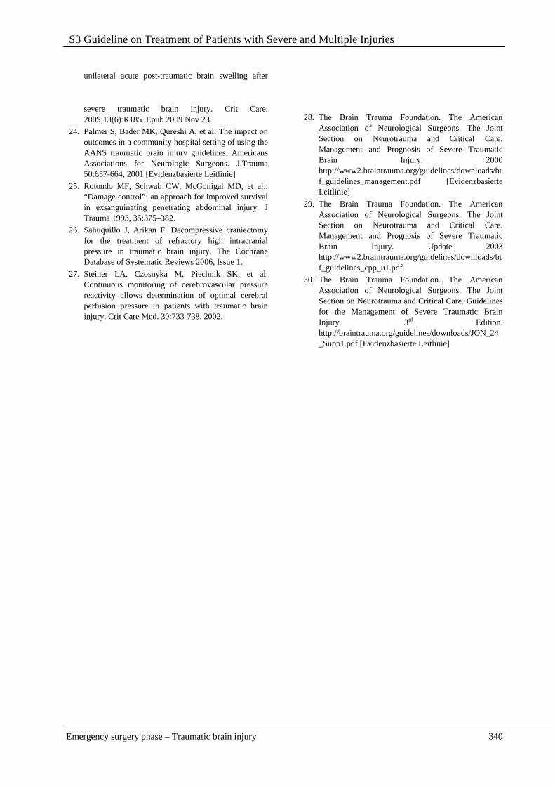
S3 Guideline on Treatment of Patients with Severe and Multiple Injuries
Emergency surgery phase – Traumatic brain injury 340
unilateral acute post‐traumatic brain swelling after
severe traumatic brain injury. Crit Care. 2009;13(6):R185. Epub 2009 Nov 23.
24. Palmer S, Bader MK, Qureshi A, et al: The impact on outcomes in a community hospital setting of using the AANS traumatic brain injury guidelines. Americans Associations for Neurologic Surgeons. J.Trauma 50:657-664, 2001 [Evidenzbasierte Leitlinie]
25. Rotondo MF, Schwab CW, McGonigal MD, et al.: “Damage control”: an approach for improved survival in exsanguinating penetrating abdominal injury. J Trauma 1993, 35:375–382.
26. Sahuquillo J, Arikan F. Decompressive craniectomy for the treatment of refractory high intracranial pressure in traumatic brain injury. The Cochrane Database of Systematic Reviews 2006, Issue 1.
27. Steiner LA, Czosnyka M, Piechnik SK, et al: Continuous monitoring of cerebrovascular pressure reactivity allows determination of optimal cerebral perfusion pressure in patients with traumatic brain injury. Crit Care Med. 30:733-738, 2002.
28. The Brain Trauma Foundation. The American Association of Neurological Surgeons. The Joint Section on Neurotrauma and Critical Care. Management and Prognosis of Severe Traumatic Brain Injury. 2000 http://www2.braintrauma.org/guidelines/downloads/btf_guidelines_management.pdf [Evidenzbasierte Leitlinie]
29. The Brain Trauma Foundation. The American Association of Neurological Surgeons. The Joint Section on Neurotrauma and Critical Care. Management and Prognosis of Severe Traumatic Brain Injury. Update 2003 http://www2.braintrauma.org/guidelines/downloads/btf_guidelines_cpp_u1.pdf.
30. The Brain Trauma Foundation. The American Association of Neurological Surgeons. The Joint Section on Neurotrauma and Critical Care. Guidelines for the Management of Severe Traumatic Brain Injury. 3rd Edition. http://braintrauma.org/guidelines/downloads/JON_24_Supp1.pdf [Evidenzbasierte Leitlinie]

S3 Guideline on Treatment of Patients with Severe and Multiple Injuries
Emergency surgery phase – Genitourinary tract 341
3.6 Genitourinary tract
Key recommendations:
Critical renal injuries (grade 5 according to the AAST classification) should be surgically explored.
GoR B
In the case of renal injuries < grade 5, a primary conservative procedure should be introduced in stable circulation conditions.
GoR B
If other injuries necessitate a laparotomy, renal injuries of average severity grade 3 or 4 can be surgically explored.
GoR 0
Explanation:
The indication to operate on renal injuries is now regarded with more restraint than a few years ago. In most cases, the decision to perform a laparotomy is already dictated by the intraabdominal concomitant injuries. However, life-threatening renal bleeding also represents an absolute indication for surgery [116]. The injury severity score according to AAST (American Association for the Surgery of Trauma) [117] has established itself as the basis of decision-making as this classification is closely correlated to the need for surgery and the possibility of salvaging the kidney [118]. Grade 5 renal injuries represent an indication for surgery because of the blood loss and/or threatening loss of renal function. In contrast, it is usually the case that grade 2 to grade 4 injuries can definitely be managed conservatively unless the patient has unstable circulation due to their renal injury; then the kidney should be surgically freed [119–133]. In the meantime, there are even individual reports by authors who have successfully treated grade 5 injuries conservatively [134, 135]. Provided pelvic or abdominal injuries require a laparotomy anyway, non-trivial renal injuries (> grade 2) can be surgically explored as this increases therapeutic certainty and may even make second interventions superfluous.
In particular, controversy has long surrounded the procedure for severe renal injuries with urine discharge and devitalized fragments. However, individual smaller studies have shown that even here non-surgical management is possible [136–138] even if the complication and revision rate here turns out to be markedly higher [139].
Pathologic pyelogram findings with additional evidence of a pulsating or expanding retroperitoneal hematoma should be surgically explored in cases where only i.v. pyelography can be carried out because of prioritizing. The evidence of hematuria and ideally also the ultrasound finding should be used here in the evaluation. Ichigi et al. showed that the size of the perirenal hematoma is closely linked to the severity of the renal injury [140] so that this criterion can also be used in the decision between a surgical or conservative procedure [4, 6, 9].

S3 Guideline on Treatment of Patients with Severe and Multiple Injuries
Emergency surgery phase – Genitourinary tract 342
Table 25: Grading classification of renal trauma according to the American Association for the Surgery of Trauma (AAST) [117]
Grade Properties
1 Renal contusion, perirenal or subcapsular hematoma, no other lesion in the imaging
2 Grade I lesion and laceration of the parenchyma up to 1 cm, collecting system not involved
3 Laceration > 1 cm without urine extravasation
4 Penetrating parenchymal lesion involving collecting system and/or hilar vessels
5 Shattered kidney and/or renal vascular pedicle avulsion, bleeding/sequestration
Key recommendation:
Selective angiographic embolization of a renal artery injury can be attempted as a therapeutic option in the patient with stable circulation.
GoR 0
Explanation:
Up till now, the importance of angiographic embolization has been documented in a few case series and case reports [95, 141, 142] but which also partly include non-traumatic bleeding of the renal arteries [143–145]). These studies also refer partly not only to the primary phase of polytrauma management but also describe the treatment of pseudoaneurysms or arteriovenous fistulas in the secondary phase [94, 146, 147]. According to these case series, bleeding is successfully arrested in about 82% [94] to 94% [95] of patients. In more recent review papers as well, the angiographic embolization of renal injuries in patients with stable circulation is increasingly accepted as the first intervention step albeit with reference to the monotrauma situation [9, 41, 47]. Usually, it involved branches of the renal artery which required embolization. It is undisputed that the selection of patients, the technical equipment, and the individual medical experience have a decisive influence on the success rate. Primarily because of the considerable amount of time required for embolization, selective angiographic embolization can only be successfully incorporated into the overall management in individual cases of multiply injured patients.

S3 Guideline on Treatment of Patients with Severe and Multiple Injuries
Emergency surgery phase – Genitourinary tract 343
Key recommendations:
Depending on the type and severity of injury and concomitant injuries, a renal injury can be surgically managed by oversewing or, if necessary, by partial renal resection and other procedures to salvage the organ.
GoR 0
Primary nephrectomy should be reserved for grade 5 injuries. GoR B
Explanation:
In the multiply injured patient with renal injury, the surgical approach is usually determined by the overall injury pattern and then normally consists of a midline laparotomy. In order to control renal bleeding, the renal pedicle is generally prepared before opening Gerota’s fascia. Individual zig-zag sutures and continuous sutures are then used to arrest the bleeding [116]. Fibrin adhesives can be advantageous here [148]. The surgical procedure for the multiply injured patient is largely identical to that for the monotrauma patient and there is no need to go into detail here.
The effort expended in reconstruction attempts should be based on the overall situation of the patient. Primary nephrectomy should be reserved for grade 5 injuries [9]. No long-term reconstruction attempts should be undertaken unless both kidneys are at risk. For reasons of time and the fewer complication possibilities, the indication for nephrectomy in the multiply injured patient should be made sooner than for the monotrauma patient [9, 41].
Ureter injuries
As ureter injuries are difficult to diagnose, if a laparotomy is being performed for another reason, it should be used for examining the ureters if such an injury is suspected [7]. Although macroscopic evaluation is also unreliable [102], it presents a huge advantage in that it allows a ureter injury to be treated early. Untreated ureter injuries lead to urine fistulas, urinomas, and infections so that the goal here should also be surgical management at the earliest opportunity [101]. The lesions are most frequently located in the proximal ureter [149]. A wide range of surgical procedures can be used [7].

S3 Guideline on Treatment of Patients with Severe and Multiple Injuries
Emergency surgery phase – Genitourinary tract 344
Bladder injuries
Key recommendations:
Intraperitoneal bladder ruptures should be surgically explored. GoR B
Extraperitoneal bladder ruptures without involvement of the neck of the bladder can be conservatively treated through suprapubic urinary diversion.
GoR 0
Explanation:
In most cases, the management of frequently numerous concomitant injuries should be given priority over a bladder injury. Numerically, extraperitoneal bladder ruptures are roughly twice common as intraperitoneal bladder ruptures [11, 60]. Combined extraperitoneal and intraperitoneal ruptures are markedly less frequently observed. Even taken on their own, intraperitoneal bladder ruptures represent a surgical indication since large tears are often found which can then lead to peritonitis and urinomas [150, 151]. The bladder should be closed and any urinomas drained.
The majority of extraperitoneal bladder injuries can be treated conservatively by means of a catheter drain, even if large retroperitoneal or scrotal extravasates are present [150]. Based on a series of 30 extraperitoneal ruptures, Cass and Luxemberg report on a 93% success rate with this non-surgical method [152]. In another series of 41 patients, almost all extraperitoneal bladder ruptures healed successfully within 3 weeks [153]. However, if the bladder neck is injured [11], bony fragments lie in the bladder or the bladder is clamped between bony pelvic fragments, a primary surgical procedure is necessary [1]. In the sequence of operations, osteosynthesis of the pelvis comes first followed by urological management [38]. Routt et al. also emphasize that good cooperation between the trauma surgeon and the urologist is essential here [38].

S3 Guideline on Treatment of Patients with Severe and Multiple Injuries
Emergency surgery phase – Genitourinary tract 345
Urethral injuries
Key recommendation:
Complete ruptures of the urethra should be treated in the emergency surgery phase by suprapubic urinary diversion.
GoR B
Urinary diversion can be supplemented by urethral re-alignment. GoR 0
Provided a pelvic fracture or another intraabdominal injury necessitates surgery anyway, urethral ruptures should be managed in the same session.
GoR B
Explanation:
It should be particularly mentioned in the management of urethral injuries that the method described here refers explicitly only to the emergency surgery phase as other principles also apply in the further management.
To date, there has been insufficient evidence as to whether primary, delayed or secondary re-anastomosing should be preferred in complete ruptures of the posterior urethra. In addition, primary and delayed urethral re-alignment is proposed [8]. The main problems in the post-traumatic course are urethral strictures, incontinence, and impotence so the treatment goal is to avoid them.
In a literature review summarizing several case series and comparative studies [20, 154–163] on the treatment of the urethral rupture, Koraitim [31, 164] describes the following rates of stricture, incontinence, and impotence: suprapubic diversion on its own 97%, 4% and 19%; primary re-alignment 53%, 5% and 36%; primary suture 49%, 21% and 56%. Accordingly, in the case of a complete urethral rupture in the male, he recommends suprapubic diversion on its own or re-alignment if there is a large gap between the ends of the urethra. However, as this literature review spans back more than 50 years, more recent studies on urethral re-alignment with better results are perhaps not sufficiently taken into account. Nevertheless, even current studies find both treatment options of equal value [165]. Accordingly, the EAST Guideline also comes to the conclusion that both primary re-alignment and also suprapubic diversion with secondary surgery are equally worthy of recommendation [10].
In the cases where surgery is necessary anyway due to other adjacent lesions, it appears expedient to manage the urethral rupture at the same time to avoid two-step management [166]. Particularly if the abdominal cavity is contaminated by large intestine injuries, primary suture of the urethra over a splinting catheter appears advisable to avoid complicating infections. Even if a conservative procedure actually appears to be possible, urethral injuries should be managed by primary surgery if the definitive osteosynthesis of the bony pelvis cannot otherwise be carried out [167].
Ruptures of the anterior urethra in the male are somewhat rarer than those of the posterior urethra. Primary surgical reconstruction may be necessary in open injuries. In the majority of cases, however, preference should also be given here to suprapubic urinary diversion followed by later reconstruction as reconstruction of the anterior urethra and the external male genitals, which are often injured as well, is usually difficult and

S3 Guideline on Treatment of Patients with Severe and Multiple Injuries
Emergency surgery phase – Genitourinary tract 346
time-consuming. However, in the case of a penile fracture with injury to the corpus spongiosum, it is recommended that the urethral injury also undergoes primary surgery [8, 168, 169]. The severity of the urological injury [170] and the overall severity of all injuries are crucial in deciding between primary surgery and conservative treatment.
Urethral injuries occur markedly less frequently in women than in men. However, when they occur, they are mostly very pronounced and associated with bladder injuries. For this reason, primary treatment should consist only of suprapubic urinary diversion if the patient has unstable circulation and/or other injuries require more urgent surgical management [67]. On the other hand, in women with less severe polytrauma, ruptures in the proximal urethra can undergo primary reconstruction via retropubic approach [69, 70, 171].
These recommendations apply similarly to children as well, whereby again a distinction should be made between the sexes. In a series of 35 boys with posterior urethra tears, Podestá et al. (1997) compared suprapubic diversion (with later urethroplasty), suprapubic diversion with urethral catheter alignment, and primary anastomosis [172]. As the continence rate after primary anastomosis only reached 50%, and in the group with catheter alignment all 10 patients still required urethroplasty later, the authors recommend suprapubic urinary diversion on its own followed by secondary urethroplasty. In a study on urethral injuries in girls with pelvic fracture and other concomitant injuries, the same authors found deferred management to be advantageous as good results could be observed despite vesical and vaginal concomitant injuries.

S3 Guideline on Treatment of Patients with Severe and Multiple Injuries
Emergency surgery phase – Genitourinary tract 347
Figure 5: Algorithm on the diagnostic and therapeutic procedure for suspected renal injuries
yes
Grade 3-
Primary
conservative
treatment
6 7
4
Emergency laparotomy
3
Suspected
blunt renal trauma
1
Circulatory shock due to abdominal bleeding?
2 yes
Gross or microscopic
hematuria?
Computer
tomography
(or other procedures)
5
yes
no
no
Pulsating retroperitoneal
hematoma? no
yes Severity grade of renal trauma
according to AAST?
Laparotomy necessary for another reason?
Grade 5 8
9
Surgical exploration
of renal injury
10

S3 Guideline on Treatment of Patients with Severe and Multiple Injuries
Emergency surgery phase – Genitourinary tract 348
References
1 Cass AS, Cass BP. Immediate surgical management of severe renal injuries in multiple-injured patients. Urology 1983: 21(2):140-145 [LoE 4].
2 Zink RA, Muller-Mattheis V, Oberneder R. [Results of the West German multicenter study "Urological traumatology"]. Urologe A 1990: 29(5):243-250.
3 Monstrey SJ, vander WC, Debruyne FM, Goris RJ. Urological trauma and severe associated injuries. Br J Urol 1987: 60(5):393-398.
4 Lynch D, Martinez-Pineiro L, las E, erafetinides E, urkeri L, ohenfellner M. Guidelines on urological trauma. http://uroweb.org/files/uploaded files/guidelines/urotrauma.pdf: Ezropean Association of Urology , 1-70. 2003 [Evidenzbasierte Leitlinie]
5 Lynch TH, Martinez-Pineiro L, Plas E, Serafetinides E, Turkeri L, Santucci RA et al. EAU guidelines on urological trauma. Eur Urol 2005: 47(1):1-15 [Evidenzbasierte Leitlinie]
6 Djakovic N, Plas E, Martinez-Pineiro L, Lynch TH, Mor Y, Santucci RA et al. Guidelines on urological trauma. http://uroweb.org/files/uploaded files/guidelines/urotrauma.pdf: Ezropean Associa-tion of Urology , 1-84. 2009 [Evidenzbasierte Leitlinie]
7 Brandes S, Coburn M, Armenakas N, McAninch J. Diagnosis and management of ureteric injury: an evidence-based analysis. BJU Int 2004: 94(3):277-289 [LoE 5].
8 Chapple C, Barbagli G, Jordan G, Mundy AR, Rodrigues-Netto N, Pansadoro V et al. Consensus statement on urethral trauma. BJU Int 2004: 93(9):1195-1202 [LoE 5].
9 Santucci RA, Wessells H, Bartsch G, Descotes J, Heyns CF, McAninch JW et al. Evaluation and management of renal injuries: consensus statement of the renal trauma subcommittee. BJU Int 2004: 93(7):937-954 [LoE 5].
10 Holevar MEJLFNKSRSJPYC. Practice management guidelines for the management of genitourinary trauma. http://www.east.org/tpg.html: Eastern Association for the Surgery of Trauma (EAST) , 1-101. 2004 [Evidenzbasierte Leitlinie]
11 Corriere JN, Jr., Sandler CM. Management of the ruptured bladder: seven years of experience with 111 cases. J Trauma 1986: 26(9):830-833 [LoE 2].
12 Glass RE, Flynn JT, King JB, Blandy JP. Urethral injury and fractured pelvis. Br J Urol 1978: 50(7):578-582.
13 Morehouse DD, Mackinnon KJ. Posterior urethral injury: etiology, diagnosis, initial management. Urol Clin North Am 1977: 4(1):69-73.
14 Nagel R, Leistenschneider W. [Urologic injuries in patients with multiple injuries]. Chirurg 1978: 49(12):731-736.
15 Medina D, Lavery R, Ross SE, Livingston DH. Ureteral trauma: preoperative studies neither predict injury nor prevent missed injuries. J Am Coll Surg 1998: 186(6):641-644.
16 Livingston DH, Lavery RF, Passannante MR, Skurnick JH, Fabian TC, Fry DE et al. Admission or observation is not necessary after a negative abdominal computed tomographic scan in patients with suspected blunt abdominal trauma: results of a prospective, multi-institutional trial. J Trauma 1998: 44(2):273-280.
17 Cotton BA, Beckert BW, Smith MK, Burd RS. The utility of clinical and laboratory data for predicting intraabdominal injury among children. J Trauma 2004: 56(5):1068-1074.
18 Allen GS, Moore FA, Cox CS, Jr., Wilson JT, Cohn JM, Duke JH. Hollow visceral injury and blunt trauma. J Trauma 1998: 45(1):69-75.
9 Guldner G, Babbitt J, Boulton M, O'Callaghan T, Feleke R, Hargrove J. Deferral of the rectal examination in blunt trauma patients: a clinical decision rule. Acad Emerg Med 2004: 11(6):635-641.
20 Porter JR, Takayama TK, Defalco AJ. Traumatic posterior urethral injury and early realignment using magnetic urethral catheters. J Urol 1997: 158(2):425-430 [LoE 4].
21 Taylor GA, Eichelberger MR, O'Donnell R, Bowman L. Indications for computed tomography in children with blunt abdominal trauma. Ann Surg 1991: 213(3):212-218.
22 Holmes JF, Sokolove PE, Brant WE, Palchak MJ, Vance CW, Owings JT et al. Identification of children with intra-abdominal injuries after blunt trauma. Ann Emerg Med 2002: 39(5):500-509.
23 Brosman SA, Paul JG. Trauma of the bladder. Surg Gynecol Obstet 1976: 143(4):605-608.
24 Iverson AJ, Morey AF. Radiographic evaluation of suspected bladder rupture following blunt trauma: critical review. World J Surg 2001: 25(12):1588-1591.
25 Demetriades D, Karaiskakis M, Toutouzas K, Alo K, Velmahos G, Chan L. Pelvic fractures: epidemiology and predictors of associated abdominal injuries and outcomes. J Am Coll Surg 2002: 195(1):1-10.
26 Dokucu AI, Ozdemir E, Ozturk H, Otcu S, Onen A, Cigdem K et al. Urogenital injuries in childhood: a strong association of bladder trauma to bowel injuries. Int Urol Nephrol 2000: 32(1):3-8.
27 Beck D, Marley R, Salvator A, Muakkassa F. Prospective study of the clinical predictors of a positive abdominal computed tomography in blunt trauma patients. J Trauma 2004: 57(2):296-300.
28 Carter CT, Schafer N. Incidence of urethral disruption in females with traumatic pelvic fractures. Am J Emerg Med 1993: 11(3):218-220.
29 Nicolaisen GS, McAninch JW, Marshall GA, Bluth RF, Jr., Carroll PR. Renal trauma: re-evaluation of the indications for radiographic assessment. J Urol 1985: 133(2):183-187.
30 Ingram MD, Watson SG, Skippage PL, Patel U. Urethral injuries after pelvic trauma: evaluation with urethrography. Radiographics 2008: 28(6):1631-1643.

S3 Guideline on Treatment of Patients with Severe and Multiple Injuries
Emergency surgery phase – Genitourinary tract 349
31 Koraitim MM. Pelvic fracture urethral injuries: evaluation of various methods of management. J Urol 1996: 156(4):1288-1291 [LoE 2b].
32 Chandhoke PS, McAninch JW. Detection and significance of microscopic hematuria in patients with blunt renal trauma. J Urol 1988: 140(1):16-18.
33 Daum GS, Krolikowski FJ, Reuter KL, Colby JM, Silva WM. Dipstick evaluation of hematuria in abdominal trauma. Am J Clin Pathol 1988: 89(4):538-542.
34 Goldner AP, Mayron R, Ruiz E. Are urine dipsticks reliable indicators of hematuria in blunt trauma patients? Ann Emerg Med 1985: 14(6):580-582.
35 Kennedy TJ, McConnell JD, Thal ER. Urine dipstick vs. microscopic urinalysis in the evaluation of abdominal trauma. J Trauma 1988: 28(5):615-617.
36 Mariani AJ, Luangphinith S, Loo S, Scottolini A, Hodges CV. Dipstick chemical urinalysis: an accurate cost-effective screening test. J Urol 1984: 132(1):64-66.
37 Frohmüller H, Theiß M. Blutungen bei Verletzungen des Harntrakts. Langenbecks Arch Chir 1993: 110(Suppl):352-357.
38 Routt ML, Simonian PT, Defalco AJ, Miller J, Clarke T. Internal fixation in pelvic fractures and primary repairs of associated genitourinary disruptions: a team approach. J Trauma 1996: 40(5):784-790 [LoE 4].
39 Zwergel T, op den WR, Zwergel U, Schwaiger R, Muhr G, Ziegler M. [Concept of interdisciplinary procedures within the scope of traumatology--the status of urology]. Unfallchirurgie 1983: 9(5):244-248.
40 Peterson NE, Schulze KA. Selective diagnostic uroradiography for trauma. J Urol 1987: 137(3):449-451.
41 Schmidlin F. [Renal trauma. Treatment strategies and indications for surgical exploration]. Urologe A 2005: 44(8):863-869 [LoE 3].
42 Pfitzenmaier J, Buse S, Haferkamp A, Pahernik S, Djakovic N, Hohenfellner M. [Kidney trauma]. Urologe A 2008: 47(6):759-767.
43 Fortune JB, Brahme J, Mulligan M, Wachtel TL. Emergency intravenous pyelography in the trauma patient. A reexamination of the indications. Arch Surg 1985: 120(9):1056-1059.
44 Klein S, Johs S, Fujitani R, State D. Hematuria following blunt abdominal trauma. The utility of intravenous pyelography. Arch Surg 1988: 123(9):1173-1177.
45 Levitt MA, Criss E, Kobernick M. Should the emergency IVP be used more selectively in blunt renal trauma? Ann Emerg Med 1985: 14(10):959-965.
46 Thomason RB, Julian JS, Mostellar HC, Pennell TC, Meredith JW. Microscopic hematuria after blunt trauma. Is pyelography necessary? Am Surg 1989: 55(3):145-150.
47 Pfitzenmaier J, Buse S, Haferkamp A, Pahernik S, Hohenfellner M. Nierentrauma. Der Unfalllchirurg 2009: 112:317-326 [LoE 3].
48 Miller KS, McAninch JW. Radiographic assessment of renal trauma: our 15-year experience. J Urol 1995: 154(2 Pt 1):352-355.
49 McAndrew JD, Corriere JN, Jr. Radiographic evaluation of renal trauma: evaluation of 1103 consecutive patients. Br J Urol 1994: 73(4):352-354.
50 Fallon B, Wendt JC, Hawtrey CE. Urological injury and assessment in patients with fractured pelvis. J Urol 1984: 131(4):712-714.
51 Mee SL, McAninch JW, Robinson AL, Auerbach PS, Carroll PR. Radiographic assessment of renal trauma: a 10-year prospective study of patient selection. J Urol 1989: 141(5):1095-1098.
52 Fuhrman GM, Simmons GT, Davidson BS, Buerk CA. The single indication for cystography in blunt trauma. Am Surg 1993: 59(6):335-337.
53 Mayor B, Gudinchet F, Wicky S, Reinberg O, Schnyder P. Imaging evaluation of blunt renal trauma in children: diagnostic accuracy of intravenous pyelography and ultrasonography. Pediatr Radiol 1995: 25(3):214-218.
54 Middlebrook PF, Schillinger JF. Hematuria and intravenous pyelography in pediatric blunt renal trauma. Can J Surg 1993: 36(1):59-62.
55 Perez-Brayfield MR, Gatti JM, Smith EA, Broecker B, Massad C, Scherz H et al. Blunt traumatic hematuria in children. Is a simplified algorithm justified? J Urol 2002: 167(6):2543-2546.
56 Stein JP, Kaji DM, Eastham J, Freeman JA, Esrig D, Hardy BE. Blunt renal trauma in the pediatric population: indications for radiographic evaluation. Urology 1994: 44(3):406-410.
57 Brandes SB, McAninch JW. Urban free falls and patterns of renal injury: a 20-year experience with 396 cases. J Trauma 1999: 47(4):643-649.
58 Carroll PR, McAninch JW. Major bladder trauma: mechanisms of injury and a unified method of diagnosis and repair. J Urol 1984: 132(2):254-257.
59 Flancbaum L, Morgan AS, Fleisher M, Cox EF. Blunt bladder trauma: manifestation of severe injury. Urology 1988: 31(3):220-222.
60 Hochberg E, Stone NN. Bladder rupture associated with pelvic fracture due to blunt trauma. Urology 1993: 41(6):531-533 [LoE 2].
61 Aihara R, Blansfield JS, Millham FH, LaMorte WW, Hirsch EF. Fracture locations influence the likelihood of rectal and lower urinary tract injuries in patients sustaining pelvic fractures. J Trauma 2002: 52(2):205-208.
62 Batislam E, Ates Y, Germiyanoglu C, Karabulut A, Gulerkaya B, Erol D. Role of Tile classification in predicting urethral injuries in pediatric pelvic fractures. J Trauma 1997: 42(2):285-287.
63 Knudson MM, McAninch JW, Gomez R, Lee P, Stubbs HA. Hematuria as a predictor of abdominal injury after blunt trauma. Am J Surg 1992: 164(5):482-485.
64 Koraitim MM, Marzouk ME, Atta MA, Orabi SS. Risk factors and mechanism of urethral injury in pelvic fractures. Br J Urol 1996: 77(6):876-880.

S3 Guideline on Treatment of Patients with Severe and Multiple Injuries
Emergency surgery phase – Genitourinary tract 350
65 Morgan DE, Nallamala LK, Kenney PJ, Mayo MS, Rue LW, III. CT cystography: radiographic and clinical predictors of bladder rupture. AJR Am J Roentgenol 2000: 174(1):89-95.
66 Palmer JK, Benson GS, Corriere JN, Jr. Diagnosis and initial management of urological injuries associated with 200 consecutive pelvic fractures. J Urol 1983: 130(4):712-714.
67 Venn SN, Greenwell TJ, Mundy AR. Pelvic fracture injuries of the female urethra. BJU Int 1999: 83(6):626-630 [LoE 4].
68 Ahmed S, Neel KF. Urethral injury in girls with fractured pelvis following blunt abdominal trauma. Br J Urol 1996: 78(3):450-453.
69 Thambi Dorai CR, Boucaut HA, Dewan PA. Urethral injuries in girls with pelvic trauma. Eur Urol 1993: 24(3):371-374 [LoE 4].
70 Black PC, Miller EA, Porter JR, Wessells H. Urethral and bladder neck injury associated with pelvic fracture in 25 female patients. J Urol 2006: 175(6):2140-2144 [LoE 4].
71 Morey AF, Iverson AJ, Swan A, Harmon WJ, Spore SS, Bhayani S et al. Bladder rupture after blunt trauma: guidelines for diagnostic imaging. J Trauma 2001: 51(4):683-686 [Evidenzbasierte Leitlinie]
72 Rehm CG, Mure AJ, O'Malley KF, Ross SE. Blunt traumatic bladder rupture: the role of retrograde cystogram. Ann Emerg Med 1991: 20(8):845-847.
73 Hsieh CH, Chen RJ, Fang JF, Lin BC, Hsu YP, Kao JL et al. Diagnosis and management of bladder injury by trauma surgeons. Am J Surg 2002: 184(2):143-147.
74 Wolk DJ, Sandler CM, Corriere JN, Jr. Extraperitoneal bladder rupture without pelvic fracture. J Urol 1985: 134(6):1199-1201.
75 Buchberger W, Penz T, Wicke K, Eberle J. [Diagnosis and staging of blunt kidney trauma. A comparison of urinalysis, i.v. urography, sonography and computed tomography]. Rofo 1993: 158(6):507-512.
76 Cass AS, Vieira J. Comparison of IVP and CT findings in patients with suspected severe renal injury. Urology 1987: 29(5):484-487.
77 Bretan PN, Jr., McAninch JW, Federle MP, Jeffrey RB, Jr. Computerized tomographic staging of renal trauma: 85 consecutive cases. J Urol 1986: 136(3):561-565.
78 Fanney DR, Casillas J, Murphy BJ. CT in the diagnosis of renal trauma. Radiographics 1990: 10(1):29-40.
79 Kinnunen J, Kivioja A, Poussa K, Laasonen EM. Emergency CT in blunt abdominal trauma of multiple injury patients. Acta Radiol 1994: 35(4):319-322.
80 Becker CD, Mentha G, Schmidlin F, Terrier F. Blunt abdominal trauma in adults: role of CT in the diagnosis and management of visceral injuries. Part 2: Gastrointestinal tract and retroperitoneal organs. Eur Radiol 1998: 8(5):772-780.
81 Rhea JT, Garza DH, Novelline RA. Controversies in emergency radiology. CT versus ultrasound in the evaluation of blunt abdominal trauma. Emerg Radiol 2004: 10(6):289-295.
82 Wah TM, Spencer JA. The role of CT in the management of adult urinary tract trauma. Clin Radiol 2001: 56(4):268-277.
83 Morey AF, McAninch JW, Tiller BK, Duckett CP, Carroll PR. Single shot intraoperative excretory urography for the immediate evaluation of renal trauma. J Urol 1999: 161(4):1088-1092.
84 Halsell RD, Vines FS, Shatney CH, Slepin MJ, Northup HM, Avara WT, III et al. The reliability of excretory urography as a screening examination for blunt renal trauma. Ann Emerg Med 1987: 16(11):1236-1239.
85 Poletti PA, Kinkel K, Vermeulen B, Irmay F, Unger PF, Terrier F. Blunt abdominal trauma: should US be used to detect both free fluid and organ injuries? Radiology 2003: 227(1):95-103.
86 Yoshii H, Sato M, Yamamoto S, Motegi M, Okusawa S, Kitano M et al. Usefulness and limitations of ultrasonography in the initial evaluation of blunt abdominal trauma. J Trauma 1998: 45(1):45-50.
87 Stengel D, Bauwens K, Porzsolt F, Rademacher G, Mutze S, Ekkernkamp A. [Emergency ultrasound for blunt abdominal trauma--meta-analysis update 2003]. Zentralbl Chir 2003: 128(12):1027-1037.
88 Stengel D, Bauwens K, Sehouli J, Porzsolt F, Rademacher G, Mutze S et al. Systematic review and meta-analysis of emergency ultrasonography for blunt abdominal trauma. Br J Surg 2001: 88(7):901-912.
89 Rose JS, Levitt MA, Porter J, Hutson A, Greenholtz J, Nobay F et al. Does the presence of ultrasound really affect computed tomographic scan use? A prospective randomized trial of ultrasound in trauma. J Trauma 2001: 51(3):545-550.
90 Lupetin AR, Mainwaring BL, Daffner RH. CT diagnosis of renal artery injury caused by blunt abdominal trauma. AJR Am J Roentgenol 1989: 153(5):1065-1068.
91 Corr P, Hacking G. Embolization in traumatic intrarenal vascular injuries. Clin Radiol 1991: 43(4):262-264.
92 Eastham JA, Wilson TG, Larsen DW, Ahlering TE. Angiographic embolization of renal stab wounds. J Urol 1992: 148(2 Pt 1):268-270.
93 Hagiwara A, Murata A, Matsuda T, Matsuda H, Shimazaki S. The usefulness of transcatheter arterial embolization for patients with blunt polytrauma showing transient response to fluid resuscitation. J Trauma 2004: 57(2):271-276.
94 Heyns CF, van VP. Increasing role of angiography and segmental artery embolization in the management of renal stab wounds. J Urol 1992: 147(5):1231-1234 [LoE 4].
95 Uflacker R, Paolini RM, Lima S. Management of traumatic hematuria by selective renal artery embolization. J Urol 1984: 132(4):662-667 [LoE 4].
96 Leppaniemi A, Lamminen A, Tervahartiala P, Haapiainen R, Lehtonen T. Comparison of high-field magnetic resonance imaging with computed tomography in the evaluation of blunt renal trauma. J Trauma 1995: 38(3):420-427 [LoE 4].
S3 Guideline on Treatment of Patients with Severe and Multiple Injuries
Emergency surgery phase – Genitourinary tract 351
97 Leppaniemi A, Lamminen A, Tervahartiala P, Salo J, Haapiainen R, Lehtonen T. MRI and CT in blunt renal trauma: an update. Semin Ultrasound CT MR 1997: 18(2):129-135.
98 Leppaniemi AK, Kivisaari AO, Haapiainen RK, Lehtonen TA. Role of magnetic resonance imaging in blunt renal parenchymal trauma. Br J Urol 1991: 68(4):355-360.
99 Ku JH, Jeon YS, Kim ME, Lee NK, Park YH. Is there a role for magnetic resonance imaging in renal trauma? Int J Urol 2001: 8(6):261-267.
100 Ambiavagar R, Nambiar R. Traumatic closed avulsion of the upper ureter. Injury 1979: 11(1):71-76.
101 Armenakas NA. Current methods of diagnosis and management of ureteral injuries. World J Urol 1999: 17(2):78-83 [LoE 4].
102 Ghali AM, El Malik EM, Ibrahim AI, Ismail G, Rashid M. Ureteric injuries: diagnosis, management, and outcome. J Trauma 1999: 46(1):150-158 [LoE 4].
103 Dobrowolski Z, Kusionowicz J, Drewniak T, Habrat W, Lipczynski W, Jakubik P et al. Renal and ureteric trauma: diagnosis and management in Poland. BJU Int 2002: 89(7):748-751.
104 Sandler CM, Goldman SM, Kawashima A. Lower urinary tract trauma. World J Urol 1998: 16(1):69-75.
105 Carroll PR, McAninch JW. Major bladder trauma: the accuracy of cystography. J Urol 1983: 130(5):887-888.
106 Festini G, Gregorutti S, Reina G, Bellis GB. Isolated intraperitoneal bladder rupture in patients with alcohol intoxication and minor abdominal trauma. Ann Emerg Med 1991: 20(12):1371-1372.
107 MacMahon R, Hosking D, Ramsey EW. Management of blunt injury to the lower urinary tract. Can J Surg 1983: 26(5):415-418.
108 Werkman HA, Jansen C, Klein JP, Ten Duis HJ. Urinary tract injuries in multiply-injured patients: a rational guideline for the initial assessment. Injury 1991: 22(6):471-474 [Evidenzbasierte Leitlinie]
109 Vaccaro JP, Brody JM. CT cystography in the evaluation of major bladder trauma. Radiographics 2000: 20(5):1373-1381.
110 Pao DM, Ellis JH, Cohan RH, Korobkin M. Utility of routine trauma CT in the detection of bladder rupture. Acad Radiol 2000: 7(5):317-324.
111 Deck AJ, Shaves S, Talner L, Porter JR. Computerized tomography cystography for the diagnosis of traumatic bladder rupture. J Urol 2000: 164(1):43-46.
112 Deck AJ, Shaves S, Talner L, Porter JR. Current experience with computed tomographic cystography and blunt trauma. World J Surg 2001: 25(12):1592-1596.
113 Peng MY, Parisky YR, Cornwell EE, III, Radin R, Bragin S. CT cystography versus conventional cystography in evaluation of bladder injury. AJR Am J Roentgenol 1999: 173(5):1269-1272.
114 Horstman WG, McClennan BL, Heiken JP. Comparison of computed tomography and conventional cystography for detection of traumatic bladder rupture. Urol Radiol 1991: 12(4):188-193.
115 Lis LE, Cohen AJ. CT cystography in the evaluation of bladder trauma. J Comput Assist Tomogr 1990: 14(3):386-389.
116 Brandes SB, McAninch JW. Reconstructive surgery for trauma of the upper urinary tract. Urol Clin North Am 1999: 26(1):183-99, [LoE 5].
117 Moore EE, Shackford SR, Pachter HL, McAninch JW, Browner BD, Champion HR et al. Organ injury scaling: spleen, liver, and kidney. J Trauma 1989: 29(12):1664-1666.
118 Santucci RA, McAninch JW, Safir M, Mario LA, Service S, Segal MR. Validation of the American Association for the Surgery of Trauma organ injury severity scale for the kidney. J Trauma 2001: 50(2):195-200 [LoE 4].
119 Wessells H, McAninch JW, Meyer A, Bruce J. Criteria for nonoperative treatment of significant penetrating renal lacerations. J Urol 1997: 157(1):24-27 [LoE 2].
120 Buckley JC, McAninch JW. Selective management of isolated and nonisolated grade IV renal injuries. J Urol 2006: 176(6 Pt 1):2498-2502 [LoE 4].
121 Baverstock R, Simons R, McLoughlin M. Severe blunt renal trauma: a 7-year retrospective review from a provincial trauma centre. Can J Urol 2001: 8(5):1372-1376 [LoE 4].
122 Cheng DL, Lazan D, Stone N. Conservative treatment of type III renal trauma. J Trauma 1994: 36(4):491-494 [LoE 4].
123 Smith EM, Elder JS, Spirnak JP. Major blunt renal trauma in the pediatric population: is a nonoperative approach indicated? J Urol 1993: 149(3):546-548 [LoE 4].
124 El-Sherbiny MT, Aboul-Ghar ME, Hafez AT, Hammad AA, Bazeed MA. Late renal functional and morphological evaluation after non-operative treatment of high-grade renal injuries in children. BJU Int 2004: 93(7):1053-1056 [LoE 4].
125 Hammer CC, Santucci RA. Effect of an institutional policy of nonoperative treatment of grades I to IV renal injuries. J Urol 2003: 169(5):1751-1753 [LoE 4].
126 Keller M. Functional outcome of nonoperatively managed renal injuries in children. J Trauma 2004: 57:108-110 [LoE 4].
127 Levy JB, Baskin LS, Ewalt DH, Zderic SA, Bellah R, Snyder HM, III et al. Nonoperative management of blunt pediatric major renal trauma. Urology 1993: 42(4):418-424 [LoE 4].
128 Moudouni SM, Hadj SM, Manunta A, Patard JJ, Guiraud PH, Guille F et al. Management of major blunt renal lacerations: is a nonoperative approach indicated? Eur Urol 2001: 40(4):409-414 [LoE 4].
129 Robert M, Drianno N, Muir G, Delbos O, Guiter J. Management of major blunt renal lacerations: surgical or nonoperative approach? Eur Urol 1996: 30(3):335-339 [LoE 4].
130 Russell RS, Gomelsky A, McMahon DR, Andrews D, Nasrallah PF. Management of grade IV renal injury in children. J Urol 2001: 166(3):1049-1050 [LoE 4].

S3 Guideline on Treatment of Patients with Severe and Multiple Injuries
Emergency surgery phase – Genitourinary tract 352
131 Santucci RA, McAninch JM. Grade IV renal injuries: evaluation, treatment, and outcome. World J Surg 2001: 25(12):1565-1572 [LoE 4].
132 Thall EH, Stone NN, Cheng DL, Cohen EL, Fine EM, Leventhal I et al. Conservative management of penetrating and blunt Type III renal injuries. Br J Urol 1996: 77(4):512-517 [LoE 4].
133 Toutouzas KG, Karaiskakis M, Kaminski A, Velmahos GC. Nonoperative management of blunt renal trauma: a prospective study. Am Surg 2002: 68(12):1097-1103 [LoE 4].
134 Bozeman C, Carver B, Zabari G, Caldito G, Venable D. Selective operative management of major blunt renal trauma. J Trauma 2004: 57(2):305-309 [LoE 2].
135 Altman AL, Haas C, Dinchman KH, Spirnak JP. Selective nonoperative management of blunt grade 5 renal injury. J Urol 2000: 164(1):27-30 [LoE 4].
136 Husmann DA, Gilling PJ, Perry MO, Morris JS, Boone TB. Major renal lacerations with a devitalized fragment following blunt abdominal trauma: a comparison between nonoperative (expectant) versus surgical management. J Urol 1993: 150(6):1774-1777 [LoE 2b].
137 Matthews LA, Smith EM, Spirnak JP. Nonoperative treatment of major blunt renal lacerations with urinary extravasation. J Urol 1997: 157(6):2056-2058 [LoE 4].
138 Moudouni SM, Patard JJ, Manunta A, Guiraud P, Guille F, Lobel B. A conservative approach to major blunt renal lacerations with urinary extravasation and devitalized renal segments. BJU Int 2001: 87(4):290-294 [LoE 4].
139 Husmann DA, Morris JS. Attempted nonoperative management of blunt renal lacerations extending through the corticomedullary junction: the short-term and long-term sequelae. J Urol 1990: 143(4):682-684 [LoE 4].
140 Ichigi Y, Takaki N, Nakamura K, Sato S, Kato A, Matsuo Y et al. Significance of hematoma size for evaluating the grade of blunt renal trauma. Int J Urol 1999: 6(10):502-508 [LoE 2].
141 Goldin AR, Funston MR. Percutaneous embolization for post-traumatic renal haemorrhage. S Afr Med J 1978: 53(26):1061-1063 [LoE 4].
142 Richman SD, Green WM, Kroll R, Casarella WJ. Superselective transcatheter embolization of traumatic renal hemorrhage. AJR Am J Roentgenol 1977: 128(5):843-846 [LoE 4].
143 Beaujeux R, Saussine C, al-Fakir A, Boudjema K, Roy C, Jacqmin D et al. Superselective endo-vascular treatment of renal vascular lesions. J Urol 1995: 153(1):14-17 [LoE 4].
144 Chatziioannou A, Brountzos E, Primetis E, Malagari K, Sofocleous C, Mourikis D et al. Effects of superselective embolization for renal vascular injuries on renal parenchyma and function. Eur J Vasc Endovasc Surg 2004: 28(2):201-206 [LoE 4].
145 Fisher RG, Ben-Menachem Y, Whigham C. Stab wounds of the renal artery branches: angiographic diagnosis and treatment by embolization. AJR Am J Roentgenol 1989: 152(6):1231-1235 [LoE 4].
146 Dinkel HP, Danuser H, Triller J. Blunt renal trauma: minimally invasive management with microcatheter embolization experience in nine patients. Radiology 2002: 223(3):723-730 [LoE 4].
147 Miller DC, Forauer A, Faerber GJ. Successful angioembolization of renal artery pseudoaneurysms after blunt abdominal trauma. Urology 2002: 59(3):444 [LoE 4].
148 Shekarriz B, Stoller ML. The use of fibrin sealant in urology. J Urol 2002: 167(3):1218-1225 [LoE 4].
149 Elliott SP, McAninch JW. Ureteral injuries from external violence: the 25-year experience at San Francisco General Hospital. J Urol 2003: 170(4 Pt 1):1213-1216 [LoE 4].
150 Morey AF, Hernandez J, McAninch JW. Reconstructive surgery for trauma of the lower urinary tract. Urol Clin North Am 1999: 26(1):49-60, viii [LoE 5].
151 Thomae KR, Kilambi NK, Poole GV. Method of urinary diversion in nonurethral traumatic bladder injuries: retrospective analysis of 70 cases. Am Surg 1998: 64(1):77-80 [LoE 4].
152 Cass AS, Luxenberg M. Features of 164 bladder ruptures. J Urol 1987: 138(4):743-745 [LoE 4].
153 Corriere JN, Jr., Sandler CM. Management of extraperitoneal bladder rupture. Urol Clin North Am 1989: 16(2):275-277 [LoE 4].
154 Asci R, Sarikaya S, Buyukalpelli R, Saylik A, Yilmaz AF, Yildiz S. Voiding and sexual dysfunctions after pelvic fracture urethral injuries treated with either initial cystostomy and delayed urethroplasty or immediate primary urethral realignment. Scand J Urol Nephrol 1999: 33(4):228-233 [LoE 2b].
155 Elliott DS, Barrett DM. Long-term followup and evaluation of primary realignment of posterior urethral disruptions. J Urol 1997: 157(3):814-816 [LoE 2b].
156 Follis HW, Koch MO, McDougal WS. Immediate management of prostatomembranous urethral disruptions. J Urol 1992: 147(5):1259-1262 [LoE 2b].
157 Herschorn S, Thijssen A, Radomski SB. The value of immediate or early catheterization of the traumatized posterior urethra. J Urol 1992: 148(5):1428-1431 [LoE 2b].
158 Husmann DA, Wilson WT, Boone TB, Allen TD. Prostatomembranous urethral disruptions: management by suprapubic cystostomy and delayed urethroplasty. J Urol 1990: 144(1):76-78 [LoE 2b].
159 Kotkin L, Koch MO. Impotence and incontinence after immediate realignment of posterior urethral trauma: result of injury or management? J Urol 1996: 155(5):1600-1603 [LoE 2b].
160 Gheiler EL, Frontera JR. Immediate primary realignment of prostatomembranous urethral disruptions using endourologic techniques. Urology 1997: 49(4):596-599 [LoE 4].
161 Singh PB, Karmakar D, Gupta RC, Dwivedi US, Tripathi VN. Result of suprapubic cystostomy only as primary management of posterior urethral rupture following pelvic fracture. Int Surg 1988: 73(1):59-62 [LoE 4].
S3 Guideline on Treatment of Patients with Severe and Multiple Injuries
Emergency surgery phase – Genitourinary tract 353
162 Mouraviev VB, Coburn M, Santucci RA. The treatment of posterior urethral disruption associated with pelvic fractures: comparative experience of early realignment versus delayed urethroplasty. J Urol 2005: 173(3):873-876 [LoE 4].
163 Culty T, Boccon-Gibod L. Anastomotic urethroplasty for posttraumatic urethral stricture: previous urethral manipulation has a negative impact on the final outcome. J Urol 2007: 177(4):1374-1377 [LoE 4].
164 Koraitim MM. Pelvic fracture urethral injuries: the unresolved controversy. J Urol 1999: 161(5):1433-1441 [LoE 2b].
165 Ku JH, Jeon YS, Kim ME, Lee NK, Park YH. Comparison of long-term results according to the primary mode of management and type of injury for posterior urethral injuries. Urol Int 2002: 69(3):227-232 [LoE 2b].
166 Brandes S, Borrelli J, Jr. Pelvic fracture and associated urologic injuries. World J Surg 2001: 25(12):1578-1587 [LoE 5].
167 Mayher BE, Guyton JL, Gingrich JR. Impact of urethral injury management on the treatment and outcome of concurrent pelvic fractures. Urology 2001: 57(3):439-442 [LoE 4].
168 Anselmo G, Fandella A, Faggiano L, Merlo F, Maccatrozzo L. Fractures of the penis: therapeutic approach and long-term results. Br J Urol 1991: 67(5):509-511 [LoE 4].
169 Nane I, Esen T, Tellaloglu S, Selhanoglu M, Akinci M. Penile fracture: emergency surgery for preservation of penile functions. Andrologia 1991: 23(4):309-311 [LoE 4].
170 Mohr AM, Pham AM, Lavery RF, Sifri Z, Bargman V, Livingston DH. Management of trauma to the male external genitalia: the usefulness of American Association for the Surgery of Trauma organ injury scales. J Urol 2003: 170(6 Pt 1):2311-2315 [LoE 4].
171 Netto Junior NR, Ikari O, Zuppo VP. Traumatic rupture of female urethra. Urology 1983: 22(6):601-603 [LoE 4].
172 Podesta ML, Medel R, Castera R, Ruarte A. Immediate management of posterior urethral disruptions due to pelvic fracture: therapeutic alternatives. J Urol 1997: 157(4):1444-1448 [LoE 2b]
S3 Guideline on Treatment of Patients with Severe and Multiple Injuries
Emergency surgery phase – Spine 354
3.7 Spine
Indication for surgery
Key recommendation:
Unstable spinal injuries with confirmed or assumed neurologic deficits, with malpositions in which neurologic deficits can probably be prevented or improved by reduction, decompression, and stabilization, should be operated on as early as possible (“day 1 surgery”).
GoR B
Explanation:
After life-threatening injuries to the body cavities and the head, and the long bones, spinal injuries occupy third place, or second place if there is a spinal cord injury, in management priority [1].
Surgical indications are atlanto-occipital dislocation, translatory atlanto-axial dislocation, unstable Jefferson fracture, unstable Dens fracture (particularly type II), Hangman fracture (rib fracture C2 and invertebral disc injury C2/C3), C3 to C7 fractures (A3, B and C types) also in terms of dislocation, and T1 to L5 fractures (A3, B and C types) also in terms of dislocation. According to prevailing opinion, an absolute primary surgical indication exists even if there is an open spinal injury [2, 3].
In addition, the indication for primary management of a spinal injury in polytrauma is assisted by the classification according to Blauth et al. (1998) into a) complex spinal injuries with an injury to essential neural pathways and organs such as the spinal cord, lung, great vessels and abdominal organs, b) unstable spinal injuries (type A3, B and C - in this case, functional treatment can lead to severe malpositions and neurologic damage), and c) stable spinal injuries. If a complex spinal injury or an unstable spinal injury is involved, the goal should be surgical stabilization at the earliest possible opportunity - in other words, on the day of the accident if none of the contraindications mentioned below are present [4].
According to Blauth et al. (1998), a complex spinal injury is a multi-level spinal injury or one accompanied by intrathoracic or intraabdominal injury or polytrauma. The fact that polytrauma makes a spinal injury a “complex” injury is substantiated inter alia by studies by Hebert and Burnham [5] which established that the length of stay in hospital was extended and the number of surgical interventions increased in these patients and that the combination of spinal injury/polytrauma is associated with an increased morbidity and mortality and an increased degree of disability. Nevertheless, according to a survey in North America by Tator et al. (1999), 1/3 of spinal injuries with neurologic injuries were still conservatively treated and of those operated on only 60% received surgery before the 5th day and 40% after [6].
The goal of primary surgical management in unstable spinal injuries with confirmed or assumed neurologic deficits or with malpositions is firstly early spinal decompression and the avoidance
S3 Guideline on Treatment of Patients with Severe and Multiple Injuries
Emergency surgery phase – Spine 355
of neurologic secondary damage and secondly to achieve positioning stability for the intensive treatment [1].
The indication for surgery to avoid neurologic damage is relatively clear in unstable fractures without spinal cord lesion. If there are spinal injuries which are unstable and which could be displaced by necessary positioning measures such as in chest trauma, the indication for primary spinal stabilization should be made [4, 7-9]. However, controversy surrounds the issue of whether early or later fracture management is advantageous in fractures where spinal cord injury has already occurred. As far as neurologic symptoms are concerned, animal experiments show advantages in spinal stabilization being carried out as early as possible [10, 11]. However, in the field of clinical research, several large systematic reviews (some with meta-analysis) could detect no clear correlation between the timing of surgery and the neurologic outcome [12-15].
Only the most recent meta-analysis by La Rosa et al. [12] revealed that early surgical decompression has advantages compared to late decompression or conservative treatment. In the early group (17 studies), an improvement in neurology could be found in 42% of patients with complete deficit, and in 90% with incomplete deficit. In the late group, the improvement quotas were 8% and 59%, in the conservative group 25% and 59%. However, as the results from the studies differ greatly, La Rosa et al. also describe early surgery only as a “practical option”.
The only randomized controlled trial on this was conducted on 62 patients with cervical spinal injury only [16]. Although the authors found no difference between early (< 72 hours) versus late stabilization (> 5 days) in the neurologic outcome, they still recommended early stabilization. Levi et al. also found indifferent results concerning the early (< 24 hours) and late (> 24 hours) stabilization in the cervical spine injury but ultimately also recommend early surgery [17]. After Wagner and Chehrazi also found no correlation between the timing of surgery and neurologic outcome in cervical spine injuries, they concluded [18] that primary medullary damage determines the prognosis. McKinley et al. draw a similar conclusion [19]. In contrast, Papadopoulos et al. observed improved neurologic outcomes after early surgery [20]. Mirz et al. also described in 1999 [21] that early stabilization (< 72 hours) of a cervical spine injury is more favorable for the neurologic outcome than later stabilization (> 72 hours). However, all these are data from studies that did not study exclusively multiply injured patients.
In addition to these studies focusing primarily on the neurologic outcome, there is a series of studies which have concentrated mainly on the non-neurologic effects of early stabilization. A study by Croce et al. found evidence in 2001 [7] that, in contrast to late stabilization (> 3 days after trauma), early stabilization (< 3 days after trauma) of the spinal injury offers advantages, especially in polytrauma (mean ISS 24) with thoracic spine injury, as the intensive care period, pneumonia rate, costs, and ventilation time can be reduced. The studies by Johnson et al. [8] also argue in favor of primary stabilization of unstable spinal fractures as this can lower the ARDS rate especially in multiply injured patients. Dai et al. also observed a reduction in pulmonary complications after early management [22]. According to results by Aebi et al. [23], the immediate surgical management of a cervical spine injury is more important for neurologic outcome than improved surgical techniques. In a study published in 2005, Kerwin et al. [24]
S3 Guideline on Treatment of Patients with Severe and Multiple Injuries
Emergency surgery phase – Spine 356
found that primary stabilization of the spine in critically injured patients (ISS > 25) shortened the length of hospital stay from 29 to 20 days.
The above-mentioned indications presume that a diagnostic study which adequately balances the injury could be performed in the emergency room phase. The patient should have stable cardiopulmonary parameters, and surgical bleeding sources should be excluded. Additional vital parameters such as intracranial pressure, core body temperature, and coagulatory function should lie within the normal range. If there is a substantiated risk that the condition of the casualty will worsen in a significantly (life-) threatening way by primary reduction, decompression, and stabilization of the spine, then spine stabilization is relatively contraindicated.
If the patient has stable intracranial pressure, pulmonary, cardiac, and circulatory function, this multiply injured patient benefits especially from early management of the spinal injury: positioning stability is achieved, thereby avoiding second hits through subsequent surgery and also reducing the antigenic load through the instability of a fracture in proximity to the trunk. On the other hand, critical conditions with hypothermia, massive transfusion, coagulation derangement, lung failure, and high catecholamine dependency constitute relative contraindications for immediate spine stabilization.
In this context, McLain and Benson (1999) [25] ascertained that immediate (< 24 hours after trauma) stabilization had the same outcomes as early stabilization (24-72 hours after trauma) of an unstable spinal fracture if the patients had multiple injuries, neurologic symptoms, and a concomitant thoracic-abdominal injury. Nevertheless, the authors recommend that stabilization is carried out as early as possible. Schlegel et al. [26] and Chipman et al. [27] also ascertained that surgical stabilization of unstable spinal fractures within 72 hours especially in polytrauma was associated with lower morbidity (fewer lung complications, fewer urinary tract infections, shorter hospitalization and intensive care stay). If there is an abdominal injury, which leads to laparotomy in up to 38% of patients with a spinal fracture [28], it must be weighed up after surgical management of the abdomen whether the unstable spinal fracture must or can be stabilized during the same session.
In contrast, in the case of a hemothorax, the condition of bleeding in the ribcage alone supports early stabilization of a thoracic spine injury [29]. The results from the study by Petitjean et al. [30] also argue in favor of early stabilization of the thoracic spine fracture inter alia secondary to simultaneous chest trauma with pulmonary contusion. If there is a primary transverse lesion or irreducible dislocation, surgery can be postponed until organ functions are stabilized during intensive care treatment.
In conclusion, therefore, there is an advantage in early surgery for the multiply injured patient particularly against the background of the publications in recent years between 2006 and 2008 [31–45] Although the neurologic outcomes appear relatively unaffected by the timing of surgery, early fracture stabilization helps to minimize general complications and the length of hospital stay. As general complications, particularly lung-related, are common in the multiply injured patient, the result is the above recommendation for surgery at the earliest possible opportunity.
Key recommendations:

S3 Guideline on Treatment of Patients with Severe and Multiple Injuries
Emergency surgery phase – Spine 357
Unstable thoracolumbar spine injuries without neurologic deficit should be surgically managed.
GoR B
Surgery should be performed on the day of the accident or alternatively later during the course.
GoR B
Explanation:
Apart from the B and C injuries, this applies particularly to A2 and A3 fractures of the thoracolumbar spine which are not displaced by positioning measures during intensive care. There is no reason here for urgently stabilizing such an injury on the day of the accident. However, according to results from Jacobs et al. [46], it generally applies that the successes of surgical treatment on unstable thoracic and lumbar spine fractures are better than those of conservative treatment in respect of reduction, neurology, mobilization, rehabilitation period, and incidence of complications [47].
Key recommendation:
Stable spinal injuries without neurologic deficit should be treated conservatively.
GoR B
Explanation:
The fracture type A1, if applicable also A2, which does not benefit from surgical stabilization, is regarded as stable [48, 49], particularly if the adjacent vertebral discs remain intact. Surgical
stabilization is not indicated in polytrauma [50].
S3 Guideline on Treatment of Patients with Severe and Multiple Injuries
Emergency surgery phase – Spine 358
Surgery technique
Key recommendation:
For injuries to the cervical spine, primary surgical methods that can be used are: 1) halo fixator, 2) ventral stabilization procedure.
GoR 0
Explanation:
The halo fixator is indicated if there are contraindications to definitive internal osteosynthesis, which is actually necessary, and a soft cervical collar is insufficient for temporary stabilization [51, 52, 53, 54].
Ventral spondylodesis is indicated particularly in C3-C7 dislocation fractures. Generally, the first-line choice is corpectomy - removal of the invertebral disc, replacement with iliac crest bone graft, if necessary, a cage, and stabilization using a plate, if necessary, a fixed-angle one [55]. In polytrauma, preference should be given to the ventral management of unstable cervical spine fractures over the dorsal stabilizing procedure particularly on the day of the accident [56].
According to Brodke et al. [57], there are no significant differences in the knitting, in the success of reduction, in neurology, and in the long-term symptoms for ventral versus dorsal cervical spine procedures but the latter requires much more effort and time, which is why it should not be recommended in polytrauma. If there is an unstable Dens fracture, ventral screwing is generally indicated; if there is an unstable Jefferson fracture, dorsal screwing or occipitocervical fusion can be indicated. However, the latter procedure does not represent a good indication for Day 1 Surgery and should be performed as an electively planned procedure.
Key recommendation:
For injuries to the thoracolumbar spine, the dorsal internal fixator should be used as the primary surgical method.
GoR B
Explanation:
Only the dorsal internal fixator can be recommended for the primary management of fractures to the thoracolumbar spine [59-61]. This procedure can achieve good reduction, decompression, and stabilization, sufficient for all positioning measures in intensive care. According to Kossmann et al., this measure is understood as damage control for the spine in polytrauma [62]. Ventral fusions are recommended only electively and then in the secondary surgery phase if they are necessary. Moreover, according to Been and Bouma, dorsal stabilization on its own can be sufficient in burst fractures of the thoracic/lumbar spine [63]. The logistic and technical effort plus surgery time must be taken into account for the various surgical methods on the spine.
Laminectomy increases instability [23, 65–68] and at best can serve as access for dorsal decompression to push forward posterior edge fragments. There is dispute over whether

S3 Guideline on Treatment of Patients with Severe and Multiple Injuries
Emergency surgery phase – Spine 359
removing bone fragments from the spinal canal (spinal clearance) is really clinically advantageous [69-71]. Insofar as there is an indication for laminectomy, it should be made very narrowly and only considered if there is neurologic deficit and compression caused by bone and invertebral disc fragments which cannot be removed ventrally.
S3 Guideline on Treatment of Patients with Severe and Multiple Injuries
Emergency surgery phase – Spine 360
References
1. Kohler A, Friedl HP, Kach K, Stocker R, Trentz O. Versorgungskonzept beim Polytraumatisierten mit Halswirbelsäulenverletzung. Helv Chir Acta. 1994 Apr;60(4):547-50 [LoE 5].
2. Richter-Turtur M. Wirbelsaulenverletzungen bei polytraumatisierten Patienten. Langenbecks Arch Chir Suppl Kongressbd. 1992;109:311-5 [LoE 5].
3. Deutsche Gesellschaft für Unfallchirurgie. Verletzungen der Halswirbelsäule. In: Stürmer KM, Deutsche Gesellschaft für Unfallchirurgie, editors. Leitlinien Unfallchirurgie (2 Auflage). Stuttgart: Thieme; 1999. p. 34-54 [LoE 5].
4. Blauth M, Knop C, Bastian L, Krettek C, Lange U. Komplexe Verletzungen der Wirbelsäule. Orthopäde. 1998 Jan;27(1):17-31 [LoE 5].
5. Hebert JS, Burnham RS. The effect of polytrauma in persons with traumatic spine injury. A prospective database of spine fractures. Spine. 2000 Jan;25(1):55-60.
6. Tator CH, Fehlings MG, Thorpe K, Taylor W. Current use and timing of spinal surgery for management of acute spinal surgery for management of acute spinal cord injury in North America: results of a retrospective multicenter study. J Neurosurg Spine. 1999 Jul;91(1):12-8 [LoE 5].
7. Croce MA, Bee TK, Pritchard E, Miller PR, Fabian TC. Does optimal timing for spine fracture fixation exist? Ann Surg. 2001 Jun;233(6):851-8 [LoE 5].
8. Johnson KD, Cadambi A, Seibert GB. Incidence of adult respiratory distress syndrome in patients with multiple musculoskeletal injuries: effect of early operative stabilization of fractures. J Trauma. 1985 May;25(5):375-84 [LoE 5].
9. Wolter D, Eggers C, Hoser H, Krumbiegel A. Wirbelsäulen- und Beckenfrakturen im Rahmen der Mehrfachverletzung. Chirurg. 1987 Oct;58(10):648-55 [LoE 5].
10. Delamarter RB, Sherman JE, Carr JB. Cauda equina syndrome: neurologic recovery following immediate, early, or late decompression. Spine. 1991 Sep;16(9):1022-9 [LoE 5].
11. Dolan EJ, Tator CH, Endrenyi L. The value of decompression for acute experimental spinal cord compression injury. J Neurosurg. 1980 Dec;53(6):749-55 [LoE 5].
12. La Rosa G, Conti A, Cardali S, Cacciola F, Tomasello F. Does early decompression improve neurological outcome of spinal cord injured patients? Appraisal of the literature using a meta-analytical approach. Spinal Cord. 2004 Sep;42(9):503-12 [LoE 2a].
13. Fehlings MG, Tator CH. An evidence-based review of decompressive surgery in acute spinal cord injury: rationale, indications, and timing based on experimental and clinical studies. J Neurosurg Spine. 1999 Jul;91(1):1-11 [LoE 2a].
14. Fehlings MG, Sekhon LH, Tator C. The role and timing of decompression in acute spinal cord injury:
what do we know? What should we do? Spine. 2001 Dec 15;26(24 Suppl):S101-10 [LoE 2a].
15. Silber JS, Vaccaro AR. Summary statement: the role and timing of decompression in acute spinal cord injury: evidence-based guidelines [comment]. Spine. 2001 Dec 15;26(24 Suppl):S110 [Evidenzbasierte Leitlinie]
16. Vaccaro AR, Daugherty RJ, Sheehan TP, Dante SJ, Cotler JM, Balderston RA, et al. Neurologic outcome of early versus late surgery for cervical spinal cord injury. Spine. 1997 Nov 15;22(22):2609-13 [LoE 1b].
17. Levi L, Wolf A, Rigamonti D, Ragheb J, Mirvis S, Robinson WL. Anterior decompression in cervical spine trauma: does the timing of surgery affect the outcome? Neurosurgery. 1991 Aug;29(2):216-22 [LoE 2b].
18. Wagner FC, Jr., Chehrazi B. Early decompression and neurological outcome in acute cervical spinal cord injuries. J Neurosurg. 1982 May;56(5):699-705 [LoE 2b].
19. McKinley W, Meade MA, Kirshblum S, Barnard B. Outcomes of early surgical management versus late or no surgical intervention after acute spinal cord injury. Arch Phys Med Rehabil. 2004 Nov;85(11):1818-25 [LoE 2b].
20. Papadopoulos SM, Selden NR, Quint DJ, Patel N, Gillespie B, Grube S. Immediate spinal cord decompression for cervical spinal cord injury: feasibility and outcome. J Trauma. 2002 Feb;52(2):323-32 [LoE 2b].
21. Mirza SK, Krengel WF, 3rd, Chapman JR, Anderson PA, Bailey JC, Grady MS, et al. Early versus delayed surgery for acute cervical spinal cord injury. Clin Orthop. 1999 Feb(359):104-14 [LoE 2b].
22. Dai LY, Yao WF, Cui YM, Zhou Q. Thoracolumbar fractures in patients with multiple injuries: diagnosis and treatment-a review of 147 cases. J Trauma. 2004 Feb;56(2):348-55 [LoE 2b].
23. Aebi M, Mohler J, Zach GA, Morscher E. Indication, surgical technique, and results of 100 surgically-treated fractures and fracture-dislocations of the cervical spine. Clin Orthop. 1986 Feb(203):244-57 [LoE 4].
24. Kerwin AJ, Frykberg ER, Schinco MA, Griffen MM, Murphy T, Tepas JJ. The effect of early spine fixation on non-neurologic outcome. J Trauma. 2005 Jan;58(1):15-21 [LoE 2b].
25. McLain RF, Benson DR. Urgent surgical stabilization of spinal fractures in polytrauma patients. Spine. 1999 Aug 15;24(16):1646-54 [LoE 2b].
26. Schlegel J, Bayley J, Yuan H, Fredricksen B. Timing of surgical decompression and fixation of acute spinal fractures. J Orthop Trauma. 1996;10(5):323-30 [LoE 2b].
27. Chipman JG, Deuser WE, Beilman GJ. Early surgery for thoracolumbar spine injuries decreases complications. J Trauma. 2004 Jan;56(1):52-7 [LoE 2b].
S3 Guideline on Treatment of Patients with Severe and Multiple Injuries
Emergency surgery phase – Spine 361
28. Beaunoyer M, St-Vil D, Lallier M, Blanchard H. Abdominal injuries associated with thoraco-lumbar fractures after motor vehicle collision. J Pediatr Surg. 2001 May;36(5):760-2 [LoE 4].
29. Dalvie SS, Burwell M, Noordeen MH. Haemothorax and thoracic spinal fracture. A case for early stabilization. Injury. 2000 May;31(4):269-70 [LoE 4].
30. Petitjean ME, Mousselard H, Pointillart V, Lassie P, Senegas J, Dabadie P. Thoracic spinal trauma and associated injuries: should early spinal decompression be considered? J Trauma. 1995 Aug;39(2):368-72 [LoE 4].
31. Cengiz SL, Kalkan E, Bayir A, Ilik K, Basefer A. Timing of thoracolomber spine stabilization in trauma patients; impact on neurological outcome and clinical course. A real prospective (rct) randomized controlled study. Arch Orthop Trauma Surg. 2008 Sep;128(9):959-66.
32. Albert TJ, Kim DH. Timing of surgical stabilization after cervical and thoracic trauma. Invited submission from the Joint Section Meeting on Disorders of the Spine and Peripheral Nerves, March 2004. J Neurosurg Spine. 2005 Sep;3(3):182-90.
33. Bagnall AM, Jones L, Duffy S, Riemsma RP. Spinal fixation surgery for acute traumatic spinal cord injury. Cochrane Database Syst Rev. 2008(1):CD004725.
34. Ball JR, Sekhon LH. Timing of decompression and fixation after spinal cord injury--when is surgery optimal? Crit Care Resusc. 2006 Mar;8(1):56-63.
35. Fehlings MG, Perrin RG. The role and timing of early decompression for cervical spinal cord injury: update with a review of recent clinical evidence. Injury. 2005 Jul;36 Suppl 2:B13-26.
36. Fehlings MG, Perrin RG. The timing of surgical intervention in the treatment of spinal cord injury: a systematic review of recent clinical evidence. Spine. 2006 May 15;31(11 Suppl):S28-35; discussion S6.
37. Frangen TM, Ruppert S, Muhr G, Schinkel C. [Respiratory failure in thoracic spine injuries. Does the timing of dorsal stabilization have any effect on the clinical course in multiply injured patients?]. Orthopade. 2007 Apr;36(4):365-71.
38. Kerwin AJ, Griffen MM, Tepas JJ, 3rd, Schinco MA, Devin T, Frykberg ER. Best practice determination of timing of spinal fracture fixation as defined by analysis of the National Trauma Data Bank. J Trauma. 2008 Oct;65(4):824-30; discussion 30-1.
39. Kishan S, Vives MJ, Reiter MF. Timing of surgery following spinal cord injury. J Spinal Cord Med. 2005;28(1):11-9.
40. McHenry TP, Mirza SK, Wang J, Wade CE, O'Keefe GE, Dailey AT, et al. Risk factors for respiratory failure following operative stabilization of thoracic and lumbar spine fractures. J Bone Joint Surg Am. 2006 May;88(5):997-1005.
41. Rutges JP, Oner FC, Leenen LP. Timing of thoracic and lumbar fracture fixation in spinal injuries: a systematic review of neurological and clinical outcome. Eur Spine J. 2007 May;16(5):579-87.
42. Schinkel C, Anastasiadis AP. The timing of spinal stabilization in polytrauma and in patients with spinal
cord injury. Curr Opin Crit Care. 2008 Dec;14(6):685-9.
43. Schinkel C, Frangen TM, Kmetic A, Andress HJ, Muhr G. [Spinal fractures in multiply injured patients: an analysis of the German Trauma Society's Trauma Register]. Unfallchirurg. 2007 Nov;110(11):946-52.
44. Schinkel C, Frangen TM, Kmetic A, Andress HJ, Muhr G. Timing of thoracic spine stabilization in trauma patients: impact on clinical course and outcome. J Trauma. 2006 Jul;61(1):156-60; discussion 60.
45. Schinkel C, Greiner-Perth R, Schwienhorst-Pawlowsky G, Frangen TM, Muhr G, Bohm H. [Does timing of thoracic spine stabilization influence perioperative lung function after trauma?]. Orthopade. 2006 Mar;35(3):331-6.
46. Jacobs RR, Asher MA, Snider RK. Thoracolumbar spinal injuries. A comparative study of recumbent and operative treatment in 100 patients. Spine. 1980 Sep-Oct;5(5):463-77 [LoE 2b].
47. Wood KB, Bohn D, Mehbod A. Anterior versus posterior treatment of stable thoracolumbar burst fractures without neurologic deficit: a prospective, randomized study. J Spinal Disord Tech. 2005 Feb;18 Suppl:S15-23 [LoE 2b].
48. Wood K, Buttermann G, Mehbod A, Garvey T, Jhanjee R, Sechriest V, et al. Operative compared with nonoperative treatment of a thoracolumbar burst fracture without neurological deficit. A prospective, randomized study. J Bone Joint Surg Am. 2003 May;85-A(5):773-81 [LoE 1b].
49. Resch H, Rabl M, Klampfer H, Ritter E, Povacz P. Operative vs. konservative Behandlung von Frakturen des thorakolumbalen Übergangs. Unfallchirurg. 2000 Apr;103(4):281-8 [LoE 2b].
50. Knight RQ, Stornelli DP, Chan DP, Devanny JR, Jackson KV. Comparison of operative versus nonoperative treatment of lumbar burst fractures. Clin Orthop. 1993 Aug(293):112-21 [LoE 2b].
51. von Gumppenberg S, Vieweg J, Claudi B, Harms J. Die primäre Versorgung der frischen Verletzungen von Brust- und Lendenwirbelsäule. Aktuelle Traumatol. 1991 Dec;21(6):265-73 [LoE 4].
52. Heary RF, Hunt CD, Krieger AJ, Antonio C, Livingston DH. Acute stabilization of the cervical spine by halo/vest application facilitates evaluation and treatment of multiple trauma patients. J Trauma. 1992 Sep;33(3):445-51 [LoE 4].
53. Hertz H, Scharf W. Stabilisierung der Halswirbelsäule mit dem Halo-Fixateur-externe für Flugtransporte. Wien Med Wochenschr. 1982 Jan 15;132(1):11-3 [LoE 4].
54. Kleinfeld F. Zur Behandlung von Frakturen der Halswirbelsäule mit dem Halo-Fixateur externe. Unfallheilkunde. 1981 Apr;84(4):161-7 [LoE 4].
55. Daentzer D, Böker DK. Operative Stabilisierung traumatischer Instabilitaten der unteren Halswirbelsäule. Erfahrungen mit einem nicht winkelstabilen ventralen Platten-Schrauben-System bei 95 Patienten. Unfallchirurg. 2004 Mar;107(3):175-80 [LoE 4].

S3 Guideline on Treatment of Patients with Severe and Multiple Injuries
Emergency surgery phase – Spine 362
56. Koivikko MP, Myllynen P, Karjalainen M, Vornanen M, Santavirta S. Conservative and operative treatment in cervical burst fractures. Arch Orthop Trauma Surg. 2000;120(7-8):448-51 [LoE 4].
57. Brodke DS, Anderson PA, Newell DW, Grady MS, Chapman JR. Comparison of anterior and posterior approaches in cervical spinal cord injuries. J Spinal Disord Tech. 2003 Jun;16(3):229-35 [LoE 1b].
58. Marshall LF, Knowlton S, Garfin SR, Klauber MR, Eisenberg HM, Kopaniky D, et al. Deterioration following spinal cord injury. A multicenter study. J Neurosurg. 1987 Mar;66(3):400-4.
59. Schweighofer F, Hofer HP, Wildburger R, Stockenhuber N, Bratschitsch G. Unstable fractures of the upper thoracic spine. Langenbecks Arch Chir. 1997;382(1):25-8 [LoE 4].
60. Pizanis A, Mutschler W. Dorsale Stabilisierung von Frakturen der Brust- und Lendenwirbelsäule durch den Fixateur interne: Technik und Ergebnisse. Zentralbl Chir. 1998;123(8):936-43 [LoE 4].
61. Knop C, Blauth M, Bühren V, Arand M, Egbers HJ, Hax PM, et al. Operative Behandlung von Verletzungen des thorakolumbalen Übergangs - Teil 3: Nachuntersuchung. Ergebnisse einer prospektiven multizentrischen Studie der Arbeitsgemeinschaft "Wirbelsäule" der Deutschen Gesellschaft für Unfallchirurgie. Unfallchirurg. 2001 Jul;104(7):583-600 [LoE 4].
62. Kossmann T, Trease L, Freedman I, Malham G. Damage control surgery for spine trauma. Injury. 2004 Jul;35(7):661-70 [LoE 5].
63. Been HD, Bouma GJ. Comparison of two types of surgery for thoraco-lumbar burst fractures: combined anterior and posterior stabilisation vs. posterior instrumentation only. Acta Neurochir (Wien). 1999;141(4):349-57 [LoE 2b].
64. Aebi M. Brust- und Lebenwirbelsäulen-Therapie. In: Witt AN, Rettig H, Schlegel KF, editors. Orthopädie in Praxis und Klinik, Spezielle Orthopädie (Wirbelsäule-Thorax-Becken). Stuttgart: Thieme; 1994. p. 3.133-8.
65. Lu WW, Luk KD, Ruan DK, Fei ZQ, Leong JC. Stability of the whole lumbar spine after multilevel fenestration and discectomy. Spine. 1999 Jul 1;24(13):1277-82 [LoE 5].
66. Degreif J, Wenda K, Runkel M, Ritter G. Die Rotationsstabilität der thorakolumbalen Wirbelsäule nach interlaminarem Schallfenster, Hemilaminektomie und Laminektomie. Eine vergleichende experimentelle Studie. Unfallchirurg. 1994 May;97(5):250-5 [LoE 5].
67. Zander T, Rohlmann A, Klockner C, Bergmann G. Influence of graded facetectomy and laminectomy on spinal biomechanics. Eur Spine J. 2003 Aug;12(4):427-34 [LoE 5].
68. Tencer AF, Allen BL, Jr., Ferguson RL. A biomechanical study of thoracolumbar spinal fractures with bone in the canal. Part I. The effect of laminectomy. Spine. 1985 Jul-Aug;10(6):580-5 [LoE 5].
69. Boerger TO, Limb D, Dickson RA. Does 'canal clearance' affect neurological outcome after thoracolumbar burst fractures? J Bone Joint Surg Br. 2000 Jul;82(5):629-35 [LoE 2a].
70. Limb D, Shaw DL, Dickson RA. Neurological injury in thoracolumbar burst fractures. J Bone Joint Surg Br. 1995 Sep;77(5):774-7 [LoE 4].
71. Wessberg P, Wang Y, Irstam L, Nordwall A. The effect of surgery and remodelling on spinal canal measurements after thoracolumbar burst fractures. Eur Spine J. 2001 Feb;10(1):55-63 [LoE 4].

S3 Guideline on Treatment of Patients with Severe and Multiple Injuries
Emergency surgery phase – Upper extremity 363
3.8 Upper extremity
Key recommendation:
Surgical management of fractures to the long bones in the upper extremities should be carried out early.
GoR B
Explanation:
There are no prospective comparative studies on the determination of the optimum timing for the surgical management of fractures of the long bones in the upper extremity in multiply injured patients. The data is based on studies which either focus on primary shaft fractures of the lower extremity in polytrauma or analyze multiply injured patients in the total collective with single fractures of the long bones of the upper extremity.
Shaft fractures of the upper extremity must be surgically managed early, if possible directly after cardio-respiratory stabilization [1].
If concerns exist over primary internal fixation, the alternative option is provided by the external fixator or, in exceptional cases, even primary plaster cast and later change in procedure [2].
After initial stabilization by external fixator, plaster cast or re-applied dressing, even fractures close to the joint can be managed well by secondary surgery if planned, if the acute problems of other injuries make this necessary [3].
Open fractures are best operated on within the first 6 hours, if necessary with temporary stabilizing measures.
In the hierarchy of urgency, however, there is also a correlation with the location of other fractures. In multiply injured patients, therefore, the priority of fractures in the upper extremity follows management of tibia, femur, pelvis, and spine but precedes complex joint reconstructions, definitive treatment of maxillofacial injuries, and soft tissue reconstructions [4].
There are no comparative studies that deal specifically with the most suitable procedure in fractures of the upper extremity in multiply injured patients. The multiply injured patient is always included in heterogeneous groups as an important indication for the surgical procedure. Thus, the conclusion by analogy is generally drawn from the totality of the patients with fractures of the long bones of the upper extremity.
However, there are no large studies here either that reflect a high level of evidence. The AO multicenter study on the humerus shaft fracture also no longer represents all current procedures [5].
In the management of fractures of the upper extremity in multiply injured patients, the focus lies on the rapid but safe stabilization of the fracture to the upper extremity. Within this context, controversy surrounds the ranking between medullary nailing and plate osteosynthesis as the
S3 Guideline on Treatment of Patients with Severe and Multiple Injuries
Emergency surgery phase – Upper extremity 364
competence of the surgeon in one or the other procedure appears to be more important than the procedure itself [3, 6–12].
In metaphyseal fractures to the humerus, radius, and ulna, specific intramedullary procedures are now also used; studies with informative value on their use in multiply injured patients are not available.
Key recommendations:
The decision to amputate or to salvage the extremity in the critical injury to the upper extremity should be made on an individual basis. The local and general condition of the patient plays a crucial role here.
GoR B
In rare cases and in extremely severe injuries, an amputation can be recommended.
GoR 0
Explanation:
In subtotal amputation injuries, fracture stabilization and reconstruction of nerves, vessels, and soft tissues should be carried out immediately after the resuscitation phase and management of vital sign injuries, if necessary also while shortening the extremity.
In the case of total amputation injuries, the availability and condition of the lost extremity are key to deciding whether it makes sense to replant or definitively amputate to create a vital stump.
Even extremely contaminated, severe open fractures do not represent per se an indication for primary amputation in multiply injured patients. Stabilization and debridement are important in this instance [15]. The literature contains mainly case histories on this subject [16].
The Mangled Extremity Severity Score (MESS) developed for the lower extremities [17] cannot be simply transferred to the upper extremity.
S3 Guideline on Treatment of Patients with Severe and Multiple Injuries
Emergency surgery phase – Upper extremity 365
Key recommendation:
Provided the severity of the overall injury permits, the surgical management of vascular injuries should be carried out at the earliest possible opportunity, i.e. directly after treating the injuries threatening the vital functions.
GoR B
Explanation:
Due to the rapid onset and poor prognosis associated with ischemic sequelae, vascular reconstruction must be carried out rapidly in polytrauma as well [18–20].
Absent pulses in the pendant parts of the extremity affected can give information about an additional vascular injury or even a vascular injury without a fracture; Doppler and duplex supplement the diagnostic study [18, 19].
Schlickewei et al. recommend the generous use of preoperative angiography in injuries to the upper extremity and the urgent surgical restoration of perfusion to the extremities to reduce the period of ischemia [20]. In the case of those injuries that required secondary amputation in conjunction with the vascular injury, the period of ischemia exceeded 6 hours in 51.8% of cases, there was severe soft tissue damage in 81.4%, and a grade III open fracture in 85.2%. However, reconstructive interventions are put to one side if vital functions are at risk. Due to low case numbers, there are only isolated case series on this [18–20].
Key recommendation:
Depending on the type of nerve damage, injuries with nerve involvement should be managed together with stabilization.
GoR B
Explanation:
The majority of multiply injured patients are ventilated and intubated on admission to hospital but the sensitivity and motor functions of the fractured upper extremity often cannot be clearly examined at the accident scene. The rate of primary non-discovered concomitant nerve damage is unclear. Provided it is not simply a question of decompression as part of the fracture management, the correct reconstruction of peripheral nerve lesions in the long bone region of the upper extremity is time-consuming and complex and should be planned and carried out in a stable environment. Thus, this should only be integrated into the primary management of multiply injured patients in exceptional cases. This does not only apply to the injury to isolated peripheral nerves but also to brachial plexus injuries [21–25].
Due to low case numbers, there are only isolated case series which are not exclusively limited to polytrauma.
Compartment syndromes associated with fractures of long bones in the upper extremity are rare. Due to deleterious sequelae occurring within a few hours, however, they require rapid

S3 Guideline on Treatment of Patients with Severe and Multiple Injuries
Emergency surgery phase – Upper extremity 366
decompression during fracture stabilization. This applies equally to multiply injured and to non-multiply injured patients and should take place within the first few hours after trauma and compartment syndrome development. Wippermann et al. [26] showed for the upper arm and Schmidt et al. [27] showed for the forearm that the prognosis depends on the totality of the injuries and is most favorable in the case of isolated compartment syndrome without fracture. Nevertheless, the conclusion of rapid action is based less on specific studies on compartment syndrome in the upper extremity in polytrauma but rather much more on the experiences with the lower extremity (Evidence Level 5). Open fractures and those with vascular injuries should thus undergo rapid surgical revision after restoration of cardiopulmonary stability. In closed fractures, those with impairment of the epiphyseal gaps represent an urgent surgical indication after stabilization of the vital functions. For logical reasons, shaft fractures in the long bone in multiply injured children are fixed outside the epiphyseal gaps by means of elastic intramedullary splinting[28]; alternatively, the external fixator can be used. Bennek [29] envisages its use particularly in open and long-segment fractures. As with Schranz [30], the case numbers are very small in this respect. Nevertheless, the procedure should be adapted to the age of the child as well as to his concomitant injuries [31, 32]. Due to low case numbers, there are only isolated case series which are not exclusively limited to polytrauma.

S3 Guideline on Treatment of Patients with Severe and Multiple Injuries
Emergency surgery phase – Upper extremity 367
References
1. Rommens, P.M., et al., [Indications, dangers and results of surgical treatment of humeral shaft fractures]. Unfallchirurg, 1989. 92(12): p. 565-70 [LoE 3b].
2. Weise, K., S. Weller, and U. Ochs, [Change in treatment procedure after primary external fixator osteosynthesis in polytrauma patients]. Aktuelle Traumatol, 1993. 23(4): p. 149-68.
3. Bleeker, W.A., M.W. Nijsten, and H.J. ten Duis, Treatment of humeral shaft fractures related to associated injuries. A retrospective study of 237 patients. Acta Orthop Scand, 1991. 62(2): p. 148-53.
4. Tscherne, H., et al., Internal fixation of multiple fractures in patients with polytrauma. Clin Orthop Relat Res, 1998(347): p. 62-78 [LoE 2a].
5. Nast-Kolb, D., W.T. Knoefel, and L. Schweiberer, [The treatment of humeral shaft fractures. Results of a prospective AO multicenter study]. Unfallchirurg, 1991. 94(9): p. 447-54.
6. Bell, M.J., et al., The results of plating humeral shaft fractures in patients with multiple injuries. The Sunnybrook experience. J Bone Joint Surg Br, 1985. 67(2): p. 293-6.
7. Blum, J., et al., [Retrograde nailing of humerus shaft fractures with the unreamed humerus nail. An international multicenter study]. Unfallchirurg, 1998. 101(5): p. 342-52.
8. Bonnaire, F. and M. Seif El Nasr, Indikation und Technik der Plattenosteosynthese am Oberarmschaft. Aktuelle Traumatol, 1997. 27: p. 86-90.
9. Brumback, R.J., et al., Intramedullary stabilization of humeral shaft fractures in patients with multiple trauma. J Bone Joint Surg Am, 1986. 68(7): p. 960-70.
10. Rommens, P.M., J. Blum, and M. Runkel, Retrograde nailing of humeral shaft fractures. Clin Orthop Relat Res, 1998(350): p. 26-39.
11. Rommens, P.M., J. Verbruggen, and P.L. Broos, Retrograde locked nailing of humeral shaft fractures. A review of 39 patients. J Bone Joint Surg Br, 1995. 77(1): p. 84-9.
12. Vander Griend, R., J. Tomasin, and E.F. Ward, Open reduction and internal fixation of humeral shaft fractures. Results using AO plating techniques. J Bone Joint Surg Am, 1986. 68(3): p. 430-3.
13. Knopp, W., K. Neumann, and G. Muhr, [Management of complicated fractures of the forearm. External fixation and early changes in procedures]. Unfallchirurg, 1988. 91(12): p. 539-44.
14. Hinsenkamp, M., F. Burny, and Y. Adrianne, External fixation of the fracture of the humerus. A review of 164 cases. Orthopaedics, 1984. 7: p. 1309-14.
15. Levin, L.S., et al., Management of severe musculoskeletal injuries of the upper extremity. J Orthop Trauma, 1990. 4(4): p. 432-40.
16. Kaleli, T. and R.A. Ozerdemoglu, Traumatic forearm amputation with avulsions of the ulnar and median nerves from the brachial plexus. Arch Orthop Trauma Surg, 1998. 118(1-2): p. 119-20.
17. Johansen, K., et al., Objective criteria accurately predict amputation following lower extremity trauma. J Trauma, 1990. 30(5): p. 568-72; discussion 572-3.
18. Karas, E.H., E. Strauss, and S. Sohail, Surgical stabilization of humeral shaft fractures due to gunshot wounds. Orthop Clin North Am, 1995. 26(1): p. 65-73 [LoE 4].
19. Richter, A., et al., [Peripheral vascular injuries in polytrauma]. Unfallchirurg, 1995. 98(9): p. 464-7 [LoE 4].
20. Schlickewei, W., et al., Upper and lower limb fractures with concomitant arterial injury. J Bone Joint Surg Br, 1992. 74(2): p. 181-8 [LoE 4].
21. Dabezies, E.J., et al., Plate fixation of the humeral shaft for acute fractures, with and without radial nerve injuries. J Orthop Trauma, 1992. 6(1): p. 10-3.
22. Kwasny, O. and R. Maier, [The significance of nerve damage in upper arm fractures]. Unfallchirurg, 1991. 94(9): p. 461-7.
23. Nast-Kolb, D., S. Ruchholtz, and L. Schweiberer, Die Bedeutung der Radialisparese für die Wahl des Behandlungsverfahrens der Humerusschaftfraktur. Aktuelle Traumatol, 1997. 27: p. 100-4.
24. Pollock, F.H., et al., Treatment of radial neuropathy associated with fractures of the humerus. J Bone Joint Surg Am, 1981. 63(2): p. 239-43.
25. Sonneveld, G.J., et al., Treatment of fractures of the shaft of the humerus accompanied by paralysis of the radial nerve. Injury, 1987. 18(6): p. 404-6.
26. Wippermann, B., U. Schmidt, and M. Nerlich, [Results of treatment of compartment syndrome of the upper arm]. Unfallchirurg, 1991. 94(5): p. 231-5 [LoE 4].
27. Schmidt, U., A. Tempka, and M. Nerlich, [Compartment syndrome of the forearm]. Unfallchirurg, 1991. 94(5): p. 236-9 [LoE 4].
28. Verstreken, L., [Orthopedic treatment of the child with multiple injuries and its current progress]. Acta Chir Belg, 1990. 90(4): p. 177-84 [LoE 4].
29. Bennek, J., The use of upper limb external fixation in paediatric trauma. Injury, 2000. 31 Suppl 1: p. 21-6 [LoE 4].
30. Schranz, P.J., C. Gultekin, and C.L. Colton, External fixation of fractures in children. Injury, 1992. 23(2): p. 80-2 [LoE 4].
31. Machan, F.G. and H. Vinz, [Humeral shaft fracture in childhood]. Unfallchirurgie, 1993. 19(3): p. 166-74 [LoE 4].
32. Von Laer, L., Frakturen und Luxationen im Wachstumsalter. 1996, Stuttgart, New York: Thieme [LoE 4]
S3 Guideline on Treatment of Patients with Severe and Multiple Injuries
Emergency surgery phase – Hand 368
3.9 Hand
Fractures and dislocations of the distal forearm, the carpals, metacarpals, and phalanges
Key recommendations:
Closed fractures and dislocations should be conservatively treated in the emergency surgery phase.
GoR B
Dislocations must be reduced and stabilized in the emergency surgery phase. GoR A
Explanation:
In polytrauma, 75% of hand injuries are closed fractures [2, 91]. In principle, closed fractures and dislocations can be reduced according to clinical criteria without too much effort and immobilized by simple means (plaster, splints). However, in unstable, extremely dislocated fractures of the distal radius, metacarpals, and phalanges, primary stabilization via an external fixator and Kirschner wires is indicated after closed reduction.
In the secondary phase (5th-12th day), the following injuries should be definitively operated on: unstable fractures and those remaining in intolerable malpositions, ligament injuries temporarily managed during the emergency surgery phase, and fractures.
Dislocations of the finger joints represent important injuries in the prognosis of hand function. In principle, reduction must be carried out immediately [23, 69]. If closed reduction is not possible, then open reduction must be carried out in the emergency surgery phase. After primary successful reduction, a stable closed finger joint dislocation without articular fracture can be treated conservatively [4, 23, 45, 64, 66, 99, 105, 126, 134].
Key recommendation:
In the case of open fractures and dislocations, primary debridement and stabilization by wires or external fixator should be carried out.
GoR B
Explanation:
Open fractures and dislocations should be managed in the emergency surgery phase. Here, the main procedure corresponds to the usual procedure for open bony injuries (dressing kept on until in surgery, wound cleaning, debridement, irrigation, fracture stabilization, soft tissue reconstruction). Fracture stabilization using the external fixator or Kirschner wires should be given preference over time-consuming primary definitive osteosynthesis (plates, screws) [5, 16, 17, 38, 81, 101]. Wound irrigation and careful debridement make a crucial contribution to infection prevention [49, 112]. Carrying out a second look after 2-3 days depends on the primary

S3 Guideline on Treatment of Patients with Severe and Multiple Injuries
Emergency surgery phase – Hand 369
local injury pattern and the clinical situation [49]. See the section on “Drug Treatment” for administration of antibiotics.
Key recommendation:
In the case of perilunar dislocation/perilunar dislocation fractures, reduction, if necessary open, must be undertaken in the emergency surgery phase.
GoR A
Explanation:
The long-term outcomes after perilunar dislocations/dislocations of the lunate bone depend on early diagnosis and correct treatment. Reduction of the dislocated carpals is undertaken early in the emergency surgery phase either closed or, if this is not possible, open. After primary closed or open reduction, stabilization must be undertaken using Kirschner wires and/or an external fixator [40, 53, 83, 95].
Definitive open reduction, internal fixation using drill wires and/or reconstruction of the torn ligaments should be undertaken in the secondary phase. Fractures as part of perilunar dislocation injuries should be managed osteosynthetically with screws or drill wires [39, 53, 56]. Whereas the injury morphology (course of fracture and dislocation line, extent of dislocation) is not important for the clinical and radiologic long-term outcome, the time until diagnosis and the accuracy and immobilization of the reduction represent relevant prognosis factors [40, 53].
Amputation injuries
Key recommendations:
Establishing the indication for replantation must be based on the overall injury severity according to the “life before limb” principle.
GoR A
In establishing the indication, the local finding and patient-related factors should be taken into account.
GoR B
Explanation:
Replantations in the hand region are possible and advisable in the multiply injured provided the severity score is 1-2 (polytrauma score [PTS]) [15, 111]. However, the indication for replantation should be kept very narrow for all those with life-threatening injuries as the surgery time is considerably extended and morbidity increased [13, 82].
Negative predictors are Crush or avulsion injuries, severe contamination, warm ischemia over 12 hours or cold ischemia over 24 hours, arteriosclerosis, and smoking [3, 8, 13, 24, 32, 37, 48, 82, 92, 120, 121]. In the case of replantations at the level of the wrist and proximal thereto, the serum potassium concentration measured 30 minutes after reperfusion in the amputated part can be used as a prognosis indicator (critical value 6.5 mmol/l) [129].

S3 Guideline on Treatment of Patients with Severe and Multiple Injuries
Emergency surgery phase – Hand 370
Key recommendation:
As with isolated hand injuries, the goal should be replantation particularly in the case of loss of thumb or several fingers, amputation at the level of metacarpals/carpals/wrist, and all amputation injuries in children.
GoR B
Explanation:
Replantations for amputations of the thumb, several fingers, metacarpals, and wrist are priority indications [13, 32, 46, 48, 82, 92, 130, 135]. Revascularizations have a somewhat more favorable prognosis as tissue bridges still in place often improve the venous outflow [90, 100].
Provided the general condition allows it, the indication for replantation should also be made in children since good functional results can be expected [28, 48, 90, 116, 136]. Positive predictors here are smooth-edged separations and a body weight exceeding 11 kg [7]. Children’s fingers tolerate markedly longer ischemic periods than those of adults [22].
Key recommendation:
Individual fingers should not be replanted if amputations are proximal of the superficial tendon insertion (middle phalanx base).
GoR B
Explanation:
The amputation level of a finger is crucial in establishing the indication for replantation. In amputations of an individual finger proximal of the superficial tendon insertion, no replantation is indicated because of the poor functional result expected as a consequence of the severe mobility restriction [24, 120, 135]. In contrast, replantations are expedient in amputations that are distally further away provided the dorsal veins can be reconstructed. Good results can be achieved on the distal phalanx even without venous reconstruction [21, 37, 47, 48, 60, 68, 113].

S3 Guideline on Treatment of Patients with Severe and Multiple Injuries
Emergency surgery phase – Hand 371
Complex hand injury
Key recommendation:
Carrying out time-consuming salvage attempts on the hand is an individual decision. It must take into account the overall injury severity and the severity of the hand injury.
GoR A
Explanation:
If there are complex hand injuries with involvement of bones, tendons, nerves, and skin, the additional strains on the patient caused by the reconstruction must be weighed up against the outlook for success and the functional gain that can be expected. Time-consuming salvage attempts in the hand region are indicated only in PTS severity grades 1 and 2 [111]. Establishing the indication for or against salvaging the hand must always take into account the individual circumstances of each patient. MESS (Mangled Extremity Severity Score), which was originally developed for the lower extremity, can serve as an additional decision aid. In prospective and retrospective studies, a positive predictor value of 100% for an amputation was also obtained for the upper extremity with a MESS value of at least 7 points [31, 52, 96].
Key recommendation:
Debridement and bony stabilization should be carried out in the emergency surgery phase.
GoR B
Explanation:
Debridement and stabilization of the hand skeleton have priority in an open injury whereas nerve, tendon, and skin reconstruction can be carried out at a later time [17, 34, 81, 102, 114]. Time-consuming definitive reconstructions of soft tissue structures should be carried out in the secondary phase. The advantages and disadvantages (time required, operative traumatization, mobilization) of drill wire osteosyntheses should be weighed up against those of stable osteosyntheses by plates and screws [19, 20, 34].
S3 Guideline on Treatment of Patients with Severe and Multiple Injuries
Emergency surgery phase – Hand 372
Skin/soft tissue injury including thermal/chemical damage
Key recommendation:
The initial treatment of circumferential skin-soft tissue damage should comprise thorough debridement followed by keeping moist the wound surfaces that cannot be closed in primary management.
GoR B
Explanation:
During the emergency surgery phase, debridement of devitalized and contaminated tissue parts should be carried out [20, 101]. Keeping the wound surfaces and deeper structures moist by means of suitable dressing techniques is more important than attempting a soft tissue graft during the initial management [17].
If the wounds are clean and free of infection, the definitive defect covering should be carried out during the secondary phase (5th-12th day). In so doing, the procedure selected should always be the least technically demanding one with a good outlook for success, i.e. free flaps are always the last treatment option [43, 72].
Key recommendations:
Thermally/chemically damaged, fully devitalized skin areas should initially be debrided.
GoR B
In the case of deep-reaching and circumferential thermal/chemical damage, an escharotomy should be carried out similar to the procedure for compartment syndrome.
GoR B
For the conservative wound treatment of superficial burns (1-2a degree), preference should be given to sulfadiazine silver ointments or synthetic dressing materials and for the temporary treatment of deep burns (2b-3 degree) preference should be given to hydrocolloid dressings or vacuum sealing.
GoR B
Explanation:
Burns require initial debridement by removing all definitely devitalized areas to prevent circulatory disorders and infections. If full skin loss is subsequently present, a primary meshed graft covering should be given preference over secondary skin grafting. The primary grafting shortens the treatment period and reduces the frequency of secondary reconstructive operations [14, 67]. In the case of deep burns, the indication for escharotomy must be monitored within the
S3 Guideline on Treatment of Patients with Severe and Multiple Injuries
Emergency surgery phase – Hand 373
first 36 hours by regularly monitoring local perfusion [1] (see section on Compartment Syndrome for indication and technique).
Silver sulfadiazine cream is suitable for treating superficial areas not requiring debridement; it should be re-applied each time after daily wound cleaning. Alternatively, synthetic dressings can be used. In the case of deeper burns, preference should be given to hydrocolloid dressings or vacuum seals as these lead to shorter healing courses and a reduction in pain [6, 93, 94, 98, 117]. In a controlled trial, faster healing of partial burns could be achieved through the use of collagenase with local antibiotics than through conventional treatment with sulfadiazine [50]. If secondary demarcated necroses occur under this treatment, they must also be removed. If healing is uncertain after 3 weeks, a skin graft, possibly after debridement again, should be carried out to avoid hypertrophic scarring and contractures [14, 67].
Tendon injuries (flexor tendons, extensor tendons)
Key recommendation:
Time-consuming tendon sutures should not be carried out as a primary procedure.
GoR B
Explanation:
Whether a severed flexor tendon should be managed by primary or delayed primary suture is surrounded by controversy [61, 62, 65, 106–110]. However, time-consuming tendon sutures can be carried out in multiply injured patients in the secondary phase (5th-7th day) without disadvantages being expected [20, 101, 102, 104, 107, 109, 131]. On the other hand, secondary flexor tendon reconstructions are disadvantageous (after weeks) [125].
The same recommendations apply in principle to the timetable for reconstruction of extensor tendon injuries as for flexor tendon injuries. However, the extent of damage to the soft tissue sheath and open joint injuries can necessitate primary definitive management [30, 124].
The choice of flexor tendon suture technique to be used depends on the preference of the surgeon as individual experience and execution are more important than the choice of suturing technique [109].
In the case of both flexor tendons being severed, reconstruction of both tendons is favored [61, 62, 71, 102, 106–110]. However, various authors prefer the sole reconstruction of the deep tendon in zone 2 because of better functional results [25, 57, 65]. In addition, there was evidence in a prospective randomized study that preference should be given to resection of the superficial flexor tendon and reconstruction only of the deep flexor tendon within zone 2 (Tang’s subdivision 2C), particularly in delayed primary management [115]. For this reason, only the deep tendon is to be reconstructed within zone 2 particularly in delayed primary flexor tendon suture. Routine administration of antibiotics is also not indicated in delayed primary flexor tendon suture. In a retrospective cohort study, Stone and Davidson [104] showed that not giving

S3 Guideline on Treatment of Patients with Severe and Multiple Injuries
Emergency surgery phase – Hand 374
antibiotics in primary or delayed primary flexor tendon reconstruction does not increase the risk of infections occurring [104]. The administration of antibiotics to the multiply injured patient depends much more on the presence of other injuries or the occurrence of infectious complications.
Nerve injuries of the hand
Key recommendation:
In assumed closed nerve injuries, time-consuming diagnostic procedures or surgical release can be dispensed with in the primary phase.
GoR 0
Explanation:
Closed nerve damage to the hand is the result of the effect of pressure or extension forces. A continuity disruption to the nerves is not to be expected. For this reason, primary surgical revision is not indicated here. The only exceptions are nerve lesions due to fractures or dislocations, where the nerve can be located and decompressed during surgical management of the skeletal injury. Thus, there is also no necessity to carry out time-consuming diagnostic measures to reveal assumed lesions while the patient is still unconscious [20]. The development of clinical symptoms and neurophysiologic parameters should be awaited.
Key recommendation:
Surgical reconstruction of open nerve injuries should be carried out as a delayed primary suture.
GoR B
Explanation:
Open nerve injuries require time-consuming microsurgical reconstruction. The best possible outcome must be achieved by initial nerve restoration [27]. For this reason, these interventions should be undertaken as delayed primary surgery in the secondary phase on 5th-7th day [18, 101, 131]. Later secondary reconstruction leads to poorer outcomes [9, 58, 59, 70, 122]. It is helpful to identify the nerve stumps and mark as atraumatic during emergency surgery [20].
S3 Guideline on Treatment of Patients with Severe and Multiple Injuries
Emergency surgery phase – Hand 375
Compartment syndrome
Key recommendation:
If there is clinical suspicion of compartment syndrome in the hand, a pressure measurement device can be used to take a measurement.
GoR 0
Explanation:
If there is compartment syndrome, it is crucial to establish an early diagnosis because irreversible damage is done to musculature and nerves after 8 hours at the latest [133]. The diagnosis is made in the primary phase according to clinical criteria [54, 55]. Normal pallor and temperature in the fingers and the presence of distal pulses [10, 33, 51, 54, 77, 133] do not exclude compartment syndrome. The cardinal symptom of pain and pain-provoking muscle extension and sensitivity tests cannot be used in the multiply injured patient who is generally unconscious or analgesic sedated. Provided compartment syndrome has not already been clinically diagnosed, the definitive diagnosis can be established using a pressure measurement device [79, 89]. Compartment pressures exceeding 30 mmHg or, in the case of hypotension, exceeding the difference pdiastolic - 30 mmHg are classed as critical values and indication for a fasciotomy in the unconscious patient [51, 73, 77, 133].
Key recommendation:
If manifest compartment syndrome is present in the hand, fasciotomy must be performed immediately.
GoR A
Explanation:
If the diagnosis of compartment syndrome has been established, an immediate fasciotomy is indicated. An early adequate dermatofasciotomy prevents ischemic contractures and represents an emergency intervention [33, 51, 54, 77, 133].
If compartment syndrome has been detected clinically or by using a device, all 10 compartments in the hand should be decompressed via 4 incisions whereas in the forearm a palmar fasciotomy is generally sufficient. In the forearm, the palmar fasciotomy is started as a parathenar carpal tunnel incision and continued up to the elbow by dividing the bicipital aponeurosis, whereby a median arch-shaped and a palmar-ulnar incision line are both equally effective [42, 133]. If this does not lead to a sufficient lowering in pressure in the dorsal compartment, additional decompression via a straight median incision line is required in the dorsal forearm [42, 89]. The 10 compartments in the hand must be decompressed via several incisions. The dorsal and palmar interosseous compartments can be accessed by dorsal incisions over metacarpals 2 and 4. The incision line for the thenar and hypothenar compartments is on the radial side of metacarpal 1 and the ulnar side of metacarpal 5, respectively [89].

S3 Guideline on Treatment of Patients with Severe and Multiple Injuries
Emergency surgery phase – Hand 376
The indication for fasciotomy on the fingers is made according to clinical criteria. As a pressure measurement device is not expedient for the fingers, the degree of swelling is used for establishing the indication for fasciotomy. The incision is made unilaterally, radial for the thumb and little finger and ulnar for the other fingers. Preference should be given to a mid-lateral incision line from the fingertip to the interdigital crease. While protecting the neurovascular bundle, the Cleland ligaments should be divided on both sides in the palmar flexor tendon canal [89].
S3 Guideline on Treatment of Patients with Severe and Multiple Injuries
Emergency surgery phase – Hand 377
References
1. Achauer BM, [The burned hand]. Operative Hand Surgery, 1999: p. 2045-2060 [LoE 2a]
2. Aldrian, S., T. Nau, et al., [Hand injury in polytrauma]. Wien Med Wochenschr, 2005. 155(9-10): p. 227-32 [LoE 4]
3. Arakaki A, Tsai TM, [Thumb replantation: survival factors and re-exploration in 122 cases]. J Hand Surg, 1993 18(2): p. 152-6 [LoE 2b]
4. Arora, R., M. Lutz, et al., [Dorsolateral dislocation of the proximal interphalangeal joint: closed reduction and early active motion or static splinting; a retrospective study]. Arch Orthop Trauma Surg, 2004. 124(7): p. 486-8 [LoE 2b]
5. Ashmead D 4th, Rothkopf DM, Walton RL, Jupiter JB, [Treatment of hand injuries by external fixation]. J Hand Surg [Am], 1992. 17(5): p. 956-64 [LoE 4]
6. Bache J., [Clinical evaluation of the use of Op-Site gloves for the treatment of partial thickness burns of the hand]. Burns Incl Therm Inj, 1988. 14(5): p. 413-6 [LoE 4]
7. Baker GL, Kleinert JM, [Digit replantation in infants and young children: determinants of survival]. Plast Reconstr Surg, 1994. 94(1): p. 139-45 [LoE 4]
8. Betancourt FM, Mah ET, McCabe SJ, [Timing of critical thrombosis after replantation surgery of the digits]. J Reconstr Microsurg, 1998. 14(5): p. 313-6 [LoE 4]
9. Birch R, Raji AR., [Repair of median and ulnar nerves. Primary suture is best]. J Bone Joint Surg Br, 1991. 73(1): p. 154-7 [LoE 2b]
10. Blount WP, [Volkmann's ischemic contracture]. Surg Gynecol Obstet, 1950. 90: p. 244-246 [LoE 5]
11. Bolton H., [Primary tendon repair]. Hand, 1970. 2(1): p. 56-7 [LoE 4]
12. Bongard FS, White GH, Klein SR, [Management strategy of complex extremity injuries]. Am J Surg, 1989. 158(2): p. 151-5 [LoE 4]
13. Boulas HJ, [Amputations of the fingers and hand: indications for replantation]. J Am Acad Orthop Surg, 1998. 6(2): p. 100-5 [LoE 5]
14. Brcic A., [Primary tangential excision for hand burns]. Hand Clin, 1990. 6(2): p. 211-9 [LoE 5]
15. Brenner P, Reichert B, Berger A., [Replantation bei Mehrfachverletzungen]. Handchir Mikrochir Plast Chir, 1995. 27(1): p. 12-6 [LoE 4]
16. Brown PW, [War wounds of the hand revisited]. J Hand Surg [Am], 1995. 20(3 Pt 2): p. 61-7 [LoE 5]
17. Brown PW, [Open injuries of the hand]. Operative Hand Surgery, 1999: p. 1607-1630 [LoE 2a]
18. Brushart TM, [Nerve repair and grafting]. Operative Hand Surgery, 1999: p. 1381-1403 [LoE 2a]
19. Büchler U., [Traumatic soft-tissue defects of the extremities. Implications and treatment guidelines.]. Arch Orthop Trauma Surg, 1990. 109(6): p. 321-9 [Evidenzbasierte Leitlinie]
20. Büchler U, Hastings H, [Combined injuries]. Operative Hand Surgery, 1999: p. 1631-50 [LoE 2a]
21. Chen CT, Wei FC, Chen HC, Chuang CC, Chen HT, Hsu WM., [Distal phalanx replantation]. Microsurgery, 1994. 15(1): p. 77-82 [LoE 4]
22. Cheng GL, Pan DD, Yang ZX, Fang GR, Gong XS, [Digital replantation in children]. Ann Plast Surg, 1985. 15(4): p. 325-31 [LoE 4]
23. Chinchalkar SJ, Gan BS. [Management of proximal interphalangeal joint fractures and dislocations]. J Hand Ther, 2003. 16(2): p. 117-28 [LoE 5]
24. Chiu HY, Shieh SJ, Hsu HY., [Multivariate analysis of factors influencing the functional recovery after finger replantation or revascularization]. Microsurgery, 1995. 16(10): p. 713-7 [LoE 2b]
25. Coenen L, Boeckx W, Gruwez JA, [The treatment of flexor tendon lesions of the fingers]. Acta Chir Belg, 1981. 80(4): p. 195-204 [LoE 4]
26. Dellinger EP, Caplan ES, Weaver LD, Wertz MJ, Droppert BM, Hoyt N, Brumback R, Burgess A, Poka A, Benirschke SK, et al, [Duration of preventive antibiotic administration for open extremity fractures]. Arch Surg, 1988. 123(3): p. 333-9 [LoE 1b]
27. De Medinaceli L, Seaber AV, [Experimental nerve reconnection: importance of initial repair]. Microsurgery, 1989. 10(1): p. 56-70 [LoE 2b]
28. Demiri E, Bakhach J, Tsakoniatis N, Martin D, Baudet J, [Bone growth after replantation in children]. J Reconstr Microsurg, 1995. 11(2): p. 113-23 [LoE 4]
29. Dittel KK, Weller S, [Zur Problematik des polytraumatisierten Patienten]. Akt Traumatol, 1981. 11: p. 35-42 [LoE 4]
30. Doyle JR, [Extensor tendons – acute injuries]. Operative Hand Surgery, 1999: p. 1950-1987 [LoE 2a]
31. Durham RM, Mistry BM, Mazuski JE, Shapiro M, Jacobs D, [Outcome and utility of scoring systems in the management of the mangled extremity]. Am J Surg, 1996. 172(5): p. 569-74 [LoE 2b]
32. Earley MJ, Watson JS, [Twenty four thumb replantations]. J Hand Surg [Br], 1984. 9(1): p. 98-102 [LoE 4]
33. Eichler GR, Lipscomb PR, [The changing treatment of Volkmann's ischemic contractures from 1955 to 1965 at the Mayo Clinic]. Clin Orthop, 1967. 50: p. 215-23 [LoE 4]
34. Elton RC, Bouzard WC, [Gunshot and fragment wounds of the metacarpus]. South Med J, 1975. 68(7): p. 833-43 [LoE 4]

S3 Guideline on Treatment of Patients with Severe and Multiple Injuries
Emergency surgery phase – Hand 378
35. Elton RC, Bouzard WC, [Management of gunshot and fragment wounds of the metacarpus]. J Bone Joint Surg , 1973. 55A: p. 887 [LoE 4]
36. Fetrow KO, [Diagnosis and management of the severed flexor tendon of the hand]. Surg Clin North Am, 1974. 54(4): p. 923-38 [LoE 5]
37. Foucher G, Norris RW, [Distal and very distal digital replantations]. Br J Plast Surg, 1992. 45(3): p. 199-203 [LoE 4]
38. Freeland AE, [External fixation for skeletal stabilization of severe open fractures of the hand]. Clin Orthopm 1987. 214: p. 93-100 [LoE 4]
39. Garcia-Elias M, [Carpal instabilities and dislocations]. Operative Hand Surgery, 1999: p. 865-928 [LoE 2a]
40. Garcia-Elias M, Irisarri C, Henriquez A, Abanco J, Fores J, Lluch A, Gilabert A, [Perilunar dislocation of the carpus. A diagnosis still often missed]. Ann Chir Main, 1986. 5(4): p. 281-7 [LoE 2b]
41. Gelberman RH, Menon J, Fronek A, [The peripheral pulse following arterial injury]. J Trauma, 1980. 20(11): p. 948-51 [LoE 4]
42. Gelberman RH, Zakaib GS, Mubarak SJ, Hargens AR, Akeson WH, [Decompression of forearm compartment syndromes]. Clin Orthop, 1978. 134: p. 225-9 [LoE 4]
43. Germann G, Sherman R, Levin LS, [Decision-making in reconstructive surgery: upper extremity]. 2000 [LoE 2a]
44. Gillespie WJ, Walenkamp G, [Antibiotic prophylaxis for surgery for proximal femoral and other closed long bone fractures]. Cochrane Database Syst Rev, 2001. 1: CD000244 [LoE 1a]
45. Glickel SZ, Barron OA, Eaton RG, [Dislocations and ligament injuries in the digits]. Operative Hand Surgery, 1999: p. 772-808 [LoE 2a]
46. Goldner RD, Nunley JA, [Replantation proximal to the wrist]. Hand Clin, 1992. 8(3): p. 413-25 [LoE 4]
47. Goldner RD, Stevanovic MV, Nunley JA, Urbaniak JR, [Digital replantation at the level of the distal interphalangeal joint and the distal phalanx]. J Hand Surg [Am], 1989. 14(2 Pt 1): p. 214-20 [LoE 4]
48. Goldner RD, Urbaniak JR, Replantation]. Operative Hand Surgery, 1999: p. 1139-1157 [LoE 2a]
49. Gonzalez MH, Jablon M, Weinzweig N, [Open fractures of the hand]. J South Orthop Assoc, 1999. 8(3): p. 193-202 [LoE 4]
50. Hansbrough JF, Achauer B, Dawson J, Himel H, Luterman A, Slater H, Levenson S, Salzberg CA, Hansbrough WB, Doré C, [Wound healing in partial-thickness burn wounds treated with collagenase ointment versus silver sulfadiazine cream]. J Burn Care Rehabil, 1995. 16(3 Pt 1): p. 241-7 [LoE 1b]
51. Hargens AR, Akeson WH, Mubarak SJ, Owen CA, Gershuni DH, Garfin SR, Lieber RL, Danzig LA, Botte MJ, Gelberman RH, [Tissue fluid pressures: from basic research tools to clinical applications]. J Orthop Res, 1989. 7(6): p. 902-9 [LoE 4]
52. Helfet DL, Howey T, Sanders R, Johansen K, [Limb salvage versus amputation. Preliminary results of the Mangled Extremity Severity Score]. Clin Orthop, 1990. 256: p. 80-6 [LoE 2b]
53. Herzberg G, Comtet JJ, Linscheid RL, Amadio PC, Cooney WP, Stalder J, [Perilunate dislocations and fracture-dislocations: a multicenter study]. J Hand Surg [Am], 1993. 18(5): p. 768-79 [LoE 2b]
54. Holden CE, [Compartmental syndromes following trauma]. Clin Orthop, 1975. 113: p. 95-102 [LoE 4]
55. Holden CE, [The pathology and prevention of Volkmann's ischaemic contracture]. J Bone Joint Surg Br, 1979. 61-B(3): p. 296-300 [LoE 4]
56. Inoue G, Tanaka Y, Nakamura R, [Treatment of trans-scaphoid perilunate dislocations by internal fixation with the Herbert screw]. J Hand Surg [Br], 1990. 15(4): p. 449-54 [LoE 4]
57. Jensen EG, Weilby A, [Primary tendon suture in the thumb and fingers]. Hand. 1974. 6(3): p. 297-303 [LoE 4]
58. Kallio PK, Vastamäki M, [An analysis of the results of late reconstruction of 132 median nerves]. J Hand Surg [Br], 1993. 18(1): p. 97-105 [LoE 2b]
59. Kallio PK, Vastamäki M, Solonen KA, [The results of secondary microsurgical repair of radial nerve in 33 patients]. J Hand Surg [Br], 1993. 18(3): p. 320-2 [LoE 2b]
60. Keller HP, Lanz U, Greulich M, [Replantation of parts of the distal phalanx without venous anastomosis]. Handchir Mikrochir Plast Chir. 1984 Mar;16(1): p. 28-30 [LoE 4]
61. Kleinert HE, Kutz JE, Atasoy E, Stormo A, [Primary repair of flexor tendons]. Orthop Clin North Am, 1973. 4(4): p. 865-76 [LoE 4]
62. Kleinert HE, Schepel S, Gill T, [Flexor tendon injuries]. Surg Clin North Am, 1981. 61(2): p. 267-86 [LoE 5]
63. Koman LA, Ruch DS, Smith BP, Smith TL, [Vascular disorders]. Operative Hand Surgery, 1999: p. 2254-2302 [LoE 2a]
64. Liss FE, Green SM, [Capsular injuries of the proximal interphalangeal joint]. Hand Clin, 1992. 8(4): p. 755-68 [LoE 4]
65. Lister GD, Kleinert HE, Kutz JE, Atasoy E, [Primary flexor tendon repair followed by immediate controlled mobilization]. J Hand Surg [Am], 1977. 2(6): p. 441-51 [LoE 2b]
66. Lutz M, Reinhart C, Kathrein A, Kralinger F, Angermann P, Gabl M, Pechlaner S, [Dorsale Luxation der PIP-Gelenke der Finger. Ergebnisse nach statischer und nach funktioneller

S3 Guideline on Treatment of Patients with Severe and Multiple Injuries
Emergency surgery phase – Hand 379
Behandlung]. Handchir Mikrochir Plast Chir, 2001. 33(3): p. 207-10 [LoE 2b]
67. Mahler D, Benmeir P, Ben Yakar Y, Greber B, Sagi A, Hauben D, Rosenberg L, Sarov B, [Treatment of the burned hand: early surgical treatment (1975-85) vs. conservative treatment (1964-74). A comparative study]. Burns Incl Therm Inj, 1987. 13(1): p. 45-8 [LoE 2b]
68. Malizos KN, Beris AE, Kabani CT, Korobilias AB, Mavrodontidis AN, Soucacos PN, [Distal phalanx microsurgical replantation]. Microsurgery, 1994. 15(7): p. 464-8 [LoE 4]
69. Mark G, [Das Schicksal des polytraumatisierten Patienten mit einer "Bagatellverletzung" an der Hand]. Handchir Mikrochir Plast Chir, 1989 . 21(1): p. 51-4 [LoE 5]
70. Marsh D, Barton N, [Does the use of the operating microscope improve the results of peripheral nerve suture?]. J Bone Joint Surg Br, 1987. 69(4): p. 625-30 [LoE 2b]
71. Massengill JB, [Primary and delayed repairs of flexor tendons in "no man's land": 18 consecutive cases]. J Med Soc N J, 1978. 75: p. 307-310 [LoE 4]
72. Massengill JB, [Treatment of skin loss in the hand]. Orthop Rev, 1987. 16(6): p. 386-93 [LoE 5]
73. McQueen MM, Court-Brown CM, [Compartment monitoring in tibial fractures. The pressure threshold for decompression]. J Bone Joint Surg Br, 1996. 78(1): p. 99-104 [LoE 2b]
74. Minami A, Kaneda K, [Repair and/or reconstruction of scapholunate interosseous ligament in lunate and perilunate dislocations]. J Hand Surg [Am], 1993. 18(6): p. 1099-1106 [LoE 2b]
75. Minami A, Ogino T, Ohshio I, Minami M, [Correlation between clinical results and carpal instabilities in patients after reduction of lunate and perilunar dislocations]. J Hand Surg [Br], 1986. 11(2): p. 213-20 [LoE 4]
76. Moore MN, [Orthopedic pitfalls in emergency medicine]. South Med J, 1988. 81(3): p. 371-8 [LoE 5]
77. Mubarak SJ, Hargens AR, [Acute compartment syndromes]. Surg Clin North Am, 1983. 63(3): p. 539-65 [LoE 5]
78. Nast-Kolb D, Keßler S, Duswald KH, Betz A, Schweiberer L, [Extremtitätenverletzungen polytraumatisierter Patienten: stufengerechte Behandlung]. Unfallchirurg, 1986. 89: p. 149-154 [LoE 4]
79. Ortiz JA Jr, Berger RA, [Compartment syndrome of the hand and wrist]. Hand Clin, 1998. 14(3): p. 405-18 [LoE 2a]
80. Partington MT, Lineaweaver WC, O'Hara M, Kitzmiller J, Valauri FA, Oliva A, Buncke GM, Alpert BS, Siko PP, Buncke HJ, [Unrecognized injuries in patients referred for emergency microsurgery]. J Trauma, 1993. 34: p. 238-241 [LoE 4]
81. Peimer CA, Smith RJ, Leffert RD, [Distraction-fixation in the primary treatment of metacarpal bone loss]. J Hand Surg [Am], 1981. 6(2): p. 111-24 [LoE 4]
82. Raskin KB, Weiland AJ, [Current concepts of replantation]. Ann Acad Med Singapore, 1995. 24(4 Suppl): p. 131-4 [LoE 5]
83. Rawlings ID, [The management of dislocations of the carpal lunate]. Injury, 1981. 12(4): p. 319-30 [LoE 2b]
84. Regel G, Seekamp A, Takacs J, Bauch S, Sturm JA, Tscherne H, [Rehabilitation und Reintregration polytraumatisierter Patienten]. Unfallchirurg, 1993. 96: p. 341-349 [LoE 4]
85. Renaud B, Langlais F, Colmar M, Thomazeau H, [Digital reimplantations and revascularizations. Preservation factors (based on 183 fingers)]. Ann Chir Main Memb Super, 1991. 10(5): p. 385-98 [LoE 4]
86. Renner A, Cserkuti F, Hankiss J, [Spätergebnisse nach Nerventransplantation an der oberen Extremität]. Handchir Mikrochir Plast Chir, 2004. 36: p. 13-18 [LoE 2b]
87. Reynolds BM, Balsano NA, Reynolds FX, [Fall from heights: A surgical experience of 200 consecutive cases]. Ann Surg, 1971. 174: p. 304-308 [LoE 4]
88. Rothkopf DM, Chu B, Gonzalez F, Borah G, Ashmead D 4th, Dunn R, [Radial and ulnar artery repairs: assessing patency rates with color Doppler ultrasonographic imaging]. J Hand Surg [Am], 1993. 18(4): p. 626-8 [LoE 4]
89. Rowland SA, [Fasciotomy: The treatment of compartment syndrome]. Operative Hand Surgery, 1999: p. 689-710 [LoE 2a]
90. Saies AD, Urbaniak JR, Nunley JA, Taras JS, Goldner RD, Fitch RD, [Results after replantation and revascularization in the upper extremity in children]. J Bone Joint Surg Am, 1994. 76(12): p. 1766-76 [LoE 2b]
91. Schaller P, Geldmacher J, [Die Handverletzung beim Polytrauma. Eine retrospektive Studie an 728 Fällen]. Handchir Mikrochir Plast Chir, 1994. 26: p. 307-312 [LoE 4]
92. Schlenker JD, Kleinert HE, Tsai TM, [Methods and results of replantation following traumatic amputation of the thumb in sixty-four patients]. J Hand Surg [Am], 1980. 5(1): p. 63-70 [LoE 2b]
93. Schrank C, Mayr M, Overesch M, Molnar J, Henkel V Donnersmarck G, Mühlbauer W, Ninkovic M. [Results of vacuum therapy (v. a.C.) of superficial and deep dermal burns]. Zentralbl Chir, 2004. 129(Suppl 1): p. 59-61 [LoE 4]
94. Schwarze H, Küntscher M, Uhlig C, Hierlemann H, Prantl L, Ottomann C, Hartmann B, [Suprathel, a new skin substitute, in the management of partial-thickness burn wounds: results of a clinical study]. Ann Plast Surg, 2008. 60(2): p. 181-5 [LoE 1b]
95. Skroudies B, Wening VJ, Jungbluth KH, [Perilunäre Luxationen und Luxationsfrakturen

S3 Guideline on Treatment of Patients with Severe and Multiple Injuries
Emergency surgery phase – Hand 380
beim Polytraumatisierten – Diagnostik und Therapie]. Unfallchirurgie, 1989. 15: p. 236-242 [LoE 4]
96. Slauterbeck JR, Britton C, Moneim MS, Clevenger FW, [[Mangled extremity severity score: an accurate guide to treatment of the severely injured upper extremity]. J Orthop Trauma, 1994. 8(4): p. 282-5 [LoE 2b]
97. Sloan JP, Dove AF, Maheson M, Cope AN, Welsh KR, [Antibiotics in open fractures of the distal phalanx?]. J Hand Surg [Br], 1987. 12(1): p. 123-4 [LoE 1b]
98. Smith DJ, McHugh TP, Phillips LG, Robson MC, Heggers JP, [Biosynthetic compound dressings--management of hand burns]. Burns Incl Therm Inj, 1988. 14(5): p. 405-8 [LoE 2b]
99. Soelberg M, Gebuhr P, Klareskov B, [Interphalangeal dislocations of the fingers treated by an elastic double-finger bandage]. J Hand Surg [Br], 1990. 15(1): p. 66-7 [LoE 4]
100. Soucacos PN, Beris AE, Touliatos AS, Korobilias AB, Gelalis J, Sakas G, [omplete versus incomplete nonviable amputations of the thumb. Comparison of the survival rate and functional results]. Acta Orthop Scand Suppl, 1995. 264: p. 16-8 [LoE 2b]
101. Spier W, [Die Handverletzung bei Mehrfachverletzten]. Med Welt, 1971. 22: p. 169-172 [LoE 4]
102. Steinberg DR, [Acute flexor tendon injuries]. Orthop Clin North Am, 1992. 23(1): p. 125-40 [LoE 4]
103. Stern PJ, [Fractures of the metacarpals and phalanges]. Operative Hand Surgery, 1999: p. 711-71 [LoE 2a]
104. Stone JF, Davidson JS, [The role of antibiotics and timing of repair in flexor tendon injuries of the hand]. Ann Plast Surg, 1998. 40(1): p. 7-13 [LoE 2b]
105. Straub G, Orthner E, [Die konservative Behandlung stabiler Mittelgelenkluxationen mit der Stack'schen Schiene]. Handchir Mikrochir Plast Chir, 1996. 28(5): p. 246-8 [LoE 4]
106. Strickland, JW., [Delayed treatment of flexor tendon injuries including grafting]. Hand Clin, 2005. 21(2): p. 219-43 [LoE 2a]
107. Strickland JW., [Flexor tendon injuries. Part 2. Flexor tendon repair]. Orthop Rev, 1986. 15(11): p. 701-21 [LoE 4]
108. Strickland JW., [Flexor tendon repair]. Hand Clin, 1985. 1(1): p. 55-68 [LoE 4]
109. Strickland JW., [Flexor tendon surgery. Part 1: Primary flexor tendon repair]. J Hand Surg [Br], 1989. 14(3): p. 261-72 [LoE 4]
110. Strickland JW., [Management of acute flexor tendon injuries]. Orthop Clin North Am, 1983. 14(4): p. 827-49 [LoE 4]
111. Südkamp N, Haas N, Flory PJ, Tscherne H, Berger A, [Kriterien der Amputation, Rekonstruktion und Replantation von
Extremitäten bei Mehrfachverletzten]. Chirurg, 1989. 60(11): p. 774-81 [LoE 5]
112. Suprock MD, Hood JM, Lubahn JD, [Role of antibiotics in open fractures of the finger]. J Hand Surg [Am], 1990. 15(5): p. 761-4 [LoE 1b]
113. Suzuki K, Matsuda M, [Digital replantations distal to the distal interphalangeal joint]. J Reconstr Microsurg, 1987. 3(4): p. 291-5 [LoE 4]
114. Swanson TV, Szabo RM, Anderson DD, [Open hand fractures: prognosis and classification]. J Hand Surg [Am], 1991. 16(1): p. 101-7 [LoE 2b]
115. Tang JB, [Flexor tendon repair in zone 2C]. J Hand Surg [Br], 1994. 19(1): p. 72-5 [LoE 1b]
116. Taras JS, Nunley JA, Urbaniak JR, Goldner RD, Fitch RD, [Replantation in children]. Microsurgery, 1991. 12(3): p. 216-20 [LoE 4]
117. Terrill PJ, Kedwards SM, Lawrence JC, [The use of GORE-TEX bags for hand burns]. Burns, 1991. 17(2): p. 161-5 [LoE 1b]
118. Tobin GR, [Closure of contaminated wounds. Biologic and technical considerations]. Surg Clin North Am. 1984. 64(4): p. 639-52 [LoE 5]
119. Tscherne H, Regel G, Sturm JA, Friedl HP, [Schweregrad und Prioritäten bei Mehrfachverletzungen]. Chirurg, 1987. 58: p. 631-640
120. Urbaniak JR, Roth JH, Nunley JA, Goldner RD, Koman LA, [The results of replantation after amputation of a single finger]. J Bone Joint Surg Am, 1985. 67(4): p. 611-9 [LoE 2b]
121. Van Adrichem LN, Hovius SE, van Strik R, van der Meulen JC, [The acute effect of cigarette smoking on the microcirculation of a replanted digit]. J Hand Surg [Am], 1992. 17(2): p. 230-4 [LoE 2b]
122. Vastamäki M, Kallio PK, Solonen KA, [The results of secondary microsurgical repair of ulnar nerve injury]. J Hand Surg [Br], 1993. 18(3): p. 323-6 [LoE 2b]
123. Verdan CE, [Practical considerations for primary and secondary repair in flexor tendon injuries]. Surg Clin North Am, 1964. 44: p. 951-970 [LoE 4]
124. Verdan CE, [Primary and secondary repair of flexor and extensor tendon injuries]. Hand Surgery, 1975: p. 144-166 [LoE 2a]
125. Verdan CE, [Primary repair of flexor tendons]. J Bone Joint Surg, 1960. 42-A: p. 647-57 [LoE 4]
126. Vicar AJ, [Proximal interphalangeal joint dislocations without fractures]. Hand Clin, 1988. 4(1): p. 5-13 [LoE 5]
127. Vloemans AF, Soesman AM, Suijker M, Kreis RW, Middelkoop E, [A randomised clinical trial comparing a hydrocolloid-derived dressing and glycerol preserved allograft skin in the management of partial thickness burns]. Burns, 2003. 29(7): p. 702-10 [LoE 1b]
128. Vossoughi, F., B. Krantz, et al, [Hand injuries as an indicator of other associated severe injuries]. Am Surg, 2007. 73(7): p. 706-8 [LoE 4]

S3 Guideline on Treatment of Patients with Severe and Multiple Injuries
Emergency surgery phase – Hand 381
129. Waikakul S, Vanadurongwan V, Unnanuntana A, [Prognostic factors for major limb re-implantation at both immediate and long-term follow-up]. J Bone Joint Surg Br, 1998. 80(6): p. 1024-30 [LoE 2b]
130. Ward WA, Tsai TM, Breidenbach W, [Per Primam thumb replantation for all patients with traumatic amputations]. Clin Orthop, 1991. 266: p. 90-5 [LoE 2b]
131. Wehner W, [Mittelhand- und Fingerfrakturen bei Mehrfachschwerverletzten]. Hefte Unfallheilkd, 1980. 141: p. 59-64 [LoE 5]
132. Welkerling H, Wening JV, Langendorff HU, Jungbluth KH, [Computergestützte Datenanalyse von Verletzten des knöchernen Bewegungsapparates beim polytraumatisierten Patienten]. Zbl Chir, 1991. 116: p. 1263-1272 [LoE 4]
133. Whitesides TE, Heckman MM, [Acute Compartment Syndrome: Update on Diagnosis and Treatment]. J Am Acad Orthop Surg, 1996. 4(4): p. 209-218 [LoE 5]
134. Wolff G, Dittmann M, Frede KD, [Klinische Versorgung des Polytraumatisierten: Indikationsprioritäten und Therapieplan]. Chirurg, 1978. 49: p. 737-744 [LoE 4]
135. Zhong-Wei C, Meyer VE, Kleinert HE, Beasley RW, [Present indications and contraindications for replantation as reflected by long-term functional results]. Orthop Clin North Am, 1981. 12: p. 849-70 [LoE 5]
136. Zuker RM, Stevenson JH, [Proximal upper limb replantation in children]. J Trauma, 1988. 28(4): p. 544-7 [LoE 4]
S3 Guideline on Treatment of Patients with Severe and Multiple Injuries
Emergency surgery phase – Lower extremity 382
3.10 Lower extremity
Key recommendations:
In polytrauma among adults, isolated and multiple shaft fractures of long bones in the lower extremity can be managed both with primary definitive as well as primary temporary and secondary definitive osteosynthesis.
GoR 0
As an exception, isolated closed shaft fractures of the tibia can also receive primary temporary stabilization with a plaster cast.
GoR 0
Explanation:
There are 2 contradictory treatment strategies for isolated shaft fractures of the long bones in the lower extremities: a) primary definitive osteosynthesis and b) the two-step osteosynthesis with secondary definitive management. Out of 65 controlled studies published on the femoral shaft fracture in polytrauma (from 1964 through 2008; with n = 18 to n = 1582 documented patients), there were 10 studies with prospective or randomized study design. However, the majority of papers were based on retrospective-clinical data. In addition to the main endpoint of case fatality rate, there were numerous subsidiary endpoints: complication rates (from pseudarthrosis rate to incidence of sepsis and organ failure), number of days in situ in the intensive care unit, ventilation parameters, cardiopulmonary changes, and length of stay in hospital. Only a few authors substantiated their treatment regimens with prospectively collected laboratory chemical findings. No paper focused on the later quality of life of the patient in the decision criteria. A late management of long bones was preferred in 20 papers whereas 37 publications regarded early management as better. Eight authors were undecided. In addition, many authors emphasized that there are certain patient groups (patients with chest and/or brain injuries) in which a method is specifically indicated or contraindicated. Specific controlled studies on the isolated lower leg fracture management strategy in polytrauma were not identified. In summary, it must be stated that the results of the literature analysis on isolated upper and lower leg shaft fractures are contradictory and do not permit any generally valid conclusion.
To date, there have been few scientific studies on the management strategy for multiple femur and lower leg shaft fractures in multiply injured patients. Although the alleged incidence of multiple femur and lower leg shaft fractures of 2-7% is suggestive of its clinical importance, there are few references to this in the literature. There was only 1 study with a prospective design (8 patients) out of 72 papers listed in the databases (MEDLINE, The Cochrane Library, and Knowledge Finder, as at 1/2004) on the research question of the surgical strategy for bilateral fracture of the lower extremity. The majority of papers were based on retrospective-clinical data (n = 42, 4–222 patients) and also case reports (n = 29). In addition to the main endpoint of case fatality rate, there were numerous subsidiary endpoints such as complication rates, number of days in situ, and concomitant injuries. The vast majority of authors see the advantages of early stabilization of fractures but the procedure and timing still remain under dispute. The high proportion of pulmonary complications in the group of multiple medullary nailing (8.2% versus

S3 Guideline on Treatment of Patients with Severe and Multiple Injuries
Emergency surgery phase – Lower extremity 383
62.5%) was noticeable in the only prospective study to date [206]. As a consequence of the results of this paper, the author recommends a multi-step management strategy. In their retrospectively collected data, other authors were unable to document any increased pulmonary risk such as that of (fat) lung embolism following multiple medullary nailings. On the other hand, others revealed a shortened convalescence and lower complication rate in surgically stabilized (pediatric) patients and advocate primary definitive stabilization. In summary, surgical stabilization is increasingly favored in the literature but the type and timing of surgical stabilization still remains a matter of controversy; a generally-valid conclusion cannot be made.
Within the context of polytrauma management, both isolated and multiple fractures of long bones of the lower extremity involve a clinically relevant research question which often has to be decided in everyday practice. Thus, there is an urgent necessity for additional prospective studies with appropriate study design to clarify the treatment strategy.
Please refer to the introductory section of the emergency surgery phase for the risk assessment (damage control) of a multiply injured patient as a decision aid in the fracture management strategy.
Key recommendations:
Proximal femoral fractures in polytrauma can be stabilized by primary osteosynthesis.
GoR 0
In justified cases, a temporary joint-bridging external fixator can be indicated. GoR 0
Explanation:
There are no controlled studies on the treatment of the proximal femoral fracture specifically in multiply injured patients. Studies cited below contain both patients with isolated femoral fracture and multiply injured patients with proximal femoral fracture [37, 103, 104]. Proximal femoral fractures are subdivided according to their location into intracapsular, extracapsular (trochanteric), and subtrochanteric fractures.
Femoral head fractures (Pipkin fractures) are rare and often associated with hip dislocations and/or acetabular fractures. Surgical management ranges from removal of small osteochondral fragments to refixation and reconstruction of the femoral head. Although femoral neck fractures are common in elderly people after relatively trivial trauma, in young people they are mostly caused by a high energy trauma which is often associated with additional multiple injuries. The favored head salvage procedure is (cannulated) screw osteosynthesis [12, 93, 131, 133–135]. Prosthetic management is listed as equivalent [86, 133-135, 140, 152, 188]. In the meta-analyses conducted by Bhandari et al. [13] and Parker et al. [132, 136, 137], the osteosynthetic management of the isolated femoral neck fracture led to a considerably higher revision rate but the infection rate, blood loss, operating time, and trend in mortality [13] were higher in the group with joint replacement. To date, no advantage has been found for the bipolar prosthesis compared to total hip replacement [34, 39, 132, 136, 137].
S3 Guideline on Treatment of Patients with Severe and Multiple Injuries
Emergency surgery phase – Lower extremity 384
The extracapsular fracture can be managed with extramedullary, fixed plate sliding hip screw (dynamic hip screw, Medoff sliding plate, etc.) or intramedullary procedure (proximal femur nail, gamma nail, etc.) [9, 29, 30, 38, 52, 65, 72, 73, 89, 99, 100, 102, 122, 130, 132–137, 139, 144, 194]. In general, surgical management of the proximal femoral fracture is regarded as the standard treatment [9, 24, 43, 54, 64, 101, 132, 136–138, 199].
There is no evidence in randomized studies on the timing of fracture management, and observational studies lead to differing conclusions [23, 45, 71, 138, 191]. Early surgical management (within 24-36 hours) after physiologic stabilization is recommended for most patients. The unnecessary delay in operating can increase the complication rate (decubitus rate, pneumonia). Emergency indications for surgery are: open fracture; fracture with vascular injury; fracture with compartment syndrome. If surgery has to be significantly delayed (> 48 hours), a joint-bridging external fixator can be temporarily (or, if applicable, permanently) attached. Complication possibilities: bleeding, infection, wound healing disorder, avascular necrosis in the femoral head, pseudarthrosis, rotational malposition, mobility restriction, prosthesis dislocation, thrombosis, embolism [128].
Please refer to the introductory section of the emergency surgery phase for the risk assessment (damage control) of a multiply injured patient as a decision aid in the fracture management strategy.
Key recommendations:
For definitive management of a femoral shaft fracture in multiply injured patients, the first-line choice of surgical procedure should be locking medullary nailing.
GoR B
Explanation:
Surgical stabilization of the femoral shaft fracture is regarded as the standard treatment (see Key Recommendation 1). Emergency indications for surgery are: open fracture; fracture with vascular injury; fracture with compartment syndrome. In a hemodynamically stable situation (see “Emergency room management”), the focus is on early definitive osteosynthesis with the intramedullary nail being preferred by most authors as the gold standard [27, 33, 96, 198]. The central argument of the proponents of the medullary nail is the early weight-bearing capacity.
Nevertheless, in a retrospective study on 255 multiply injured patients with femoral fracture, Neudeck et al. [119] showed that, taking account of injury severity, injury pattern, and clinical course, only 29% of these patients could benefit from the advantage of early weight-bearing capacity after primary medullary nailing. Thus, the choice of primary surgical procedure (nailing versus plate osteosynthesis) in the multiply injured patient is also treated as a matter for debate by a few authors [6, 18, 20, 83, 90, 126, 159, 168, 174]. Bone et al. [18] showed that the incidence of pulmonary complications does not depend on the type of stabilization (nail/plate) of the femoral fracture but is solely caused by the lung injury. In a retrospective study on 217 patients with drilled femur nailing and 206 patients with plate osteosynthesis, Bosse et al. [20]

S3 Guideline on Treatment of Patients with Severe and Multiple Injuries
Emergency surgery phase – Lower extremity 385
likewise found no differences in the incidence of lung failure (ARDS) in multiply injured patients with and without chest trauma. In a retrospective study for primary plate osteosynthesis, Auf´m Kolk et al. [6] also found evidence of no increase in case fatality rate and morbidity in patients with and without chest trauma (AIS thorax ≥ 3). In support of this, several animal models, including one by Wozasek et al. [200], found evidence of no significant pulmonary-hemodynamic effect between medullary nailing and plate osteosynthesis. There is no dispute surrounding the issue of fat embolization due to elevated intramedullary pressure as a result of medullary nailing, and there is evidence of this, particularly by echocardiography, in many clinical and animal experimental studies [145]. Ultimately, the question of clinical relevance still remains unclarified and thus also the question whether preference should be given to (non)drilled medullary nailing. Accordingly, several prospective randomized studies comparing drilled and nondrilled medullary nailing found evidence of no differences in the ARDS rate, pulmonary complications, and the survival rate [5, 35].
Open grade 3 femoral fractures with vascular involvement are regarded as contraindications of primary medullary nailing in hemodynamically stable patients [51, 119, 182]. In these cases, alternative procedures such as the external fixator are used as a type of stabilization [166].
Femoral shaft fractures are characterized by good callus formation and a low complication rate [26]. Ten to twenty percent of femoral shaft fractures are associated with ligamentous injuries in the knee joint. Complication possibilities are: bleeding, infection, wound healing disorder, avascular necrosis in the femoral head, pseudarthrosis, rotational malposition, mobility restriction, thrombosis, embolism.
Please refer to the introductory section of the emergency surgery phase for the risk assessment (damage control) of a multiply injured patient as a decision aid in the fracture management strategy.
S3 Guideline on Treatment of Patients with Severe and Multiple Injuries
Emergency surgery phase – Lower extremity 386
Key recommendation:
Unstable distal femoral fractures in polytrauma can be stabilized by primary surgery.
GoR 0
Explanation:
There are no controlled studies on the treatment of the distal femoral fracture specifically in polytrauma. Studies cited below contain both patients with isolated femoral fracture and multiply injured patients with distal femoral fracture. Surgical management of the distal femoral fracture is regarded as the standard treatment. Emergency indications for surgery are: open fracture; fracture with vascular injury; fracture with compartment syndrome. In a hemodynamically stable situation, the focus is on early definitive osteosynthesis. Depending on the fracture type, both intra-articular fractures and fractures without intra-articular involvement of the distal femur can be managed by open or closed reduction and osteosynthesis by means of a plate (Less Invasive Stabilization System [LISS], angled plate, etc.) or retrograde nailing [67, 79, 88, 125, 169, 179, 207]. A joint-bridging external fixator can be temporarily attached in a hemodynamically unstable situation or as part of a damage control strategy.
Complication possibilities: bleeding, infection, wound healing disorder, pseudarthrosis, rotational malposition, mobility restriction, thrombosis, embolism, early arthrosis.
Please refer to the introductory section of the emergency surgery phase for the risk assessment (damage control) of a multiply injured patient as a decision aid in the fracture management strategy.
Key recommendations:
Knee dislocations must be reduced at the earliest possible opportunity. GoR A
Knee dislocations must be stabilized at the earliest possible opportunity. GoR B
Explanation:
There are no controlled studies on the treatment of knee dislocation specifically in polytrauma. Studies cited below contain both patients with isolated knee dislocation and multiply injured patients with knee dislocation. The highest management priority is given to any vascular injury (popliteal artery), which must be treated. The study by Green and Allen [56] with 245 patients with knee dislocation showed a vascular injury in 32% of cases. In 86% of patients who received vascular reconstruction beyond the 8-hour period, an amputation had to be performed; 2/3 of the remaining patients retained an ischemic contracture. Compartment release is recommended if the ischemia period exceeds the 6-hour limit and if there is a threat of compartment syndrome.
S3 Guideline on Treatment of Patients with Severe and Multiple Injuries
Emergency surgery phase – Lower extremity 387
In the hemodynamically stable and unstable multiply injured patient, the knee dislocation should be reduced at the earliest possible opportunity. If closed reduction is not successful, the dislocated joint is open reduced [77]. In planned conservative treatment and in planned early cruciate ligament reconstruction, the stabilization of the reduction result can be carried out by means of external fixator and transfixation with Steinmann nail or with brace/plaster. According to expert opinion, the external fixator reveals advantages over other methods [91].
Ligamentous injuries after knee dislocation can be treated by surgery or conservatively. The meta-analysis by Dedmond and Almekinders [40] studied the results from 12 retrospective and 3 prospective studies on 132 surgically treated and 74 conservatively treated knee dislocations with respect to the clinical outcome. The surgically managed patients showed significantly better results in range of motion (123 ° versus 108 °), in the Lysholm score (85.2 versus 66.5), and a reduced flexion contracture (0.5 ° versus 3.5 °). Randomization of the treatment groups did not take place and the indication for surgical or conservative procedure is not substantiated. Two more retrospective studies also showed superiority in surgical compared to non-surgical treatment [113, 158].
Direct suture or cruciate ligament replacement is available for the surgical management of cruciate ligaments after knee dislocation. Regarding stability and range of motion, the retrospective study by Mariani et al. [105] with a small number of cases of knee dislocations showed superiority in anterior and posterior cruciate ligament reconstruction with patellar tendon or semitendinosus tendon compared to direct suture [105].
Key recommendation:
Unstable proximal tibial fractures and tibial head fractures can undergo primary stabilization.
GoR 0
Explanation:
There are no controlled studies on the treatment of proximal tibial fracture specifically in polytrauma. Studies cited below contain both patients with isolated proximal tibial fracture and multiply injured patients with proximal tibial fracture.
Primary management can be carried out by splint immobilization. Non-dislocated fractures are conservatively treated by decompression and functional treatment. If necessary, surgical fixation can be performed to prevent secondary dislocation. Surgical management of the dislocated proximal tibial fracture is regarded as the standard treatment [75, 114]. Rival procedures are plate systems (conventional, fixed-angle Less Invasive Stabilization System – LISS, etc.), tibia nails, screws, and fixator systems [10, 84, 121, 153], which are used depending on the complexity and joint surface involvement of the fracture. Requirements of osteosynthesis are the option for joint surface reconstruction and permanent fracture stabilization along with mobilization treatment while minimizing the perioperative soft tissue damage. In the case of minor dislocation, arthroscopically assisted, radiologically controlled reduction and percutaneous screw fixing can be carried out [58]. Emergency indications for surgery are: open fracture;

S3 Guideline on Treatment of Patients with Severe and Multiple Injuries
Emergency surgery phase – Lower extremity 388
fracture with vascular injury; fracture with compartment syndrome. If necessary, an external fixator can be attached until the soft tissue conditions permit definitive management. In the hemodynamically stable situation, the focus is on early definitive elective osteosynthesis after initial subsidence in swelling (e.g., after 3-5 days). Tibial plateau fractures are associated with meniscus injuries in up to 50% of cases and with ligamentous injuries in up to 25% of cases [11].
Complication possibilities [205]: bleeding, infection, wound healing disorder, pseudarthrosis, rotational malalignment, mobility restriction, thrombosis, embolism, early arthrosis.
Please refer to the introductory section of the emergency surgery phase for the risk assessment (damage control) of a multiply injured patient as a decision aid in the fracture management strategy.
Key recommendation:
Tibial shaft fractures should undergo surgical stabilization. GoR B
Explanation:
There are no controlled studies on the best management procedure specifically for a tibial shaft fracture occurring in polytrauma. The core requirement is adapted management in relation to the overall condition. Due to the marginal soft tissue situation in the distal half of the tibia, the treatment strategy is often not dictated by the fracture per se but by the existing soft tissue situation.
Stable fractures with minimum dislocation can be conservatively treated with plaster immobilization [164]. Surgical management of the unstable tibial shaft fracture is regarded as the standard treatment, usually by intramedullary nailing [159, 197, 201]. Emergency indications for surgery are: open fracture; fracture with vascular injury; fracture with compartment syndrome. In a hemodynamically stable situation, the focus is on early definitive osteosynthesis. If surgery has to be significantly delayed (> 48 hours) or there is an extensive open injury with severe contamination, an external fixator can be temporarily (or if necessary permanently) attached [80].
In a meta-analysis by Bhandari et al. [15], the treatment of open tibial shaft fractures was studied. The results showed that, compared to the external fixator, non-drilled medullary nails reduced the risk of re-operation, pseudarthrosis, and superficial infection. A smaller re-operation risk was revealed with drilled nails in comparison with non-drilled nails. In a prospective randomized study, evidence was also found in closed fractures of a lower rate of secondary operations and pseudarthroses after a drilled medullary nail compared to a non-drilled medullary nail [94]. Tibial shaft fractures are associated with ligamentous injuries in up to 22% of cases. Complication possibilities: bleeding, infection, wound healing disorder, soft tissue necrosis with the necessity of a skin graft (dermatoplasty), pseudarthrosis, rotational malalignment, mobility restriction, thrombosis, embolism. Please refer to the introductory section of the emergency

S3 Guideline on Treatment of Patients with Severe and Multiple Injuries
Emergency surgery phase – Lower extremity 389
surgery phase for the risk assessment (damage control) of a multiply injured patient as a decision aid in the fracture management strategy.
Key recommendation:
Distal lower leg fractures including articular distal tibial fractures should undergo surgical stabilization.
GoR B
Explanation:
There are no controlled studies on the isolated treatment of the distal tibial fracture specifically in polytrauma. Studies cited below contain both patients with isolated distal tibial fracture and multiply injured patients with distal tibial fracture.
Surgical management of the distal tibial fracture is regarded as the standard treatment. Due to the marginal soft tissue situation in the distal tibia (and in the pilon), the treatment strategy is often not dictated by the fracture per se but by the existing soft tissue situation. Emergency indications for surgery are: open fracture; fracture with vascular injury; fracture with compartment syndrome. In a hemodynamically stable situation, the focus is on early definitive osteosynthesis. Distal tibial fractures without pilon involvement can be managed by medullary nail osteosynthesis. In addition to medullary nailing, fixed-angle plate osteosynthesis should be mentioned as a procedure option, particularly as an inserted plate. In the case of a distal fibular fracture as well, additional plate osteosynthesis of the fibula is recommended (in order to build a frame and to prevent distal axial deviation) [19, 41, 63, 97, 151, 155, 176, 186, 195]. In the case of pilon involvement, open reduction and osteosynthesis are regarded as the standard treatment [26, 69, 184, 202]. If the operation has to be significantly delayed (> 48 hours) (e.g., if there is severe swelling or open contamination), a joint-bridging external fixator can also be attached temporarily (or if necessary permanently), if necessary with percutaneous fixation of the joint surface (screws, K wires). Complication possibilities are: bleeding, infection, wound healing disorder, soft tissue necrosis with the necessity of a skin graft (dermatoplasty), pseudarthrosis, rotational malalignment, mobility restriction, thrombosis, embolism, early arthrosis. Please refer to the introductory section of the emergency surgery phase for the risk assessment (damage control) of a multiply injured patient as a decision aid in the fracture management strategy.

S3 Guideline on Treatment of Patients with Severe and Multiple Injuries
Emergency surgery phase – Lower extremity 390
Key recommendation:
Ankle fractures should undergo primary stabilization. GoR B
Explanation:
There are no controlled studies on the isolated treatment of ankle fractures specifically in polytrauma. Studies cited below contain both patients with isolated ankle fracture and multiply injured patients with ankle fracture.
Surgical management in general and the type of osteosynthetic management of the fibula fracture depend not least on the rest of the injury pattern in the multiply injured patient. Thus, some authors prefer external fixation at an injury severity of ISS > 25 or 29 points and/or with a chest trauma of AIS > 3 [4, 118, 164]. In addition, the type of fracture determines the choice of osteosynthesis material.
Proximal fibula: In Maisonneuve injuries, the distal fibula should be surgically fixed to the tibia in the upper ankle [47]. Here, 2 syndesmotic screws should be attached as, being tricortical, these screws have 5-fold greater tear and rotational strength than the sole suture of the syndesmosis [55, 203].
Fibula shaft: High fibula fractures in terms of a pronation-eversion injury according to Lauge-Hansen type III or IV should be surgically managed (plate osteosynthesis). The complex dislocation mechanism may have additionally led to other bony (medial malleolus fractures) and ligamentous injuries (syndesmoses, medial/lateral capsular ligament apparatus) [157].
Distal fibula: “Stable” and “unstable” fractures must be differentiated between in isolated lateral malleolus fractures. “Stable” fractures are those at the level of the syndesmosis (Weber B1) and supination-eversion fractures type SE II according to Lauge-Hansen [25, 44, 156, 204]. A stable lateral malleolus fracture exists if there is no fibula shortening, no fracture dislocation > 2 mm, no axis deflection, and an intact posterior syndesmosis [44, 156]. Stable lateral malleolus fractures can be conservatively immobilized, e.g., in a plaster cast or orthotic device manufactured from synthetic material. Types of fracture that deviate from this must be surgically managed.
The type of osteosynthesis also depends on the concomitant soft tissue injury (contusion, swelling, compartment syndrome) [146]. In the case of relatively severe soft tissue damage or more complex types of fracture (e.g., dislocation fractures), the first goal must be external fixation irrespective of the extent of the remaining injuries in order to prevent imminent neurovascular damage [22]. In the case of stable lateral malleolus fractures and lateral malleolus fractures that have been made stable by osteosynthesis, a follow-up treatment strategy that provides early functionality and early weight-bearing capacity shows a significant improvement in the ankle’s range of motion and requires a shorter rehabilitation phase [148].

S3 Guideline on Treatment of Patients with Severe and Multiple Injuries
Emergency surgery phase – Lower extremity 391
Please refer to the introductory section of the emergency surgery phase for the risk assessment (damage control) of a multiply injured patient as a decision aid in the fracture management strategy.
Key recommendation:
Perioperative antibiotic prophylaxis must be carried out in the surgical management of both close and open fractures of the lower extremity.
GoR A
Explanation:
In open fractures, there is preoperative bacterial contamination in 48-60% of all wounds and in 100% of all severe wounds [98].
Antibiotic administration in closed fractures:
In the surgical management of closed fractures, the administration of antimicrobial prophylaxis (normally a single shot of a long-acting first-generation cephalosporin) is generally recommended when implanting foreign material [3, 78]. There is EL 1 data on the management of femoral neck fractures which confirm a significant reduction in postoperative wound infections through perioperative antibiotic treatment [21, 31, 32, 78]. The Cochrane Review of 2003, which analyzes data from 8,307 patients from 22 studies, reveals a significant reduction in postoperative wound infections as well as also in infections of the genitourinary and respiratory tracts by preoperative single shot antibiosis during the surgical management of fractures to the long bones. Both in the Cochrane Review by Gillespie et al. [53] and in the meta-analysis by Slobogean et al. [173], no evidence could be found of further advantages from multi-dose compared to single shot antibiosis.
Antibiotic administration in open fractures:
The presence of open fractures provides sufficient evidence that antimicrobial prophylaxis should be carried out. According to the guideline of EAST (Eastern Association for the Surgery of Trauma), in addition to careful wound debridement - if possible within 6 hours of the trauma - it is recommended that coverage of gram-positive organisms is also started as early as possible. For fractures of grade 3 according to Gustilo, additional treatment for gram-negative triggers and also high-dose penicillin should be administered for farm-related injuries as a prophylaxis against clostridial infections. The treatment should be continued up to 24 hours after primary defect covering. With grade 3 fractures, the antibiotic treatment should be continued up to 72 hours after the trauma and not more than 24 hours after soft tissue has been covered [98]. As with a series of other studies [68], Dellinger et al. [42] could not show any significant difference in infection rate in 248 patients in relation to the period of antibiotic prophylaxis (1 versus 5 days).
Although some authors recommend the administration of antibiotic-impregnated beads for local infection prophylaxis in addition to i.v. antibiosis, there is no supporting literature of EL 1 on this either [68, 70, 124, 170].

S3 Guideline on Treatment of Patients with Severe and Multiple Injuries
Emergency surgery phase – Lower extremity 392
Key recommendation:
Provided the severity of the overall injury permits, the surgical management of vascular injuries in the lower extremity should be carried out at the earliest possible opportunity, i.e. directly after treating the injuries threatening the vital functions.
GoR B
Explanation:
There are only few confirmed data on the incidence of arterial and venous vascular injuries of the lower extremity in multiply injured patients. There is wide variation worldwide among the individual collectives in degree of severity, the mechanism of generation, the location of the vascular injury (and of the other injuries), and the quality of the preoperative diagnostic study and management [147, 177, 181, 185, 190]. The morphologic damage to the vessels in relation to the mechanism of generation is accurately described in its importance for the type of management [192].
The management recommendations listed here are based predominantly on the experiences and recommendations of experts who have published their results and conclusions from individual collectives. Only one publication is based on a controlled randomized trial [193]. However, the published recommendations from different subdivisions of trauma surgery and vascular surgery permit only qualified conclusions on the treatment of severe injuries in the lower extremity with vascular involvement in multiply injured patients. Ultimately, therefore, it is an individual decision for the individual patient.
Provided the severity of the overall injury permits, the surgical management of arterial and venous injuries in the lower extremity should also be carried out in multiply injured patients at the earliest possible opportunity, i.e. directly after treating the injuries threatening the vital functions. There is no consensus in the literature on whether a fracture must be stabilized first followed by vascular reconstruction or whether the reverse sequence is advantageous. Discussion also surrounds interim solutions (primary shunt insertion to preserve blood supply, fracture stabilization and later definitive vascular reconstruction or in terms of damage control through to physiologic recompensation of the patient after severe trauma) [81, 108, 117, 120, 123, 127, 143, 180]. In complex trauma with a high prediction probability of vascular injury, primary vascular revision should be carried out with, if necessary, immediate vascular reconstruction [193]. The resources, principles of surgery, and operative techniques available correspond to those for non-trauma-induced management of arterial and venous reconstructions and partly exceed the indication range.
Arterial injuries of the iliac and femoral vessels should be reconstructed and are usually technically easily accessible. An isolated crural artery injury can be ligated if the openness of the other distal main arteries is confirmed. If at least 2 vessels are affected, there is almost always a critical vascular disorder which requires primary revascularization. The combination with venous injuries increases the amputation rate, which is why the indication for venous reconstruction should be made broadly in combination injuries [50, 177, 181]. Arterial injuries of the lower
S3 Guideline on Treatment of Patients with Severe and Multiple Injuries
Emergency surgery phase – Lower extremity 393
extremity should be managed (in descending order) by means of direct suture, insertion of a continuity-preserving anastomosis, patch angioplasty (autologous, synthetic material) or bypass reconstruction (autologous, synthetic material, composite) [46, 190]. Venous injuries of the lower extremity should be managed (in descending order) by means of patch graft, autologous vein interposition graft, PTFE (polytetrafluorethylene) interposition graft or primary ligature [1, 111, 129, 141, 142, 154, 183].
The indication for fasciotomy should be made early; if necessary it should be carried out even before vascular reconstruction [49, 177].
Endovascular treatment of arterial injuries represents another option for managing arterial injuries of the lower extremity even in the multiply injured patient. Established procedures applied proximal to the extremity (coiling, covered stents) can also be used peripherally in individual cases. The goal can even be temporary revascularizations until definitive surgical management [106, 116, 149, 167].
Key recommendation:
In the case of compartment syndrome in the lower extremity, immediate compartment decompression and fixation of a concomitant fracture must be carried out.
GoR A
Explanation:
Compartment syndromes in relation to fractures of long bones in the lower extremity and particularly in the tibia are not rare. Due to the deleterious sequelae within a few hours, however, they require rapid decompression (fasciotomy) during fracture stabilization. Van den Brand et al. [187] even support prophylactic versus therapeutic fasciotomy. Establishing an early diagnosis is essential if there is compartment syndrome because irreversible damage to musculature and nerves results after 8 hours at the latest [196]. The diagnosis is made in the primary phase according to clinical criteria [74]. Normal pallor and skin temperature and the presence of distal pulses [66, 74, 115, 196] do not exclude a compartment syndrome. The cardinal symptom of pain and pain-provoking muscle extension and sensitivity tests are not usable in the multiply injured patient who is generally unconscious or analgesic sedated. For this reason, according to Rowland et al. [162], the definitive diagnosis must be established using a pressure measurement device. Compartment pressures exceeding 30 mmHg or, in the case of hypotension, exceeding the difference pdiastolic - 30 mmHg are classed as critical values and indication for fasciotomy [66, 92, 110, 115, 196]. If the diagnosis of compartment syndrome has been established, immediate fasciotomy (emergency intervention) is indicated [66, 74, 115, 196]. All 4 compartments in the lower leg should be opened. The prognosis depends on the totality of the injuries and is most favorable in the case of isolated compartment without fracture. If there is a concomitant fracture, stable osteosynthesis should be carried out in addition to the fasciotomy. The preferred stable osteosynthesis is intramedullary nailing [48, 189] as, compared to other procedures, it causes the least irritation to the soft tissue and avoids the necessity of pin transfixation of the tissue. In a meta-analysis by Bhandari et al. [14], the drilled medullary nail was compared to the non-drilled
S3 Guideline on Treatment of Patients with Severe and Multiple Injuries
Emergency surgery phase – Lower extremity 394
medullary nail with regard to the relative risk of compartment syndrome. Although not significant (relative risk 0.45; 95% CI: 0.13-1.56), the authors concluded that the drilling of the medullary nail appears to lower the risk of compartment syndrome. Nevertheless, the conclusion of rapid action is based less on specific studies on compartment syndrome in polytrauma but rather much more on experience.
Key recommendation:
The decision to amputate or to salvage the extremity in the critical injury to the lower extremity should be made on an individual basis. The local and general condition of the patient plays a crucial role here.
GoR B
Explanation:
The critical injury to the lower extremity can represent a complex problem in the treatment of polytrauma. The critical decision between amputation and salvaging the extremity can be necessary here. The literature shows that loss of neurologic function is correlated with delayed amputation and increased morbidity as well as mortality [2]. Early amputation should be considered if there is a loss of function and sensitivity in the foot/extremity. Conversely, if there is function and sensitivity in the foot/extremity, the goal should be to salvage [2]. Thus, the focus should be on amputation for all patients, for example, with a type III-C fracture and a completely severed sciatic or tibial nerve. In the case of significant nerve severance, no studies have shown an advantage in salvaging the extremity compared to early amputation [17, 109, 163].
Vascular integrity increases the probability of salvaging the extremity [161]. The vascular disorder should be remedied as quickly as possible. An ischemic period of > 6 hours was correlated with irreversible nerve damage and loss of function [95, 178]. For logical reasons, necrotic extremities (or parts thereof) should be amputated. A delay in amputation leads to a significant increase in sepsis, immobility, number of surgical interventions required, mortality, and costs [17, 109, 163].
Many reports have been published about objective criteria for the decision to amputate or salvage the extremity [36, 57, 76, 85]. However, to date, no study could define guaranteed prediction instruments for this decision. Scoring systems (e.g., Predictive Salvage Index, Mangled Extremity Severity Score [MESS], Limb Salvage Score, NISSSA [Nerve Injury, Ischemia, Soft Tissue injury, Skeletal injury, Shock and Age of Patient] Scoring Index) can be used to supplement the clinical assessment. Thus, it is absolutely essential that an individual decision is taken for each patient and for each injury. The decision to amputate or to salvage the extremity should never be taken solely on the basis of a protocol or algorithm [16].
In summary, the primary and secondary amputation rate in injuries of the lower extremity (without them being predictable, e.g.,, through scoring systems) thus depends on the number and location level of the simultaneously injured arterial and venous vessels, the injured nerves, the overall severity of the injuries, and the extent of the concomitant soft tissue damage [7, 49, 50, 82, 87, 107, 112, 123, 150, 171, 172, 175, 177, 181, 190].

S3 Guideline on Treatment of Patients with Severe and Multiple Injuries
Emergency surgery phase – Lower extremity 395
References
1. Aitken RJ, Matley PJ, Immelman EJ Lower limb vein trauma: a long-term clinical and physiological assessment. Br J Surg 1989; 76(6): 585-8
2. Alexander J, Piotrowski J, Graham D, Franceschi D, King T. Outcome of complex vascular and orthopaedic injuries of the lower extremity. Am J Surg 1991;162:111-116 [LoE 3]
3. American Society of Health-System Pharmacists ASHP. Therapeutic Guidelines on Antimicrobial Prophylaxis in Surgery. Am J Health Sys Pharm 1999. 56:1839-1888 [Evidenzbasierte Leitlinie]
4. Ankin NL. Surgical treatment of fractures of long bones in a patient with multiple injuries. Klin Khir (1998) 7:41-44
5. Anwar IA, Battistella FD, Neiman R, Olson SA, Chapman MW, Moehring HD. Femur fractures and lung complications: a prospective randomized stuy of reaming. Clin Orthop Relat Res (2004); 422:71-76 [LoE 1]
6. Auf´m Kolk M, Neudeck F, Voggenreiter G, Schneider K, Obertacke U, Schmit-Neuerburg KP: Einfluß der primären Oberschenkelplatten-osteosynthese auf den Verlauf polytraumatisierter Patienten mit und ohne Thoraxtrauma. Unfallchirurg 1998; 101: 433-439 [LoE 3]
7. Ballard JL, Bunt TJ, Malone JM Management of small artery vascular trauma. Am J Surg 1992; 164(4):316-9
8. Baumgaertner MR, Curtin SL, Lindskog DM, Keggi JM. The value of the tip-apex distance in predicting failure of fixation of peritrochanteric fractures of the hip. J Bone Joint Surg 1995;77A:1058-1064.
9. Baumgaertner MR, Curtin SL, Lindskog DM. Intramedullary versus extramedullary fixation for the treatment of intertrochanteric hip fractures. Clin Orthop (1998) 348:87-94.
10. Beck M, Gradl G, Gierer P, Rotter R, Witt M, Mittlmeier T. Versorgung der komplikations-belasteten proximalen Tibia-Etagenfraktur mit dem winkelstabilen Less-invasive-stabilization-System. Unfallchirurg 2008; 111:493-498.
11. Bennett WF, Browner B. Tibial plateau fractures: a study of associated soft tissue injuries. J Orthop Trauma 1994;8:183-188 [LoE 2b]
12. Benterud JG, Husby T, Nordsletten L, Alho A. Fixation of displaced femoral neck fractures with a sliding screw plate and a cancellous screw or two Olmed screws. A prospective, randomized study of 225 elderly patients with a 3-year follow-up. Ann Chir Gynaecol (1997) 86:338-342.
13. Bhandari M, Devereaux PJ, Swiontkowski MF, Tornetta P, Obremskey W, Koval KJ, Nork S, Sprague S, Schemitsch EH, Guyatt GH. Internal fixation compared with arthroplasty for displaced fractures of the femoral neck. JBJS (2003) 85-A:1673-1681.
14. Bhandari M, et al. Reamed versus nonreamed intramedullary nailing of lower extremity long bone
fractures: a systematic overwiew and metaanalysis. J Orthop Trauma 2000; 14:2-9 [LoE 1]
15. Bhandari M, Guyatt GH, Swiontkowski MF, Schemitsch EH. Treatment of open fractures of the shaft of the tibia. J Bone Joint Surg (Br) (2001) 83:62-68 [LoE 1]
16. Bonanni F, Rhodes M, Lucke J. The futility of predictive scoring of mangled lower extremities. J Trauma 1993;34:99-104 [LoE 5]
17. Bondurant FL, Cotler HB, Buckle R, Miller-Crotchett P, Browner BD. Medical and economic impact of severely injured lower extremities. J Trauma 1988;28:1270-1273 [LoE 3]
18. Bone LB, Babikian G, Stegemann PM: Femoral canal reaming in the polytrauma patient with chest injury: A clinical perspective. Clin Orthop 1995; 318: 91-94 [LoE 3]
19. Boos N, Bugyi I. Results of locking intramedullary nailing in distal tibial shaft fractures. Unfallchirurg 1989;92:453-458.
20. Bosse MJ, Machenzie EJ, Riemer BL, Brumback RJ, McCarthy ML, Burgess AR: Adult respiratory distress syndrome, pneumonia and mortality following thoracic injury and femoral fracture treated either with intramedullary nailing with reaming or with a plate. A comparative study. J Bone Joint Surg Am, 1997; 79: 799-809 [LoE 2a]
21. Boyd RJ, Burke JF, Colton T. A double blinded clinical trial of prophylactic antibiotics in hip fractures. J Bone Joint Surg 1973. 55:1251-1258.
22. Bray TJ, Endicott M, Capra SE. Treatment of open ankle fractures: immediate internal fixation versus closed immobilization and delayed fixation. Clin Orthop 1989; 240:47-52 [LoE 3]
23. Bredahl C, Nyholm B, Hindsholm KB, et al. Mortality after hip fracture: results of operation within 12 hours of admission. Injury (1992) 23:83-86.
24. Brien WW, Wiss DA, Becker V, Lehman T. Subtrochanteric femur fractures: a comparison of the Zickel nail, 95 degrees blade plate, and interlocking nail. J Orthop Trauma 1991;2:458-464.
25. Brink O, Staunstrup H, Sommer J. Stable lateral malleolar fractures treated with Aircast Ankle Brace and Don Joy R.O.M.-Walker Brace: A prospective randomized study. Foot Ankle Int (1996) 17: 679-684 [LoE 1]
26. Brumback RJ, McGarvey WC. Fractures of the tibial plafond: the pilon fracture. Evolving treatment concepts. Orthop Clin North Am 1995;26:273-285 [LoE 5]
27. Brumback RJ, ToalTR, Murphy-Zane MS, Novak VP, Belkoff SM. Immediate weight-bearing after treatment of a comminuted fracture of the femoral shaft with a statically locked intramedullary nail. J Bone Joint Surg 1999;81A:1538-1544 [LoE 2b]
28. Brumback RJ, Uwagie-Ero S, Lakatos RP, Poka A, Bathon GH, Burgess AR. Intramedullary nailing of femoral shaft fractures. Part II. Fracture-healing with

S3 Guideline on Treatment of Patients with Severe and Multiple Injuries
Emergency surgery phase – Lower extremity 396
static interlocking fixation. J Bone Joint Surg 1988;70A:1453-1462 [LoE 2b]
29. Buciuto R, Hammer R. RAB-plate versus sliding hip screw for unstable trochanteric hip fractures: stability of the fixation and modes of failure – radiographic analysis of 218 fractures. J Trauma (2001) 50:545-550.
30. Buciuto R, Uhlin B, Hammerby S, Hammer R. RAB-plate vs Richards CHS plate for unstable trochanteric hip fractures. A randomized study of 233 patients with 1-year follow up. Acta Orthop Scand (1998) 69:25-28.
31. Buckley R, Huges G, Snodgrass T, Huchcroft SA. Perioperative cefazolin prophylaxis in hip fracture surgery. Can J Surg 1990. 33:122-127.
32. Buckley SL, Smith GR, Sponseller PD, Thompson JD, Griffin PP. Open fractures of the tibia in children. J Bone Joint Surg 1990;72A:1462-1469.
33. Butler MS, Brumback RJ, Ellison TS, Poka A, Bathon GH, Burgess AR. Interlocking intramedullary nailing for ipsilateral fractures of the femoral shaft and distal part of the femur. J Bone Joint Surg 1991;73A:1492-1502.
34. Calder SJ, Anderson GH, Jagger C, et al. Unipolar or bipolar prosthesis for displaced intracapsular hip fracture in octogenarians: a randomised prospective study. J Bone Joint Surg Br (1996) 78:391-394.
35. Canadian Orthopaedic Trauma Society. Reamed versus unreamed intramedullary nailing of the femur: comparison of the rate of ARDS in multiple injured patients. J Orthop Trauma (2006); 20:384-387 [LoE 1]
36. Chih-Hung L, Fu-Chan W, Levin S, Jun-I S, Wen-Lin Y. The functional outcome of lower-extremity fractures with vascular injury. J Trauma 1997;43:480-485.
37. Chilov MN, Cameron ID, March LM. Evidence-based guidelines for fixing broken hips: an update. MJA (2003) 179:489-493 [Evidenzbasierte Leitlinie]
38. Chinoy MA, Parker MJ. Fixed nail plates versus sliding hip systems for the treatment of trochanteric femoral fractures: a meta analysis of 14 studies. Injury (1999) 30:157-163.
39. Cornell CN, Levine D, O`Doherty J, Lyden J. Unipolar versus bipolar hemiarthroplasty for the treatment of femoral neck fractures in the elderly. Clin Orthop (1998) 348:67-71.
40. Dedmond BT, Almekinders LC. Operative versus nonoperative treatment of knee dislocations – A Meta-Analysis. Am J Knee Surg 2001; 14: 33-38 [LoE 2c]
41. DeLee JC, Heckman JD, Lewis AG. Partial fibulectomy for ununited fractures of the tibia. J Bone Joint Surg Am 1981;63:1390-1395.
42. Dellinger EP, Caplan ES, Weaver LD, Wertz MJ, Droppert BM, Hoyt N, Brumback R, Burgess A, Poka A, Benirschke SK. Duration of preventive antibiotic administation for open extremity fractures. Arch Surg 1988. 123:333-339 [LoE 1]
43. Desjardins AL, Roy A, Paiement G, Newman N, Pedlow F, Desloges D, Turcotte RE. Unstable
intertrochanteric fracture of the femur: a prospective randomized study comparing anatomical reduction and medial displacement osteotomy. J Bone Joint Surg 1993;75B:445-447.
44. Dietrich A, Lill H, Engel T, Schönfelder M, Josten C. Conservative functional treatment of ankle fractures. Arch Orthop Trauma Surg (2002) 122: 165–168 [LoE 2b]
45. Dolk T. Operation in hip fracture patients – analysis of the time factor. Injury (1990) 21:369-372 [LoE 3]
46. Dorweiler B, Neufang A, Schmiedt W, Hessmann MH, Rudig L, Rommens PM, Oelert H Limb trauma with arterial injury: long term performance of venous interposition grafts. Thorac Cardiovasc Surg 2003; 51(2):67-72
47. Duchesneau S, Falat LM. The Maisonneuve fracture. J Foot Ankle Surg (1995) 34: 422-428 [LoE 5]
48. Echtermeyer V. Kompartment Syndrom. Prinzipien der Therapie. Unfallchirurg 1991; 94:225-230.
49. Fainzilber G, Roy-Shapira A, Wall MJ, Jr., Mattox KL Predictors of amputation for popliteal artery injuries. Am J Surg 1995;170(6): 568-70
50. Faris IB, Raptis S, Fitridge R Arterial injury in the lower limb from blunt trauma. Aust N Z J Surg 1997;67(1): 25-30
51. Friedl HP, Trentz O: Marknagelung im Rahmen der Versorgung bei Polytraumatisierten. Op J (1995) 11: 308-312
52. Fritz T, Hiersemann K, Krieglstein C, Friedl W. Prospective randomized comparison of gliding nail and gamma nail in the therapy of trochanteric fractures. Arch Orthop Trauma Surg (1999) 119:1-6.
53. Gillespie WJ, Walenkamp G. Antibiotic prophylaxis for surgery for proximal femoral and other closed long bone fractures (Cochrane Review). In: The Cochrane Library, Issue 1, 2003. Oxford: Update Software [LoE 1]
54. Goldhagen PR, O`Conner DR, Schwarze E. A prospective comparative study of the compression hip screw and the gamma nail. J Orthop Trauma 1994;5:367-372.
55. Grass R, Herzmann K, Biewener A, Zwipp H. Verletzungen der unteren tibiofibularen Syndesmose.Unfallchirurg (2000) 103:520–532 [LoE 5]
56. Green NE, Allen BL. Vascular injuries associated with dislocation of the knee. J Bone Joint Surg Am 1977; 59: 236-9 [LoE 2c]
57. Gregory R, Randolph G, Peclet M, Wagner JS, Gilbert DA, Wheeler JR, Snyder SO, Gayle RG, Schwab CW. The mangled extremity syndrome (MES): a severity grading system for multisystem injury of the extremity. J Trauma 1985;25:1147-1150 [LoE 3]
58. Guanche CA, Markman AW. Arthroscopic management of tibial plateau fractures. Arthroscopy 1993;9:467-471 [LoE 3]
59. Gustilo RB, Anderson JT. Prevention of infection in the treatment of one thousand and twenty-five open fractures of long bones:retrospective and prospective analyses. J Bone Joint Surg 1976;58A:453-458.

S3 Guideline on Treatment of Patients with Severe and Multiple Injuries
Emergency surgery phase – Lower extremity 397
60. Gustilo RB, Gruninger RP, Davis T. Classification of type III (severe) open fractures relative to treatment and results. Orthopedics 1987;10:1781-1788.
61. Gustilo RB, Mendoza RM, Williams DN. Problems in the management of type III (severe) open fractures: A new classification of type III open fractures. J Trauma 1984;24:742-746.
62. Gustilo RB. Use of antimicrobials in the management of open fractures. Arch Surg 1979;114:805-808.
63. Habernek H, Kwasny O, Schmid L, Ortner F. Complications of interlocking nailing for lower leg fractures: a 3-year follow up of 102 cases. J Trauma 1992;33:863-869.
64. Haentjens P, Casteleyn P, DeBoeck H, Handelberg F, Opdecam P. Treatment of unstable intertrochanteric and subtrochanteric fractures in the elderly patients. J Bone Joint Surg 1989;71A:1214-1225.
65. Hardy DC, Descamps PY, Krallis P, et al. Use of an intramedullary hip-screw compared with a compression hip-screw with a plate for intertrochanteric femoral fractures. A prospective, randomized study of one hundred patients. J Bone Joint Surg Am (1998) 80:618-630.
66. Hargens AR, Akeson WH, Mubarak SJ, Owen CA, Gershuni DH, Garfin SR, Lieber RL, Danzig LA, Botte MJ, Gelberman RH. Tissue fluid pressures: from basic research tools to clinical applications. J Orthop Res. 1989;7:902-9.
67. Hartin NL, Harris I, Hazratwala K. Retrograde nailing versus fixed-angle blade plating for supracondylar femoral fractures: a randomized controlled trials. ANZ J Surg 2006;76:290-294.
68. Hauser CJ, Adams CA, Eachempati SR, Council of the Surgical Infection Society. Surgical Infection Society guideline: prophylactic antibiotic use in open fractures: an evidence-based guideline. Surg Infect 2006; 7:379-405 [Evidenzbasierte Leitlinie]
69. Helfet DL, Koval K, Pappas J, Sanders RW, DiPasquale T. Intraarticular „pilon“ fracture of the tibia. Clin Orthop 1994;298:221-228.
70. Henry SL, Ostermann PA, Seligson D. The prophylactic use of antibiotic impregnated beads in open fractures. J Trauma 1990;30:1231-1238 [LoE 3]
71. Hoerer D, Volpin G, Stein H. Results of early versus delayed surgical fixation of hip fractures in the elderly: a comparative, retrospective study. Bull Hosp Jt Dis (1993) 53:29-33.
72. Hoffman CW, Lynskey TG. Intertrochanteric fractures of the femur: a randomized prospective comparison of the Gamma nail and the Ambi hip screw. Aust NZ J Surg (1996) 66:151-155.
73. Hoffmann R, Schmidtmaier G, Schulz R, et al. Classic nail versus DHS. A prospective randomised study of fixation of trochanteric femur fractures. Unfallchirurg (1999) 102:182-190.
74. Holden CE. Compartmental syndromes following trauma. [LoE 5]
75. Honkonen SE. Indications for surgical treatment of tibial condyle fractures. Clin Orthop 1994;302:199-205 [LoE 2b]
76. Howe HR, Poole GV, Hansen KJ, Clark T, Plonk GW, Koman LA, Pennell TC. Salvage of lower extremity following combined orthopaedic and vascular trauma. A predictive salvage index. Am Surg 1987;53:205-208 [LoE 3]
77. Huang FS, Simonian PT, Chansky HA. Irreducible posterolateral Dislocation of the knee [LoE 5]
78. Hunfeld KP, Wichelhaus TA, Schäfer V, Rittmeister M. Perioperative Antibiotikaprophylaxe bei aseptischen Eingriffen in der Orthopädie. Orthopäde 1998. 32:1070-1077 [LoE 2]
79. Iannacone WM, Bennett FS, DeLong WG, Born CT, Dalsey RM. Initial experience with the treatment of supracondylar femoral fractures using the supracondylar intramedullary nail: a preliminary report. J Orthop Trauma 1994;8:322-327.
80. Inan M, Halici M, Ayan I, Tuncel M, Karaoglu S. Treatment of type IIIA open fractures of tibial shaft with Ilizarov external fixator versus unreamed tibial nailing. Arch Orthop Trauma Surg 2007; 127:617-623.
81. Jaggers RC, Feliciano DV, Mattox KL, Graham JM, DeBakey ME Injury to popliteal vessels. Arch Surg 1982; 117(5):657-61
82. Jarde O, Abet D, Pietri J [Bone and vascular injuries of the popliteal fossa. Apropos of 21 cases] Phlebologie 1985; 38(2): 347-52
83. Jekic IM, Jekic ML. The status of plate osteosynthesis in the treatment of femur shaft fracture in polytrauma patients. Helv Chir Acta (1994) 60:611-613.
84. Jiang R, Luo CF, Wang MC, Yang TY, Zeng BF. A comparative study of Less Invasive Stabilization System (LISS) fixation and two-incision double plating for the treatment of bicondylar tibial plateau fractures. Knee 2008; 15:139-143.
85. Johansen K, Daines M, Howey T, Helfet D, Hansen ST. Objective criteria accurately predict amputation following lower extremity trauma. J Trauma 1990;30:568-573 [LoE 3]
86. Johansson T, Jacobsson SA, Ivarsson I, et al. Internal fixation versus total hip arthroplasty in the treatment of displaced femoral neck fractures: a prospective randomized study of 100 hips. Acta Orthop Scand (2000) 71:597-602.
87. Katzman SS, Dickson K, Determining the prognosis for limb salvage in major vascular injuries with associated open tibial fractures. Orthop Rev 1992; 21(2): 195-9
88. Kayali C, Agus H, Turgut A. Successful results of minimally invasive surgery for comminuted supracondylar femoral fractures with LISS: comparative study of multiply injured and isolated femoral fractures. J Orthop Sci 2007;12:458-465.
89. Koval KJ. Intramedullary nailing of proximal femur fractures. Am J Orthop (2007); 36(4Suppl):4-7.
90. Kregor PJ, Song KM, Routt ML, Sangeorzan BJ, Liddell RM, Hansen ST. Plate fixation of femoral shaft fractures in multiply injured children. J Bone Joint Surg Am (1993) 75:1774-1780.
S3 Guideline on Treatment of Patients with Severe and Multiple Injuries
Emergency surgery phase – Lower extremity 398
91. Krettek C, Schandelmaier P, Lobenhoffer P, Tscherne H. Komplextrauma des Kniegelenks. Unfallchirurg 1996; 99: 616-627
92. Köstler W, Strohm PC, Südkamp NP. Acute compartment syndrome of the limb. Injury 2004; 35:1221-1227.
93. Lagerby M, Asplund S, Ringqvist I. Cannulated screws for fixation of femoral neck fractures. No difference between Uppsala screws and Richards screws in a randomized prospective study of 268 cases. Acta Orthop Scand (1998) 69:387-391.
94. Larsen LB, Madsen JE, Hoiness PR, Ovre S. Should insertion of intramedullary nails for tibial fractures be with or without reaming? A prospective, randomized study with 3.8 years’ follow up. J Orthop Trauma 2004; 18:144-149.
95. Lazarides MK, Arvanitis DP, Kopadis GC, Tsoupanos SS, Dayantas JN. Popliteal artery and trifurcation injuries: is it possible to predict the outcome? Eur J Vasc Surg 1994;8:226-230.
96. Lhowe DW, Hansen ST. Immediate nailing of open fractures of the femoral shaft. J Bone Joint Surg 1988;70A:812-820.
97. Lottes JO. Intramedullary nailing of the tibia. Instr Course Lect 1958;15:65-77.
98. Luchette FA, Bone LB, Born CT, DeLong WG, Hoff WS, Mullins D, Palumbo F, Pasquale MD. EAST practice management Guidelines work group: Practice management guidelines for prophylactic antibiotic use in open fractures. www.east.org; 2000 [Evidenzbasierte Leitlinie]
99. Lunsjo K, Ceder L, Thorngren KG, et al. Extramedullary fixation of 569 unstable intertrochanteric fractures: a randomized multicenter trial of the Medoff sliding plate versus three other screw-plate systems. Acta Orthop Scand (2001) 72:133-140.
100. Lunsjo K, Ceder L, Tidermark J, et al. Extramedullary fixation of 107 subtrochanteric fractures: a randomized multicenter trial of the Medoff sliding plate versus 3 other screw-plate systems. Acta Orthop Scand (1999) 70:459-466.
101. Lu-Yao GL, Keller RB, Littenberg B, Wennberg JE. Outcomes after displaced fractures of the femoral neck: a metaanalysis of one hundred and six published reports. J Bone Joint Surg 1994;76A:15-25.
102. Madsen JE, Naess L, Aune AK, et al. Dynamic hip screw with trochanteric stabilizing plate in the treatment of unstable proximal femoral fractures: a comparative study with the Gamma nail and compression hip screw. J Orthop Trauma (1998) 12:241-248.
103. March LM, Cameron ID, Cumming RG, et al. Mortality and morbidity after hip fracture: can evidence based clinical pathways make a difference? J Rheumatol (2000) 27:2227-2231.
104. March LM, Chamberlain AC, Cameron ID et al. How best to fix a broken hip. Fractured Neck of Femur Health Outcomes Project Team. Med J Aust (1999) 170:489-494.
105. Mariani PP, Santoriello P, Iannone S, Condello V, Adriani E. Comparison of surgical treatments for knee dislocation. Am J Knee Surg 1999; 12: 214-221
106. Marin ML, Veith FJ, Cynamon J et al. Initial experience with transluminally placed endovascular grafts for the treatment of complex vascular lesions. Ann Surg 1995; 222(4):449-469
107. Martin LC, McKenney MG, Sosa JL, Ginzburg E, Puente I, Sleeman D, Zeppa R Management of lower extremity arterial trauma. J Trauma 1994; 37(4): 591-8
108. McHenry TP, Holcomb JB, Aoki N, Lindsey RW Fractures with major vascular injuries from gunshot wounds: implications of surgical sequence. J Trauma 2002 Oct; 53(4):717-21
109. McNamara MG, Heckman JD, Corley FG. Severe open fractures of the lower extremity: a retrospective evaluation of the Mangled Extremity Severity Score (MESS). J Orthop Trauma 1994;8:81-87 [LoE 3]
110. McQueen MM, Court-Brown CM. Compartment monitoring in tibial fractures. The pressure threshold for decompression. J Bone Joint Surg Br. 1996; 78:99-104 [LoE 2b]
111. Miranda Junior F, Francisco Junior J, Burihan E The management of venous trauma: early and late results. Int angiol 1991; 10(3): 146-51
112. Moniz MP, Ombrellaro MP, Stevens SL, Freeman MB, Diamond DL, Goldman MH Concomitant orthopaedic and vascular injuries as predictors for limb loss in blunt lower extremity trauma. Am Surg 1997; 63(1):24-8
113. Montgomery TJ, Savoie FH, White JL, Roberts TS, Hughes JL. Orthopedic management of knee dislocations. Comparison of surgical reconstruction and immobilisation. Am J Knee Surg 1995; 8: 97-103
114. Moore TM, Patzakis MJ, Harvey JP. Tibial plateau fractures: definition, demographics, treatment rationale, and long-term results of closed traction management or operative reduction. J Orthop Trauma 1987;1:97-119.
115. Mubarak SJ, Hargens AR. Acute compartment syndromes. Surg Clin North Am. 1983; 63:539-65 [LoE 5]
116. Naidoo NM, CorrPD, Robbs JV, Maharaj, Nair R Angiographic Embolisation in Arterial trauma. Eur J Vasc Endovasc Surgery 2000; 19:77-81
117. Nanobashvili J, Kopadze T, Tvaladze M, Buachidze T, Nazvlishvili G War injuries of major extremity arteries. Word J Surg 2003 Feb; 27(2):134-9
118. Nast-Kolb D. Marknagelung beim Polytrauma. Für und Wider der Frühversorgung. Unfallchirurg (1997) 100: 80-84 [LoE 3]
119. Neudeck F, Aufmkolk M, Voggenreiter G, Olivier LC, Majetschak M, Obertacke U: Wieviel schwermehrfachverletzte Patienten können den Vorteil der Frühbelastbarkeit nach Femurmarknagelung nutzen? Unfallchirurg (Germany), 1998, 101(10), p769-774 [LoE 3]
120. Niedermeier HP, Gefäßverletzungen an den Extremitäten. Gefässchirurgie 2002 7: 229-232
S3 Guideline on Treatment of Patients with Severe and Multiple Injuries
Emergency surgery phase – Lower extremity 399
121. Nork SE, Barei DP, Schildhauer TA, Agel J, Holt SK, Schrick JL, Sangeorzan BJ. Intramedullary nailing of proximal quarter tibial fractures. J Orthop Trauma 2006; 20:523-528.
122. Olsson O, Ceder L, Hauggaard A. Femoral shortening in intertrochanteric fractures. A comparison between the Medoff sliding plate and the compression hip screw. J Bone Joint Surg Br (2001) 83:572-578.
123. Orcutt MB, Levine BA, Root HD, Sirinek KR The continuing challenge of popliteal vascular injuries. Am J Surg 1983; 146 (6): 758-61
124. Ostermann PA, Henry SL, Seligson D. The role of local antibiotic therapy in the management of compound fractures. Clin Orthop 1998. 295:102-111.
125. Ostermann PA, Neumann K, Ekkernkamp A, Muhr G. Long-term results of unicondylar fractures of the femur. J Orthop Trauma 1994;8:142-146 [LoE 2b]
126. Paar O, Kasperk R, Schubert T. Plate osteosynthesis of the femur in patients with multiple and mono-trauma. Aktuelle Traumatol (1990) 20:171-175.
127. Padberg FT, Jr., Rubelowsky JJ, Hernandez-Maldonado JJ, Milazzo V, Swan KG, Lee BC, Hobson RW, 2nd Infrapopliteal arterial injury: prompt revascularisation affords optimal limb salvage. J Vasc Surg 1992; 16(6):877-85
128. Pagnani MJ, Lyden JP. Post-operative femoral fracture after intramedullary fixation with a gamma nail: a case report and review of the literature. J Trauma 1994;37:133-137.
129. Pappas PJ, Haser PB, Teehan EP, Noel AA, Silva MB, Jr., Jamil Z, Swan KG, Padberg FT,Jr., Hobson RW,2nd Outcome of complex venous reconstructions in patients with trauma. J Vasc Surg 1997; 25(2):398-404
130. Park SR, Kang JS, Kim HS, et al. Treatment of intertrochanteric fracture with Gamma AP locking nail or by a compression hip screw – a randomised prospective trial. Int Orthop (1998) 22:157-160.
131. Parker MJ, Blundell C. Choice of implant for internal fixation of femoral neck fractures. Meta-analysis of 25 randomised trials including 4925 patients. Acta Orthop Scand (1998) 69:138-143 [LoE 1a]
132. Parker MJ, Gurusamy K. Internal fixation versus arthroplasty for intracapsular proximal femoral fractures in adults. Cochrane Database Syst Rev (2006) CD001708 [LoE 1]
133. Parker MJ, Handoll HH, Bhonsle S, Gillespie WJ. Condylocephalic nails versus extramedullary implants for extracapsular hip fractures. Cochrane Database Syst Rev (2002) CD000338 [LoE 1]
134. Parker MJ, Handoll HH, Chinoy MA. Extramedullary fixation implants for extracapsular hip fractures. Cochrane Database Syst Rev (2002) CD000339 [LoE 1]
135. Parker MJ, Handoll HH. Gamma and other cephalocondylic intramedullary nails versus extramedullary implants for extracapsular hip fractures. Cochrane Database Syst Rev (2002) CD000093 [LoE 2]
136. Parker MJ, Handoll HH. Intramedullary nails for extracapsular hip fractures in adults. Cochrane Database Syst Rev (2006) CD004961 [LoE 1]
137. Parker MJ, Handoll HH. Replacement arthroplasty versus internal fixation for extracapsular hip fractures. Cochrane Database Syst Rev (2006) CD000086 [LoE 1]
138. Parker MJ, Myles JW, Anand JK, Drewett R. Cost-benefit analysis of hip fracture treatment. J Bone Joint Surg 1992;74B: p. 261-264 [LoE 2b]
139. Parker MJ, Pryor GA. Gamma versus DHS nailing for extracapsular femoral fractures. Meta-analysis of ten randomised trials. Int Orthop (1996) 20: p. 163-168 [LoE 2]
140. Parker MJ, Pryor GA. Internal fixation or arthroplasty for displaced cervical hip fractures in the elderly: a randomised controlled trial of 208 patients. Acta Orthop Scand (2000) 71: p. 440-446 [LoE 1b]
141. Parry NG, Feliciano DV, Burke RM, Cava RA, Nicholas JM, Dente CJ, Rozycki GS Management and short-term patency of lower extremity venous injuries with various repairs. Am J Surg 2003; 186(6):631-5
142. Pasch AR, Bishara RA, Schuler JJ, Lim LT, Meyer JP, Merlotti G, Barett JA, Flanigan DP Results of venous reconstruction after civilian vascular trauma. Arch Surg 1986;121(5): 607-11
143. Peck JJ, Eastman AB, Bergan JJ, Sedwitz MM, Hoyt DB, McReynolds DG Popliteal vascular trauma. A community experience. Arch 1990; 125 (10): 1339-43
144. Pelet S, Arlettaz Y, Chevalley F. Osteosynthesis of per- and subtrochanteric fractures by blade plate versus gamma nail. A randomized prospective study. Swiss Surg (2001) 7:126-133.
145. Pell AC, Christie J, Keating JF, Sutherland GR: The detection of fat embolism by transoesophageal schocardiography during reamed intramedullary nailing. A study of 24 patients with femoral and tibial fractures. J Bone Joint Surg Br, 1993, 75: 921-925 [LoE 5]
146. Phillips WA, Schwartz HS, Keller CS, et al. A prospective randomized study of the management of severe ankle fractures. J Bone Joint Surg 1985; 67A:67-78 [LoE 1b]
147. Piatek S, Bürger T, Halloul Z, Westphal T, Holmenschlager F, Winckler S. Arterielle Gefäßverletzungen bei Frakturen und Luxationen. Zentralbl Chir 2001; 126:379-384.
148. Port AM, McVie JL, Naylor G, Kreibbich DN. Comparison of two conservative methods of treating an isolated fracture of the lateral malleolus. JBJS 1996; 78 (B) : 568-572 [LoE 3]
149. Pretre R, Bruschweiler I, rossier J et al. Lower limb trauma with injury to the popliteal vessels. J Trauma 1996; 40(4): 595-601
150. Radonic V, Baric D, Petricevic A, Andric D, Radonic S Military injuries to the popliteal vessels in Croatia. J Cardiovasc Surg (Torino) 1994; 35(1): 27-32
151. Rankin EA, Metz CW. Management of delayed union in early weight-bearing treatment of the fractured tibia. J Trauma 1970;10:751-759.
S3 Guideline on Treatment of Patients with Severe and Multiple Injuries
Emergency surgery phase – Lower extremity 400
152. Ravikumar KJ, Marsh G. Internal fixation versus hemiarthroplasty versus total hip arthroplasty for displaced subcapital fractures of femur – 13 year results of a prospective randomised study. Injury (2000) 31:793-797.
153. Ricci WM, Rudzki JR, Borrelli J Jr. Treatment of complex proximal tibia fractures with the less invasive skeletal stabilization system. J Orthop Trauma 2004; 18:521-527.
154. Rich NM Principles and indications for primary venous repair. Surg 1982;91(5):492-6
155. Richter D, Hahn MP, Laun RA, Ekkernkamp A, Muhr G, Ostermann PAW. Der Sprunggelenksnahe Unterschenkelbruch – ist die Osteosynthese mit ungebohrtem Marknagel ausreichend? Chirurg 1998;69:563-570 [LoE 2b]
156. Richter J, Langer Ch, Hahn JP, Josten C, Muhr G. Ist die funktionell konservative Behandlung stabiler Aussenknöchelfrakturen gerechtfertigt? Chirurg (1996) 67: 1255-1260 [LoE 2b]
157. Richter J. Sprunggelenksfrakturen des Erwachsenen. Chirurg (2000) 71: 489-502 [LoE 5]
158. Richter M, Bosch U, Wippermann B, Hofmann A, Krettek C. Comparison of surgical repair or reconstruction of the cruciate ligaments versus nonsurgical treatment in patients with traumatic knee dislocations. Am J Sports Med 2002; 30: 718-27
159. Riemer BL, Butterfield SL, Burke CJ, Mathews D. Immediate plate fixation of highly comminuted femoral diaphyseal fractures in blunt polytrauma patients. Orthopedics (1992) 15:907-916 [LoE 2b]
160. Riemer BL, Miranda MA, Butterfield SL, Burke CJ. Nonreamed nailing of closed and minor open tibial fractures in patients with blunt polytrauma. Clin Orthop 1995; 320:119-124 [LoE 3]
161. Roessler M, Wisner D, Holcroft W. The mangled extremity. When to amputate? Arch Surg 1991;26:1243-1249.
162. Rowland SA. Fasciotomy: The treatment of compartment syndrome. In Green,DP,Hotchkiss,RN, Pederson WC (Hrsg.): Operative Hand Surgery Churchill Livingstone, New York, Edinburgh, London, Melbourne, Tokyo 1999, p. 689-710
163. Russell W, Sailores D, Whittle T. Limb salvage versus traumatic amputation. Ann Surg 1991;213:473-481 [LoE 3]
164. Rüter A, Trentz O, Wagner M (Hrsg). Unfallchirurgie, 1. Auflage, Urban Schwarzenberg, München-Wien-Baltimore (1995).
165. Sarmiento A, Gersten LM, Sobol PA, Shankwiler JA, Vangsness CT. Tibial shaft fractures treated with functional braces: experience with 780 fractures. J Bone Joint Surg 1989;71B:602-609 [LoE 3]
166. Scalea TM, Boswell SA, Scott JD, Mitchell KA, Kramer ME, Pollak AN. External fixation as a bridge to intramedullary nailing for patients with multiple injuries and with femur fractures: Damage Control orthopedics. J Trauma 2000;48:613-623 [LoE 2b]
167. Scheinert D, Schroder M, Steinkamp H et al. Percutane Therapie katheterinduzierter traumatischer
Gefäßläsion mit Dacronummantelten Nitinolstents. Zentralbl Chir 2000; 125 (1): 27-33
168. Schmidtmann U, Knopp W, Wolff C, Stürmer KM. Ergebnisse der elastischen Plattenosteosynthese einfacher Femurfrakturen beim Polytrauma. Unfallchirurg (1997) 100:949-956 [LoE 2b]
169. Schütz M, Muller M, Regazzoni P, Höntzsch D, Krettek C, Van der Werken, Haas N. Use of the less invasive stabilization system (LISS) in patients with distal femoral (AO33) fractures: a prospective multicenter study. Arch Orthop Trauma Surg 2005;125:102-108.
170. Seligson D, Ostermann PA, Henry SL, Wolley T. The management of open fractures associated with arterial injury requiring vascular repair. J Trauma 1994;37:938-940.
171. Sfeir RE, Khoury GS, Haddad FF, Fakih RR, Khalifeh MJ Injury to the popliteal vessels: the Lebanese war experience. World J Surg 1992; 16(6): 1156-9
172. Shap PM, Wapnir I, Babu S, Stahl WM, Clauss RH Compartment syndrome in combined arterial and venous injuries of the lower extremity. A J Surg 1989; 158(2): 136-40
173. Slobogean GP, Kennedy SA, Davidson D, O’Brien PJ. Single- versus multiple-dose antibiotic prophylaxis in the surgical treatment of closed fractures: a meta-analysis. J Orthop Trauma 2008; 22:264-269 [LoE 1]
174. Smrke D, Princic J. Platten- und Schraubenosteosynthese bei Femurschaftfrakturen. Unfallchirurg (2000) 103:110-114 [LoE 3]
175. Snyder WH, 3rd Popliteal and shank arterial injury. Surg Clin North Am 1988; 68(4): 787-807
176. Sorensen KH. Treatment of delayed union and non-union of the tibia by fibular resection. Acta Orthop Scand 1969;40:92-104.
177. Sriussadaporn S Arterial injuries of the lower extremity from blunt trauma. J Med Assoc Thai 1997; 80(2): 121-9
178. Starr AJ, Hunt JL, Rienhart CM. Treatment of femur fractures with associated vascular injury. J Trauma 1996;40:17-21 [LoE 3]
179. Stocker R, Heinz T, Vecsei V. Results of surgical management of distal femur fractures with joint involvement. Unfallchirurg (1995) 98:392-397 [LoE 2b]
180. Subramanian A, Vercruysse G, Dente C, Wyrzykowski A, King E, Feliciano DV. A decade’s experience with temporary intravascular shunts at a civilian level I trauma center. J Trauma 2008; 65:316-324.
181. Sultanov DD, Usmanov NU, Kurbanov UA, Baratov AK, Kurbanov NR Surgical management of traumatic injuries to the tibial arteries. Angiol Sosud Khir 2003; 9(2):111-7
182. Südkamp N, Haas N, Flory PJ, Tschnerne H, Berger A: Kriterien der Amputation, Rekonstruktion und Replantation von Extremitäten bei Mehrfachverletzten. Chirurg (1989) 60: 774-781 [LoE 5]

S3 Guideline on Treatment of Patients with Severe and Multiple Injuries
Emergency surgery phase – Lower extremity 401
183. Timberlake GA, Kerstein MD, Venous injury: to repair or ligate, the dilemma revisted. Am Surg 1995; 61(2) :139-45
184. Tornetta P III, Weiner L, Bergmen M, Watnek N, Steuer J, Kelley M, Yang E. Pilon fractures: treatment with combined internal and external fixation. J Orthop Trauma 1993;7:489-496.
185. Usmanov NU, Sultanov DD, Baratov AK, Kurbanov NR [Surgical strategy in traumatic lesions of the popliteal artery]. Vestn Khir Im II Grek 2003; 162(3):64-8
186. Vallier HA, Le TT, Bedi A. Radiographic and clinical comparisons of distal tibia shaft fractures (4 to 11 cm proximal to the plafond): plating versus intramedullary nailing. J Orthop Trauma 2008; 22:307-311.
187. Van den Brand JGH, Sosef NL, Verleisdonk EJMM, Van der Werken C. Acute compartment syndrome after lower leg fracture – long-term results of prophylactic and therapeutic fasciotomy. Eur J Trauma 2004; 30:93-97.
188. Van Dortmont LM, Douw CM, van Breukelen AM, et al. Cannulated screws versus hemiarthroplasty for displaced intracapsular femoral neck fractures in demented patients. Ann Chir Gynaecol (2000) 89:132-137.
189. Van Essen GJ, McQueen MM. Compartment syndrome in the lower limb. Hosp Med 1998; 59:294-297.
190. Velinovic MM, Davidovic BL, Lotina IS, Vranes RM, Djukic LP, Arsov JV, Ristic VM, Kocica JM, Petrovic LP Complications of operative treatment of injuries of peripheral arteries. Cardiovasc Surg 2000; 8(4):256-64
191. Villar RN, Allen SM, Barnes SJ. Hip fractures in healthy patients: operative delay versus prognosis. BMJ (1986) 293:1203-1204 [LoE 2b]
192. Vollmar J Verletzungen der Arterien. In: Vollmar (Hrsg) Rekonstruktive Chirurgie der Arterien. Thieme, Stuttgart 1995, pp70-95
193. Waikakul S, Sakkarnkosol S, Vanadurongwan V Vascular injuries in compound fractures of the leg with initially adequate circulation. J Bone Joint Surg Br 1998; 80(2): 254-8
194. Watson JT, Moed BR, Cramer KE, Karges DE. Comparison of the compression hip screw with the Medoff sliding plate for intertrochanteric fractures. Clin Orthop (1998) 348:79-86.
195. Weber TG, Harrington RM, Bradford Henley M, Tencer AF. The role of fibular fixation in combined fractures of the tibia and fibula: a biomechanical investigation. J Orthop Trauma 1997;11:206-211 [LoE 3]
196. Whitesides TE, Heckman MM. Acute Compartment Syndrome: Update on Diagnosis and Treatment. J Am Acad Orthop Surg. 1996; 4:209-218Acello AN, Wallace GF, Pachuda NM. Treatment of open fractures of the foot and ankle: a preliminary report. J Foot Ankle Surg 1995;34:329-346 [LoE 5]
197. Whittle AP, Russell TA, Taylor JC, Lavelle DG. Treatment of open fractures of the tibial shaft with the use of interlocking nailing without reaming. J Bone Joint Surg 1992;74A:1162-1171 [LoE 2b]
198. Winquist RA, Hansen ST, Clawson DK. Closed intramedullary nailing of femoral fractures: a report of five hundred and twenty cases. J Bone Joint Surg 1984;66A:529-539.
199. Wiss DA, Brien WW. Subtrochanteric fractures of the femur: results of treatment by interlocking nailing. Clin Orthop 1992;283:231-236
200. Wozasek GE, Simon P, Redl H et al.: intramedullary pressure changes and fat intravasation during intramedullary nailing: An experimental study in sheep. J Trauma 1994; 36:202-207 [LoE 5]
201. Wu CC, Shih CH. Complicated open fractures of the distal tibia treated by secondary interlocking nailing. J Trauma 1993;34:792-796 [LoE 2b]
202. Wyrsch B, McFerran MA, McAndrew M, Limbird TJ, Harper MC, Johnson KD, Schwartz HS. Operative treatment of fractures of the tibial plafond. A randomized, prospective study. J Bone Joint Surg Am 1996. 78:1646-1657 [LoE 1b]
203. Xenos JS, Hopkinson WJ, Mulligan ME,Olson EJ, Popovic DM. The tibiofibular syndesmosis. Evaluation of the ligamentous structures. Methods of fixation, and radiographic assessment. J Bone Joint Surg Am (1995) 77:847–856
204. Yde J, Kristensen KD. Ankle fractures. Supination-eversion fractures stage II. Primary and late results of operative and non-operative treatment. Acta Orthop Scand (1981) 51: 695-702
205. Young MJ, Barrack RL. Complications of internal fixation of tibial plateau fractures. Orthop Rev 1994;23:149-154 [LoE 3]
206. Zalavras C, Velmahos G, Tan Chan L, Patzakis MJ, Demetriades D: Multiple intramedullary nailing: A risk factor for respiratory compromise following femur fractures; Abstract (Paper No.: 249) of the Annual Meeting of AAOS February 2003 in New Orleans [LoE 2b]
207. Zehntner MK, Marchesi DG, Burch H, Ganz R. Alignment of supracondylar /intracondylar fractures of the femur after internal fixation by AO /ASIF technique. J Orthop Trauma 1992;6:318-326 [LoE 2b]
S3 Guideline on Treatment of Patients with Severe and Multiple Injuries
Emergency surgery phase – Foot 402
3.11 Foot
The affected patients often have residual pains and restricted function in the foot after polytrauma management requiring a high level of staff and material resources. The reasons for foot injuries in polytrauma being missed or underestimated are more eye-catching and life-threatening injuries, deficient radiography technique in the emergency situation, extremely variable clinical standards in the analgesic sedated patient, lack of experience on the part of the examiner in less common foot injuries, and breakdown in communication in the treatment of the multiply injured due to several teams working together [45, 60].
The number of studies with relatively high evidence on the topic of management of foot injuries in the multiply injured patient is remarkably small. This is all the more remarkable as the presence of foot injuries has a significant negative influence on the prognosis of multiply injured patients [84]. For the reasons cited, repeated attempts have been made to compile experience-based treatment guidelines for these patient groups [52, 60, 76, 77, 92, 93], which, in the absence of controlled studies, form the basis of the following draft. The aim of this guideline section is therefore to provide an aid based on the available study data for the timely and appropriate treatment of foot injuries which is adapted to the extent of injury in the multiply injured patient.
Emergency indications
The necessity of emergency management of open fractures, neurovascular injuries, compartment syndrome, and an extreme soft tissue hazard is no different from the emergency indication in the remaining skeletal sections [16, 76, 77]. Accordingly, reference is made to the appropriate guideline parts.
Topographic features in the foot arise from the danger of avascular necrosis even in closed dislocation fractures of the talus [15, 25, 34, 77], to a lesser degree also of the navicular bone [70], and in Lisfranc dislocation fractures and calcaneus fractures, which hold an increased danger of compartment syndrome [46, 51, 54, 64, 94]. In addition, the closed reduction of dislocation fractures of the talus and of the Chopart and Lisfranc joint is only possible in exceptional cases. The cited injuries should be managed immediately following the initial stabilization of the multiply injured patient.

S3 Guideline on Treatment of Patients with Severe and Multiple Injuries
Emergency surgery phase – Foot 403
Compartment syndrome in the foot
Key recommendations:
If a manifest compartment syndrome is present in the foot, a fasciotomy must be performed immediately.
GoR A
If there is clinical suspicion of compartment syndrome in the foot, a pressure measurement device can be used to take a measurement.
GoR 0
Explanation:
Calcaneus fractures, Lisfranc dislocation fractures, and in general severe crush injuries are particularly at risk from compartment syndrome [39, 46, 51, 54, 76]. Most authors recommend a fasciotomy from a threshold of 30 mmHg [47, 50, 51, 87]. In contrast to the lower leg, other authors recommend compartment splitting even from a threshold of 25 mmHg as blisters develop more rapidly on the foot and the tolerance of the small foot muscles and terminal branches of the nerves and vessels is less compared to comparable pressures in the lower leg [93, 94].
In compartment syndrome of the lower leg, attention should be paid to a concomitant foot compartment syndrome as established by Manoli et al. [40] in a series of 8 cases. There were multiple injuries in 7 out of 8 cases. In experimental and clinical studies, both the dorsomedial and the medial fasciotomy (modified Henry approach) permit sufficient decompression of all foot compartments [40, 50]. In addition, 2 parallel dorsal incisions and a “three-incision decompression” with additional plantar fasciotomy are described which, however, offer no obvious advantage.
Open injuries
Soft tissue damage in the foot has a crucial effect on the functional outcome [26, 27, 86]. Aggressive debridement of contaminated and hypoperfused tissue and early soft tissue covering are essential in the treatment of open fractures in the foot in order to prevent prolonged infection courses [12, 16, 27, 35, 74, 75, 96].
Bones, joint cartilage, and tendons are at risk even if there is primary vitality if they are not sufficiently covered by tissue. Artificial skin products can guarantee a temporary closure if a secondary skin closure is expected after swelling has subsided and consolidation of the soft tissues or an additional second look is necessary due to severe contamination (farm-related injuries) [29]. Secondary split thickness skin grafts are suitable for superficial defects in non-weight-bearing regions. These require a clean (not sterile) wound surface without exposed bones, joint cartilage or tendons. In children, the requirements for the wound surface are disproportionately less [1]. The problems of marginal hyperkeratosis in the border region between transplant and local foot skin are still unresolved [13]. In degloving injuries, the upper layer (approx. 0.3 mm) of the hypoperfused and potentially avitalized abraded skin can be detached with the dermatome and be used for covering adjacent sections with vitalized wound
S3 Guideline on Treatment of Patients with Severe and Multiple Injuries
Emergency surgery phase – Foot 404
substrate (split-thickness skin excision [89]). In addition, the extent of bleeding after raising the transplant permits a reliable conclusion to be made on its viability.
Multi-layer defects require a local or free flap transfer [35, 44, 68]. The flap choice here depends on the size of defect and the blood supply pattern and takes into account the functional-anatomic foot zone divisions and the like with like principle [1, 28, 44]. Local pedicled flaps are suitable for covering smaller lateral, medial or plantar defects due to their limited action range [20]. Free flaps with microvascular anastomosis require an intact attachment point and attention must be paid to the type of shoe and cosmetic aspects in addition to technical feasibility [66]. Preoperative angiography (if necessary also phlebography) should generally be carried out [35]. More extensive defects on the flat dorsum of foot benefit from free fasciocutaneous flaps whereas deep, contaminated defect cavities need to be filled with muscle flaps covered with split thickness skin grafts (e.g., latissimus dorsi). The latter are less bulky than myocutaneous flaps [41]. The pedicle-rotated sural flap is a suitable salvage procedure in inadequate main vessels [6, 12, 38].
Even in successful extremity salvage, considerable functional deficits often remain after open pilon, talus, and calcaneus fractures [27, 68, 73]. This is partly explained by arthrogenous and tendogenic fibroses with corresponding mobility deficit after the necessary longer immobilization. In open grade 2 and grade 3 lower leg fractures, early defect covering with free flap transfer is a proven technique compared to delayed covering [11, 17, 19]. In this regard, reference is made to the section “Open and closed soft tissue damage to the extremities”.
The experiences with the foot are fewer due to smaller patient numbers. In initial series, patients with larger, contaminated defects due to open foot trauma achieved good functional outcomes through early flap coverage within 24-120 hours with primary stable osteosynthesis [12, 48]. However, this procedure is only possible in a patient in a stable general condition; in terms of an optimum functional outcome, all reconstructive options should then be exhausted for the multiply injured patient as well [56].
As with open fractures in other extremity sections, a single shot antibiotic prophylaxis is also recommended for open fracture in the foot to supplement the surgical debridement; depending on the expected, predominantly gram-positive bacteria range, first or second generation cephalosporins or an antibiotic with comparable effect range in terms of the calculated antibiosis are used [10, 14, 24, 27, 52, 53].
S3 Guideline on Treatment of Patients with Severe and Multiple Injuries
Emergency surgery phase – Foot 405
Complex trauma of the foot
Key recommendations:
The decision to amputate the foot should be made on an individual basis. GoR B
Replantation of the foot generally cannot be recommended in polytrauma. GoR 0
Explanation:
The definition of complex foot trauma is based both on the regional extent of the injury across the 5 anatomic-functional levels of the foot and on the extent of soft tissue damage [92]. According to Tscherne und Oestern [81], 1 point is awarded for each injured foot region and for each grade of soft tissue damage; the definition of a complex foot trauma is when 5 or more points are awarded. The absolute score simultaneously permits a prognostic statement [92].
If there is complex foot trauma, the criteria for amputation in relation to the overall injury severity in polytrauma are not defined precisely. Tscherne [81] recommends primary amputation with a PTS value (Hannover polytrauma key [65]) of 3-4, and an individual decision if the PTS is 2. Validated scores such as the Hannover fracture scale (HFS [83]), the MESS (Mangled Extremity Severity Score [32]) and the NISSSA Score (Nerve Injury, Ischemia, Soft Tissue Injury, Skeletal Injury, Shock and Age of Patient Score [43]), the Predictive Salvage Index (PSI) [30], and the Limb Salvage Index (LSI) [67] offer a certain amount of help in decision-making. In a prospective multicenter study on 601 patients with complex injuries of the lower extremity (Lower Extremity Assessment Project [LEAP]), a high specificity of all scores (HFS, MESS, NISSSA, PSI, LSI) was found but with low to moderate sensitivity [7]. This means that a low score can certainly reliably predict limb salvage but a high score is not predictive for an amputation. The authors therefore warn against an uncritical application of the scores in deciding in favor of amputation [7]. In addition, such scores cannot replace in particular individual consideration of the overall course in the polytrauma and the specific local injury pattern in the foot [94, 95].
In addition to general criteria such as age, concomitant diseases, and concomitant injuries, the following points regarding the foot are important in the decision to amputate: the loss of large parts of the sole of the foot with its unique chambered profile cannot be replaced by equivalent tissue and is potentially more serious than defects on the dorsum of foot. Vascular injuries endanger the vitality of distal foot sections and make the restoration of foot function considerably more difficult [8, 23, 72, 94]. The loss of the protective sensitivity of the sole of the foot due to a traumatic tibial nerve lesion involves a greater potential for soft tissue-induced late complications even if sensitivity can be regained within 2 years in about half the cases of blunt injury of the tibial nerve [9].
Severe comminutions of the bony foot skeleton and joint destruction which necessitate primary arthrodesis to support osteosynthesis potentially lead to a more rigid foot with non-physiologic pressure distribution on the sole of the foot, which is often compromised by the trauma anyway. The traumatic loss of the talus or its joint surfaces through the necessary tibiotalar,
S3 Guideline on Treatment of Patients with Severe and Multiple Injuries
Emergency surgery phase – Foot 406
tibiotalocalcaneous or pantalar arthrodesis leads to a rigid foot with considerable functional impairment even if bones and wounds heal without problems [21, 68, 70]. In all cited cases, the indication for amputation should be verified early even if there are no life-threatening concomitant injuries [23, 52, 68]. In these cases, the Pirogoff amputation at least still permits the original sole of the foot to bear weight; it is also suitable in critical blood supply conditions [92].
In the LEAP Study on 8 North American Level I trauma centers, the most important amputation criteria in severe high-energy injuries of the lower leg and foot were severe muscle injury (OR 8.74), severe vein injury (OR 5.72), absence of plantar sensitivity (OR 5.26), open foot fracture (OR 3.12), and absence of foot pulses (OR 2.02). Patient-related factors that influenced the decision in favor of amputation were hemorrhagic shock and concomitant diseases whereas the general injury severity (ISS) had no significant influence in this series [78].
In contrast to the vascular-surgical principles of waiting until hypoperfused extremity sections are demarcated, an early decision on the final amputation level is recommended in fresh trauma for an early definitive soft tissue closure [92, 93]. In principle, there should be no blood arrest during surgery in order to assess correctly the vitality of bones and musculature [52, 63].
The experiences with replantation are disproportionately fewer in the foot than in the hand and are restricted to case reports and small case series [4, 5, 18, 88]. The outlook for successful replantation is markedly greater in children than in adults [3, 31]. Essentially, the attempt should only be undertaken if a plantigrade, stable foot with protective sensitivity of the sole of the foot can be regarded as a realistic endpoint of treatment without endangering the patient. Important criteria for successful replantation are an anoxia period of less than 6 hours and high patient compliance in the face of slow, difficult rehabilitation [18]. It is almost impossible to estimate these criteria in the multiply injured patient and replantation that lasts several hours within the critical ischemia period is generally not indicated due to the general condition of the patient [72].
S3 Guideline on Treatment of Patients with Severe and Multiple Injuries
Emergency surgery phase – Foot 407
Specific injuries
Key recommendation:
Dislocations and dislocated fractures of the tarsal bones and metatarsals should be reduced and stabilized as soon as possible.
GoR B
Explanation:
Central dislocation fractures of the talus (aviator’s astragalus) are associated with a polytrauma with above-average frequency (according to the AO multicenter study in 52% of cases [34]). The relationship between the occurrence of avascular necrosis of the talus and initial dislocation extent has been confirmed in several large clinical series [15, 25, 34]. Closed reduction is only rarely possible in dislocated fractures of the talus, and repeated attempts damage the soft tissues which are compromised anyway. For this reason, the goal is immediate open reduction and (mostly minimally invasive) stabilization in dislocated fractures of the talus (if permitted by the general condition of the multiply injured patient) in order not to endanger further the vitality of the skin and the talus itself [15, 25, 62, 79]. The definitive management and osteosynthesis of minor dislocated talus fractures can be carried out later if the patient is in a stable general condition without there being an increased risk of avascular necrosis of the talus developing [36, 85, 86].
Calcaneus fractures with open wound, manifest compartment syndrome, and incarcerated soft tissues should be managed by emergency surgery. After the diagnostic study in the case of open injuries, initial debridement, if necessary artificial skin covering, temporary percutaneous Kirschner wire osteosynthesis or medial transfixation (each with a Schanz screw in the distal tibia, in the tuber calcanei and metatarsal I) are carried out to prevent soft tissue retraction [27, 63, 95]. In extensive, bony defects, insertion of PMMA (polymethyl methacrylate) beads is recommended. A second look operation must generally be carried out within 48-72 hours. The indication for early flap coverage should be made broadly [12, 48].
In closed grade 3 fractures with manifest compartment syndrome, the emergency dermatofasciotomy is performed in polytrauma via an extended dorsomedial approach with insertion of a triangular medial external fixator [63, 95]. The clinical relevance of the plantar calcaneal compartment, which has been presented in injection studies and in which an isolated increase in pressure can occur, is not finally clarified but the occurrence of hammer toe malalignments after isolated calcaneal fractures indicates this problem [39, 54, 96].
In the vast majority of fractures (closed soft tissue damage grade 1 and 2), osteosynthesis is recommended 6-10 days later after the swelling in the soft tissue has subsided [2, 58, 68, 71, 91, 95]. Elevating the extremity by more than 10 cm above the level of the heart is not recommended so as to prevent ischemia [16]. A good indicator for surgery time is the onset of skin creasing due to the subsiding edematous swelling [68]. A surgery time beyond the 14th day is associated with an increased risk of complications if no reduction and transfixing has been carried out initially [58, 80]. Local contraindications for osteosynthesis exist if there are critical soft tissue
S3 Guideline on Treatment of Patients with Severe and Multiple Injuries
Emergency surgery phase – Foot 408
conditions with high risk of infection such as tension blisters and skin necroses and advanced arterial or venous vascular disorders; general contraindications are lack of compliance and a manifest immune weakness [58, 92, 95]. Conservative treatment is indicated in these cases due to the risk of wound healing disorders and deep infections.
Injuries at the level of the Chopart and Lisfranc joint are associated with multiple injuries with an above-average frequency (50-80%) [33, 64, 90]. They belong to the most commonly missed injuries of all, particularly in polytrauma [22, 33, 37, 57, 92].
The closed reduction of Chopart and Lisfranc dislocated fractures is generally not possible so that there is an indication for emergency surgery in most cases [37, 49]. Lisfranc dislocated fractures are also associated with an increased risk of compartment syndrome in the foot [51, 64]. If the general condition of the patient permits no definitive osteosynthesis, the goal is Kirschner wire transfixation and/or the insertion of a tibiometatarsal external fixator; definitive management should be carried out later [57, 61, 63, 93].
After stabilization of the general condition of the multiply injured patient, fractures of the metatarsals and toes can be managed later by osteosynthesis according to the general treatment principles; the above-cited general principles apply to open injuries of the forefoot [59].
S3 Guideline on Treatment of Patients with Severe and Multiple Injuries
Emergency surgery phase – Foot 409
References
1. Attinger CE, Evans KK, Bulan E, Blume P, Cooper P (2006) Angiosomes of the foot and ankle and clinical implications for limb salvage: reconstruction, incisions, and revascularization. Plast Reconstr Surg 117: 261S-293S [LoE 5]
2. Benirschke SK, Sangeorzan BJ (1993) Extensive intraarticular fractures of the foot. Surgical management of calcaneal fractures. Clin Orthop: 128-134 [LoE 4]
3. Beris AE, Soucacos PN (1994) Major limb replantation in children. Microsurgery 15: 474-478 [LoE 5]
4. Berger A, Hierner R, Becker MH, Rieck B, Lassner F (1997) Replantationsschirurgie. Unfallchirurg 100: 694-704 [LoE 4]
5. Betz AM, Stock W, Hierner R, Schweiberer L (1996) Cross-over replantation after bilateral traumatic lower-leg amputation: a case report with a six-year follow-up. J Reconstr Microsurg 12: 247-255 [LoE 4]
6. Boack DH, Bogutsch G, Mittlmeier T, Haas NP (2005) Der modifizirte erweiterte distal gestielte sapheno-fasziokutane Suralislappen. Fuß Sprungg 3: 9-18 [LoE 4]
7. Bosse MJ, MacKenzie EJ, Kellam JF et al. (2001) A prospective evaluation of the clinical utility of the lower-extremity injury-severity scores. J Bone Joint Surg Am 83-A: 3-14 [LoE 2]
8. Bosse MJ, MacKenzie EJ, Kellam JF et al. (2002) An analysis of outcomes of reconstruction or amputation after leg-threatening injuries. N Engl J Med 347: 1924-1931 [LoE 2]
9. Bosse MJ, McCarthy ML, Jones AL et al. (2005) The insensate foot following severe lower extremity trauma: an indication for amputation? J Bone Joint Surg Am 87: 2601-2608 [LoE 2]
10. Braun R, enzler MA, Rittmann WW (1987) A double-blind clinical trial of prophylactic cloxacillin in open fractures. J Orthop Trauma 1:12-17 [LoE 1]
11. Bray TJ, Endicott M, Capra SE (1989) Treatment of open ankle fractures. Immediate internal fixation vs. closed immobilization and delayed fixation. Clin Orthop 240: 47-52 [LoE 3]
12. Brenner P, Rammelt S, Gavlik JM, Zwipp H (2001) Early soft tissue coverage after complex foot trauma. World J Surg 25: 603-609 [LoE 4]
13. Brenner P, Rammelt S (2002) Abdominal wall and foot reconstruction after extensive desmoid tumor resection. Langenbeck's Arch Surg 386: 592-597 [LoE 5]
14. Brown PW (1973) The prevention of infection in open wounds. Clin Orthop 96: 42-50 [LoE 3]
15. Canale ST, Kelly FB, Jr. (1978) Fractures of the neck of the talus. J Bone Joint Surg (Am) 60: 143-156 [LoE 4]
16. Chapman MW, Olson SA (1996) The treatment of open fractures. In: Rockwood CA, Green DP, Bucholz RW eds) Fractures in adults. J B Lippincott, Philadelphia, 347 ff [LoE 5]
17. Cierny G 3rd, Byrd HS, Jones RE (1983) Primary versus delayed soft tissue coverage for severe open
tibial fractures. A comparison of results. Clin Orthop. 1983 178: 54-63 [LoE 3]
18. Daigeler A, Fansa H, Schneider W (2003) Orthotopic and heterotopic lower leg reimplantation. Evaluation of seven patients. J Bone Joint Surg Br 85: 554-558 [LoE 4]
19. Godina M (1986) Early microsurgical reconstruction of complex trauma of the extremities. Plast Reconstr Surg 78: 285 [LoE 3]
20. Gould J (1987) Reconstruction of soft tissue injuries of the foot and ankle with microsurgical techniques. Orthopedics 10: 151 [LoE 4]
21. Grob D, Simpson LA, Weber BG, Bray T (1985) Operative treatment of displaced talus fractures. Clin Orthop 199: 88-96 [LoE 4]
22. Haapamaki V, Kiuru M, Koskinen S (2004) Lisfranc fracture-dislocation in patients with multiple trauma: diagnosis with multidetector computed tomography. Foot Ankle Int 25: 614-619 [LoE 4]
23. Hansen ST, Jr. (2001) Salvage or amputation after complex foot and ankle trauma. Orthop Clin North Am 32: 181-186 [LoE 5]
24. Hauser CJ, Adams CA, Eachempati SR (2006) Surgical Infection Societay Guidline. Prophylactic antibiotic use in open fractures: An evidence-based guideline. Surg Infect 7: 379-405 [Evidenzbasierte Leitlinie]
25. Hawkins LG (1970) Fractures of the neck of the talus. J Bone Joint Surg [Am] 52: 991-1002 [LoE 4]
26. Heckman JD, Champine M (1989) New techniques in the management of foot trauma. Clin Orthop 240: 105 [LoE 5]
27. Heier KA, Infante AF, Walling AK, Sanders RW (2003) Open fractures of the calcaneus: soft-tissue injury determines outcome.´J Bone Joint Surg Am 85-A: 2276-82 [LoE 3]
28. Hidalgo DA, Shaw WW (1986) Anatomical basis of plantar flap design. Plast Reconstr Surg 78: 627-636 [LoE 4]
29. Horowitz JH, Nichter LS, Kenney JG, et al. (1985) Lawnmower injuries in children. Lower extremity reconstruction. J Trauma 25: 1138 [LoE 4]
30. Howe HR, Jr., Poole GV, Jr., Hansen KJ et al. (1987) Salvage of lower extremities following combined orthopedic and vascular trauma. A predictive salvage index. Am Surg 53: 205-208 [LoE 2]
31. Hsiao CW, Lin CH, Wei FC (1994) Midfoot replantation: case report. J Trauma 36: 280-281 [LoE 5]
32. Johansen K, Daines M, Howey T, Helfet D, Hansen ST (1990) Objective criteria accurately predict amputation following lower extremity trauma. J Trauma 30: 568-573 [LoE 2]
33. Kotter A, Wieberneit J, Braun W, Ruter A (1997) Die Chopart-Luxation. Eine häufig unterschätzte Verletzung und ihre Folgen. Eine klinische Studie. Unfallchirurg 100: 737-741 [LoE 4]
34. Kuner EH, Lindenmaier HL (1983) Zur Behandlung der Talusfraktur. Kontrollstudie von 262 Behandlungsfällen. Unfallchirurgie 9: 35-40 [LoE 4]
S3 Guideline on Treatment of Patients with Severe and Multiple Injuries
Emergency surgery phase – Foot 410
35. Levin LS, Nunley JA (1993) The management of soft tissue problems associated with calcaneal fractures. Clin Orthop 290: 151-160 [LoE 4]
36. Lindvall E, Haidukewych G, DiPasquale T, Herscovici D, Jr., Sanders R (2004) Open reduction and stable fixation of isolated, displaced talar neck and body fractures. J Bone Joint Surg Am 86-A:2229-2234
37. Main BJ, Jowett RL (1975) Injuries of the midtarsal joint. J Bone Joint Surg [Br] 57: 89-97 [LoE 4]
38. Mainard D, Wepierre G, Cronier B, Delagoutte JP (1995) [Double use of sural fascio-cutaneous flap with distal pedicle to cover loss of substance of ankle or heel]. Rev Chir Orthop Reparatrice Appar Mot 80: 73-77 [LoE 4]
39. Manoli A, Weber TG (1990) Fasciotomy of the foot: an anatomical study with special reference to release of the calcaneal compartment. Foot Ankle 10: 267-275 [LoE 4]
40. Manoli A, Fakhouri AJ, Weber TG (1993) Concurrent compartment syndromes of the foot and leg. Foot Ankle 14: 339 [LoE 4]
41. May JW, Jr., Rohrich RJ (1986) Foot reconstruction using free microvascular muscle flaps with skin grafts. Clin Plast Surg 13: 681-689 [LoE 4]
42. McCormack RG, Leith JM (1998) Ankle fractures in diabetics. Complications of surgical management. J Bone Joint Surg Br 80: 689-692
43. McNamara MG, Heckman JD, Corley FG (1994). Severe open fractures of the lower extremity: a retrospective evaluation of the Mangled Extremity Severity Score (MESS). J Orthop Trauma 8:81-87 [LoE 3]
44. Menke H, Baumeister S, Erdmann D, Sauerbier M, Germann G (2000) Therapeutische Optionen zur Behandlung von Fersendefekten. Chirurg 71: 311-318 [LoE 4]
45. Metak G, Scherer MA, Dannohl C (1994) Übersehene Verletzungen des Stütz- und Bewegungsapparates beim Polytrauma – eine retrospective Studie. Zentralbl Chir 119: 88-94 [LoE 4]
46. Mittlmeier T, Machler G, Lob G, Mutschler W, Bauer G, Vogl T (1991) Compartment syndrome of the foot after intraarticular calcaneal fracture. Clin Orthop 269: 241-248 [LoE 4]
47. Mubarak SJ, Owen CA (1975) Compartmental syndrome and its relation to the crush syndrome: a spectrum of disease - a review of 11 cases of prolnged limb compression. Clin Orthop 113: 81-89 [LoE 4]
48. Musharrafieh R, Osmani O, Saghieh S, El-Hassan B, Atiyeh B (1999) Microvascular composite tissue transfer for the management of type IIIB and IIIC fractures of the distal leg and compound foot fractures. J Reconstr Microsurg 15: 501-507 [LoE 4]
49. Myerson MS, Fisher RT, Burgess AR, Kenzora JE (1986) Fracture dislocations of the tarsometatarsal joints: end results correlated with pathology and treatment. Foot Ankle 6: 225-242 [LoE 4]
50. Myerson MS (1988) Experimental decompression of fascial compartments of the foot: the basis for fasciotomy in acute compartment syndromes. Foot Ankle 8: 308-314 [LoE 5]
51. Myerson MS (1991) Management of compartment syndromes of the foot. Clin Orthop 271: 239 [LoE 5]
52. Myerson MS (1999) Soft tissue trauma: acute and chronic management. In: Mann RA, Coughlin MJ eds) Surgery of the foot and ankle. Vol. II. Mosby, St Louis, 1330-1372 [LoE 5]
53. Patzakis MJ, Harvey JPJ, Ivler D (1974) The role of antibiotics in the management of open fractures. J Bone Joint Surg Am 56: 532-541 [LoE 1]
54. Pisan M, Klaue K (1994) Compartment syndrome of the foot. Eur J Foot Ankle Surg 1: 29-36 [LoE 4]
55. Rainer C, Schwabegger AH, Bauer T, et al. (1999) Free flap reconstruction of the foot. Ann Plast Surg 42: 595-606
56. Rammelt S, Grass R, Brenner P, Zwipp H (2001) Septische Talusnekrose nach drittgradig offener Talusfraktur im Rahmen eines komplexen Fußtraumas (floating talus). Trauma Berufskrankh 3 (Suppl. 2): 230-235 [LoE 5]
57. Rammelt S, Grass R, Schikore H, Zwipp H (2002) Verletzungen des Chopart-Gelenks. Unfallchirurg 105: 371-385 [LoE 5]
58. Rammelt S, Barthel S, Biewener A, Gavlik JM, Zwipp H (2003) Calcaneusfrakturen. Offene Reposition und interne Stabilisierung. Zbl Chir 128: 517-528 [LoE 4]
59. Rammelt S, Heineck J, Zwipp H. (2004) Metatarsal fractures. Injury 35 (2 Suppl): 77-86
60. Rammelt S, Biewener A, Grass R, Zwipp H (2005) Verletzungen des Fußes beim polytraumatisierten Patienten. Unfallchirurg 108: 858-865 [LoE 5]
61. Rammelt S, Schneiders W, Schikore H, Holch M, Heineck J, Zwipp H (2008) Primary open reduction and fixation compared with delayed corrective arthrodesis in the treatment of tarsometatarsal (Lisfranc) fracture-dislocation. J Bone Joint Surg (Br) 90: 1499-1506 [LoE 4]
62. Rammelt S, Zwipp H (2009) Talar neck and body fractures. Injury 40: 120-135
63. Randt T, Zwipp H (1997) Polytrauma: Indikation und Technik der Osteosynthese am Fuß. OP-Journal 13: 192-198 [LoE 5]
64. Randt T, Dahlen C, Schikore H, Zwipp H (1998) Luxationsfrakturen im Mittelfußbereich - Verletzungen des Chopart- und Lisfranc-Gelenkes. Zentralbl Chir 123: 1257-1266 [LoE 4]
65. Regel G, Lobenhoffer P, Lehmann U, Tscherne H (1993) Ergebnisse in der Behandlung Polytraumatisierter. Eine vergleichende Analyse von 3406 Fällen zwischen 1972 und 1991. Unfallchirurg 96: 350-362 [LoE 5]
66. Reigstad A, Hetland KR (1994) Free flap coverage in the reconstruction of foot injury. Acta Orthop Scand 65: 103-106 [LoE 4]
67. Russell WL, Sailors DM, Whittle TB, Fisher DF, Jr., Burns RP (1991) Limb salvage versus traumatic amputation. A decision based on a seven-part predictive index. Ann Surg 213: 473-481 [LoE 2]
68. Sanders R, Pappas J, Mast J, Helfet D (1992) The salvage of open grade IIIB ankle and talus fractures. J Orthop Trauma 6: 201-208 [LoE 4]
S3 Guideline on Treatment of Patients with Severe and Multiple Injuries
Emergency surgery phase – Foot 411
69. Sanders R, Fortin P, DiPasquale A, Walling A, Helfet D, Ross E (1993) The results of operative treatment of displaced intra-articular calcaneal fractures using a CT scan classification. In: Tscherne H, Schatzker J eds) Major fractures of the pilon, the talus and the calcaneus. Springer Verlag, Berlin, Heidelberg, New York, 175-189
70. Sangeorzan BJ, Benirschke SK, Mosca VEA (1989) Displaced intraarticular fractures of the tarsal navicular. J Bone Joint Surg (Am) 71: 1504-1510 [LoE 5]
71. Sangeorzan BJ, Benirschke SK, Carr JB (1995) Surgical management of fractures of the os calcis. Instr Course Lect 44: 359-370 [LoE 5]
72. Seiler H, Braun C, op den Winkel R, Zwank L (1986) Makro- und Mikroreplantationen an Unterschenkel und Fuß. Langenbecks Arch Chir 369: 625-627 [LoE 4]
73. Siebert CH, Hansen M, Wolter D (1998) Follow-up evaluation of open intra-articular fractures of the calcaneus. Arch Orthop Trauma Surg 117: 442-447 [LoE 4]
74. Simpson AH, Deakin M, Latham JM (2001) Chronic osteomyelitis. The effect of the extent of surgical resection on infection-free survival. J Bone Joint Surg Br 83: 403-407 [LoE 3]
75. Sirkin M, Sanders R, DiPasquale T, Herscovici D, Jr. (1999) A staged protocol for soft tissue management in the treatment of complex pilon fractures. J Orthop Trauma 13: 78-84 [LoE 4]
76. Stiegelmar R, McKee MD, Waddell JP, Schemitsch EH (2001) Outcome of foot injuries in multiply injured patients. Orthop Clin North Am 32: 193-204 [LoE 5]
77. Swiontkowski MF (1996) The multiply-injured patient with musculoskeletal injuries. In: Rockwood CA, Green DP, Bucholz RW eds) Fractures in adults. J B Lippincott, Philadelphia, 130-157 [LoE 5]
78. Swiontkowski et al. (2002) J Trauma [LoE 2]
79. Szyszkowitz R, Reschauer R, Seggl W (1985) Eighty-five talus fractures treated by ORIF with five to eight years of follow-up study of 69 patients. Clin Orthop 199: 97-107
80. Tennent T, Calder P, Salisbury R et al. (2001) The operative management of displaced intra-articular fractures of the calcaneum: a two-centre study using a defined protocol. Injury 32: 491-496 [LoE 4]
81. Tscherne H, Oestern HJ (1982) Die Klassifizierung des Weichteilschadens bei offenen und geschlossenen Frakturen. Unfallheilkunde 85: 111-115
82. Tscherne H (1986) Management der Verletzungen am distalen Unterschenkel und Fuß. Langenbecks Arch Chir 369: 539-542
83. Tscherne H, Regel G, Sturm JA, Friedl HP (1987) Schweregrad und Prioritäten bei Mehrfachverletzungen. Chirurg 58: 631-640 [LoE 3]
84. Turchin DC, Schemitsch EH, McKee MD, Waddell JP (1999) Do foot injuries significantly affect the functional outcome of multiply injured patients? J Orthop Trauma 13: 1-4 [LoE 2a]
85. Vallier HA, Nork SE, Benirschke SK, Sangeorzan BJ (2003) Surgical treatment of talar body fractures. J Bone Joint Surg Am 85-A:1716-1724
86. Vallier HA, Nork SE, Barei DP, Benirschke SK, Sangeorzan BJ (2004) Talar neck fractures: results and outcomes. J Bone Joint Surg Am 86-A:1616-1624 [LoE 4]
87. Whitesides TEJ, Haney TC, Morimoto K (1975) Tissue pressure measurements as a determinant for the need of fasciotomy. Clin Orthop 113: 43-51 [LoE 3]
88. Yuksel F (2000) Replantation of an avulsive amputation of a foot after recovering the foot from the sea. Plast Reconstr Surg. 105: 1435-1437 [LoE 5]
89. Zeligowski AA, Ziv I (1987) How to harvest skin graft from the avulsed flap in degloving injuries. Ann Plast Surg 19: 89-90 [LoE 4]
90. Zwipp H, Scola E, Schlein U, Riechers D (1991) Verrenkungen der Sprunggelenke und der Fußwurzel. Hefte Unfallheilkunde 220: 81-82 [LoE 5]
91. Zwipp H, Tscherne H, Thermann H, Weber T (1993) Osteosynthesis of displaced intraarticular fractures of the calcaneus. Results in 123 cases. Clin Orthop. 290: 76-86 [LoE 4]
92. Zwipp H (1994) Chirurgie des Fußes. Springer-Verlag, Wien - New York [LoE 5]
93. Zwipp H, Dahlen C, Randt T, Gavlik JM (1997) Komplextrauma des Fußes. Orthopäde 26: 1046-1056 [LoE 5]
94. Zwipp H, Rammelt S (2002) Frakturen und Luxationen. In: Wirth CJ, Zichner, L. (eds.): Orthopädie und Orthopädische Chirurgie. Vol. 8. Georg Thieme Verlag, Stuttgart, New York, 531-618 [LoE 5]
95. Zwipp H, Rammelt S, Barthel S. Kalkaneusfraktur. Unfallchirurg 108: 737-748, 2005 [LoE 5]
96. Zwipp H, Sabauri G, Amlang M (2008) Zur chirurgischen Behandlung des Pes equino varus als Folge eines Kompartment- und /oder Postischämiesyndromes der tiefen Flexorenloge des Unterschenkels. Unfallchirurg [LoE 4]
S3 Guideline on Treatment of Patients with Severe and Multiple Injuries
Emergency surgery phase – Mandible and midface 412
3.12 Mandible and midface
Securing the airways; bleeding
Key recommendation:
In mandible and maxillofacial injuries, primary securing of the airways and hemostasis in the oral and maxillofacial region must be carried out.
GoR A
Explanation:
The immediate securing of the airways and management of intense bleeding are vital to life [44]. There is often a danger of suffocation due to foreign bodies (e.g., dental prostheses, tooth and bone fragments, blood clots, mucus, vomit). This danger should be eliminated by manually cleaning the oral cavity and the throat and by suctioning the deeper airways [2]. If there is instability in the mandible as a result of comminutions or erosion of the middle piece, this can cause the tongue to fall back and displace the airways. The hazardous situation can be remedied by reduction and stabilization of the mandible with wire ligatures attached to available teeth [2]. If the airways in the craniocervical region are disabled by intense bleeding, tongue swelling and displacement, then intubation, a tracheotomy or coniotomy (cricothyroidotomy) is necessary depending on the urgency and feasibility [3, 28].
If larger vessels are affected (generally the origins of the external carotid artery), surgical hemostasis will be necessary. Open surgical hemostasis with vascular ligation and bipolar electrocoagulation or embolization with angiography is recommended [16, 18, 28]. The exact source of bleeding should be located for effective hemostasis [38]. Epistaxis is one of the commonest types of bleeding. Most bleeding can be arrested by primary compression using tamponades [26, 40]. In the case of persistent bleeding in the nasopharyngeal space, it is necessary to insert Bellocq packing or a balloon catheter [15]. In the case of bleeding from the maxillofacial region, particularly the maxillary artery, an attempt can be made to arrest the bleeding by compressing the maxilla dorsocranially against the base of the skull (e.g., spatula head bandage, dental impression tray with extraoral brace) [2]. In the case of sagittal maxillary fractures, compression can be necessary, e.g., by a transverse wire suture from the molars on one side to the molars on the contralateral side [2, 37]. Reduction and fixing of craniofacial fractures often represent the best causal treatment even for severe hemorrhages [15].
S3 Guideline on Treatment of Patients with Severe and Multiple Injuries
Emergency surgery phase – Mandible and midface 413
Soft tissue facial injuries
Key recommendation:
Soft tissue injuries should be managed during the emergency surgery phase. GoR B
Explanation:
Injuries to the facial soft tissues occur either isolated in the form of abrasion, gash, cuts, crush, and defect wounds or in severe trauma in combination with craniofacial fractures. Gash and crush wounds are the commonest facial soft tissue injuries [43]. Soft tissue injuries, particularly those with exposed cartilage and/or bone surfaces, for example, should be managed at the earliest opportunity. Ideally, this can take place in the emergency room [20]. Rapid management of soft tissue injuries also contributes towards achievement of improved esthetic and functional outcomes [5, 17, 27, 31, 41, 45].
Appropriate hemostasis and cerebral decompression if there is intracranial pressure are the most important principles in the first hours after the trauma [24]. Craniofacial and soft tissue injuries are managed in the secondary phase [42]. In the case of combined soft tissue injuries with craniofacial fractures, definitive soft tissue management should be carried out after reconstruction of the bony structures if possible (“from inside to outside”) [22]. Functional structures such as eyelids, lips, the facial nerve, and the parotid gland should be reconstructed during primary wound management [39]. Gently cleansing the wound and removing foreign bodies should be carried out before reconstructive graft work so that good esthetic and functional results can be achieved later [21]. Bigger reconstructive measures or microvascular reconstructions are generally undertaken in two steps [32].
Tooth injuries, alveolar process fractures
Key recommendation:
The goal should be immediate management, if necessary rapid management of the tooth-alveolar process trauma.
GoR B
Explanation:
The treatment goal for tooth injuries and alveolar process fractures consists of restoring shape and function (esthetics, occlusion, articulation, phonation). This entails attempting to salvage both the tooth structure and the alveolar process.
The treatment depends on the general salvage worth and vitality of the teeth [1].
The prognosis for preserving a tooth long-term after avulsion depends on the length of time and storage of the tooth (e.g., cell culture medium/Dentosafe, cold milk, physiologic saline solution, oral cavity) until successful replantation [9, 10]. The most favorable replantation results can be
S3 Guideline on Treatment of Patients with Severe and Multiple Injuries
Emergency surgery phase – Mandible and midface 414
achieved within the first 30 minutes [46]. Avulsed teeth, which have been preserved dry for several hours, have the most unfavorable prognosis although successful replantations have also been reported in individual case reports. For this reason, a replantation attempt after a longer interval can also be justified in individual cases [13].
Management of alveolar process fractures should also be undertaken at the earliest opportunity [6, 46].
Acute treatment should be carried out within a few hours for extrusion, lateral dislocation or avulsion of a tooth, an alveolar process fracture or a root fracture [1, 6]. Careful handling of the periodontal ligament and swift fixation via splints or splint bandaging protect from infections and permanent tooth loss [9, 10, 48].
Managing complicated crown fractures after 3 hours and uncomplicated crown fractures with exposed dentine after 48 hours worsen the prognosis of vital teeth [6].
Mandible and midface
Key recommendation:
Depending on the overall injury severity, maxillofacial and mandible fractures can be managed in the emergency surgery phase or secondary phase.
GoR 0
Explanation:
The treatment goal consists of restoring shape and function. Particular value is placed on restoring occlusion, articulation, joint function, esthetics, and motor and/or sensory nerve function. Treatment strategies, surgery techniques, and procedure are comparable with those for isolated fractures or combination fractures of the mandible and/or midface.
Ideally, maxillofacial and mandible fractures receive one-step early primary management [7, 36]. In maxillofacial fractures, early management with anatomic reduction and fixation led to a reduction in edema formation and better recontouring of the facial soft tissues [12, 23, 34]. However, the timing was very imprecisely stated by the authors with “immediately” or “within the first few days”. Bos et al. [4] require surgical management of maxillofacial fractures with open reduction and fixation within 48-72 hours in order to achieve a good esthetic and functional outcome and to avoid secondary corrections. Better reduction of bone fragments and faster healing and thus also more favorable esthetic results were observed in children with maxillofacial fractures who were operated on within a week after trauma [19].
With reference to a concomitant traumatic brain injury (TBI), the Glasgow Coma Scale (GCS) provides valuable information on the prognosis of the injured person. However, this does not mean that patients with a low GCS have to be automatically excluded from management of craniofacial fractures. Manson [23] reports that patients with head injuries can undergo surgery without increased complication rates provided that the intracranial pressure is kept below a value of 25 mmHg during the intervention. In a retrospective study on 49 patients with mandible and/or maxillofacial fractures with additional traumatic brain injury, Derdyn et al. [8] observed

S3 Guideline on Treatment of Patients with Severe and Multiple Injuries
Emergency surgery phase – Mandible and midface 415
that patients with intracranial pressure below 15 mmHg after early surgical management (0-3 days after the accident) had comparable survival rates to comparison groups after medium-term (4-7 days) or later (> 7 days) surgical management. There were no significant differences in postoperative complications between the comparative patient collectives receiving early, medium-term, and late surgery. In contrast, craniofacially-injured patients with low GCS, intracranial bleeding, and shift in median brain structures after lateral and multisystem trauma had a significantly worse prognosis.
Due to improvements in functional and esthetic outcomes through the use of mini- and microplates and by less invasive surgical techniques [14], early management within 24-72 hours is becoming increasingly controversial.
If higher priority is given to the general condition or other injuries, then the definitive management of craniofacial injuries can be postponed by 7-10 days after the trauma event after management of soft tissue injuries and temporary stabilization (e.g., with splint bandaging, wire ligatures, splints) of fractures [7]. Ideally, soft tissue management and temporary stabilizations can take place in the emergency room [20].
In a retrospective study with comparable groups on a total of 82 multiply injured patients with mandible and/or maxillofacial fractures, Weider et al. [47] showed that delayed management (≥ 48 hours) did not lead to any extension in treatment time in the intensive care unit and in hospitalization. The infection rate was negligible and the complication rate comparable with that of patients who had been operated on within 48 hours. Schettler [35] did not observe any disadvantages in definitive management of maxillofacial fractures within 14 days. Neither infections nor residual disorders in eye motility were observed to a greater extent compared to immediate treatment. On the other hand, after the initial severe edema subsided, the complicated rejoining of even the smallest bone fragments was much easier to carry out. He regards the most favorable timing for definitive management as between the 5th and 10th day after the trauma. Kühne et al. [20] retrospectively analyzed a total of 78 patients with mandible and/or maxillofacial fractures who received surgery in the emergency room. There was a comparatively identical postoperative complication rate among the patients who received early primary (within 72 hours) or delayed (after 72 hours) surgery. The group of patients who received delayed surgery had a markedly higher overall injury severity than those who received early primary management.
Exceptions for delayed management are non-arrestable bleeding from fractures which require immediate reduction and osteosynthesis, and intraorbital or intracranial damage to the optic nerve, which necessitates therapeutic action within a few hours [7]. Retrobulbar hematomas, elevated eye pressure or direct compression on the optic nerve secondary to vision deterioration can necessitate the immediate introduction of a megadose of cortisone treatment over 48 hours (30 mg Urbason/kg BW i. v. as bolus and 5.4 mg Urbason/kg BW hourly over the following 47 hours) and/or immediate surgical decompression of the optic nerve [7, 11, 30, 46].
In injuries covering several disciplines, the appropriate specialist disciplines must be involved in the treatment planning and treatment [25]. Depending on the injury severity, the sequence of measures to be taken should be established on an interdisciplinary basis [20, 25, 47].

S3 Guideline on Treatment of Patients with Severe and Multiple Injuries
Emergency surgery phase – Mandible and midface 416
References
1. Andreasen JO, Andreasen FM, Bakland LK, Flores MT: Traumatic dental injuries. Blackwell Munksgaard, Copenhagen, 2003 [LoE 4]
2. Austermann KH: Frakturen des Gesichtsschädels. In: Schwenzer N und Ehrenfeld M.: Zahn-Mund-Kiefer-Heilkunde, Band 2: Spezielle Chirurgie. Georg Thieme Verlag, Stuttgart, New York, 275-366, 2002 [LoE 5]
3. Bootz F: Immediate management of rupture and soft tissue injuries the area of head and neck. HNO 41(11): 542-52, 1993 [LoE 4]
4. Bos RR, Jansma J, Vissink A: Fractures of the midface. Ned Tijdschr Tandheelkd. 104(11): 440-443, 1997 [LoE 5]
5. Bruce D: Craniofacial trauma in children. J Craniomaxillofac Trauma 1(1): 9-19, 1995 [LoE 4]
6. Dale RA: Dentoalveolar trauma. Emerg Med Clin North Am 18(3): 411-48, 2000 [LoE 4]
7. Dempf HP, Hausamen JE: Gesichtsschädelfrakturen. Unfallchirurg 103: 301-13, 2000 [LoE 5]
8. Derydn C, Persing JA, Broaddus WC, Delashaw JB, Jane M, Levine PA, Torner J: Craniofacial trauma: an assessment of risk related to timing of surgery. Plast Reconstr Surg 86: 238-45, 1990 [LoE 2]
9. Flores MT, Andersson L, Andreasen JO, Bakland LK, Malmgren B, Barnett F, Bourguignon C, DiAngelis A, Hicks L, Sigurdsson A, Trope M, Tsukiboshi M, von Arx T: Guidelines for the management of traumatic dental injuries. I. Fractures and luxations of permanent teeth. Dent Traumatol 23(2): 66-71, 2007a [Evidenzbasierte Leitlinie]
10. Flores MT, Andersson L, Andreasen JO, Bakland LK, Malmgren B, Barnett F, Bourguignon C, DiAngelis A, Hicks L, Sigurdsson A, Trope M, Tsukiboshi M, von Arx T: Guidelines for the management of traumatic dental injuries. II. Avulsion of permanent teeth. Dent Traumatol 23(3): 130-36, 2007b [Evidenzbasierte Leitlinie]
11. Gellrich NC, Gellrich MM, Zerfowski M, Eufinger H, Eysel UT: Clinial and experimental study on traumatic optic nerve demage. Der Ophthalmologe , 94(11): 807-14, 1997 [LoE 4]
12. Gruss JS: Complex craniomaxillofacial trauma: evolving concepts in management: a trauma unit‘s experience – 1989 Fraser B. Gurd Lecture. J Trauma 30: 377-83, 1990 [LoE 5]
13. Gutwald R, Gellrich NC, Schmelzeisen R. Einfürung in die zahnärztliche Chirurgie. Urban&Fischer Verlag, München, Jena, 191-203, 2003 [LoE 4]
14. Hartel J, Hoppe H (1991) Erfolgsrate in der Behandlung von Mittelgesichtsfrakturen. Dtsch Z Mund Kiefer Gesichts Chir 15: 111–115, 1991 [LoE 4]
15. Hausamen JE, Schmelzeisen R: Behandlung von Blutungen im Kiefer- und Gesichtsbereich. Langenbecks Arch Chir Suppl (Kongressbericht), 333-337, 1993 [LoE 4]
16. Heymans O, Nelissen X, Gilon Y, Damme HV, Flandroy P: Vascular complications after cranio-facial
trauma. Rev Stomatol Chir Maxillofac 103(5): 281-7, 2002 [LoE 4]
17. Jones WD 3rd, Whitaker LA, Murtagh F: Applications of reconstructive craniofacial techniques to acute craniofacial trauma. J Trauma 17(5): 339-43, 1977 [LoE 4]
18. Kirichenko MN, Mosunov AI, Bronnikov NM, Astakhov SI: Surgical treatment of blood vessel injuries in a trauma center. Vestn Khir Im I I Grek 131(10): 88-91, 1983 [LoE 4]
19. Kos M, Luczak K, Godzinski J, Rapala M, Klempours J: Midfacial fractures in children. Eur J Pediatr Surg 12 (4): 218-225, 2002 [LoE 3]
20. Kühne CA, Krueger C, Homann M, Mohr C, Ruchholtz S: Mund Kiefer Gesichtschir 11(4): 201-8, 2007 [LoE 3]
21. Leach J: Propper handling of soft tissue in the acute phase. Facial Plast Surg 17(4): 227-238, 2001 [LoE 4]
22. Lewandowski B, Brodowski R, Blajer P: Primary management of facial skeleton injuries in patients treated at the maxillofacial surgery ward. Pol Merkuriusz Lek 8(45): 136-140, 2000 [LoE 2]
23. Manson PN: Management of facial fractures. Perspect Plast Surg 2:1, 1988 [LoE 5]
24. Marzi I, Mutschler W: Strategie der operativen Versorgung des Polytraumas. Zentralbl Chir 121 (11): 950-962, 1996 [LoE 4]
25. Mathiasen RA, Eby JB, Jarrahy R, Shahinian HK, Margulies DR: A dedicated craniofacial trauma team improves efficiency and reduces cost. J Surg Res 97(2): 138-43, 2001 [LoE 2]
26. McGarry GW, Moulton C: The first aid management of epistaxis by accident and emergency department staff. Arch Emerg Med 10(4): 298-300, 1993 [LoE 4]
27. Merville LC, Diner PA, Blomgren I: Craniofacial trauma. World J Surg 13(4): 419-39, 1989 [LoE 4]
28. Perry M, O'Hare J, Porter G: Advanced Trauma Life Support (ATLS®) and facial trauma: can one size fit all? Part 3: Hypovolaemia and facial injuries in the multiply injured patient. Int J Oral Maxillofac Surg 37(5): 405-14, 2008 [LoE 3]
29. Perry M, Morris C: Advanced Trauma Life Support (ATLS®) and facial trauma: can one size fit all? Part 2: ATLS®, maxillofacial injuries and airway management dilemmas. Int J Oral Maxillofac Surg 37(4): 309-20, 2008
30. Perry M, Moutray T: Advanced Trauma Life Support (ATLS®) and facial trauma: can one size fit all? Part 4: 'can the patient see?' Timely diagnosis, dilemmas and pitfalls in the multiply injured, poorly responsive/unresponsive patient. Int J Oral Maxillofac Surg 37(6): 505-14, 2008 [LoE 3]
31. Robotti E, Forcht Dagi T, Ravegnani M, Bocchiotti G: A new prospect on the approach to open, complex, craniofacial trauma. J Neurosurg Sci 36(2): 86-99, 1992 [LoE 4]
32. Rodriguez ED, Martin M, Bluebond-Langner R, Khalifeh M, Singh N, Manson PN: Microsurgical reconstruction of posttraumatic high-energy maxillary
S3 Guideline on Treatment of Patients with Severe and Multiple Injuries
Emergency surgery phase – Mandible and midface 417
defects: establishing the effectiveness of early reconstruction. Plast Reconstr Surg 120(7 Suppl 2): 103S-17S, 2007 [LoE 4]
33. Sackett DL, Richardson WS, Rosenberg W, Haynes RB: Evidence-based medicine: How to practice and teach EBM. Churchill Livingstone, London, 1997
34. Sargent LA, Rogers GF: Nasoethmoid orbital fractures: Diagnosis and management. J Craniomaxillofac Trauma 5 (1): 19-27, 1999 [LoE 5]
35. Schettler D: Zeitpunkt der definitiven Versorgung schwerer Orbita- und Mittelgesichtsverletzungen. Fortschr Kiefer Gesichtschir 36: 39-41, 1991 [LoE 4]
36. Schierle HP, Hausamen JE: Moderne Prinzipien in der Behandlung komplexer Gesichtsschädelverletzungen. Unfallchirurg 100: 330-7, 1997 [LoE 5]
37. Schubert J: Wundlehre. In Schwenzer N, Ehrenfeld M: Zahn-Mund-Kiefer-Heilkunde, Band 1: Allgemeine Chirurgie, Georg Thieme Verlag, Stuttgart, New York, 1-26, 2000 [LoE 5]
38. Sparacino LL: Epistaxis management: what´s new and what´s noteworthy. Lippincotts. Prim Care Pract 4(5): 498-507, 2000 [LoE 4]
39. Spauwen PH: Soft tissue injuries of the face. Ned Tijdschr Tandheelkd 104 (11): 421-424, 1997 [LoE 4]
40. Strachan D, England J: First-aid treatment of epistaxis-confirmation of widespread ignorance. Postgrad Med J 74(868): 113-4, 1998 [LoE 4]
41. Stranc MF, Harrison DH: Primary treatment of craniofacial injuries. Rev Stomatol Chir Maxillofac 79(5): 363-71, 1978 [LoE 4]
42. Tscherne H, Regel G, Pape HC, Pohlemann T, Krettek C: Internal fixation of multiple fractures in patients with polytrauma. Clin Orthop (347): 62-78, 1998 [LoE 4]
43. Tu Ah, Girotto JA, Singh N, Dufresne CR, Robertson BC, Seyfer AE, Manson PN, Iliff N: Facial fractures from dog bite injuries. Plast Reconstr Surg 109(4): 1259-65, 2002 [LoE 4]
44. Tung TC, Tseng WS, Chen CT, Lai JP, Chen YR: Acute life-threatening injuries in facial fracture patients: a review of 1,025 patients. J Trauma 49(3): 420-4, 2000 [LoE 4]
45. Vigneul JC, Le Flem P, Princ G: Craniofacial trauma. Value and methods of early treatment 70 cases. Rev Stomatol Chir Maxillofac 80(5): 280-98, 1979 [LoE 3]
46. Ward Booth P, Eppley LB, Schmelzeisen R: Maxillofacial trauma and esthetic facial reconstruction. Elsevier Science, Churchill Livingstone, London, 2003 [LoE 4]
47. Weider L, Hughes K, Ciarochi J, Dunn E: Early versus delayed repair of facial fractures in the multiply injured patient. Am Surg 65: 790–793, 1993 [LoE 2]
48. Yang D, Shi Z: Clinical retrospect on autoimplantation of traumatically dislocated teeth. Hua Xi Kou Qiang Yi Xue Za Zhi, 2000 [LoE 4]

S3 Guideline on Treatment of Patients with Severe and Multiple Injuries
Emergency surgery phase – Neck 418
3.13 Neck
Key recommendations:
Provided no intubation or tracheotomy has been carried out beforehand, all findings related to the airways must be examined and assessed before induction of intubation anesthesia.
GoR A
Intubation aids and a coniotomy set must be kept immediately available. Difficult airway algorithms must be heeded here.
GoR A
A coniotomy carried out previously must be surgically closed; if necessary, a tracheotomy must be carried out.
GoR A
Penetrating trauma to the esophagus should undergo primary reconstructive treatment within 24 hours.
GoR B
Explanation:
If the upper airways are involved in a polytrauma, intubation difficulties from swelling, displacement and/or secretion and blood are to be expected.
In the case of tracheal tears or avulsions or open tracheal injuries, surgical exploration with insertion of a tracheostoma or direct reconstruction is recommended [1]. The same applies to trauma in the region of the larynx.
There is controversy surrounding conservative treatment of tracheal tears. Conservative treatment can be considered for non-gaping, short-segment lesions that can be bridged by the tube [3]. The majority of studies argue in favor of surgical reconstruction at the earliest opportunity via transcervical approach, thoracotomy or, as an exception, a transcervical-transtracheal approach. The single-layer suture with absorbable material and single knot sutures is recommended [1, 2, 4–7]. The decision must be made on a case-by-case basis as to whether a tracheotomy in the conventional sense, in other words an epithelized tracheostoma, or a puncture tracheotomy is used. On the one hand, the exclusion criteria for a puncture tracheotomy must be heeded and, on the other hand, the risk of iatrogenic injury to adjacent structures [5]. The fact that cannula replacement is simpler is a particular advantage of the epithelized tracheostoma. In laryngeal trauma, attempts should be made to effectuate early reconstruction. There are no literature sources to be found which focus on a purely conservative treatment of laryngeal trauma [1, 2, 4–7], particularly against the background of preventing stenoses and voice disorders. In addition to removing stenoses and covering cartilage defects, the insertion of indwelling laryngeal stents for several weeks is recommended in order to prevent stenoses, strictures, and webbing [2, 4, 5].
An elective tracheotomy should be considered if ventilation treatment is expected to continue longer. Historical studies have shown that, even after 48 hours, orotracheal intubation can lead to
S3 Guideline on Treatment of Patients with Severe and Multiple Injuries
Emergency surgery phase – Neck 419
irreversible damage to the larynx and tracheal cartilage with blood pressure, tube materials, and the use of vasoactive substances being important determinants. The main critical place is the cricoid cartilage; using modern cuffs (low-pressure high-volume), the risk of a tracheal stenosis can be lowered with simultaneous monitoring of the cuff pressure. Early tracheotomy thus serves primarily to prevent cricoid cartilage stenosis.
Damage to the recurrent laryngeal nerve or the vagus nerve can be most easily detected by evaluating vocal cord mobility using a laryngoscope (direct and indirect) or stroboscope. There is no evidence in the literature for emergency surgical treatment for a suspected recurrent paresis as part of polytrauma. Here, the focus is on confirming airway stenosis possibly caused by posttraumatic vocal cord paralysis. No studies have been found on traumatically induced laryngeal paralysis. The conclusions are based on postoperative pareses after struma surgery. Here, contradictory successes in surgical decompressions and reconstructions are reported. A noticeable improvement in the situation for the patient cannot be deduced from the literature. Following on from the endoscopic functional diagnostic study (laryngoscopy/stroboscopy), imaging procedures such as computed tomography can provide evidence on the location of the damage [9, 10].
As an alternative to surgery, conservative treatment using antibiotic protection can be considered for localized perforations lying in the cervical section of the esophagus [11]. According to case series, a direct suture of all layers within the first 24 hours offers the best prognosis for the clinical course [12, 13]. According to the literature, intrathoracic esophageal injuries should always undergo surgical treatment; no studies have been found which support conservative treatment. For esophageal perforations not accessible by direct suture, partial resections, if necessary with interposition grafts, are recommended [12-18]; alternatively, an endoluminal bond with fibrin adhesive can be considered. With all these recommendations, it should be noted that no clinical studies have been found, only case series and individual reports.
This should be carried out as surgical reconstruction, if necessary with interposition grafts of the arterial vessels. However, injuries not occluding the lumen can also be treated conservatively (e.g., dissections). A reconstruction of venous vessels must not be carried out/is not indicated.
Angiography, computed tomography and duplex or Doppler ultrasonography represent the first line choice of examination procedures for injuries to the neck vessels [21]; this applies without restriction in zones I and III according to Roon and Christensen [23]. Surgical exploration is additionally recommended for zone II. Although this is hotly debated in the literature, it is not in dispute that 100% of defects can be detected by this method and if necessary treated [21, 23]. The largest clinically controlled study is by Weaver et al. [24] and comes to the conclusion that reconstructions of arterial vessels offer the best outcome for penetrating injuries. The restoration of arterial vessels must be carried out within a timeframe of 120 minutes [20]. However, injuries not occluding the lumen can be treated conservatively with success by duplex ultrasonography monitoring [24].
In addition to a surgical intervention, there is also the possibility of neuroradiologic endovascular treatment for pseudoaneurysms or fistulas [19]. No studies have been found that support reconstruction of injured venous vessels [22].
S3 Guideline on Treatment of Patients with Severe and Multiple Injuries
Emergency surgery phase – Neck 420
References
1. Dienemann H, Hoffmann H (2001). Tracheobronchial injuries and fistulas. Chirurg 72 (2001): 1131-1136
2. Donald PJ. Trachealchirurgie: Kopf – und Hals Chirurgie (H.H. Naumann et al) 1998 Georg Thieme Verlag (S. 243 - 57)
3. Gabor S, Renner H, Pinter H, Sankin O, Maier A, Tomaselli F, Smolle Juttner FM. Indications for surgery in tracheobronchial ruptures. Eur J Cardiothorac Surg 20 (2001): 399-404
4. Pitcock, J. Traumatologie der Halsweichteile: Kopf – und Hals Chirurgie (H.H. Naumann et al) 1998 Georg Thieme Verlag (S. 459 - 75)
5. Welkoborsky HJ. Verletzungen der Halsregion und der Halswirbelsäule. Praxis der HNO – Heilkunde, Kopf- und Halschirurgie (J. Strutz, W. Mann) 2010 Georg Thieme Verlag
6. Hwang SY, Yeak SC. Management dilemmas in laryngeal trauma. J Laryngol Otol 118 (2004) 325-328
7. Bell RB, Verschueren DS, Dierks, EJ. Management of laryngeal trauma. Oral Maxillofac Surg Clin N Am 20 (2008) 415-430.
8. Robinson S, Juutilainen M, Suomalainen A. Multidetector row computed tomography of the injured larynx after trauma. Semin Ultrasound CT MR 30 (2009) 188-194
9. Thermann M, Feltkamp M, Elies W, Windhorst T. Recurrent laryngeal nerve paralysis after thyroid gland operations. Etiology and consequences. Chirurg. 1998 Sep;69(9):951-6
10. Welkoborsky HJ. Verletzungen der Halsregion und der Halswirbelsäule. Praxis der HNO – Heilkunde, Kopf- und Halschirurgie (J. Strutz, W. Mann) 2010 Georg Thieme Verlag
11. Demetriades D, Velmahos GG, Asensio JA. Cervical pharyngoesophageal and laryngotracheal injuries. World . J Surg 25 (2001): 1044-1048.
12. Eroglu A, Can Kurkcuogu I, Karaoganogu N, Tekinbas C, Yimaz O, Basog M. Esophageal perforation: the importance of early diagnosis and primary repair. Dis Esophagus. 17 (2004): 91-94
13. Kotsis L, Kostic S, Zubovits K. Multimodality treatment of esophageal disruptions. Chest. 112 (1997): 1304-1309
14. Lamesch P, Dralle H, Blauth M, Hauss J, Meyer HJ.Perforation of the cervical esophagus after ventral fusion of the cervical spine. Defect coverage by muscle-plasty with the sternocleidomastoid muscle: case report and review of the literature. Chirurg 68 (1997): 543-547.
15. Mai C, Nagel M, Saeger HD. Surgical therapy of esophageal perforation. A determination of current status based on 4 personal cases and the literature. Chirurg 68 (1997): 389-394
16. Pitcock, J. Traumatologie der Halsweichteile. Kopf – und Hals Chirurgie (H.H. Naumann et al) 1998 Georg Thieme Verlag (S. 459 - 475).
17. Strohm PC, Muller CA, Jonas J, Bahr R. Esophageal perforation. Etiology, diagnosis, therapy. Chirurg 73(2002): 217-222
18. Sung SW, Park JJ, Kim YT, Kim JH. Surgery in thoracic esophageal perforation: primary repair is feasible. Dis Esophagus 15(2002): 204-209
19. Diaz-Daza O, Arraiza FJ, Barkley JM, Whigham CJ. Endovascular therapy of traumatic vascular lesions of the head and neck. Cardiovasc Intervent Radiol 26 (2003): 213-221.
20. Etl S, Hafer G, Mundinger A. Cervical vascular penetrating trauma. Unfallchirurg 103 (2000): 64-67.
21. Ginzburg E, Montalvo B, LeBlang S, Nunez D, Martin L. The use of duplex ultrasonography in penetrating neck trauma. Arch Surg. 131 (1996): 691-693.
22. Pitcock J. Traumatologie der Halsweichteile Kopf – und Hals Chirurgie (H.H. Naumann et al) 1998 Georg Thieme Verlag (S. 459 - 475).
23. Roon AJ, Christensen N. Evaluation and treatment of penetrating cervical injuries. J Trauma 19 (1979): 391-397
24. Weaver FA, Yellin AE, Wagner WH, Brooks SH, Weaver AA, Milford MA. The role of arterial reconstruction in penetrating carotid injuries. Arch Surg 123(1988): 1106-1111.

S3 Guideline on Treatment of Patients with Severe and Multiple Injuries
Emergency surgery phase 421
Date published: 2002
Date revised: July 2011
Next revision planned for: December 2014
©Deutsche Gesellschaft für Unfallchirurgie Authorized for electronic publication: AWMF online
The “guidelines” of the Scientific Medical Societies are systematically developed aids for physicians in decision-making in specific situations. Based on current scientific knowledge and on procedures proven in practice, they ensure a greater degree of safety in medicine yet are also intended to cover economic aspects. The “guidelines” are not legally binding for physicians and therefore neither substantiate liability nor exempt from liability.
The AWMF compiles and publishes the guidelines of the medical societies with the utmost care. Notwithstanding, the AWMF accepts no responsibility for the accuracy of the content. For information on dosages, in particular, the manufacturer’s data should always be heeded.