2019 hrs expert consensus statement on evaluation, risk
TRANSCRIPT

2019 HRS expert consensus statement on evaluation,risk stratification, and management ofarrhythmogenic cardiomyopathy
Jeffrey A. Towbin, MS, MD (Chair),1,2 William J. McKenna, MD, DSc (Vice-Chair),3
Dominic J. Abrams, MD, MRCP, MBA,4 Michael J. Ackerman, MD, PhD,5,*Hugh Calkins, MD, FHRS, CCDS,6 Francisco C.C. Darrieux, MD, PhD,7,†
James P. Daubert, MD, FHRS,8 Christian de Chillou, MD, PhD,9,‡
Eugene C. DePasquale, MD,10,x Milind Y. Desai, MD,11,{
N.A. Mark Estes III, MD, FHRS, CCDS,12 Wei Hua, MD, FHRS,13,#
Julia H. Indik, MD, PhD, FHRS,14 Jodie Ingles, MPH, PhD, FHRS,15,**Cynthia A. James, ScM, PhD, CGC,6 Roy M. John, MBBS, PhD, CCDS, FHRS,16
Daniel P. Judge, MD,17,†† Roberto Keegan, MD,18,19,‡‡ Andrew D. Krahn, MD, FHRS,20
Mark S. Link, MD, FHRS,21,xx Frank I. Marcus, MD,14
Christopher J. McLeod, MBChB, PhD, FHRS,5 Luisa Mestroni, MD,22
Silvia G. Priori, MD, PhD,23,24,25 Jeffrey E. Saffitz, MD, PhD,26
Shubhayan Sanatani, MD, FHRS, CCDS,27,{{ Wataru Shimizu, MD, PhD,28,##
J. Peter van Tintelen, MD, PhD,29,30 Arthur A.M. Wilde, MD, PhD,24,29,31
Wojciech Zareba, MD, PhD32
Document Reviewers: Peter Aziz, MD; Mina K. Chung, MD, FHRS; Shriprasad Deshpande,MBBS, MS; Susan Etheridge, MD, FACC; Marcio Jansen de Oliveira Figueiredo, MD; John GorcsanIII, MD, FASE; Denise Tessariol Hachul, MD; Robert Hamilton, MD; Richard Hauer, MD; MinoruHorie, MD, PhD; Yuki Iwasaki, MD, PhD; Rajesh Janardhanan, MD, MRCP, FACC, FASE; NealLakdawala, MD; Andrew P. Landstrom, MD, PhD; Andrew Martin, MBChB, CCDS; Ana Morales,MS; Brittney Murray, MS; Santiago Nava Townsend, MD; Stuart Dean Russell, MD; FredericSacher, MD, PhD; Mauricio Scanavacca, MD; Kavita Sharma, MD; Yoshihide Takahashi, MD;Harikrishna Tandri, MBBS, MD; Gaurav A. Upadhyay, MD, FACC; Christian Wolpert, MD
From the 1Le Bonheur Children’s Hospital, Memphis, Tennessee, 2University of Tennessee Health Science
Center, Memphis, Tennessee, 3University College London, Institute of Cardiovascular Science, London,United Kingdom, 4Boston Children’s Hospital, Boston, Massachusetts, 5Mayo Clinic, Rochester,Minnesota, 6Johns Hopkins University, Baltimore, Maryland, 7Universidade de S~ao Paulo, Instituto doCorac~ao HCFMUSP, S~ao Paulo, Brazil, 8Duke University Medical Center, Durham, North Carolina,9Nancy University Hospital, Vandoeuvre-l�es-Nancy, France, 10University of California Los Angeles,Los Angeles, California, 11Cleveland Clinic, Cleveland, Ohio, 12University of Pittsburgh MedicalCenter, Pittsburgh, Pennsylvania, 13Fu Wai Hospital, Beijing, China, 14University of Arizona, SarverHeart Center, Tucson, Arizona, 15Agnes Ginges Centre for Molecular Cardiology at CentenaryInstitute, The University of Sydney, Sydney, Australia, 16Vanderbilt University Medical Center,Nashville, Tennessee, 17Medical University of South Carolina, Charleston, South Carolina, 18HospitalPrivado Del Sur, Buenos Aires, Argentina, 19Hospital Espa~nol, Bahia Blanca, Argentina, 20TheUniversity of British Columbia, Vancouver, Canada, 21UT SouthwesternMedical Center, Dallas, Texas,22University of Colorado Anschutz Medical Campus, Aurora, Colorado, 23University of Pavia, Pavia,Italy, 24European Reference Network for Rare and Low Prevalence Complex Diseases of the Heart(ERN GUARD-Heart), 25ICS Maugeri, IRCCS, Pavia, Italy, 26Beth Israel Deaconess Medical Center,Boston, Massachusetts, 27Children’s Heart Center, Vancouver, Canada, 28Department of1547-5271/$-see front matter © 2019 Heart Rhythm Society. All rights reserved. https://doi.org/10.1016/j.hrthm.2019.05.007

e302 Heart Rhythm, Vol 16, No 11, November 2019
Cardiovascular Medicine, Nippon Medical School, Tokyo, Japan, 29University of Amsterdam,Academic Medical Center, Amsterdam, the Netherlands, 30Utrecht University Medical Center Utrecht,University of Utrecht, Department of Genetics, Utrecht, the Netherlands, 31Department of Medicine,Columbia University Irving Medical Center, New York, New York, and 32University of RochesterMedical Center, Rochester, New York.
*Representative of the American College of Cardiology (ACC
)†Representative of the Sociedade Brasileira de Arritmias Cardíacas (SOBRAC)‡Representative of the European Heart Rhythm Association (EHRA)xRepresentative of the International Society for Heart & Lung Transplantation (ISHLT){Representative of the American Society of Echocardiography (ASE)#Representative of the Asia Pacific Heart Rhythm Society (APHRS)**Representative of the National Society of Genetic Counselors (NSGC)††Representative of the Heart Failure Society of America (HFSA)‡‡Representative of the Latin American Heart Rhythm Society (LAHRS)xxRepresentative of the American Heart Association (AHA){{Representative of the Pediatric & Congenital Electrophysiology Society (PACES)##Representative of the Japanese Heart Rhythm Society (JHRS)cardiomyopathy; AP5 action potential; ARB5 angiotensinreceptor blocker; ARVC5 arrhythmogenic right ventricular cardio-myopathy; AV5 atrioventricular; BrS5 Brugada syndrome; CMR5cardiac magnetic resonance imaging; COR5 Class of Recommenda-tion; CPVT5 catecholaminergic polymorphic ventricular tachy-cardia; CRBBB5 complete right bundle branch block; CT5computed tomography; DCM5 dilated cardiomyopathy; ECG5electrocardiogram; EPS5 electrophysiology study; FAO5fatty-acid oxidation; GJ5 gap junction; HCM5 hypertrophic car-diomyopathy; HF5 heart failure; HFmrEF5 heart failure withmid-range ejection fraction; HFrEF5 heart failure with reducedejection fraction; HR5 hazard ratio; ICCD5 isolated cardiac con-duction disease; ICD5 implantable cardioverter defibrillator;ID5 intercalated disc; IF5 intermediate filament; JUP5 junctionplakoglobin; KSS5 Kearns-Sayre syndrome; LBBB5 left bundlebranch block; LDB35 LIM domain binding 3; LGE5 late gadolin-ium enhancement; LM5 lateral membrane; LOE5 Level of Evi-dence; LQT15 long QT syndrome type 1; LQT35 long QTsyndrome type 3; LQTS5 long QT syndrome; LTCC5 L-type calciumchannel; LV5 left ventricle; LVEF5 left ventricular ejection frac-tion; LVNC5 left ventricular noncompaction; MELAS5mitochon-drial encephalopathy, lactic acidosis, and stroke;MERRF5myoclonic epilepsy with ragged red fibers;MET5metabolic equivalent; MLP5muscle LIM protein;MRI5magnetic resonance imaging; NCX5Na1/Ca21 exchanger;NGS5 next-generation sequencing; NSVT5 nonsustained ventric-ular tachycardia; NYHA5New York Heart Association;PFHB15 progressive familial heart block type I; PVC5 premature
AbstractArrhythmogenic cardiomyopathy (ACM) is an arrhythmogenic disor-der of the myocardium not secondary to ischemic, hypertensive, orvalvular heart disease. ACM incorporates a broad spectrum of genetic,systemic, infectious, and inflammatory disorders. This designationincludes, but is not limited to, arrhythmogenic right/left ventricularcardiomyopathy, cardiac amyloidosis, sarcoidosis, Chagas disease,and left ventricular noncompaction. The ACM phenotype overlapswith other cardiomyopathies, particularly dilated cardiomyopathywith arrhythmia presentation that may be associated with ventriculardilatation and/or impaired systolic function. This expert consensusstatement provides the clinician with guidance on evaluation andmanagement of ACM and includes clinically relevant information ongenetics and disease mechanisms. PICO questions were utilized toevaluate contemporary evidence and provide clinical guidancerelated to exercise in arrhythmogenic right ventricular cardiomyopa-thy. Recommendations were developed and approved by an expertwriting group, after a systematic literature search with evidence ta-bles, and discussion of their own clinical experience, to present thecurrent knowledge in the field. Each recommendation is presented us-ing the Class of Recommendation and Level of Evidence systemformulated by the American College of Cardiology and the AmericanHeart Association and is accompanied by references and explanatorytext to provide essential context. The ongoing recognition of the ge-netic basis of ACM provides the opportunity to examine the diversetriggers and potential common pathway for the development of dis-ease and arrhythmia.
KEYWORDS Arrhythmogenic cardiomyopathy; Arrhythmogenic left ven-tricular cardiomyopathy; Arrhythmogenic right ventricular cardiomyopathy;Cascade family screening; Catheter ablation; Diagnosis of arrhythmogeniccardiomyopathy; Disease mechanisms; Electrophysiology; Exercise restric-tion; Genetic testing; Genetic variants; ICD decisions; Left ventricularnoncompaction; Risk stratification; Treatment of arrhythmogeniccardiomyopathy.
ABBREVIATIONS ACE5 angiotensin-converting enzyme;ACM5 arrhythmogenic cardiomyopathy; AJ5 adherens junction;AL5 amyloid light-chain; ALVC5 arrhythmogenic left ventricular
ventricular contraction; RBBB5 right bundle branch block;RCM5 restrictive cardiomyopathy; RV5 right ventricle; RVEF5 right ventricular ejection fraction; RVOT5 right ventricularoutflow tract; SCD5 sudden cardiac death; SCN5A5 sodiumvoltage-gated channel alpha subunit 5; SQTS5 short QT syndrome;SR5 sarcoplasmic reticulum; TAD5 terminal activation duration;TRPM45 transient receptor potential melastatin 4; TWI5 T waveinversion; VF5 ventricular fibrillation; VFL5 ventricular flutter;VT5 ventricular tachycardia; VUS5 variant of uncertain signifi-cance; WES5whole exome sequencing; WGS5whole genomesequencing; ZASP5 Z-band alternatively spliced PDZ-motif (HeartRhythm 2019;16:e301–e372)

Towbin et al Evaluation, Risk Stratification, and Management of ACM e303
TABLE OF CONTENTS
Section 1 Introduction ............................................ e304 Section 2 Arrhythmogenic cardiomyopathy .......... e304ofAmRhy(EHSocRhy(LA
2.1. Arrhythmogenic cardiomyopathy ..............
e304 2.2. Arrhythmogenic right ventricularcardiomyopathy ..........................................
e306 2.3. Arrhythmogenic left ventricularcardiomyopathy ..........................................
e308 2.4. Final common pathways in arrhythmogeniccardiomyopathy ..........................................
e308 Section 3 Diagnosis and treatment ofarrhythmogenic cardiomyopathy ............................ e3093.1. Diagnosis of arrhythmogeniccardiomyopathy ..........................................
e3093.2. Evaluation overview ..................................
e309 3.3. Family history ............................................ e310 3.4. Electrocardiogram features inarrhythmogenic right ventricularcardiomyopathy ..........................................
e310DevelCardioericanthmRA),iety fothmHRS)
3.4.1. Repolarization abnormalities ..........
e310 3.4.2. Depolarization and conductionabnormalities ...................................
e312 3.4.3. Electrocardiogram abnormalities inarrhythmogenic cardiomyopathiesother than arrhythmogenic rightventricular cardiomyopathy ............
e3133.4.4. Ambulatory electrocardiogrammonitoring .......................................
e3133.4.5. Signal-averaged electrocardiogram .
e313 3.5. Cardiac imaging ......................................... e313 3.6. Electrophysiology testing .......................... e314 3.7. Endomyocardial biopsy ............................. e314 3.8. Genetic testing ........................................... e3143.8.1. Genetic testing methods .................
e314 3.8.2. Variant and gene interpretation ...... e315 3.8.3. Which test to use ............................ e315 3.8.4. Advantages and disadvantages ofvarious methods ..............................
e316 3.8.5. Who to study .................................. e317 3.8.6. The role of genetic testing inarrhythmogenic cardiomyopathies ..
e317 3.8.7. The use of a genetic test in riskstratification and management ........
e318 3.8.8. Limitations of genetic testing ......... e3193.9. Cascade family screening ..........................
e319 3.9.1. Cascade family screening: screeningrecommendations in children andadults ...............................................
e319oped in collaboration with and endorsed by the American Collegelogy (ACC), the American Heart Association (AHA), theSociety of Echocardiography (ASE), the Asia Pacific Heart
Society (APHRS), the European Heart Rhythm Associationthe Heart Failure Society of America (HFSA), the Internationalr Heart & Lung Transplantation (ISHLT), the Japanese HeartSociety (JHRS), the Latin American Heart Rhythm Society, the National Society of Genetic Counselors (NSGC), the Pedi-
atriBrapleacommenexppermpoli132clin
3.10. Risk stratification and implantablecardioverter defibrillator decisions ..........
e3223.11. Management of ventricular arrhythmiaand dysfunction .......................................
e325c & Consileira dse cont). Permt, and/oress perission
cies/cop5 G Stricaldocs
3.11.1. Medications includingangiotensin-converting enzymeinhibitors, beta-blockers, andantiarrhythmic drugs ...................
e3253.11.2. Role of catheter ablation ............
e328 3.12. Prevention of disease progression ........... e3293.12.1. Clinical exercise questions todirect a literature search .............
e3303.12.2. Exercise definitions .....................
e331 3.12.3. Exercise increases age-relatedpenetrance among genotype-positive relatives .........................
e3323.12.4. Exercise and otherarrhythmogeniccardiomyopathies ........................
e333Section 4 Disease mechanisms ...............................
e334 4.1. Desmosomal defects .................................. e334 4.2. Ion channel defects .................................... e3364.2.1. SCN5A ............................................
e336 4.3. Cytoskeletal defects ................................... e3374.3.1. Myofibrillar cytoskeleton ................
e338 4.3.2. ZASP/LDB3 .................................... e339 4.3.3. a-actinin-2 ....................................... e340 4.3.4. Filamin-C ........................................ e340 4.3.5. Extramyofibrillar cytoskeleton ........ e3414.4. Sarcomeric defects .....................................
e342 4.5. Metabolic defects ....................................... e342 4.6. Mitochondrial forms .................................. e3434.6.1. Kearns-Sayre syndrome ..................
e344 4.7. Histiocytoid (oncocytic) cardiomyopathy . e344Section 5 Other disorders .......................................
e344 5.1. Infiltrative cardiomyopathies: amyloidosis e344 5.2. Brugada syndrome ..................................... e347 5.3. Potassium channels: KCNQ1, KCNH2, andTRMP4 ......................................................
e347 5.3.1. KCNQ1 ........................................... e347 5.3.2. KCNH2 ........................................... e348 5.3.3. TRPM4 ........................................... e3485.4. Phospholamban ..........................................
e349 5.5. Left ventricular noncompaction ................. e3505.5.1. Diagnostic methods and criteria .....
e352 5.5.2. Treatment ........................................ e352Section 6 Future directions and researchrecommendations ....................................................
e354genital Electrophysiology Society (PACES), and the Sociedadee Arritmias Cardíacas (SOBRAC). For copies of this document,act the Elsevier Inc. Reprint Department ([email protected]: Multiple copies, modification, alteration, enhance-r distribution of this document are not permitted without themission of the Heart Rhythm Society. Instructions for obtainingare located at https://www.elsevier.com/about/our-business/yright/permissions. Correspondence: Heart Rhythm Society,eet NW, Suite 400, Washington, DC 20005. E-mail address:@hrsonline.org.

e304 Heart Rhythm, Vol 16, No 11, November 2019
Appendix. Supplementary Data .............................
e355References ........................................................... e355Appendix 1 Author Disclosure Table .................. e366Appendix 2 Peer Reviewer Disclosure Table ........... e370
Section 1 IntroductionThis international consensus statement is intended to helpcardiologists and other health care professionals involvedin the care of adult and pediatric patients with arrhythmo-genic cardiomyopathy (ACM), which encompasses a broadrange of disorders, by providing recommendations for evalu-ation and management and supporting shared decision mak-ing between health care providers and patients in a documentformat that is also useful at the point of care.
This consensus statement was written by experts in the fieldchosen by the Heart Rhythm Society (HRS) and collaboratingorganizations. Twelve societies collaborated with the HRS inthis effort: the American College of Cardiology (ACC), theAmerican Heart Association (AHA), the Asia Pacific HeartRhythmSociety (APHRS), theAmericanSociety ofEchocardi-ography (ASE), the European Heart Rhythm Association(EHRA), the Heart Failure Society of America (HFSA), the In-ternational Society for Heart & Lung Transplantation (ISHLT),the JapaneseHeartRhythmSociety (JHRS), theLatinAmericanHeart Rhythm Society (LAHRS), the National Society ofGenetic Counselors (NSGC), the Pediatric & Congenital Elec-trophysiology Society (PACES), and the Sociedade Brasileirade Arritmias Cardíacas (SOBRAC).
In accordance with the policies of the HRS, disclosure ofany relationships with industry and other entities wasrequired from the writing committee members (Appendix1) and from all peer reviewers (Appendix 2). Of the 30 com-mittee members, 16 (53%) had no relevant relationships withindustry, including the document Chair and Vice-Chair. Sec-tions that contain recommendations were written by commit-tee members who were free of any relevant relationships withindustry.
The writing committee reviewed evidence gathered byelectronic literature searches (MEDLINE/PubMed, Em-base, Cochrane Library). No specific year was chosenfor the oldest literature. Search terms included but werenot limited to the following: arrhythmogenic right ventric-ular cardiomyopathy, arrhythmogenic cardiomyopathy,dilated cardiomyopathy, lamin, ventricular tachycardia,ventricular arrhythmia, Fabry, noncompaction, phospho-lamban, cardiac amyloidosis, amyloid heart, heart failure,right ventricular failure, ARVC therapy, ARVC amiodar-one, ARVC sotalol, ARVC flecainide, ablation, familyscreening, family risk, family member, relative, and elec-trocardiography. Evidence tables were constructed todescribe the evidence, including study type, with observa-tional cohorts representing the predominant form of evi-dence. Case reports were not used to supportrecommendations. This document also used a PICO
question to focus the search for evidence in Section 3.12.A member of the writing committee, free of relationshipswith industry and educated in evidence-based medicineand clinical practice document methodology, oversawthe evaluation of the evidence and determination of theLevel of Evidence (LOE) for each recommendation.
Recommendations were formulated using the Class ofRecommendation (COR) and LOE system formulated bythe ACC and AHA (Figure 1). This system provides a trans-parent mechanism to judge benefit relative to risk using aclassification scheme (I, IIa, IIb, and III), supported by ev-idence quality and quantity using an LOE rating (A, B-R,B-NR, C-LD, C-EO); all recommendations are listed witha COR and LOE rating. For clarity and usefulness, eachrecommendation contains the specific references from theliterature used to justify the LOE rating, which are alsosummarized in the evidence tables (Appendix 3). Recom-mendations based solely on the writing committee opinionare given an LOE rating of C-EO. Each recommendation isaccompanied by explanatory text or knowledge “byte.”Flow diagrams and appropriate tables provide a summary ofthe recommendations, intended to assist health care providersat the point of care. A comprehensive discussion (Section 4) ispresented to further the understanding of molecular mecha-nisms underlying ventricular dysfunction and arrhythmogene-sis in ACM. For additional information on HRS clinicalpractice document development, please refer to the HRSmeth-odology manual.1 Clinical practice documents that are rele-vant to this document are listed in Table 1.
To reach consensus, the writing committee membersparticipated in surveys, requiring a predefined threshold of75% approval for each recommendation, with a quorum oftwo-thirds of the writing committee. An initial failure toreach consensus was resolved by subsequent discussions, re-visions as needed, and re-voting. The mean consensus overall recommendations was 94%.
An industry forum was conducted to achieve a struc-tured dialogue to address technical questions and gain abetter understanding of future directions and challenges.Because of the potential for actual or perceived bias,HRS imposes strict parameters for information sharing toensure that industry participates only in an advisory capac-ity and has no role in either the writing or review of thedocument. This consensus statement underwent internalreview by the HRS Scientific and Clinical DocumentsCommittee and was approved by the writing committee.Public comment on recommendations was obtained. Thedocument underwent external peer review by reviewersappointed by HRS and each of the collaborating societies,and revisions were made by the chairs.
Section 2 Arrhythmogenic cardiomyopathy2.1. Arrhythmogenic cardiomyopathyACM is defined as an arrhythmogenic heart muscle disordernot explained by ischemic, hypertensive, or valvular heart

Figure 1 ACC/AHA Recommendation System: Applying Class of Recommendation and Level of Evidence to Clinical Strategies, Interventions, Treatments,and Diagnostic Testing in Patient Care.* Reproduced with permission of the American College of Cardiology and the American Heart Association.2
Towbin et al Evaluation, Risk Stratification, and Management of ACM e305
disease. ACM may present clinically as symptoms or docu-mentation of atrial fibrillation, conduction disease, and/orright ventricular (RV) and/or left ventricular (LV) arrhythmia(Figure 2).
The etiology may be part of a systemic disorder (eg,sarcoidosis, amyloidosis), an apparently isolated cardiac ab-normality (eg, myocarditis), an infection (eg, Chagas dis-ease), or be genetic (eg, desmosomal arrhythmogenic rightventricular cardiomyopathy [ARVC] or arrhythmogenic left
ventricular cardiomyopathy [ALVC], lamin A/C, filamin-C,phospholamban) with particular phenotypic (cardiac, cuta-neous, immunologic) features (Figure 3). Ion channel dis-ease, which can also cause ACM, is considered in Section4 Disease Mechanisms and is discussed in other clinical prac-tice documents. Similarly, sarcoidosis and Chagas disease,which are important causes of ACM, are discussed onlybriefly because they are the subject of other clinical practicedocuments. In contrast, the arrhythmic management of

Table 1 Relevant clinical practice documents
Title Organization Publication year
2017 AHA/ACC/HRS Guideline for Management of Patients with VentricularArrhythmias and the Prevention of Sudden Cardiac Death3
AHA, ACC, HRS 2017
ACC/AHA/HRS 2008 Guidelines for Device-Based Therapy of CardiacRhythm Abnormalities4
ACC, AHA, HRS 2008
HRS/EHRA Expert Consensus Statement on the State of Genetic Testing forthe Channelopathies and Cardiomyopathies5
HRS, EHRA 2011
HRS/EHRA/APHRS Expert Consensus Statement on the Diagnosis andManagement of Patients with Inherited Primary Arrhythmia Syndromes6
HRS, EHRA, APHRS 2013
2016 ACC/AHA/HFSA Focused Update on New Pharmacological Therapy forHeart Failure: An Update of the 2013 ACCF/AHA Guideline for theManagement of Heart Failure7
ACC, AHA, HFSA 2016
2013 ACCF/AHA Guideline for the Management of Heart Failure8 ACC, AHA 20132016 ESC Guidelines for the Diagnosis and Treatment of Acute and ChronicHeart Failure9
ESC 2016
Marcus et al. Diagnosis of Arrhythmogenic Right VentricularCardiomyopathy/Dysplasia: Proposed Modification of the Task ForceCriteria10
NA 2010
Hershberger et al. Genetic Evaluation of Cardiomyopathy—A Heart FailureSociety of America Practice Guideline11
HFSA 2018
Corrado et al. Treatment of Arrhythmogenic Right VentricularCardiomyopathy/Dysplasia: An International Task Force ConsensusStatement12
NA 2015
e306 Heart Rhythm, Vol 16, No 11, November 2019
patients with amyloidosis is comprehensively discussed inSection 5.1, since this topic has not been adequately ad-dressed in previous clinical practice documents.
A distinguishing feature of ACM is the clinical presenta-tion with documented and/or symptomatic arrhythmia. TheACM phenotype can overlap with other cardiomyopathies,particularly dilated cardiomyopathy (DCM), in which thearrhythmia presentation may be associated with moderateto severe ventricular dilatation and/or impaired systolic func-tion (eg, ARVC or ALVC caused by DSP, FLNC, SCN5A orPLN variants) (Figure 3 and Figure 4). As with all forms ofgenetically based cardiovascular disease, the mechanismsresponsible for the phenotype that develops rely on dysfunc-tion of final common protein pathways. For instance, DCM istypically caused by variants in genes encoding structural pro-teins such as cytoskeletal and sarcomeric proteins and, in thiscase, usually presents with features of heart failure (HF). Ar-rhythmias, which are most commonly caused by variants ingenes encoding ion channels when isolated, may also be alate manifestation in DCM or other forms of cardiomyopathy.These “final common pathways” can interact as overlappingpathways through protein-protein binding and, in these cases,can provide complex phenotypes, such as DCM with signif-icant arrhythmia potential. This distinction between anarrhythmic vs a HF presentation in patients who fulfill currentDCM diagnostic criteria is important because the genetic ba-sis, sudden death risk, prognosis, and focus of managementare different in these two scenarios. Although rare, ACMcan also overlap with hypertrophic cardiomyopathy (HCM;final common pathway, the sarcomere), restrictive cardiomy-opathy (RCM; final common pathway, the sarcomere), or LVnoncompaction (LVNC; final common pathway, the sarco-mere and cytoskeleton). Troponin T variants, unlike other
sarcomeric disease-causing genes, may present with cardiacarrest or sudden death despite mild or even absent LV hyper-trophy, whereas troponin I variants may cause a restrictivephenotype in which the dominant clinical presentation isatrial fibrillation.13–15 Nonsarcomeric HCM (eg, Anderson-Fabry disease), caused by alpha-galactosidase A variants,may also initially present with arrhythmia, though not inthe absence of diagnostic phenotypic features.
Clinical evaluation to diagnose and manage ACM in adultsand children should consider genetic and nongenetic causeswith an assessment of electrocardiographic and structural ab-normalities and arrhythmic risk.Thepedigree evaluation shouldinclude a 3-generation family tree with an emphasis on prema-ture cardiovascular events (eg, sudden death, HF) and associ-ated cardiac (eg, arrhythmias, conduction disease) andnoncardiac (eg, skeletalmyopathy, renal failure, auditory/visualdefects) phenotypes. Mutation analysis, endomyocardial bi-opsy, and electrophysiology studies (EPSs) are indicated inthe particular clinical circumstances discussed below.
2.2. Arrhythmogenic right ventricularcardiomyopathyARVC is the best characterized of the ACMs, with earlyclinical reports16–18 leading to internationally agreed-upondiagnostic10,18,19 and management guidelines.12 The pre-dominant RV involvement with left bundle branch block(LBBB) ventricular tachycardia (VT) and fibrous or fibro-fatty replacement of RV myocardium is distinct from theLV predominance of most cardiac conditions and otherACMs. ARVC is most often familial, with autosomal domi-nant inheritance. Studies of one of the uncommon recessiveforms20,21 with a cardiocutaneous phenotype led to theidentification of the first disease-causing gene22 and the

Patient has ACM
Ventricular dysfunction*
Is arrhythmia** in the clinical presentation?
Yes
Is another disorder present?
Other systemic disorders to consider:
Sarcoidosis Myocarditis
Chagas diseaseAmyloidosis
Patient does not have ACMNo
Yes
Other acquired conditions and cardiomyopathies
to consider: Ischemic heart disease
Hypertensive heart diseaseValvular heart disease
Dilated cardiomyopathy
*Not explained by ischemic, hypertensive, or valvular heart disease**Arrhythmia includes conduction disease, atrial arrhythmias, ventricular arrhythmias
Figure 2 Algorithm to consider the presence of an arrhythmogenic cardiomyopathy (ACM).
Genotype Phenotype
Desmosomal ARVC/ALVC, hair/skin abnormali�es
Lamin A/C Conduc�on disease, ventricular arrhythmia/sudden death, DCM, lipodystrophy, muscular dystrophy
SCN5A Brugada syndrome, conduc�on disease, AF, VT/VF, DCM
PLN Low-voltage ECG, VT/VF, DCM, HCM, ARVC
TMEM43 Sudden death M >F, DCM
FLNC Sudden death, DCM
RBM20 DCM, AF; ventricular arrhythmia/sudden death uncommon as an early feature
Desmin Skeletal myopathy, DCM; arrhythmia uncommon as an early feature
Figure 3 Arrhythmogenic cardiomyopathy (ACM): phenotypes associated with the most common genetic causes of ACM. AF5 atrial fibrillation; ALVC5arrhythmogenic left ventricular cardiomyopathy; ARVC5 arrhythmogenic right ventricular cardiomyopathy; DCM5 dilated cardiomyopathy; ECG5 electro-cardiogram; F5 female; FLNC5 filamin-C; M5male; HCM5 hypertrophic cardiomyopathy; PLN5 phospholamban; RBM205 RNA binding motif protein20; VF5 ventricular fibrillation; VT5 ventricular tachycardia; SCN5A5 sodium voltage-gated channel alpha subunit 5; TMEM435 transmembrane protein 43.
Towbin et al Evaluation, Risk Stratification, and Management of ACM e307

Ventricular Dysfunction in ACM (not due to systemic disorders)
Right (ARVC)Right and Left (Biventricular) Left (ALVC)
Common Pathways
Genetic Variants
DesmosomeIntercalated Disc
Ion Channel
CytoskeletonSarcoplasmic Reticulum
SarcomereIon ChannelMitochondria
PKP2, JUP
DSC2, DSG2,
DSP, SCN5A
PLN
LMNA, DSP, FLNC, TMEM43, LDB3, Desmin,
α-actinin, BAG3, NKX2-5,
RBM20, SCN5A, KCNQ1,
KCNH2, TRPM4, Mitochondrial Mutations
Figure 4 Approach to understanding the common pathway and genetic variants in a patient with arrhythmogenic cardiomyopathy (ACM) according to the predom-inant ventricular dysfunction. See also Table 3. ALVC5 arrhythmogenic left ventricular cardiomyopathy; ARVC5 arrhythmogenic right ventricular cardiomyopathy;BAG35 BCL2 associated athanogene 3; DSC25 desmocollin-2; DSG25 desmoglein-2; DSP5 desmoplakin; FLNC5 filamin-C; JUP5 junction plakoglobin;KCNH25 potassiumvoltage-gated channel subfamilyHmember 2;KCNQ15 potassiumvoltage-gated channel subfamilyQmember1;LDB35LIMdomain binding3; LMNA5 lamin A/C;NKX2-55NK2 homeobox 5; PKP25 plakophilin-2; PLN5 phospholamban; RBM205 RNA binding motif protein 20; SCN5A5 sodiumvoltage-gated channel alpha subunit 5; TMEM435 transmembrane protein 43; TRPM45 transient receptor potential melastatin 4.
e308 Heart Rhythm, Vol 16, No 11, November 2019
recognition that most ARVC is caused by variants in one ofseveral desmosomal genes (see Section 3.8 Genetic Testing,below).23–26
Autosomal dominant inheritance predominates and mostpatients will have one or more pathogenic variants in genesencoding desmosomal proteins. The disease is thereforeconsidered to have desmosome dysfunction as its final com-mon pathway; in other words, ARVC is a disease of thedesmosome or desmosomopathy.27–29 However, there aredisease-causing genes that cause “classic” ARVC that donot encode for desmosomal proteins. In most of these cases,the proteins encoded by the mutated gene are either bindingpartners of desmosomal proteins or proteins whose functionis disturbed due to desmosomal protein dysfunction or viceversa, such as ion channels. Recently, pathogenic gene vari-ants have been identified in patients and families, which sug-gests that more than just the desmosome is involved, but infact the intercalated disc (ID) as a whole is involved.27–29
LV ACM would similarly follow this “final commonpathway” model.27–29
2.3. Arrhythmogenic left ventricularcardiomyopathyThe distinctive phenotypic presentation of ARVC withLBBB VT associated with RV structural abnormalitiesovershadowed recognition that most patients with ARVCdevelop LV involvement, especially when evaluated withsensitive imaging modalities such as cardiac magnetic
resonance imaging (CMR) (biventricular ACM). With theidentification of desmosomal disease-causing variants, indi-viduals and families with predominantly LV arrhythmiaand structural abnormalities were recognized30,31, as werepatients with nondesmosomal arrhythmia-associated variants(eg, lamin A/C,32 phospholamban,33 filamin-C34 who hadACMwith predominantly left (but also right) or biventricularphenotypes. The term “ALVC” has been proposed to recog-nize ACM of LV origin as distinct from ARVC and to rectifythe relative lack of diagnostic and prognostic data, whichcontrasts with multiple international clinical practicedocuments10,12,19 generated for ARVC. In time, a betterunderstanding will hopefully be gained of why particularvariants (eg, desmosomal, lamin A/C [LMNA], sodiumvoltage-gated channel alpha subunit 5 [SCN5A], desmin[DES]) cause diverse phenotypes, and the clinical distinctionbetween ARVC and ALVC will be viewed from a pathoge-netic rather than a phenotypic basis under an umbrella of ge-netic and acquired ACM. For the present, however, definingthe diagnostic criteria and phenotypic features of ALVC inrelation to outcome will be important in understanding thegenetic basis and pathogenesis of the genetic and nongeneticconditions encompassed by ACM.
2.4. Final common pathways in arrhythmogeniccardiomyopathyThe “final common pathway” hypothesis,35–37 which statesthat hereditary cardiovascular diseases with similar
Towbin et al Evaluation, Risk Stratification, and Management of ACM e309
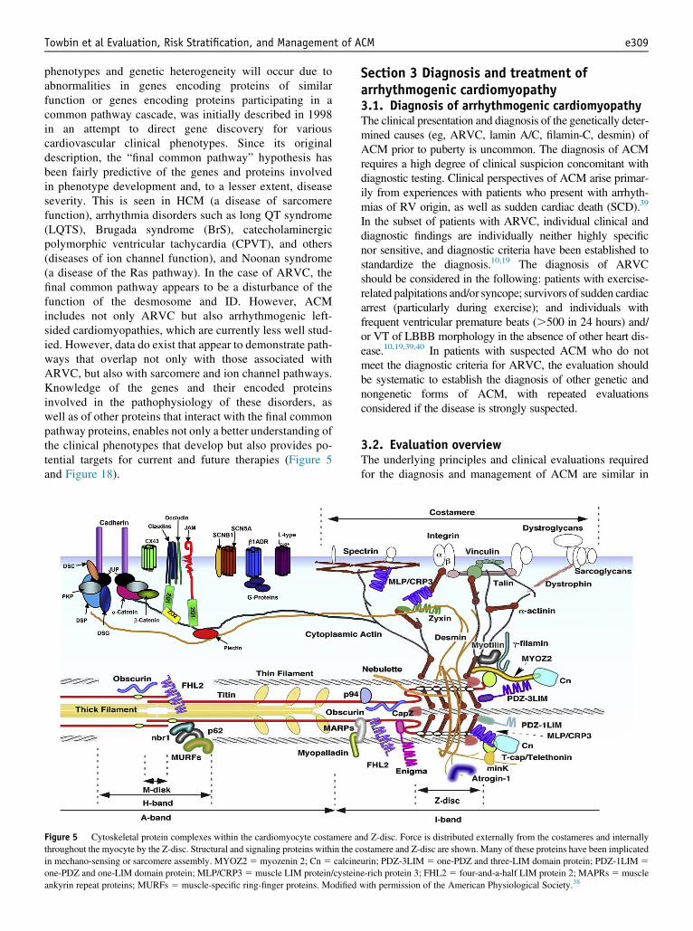
phenotypes and genetic heterogeneity will occur due toabnormalities in genes encoding proteins of similarfunction or genes encoding proteins participating in acommon pathway cascade, was initially described in 1998in an attempt to direct gene discovery for variouscardiovascular clinical phenotypes. Since its originaldescription, the “final common pathway” hypothesis hasbeen fairly predictive of the genes and proteins involvedin phenotype development and, to a lesser extent, diseaseseverity. This is seen in HCM (a disease of sarcomerefunction), arrhythmia disorders such as long QT syndrome(LQTS), Brugada syndrome (BrS), catecholaminergicpolymorphic ventricular tachycardia (CPVT), and others(diseases of ion channel function), and Noonan syndrome(a disease of the Ras pathway). In the case of ARVC, thefinal common pathway appears to be a disturbance of thefunction of the desmosome and ID. However, ACMincludes not only ARVC but also arrhythmogenic left-sided cardiomyopathies, which are currently less well stud-ied. However, data do exist that appear to demonstrate path-ways that overlap not only with those associated withARVC, but also with sarcomere and ion channel pathways.Knowledge of the genes and their encoded proteinsinvolved in the pathophysiology of these disorders, aswell as of other proteins that interact with the final commonpathway proteins, enables not only a better understanding ofthe clinical phenotypes that develop but also provides po-tential targets for current and future therapies (Figure 5and Figure 18).
Figure 5 Cytoskeletal protein complexes within the cardiomyocyte costamere athroughout the myocyte by the Z-disc. Structural and signaling proteins within the cin mechano-sensing or sarcomere assembly. MYOZ2 5 myozenin 2; Cn 5 calcineone-PDZ and one-LIM domain protein; MLP/CRP35 muscle LIM protein/cysteinankyrin repeat proteins; MURFs 5 muscle-specific ring-finger proteins. Modified
Section 3 Diagnosis and treatment ofarrhythmogenic cardiomyopathy3.1. Diagnosis of arrhythmogenic cardiomyopathyThe clinical presentation and diagnosis of the genetically deter-mined causes (eg, ARVC, lamin A/C, filamin-C, desmin) ofACM prior to puberty is uncommon. The diagnosis of ACMrequires a high degree of clinical suspicion concomitant withdiagnostic testing. Clinical perspectives of ACM arise primar-ily from experiences with patients who present with arrhyth-mias of RV origin, as well as sudden cardiac death (SCD).39
In the subset of patients with ARVC, individual clinical anddiagnostic findings are individually neither highly specificnor sensitive, and diagnostic criteria have been established tostandardize the diagnosis.10,19 The diagnosis of ARVCshould be considered in the following: patients with exercise-related palpitations and/or syncope; survivors of sudden cardiacarrest (particularly during exercise); and individuals withfrequent ventricular premature beats (.500 in 24 hours) and/or VT of LBBB morphology in the absence of other heart dis-ease.10,19,39,40 In patients with suspected ACM who do notmeet the diagnostic criteria for ARVC, the evaluation shouldbe systematic to establish the diagnosis of other genetic andnongenetic forms of ACM, with repeated evaluationsconsidered if the disease is strongly suspected.
3.2. Evaluation overviewThe underlying principles and clinical evaluations requiredfor the diagnosis and management of ACM are similar in
nd Z-disc. Force is distributed externally from the costameres and internallyostamere and Z-disc are shown. Many of these proteins have been implicatedurin; PDZ-3LIM 5 one-PDZ and three-LIM domain protein; PDZ-1LIM 5e-rich protein 3; FHL2 5 four-and-a-half LIM protein 2; MAPRs 5 musclewith permission of the American Physiological Society.38

e310 Heart Rhythm, Vol 16, No 11, November 2019
ARVC and ALVC with respect to excluding acquired causesfor the cardiomyopathy, ensuring a probable or definitivediagnosis and characterizing arrhythmia in relation to treat-ment and prognosis. Genetic causes of isolated or predomi-nantly RV arrhythmia and structural abnormalities are mostcommonly associated with desmosomal gene variants. Theremay be additional cutaneous phenotypes that manifest withautosomal dominant desmoplakin variants and are oftenflorid in recessive desmosomal disease.20,23 The geneticcauses of arrhythmia and structural disease of LV origin,however, typically manifest with additional cardiac (eg,conduction disease, atrial fibrillation) or systemic (eg,muscular dystrophy, lipodystrophy) phenotypes. Familialevaluation should therefore focus on arrhythmic disease,but also consider associated phenotypes. Several of theALVC disease-causing gene variants have been reported inpatients with LV or biventricular arrhythmia and LV dilata-tion and/or impaired function (eg, PLN, FLNC, LMNA,SCN5A). The diagnostic distinction here is from DCM andits genetic causes.28,41,42 In ACM, the clinical presentationin the proband and/or family members is typically witharrhythmia rather than HF, although both may be present inadvanced disease.
In patients with suspected ACM, the initial evaluation in-cludes clinical history, physical examination, detailed familyhistory, 12-lead electrocardiogram (ECG), 2D echocardiogra-phy, ambulatory ECGmonitoring, and CMR.10 Most patientswith suspected ACM presenting with arrhythmia can be diag-nosed using noninvasive imaging and electrocardiographicassessment. If the initial testing is nondiagnostic, additionaltesting may include signal-averaged ECG, exercise ECG,pharmacological testingwith isoproterenol,43 endomyocardialbiopsy, and EPS. In a series of 48 older children (aged 13–15years) presenting with possible ACM, a comprehensive clin-ical and genetic evaluation in the context of the adult TaskForce Criteria for the diagnosis of ARVC revealed that 46%of the children had features consistent with a diagnosis ofHCM, DCM, or ion channel disease, while 25% had featuresconsistent with ARVC.44
The diagnosis of ALVC relies on documenting arrhythmiaof isolated or predominantly LV origin in a proband or familymember with cardiomyopathy (eg, arrhythmia) not caused byischemic, valvular, or hypertensive heart disease. ImpairedLV function and/or structural abnormalities as determined by2D ECG and CMR can be absent, mild, or severe. Typically,arrhythmia is an early manifestation of disease. Internationallyaccepted diagnostic criteria analogous to those established forARVC10 are required; however, an issue is the diagnosis ofACM in the presence of other potential causes for which coex-istence vs causality may be difficult to determine. Given thecurrently incomplete knowledge of the genetic basis ofACM, particularly of the ALVC and biventricular forms, thedevelopment of clinical diagnostic criteria is needed.
After the original clinical description of RV dysplasia,17 itbecame clear that the diagnosis of this condition would bedifficult to establish, particularly in the early stages of thedisease when RV dilation or segmental dilatation is mild.
Therefore, differentiating RV dysplasia from the normalheart could be equivocal. A task force was subsequentlyassembled to consider criteria for the diagnosis of arrhythmo-genic RV dysplasia/cardiomyopathy, the results of whichwere published in 1994.19 The task force concluded that thereis no single gold standard for the diagnosis and that diseaseand the diagnosis require a combination of major and minorcriteria encompassing structural, histological, electrocardio-graphic, arrhythmogenic, and genetic factors. LV diseasewas excluded from these criteria. The revision of the TaskForce Criteria in 2010 included LV disease and addedCMR for the diagnosis; the criteria are listed in Figure 6.10
Diagnostic criteria for ARVC in the pediatric populationremain to be established since disease expression in childrenis uncommon. In a series of 16 patients, clinical presentationwas life-threatening arrhythmia in 10 (median age of 14years). In all 16 patients, LV and/or RV dysfunction wascommon and associated with the histopathological featuresof ARVC.45 Recently, a diagnostic and prognostic role hasbeen proposed for the presence of anti-desmoglein-2(DSG2) antibodies, which were present in patients withARVC but not in controls; this work is potentially importantand warrants confirmation in a larger number of patients andin other forms of ACM (eg, cardiac sarcoidosis).46,47
3.3. Family historyA detailed family history covering at least 3 generations andthe clinical evaluation of relatives are important in the diag-nostic assessment for ACM. In a patient with suspectedACM, a family history focusing on unexplained prematuredeaths, arrhythmias, and conduction disease may identify fa-milial disease. The presence of associated noncardiac pheno-types (eg, skeletal myopathy, other organ disease) can alsoprovide clues to the underlying diagnosis for both genetic(eg, desmin or lamin myopathy) and nongenetic (eg, Chagasdisease) causes.
The 12-lead ECG is an important part of the diagnosticevaluation of patients with suspected ACM. Reports on theECG findings of patients who meet the diagnostic criteriafor ARVC have shown that the majority (.85%) demon-strate at least one characteristic ECG feature of ARVC buta normal ECG has been reported in up to 12%.49–51 ARVCis a progressive disease, which is reflected in the well-documented dynamic ECG changes associated with diseaseprogression that have been demonstrated in several cohortsof patients with ARVC.49–54 Over time, the ECG mayevolve with further prolongation of the S wave upstroke,increased QRS duration, and development of bundlebranch block and precordial T wave inversion (TWI).53,54
3.4. Electrocardiogram features in arrhythmogenicright ventricular cardiomyopathy3.4.1. Repolarization abnormalitiesThe prevalence of TWI in leads V1–V3 (the characteristicECG finding in patients with ARVC) varies from 19% to67%,55–57 presumably due to the difference in study

Major Minor
Regional RV akinesia, dyskinesia, or aneurysm and 1 of the following (end diastole):
Regional RV akinesia, dyskinesia, or aneurysm and 1 of the following (end diastole):
a) PLAX RVOT ≥32 mm (PLAX/BSA ≥19 mm/m2)a) PLAX RVOT ≥29 mm to <32 mm (PLAX/BSA ≥16 to <19 mm/m2)
b) PSAX RVOT ≥36 mm (PSAX/BSA ≥21 mm/m2)b) PSAX RVOT ≥32 to <36 mm (PSAX/BSA ≥18 to <21 mm/m2)
c) Frac�onal area change ≤33% c) Frac�onal area change >33 to ≤40%
Regional RV akinesia or dyskinesia or dyssynchronous RV contrac�on and 1 of the following:
Regional RV akinesia or dyskinesia or dyssynchronous RV contrac�on and 1 of the following:
a) Ra�o RVEDV/BSA ≥110 mL/m2 (male), ≥100 mL/m2 (female)
a) Ra�o RVEDV/BSA ≥100 to <110 mL/m2 (male), ≥90 to 100 mL/m2 (female)
b) RVEF ≤40% b) RVEF >40 to ≤45%RV angiography Regional RV akinesia, dyskinesia, or aneurysm
Endomyocardial biopsy showing fibrous replacement of the RV free wall myocardium in ≥1 sample, with or without fa�y replacement and with:
Residual myocytes <60% by morphometric analysis (or <50% if es�mated)
Residual myocytes 60% to 75% by morphometric analysis (or 50% to 65% if es�mated)
I. Inverted T waves in leads V1 and V2 in individuals >14 years of age (in the absence of complete RBBB) or in V4, V5, or V6.II. Inverted T waves in leads V1, V2, V3, and V4 in individuals >14 years of age in the presence of complete RBBB
I. Late poten�als by SAECG in ≥1 of 3 parameters in the absence of QRS dura�on of ≥110ms on the standard ECG:
a) Filtered QRS dura�on (fQRS) ≥114 msb) Dura�on of terminal QRS <40 μV (low-amplitude signal dura�on) ≥38 msc) Root-mean-square voltage of terminal 40 ms ≤20 μV
II. Terminal ac�va�on dura�on of QRS ≥55 ms measured from the nadir of the S wave to the end of the QRS, including R’ in V1, V2, or V3 in the absence of complete RBBB
I. Nonsustained or sustained VT or RV ou�low configura�on, LBBB morphology with inferior axis (posi�ve QRS in II, III and aVF and nega�ve in lead aVL) or of unknown axis
II. >500 ventricular extrasystoles per 24 hours (Holter)
I. ARVC confirmed in a first-degree rela�ve who meets current Task Force Criteria
I. History of ARVC in a first-degree rela�ve in whom it is not possible or prac�cal to determine whether the family member meets current Task Force Criteria
II. ARVC confirmed pathologically at autopsy or surgery in a first-degree rela�ve
II. Premature sudden death (<35 years of age) due to suspected ARVC in a first-degree rela�ve
III. Iden�fica�on of a pathogene�c muta�on categorized as associated or probably associated with ARVC in the pa�ent under evalua�on
III. ARVC confirmed pathologically or by current Task Force Criteria in second-degree rela�ve
Modified Task Force Criteria for ARVC – Diagnostic Categories Major and Minor CriteriaDefinite: 2 major OR 1 major and 2 minor, OR 4 minor criteria from different categories
Borderline: 1 major and 1 minor, OR 3 minor criteria from different categoriesPossible: 1 major, OR 2 minor criteria from different categories
Global or regional dysfunc�on and structural altera�ons determined by echo, MRI, or RV angiography:
Echo
MRI
Tissue characteriza�on of wall
Repolariza�on abnormali�es
ECGInverted T waves in right precordial leads (V1, V2, and V3) or beyond in individuals >14 years of age (in the absence of complete RBBB QRS ≥120ms)
ECGEpsilon wave (reproducible low-amplitude signals between end of QRS complex to onset of the T wave) in the right precordial leads (V1 to V3)
Depolariza�on/conduc�on abnormali�es
Arrhythmias
Nonsustained or sustained VT of LBBB with superior axis (nega�ve or indeterminate QRS in leads II, III, and aVF and posi�ve in lead aVL)
Family history
Figure 6 Modified Task Force Criteria for arrhythmogenic right ventricular cardiomyopathy (ARVC) showing the diagnostic categories for major and minorcriteria according to the 2010 ARVC Task Force Criteria. These criteria are sensitive and specific in differentiating patients with ARVC from control populationsbut have not been adequately tested in relation to other arrhythmogenic cardiomyopathies (ACMs) with overlapping phenotypes (eg, cardiac sarcoidosis, myocar-ditis).48 BSA 5 body surface area; ECG 5 electrocardiogram; echo 5 echocardiogram; MRI 5 magnetic resonance imaging; PLAX 5 parasternal long-axis;PSAX5 parasternal short-axis; RBBB5 right bundle branch block; RV5 right ventricular; RVEDV5 right ventricular end-diastolic volume; RVEF5 rightventricular ejection fraction; RVOT 5 right ventricular outflow tract; SAECG 5 signal-averaged electrocardiogram; VT 5 ventricular tachycardia.
Towbin et al Evaluation, Risk Stratification, and Management of ACM e311

e312 Heart Rhythm, Vol 16, No 11, November 2019
populations. TWI in the precordial leads beyond V2 isrelatively common in Afro-Caribbean individuals,58
although it is rare (1% in females and 0.2% in males) inasymptomatic white individuals.59 TWI in patients youngerthan 14 years of age is more frequently observed in athletes(the so-called juvenile pattern).60 TWI is reasonably specificin patients older than 14 years of age and is considered a ma-jor diagnostic abnormality in ARVC. TWI in leads V1–V4 inindividuals older than 14 years associated with completeright bundle branch block (CRBBB) is a minor criterion forthe diagnosis of ARVC (Figure 7). The presence of TWI inlateral and/or inferior leads suggests LV involvement in pa-tients with ARVC (Figure 7).61
3.4.2. Depolarization and conduction abnormalities3.4.2.1. Epsilon waveThe epsilon wave is defined as a reproducible low-ampli-tude deflection located between the end of the QRS andthe onset of the T wave in leads V1–V3 (Figure 7).10,56
Epsilon waves reflect delayed conduction in the RV(Figure 7). The prevalence of the epsilon wave in Europeanand American registries varies from 0.9% to 25%.62 Elec-troanatomical mapping in patients with ARVC and an
IRBBB(QRS=110 ms)
*
*
*
*
I
II
III
aVR
aVL
aVF
V1
V2
V3
V4
V5
V6
CR(QRS
I
II
III
aVR
aVL
aVF
Figure 7 Representative 12-lead electrocardiogram (ECG) obtained from patientsplete right bundle branch block (IRBBB) and complete right bundle branch blockrespectively. The closed arrow indicates an epsilon wave, which was defined as lof the T wave in leads V1–V3. The asterisk indicates the T wave inversion recorde
epsilon wave has shown that the timing of the epsilon waveon the surface ECG corresponded to activation of the basal(peri-tricuspid) RV region of the epicardium. Epsilon waveshave been associated with severe conduction delay due toextensive endocardial and epicardial scarring at that site.63
Epsilon waves may reflect short-term arrhythmia risk butare of limited diagnostic utility because they are variable,have low sensitivity and specificity (seen in other condi-tions), and are dependent on ECG filter setting andmagnification.54,62,64,65
3.4.2.2. Prolonged terminal activation durationProlonged terminal activation duration (TAD) is measuredfrom the nadir of the S wave to the end of all depolarizationdeflections (Figure 8). A TAD �55 ms in any of the V1–V3
leads in the absence of CRBBB is defined as a prolongedTAD.55,66 Prolonged TAD in leads V1–V3 has beenreported to aid in differentiating ARVC from rightventricular outflow tract (RVOT)-VT.67 Prolonged TADwas confirmed in 30 of 42 patients with ARVC and in only1 of 27 patients with idiopathic RVOT-VT.55 Moreover,TAD prolongation was the sole ECG abnormality in 4 of 7gene-positive family members with ARVC,68 suggesting arole in the early recognition of “at-risk” individuals.
BBB=140 ms)
*
*
*
*
V1
V2
V3
V4
V5
V6
* T wave inversion
Epsilon wave
with arrhythmogenic right ventricular cardiomyopathy (ARVC) with incom-(CRBBB). QRS duration of IRBBB and CRBBB was 110 ms and 140 ms,ow-amplitude deflection located between the end of the QRS and the onsetd in V1–V4 in patients with ARVC and IRBBB or CRBBB.

Figure 8 Terminal activation duration (TAD) is measured from the nadir ofthe S wave to the end of all depolarization deflections and is prolonged if �55ms in any of the V1–V3 leads in the absence of complete right bundle branchblock (CRBBB). Modified with permission of Oxford University Press onbehalf of the European Society of Cardiology.69
Towbin et al Evaluation, Risk Stratification, and Management of ACM e313
3.4.3. Electrocardiogram abnormalities in arrhythmogeniccardiomyopathies other than arrhythmogenic right ventricularcardiomyopathyCharacterization of ECG findings in other ACMs is lessdetailed. The 12-lead ECG abnormalities include invertedT waves in leads I, aVL, and V4–V6; other repolarization ab-normalities; generalized low-voltage; increased QRS dura-tion; and isolated ectopy of LV origin. A completelynormal ECG is uncommon. Variants in lamin A/C may beassociated with progressive conduction disease (eg, PR pro-longation to atrioventricular [AV] block), variants indesmosomal genes and phospholamban with a lowvoltage ECG, and in filamin-C with minor repolarizationchanges only. In contrast to ARVC associated with desmo-somal variants, ECG abnormalities do not appear to be anearly marker of disease in FLNC and desmin-relatedACM. In ACMs associated with systemic disease, conduc-tion abnormalities are often early features (eg, sarcoidosisand Chagas disease).70,71
3.4.4. Ambulatory electrocardiogram monitoringAmbulatory ECGmonitoring (24 to 48 hours) is important forcharacterizing all patients for whom the diagnosis of ACM isbeing considered. The presence of .500 ventricular prema-ture beats per 24-hourmonitoring period is aminor diagnosticcriterion for ARVC. In a study of 40 patients meeting ARVCTask Force Criteria who underwent ambulatory ECG moni-toring for an average of 159 hours, the average ventricular pre-mature beat count (per 24 hours) was 1091, with significantday-to-day variation. Despite this variation, the 24-hourburdenwas accurate 89.6% of the time to the correct groupingbased on the revised Task Force Criteria.72,73
Documentation of ventricular arrhythmia with amorphology consistent with an LV origin is required forthe diagnosis of ALVC. Precise definitions relating to charac-teristics VT and/or frequency of ventricular ectopy remain tobe established for forms of ACM other than ARVC. Thearrhythmia may be asymptomatic or associated with palpita-tions and/or impaired consciousness.
3.4.5. Signal-averaged electrocardiogramAlthough an abnormal signal-averaged ECGwas a minor cri-terion in the 2010 Task Force Criteria, its use has declinedlargely due to its limited sensitivity and specificity, as wellas its limited availability in many medical centers.10,74
3.5. Cardiac imagingEchocardiography and other noninvasive imaging modalitiesare important for evaluating patients suspected of ACM toassess structural and functional abnormalities and aid in diag-nosis.75,76
For many patients with suspected ACM, 2D echocardiog-raphy provides adequate visualization, enabling a systematicqualitative and quantitative assessment of ventricular func-tion and cavity dimensions, although there may be limitationswhen imaging the RV. Additional imaging with CMR pro-vides accurate measurements of volumes and also regionaland global ventricular function.52 If CMR is contraindicatedor not available, multidetector computed tomography (CT),RV angiography or radionuclide angiography are alterna-tives, but are currently less frequently used to assess ventric-ular function. The Task Force Criteria for ARVC include thepresence of RV akinesia, dyskinesia, or aneurysms, togetherwith an assessment of RVOT diameter and RV-fractionalarea change. Emerging echocardiographic parameters in theevaluation of patients with suspected or established ARVCinclude the measurement of tricuspid annular plane systolicexcursion, RV basal diameter, global longitudinal strain(RV and LV), mechanical dispersion (RV and LV), and theuse of 3D echocardiography.77,78 However, prospectivestudies are needed before these assessments arerecommended for routine use.
The 2010 Task Force Criteria for ARVC included CMR pa-rameters for RV global and regional dysfunction and RV vol-ume.10 The major criterion requires a regional RV wall motionabnormality and either increased RV end-diastolic volume(�110 mL/m2 in men; �100 mL/m2 in women) or depressedright ventricular ejection fraction (RVEF) �40% (sensitivity:men 76%, women 68%; specificity: men 90%, women 98%).The CMR minor criterion also requires regional RV wall mo-tion abnormality with lesser degrees of RV enlargement(�100 mL/m2 in men; �90 mL/m2 in women).10 The TaskForce Criteria did not include CMR measures of RV myocar-dial fat or late gadolinium enhancement (LGE); however, thesewere not considered reliable measurements at the time the TaskForce Criteria were developed (2010).
The 2010 Task Force Criteria for ARVC do not definediagnostic criteria for LV involvement. If present, LGE is

e314 Heart Rhythm, Vol 16, No 11, November 2019
typically found in a subepicardial or mid-wall distributionconfined to the LV. LV dominant disease may be underdiag-nosed and attributed to other disorders.78 The potential ofCMR to diagnose and risk stratify patients with ACM re-mains to be fully exploited. LV LGE has been identified asthe sole imaging abnormality in patients with desmoplakindisease who have arrhythmia of LV origin and a normalECG.31 In general, ECG abnormalities and arrhythmia areconsidered the earliest manifestations54,79; however, Sen-Chowdhry et al have also demonstrated that CMR may besensitive to detecting early changes in ARVC. The role ofCMR in the early diagnosis of ACM of nondesmosomalorigin, for other genetic and acquired causes, warrants eval-uation.30,80 CMR expertise will be particularly important inthe early diagnosis in the absence of ECG or other imagingabnormalities, given the risk that epicardial fat may bemisinterpreted as delayed enhancement.
LV structural and functional abnormalities will relate toparticular genetic abnormalities and disease stage. Currentgenotype-phenotype relations are based on small data setsbut suggest that ACM with clinically significant LV ar-rhythmias (eg, ALVC) may occur with “normal” toseverely impaired LV function. Experience is greatestwith lamin A/C disease, in which phenotypes includeEmery-Dreifuss muscular dystrophy, generalized lipodys-trophy, DCM with HF, progressive conduction diseasewith late-onset DCM, and ALVC with or without signifi-cant LV impairment. ALVC caused by desmoplakin vari-ants can also be present with absent to severe LVdysfunction and may present with sudden death.81 Prelim-inary experience indicates that LGE on CMR can be pre-sent in the absence of LV dysfunction and may providean early diagnostic feature when LV arrhythmia appearsto have occurred in isolation.31
3.6. Electrophysiology testingElectrophysiology testing in ACM is often unnecessary forthe diagnostic evaluation of patients with suspected ARVCor ALVC.12 Multicenter studies of patients with ARVCwho received an implantable cardioverter defibrillator(ICD) have demonstrated the low predictive accuracy of elec-trophysiology testing in identifying those at risk of SCD and/or life-threatening arrhythmia.82,83 The reported incidence of“life-saving” ICD discharges for treatment of fast VT/ventricular fibrillation (VF) was not significantly differentbetween those who were and those were not inducible.Corrado et al studied 106 patients with ARVC whoreceived an ICD as primary prevention. The positive andnegative predictive value for VT/VF inducibility was 35%and 70%, respectively.82 Electrophysiology testing, however,may be beneficial in patients with refractory ventriculararrhythmias for ablation consideration and differentiationfrom RVOT tachycardia. In this setting, electrophysiologytesting with high-dose isoproterenol may help differentiate pa-tients with idiopathic VT or ventricular premature beats fromthose with ARVC.84
3.7. Endomyocardial biopsyBiopsy can be particularly useful in identifying systemic orinflammatory conditions that cause ACM (eg, sarcoidosis,myocarditis). However, endomyocardial biopsy (one of theTask Force Criteria for the diagnosis of ARVC) is invasive,lacks sensitivity and specificity, has low diagnostic yield,and, therefore, is now rarely performed in the initial diagnosisof ARVC. The characteristic histological feature is the pres-ence of transmural fibrofatty replacement of the RV myocar-dium, with major and minor criteria differentiated by degreeof replacement (,60% vs 60%–75% myocytes by morpho-metric analysis).10 Diagnosis by biopsy is limited due to falsenegatives secondary to patchy involvement and sampling er-ror.85,86 Electroanatomical voltage mapping may improve theyield of endomyocardial biopsy by identifying areas of lowvoltage.87 Endomyocardial biopsy is associated with therisk of perforation, which is increased with RV free wall bi-opsy.85,88 Septal biopsy is generally not helpful because it istypically the least affected area of the myocardium inARVC.86 Novel immunohistochemical analysis in patientswith ARVC with desmosomal variants demonstrated alteredplakoglobin and connexin43 signal as a marker of diseaseexpression79,89–91; however, this has not proven to be ofdiagnostic utility. Sarcoidosis, for which treatment mayinclude steroids, is important in the differential diagnosis ofARVC, but similar limitations with regard to samplingerror and risk are present. Myocardial tissue obtained frompostmortem and explanted hearts will have the value butnot the limitations of endomyocardial biopsy and should besought and examined whenever feasible.
3.8. Genetic testingGeneral concepts on the role of genetic testing in the diag-nosis and management of ARVC and other ACMs are out-lined below, with recommendation flow diagrams shown inFigure 10 and Figure 11.
3.8.1. Genetic testing methodsSeveral methods are available to identify the genetic basis ofan ACM. Single genes are usually analyzed by Sangersequencing, which has been proven to be a reliable techniqueto identify variants underlying genetic disease and has beenthe gold standard for decades. With increasing numbers ofgenes identified as underlying a specific cardiac disorder (ge-netic heterogeneity) and the fact that more than one gene and/or variant (digenic inheritance or polygenic inheritance) cancontribute to the disease phenotype,75,92 next-generationsequencing (NGS)–based methods enable the parallelsequencing of several targeted genes (a panel, eg,cardiomyopathy-panel) at the same time and at relativelylow cost.93 In addition to these targeted NGS panels,sequencing of all protein coding genes (exome) of the humangenome (whole exome sequencing [WES]) or even all DNAnucleotides (whole genome sequencing [WGS]) can beperformed.

Towbin et al Evaluation, Risk Stratification, and Management of ACM e315
3.8.2. Variant and gene interpretationDNA sequences normally vary in the general populationwhen comparing different individuals. However, evenwhen they reside in bona fide ACM-susceptibility genes,not every DNA variant contributes to the disease.94 The ma-jor challenge is to correctly assign potential pathogenicity tothese DNA variants. The American College of Medical Ge-netics and Genomics (ACMG) has published guidelines forinterpreting genetic variants and proposed a classificationbased on the likelihood that a variant is related to disease(Table 2): pathogenic (class 5), likely pathogenic (class 4),uncertain significance (class 3), likely benign (class 2), orbenign (class 1), in which a “likely pathogenic” and “likelybenign” variant are used to mean greater than 90% certaintyof a variant being either disease-causing or benign, respec-tively.95
The importance of correctly interpreting an identified var-iant’s pathogenicity is now considered the most critical stepin genetic testing, especially considering that there appearsto be substantial interreviewer disagreement over variantinterpretation.97–100 Ethnicity information is essential forinterpreting the data.101 Within the ACMs, examples ofincorrect classification of variants in major ARVC-relatedgenes have been published.102–106 Besides variantadjudication and the vexing variant of uncertainsignificance (VUS), many alleged and published ACM-susceptibility genes are being re-analyzed as to the strengthof their disease-gene association and, over time, several pub-lished ACM-susceptibility genes may be demoted to genes ofuncertain significance. Accordingly, when evaluating pa-tients suspected of an ACM, it is critical that the genetic testsconducted as part of the evaluation and the interpretation ofthe genetic test results be conducted by comprehensive teamswith expertise in these disorders.107
Several genes have been implicated in ACM, with varyingevidence strength (Table 3). The ClinGen CardiovascularClinical Domain Working Group for cardiovascular disor-ders is curating genes in relation to specific disorders.108
One of the first efforts in adapting the ACMG 2015 guide-lines for variant interpretation in genes related to cardioge-netic disease has recently been published, and this processis also underway for ACM.109
Depending on the reason for using the results of a genetictest, a certain amount of evidence for pathogenicity is neces-
Table 2 Classification of likelihood of pathogenicity of avariant
Classification ofvariant Description
Likelihood of beingpathogenic
Class 5 Pathogenic .95%Class 4 Likely pathogenic .90%Class 3 Variant of uncertain
significance10–90%
Class 2 Likely benign ,10%Class 1 Benign ,5%
Adapted from Plon et al.96
sary; for prenatal diagnostics or a pre-implantation geneticdiagnosis, the evidence for pathogenicity must be strong,and only class 5 variants are used. For genetic cascadescreening in family members, only class 4 and 5 variantsare used; family members negative for the family’s class 5variant are dismissed from regular cardiologic follow-up,whereas those relatives who test negative for a given family’sclass 4 variant remain in the cardiogenetic clinics, albeit forlonger follow-up intervals. The frequency and duration offollow-up for family members who are negative for a class4 variant should be individualized at the discretion of the clin-ical team. Class 3 variants (ie, a VUS) should be deemed“nonactionable.” Given both incomplete penetrance andage-dependent penetrance, clinically unaffected family mem-bers should not be tested to determine their status for a class 3variant found in the family unless additional evidence (suchas various functional validation assays and/or demonstrationof co-segregation among clinically affected family members)has been obtained that would prompt a variant promotionfrom an ambiguous class 3 variant (VUS) to a clinicallyactionable class 4 or class 5 variant.
3.8.3. Which test to useWith the availability of NGS, the number of genes that can bestudied in a single patient rapidly increases. However, thevalue of including a greater number of genes in a panelshould be weighed against the drawback of adding genesthat have insufficient evidence (or none) of being related tothe patient’s disease or that account for only a small percent-age of the genotyped patients and are therefore more prone toerrors in attributing the pathogenic role of the identified var-iants.
Therefore, a list of core genes can focus on those with suf-ficient evidence to be disease-related. The ClinGen workinggroup for cardiovascular disorders is responsible for review-ing clinical, genetic, and experimental data to establish thestrength of evidence supporting gene-disease associations inheart disease. Gene curation for HCM was recentlycompleted, and curation for ARVC and DCM is under-way.110,111 Until the official ClinGen-approved results ofthese gene curation efforts are available, we anticipate thatthe genes listed in Table 3 will likely be retained as ACM-susceptibility genes with sufficient evidence to merit theirdisease–gene association and will be useful in clinical prac-tice. These recognized genes should therefore be prioritizedfor patients and families with a clinical diagnosis of ACMor its subforms. If other genes are included in the analysis,identifying a pathogenic or likely pathogenic variant in oneof the non-ACM related genes should not automatically orreflexively be considered an explanation for the patient’sACM phenotype. In other words, a pathogenic or likely path-ogenic variant in KCNH2 (a gene in which P/LP variantscause abnormalities in the QTc without structural heart dis-ease) does not carry the same intrinsic probability of pathoge-nicity for ACM as a plakophilin-2 (PKP2) variant that hasbeen graded as a pathogenic or likely pathogenic variant.

Table 3 Minimum set of genes to be prioritized in arrhythmogenic cardiomyopathy (ACM)
Gene Protein type
Predominanttype ofmutation OR/EF100
Signal:Background94 Remarks References
BAG3 Chaperone Truncating andmissense
NA NA Also causes myofibrillarmyopathy
121
DES IF Truncating andmissense
NA NA Also causes myofibrillarmyopathy
122
DSC2 Desm Truncating andmissense
NT 2.15 (EF 0.53);T 21.5* (EF 0.95)
nsns
Rare 26
DSG2 Desm Truncating andmissense
NT 2.83* (EF 0.65)T 19.8* (EF 0.95)
2:1* (NT/T) Rarely recessive 123
DSP Desm Truncating andmissense
NT 2.1* (EF 0.52)T 89.9* (EF 0.99)
nsns
Recessive: Carvajalsyndrome
23,124
FLNC Actin crosslink Truncating andmissense
NA NA Also causes myofibrillarmyopathy
34
JUP Desm Missense NT 7.8* (EF 0.87)T 28.1 (EF –)
Recessive: Naxos syndrome 22,125
LDB3 Z-band Missense NA NA Cypher/ZASP 126
LMNA NE Truncating andmissense
NA NA AV block; CD 127
NKX2-5 Homeobox Truncating andmissense
NA NA AV block, CD, CHD 128
PKP2 Desm Truncating NT 1.3 (EF 0.23)T 484.7* (EF 1.0)
10:1*42:1*
Large deletions 1-2% 24
PLN Ca Missense,nonsense, anddeletion
NA NA Predominantly R14del 33,129
RBM20 Splice factor Missense NA NA Mostly in exon 9 130
SCN5A Sodium channel Mostly missense NA NA Brugada, SND, CD 131
TMEM43 NE Missense NT 0.76 (EF–)T 13 (EF–)
ns p.S358L disease-causing;also called LUMA
132
These genes have multiple lines of evidence indicating involvement in ACM and its subtypes (arrhythmogenic left ventricular cardiomyopathy, arrhythmo-genic right ventricular cardiomyopathy). OR/EF and Signal:Background data are largely derived from cohorts with western European ancestry, and other ethnic-ities can be different.
AV5 atrioventricular; BV5 biventricular; Ca5 calcium handling; CD5 conduction delay; CHD5 congenital heart disease; CPVT5 catecholaminergic poly-morphic ventricular tachycardia; DES5 desmin; Desm5 desmosomal; DSC25 desmocollin-2; DSG25 desmoglein-2; EF5 etiological fraction; IF5 intermediatefilament; LD5 left dominant; NA5 data not available; NE5 nuclear envelope; ns5 not significant; NT5 nontruncating variants; OR5 odds ratio; RD5 rightdominant; SND 5 sinus node dysfunction; T 5 truncating variants.*Genes with significant excess in cases over ExAc reference samples.100 Other genes that have been identified in ACM with insufficient or conflicting evidence areABCC9,112 TGFB3,113 TTN,114 CTNNA3,115 sarcomeric genes (MYH7, MYBPC3),116,117 SCN3B,117 CDH2,118,119 TJP1.120
e316 Heart Rhythm, Vol 16, No 11, November 2019
A recent viewpoint paper by the European Society of Car-diology (ESC) working group on myocardial and pericardialdiseases emphasized that, in a diagnostic setting, only recog-nized genes associated with the condition should be investi-gated in patients who meet the diagnostic criteria of a specificcardiovascular condition. WES and WGS should be used forgenetic diagnosis only if filtered against recognized disease-causing genes. The coverage should enable the identificationof all exonic variants in these genes.107
Table 4 Different methods for screening genes
Target
Sanger sequencing Single gene(s)Targeted NGS panel Panel of genes of interestWES filtered against genes of interest Set of genes of interestWES All genesWGS All genes and intronic sequ
CNVs 5 copy number variations; IE 5 inefficient (expensive for large amountssequencing; WES5 whole exome sequencing; WGS5 whole genome sequencing;1
3.8.4. Advantages and disadvantages of various methodsThe various techniques that can be used for genetic testingeach have their own advantages and disadvantages, as sum-marized in Table 4. Coverage of the genomic regions of inter-est, the possibility of identifying large deletions/duplications,flexibility, and costs are important factors to consider whenordering a genetic test.
Sanger sequencing is a reliable method with goodcoverage of the nucleotides that need to be studied,
Coverage CNVs Flexibility Costs
11 22 2 IE1 1 2 1/21/2 1/2 1 11/2 1/2 1 1
ences 1 1 1 11
of sequencing but inexpensive for a small amount); NGS 5 next-generation15 very high;15 high;1/25 intermediate;25 low;225 very low.

Towbin et al Evaluation, Risk Stratification, and Management of ACM e317
particularly for evaluating a single or a small number ofgenes. Sanger sequencing is also appropriate for cascadetesting in at-risk family members, clinical confirmation ofresearch genetic results, and cosegregation studies. However,large deletions and duplications of genes can be missed whenusing Sanger sequencing. It is well known that larger dele-tions and/or duplications (eg, in PKP2) are a known causeof ACM68,133,134 and can be identified in a smallpercentage of cases.
Targeted NGS panels have the advantage that theyare well validated, and it is well known which parts areinsufficiently covered. Additional Sanger sequencing exper-iments are frequently used to evaluate the insufficientlycovered regions.93 Bioinformatic tools must be added to
COR LOE Recommendations
I C-EO For individuals and decedentdiagnosis of ACM, geneticsusceptibility genes is rec
I C-EO For genetic testing of the escomprehensive analysis ofis recommended.
A genetic test is generally performed in an index patient with either a clquestion or when there is at least a reasonable index of suspicion for tsubsequent genetic test interpretation should be strongly influenced bynonspecific syncope or TWIs confined to only precordial lead V1, for examtest, the available evidence that a specific gene is related to ACM shouldto identify variants in these genes. This may entail additional tests toidentify deletions and duplications.
For individuals who have died suddenly with a postmortem (likely) diagnosagain include those disease genes implicated in the necropsy diagnositissue, fibroblasts from a skin biopsy, and even formalin-fixed paraffinACM-associated genes can also be evaluated in autopsy-negative SCD cstructural abnormalities.138
Table 3 lists the minimum set of genes to be evaluated.
the bioinformatics pipeline to identify deletions and/or dupli-cations in the genes of interest in targeted panel screening, arelatively inexpensive, fast, and reliable method to studylarger series of genes.
The results of exome sequencing, a relatively fast test,can be filtered against the set of core genes rather thanevaluating all 20,0001 human genes. This reduces thechance of incidental findings. The major advantage ofexome sequencing is that novel or additional genes canbe easily added by “opening” the data whenever new dis-ease genes are established. On the downside, the qualityand/or coverage of some parts of the “core genes” maybe insufficient, and larger deletions and/or duplicationscan easily be missed.
3.8.5. Who to study
References
s with either a clinical or necropsytesting of the established ACM-ommended.tablished ACM-susceptibility genes,all established genes with full coverage
inical diagnosis that fulfills the clinical criteria for the disease inhat specific disorder. Both the selected disease gene panel and thethe veracity of the phenotype. The genetic testing of patients withple, should be strongly discouraged.135 When interpreting a geneticbe taken into account. The test used should be of sufficient quality
cover all exons and additional bioinformatic and laboratory tests to
is of ACM or one of its subforms, postmortem genetic testing shoulds. Various sources to isolate DNA can be used, such as blood, frozen-embedded tissue.136,137
ases because ventricular arrhythmias leading to SCD may precede
3.8.6. The role of genetic testing in arrhythmogeniccardiomyopathiesA positive genetic test result (ie, likely pathogenic, class 4 orpathogenic variant, class 5) can (1) genetically confirm theclinical diagnosis and provide disease–gene-specific riskstratification and tailoring of therapies139 and (2) enablevariant-specific cascade genetic testing of appropriate familymembers and relatives (see Section 3.9 Cascade FamilyScreening), including the potential for prenatal or preimplan-tation genetic diagnostics (a topic beyond the scope of thisconsensus statement).
In the current Task Force Criteria for ARVC,10 the“Identification of a pathogenic mutation categorized asassociated or probably associated with ARVC in the pa-tient under evaluation” is weighted as a major criterion
in the “family history” section. A pathogenic mutation(now classified as either a class 4 or class 5 variant perACMG nomenclature) is defined as “a DNA alterationassociated with ARVC that alters or is expected to alterthe encoded protein, is unobserved or rare in a largenon-ARVC control population, and either alters or is pre-dicted to alter the structure or function of the protein orhas demonstrated linkage to the disease phenotype in aconclusive pedigree.” Since a positive genetic test resultis regarded as a major criterion, it will contribute up to50% to the diagnosis of ARVC, thus highlighting theimportance of an experienced genetic team. Nevertheless,there is the question of whether to put this much weighton a genetic result for which the true characteristics suchas penetrance are generally not well known.

Figure 9 Cumulative prevalence of disease expression in family membersat risk of arrhythmogenic right ventricular cardiomyopathy (ARVC).147
e318 Heart Rhythm, Vol 16, No 11, November 2019
3.8.7. The use of a genetic test in risk stratification andmanagementWhether the result of a genetic test can be used for risk strat-ification or management depends on the known relationshipbetween genotype and phenotype. In general, there is limitedevidence for a clinically actionable relationship between ge-notype and phenotype, with a few exceptions presented in thefollowing subsections.
3.8.7.1. Left ventricular dysfunctionLV dysfunction is most often present in patients with ACMwith pathogenic or likely pathogenic variants in LMNA,BAG3, or one of the founder variants in the PLN andTMEM43 genes, followed by variants in DSP, DSG2/DSC2and the lowest frequency in PKP2/JUP. This holds true forboth index patients and family members.140,141
3.8.7.2. Multiple variantsApproximately 3%–6% of patients have more than 1pathogenic or likely pathogenic variant contributing tothe disease phenotype. Patients with multiple pathogenicvariant-mediated ACM have more severe disease, asreflected by an earlier age at disease onset92 and the pres-ence of VTs (,20 years vs 35 years for patients with asingle ACM-causative variant),68 a higher lifetime riskof arrhythmia142 or SCD,143 and earlier progression tocardiomyopathy.141,144,145
3.8.7.3. Specific variants and genes3.8.7.3.1. Desmosomal genesDisease expression reaching diagnostic criteria is most com-mon between 20 and 50 years of age (40%; 95% CI, 34%–
46%),146 although in one series, 9 of 40 pediatric desmo-somal gene-positive patients had the disease at a mean ageof 17.8 6 5.1 years.147 LGE identified by CMR, mostfrequently seen in the LVmyocardium, was the first evidenceof disease expression in a small subset of individuals.75 In acomprehensive evaluation of 274 family members, the inci-dence of a new diagnosis (as per 2010 Task Force Criteria)in those aged 10–20 years was 0.5 per 100 person-years,and the odds ratio of a diagnosis in those aged ,18 yearsin the multivariate analysis was 0.37 (0.14–0.93), with nodiagnosis reached under the age of 14 years. Likewise, anew diagnosis in relatives older than 60 years is less com-mon.146 The cumulative prevalence by decade is shown inFigure 9 based on data from Quarta et al.147
Overall, relatives have less severe disease compared withprobands, are more commonly asymptomatic, and show dis-ease onset at an older age.145 Arrhythmic events in familymembers appear to occur only in the presence of manifestelectrocardiographic and structural changes.146,148 Similarenhanced disease activity is observed in pediatric probandscompared with their age-matched relatives.75
3.8.7.3.1.1. Desmoplakin (DSP)Pathogenic variants inDSP-encoded desmoplakin are asso-ciated with a spectrum of disorders, including cardio-cutaneous syndromes. For patients with likely pathogenic(class 4) or pathogenic (class 5) variants in DSP over
50% of index-patients and 17% of family members havean arrhythmic phenotype with LV dysfunction(Table 2).141 In addition to biventricular forms, left domi-nant forms are also present and extensive fibrotic patternscan be identified by CMR (see Section 5.5 Left VentricularNoncompaction).140,158
3.8.7.3.2. Lamin A/C (LMNA)The cardiac phenotype for LMNA-mediated ACM is char-acterized by atrial fibrillation and cardiac conduction dis-ease, which can precede the development of ventriculararrhythmias and cardiomyopathy by decades.149,150
LMNA variants have also been identified in patientsdiagnosed with ARVC151–153 or more biventricularand left-dominant forms of the disease.127,154 Riskstratification has been reported from Asian and Europeanpopulations.155,156 In the European study, nonsustainedventricular tachycardia (NSVT), left ventricular ejectionfraction (LVEF) ,45% at first clinical contact, male sex,and nonmissense variants have been reported to be risk fac-tors for malignant ventricular arrhythmias.156 Patients witha LMNA variant who are in need of a pacemaker oftenreceive an ICD, which is effective in treating possibly lethaltachyarrhythmias.157
3.8.7.3.3. Transmembrane protein 43 (TMEM43)The p.S358L mutation in transmembrane protein 43(TMEM43) is a specific founder variant that has been identi-fied in a large number of patients diagnosed with ARVC fromEurope and Canada (Newfoundland).132,159 Its clinicalphenotype is characterized by poor R wave progression inprecordial leads and LV enlargement in 43% of affectedindividuals, with 11% meeting the criteria for DCM.160 Astudy involving nearly 150 p.S358L-TMEM43-positive indi-viduals concluded that survival was greater for those treatedwith an ICD than for those with conventional, non-ICDcare.160
3.8.7.3.4. Phospholamban (PLN)The pathogenic p.R14del-PLN variant has been identified in1% of patients with ARVC in the United States and 12% ofDutch patients with ARVC,33 as well as in patients from

Towbin et al Evaluation, Risk Stratification, and Management of ACM e319
several other countries (Spain, Germany, Greece, Canada,Norway). Patients with this variant frequently have low-voltage ECGs and are considered to be at high risk for
COR LOE Recommendations
IIa C-EO The interpretation of aproviders with experuseful.
Performing a genetic test on an index patient or relative has several aspectteam. There are specific test-related “technical” aspects that result in sAdvantages and Disadvantages of Various Methods). The interpretationexample, class 1, 2, and 3 variants are not considered as actionable. Thdepends greatly on the precise clinical characterization of the phenotynecessarily predict the disease severity in that specific individual. Wheincidental findings may be identified, such as likely pathogenic or patphenotype than the one that motivated the referral.
Genetic testing can cause a mixture of positive and negative emotions fornavigate these feelings and learn to live with this inherited condition. Gvariants in ways that alleviate anger, anxiety, fear, and guilt that are
This expert team should therefore consist, at minimum, of cardiologists, cliniindividuals with expertise that encompass these subspecialties.
Genetic Testing
Who How
For genetic testing of the established ACM-susceptibility
genes, comprehensive analysis of all established genes with full
coverage is recommended (COR I, LOE C-EO).
For individuals and decedents with either a clinical or necropsy
diagnosis of ACM, genetic testing of the established
ACM-susceptibility genes is recommended
(COR I, LOE C-EO).*
The interpretation of a cardiac genetic test by a team of providers
with expertise in genetics and cardiology can be useful (COR IIa, LOE C-EO).
*Cascade Family Screening
Figure 10 Genetic testing recommendations. * 5 Cascade familyscreening: see Section 3.9 Cascade Family Screening. ACM 5 arrhythmo-genic cardiomyopathy; COR 5 Class of Recommendation; LOE 5 Levelof Evidence. Colors correspond to COR in Figure 1.
malignant ventricular arrhythmias and end-stage HF, withLVEF ,45% and sustained VT or NSVT as independentrisk factors (see Section 5.4 Phospholamban).161
3.8.8. Limitations of genetic testing
References
cardiac genetic test by a team oftise in genetics and cardiology can be
s that must be considered and thus requires a comprehensive, expertome variants not being detected by certain tests (see Section 3.8.4of a genetic test requires an accurate interpretation of variants. Fore interpretation is also influenced by the pretest probability, whichpe. Additionally, the identification of a genetic defect does notn using a panel with more genes that underlay other phenotypes,hogenic variants (class 4 and 5) that could lead to a different
the patient. Genetic counselors can help patients and their familiesenetic counselors can explain the implications of identified geneticlikely to occur in patients and their families.cal andmolecular geneticists, genetic counselors, and pathologists, or
The ACMGhas issued an updated list of over 50 actionablegenes.162Laboratories performingWESorWGS(generally fordiagnostic odyssey cases) should report the presence of patho-genic or likely pathogenic variants residing in these genes, un-less the individualwho is being tested has chosen not to receivethese results. This list includes 5 of the established ARVC-susceptibility genes. For these incidental findings, however,the frequency of related clinical phenotypes in unselected pa-tient populations is generally not well established. When vari-ants in a knownARVC-susceptibility gene are identified in thecontext of a nonphenotype-driven incidentalfinding, the likeli-hood that this variant (even if graded as a class 4 or 5) portends
the presence of ARVC or the risk of developing ARVC in thefuture is considered low, as was recently established forarrhythmia and ARVC-related genes.98,163
3.9. Cascade family screeningSee Evidence Table: Cascade Family Screening. Flow dia-gram of recommendations is shown in Figure 11.
3.9.1. Cascade family screening: screening recommendationsin children and adultsClinical cascade testing refers to the cardiovascular andgenetic evaluation of first-degree family members of an in-dividual (proband) with a confirmed diagnosis of ACM andis ideally performed within the confines of a multidisci-plinary cardiovascular genetics program, familiar with theclinical and genetic complexities of the condition.164 Theunderlying etiology of ACM in many cases is due to alter-ations in cardiac genes that encode proteins critical tonormal heart development and/or function. For the mostpart, these are inherited as an autosomal-dominant trait,such that first-degree family members have a 50% a prioririsk of developing ACM, although the penetrance and dis-ease severity are typically less in family members comparedwith probands.145 Detailed clinical and genetic familialevaluation, both at the time of diagnosis and duringfollow-up, will help determine the inheritance patternsand likelihood of consanguinity.
Desmosomal variants are relatively common in controlpopulations and may erroneously be considered disease-causing,94 although certain variants have a well-recognizedassociation with the condition, and targeted genetic testingcan be used in isolation within specific families.

e320 Heart Rhythm, Vol 16, No 11, November 2019
3.9.1.1. Family history
COR LOE Recommendations References
I C-EO It is recommended that a genetic counselor or appropriatelyexperienced clinician obtain a comprehensive 3-generationfamily history.
A detailed �3-generation family history collected from the proband at their initial consultation is vital and should be obtained by a geneticcounselor or an appropriately experienced clinician.165–168 The family history can be used to determine the existence of familial disease,provide important data regarding the full phenotypic spectrum within the family, and identify relatives who should be informed of the needfor cardiac evaluation.
3.9.1.2. Cardiac evaluationThe yield of cardiac screening is highly varied due to age-related and typically incomplete penetrance, and the diseasespectrum can be diverse, even within families harboring the
COR LOE Recommendations
I B-NR It is recommended thatclinical evaluation ev10–12 years of age.
ACM variants can display incomplete penetrance and varied expression. In Athe third and fourth decade of life, although this may vary with the unexpression is, however, recognized in adolescents, although it is extreprobands.75,145,171 At-risk relatives who undergo clinical evaluation mpenetrance), or be clinically unaffected. Serial evaluation can define ofamilies with ARVC, the highest probability of a diagnosis of ARVC occ
COR LOE Recommendations
I B-NR Cardiovascular evaluatioambulatory ECG, and
Evaluation of all at-risk family members should include a 12-lead ECG, 24modality (echocardiogram, CMR, or CT) can vary depending on availabilcausal variant identified in the proband showed that approximately one-one-third were unaffected,172 although other studies have shown a mudemonstrate disease features, electrocardiographic changes typically ocsubtle structural abnormalities can be identified by detailed echocardioLV myocardium, was the first evidence of disease expression in a small
COR LOE Recommendations
IIb C-LD Exercise stress testing (considered as a usefu
In addition, exercise stress testing may expose a latent phenotype by initiaor palpitations should initiate an urgent evaluation.
same variant, incorporating right-sided, left dominant, andbiventricular phenotypes. Family members may display arelatively mild or incomplete phenotype, including subtleelectrocardiographic or structural abnormalities.
3.9.1.3. Age-related penetrance of disease in at-riskrelatives
References
first-degree relatives undergoery 1–3 years starting at
34,75,146,
160,161,
169,170
RVC there is age-related penetrance with onset typically observed inderlying etiology and specific familial characteristics. Diseasemely rare under the age of 10 and is almost exclusively seen inay be clinically affected, have borderline disease (incompletengoing disease expression and risk stratification.50,147 In a study ofurred between 20–50 years of age (40%; 95% CI, 34%–46%).146
References
n should include 12-lead ECG,cardiac imaging.
21,75,145–147,
172–174
-hour Holter monitoring, and cardiac imaging. The exact imagingity and institutional expertise. A study of relatives harboring a PKP2third had a diagnosis of ARVC, one-third had borderline disease, andch lower diagnostic rate among family members.173 In relatives whocur earlier and more commonly than structural changes,174 althoughgraphic analysis.77,175 LGE on CMR, most frequently observed in thesubset.75
3.9.1.4. Cascade cardiac investigations
References
arrhythmia provocation) may bel adjunct to cardiovascular evaluation.
148
ting ventricular ectopy or arrhythmia.148 Symptoms such as syncope

Towbin et al Evaluation, Risk Stratification, and Management of ACM e321
3.9.1.5. Cascade genetic testingWhena likelypathogenic or pathogenic genetic varianthas beenidentified in the proband, cascade genetic testing can be offeredto first-degree at-risk relatives. Cascade genetic testing shouldonly be offered in the context of comprehensive pretest geneticcounseling, with the goal to discuss the process of genetictesting; the implications of the results for patients and their fam-ily members; social, lifestyle, and insurance implications; andan examination of patients’ feelings about either a positive ornegative result.166,176 Inappropriate use of genetic testing in afamily has the potential to introduce unnecessary worry andfear, as well as potential harms related to the misinterpretationof genetic variants.166,176 Cascade genetic testing is therefore
COR LOE Recommendations
IIb C-EO In families with a varianreasonable for asympthave the familial varievaluation to be releaeducated to return if
At present, the key role of genetic testing for many ACM conditions is tosurveillance or gene-negative relatives who are unlikely to develop discardiovascular and genetic investigation will also help confirm variantcomprehensively evaluated and who do not carry the pathogenic variashould be educated regarding specific symptoms and advised to seek f
+
++
CARDIOVASCULAR AND GENETIC EVALUATION
-
-
THREE-GENERATION PEDIGREE
* * * * *
* *
+
-
Figure 11 Summary of family screening recommendations. COR5Class of Reccorrespond to COR in Figure 1.
only offered to family members where a likely pathogenic orpathogenic variant in a known disease-associated gene is iden-tified in the proband and can be interpreted with an appropriatelevel of expertise. Consideration must also be given to the fam-ily members’ psychosocial wellbeing.
Efforts to ensure ongoing reclassification of variants arecritically important for cascade genetic testing and familiesbenefit from beingmanaged in a specializedmultidisciplinarycardiac genetic service. Ideally, systematic processes or acombined approach of relying on new information from thetesting laboratories and review of family variants triggeredby a family member returning for routine follow-up shouldbe in place.
References
t classified as pathogenic, it may beomatic members of a family who do notant and have a normal cardiovascularsed from regular screening anddisease symptoms occur.
identify asymptomatic carriers who can be targeted for closerease and can be released from future screening.169 Comprehensivestatus within the wider family. Family members who arent may be released from further regular evaluation, although theyurther evaluation should these occur.
3.9.1.6. Cascade genetic testing in minors
Cascade testing for familial variants in children remains contro-versial, given the complex medical, legal, and psychological is-sues involved. Testing is typically deferred until an age whenclinical features are more likely, although this can be affectedby the clinical disease spectrum and segregation of the variantin other family members, coupled with the specific preferencesof the child and family. Genetic testing should always be guided
a. The use of genetic testing assumes prior identification of a pathogenic variant in the proband.b. May vary with age, lifestyle, and family history.c. Unless family history of disease suggests potential for earlier onset or presence of pathogenic variant in family supports presymptomatic genetic testing.
a
.
It is recommended that first-degree relatives undergo clinical evaluation every 1–3 years starting at 10–12 years
of age (COR I, LOE B-NR).
Cardiovascular evaluation should include 12-lead ECG, ambulatory ECG, and cardiac imaging (COR I, LOE B-NR).
Exercise stress testing (arrhythmia provocation) may be considered as a useful adjunct to cardiovascular
evaluation (COR IIb, LOE C-LD).
In families with a variant classified as pathogenic, it may be reasonable for asymptomatic members of a family who
do not have the familial variant and have a normal cardiovascular evaluation to be released from regular screening and educated to return if disease symptoms
occur (COR IIb, LOE C-EO).
It is recommended that a genetic counselor or appropriately experienced clinician obtain a comprehensive
3-generation family history (COR I, LOEC-EO).
ommendation; ECG5 electrocardiogram; LOE5 Level of Evidence. Colors

e322 Heart Rhythm, Vol 16, No 11, November 2019
by the child’s best interest and performed by a multidisciplinaryteam including specialist cardiologists, geneticists, genetic coun-selors, and psychologists with expertise in genetic counseling,variant interpretation, and disease management, when feasible.
3.10. Risk stratification and implantablecardioverter defibrillator decisionsSee Evidence Table: Risk Stratification and ICD Decisions.The recommendation flow diagram is shown in Figure 12.
SCD is the most feared consequence of ACM. In a seriesof SCD occurrences in young individuals, ARVC accountsfor up to 20% of the cases, particularly in certain geneticethnic populations. There are fewer data on the contributionof other ACMs to SCD, likely a result of the difficulty ofdiagnosing these diseases postmortem. Prevention of SCDis possible with ICDs; identifying patients at risk of SCD isnecessary to target those who should receive ICDs.
Risk stratification is limited by the available data, nearlyall of which are retrospective in nature and obtained from pa-tients referred to tertiary care centers. Also, some of thelarger, more recent registry data almost certainly contain pa-tients who were previously reported in prior publicationsfrom the same center. Thus, the largest, most recent registriesare the most reliable in terms of risk assessment.
Most series include patients with ICDs; in fact, in someseries an ICD is a requirement for entry into the registry.
COR LOE Recommendations
I C-EO The decision to implantbe a shared decisiontaking into account thpotential longevity o
A shared decision-making process is essential to clarify the anticipated btherapy and the evidence supporting them are discussed to enable pat
COR LOE Recommendations
I B-NR In individuals with ACMor VF, an ICD is recom
I B-NR In individuals with ACMhemodynamically tole
As with other diseases, previous sustained ventricular arrhythmia is undoCohort studies that included patients with ARVC and ventricular arrhytthese arrhythmic events are a strong predictor of future life-threatenindevice.83,178–180
COR LOE Recommendations
IIa B-NR In individuals with ACMventricular arrhythmia
Syncope is a common symptom in young individuals, and it is importantcohort studies, syncope is an independent predictor of future ventricul301 patients followed for a mean of 5.8 years had a clinical outcome ofmortality,183 with a history of syncope being an independent predictor312 patients followed for 8.8 6 7.3 years had a clinical outcome of VT
Appropriate therapies for VT, ventricular flutter (VFL),and VF are included as endpoints. ICD-treated arrhythmiasare used as a surrogate for SCD, but there is abundant ev-idence that not all ICD-treated arrhythmias would have ledto SCD. To make the SCD endpoint more specific anddetailed, a number of studies have the separate endpointsof potentially life-threatening arrhythmias and all ventric-ular arrhythmias. In these studies, life-threatening arrhyth-mias are generally defined as SCD or hypotensive VT inpatients without ICDs and ICD-treated VF or VFL �240bpm in those with ICDs. All arrhythmias are generallydefined as any sustained arrhythmia (.30 seconds) thatspontaneously occurs and any ventricular arrhythmiatreated by the ICD, including treatment withantitachycardia pacing or shock. Some registries includecardiovascular death, heart transplantation, and ventriculararrhythmias for a composite endpoint. An internationalcollaboration of 18 centers from Europe and North Amer-ica developed a risk model,177 where male sex, relativeyouth, ECG, imaging features reflecting more extensiveRV disease, and the severity of ventricular arrhythmiawere the most accurate identifiers of the high-risk cohortstudied. The model provides 1- to 5-year ventriculararrhythmia event-free survival rates for the predictedhigh-risk group and the potential to determine 5-yearrisk in the individual patient.
References
an ICD in an individual with ACM shouldbetween the patient and the physician,e risks and benefits of the ICD over thef the patient.
enefits of an ICD for each individual patient. Potential options forients to make an informed decision.
References
who have suffered a cardiac arrest with VTmended.
83,178–182
who have sustained VT notrated, an ICD is recommended.
83,178–180,
182
ubtedly the strongest predictor of recurrent ventricular arrhythmia.hmic (VT or VF) events prior to enrollment have shown thatg ventricular arrhythmias; an ICD can therefore be a life-saving
References
and syncope suspected to be due to a, an ICD is reasonable.
82,83,178–180,
183–189
to clarify that syncope is likely due to a ventricular arrhythmia. Inar arrhythmic events.82,83,178–180,183,184 In the Pavia Registry, 73 ofSCD, aborted SCD, syncopal VT or electrical storm, or cardiovascular(hazard ratio [HR]: 4.38; P5 .002). In the Hopkins Registry, 186 ofor VF with syncope as a univariate predictor (HR: 1.85; P 5 .021).

Decision for an ICD
The decision to implant an ICD in an individual with ACM should be a shared decision between the patient and the physician, taking into account the risks and benefits of the ICD over the potential longevity of the patient (COR I, LOE C-EO).
Cardiac arrest or sustained VT not hemodynamically
tolerated?
Hemodynamically tolerated sustained VT or syncope suspected due to
ventricular arrhythmia?
Phospholamban, FLNC mutation, or lamin
A/C ACM with LVEF <45%?
LVEF ≤35%?
Major and minor criteria present?*
In individuals with ACM who have suffered a cardiac arrest
with VT or VF, an ICD is recommended
(COR I, LOE B-NR).
In individuals with ACM who have sustained VT not
hemodynamically tolerated, an ICD is recommended
(COR I, LOE B-NR).
Yes
No
Yes
In individuals with ACM (other than ARVC) and
hemodynamically tolerated VT, an ICD is recommended
(COR I, LOE B-NR).
In individuals with ACM and syncope suspected to be due to a ventricular arrhythmia, an
ICD is reasonable (COR IIa, LOE B-NR).
In individuals with ARVC with hemodynamically tolerated
sustained VT, an ICD is reasonable
(COR IIa, LOE B-NR).
No
In individuals with phospholamban
cardiomyopathy and LVEF <45% or NSVT, an ICD is
reasonable (COR IIa, LOE B-NR).
Yes
In individuals with lamin A/C ACM and two or more of the
following: LVEF <45%, NSVT, male sex, an ICD is
reasonable (COR IIa, LOE B-NR).
No
No
In individuals with lamin A/C ACM and an indication for pacing, an ICD with pacing capabilities is reasonable
(COR IIa, LOE C-LD).
In individuals with FLNCACM and an LVEF <45%, an
ICD is reasonable (COR IIa, LOE C-LD).
Yes
ICD implantation may be reasonable for individuals with ARVC and two major,
one major and two minor, or four minor risk factors for ventricular arrhythmia* (COR IIb, LOE B-NR).
ICD implantation is reasonable for individuals
with ARVC and three major, two major and two minor, or
one major and four minor risk factors for ventricular
arrhythmia* (COR IIa, LOE B-NR).
Yes
In individuals with ACM with LVEF 35% or lower and
NYHA class I symptoms and an expected meaningful survival of greater than 1
year, an ICD is reasonable (COR IIa, LOE B-R).
In individuals with ACM with LVEF 35% or lower and
NYHA class II-III symptoms and an expected meaningful
survival of greater than 1 year, an ICD is recommended
(COR I, LOE B-R).
*Major criteria: NSVT, inducibility to VT at EPS, LVEF ≤49%. Minor criteria: male sex, >1000 PVCs/24 h, right ventricular dysfunction (as per major criteria of the 2010 Task Force Criteria), proband status, two or more desmosomal variants. If both NSVT and PVC criteria are present, then only NSVT can be used.
Figure 12 Implantable cardioverter defibrillator (ICD) recommendations. See Section 5 Other Disorders for recommendations regarding left ventricular non-compaction. ACM5 arrhythmogenic cardiomyopathy; ARVC5 arrhythmogenic right ventricular cardiomyopathy; COR5 Class of Recommendation; EPS5electrophysiology studies; FLNC 5 filamin-C; LOE 5 Level of Evidence; LVEF 5 left ventricular ejection fraction; NSVT 5 nonsustained ventricular tachy-cardia; NYHA 5 New York Heart Association; PVC 5 premature ventricular contraction; VF 5 ventricular fibrillation; VT 5 ventricular tachycardia. Colorscorrespond to COR in Figure 1.
Towbin et al Evaluation, Risk Stratification, and Management of ACM e323

COR LOE Recommendations References
IIa B-NR In individuals with ARVC with hemodynamically toleratedsustained VT, an ICD is reasonable.
178–180,183
Hemodynamically tolerated VT has also been associated with adverse arrhythmic outcomes. In the Pavia registry, 73 of 301 patients followedfor a mean of 5.8 years had a clinical outcome of SCD, aborted SCD, syncopal VT or electrical storm, or cardiovascular mortality.183
Hemodynamically tolerated monomorphic VT was an independent predictor (HR: 2.19; P5 .023). In the Hopkins registry, VT at presentationwas a univariate predictor for VT (HR: 1.86; P ,.001).
COR LOE Recommendations References
IIa B-NR ICD implantation is reasonable for individuals with ARVC andthree major, two major and two minor, or one major and fourminor risk factors for ventricular arrhythmia.*
179,180,183,
190
IIb B-NR ICD implantation may be reasonable for individuals with ARVCand two major, one major and two minor, or four minor riskfactors for ventricular arrhythmia.*
179,180,183,
190
*Major criteria: NSVT, inducibility to VT at EPS, LVEF �49%. Minor criteria: male sex, .1000 premature ventricular contractions (PVCs)/24hours, RV dysfunction (as per major criteria of the 2010 Task Force Criteria, see Figure 6), proband status, 2 or more desmosomal variants. Ifboth NSVT and PVC criteria are present, then only NSVT can be used.
The variables associated with VT/VF in more than one cohort include younger age at presentation (significant in 4 series) 83,179,180,190 and malesex (significant in 2 series).183,190 The variables associated with VT/VF in only one study include NSVT,82 PVC frequency.1000/24 hours,180
VT inducibility at EPS,180 atrial fibrillation,183 hemodynamically tolerated monomorphic VT,183 participation in strenuous exercise,183 andreduced LVEF.83
COR LOE Recommendations References
I B-R In individuals with ACM with LVEF 35% or lower and NYHA classII-III symptoms and an expected meaningful survival ofgreater than 1 year, an ICD is recommended.
182,185–188,
191
ACM can be secondary to a wide variety of genetic defects and acquired abnormalities. Some may have structural and functional abnormalitiesthat overlap with DCM. Etiologies are more likely to present early in their clinical course with ventricular arrhythmias, particularly theinherited cardiomyopathies caused by pathogenic variants in PLN, LMNA, FLNC, TMEM43, RBM20, and DES. In large randomized controlledtrials that enrolled patients with DCM, ICDs improved survival. Patients enrolled in these trials had New York Heart Association (NYHA) classII or III symptoms and were undergoing guideline-directed medical therapy for HF.
COR LOE Recommendations References
IIa B-R In individuals with ACMwith LVEF 35% or lower and NYHA class Isymptoms and an expected meaningful survival of greaterthan 1 year, an ICD is reasonable.
187
The Defibrillators in Non-Ischemic Cardiomyopathy Treatment Evaluation (DEFINITE) trial included patients with nonischemic DCM and NYHA Isymptoms, comprising 99 of 458 patients who were randomized to an ICD vs medical therapy for the prevention of SCD.
COR LOE Recommendations References
I B-NR In individuals with ACM (other than ARVC) andhemodynamically tolerated VT, an ICD is recommended.
156,161
IIa B-NR In individuals with phospholamban cardiomyopathy and LVEF,45% or NSVT, an ICD is reasonable.
161
IIa B-NR In individuals with lamin A/C ACM and two or more of thefollowing: LVEF,45%, NSVT, male sex, an ICD is reasonable.
156
In a cohort of 403 patients from the Netherlands with the founder R14del variant in PLN, independent variables associated with malignantarrhythmias included LVEF ,45%, sustained VT or NSVT.161 Other variables were associated with malignant arrhythmias, but none of theseremained significant after multivariate analysis. Although sustained VT was not studied in the lamin A/C cohorts, a finding of NSVT on Holtermonitoring was a significant predictor for spontaneous VT/VF in a cohort of 269 patients.156
In patients with lamin A/C, several clinical variables are associated with the risk of spontaneous VT/VF or ICD-treated VT/VF. In a cohort of 269patients from a European registry, NSVT on Holter monitoring, LVEF ,45%, and male sex were associated with VT/VF, but only if a patienthad 2 or more of these factors.156 In an international registry of 122 patients, male sex, LVEF �50% at the first clinical contact, andnonmissense variants were independent predictors of arrhythmias.32 In this study, the risk of arrhythmia increased exponentially as thenumber of these predictors increased. During the 7-year follow-up, the incidence of sustained VT/VF was 9% with 1 of these risk factors,increasing to 28% with 2, 47% with 3, and 69% with 4 risk factors.
e324 Heart Rhythm, Vol 16, No 11, November 2019

COR LOE Recommendations References
IIa C-LD In individuals with FLNC ACM and an LVEF ,45%, an ICD isreasonable.
34
Variants in FLNC are associated with several skeletal and cardiac myopathies. Variants in FLNC are associated with several skeletal and cardiacmyopathies. Pathogenic variants in FLNC were recently recognized to cause ACM, resulting, in part, from the identification of truncationvariants in 28 unrelated cardiomyopathy patients referred to a gene testing laboratory in Spain.34 Familial evaluation led to theidentification of 54 individuals with FLNC variants. SCD and arrhythmias treated by an ICD were frequent. In the 12 patients with SCD, themean LVEF was 39.6% 1 12%.
COR LOE Recommendations References
IIa C-LD In individuals with lamin A/C ACM and an indication for pacing,an ICD with pacing capabilities is reasonable.
149,170,189
In some cohort studies,149,170,189 AV block was a univariate predictor for VT or VF, thereby justifying consideration of an ICD if pacing is needed.
Towbin et al Evaluation, Risk Stratification, and Management of ACM e325
3.11. Management of ventricular arrhythmia anddysfunction3.11.1. Medications including angiotensin-converting enzymeinhibitors, beta-blockers, and antiarrhythmic drugsSee Evidence Table: Medical Therapy for VentricularArrhythmia and Dysfunction. Recommendation flow dia-grams are shown in Figure 13 and Figure 14.
The aim of medical therapy in ACM is to control the ven-tricular dimensions and function, manage the congestivesymptoms, and prevent and treat the arrhythmia. The man-agement of HF in ACM involves two different aspects ofmyocardial dysfunction: LV failure and RV failure.
3.11.1.1. Medical therapies for left ventricular failureALVC that phenotypically overlaps with classic DCM pre-dominantly affects the LV. In this case, the treatment ofsymptomatic and asymptomatic HF with reduced ejectionfraction (HFrEF) in the LV follows the current 2013 (updatedin 2016) AHA/ACC7,8 and ESC guidelines.9 Guideline-directed therapies include angiotensin-converting enzyme(ACE) inhibitors and angiotensin receptor blockers(ARBs), beta-blockers, aldosterone antagonists, and, in
COR LOE Recommendations
IIa C-EO In individuals with ACMuse of ACE inhibitorsaldosterone antagonis
IIb C-EO In symptomatic individuuse of isosorbide diniconsidered.
The therapy to reverse ventricular remodeling in RV failure (typical of ARVpatients with ARVC. In an ARVC model of plakoglobin knockdown in micand isosorbide dinitrate prevented the development of ARVC induced bbenefit of preload-reducing therapy in early stages of RV remodeling.
selected cases, vasodilators (hydralazine and isosorbide dini-trate).8,9,192 The 2016 recommendations of the AHA/ACC7
and ESC guidelines9 include new drugs: the angiotensinreceptor-neprilysin inhibitor (valsartan/sacubitril193) and thesinoatrial modulator, ivabradine.193,194 The therapy forcongestive symptoms includes loop diuretics and volumecontrol, with recommendations for a low-sodium diet.8,9
The benefit of digitalis for symptoms in patients with sinusrhythm has been debated; however, a recent retrospectiveanalysis of the randomized Digitalis Investigation Grouptrial suggested that patients with LVEF ,40% (HFrEF)and patients with LVEF 40%–50% (HF with mid-range ejec-tion fraction, HFmrEF) had a benefit in terms of mortalityand hospitalization (HFmrEF) or hospitalization only(HFrEF) from digitalis therapy.195 Additionally, patientswith reduced LVEF may benefit from cardiac resynchroniza-tion therapy,196 LV-assist devices, and heart transplanta-tion.8,9 In a systematic review of 4 studies evaluating theuse of digitalis for RV failure, which were limited topatients with cor pulmonale, there was no evidence ofbenefit in terms of improvement in RVEF, exercisecapacity, or NYHA class.
3.11.1.2. Medical therapies for right ventricular failure
References
and symptomatic RV dysfunction, theor ARBs, as well as beta-blockers,ts, and diuretics, is reasonable.als with ACM and RV dysfunction, thetrate to reduce preload may be
C) is less established due to the lack of trials specifically addressinge, the preload-reducing treatment using a combination of diureticsy endurance exercise training.197 These data suggest a potential

In symptomatic individuals with ACM and RV dysfunction, the use of isosorbide dinitrate to reduce
preload may be considered (COR IIb, LOE C-EO).
In individuals with ACM and symptomatic RV dysfunction, the use of ACE inhibitors or ARBs, as
well as beta-blockers, aldosterone antagonists, and
diuretics, is reasonable (COR IIa, LOE C-EO).
Atrial fibrillation, intracavitary thrombosis,
or venous/systemic thromboembolism?
RV dysfunction?
Symptoms due to RV dysfunction?
Yes
Yes
LV or RV aneurysm?
Yes
Antithrombotic therapy may be reasonable in individuals with
LV or RV aneurysm (COR IIb, LOE C-EO).
In individuals with ACM, in the presence of atrial fibrillation, intracavitary thrombosis, or
venous/systemic thromboembolism, anticoagulant
therapy is recommended (COR I, LOE B-NR).
Yes
Figure 13 Recommendations for ventricular dysfunction and antithrombotic medical therapy in individuals with arrhythmogenic cardiomyopathy (ACM).ACE 5 angiotensin-converting enzyme; ARB 5 angiotensin receptor blocker; COR 5 Class of Recommendation; LOE 5 Level of Evidence; LV 5 leftventricular; RV 5 right ventricular. Colors correspond to COR in Figure 1.
e326 Heart Rhythm, Vol 16, No 11, November 2019
3.11.1.3. Antithrombotic therapy in arrhythmogeniccardiomyopathy
COR LOE Recommendations References
I B-NR In individuals with ACM, in the presence of atrial fibrillation,intracavitary thrombosis, or venous/systemicthromboembolism, anticoagulant therapy is recommended.
198
Patients with ARVC can develop “atrial disease” and predisposition to atrial tachyarrhythmias. In the absence of atrial fibrillation, however,there is no clear evidence of a benefit from anticoagulation compared with placebo or aspirin in HF.199,200 Specifically in the ARVCpopulation, a study of 126 patients with ARVC found a relatively lower risk of thromboembolism in ARVC compared with LV HF; however,patients with severely dilated and hypokinetic RVs with slow blood flow and spontaneous echocardiographic contrast were at higher risk.198
Overall, anticoagulation is appropriate for the ACM population (ALVC and ARVC) to reduce the stroke risk in patients with atrial fibrillation inaccordance with the current ACC/AHA and ESC guidelines for the management of atrial fibrillation,201,202 intracavitary thrombosis, andvenous or systemic thromboembolism. In the absence of these factors, however, there is no evidence of a benefit from anticoagulationcompared with placebo or aspirin.
COR LOE Recommendations References
IIb C-EO Antithrombotic therapy may be reasonable in individuals withLV or RV aneurysm.
ACM may carry an increased risk of thromboembolic events. In ALVC risk is increased by intraventricular thrombus formation in severe LVdysfunction, and in ARVC by not only RV dysfunction but also localized aneurysms and sacculations. Furthermore, some patients with ARVCcan develop “atrial disease” and predisposition to atrial tachyarrhythmias. There are no data to indicate antithrombotic therapy in isolatedRV dysfunction.

Towbin et al Evaluation, Risk Stratification, and Management of ACM e327
3.11.1.4. Arrhythmia management
COR LOE Recommendations References
I C-LD Beta-blocker therapy is recommended in individuals with ACMwith inappropriate ICD interventions resulting from sinustachycardia, supraventricular tachycardia, or atrialfibrillation/flutter with high ventricular rate.
203
Beta-blocker therapy may prevent the occurrence of supraventricular arrhythmias within the programmed VT detection zone. InappropriateICD shocks, which are typically due to supraventricular arrhythmias, are to be avoided, and studies of HF patients have demonstratedimproved outcomes when the number of inappropriate shocks is reduced.204–206 There are no randomized studies on specific beta-blockers in ACM. In the general population with HF, a nonrandomized post hoc substudy of the Multicenter Automatic DefibrillatorImplantation with Cardiac Resynchronization Therapy (MADIT-CRT) trial showed the effectiveness of beta-blockers (carvedilol inparticular) in reducing the number of inappropriate ICD therapies for patients who received an ICD with or without biventricularpacing.203
COR LOE Recommendations References
IIa C-EO Beta-blocker therapy is reasonable in individuals with ACM whodo not have an ICD.
In patients clinically affected by ACM, beta-blockers can prevent adrenergic arrhythmias, exercise-induced arrhythmias, and ventricularremodeling, although there are no controlled clinical trials to unequivocally demonstrate the drugs’ benefit. In a cohort of well-characterizedindividuals with ARVC, beta-blockers were not significantly effective.183 In unaffected carriers (genotype-positive or phenotype-negative),the lack of information currently does not support long-term beta-blocker therapy.
COR LOE Recommendations References
IIb B-NR Amiodarone (LOE B-NR) and sotalol (LOE C-LD) may be reasonable inindividuals with ACM for control of arrhythmic symptoms or toreduce ICD shocks.
183,207,208
C-LD
In patients with ventricular arrhythmias, antiarrhythmic therapy can be used to control symptoms. In a study of 95 patients with ARVC, themost effective drug appeared to be amiodarone,207 whereas there was no significant evidence for the efficacy of sotalol and beta-blockers. Ina more recent series of 301 patients with ARVC, however, neither beta-blocker, amiodarone, nor sotalol reduced life-threatening arrhythmicevents.183
Given that patients with ARVC are predominantly younger than conventional HF patients, sotalol therapy before amiodarone in the earlierphases of the disease can be justified to avoid long-term use and prevent the adverse extracardiac effects of amiodarone, although there areno robust data to support this approach in this ARVC patient population.
The Optimal Pharmacological Therapy in Implantable Cardioverter Defibrillator Patients (OPTIC) trial randomized 412 patients with an ICD (butnot specifically ACM) and inducible or spontaneous VT or VF to treatment with amiodarone with a beta-blocker, sotalol alone, or a beta-blocker alone.208 Sotalol showed a trend to reduce all-cause ICD shocks at 1 year from 38.5% to 24.3% (HR: 0.61; P5 .055). Patients treatedwith sotalol should have a normal or near-normal QT interval at baseline, and normal or near-normal renal function. Compared with beta-blocker therapy alone, amiodarone reduced the number of ICD shocks (HR 0.27; P ,.001),208 but this came at the cost of more adverseeffects.43
COR LOE Recommendations References
IIb C-LD Flecainide in combination with beta-blockers and in the absenceof other antiarrhythmic drugs may be reasonable inindividuals with ACM, an ICD, and preserved LV and RVfunction for control of ventricular arrhythmias that arerefractory to other therapies.
209
In a small series of patients, the addition of flecainide in combination with sotalol or metoprolol was found to be effective for controllingventricular arrhythmias in patients with an ICD and ARVC refractory to single-agent therapy and/or catheter ablation.209 Data from patientswith CPVT, including a recent randomized trial,210 also suggest the efficacy of flecainide in these patients, which could be extrapolated to thepopulation with ARVC.211 Overall, these findings suggest the potential benefit of flecainide in combination with beta-blockers for patientswith refractory ventricular arrhythmias.

ICD?
Inappropriate ICD therapies?
Yes
Yes
Symptomatic arrhythmias?
Beta-blocker therapy is recommended in ACM individuals with inappropriate ICD interventions resulting from sinus
tachycardia, supraventricular tachycardia, or atrial fibrillation/flutter
with high ventricular rate (COR I, LOE C-LD).
Beta-blocker therapy is reasonable in ACM
individuals who do not have an ICD
(COR IIa, LOE C-EO).
No
PreservedRV and LV function?
Continued symptoms due to
ventricular arrhythmias?
Yes
Yes
Flecainide in combination with beta-blockers and in the absence of other antiarrhythmic drugs may be
reasonable in individuals with ACM, an ICD, and preserved LV and RV function for control of ventricular arrhythmias that
are refractory to other therapies (COR IIb, LOE C-LD).
Amiodarone (LOE B-NR) and sotalol (LOE C-LD) may be reasonable in individuals with ACM for control of
arrhythmic symptoms or to reduce ICD shocks (COR IIb).
Yes
Figure 14 Medical therapy recommendations for arrhythmias. ACM5 arrhythmogenic cardiomyopathy; COR5 Class of Recommendation; ICD5 implant-able cardioverter defibrillator; LOE 5 Level of Evidence; LV = left ventricular; RV = right ventricular. Colors correspond to COR in Figure 1.
e328 Heart Rhythm, Vol 16, No 11, November 2019
3.11.2. Role of catheter ablationSee Evidence Table: Catheter Ablation. A recommendationflow diagram is shown in Figure 15.
COR LOE Recommendations References
IIa B-NR In individuals with ACM and recurrent sustained monomorphicVT who have failed or are intolerant of amiodarone, catheterablation is reasonable for reducing recurrent VT and ICDshocks.
212–222
Catheter ablation is a well-studied therapy for almost all forms of cardiomyopathy, especially for patients with ischemic scars and those withidiopathic dilated cardiomyopathies.223–225 Catheter ablation is recognized as a central treatment option for patients with ventriculararrhythmias who have received therapies from their ICDs, and in the context of failure or intolerance of antiarrhythmic drugs.213,214 ForARVC, evidence from single-center and multicenter cohorts has demonstrated the effectiveness of ablation in reducing the incidence ofrecurrent VT events and ICD shocks.218–222
(Continued )

Although the outcomes are dictated more by the underlying arrhythmogenic substrate and the disease process, there are neverthelesssimilarities in the pathophysiology and strategies for catheter ablation across all forms of structural heart disease. Compared with patientswith healthy hearts, patients with structural heart disease (including those with ischemic heart disease, DCMs, and all forms of ACM) allretain a diseased ventricular myocardium and various degrees of fibrosis or scars. These are fundamental substrates for reentrant ventriculararrhythmia and can therefore be targeted if the patient presents with monomorphic VT.226–229 Ablation for all forms of structural heartdisease is aimed at removing or ameliorating this arrhythmogenic element, and extrapolation is therefore employed in this section, given thelimited data for the rare or less-defined cardiomyopathies. Catheter ablation of VT associated with LMNA cardiomyopathy is associated withpoor outcomes, including a high rate of arrhythmia recurrence, progression to end-stage HF, and high mortality.230 There are only isolatedcase reports for catheter ablation of VT in patients with LVNC,231,232 cardiac amyloidosis,233,234 and Fabry disease235; the bulk of the dataconcern procedural approaches and outcomes for patients with arrhythmogenic RV dysplasia or cardiomyopathy.216,218–222,236,237
COR LOE Recommendations References
IIa B-NR In individuals with ACM and recurrent symptomatic sustained VTin whom antiarrhythmic medications are ineffective or nottolerated, catheter ablation with availability of a combinedendocardial/epicardial approach is reasonable.
216,218–222
IIa C-EO In symptomatic individuals with ACM and a high burden ofventricular ectopy or nonsustained VT in whom beta-blockersand/or antiarrhythmic medications are ineffective or nottolerated, catheter ablation with availability of a combinedendocardial/epicardial approach is reasonable.
Unlike many ischemic cardiomyopathies in which the diseased substrate is easily accessed transvenously, arrhythmogenic RV dysplasia andcardiomyopathy frequently require an epicardial approach, which is directly related to the location of the diseased tissue.216,218–222,236,237 Thisparticular approach has been relatively well-studied in terms of outcomes and technical approach. Freedom from ventricular arrhythmias andICD therapies is definitively improved with combined endocardial and epicardial ablation. Interrupting the diseased substrate and targeting theclinical VT have provided higher long-term success rates of approximately 60%–80%. Therefore, a combined endocardial and epicardialapproach is helpful when targeting symptomatic ventricular arrhythmias. These recommendations do not address the separate question of howto approach a patient who has already failed an endocardial approach or whether an epicardial approach should be employed as the first line.
COR LOE Recommendations References
IIb C-LD Individuals with ACM and recurrent symptomatic sustained VT inwhom medical therapy has not failed may be considered forcatheter ablation.
216,218,220
In some catheter ablation studies of patients with ACM, antiarrhythmic drug therapy was not mandated for inclusion. However, the number ofsuch patients included in these studies was limited. This recommendation addresses patients with recurrent symptomatic sustained VT whodesire ablation either as first-line treatment or to reduce or avoid medical therapy that has been effective.
(Continued )
Towbin et al Evaluation, Risk Stratification, and Management of ACM e329
Current technology and techniques suggest that electroa-natomical mapping supports better outcomes, which isroutinely employed at all major centers. These methods areroutinely employed to more accurately define scars and dis-ease. Ablations for the more unusual cardiomyopathies areperformed at high-volume referral centers, which are moreaccustomed to the idiosyncrasies of each cardiomyopathysubtype. These centers provide highly trained operatingroom staff, anesthesiologists, and surgical backup.
Catheter ablation for patients with ARVC should not beconsidered curative for the underlying arrhythmogenic sub-strate and is ultimately aimed at improving quality of lifeby limiting symptomatic ectopy, sustained arrhythmia, andespecially ICD therapies. There are insufficient data showingdisease progression is affected, sudden death is prevented, ormortality is reduced.
3.12. Prevention of disease progressionSee Evidence Table: Exercise Restriction. A recommenda-tion flow diagram is shown in Figure 16.
Clinicians have long recognized that patients with ARVCwere disproportionately athletes238 and that athletic patientswith ARVC have a high risk of SCD.239 A seminal reviewof autopsies in Italy showed that participation in competitiveathletics resulted in a more than 5-fold increase in SCD riskamong adolescents and young adults with ARVC240 and thatimplementing a preparticipation screening program resultedin a sharp decline in deaths.241
The discovery that pathogenic variants in genes encodingthe cardiac desmosome were present in up to 60% of patientswith ARVC provided insight into the connection between ex-ercise and ARVC.145 Murine ARVC models with abnormalexpression of desmosomal proteins have consistently shownexercise-induced or exercise-exacerbated cardiovascularphenotypes.242–246 Defining the molecular mechanisms ofthis process is an active area of research.
These discoveries also prompted research to more preciselydefine the role of exercise in penetrance, arrhythmic risk, andstructuralprogression inpatientswithARVCand their at-risk rel-atives. These studies (reviewed below) make a compelling case

Yes
NoSustained
monomorphic VT?
In individuals with ACM and recurrent sustained
monomorphic VT who have failed or are intolerant of
amiodarone, catheter ablation is reasonable for reducing
recurrent VT and ICD shocks (COR IIa, LOE B-NR).
In individuals with ACM and recurrent symptomatic sustained VT in whom
antiarrhythmic medications are ineffective or not tolerated,
catheter ablation with availability of a combined
endocardial/epicardial approach is reasonable (COR IIa, LOE B-NR).
In symptomatic individuals with ACM and a high burden of
ventricular ectopy or nonsustained VT in whom
beta-blockers and/or antiarrhythmic medications are
ineffective or not tolerated, catheter ablation with
availability of a combined endocardial/epicardial
approach is reasonable (COR IIa, LOE C-EO).
Individuals with ACM and recurrent symptomatic sustained VT in whom
medical therapy has not failed may be considered for catheter ablation
(COR IIb, LOE C-LD).*
Amiodarone ineffective or not
tolerated?
Nonsustained VT or high burden of ventricular ectopy?
Antiarrhythmic medications ineffective or
not tolerated?
Beta-blockers and/or antiarrhythmic
medications ineffective or not tolerated?
YesYes
Yes
*This recommendation does not exclude the choice to continue medical therapy that has not failed and not proceed with ablation
Decision for catheter ablation
Yes
Figure 15 Catheter ablation recommendations for individuals with arrhythmogenic cardiomyopathy (ACM). COR 5 Class of Recommendation; ICD 5implantable cardioverter defibrillator; LOE 5 Level of Evidence; VT 5 ventricular tachycardia. Colors correspond to COR in Figure 1.
e330 Heart Rhythm, Vol 16, No 11, November 2019
that (1) there is a dose-dependent relationship between exerciseexposure and ARVC onset (penetrance) and severity; and (2)frequent high-intensity or competitive exercise in patients withestablished ARVC is associated with worse clinical outcomes.
3.12.1. Clinical exercise questions to direct a literature searchIn this section, we used the PICO format to construct ques-tions to direct a literature search. The following questionswere analyzed:

Clinicians should counsel adolescent and adult individuals with a positive genetic test for ARVC but who are
phenotype-negative that competitive or frequent high-intensity endurance exercise is associated with increased
likelihood of developing ARVC and ventricular arrhythmias
(COR I, LOE B-NR).
Meets diagnostic criteria for ARVC?
Genotype-positive family member?
Yes
Individuals with ARVC should not participate in competitive or
frequent high-intensity endurance exercise as this is associated
with increased risk of ventricular arrhythmias and promoting
progression of structural disease (COR III: Harm, LOE B-NR).
No
Yes
Competitive exercise: Includes regular competition and systematic intense training.Endurance exercise: Class B (moderate): such as downhill skiing, figure skating, running (sprint), volleyball. Class C (high): such as long-distance running, cross-country skiing, rowing, basketball.
Figure 16 Exercise recommendations for individuals with arrhythmogenic right ventricular cardiomyopathy (ARVC). COR 5 Class of Recommendation;LOE 5 Level of Evidence. Colors correspond to COR in Figure 1.
Towbin et al Evaluation, Risk Stratification, and Management of ACM e331
1. Should a family member who is mutation-positive butphenotype-negative be restricted from strenuous exerciseto prevent ARVC disease expression?
2. Should patients who meet Task Force Criteria for thediagnosis of ARVC, regardless of symptoms or diseaseseverity, be restricted from strenuous exercise, comparedto no restriction, to prevent VT or VF?
3. Should patients who meet Task Force Criteria for the diag-nosis of ARVC, regardless of symptoms or disease severity,be restricted from strenuous exercise, compared to no re-striction, to prevent progression of RV or LV dysfunction?
3.12.2. Exercise definitionsTo best translate the results of these studies to clinical prac-tice, it is important to consider how each study collects ex-ercise history and defines an individual as an athlete.Physical activity has 4 broad dimensions: (1) mode ortype of activity, (2) frequency, (3) duration, and (4) inten-sity.247 Activity can be considered recreational or compet-itive and categorized based on peak static and dynamicdemand. Here, we define “endurance” exercise as thatwith a moderate or high dynamic demand as per the
AHA/ACC Scientific Statement for Eligibilityand Disqualification Recommendations for CompetitiveAthletes with Cardiovascular Abnormalities248 (class Cand B activities). Similarly, we define “competitive exer-cise” as “participation in an organized team or individualsport that requires regular competition against others as acentral component, places a high premium on excellenceand achievement and requires some form of systematic(and usually intense) training,” consistent with these guide-lines.249
Intensity, duration, and frequency of aerobic physical activitycan be integrated into one measure (metabolic equivalent[MET]-minutes/week) for an exercise “dose.” For instance, theAHA minimum recommended exercise for healthy adults is450–750 MET-minutes weekly.250 A MET is the ratio of thework metabolic rate to the resting metabolic rate. Vigorous-intensity activities are generally considered those requiring �6METs.251 The 2011 Adult Compendium of Physical Activitiesprovides a comprehensive listingof theMETsassociatedwith a va-riety of physical activities (https://sites.google.com/site/compendiumofphysicalactivities/).252 Figure 17 includes examplesof METs associated with common types of endurance exercise.

METs Examples of METs associated with endurance exercise
16 Competitive cycling
15 Cross-country ski racing (>8.0 mph)
12 Canoeing, rowing, crew in competition
10 Soccer, competitive
9.8 Running–6 mph (10 minutes/mile)
8 Basketball game
7 Racquetball
5.8 Swimming laps, freestyle–light-moderate effort
5.3 Downhill skiing–moderate effort
5 Walking for exercise–4 mph (very brisk pace, level, firm surface)
4.8 Golf
3.5 Walking for pleasure or transportation
3.3 Sailing (boat and board sailing, windsurfing, ice sailing)
3 Canoeing/rowing for pleasure
2.5 Yoga
Intensity
High
Low
Frequency
Never/ Rare
Regular
Figure 17 Metabolic equivalents (METs) associatedwith common types of endurance exercise (https://sites.google.com/site/compendiumofphysicalactivities/).247,252
Inverse association between intensity of exercise (METs) and recommended frequency of participation among patients with arrhythmogenic right ventricularcardiomyopathy (ARVC). Aiding patients and at-risk family members in making choices about participation in various types of exercise involves ongoingdiscussion and shared decision making. Based on data suggesting that higher exercise intensity and doses (intensity*duration) are associated with poorer out-comes,254,255,258,259 vigorous-intensity activities (red/orange) should be performed rarely if at all, and lower-intensity activities (green) more regularly. This figure isprovided to aid the clinician in understanding METs associated with a variety of common activities252 and to aid in discussions with patients and families.
e332 Heart Rhythm, Vol 16, No 11, November 2019
3.12.3. Exercise increases age-related penetrance amonggenotype-positive relativesEvidence from several retrospective studies suggests there is adose-dependent relationship between endurance exercise andthe likelihood of developing ARVC. A study of 87 carriers ofheterozygous desmosomal variants showed that participationin vigorous endurance athletics and a longer duration of annualexercise were associated with an increased likelihood of ARVCdiagnosis and of developing sustained ventricular arrhyth-mias.253 Endurance athletes were defined as participants in asport with a high dynamic demand248 for at least 50 hours peryear at vigorous intensity. A separate analysis using the samedefinitions171 further showed that patients with ARVC withadolescent onset were significantly more likely to have beenendurance athletes during their youth than were patients withARVC diagnosed as adults. Finally, a third study confirmedthat in 10 families with a segregating PKP2 variant, familymembers who developed ARVCwere more likely to be athletesand to have engaged in a significantly higher exercise doseacross their lifespan than family members without disease.254
Consistent with this, Saberniak et al 255 showed that athletes(1440 MET-minutes/week for a minimum of 6 years) weremore likely to be diagnosedwith ARVC, and the age of startingathletic training was correlated with age of ICD implantation,suggesting a temporal relationship between the timing of exer-cise exposure and disease onset. This study also illustrated alinear relationship between the amount of physical activity
and the extent of RV and LV dysfunction in patients and at-risk family members. Among asymptomatic family members,athletes had worse LV function and more RV abnormalities.It is important to recognize that most of these data are from car-riers of PKP2 variants, and the association between exerciseand penetrance in carriers of other desmosomal and nondesmo-somal ARVC-related variants awaits confirmation.
Nonetheless, taken together, these studies establish that thelikelihood that genotype-positive relatives of patients withARVCwill develop disease is strongly associatedwith frequentendurance exercise. Thus, presymptomatic genetic testing notonly facilitates early diagnosis but also provides the opportu-nity to decrease the risk of developing ARVC through lifestylechanges. Clinicians should counsel these patients that compet-itive or frequent high-intensity endurance exercise is associatedwith an increased likelihood of developing ARVC.
3.12.3.1. Exercise for carriers of pathogenic variantsdetected incidentallyIt is important to recognize that these data are from genotype-positive patients who are also relatives of patients withARVC. ARVC-associated pathogenic variants are increas-ingly identified through population-based sequencing studiesand direct-to-consumer genetic testing.11 The desmosomalgenes are also included in the list of 59 genes recommendedby the ACMG for return when discovered as secondary find-ings.162 Research suggests that the penetrance of variants
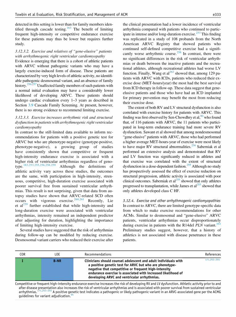
Towbin et al Evaluation, Risk Stratification, and Management of ACM e333
detected in this setting is lower than for family members iden-tified through cascade testing.163 The benefit of limitingfrequent high-intensity or competitive endurance exercisefor these patients may thus be lower but requires furtherstudy.
3.12.3.2. Exercise and relatives of “gene-elusive” patientswith arrhythmogenic right ventricular cardiomyopathyEvidence is emerging that there is a cohort of athletic patientswith ARVC without pathogenic variants who may have alargely exercise-induced form of disease. These patients arecharacterized by very high levels of athletic activity, no identifi-able pathogenic desmosomal variant, and an absence of familyhistory.256,257Unaffected familymembers of such patients witha normal initial evaluation may have a considerably lowerlikelihood of developing ARVC. These patients shouldundergo cardiac evaluation every 1–3 years as described inSection 3.9 Cascade Family Screening. At present, however,there is no strong evidence to recommend limiting exercise.
3.12.3.3. Exercise increases arrhythmic risk and structuraldysfunction in patients with arrhythmogenic right ventricularcardiomyopathyIn contrast to the still-limited data available to inform rec-ommendations for patients with a positive genetic test forARVC but who are phenotype-negative (genotype-positive,phenotype-negative), a growing group of studieshave consistently shown that competitive or frequenthigh-intensity endurance exercise is associated with ahigher risk of ventricular arrhythmias regardless of geno-type.183,253,255,256,258,259 Although the definitions ofathletic activity vary across these studies, the outcomesare the same, with participation in high-intensity, stren-uous, competitive, high-duration exercise associated withpoorer survival free from sustained ventricular arrhyth-mias. This result is not surprising, given that data from au-topsy studies have shown that ARVC-related SCD oftenoccurs with vigorous exercise.260,261 Recently, Lieet al259 further established that while high-intensity andlong-duration exercise were associated with ventriculararrhythmias, intensity remained an independent predictorafter adjusting for duration, highlighting the importanceof limiting high-intensity exercise.
Several studies have suggested that the risk of arrhythmiasduring follow-up can be modified by reducing exercise.Desmosomal variant carriers who reduced their exercise after
COR LOE Recommendations
I B-NR Clinicians should counsea positive genetic tesnegative that competiendurance exercise isdeveloping ARVC and
Competitive or frequent high-intensity endurance exercise increases the riafter disease presentation also increases the risk of ventricular arrhythmarrhythmias.171,253–255 A positive genetic test indicates a pathogenic oguidelines for variant adjudication.95
the clinical presentation had a lower incidence of ventriculararrhythmia compared with patients who continued to partic-ipate in intense and/or long-duration exercise.253 This findingwas replicated in a study of 108 probands from the NorthAmerican ARVC Registry that showed patients whocontinued self-defined competitive exercise had a signifi-cantly worse arrhythmic course.258 In contrast, there wereno significant differences in the risk of ventricular arrhyth-mias or death between the inactive patients and the recrea-tional athletes, although recreational athletes had worse LVfunction. Finally, Wang et al262 showed that, among 129 pa-tients with ARVC with ICDs, patients who reduced their ex-ercise dose (MET-hours/year) the most had the best survivalfrom ICD therapy in follow-up. These data suggest that gene-elusive patients and those who have had an ICD implantedfor primary prevention may benefit the most from reducingtheir exercise dose.
The extent of both RV and LV structural dysfunction is alsocorrelated with exercise history for patients with ARVC. Thisfindingwas first observed by Sen-Chowdhry et al,80 who foundthat, of 116 patients with ARVC, the 11 patients who partici-pated in long-term endurance training had more severe RVdysfunction. Sawant et al showed that among nondesmosomal“gene-elusive” patients with ARVC, those who had performeda higher average MET-hours-year of exercise were most likelyto have major RV structural abnormalities.256 Saberniak et alperformed an extensive analysis and demonstrated that RVand LV function was significantly reduced in athletes andthat exercise was correlated with the extent of structuraldysfunction in a dose-dependent fashion.255 Although no studyhas prospectively assessed the effect of exercise reduction onstructural progression, athletic activity is associated with poorclinical outcomes. Saberniak et al255 showed that only athletesprogressed to transplantation, while James et al253 showed thatonly athletes developed class C HF.
3.12.4. Exercise and other arrhythmogenic cardiomyopathiesIn contrast to ARVC, there are limited genotype-specific datafrom which to make exercise recommendations for otherACMs. Similar to desmosomal and “gene-elusive” ARVCpatients, ventricular arrhythmias occur disproportionatelyduring exercise in patients with the R14del PLN variant.161
Preliminary studies suggest, however, that a history ofathletics is not associated with disease penetrance in thesepatients.
References
l adolescent and adult individuals witht for ARVC but who are phenotype-tive or frequent high-intensityassociated with increased likelihood ofventricular arrhythmias.
171,253–255
sk of developing RV and LV dysfunction. Athletic activity prior to andias and is associated with poorer survival from sustained ventricularr likely pathogenic variant in an ARVC-associated gene per the ACMG

COR LOE Recommendations References
III: Harm B-NR Individuals with ARVC should not participate in competitive orfrequent high-intensity endurance exercise as this isassociated with increased risk of ventricular arrhythmias andpromoting progression of structural disease.
80,171,183,253–255,258
Competitive or frequent high-intensity endurance exercise is related to the extent of RV and LV dysfunction in patients with ARVC. Additionally,such exercise is associated with poorer outcomes for ventricular arrhythmias, whereas reducing exercise has a more favorable arrhythmicprognosis. Aiding patients and at-risk family members in making choices about participation in various types of exercise involves ongoingdiscussion and shared decision making.
e334 Heart Rhythm, Vol 16, No 11, November 2019
Competitive exercise includes participation in “an orga-nized team or individual sport that requires regular competi-
tion against others as a central component, places a highpremium on excellence and achievement, and requiressome form of systematic (and usually intense) training” asdefined by the AHA/ACC Scientific Statement for Eligibilityand Disqualification Recommendations for Competitive Ath-letes with Cardiovascular Abnormalities.249Endurance exercise includes class C and B sports inthese guidelines.248 Data on the effect of static exercise(Class A) on outcomes are largely absent from the literature.Intensity is typically measured in METs.252
For the basic science details of the mechanisms respon-sible for the forms of ACM, please see Section 4 DiseaseMechanisms.
Section 4 Disease mechanismsAn overview of some of the disease mechanisms for ACM isshown in Figure 18.
4.1. Desmosomal defectsThe cardiac ID is a highly organized structure that connectsadjacent cardiomyocytes and is classically comprised of threemain structures: (1) gap junctions (GJs), which metabolicallyand electrically connect the cytoplasm of adjacent cardiomyo-cytes; (2) adherens junctions (AJs), which connect the actincytoskeleton of adjacent cells; and (3) desmosomes, whichfunction as cell anchors and connect intermediate filaments(IFs). In addition, ion channels reside in the ID. Pathologic ge-netic variants in ID proteins have been associated with cardiacarrhythmias, such as BrS, ARVC, and other genetically deter-mined ACMs.263,264 However, rather than being independent,all ID components work closely together by partnering withmultifunctional proteins such as ZO-1, ankyrin G, and b-cate-nin, allowing the ID to integratemechanical and electrical func-tions. GJs form a plaque surrounded by the perinexus in whichfree connexons reside; the connexome integrates sodium (NaV)channels, the desmosome, and GJs; and the area compositahosts AJs and desmosomes, also integrated as adhering junc-tions. Furthermore, the transitional junction connects sarco-meres to the plasma membrane. The ID ensures rapidpropagation of the electrical signal that initiates contractionthroughout the heart and allows the cardiomyocytes to with-stand the strong mechanical forces imposed by the beating of
the heart. AJs, desmosomes, GJs, and ion channels form a func-tional unit as the area composita. Furthermore, GJs and ionchannels likely create and propagate action potentials (APs)together. Some structural components of cell–cell junctionscan also interact with other ID proteins or function in signalingpathways, such as Cx43 and b-catenin. Protein deficiencies canultimately lead not only to mechanical dysfunction (eg, AJdysfunction) but also to arrhythmias, often via GJ remodeling,thereby illustrating the interdependency of ID components andthe coupling of mechanical and electrical elements.
The lateral membrane (LM) of cardiomyocytes has adifferent makeup compared to the ID, hosting, among others,costameres and focal adhesions and linking sarcomeres to theextracellular matrix. The ID and LM have several proteins incommon, such as vinculin and a-actinin, and ion channels.
The AJ is the primary anchor for myofibrils and connectsactin filaments from adjacent cells, which allows the cell toretain its shape under mechanical stress. Furthermore, the AJtransduces signals concerning the actin cytoskeleton and sensesmechanical forces on the cell. The transmembrane protein N-cadherin is the main constituent of AJs and homodimerizeswith N-cadherins from adjacent cells in the extracellular space,acting as an intercellular zipper. This action provides tissuespecificity during development, allowing cells to interact onlywith cells expressing the same cadherin. Calcium ions ensurethe rod shape ofN-cadherin, the intracellular domainwhich pri-marily binds b-catenin. N-cadherin also possesses regulatoryfunctions including a role in mechanosensing. b-catenindirectly interacts with the C-terminal cytoplasmic domain ofN-cadherin. By associating with a-catenin and vinculin, b-cat-enin connects AJs to the actin cytoskeleton.
b-catenin also plays a central role in cadherin-mediatedsignaling and can activate the canonical Wnt signalingpathway. b-catenin translocates to the nucleus whenWnt bindsits Frizzled receptor, to initiate transcription of transcriptionfactors of the T-cell factor/lymphoid enhancer-binding factorfamily. The canonical Wnt pathway is crucial in cardiac devel-opment but has also been proposed as the key mechanism incertain cardiomyopathies (ie, activation induces cardiac hyper-trophy). Therefore, N-cadherin has been thought to sequesterb-catenin to prevent Wnt activation. Activation of the Wntpathway increases expression of the GJ protein Cx43, andthe C-terminus of Cx43 can interact with b-catenin. WhenWnt is not present, cytoplasmic b-catenin is targeted for degra-dation by the proteasome.

Figure 18 Disease mechanisms for arrhythmogenic cardiomyopathy. Cardiomyocyte showing the extracellular matrix, sarcolemma, sarcomere, nucleus, andkey proteins that provide structure for ventricular function and cardiac rhythm. See the description of the functions of these proteins in Section 4.1 DesmosomalDefects.
Towbin et al Evaluation, Risk Stratification, and Management of ACM e335
Although AJs also transduce forces to the cytoskeleton,desmosomes are more robust, thanks to their connection tomechanically resilient IFs. The intercellular part of the car-diac desmosome is built up by the cadherins desmoglein-2(DSG2) and desmocollin-2 (DSC2), which bind in a heterol-ogous way. The armadillo proteins junction plakoglobin(JUP) and plakophilin-2 (PKP2), and desmoplakin (DSP)(a member of the plakin superfamily), connect desmin tothe desmosome. When DSC2 and DSG2 are bound, the hy-peradhesive state of the desmosome depends on the presenceof calcium ions.
Considering the major desmosomal proteins, PKP2 isassociated with GJs and is required for the organization ofID and desmosomal function. Together with JUP, PKP2 me-diates attachment to IFs. PKP2 knockdown causes a decreasein conduction velocity and an increased propensity todevelop re-entry arrhythmias, whereas PKP2 variants aremost common in hereditary ACM. Plakoglobin is presentin both desmosomes and AJs. Desmoplakin connects the des-mosomes to the type III IF protein desmin, and its N-terminaland C-terminal domains and the a-helical domain in betweenare each almost 1000 amino acids long and interaction with
PKP2 occurs at their N-terminal domains. DSG2 pathogenicgene variants are, like all other cardiac desmosomal proteins,associated with ACM.
The most prominent ACM desmosomal gene mutationsinclude PKP2 and DSP, with desmosomal cadherins DSG2and DSC2, and JUP being less common.14,25,169,265 Themajority of these genes primarily cause ARVC, althoughpathogenic variants in DSP cause a substantial amount ofALVC. Other “nondesmosomal” genes, such asTGFB3 and TMEM43, disrupt the function ofdesmosomes.132,266,267 One of the more recently describedcausative genes is CDH2, encoding N-cadherin, anothermember of the cadherin superfamily of predominantlyCa21-dependent cell surface adhesion proteins.118,119 In thereport by Mayosi et al, the affected family members allpresented with ventricular arrhythmias and demonstratedimaging features of ARVC. The study by Turkowski et al,however, described a family with an arrhythmogenicpresentation who all showed a cardiomyopathy in theimaging study.119 In desmosomes, desmosomal cadherins(desmocollin and desmoglein) are mainly anchored to theIFs of the cytoskeleton through numerous intracellular

e336 Heart Rhythm, Vol 16, No 11, November 2019
protein partners, whereas in fascia AJs, the classical cadherinN-cadherin is primarily anchored to the actin microfilamentsof the cytoskeleton and promotes cell–cell adhesionthrough extracellular associations of its cadherin repeat do-mains.268–270 Interestingly, the protein components ofdesmosomes and fascia AJs are not mutuallyexclusive.270,271 In fact, the mechanical junctions of the IDare an admixture of desmosomal and fascia adherensproteins that form a hybrid functional zone, the areacomposita.269,272,273 Therefore, even if ARVC has beentraditionally considered as a desmosomal disease, it is nowreasonable to consider that the mechanistic basis of ARVCmay extend beyond the strict functional zone of thedesmosome, to that of the area composita. Supporting thisconcept, pathologic variants in CTNNA3 (another gene inthe area composita), which encodes for aT-catenin, hasalso been identified in patients with ARVC who werenegative for pathologic variants in the main desmosomalgenes.115 a-catenins are natural partners of the cytoplasmicdomain of classical cadherins, that is, N- and E-cadherins,and, in the case of N-cadherin, act as its go-between foranchoring to the actin cytoskeleton.
The fact that cadherin-2, like its desmosomal cadherincounterparts, is a major player in the ID is also supportedby the Cdh2 cardiac-specific mouse model with deletion ofN-cadherin in the adult mouse heart causing dissolution ofthe ID structure, including loss of both desmosomes andAJs, demonstrating that desmosome integrity is alsocadherin-2 dependent.274 These mice also exhibited modest,albeit atypical, DCM and spontaneous ventricular arrhyth-mias that resulted in SCD.274 This increased arrhythmic pro-pensity (all mice experienced SCD approximately 2 monthsafter deleting N-cadherin from the heart) was probably dueto a reduced and heterogeneously distributed connexin-43,causing loss of functional GJs and partial cardiomyocyte un-coupling and highlighting the prominent role of cadherin-2 inall types of functional junctions in the ID.275 GJ decreases innumber and size, with concomitantly increased arrhythmiasusceptibility, have also been demonstrated in the contextof N-cadherin heterozygous null mice, with 30%–60% ofthese mice developing VT, suggesting that cadherin-2 hap-loinsufficiency might create an important arrhythmogenicsubstrate.276 ID remodeling with concomitant reduction oflocalization of desmosomal proteins, connexin-43, andcadherin-2 has also been demonstrated in ventricular tissuesof transplanted hearts of patients with ARVC, further sup-porting the involvement of cadherin-2 in ARVC pathogen-esis.277
4.2. Ion channel defectsCardiac cells are excitable cells that can generate and propa-gate an AP, the electrical signal that induces cardiomyocytecontraction. The cardiac AP is generated by ions movingacross the cell membrane that, by depolarization, takes thecell from the resting state to an activated state and then, byrepolarization, back to the resting membrane potential.278
All phases of the cardiac AP occur via the synergistic activa-tion and inactivation of several voltage-dependent ion chan-nels. In contractile cardiomyocytes, APs are triggered by theacute entrance of sodium ions (Na1) inside the cell, resultingin an inward current (INa) (SCN5A) that shifts the membranepotential from its resting state (290 mV) to a depolarizationstate (120 mV). This phase is followed by the efflux of po-tassium (K1) ions through an outward current named Ito,which initiates cell repolarization. This in turn is followedby the plateau phase, a short period of constant membrane po-tential due to the balance between inward calcium (Ca21)currents (ICaL) through the voltage-dependent L-type calciumchannels (LTCCs) and time-dependent delayed-rectifier out-ward K1 currents (mainly slow delayed-rectifier IKs[KCNQ1] and rapid delayed-rectifier IKr [KCNH2]). At thispoint, the Ca21 entry through the LTCC triggers a muchlarger release of Ca21 from sarcoplasmic reticulum (SR)stores through the ryanodine receptor channel type 2, produc-ing a systolic increase in the intracellular Ca21 needed forcell contraction. Upon LTCC inactivation, the net outwardK1 currents repolarize the cell and bring the membrane po-tential to its resting state. The balance between Ca21 andK1 currents therefore determines the AP duration. The basaland acetylcholine-dependent inwardly rectifying K1 currents(IK1 and IKACh) control the final repolarization and determinethe resting membrane potential. Ca21 is then extruded fromthe cell through the Na1/Ca21 exchanger (NCX) type 1and taken back into the SR through the SR Ca21-ATPasetype 2a, thereby restoring low intracellular Ca21 levels, al-lowing cell relaxation during diastole.
Pacemaker cells are distinct from other cell types inshowing automaticity, a property resulting from bothvoltage-dependent and calcium-dependent mechanisms.279
The former involves the funny current (If) carried byhyperpolarization-activated cyclic nucleotide-gated chan-nels,280 which have several unusual characteristics, such asactivation on hyperpolarization, permeability to sodium andpotassium ions, modulation by intracellular cyclic adenosinemonophosphate, and a small single-channel conductance.The latter involves spontaneous calcium release from theSR,281 which activates INCX. The crucial role of calcium-dependent mechanisms has been demonstrated in mice withcomplete atrial-specific knockout of NCX, which has shownno pacemaker activity.282 Both mechanisms result in sponta-neous depolarization responsible for the rising slope of themembrane potential. When the proper ion current flows aredisturbed, electrical abnormalities in the form of arrhythmiasoccur.
4.2.1. SCN5AThe SCN5A gene, which encodes the alpha subunit of thevoltage-gated sodium channel Nav1.5, is responsible for theinward sodium current (INa).
283 This current is the maincomponent of rapid depolarization in cardiomyocytes and isresponsible for the AP upstroke, which subsequently initiatesthe multistep excitation–contraction coupling cascade.284

Towbin et al Evaluation, Risk Stratification, and Management of ACM e337
This periodic depolarization underlies the synchronous andrhythmic contraction of the heart chambers.284
Pathologic variants in genes encoding for ion channelproteins are well-known causes of inherited arrhythmia dis-orders, such as LQTS, short QT syndrome (SQTS), BrS,CPVT, and AV block, to name a few. The involved genesinclude those encoding for the cardiac sodium channel, po-tassium channels, and calcium channels. In these disorders,pathologic variants in the affected gene result in distur-bance of the function of the encoded ion channel protein,leading to abnormalities in the function of the AP. Inseveral of these genes, pathologic variants can cause a het-erogeneous array of clinical features, at times differingeven within the same family. For instance, pathologic var-iants in the cardiac sodium channel gene SCN5A areresponsible for LQTS type 3 (LQT3), which developsdue to a gain in channel function. On the other hand, path-ologic SCN5A variants also cause BrS, an electrocardio-graphically distinguishable disorder compared withLQT3, which occurs due to a loss of sodium channel func-tion.285 In addition to causing ventricular tachyarrhyth-mias, atrial fibrillation, atrial standstill, and AV block,286
SCN5A is known to cause an arrhythmogenic form ofDCM and an arrhythmogenic form of LVNC.287
The 2006 Scientific Statement on “Contemporary defini-tions and classification of the cardiomyopathies,” endorsedby the AHA, placed “ion channel disorders” under the classi-fication of primary genetic cardiomyopathies.288 This deci-sion was largely based on data regarding the role ofpathologic variants in genes encoding defective ion channelproteins, governing cell membrane transit of sodium, potas-sium, and calcium ions leading to ion channel-relatedarrhythmia disorders, including LQTS, SQTS, BrS, andCPVT and the role of these disorders in the development ofcardiomyopathies.288 This classification scheme hascontinued to be evaluated, and the list of overlappingcardiomyopathy–arrhythmia phenotypes has grown overtime, with primary and secondary causes of ion channeldysfunction seen in many cardiomyopathies. Pathogenic var-iations in the SCN5A gene, resulting in electrical and struc-tural cardiac remodeling, (ie, arrhythmogenic DCM), wasfirst described in 2003 by Groenewegen et al in a large familywith atrial standstill, a rare form of atrial cardiomyopathy.289
At the same time, it was shown that the clinical spectrum ofrare SCN5A pathologic genetic variants could be expanded toARVC and DCM, accompanied by arrhythmias and conduc-tion disorders.131,290 In 2008, new evidence showed thatpathologic variants in the SCN5A gene might represent arisk factor for rhythm disturbances in LVNC.291 These disor-ders are inherited as autosomal dominant traits. The fre-quency of SCN5A-mediated cases in patients with ACMs isapproximately 2%;292 however, when pathologic variantsin the SCN5A and LMNA genes are taken together, they ac-count for up to 5%–10% when considering only patientswith DCM with progressive cardiac conduction defects andsupraventricular and/or ventricular arrhythmias. Both RVand LV dilation and dysfunction can occur, as can broad
and heterogeneous electrical abnormalities, including atrialstandstill, progressive AV block, atrial fibrillation, sick sinussyndrome, VT, torsades de pointes, and VF, resulting inarrhythmic sudden death in some cases.
SCN5Amay also play a role in ACMwithout having path-ogenic gene variants. In ARVC, it is clear that when patho-logic variants in genes encoding the cardiac desmosomeare identified, Nav1.5, which has been shown to co-precipitate with the desmosomal protein PKP2, can be dis-rupted and dysfunctional. The loss of PKP2 expression hasbeen shown to alter the amplitude and kinetics of the sodiumcurrent (INa).
293 In addition, pathologic variants in PKP2have been associated with a sodium channelopathy pheno-type, whereas decreased immunoreactive Nav1.5 proteinhas been detected in the majority of human ARVC heart sam-ples.294 These observations indicate a close functional asso-ciation between Nav1.5 and mechanical junction proteins,which is further supported by the finding that Nav1.5 copre-cipitates with the AJ protein N-cadherin295 and demon-strating the presence of “adhesion/excitability” nodesformed by aggregates of Nav1.5 and N-cadherin.296 Leo-Macias et al described the presence of these adhesion/excit-ability nodes in cardiac myocytes and demonstrated that (1)the AJ protein N-cadherin serves as an attractor for Nav1.5clusters, (2) the Nav1.5 in these clusters are major determi-nants of the cardiac sodium current, and (3) clustering ofNav1.5 facilitates its regulation by molecular partners.296
Te Riele et al further demonstrated that Nav1.5 is in a func-tional complex with cell adhesion molecules and that a pri-mary Nav1.5 defect can affect N-cadherin biology,resulting in reduced size and density of N-cadherin clustersat the ID.295
The finding that Nav1.5 coprecipitates with the AJ proteinN-cadherin demonstrates the link to the junction/ID/desmo-some and supports the hypothesis that sodium channeldysfunction can occur via disruption of binding partners be-ing mutated (ie, supporting the PKP2 and arrhythmia sce-nario). Therapy for this disorder has not been well studiedand is not standardized. Pacemakers and ICDs have beenused for some individuals with varying outcomes. Pharmaco-logical therapies have been disappointing, and no specificpharmacotherapy has thus far been recommended for thesepatients.
4.3. Cytoskeletal defectsThe cytoskeleton is the cell’s basic scaffold inwhich other sub-cellular components are spatially arranged so as to communi-cate efficiently between the cell’s internal and externalenvironments. In striated muscle cells, the cytoskeleton con-sists of myofibrillar and extramyofibrillar portions. The myofi-brillar cytoskeleton is composedof thin and thickmyofilamentsand titin filaments, providing the foundation for myocytecontraction and relaxation. The extramyofibrillar cytoskeletonconsists of microfilaments, microtubules, and IFs.
IFs serve as a scaffold connecting the sarcomere to otherorganelles (such as mitochondria or the nucleus) to maintain

e338 Heart Rhythm, Vol 16, No 11, November 2019
cellular integrity and contribute to mechanotransduction. Thesarcomere is tethered to the sarcolemma (the membrane sur-rounding the myofibril) by another cytoskeletal assembly—the costamere. Costameres link the sarcomere to the sarco-lemma via the Z-disc and M-band. Individual heart cellsare connected by IDs, which synchronize muscle contraction.The myofibrils are linked to the plasma membrane at the Z-discs via the costameres. There are specific membrane invag-inations (T-tubules) at the Z-disc, which associate with flank-ing SR to the dyad. At the ID, desmosomes and AJs linkneighboring cardiomyocytes mechanically, and GJs provideion channels for intercellular communication. Desmosomeslink to the IF cytoskeleton (composed of desmin), whereasAJs anchor actin filaments (the myofibrils). The border ofthe last sarcomere before the plasma membrane is definedas the transitional junction.
The cytoskeletal structure is continually remodeled toaccommodate normal cell growth and respond to pathophys-iological cues. The cytoskeleton maintains the structuralintegrity and morphology of cardiomyocytes. Cytoskeletoncomponents are also involved in a variety of cellular pro-cesses, such as cell growth and division, cell movement,vesicle transport, cellular organelle location and function,localization and distribution of membrane receptors, andcell–cell communications. The cytoskeleton in cardiac myo-cytes is also believed to play an important role in the transduc-tion of mechanical signals, based upon the unique distributionof the extensive cytoskeletal network as well as the juxtaposi-tion of ion channels, signaling transducers, and network mes-sengers. Cytoskeletal modifications and cardiac myocyteremodeling are causally linked to cardiac hypertrophy andfailure. Abnormalities in cytoskeletal components not onlycause structural defects but also impair mechanotransduction.The cytoskeleton not only interacts with the extracellular ma-trix via transmembrane proteins such as integrins but also reg-isters adjacent Z-discs to one another, to the cell membrane,and to the nuclear envelope through a delicate network. Anumber of signaling partners bind to the network eitherdirectly or via linker proteins. For example, the muscle LIMprotein (MLP) gene encodes a muscle-specific cytoskeletalprotein interacting with titin and telethonin (T-cap). Studiesin genetically engineered mice with targeted ablation ofMLP suggest that the titin–telethonin–MLP complex mayserve as a stretch sensor in cardiac muscle cells. There isgrowing interest in examining the role of cytoskeletal compo-nents in ion channel regulation under physiological andpathological conditions.
DCM characterized by ventricular dilatation and dimin-ished contractile function accounts for more than 80% ofnon-HCMs. DCM has a population prevalence of approxi-mately 1 in 500 and is associated with prognostically adversearrhythmias at initial disease presentation in up to one-thirdof patients.297 While increased age, male sex, and impairedventricular function are established arrhythmic risk factors,arrhythmias also occur in patients with no known risk factors.Approximately 20%–35% of DCM cases are familial.Although impaired force generation, energy shortage, and
compromised calcium homeostasis could cause DCM,impaired force transmission and/or defective mechanotrans-duction caused by defects in cytoskeletal proteins such asdesmin, lamin A/C, a-actin, d-sarcoglycan, dystrophin, pla-koglobin, desmoplakin, MLP, and telethonin appear to be aprevalent mechanism underlying DCM.
4.3.1. Myofibrillar cytoskeletonThe myofibrillar cytoskeleton is composed of thin and thickmyofilaments of the sarcomere as well as titin filaments,providing the foundation for myocyte contraction and relaxa-tion. The basic unit of a myofibril is called the sarcomere and isdefined as the region between two Z-discs. The actin cross-linker protein a-actinin is a classical marker for Z-discs; how-ever, Z-discs house a large number of other cytoskeletal andsignaling proteins. The sarcomere, which is the smallest con-tractile unit of striated muscle, has its lateral boundariesdefined by the protein-dense Z-discs that cross-link the barbedends of actin-based thin filaments from adjacent sarcomeresvia a-actinin and are bordered by the I-band, the region oneither side of a Z-disc that is devoid ofmyosin-containing thickfilaments. The A-band comprises the region extending theentire length of the thick filaments, and the M-band residesat the center of the A-band. The force of muscle contractionoccurs when the myosin motor protein attaches to the actinfilament and pulls the Z-discs toward the M-band. The sarco-mere is not a static structure and responds to alterations inmus-cle load and injury. Z-discs also serve as an anchor site for theN-terminus of titin and nebulin and nebulette filament systems,making it indispensable for transmitting contractile force.
Z-discs anchor the thin filaments, which are composed ofactin, tropomyosin, and the troponin complex. Tropomyosinand the troponin complex are crucial for contraction regula-tion at the thin filament level, which is triggered by calcium.The thick filaments are composed of myosin dimers (amyosin consists of a myosin heavy chain and two myosinlight chains), which are arranged in bipolar filaments, withthe myosin tails making up the central region of the sarco-mere and the head interdigitating with the thin filaments.Myosin-binding protein C is associated with a subset of themyosin heads and contributes to controlling contraction atthe thick filament level. The third filament system is calledthe elastic filaments and consists of titin.
Variants in Z-disc–associated proteins are linked tonumerous cardiomyopathies and skeletal myopathies.298–301
a-actinin is the predominant Z-disc protein. There are fourvertebrate a-actinin genes with overlapping functions; howev-er, only ACTN2 is found in cardiac muscle.302 The N-terminalactin-binding domain is linked to an a-actinin-2 homodimercross-linking two antiparallel actin filaments of adjacent sarco-meres, forming a flexible tetragonal lattice.303 This lattice isessential for the rigidity that the Z-disc requires to serve as astructural anchor site, while still allowing for the flexibilityneeded to conform to contractile forces.
a-actinin has a myriad of binding partners, with each inter-action serving a distinct role in the production of concerted

Towbin et al Evaluation, Risk Stratification, and Management of ACM e339
contractile action. The major Z-disc proteins that interactwith ACTN2 include actinin-associated LIM protein, muscleLIM protein, the N-terminus of titin, myotilin, CapZ, Z-bandalternatively spliced PDZ-motif protein (ZASP), filamin,a-actinin, and telethonin-binding protein at the Z-disc, myo-palladin, and myopodin.304–306 Independent studies havereported that human variants in the ACTN2 gene areassociated with DCM, HCM, idiopathic VF, LVNC, andatrial arrhythmias.307
Filamin protein family members also bind and cross-linkactin. There are three filamin proteins: filamin-A (a iso-form), filamin-B (b isoform), and striated muscle-specificfilamin-C (g isoform). Filamin-C (g-filamin) is one of themajor proteins that serves as a link between the costamereand Z-disc and is involved in signal transduction with integ-rins. Filamin-C functions through interactions with sarco-lemmal muscle cell membrane proteins such as g- andd-sarcoglycans of the dystrophin glycoprotein complex,308
the b1A-subunit of the integrin receptor complex,309 andZ-disc proteins (such as myotilin310 and FATZ309,311,312).An autosomal dominant nonsense variant, p.Trp2710*, inthe last exon of the human filamin-C gene interferes withits dimerization process and causes filamin-C to aggregatewithin skeletal muscle fibers, a phenomenon that eventuallyleads to myofibrillar myopathy.313,314
Many of the proteins within the myofibrillar cytoskeletonhave been shown to cause cardiac and/or skeletal myopathy.Review of the details on the patients with pathologic variantsin the genes encoding these proteins with disturbance of pro-tein function has demonstrated a significant association withearly-onset arrhythmias, conduction system disease, and sud-den cardiac arrest or death, consistent with an arrhythmo-genic form of cardiomyopathy.
4.3.2. ZASP/LDB3ZASP/LIM domain binding 3 (LDB3) is one of the majorcomponents of the Z-disc proteins in cardiac muscle315 andplays an important role in stabilizing the Z-disc structurethrough its PDZ-mediated interaction with a-actinin-2, themain component of the Z-disc actin cross-linker, and F-actin,the main cytoarchitectural protein of cardiomyocytes.316
Global ablation of the murine ZASP homolog cypher candisorganize the sarcomere and cytoskeleton, leading to se-vere cardiomyopathy and skeletal myopathy in mice and hu-mans,317 whereas cardiac-specific ablation of cypher cancause DCM and SCD.318 The product of SCN5A, theNav1.5 current, localizes at the cardiomyocyte membranealong the sarcomeric Z-lines via a-actinin-2, thus connectingNav1.5 to actin filaments.319 ZASP/telethonin contributes tolocalizing Nav1.5 to the T-tubule membrane at the Z-line,creating a multiprotein complex associated with a-actinin-2. Variants in the ZASP/LDB3 gene have been shown tocause abnormalities in sodium channel function.
Vatta et al were the first to describe pathologic variants inZASP/LDB3 in patients with DCM and LVNC, identifying6 (6%) of 100 probands screened.126 Pathologic variants in
ZASP/LDB3 were identified in 2 families and 4 sporadiccases. Of the 9 familial and sporadic patients affected, 3had early-onset conduction system abnormalities and ven-tricular arrhythmias, including sinus bradycardia, second-degree AV block, PVCs, VT, intraventricular conductiondelay, ventricular bigeminy, and LBBB. Subsequent reportson patients with arrhythmias and conduction disease associ-ated with DCM and LVNC have supported the causativeconnection with variants in ZASP/LDB3. Arimura et al320 re-ported on a family with 6 affected members who developedDCM between 50 and 69 years of age, consistent with late-onset DCM, 3 of whom died suddenly. Xi et al321 studiedone of the original ZASP/LDB3 pathologic variants reportedby Vatta et al126 and demonstrated several underlying mech-anisms by which the ZASP-D117N variant (a ZASP/LDB3variant identified in patients with DCM/LVNC associatedwith intraventricular conduction delay, ventricularbigeminy, and LBBB) can cause intraventricular conductiondelay: (1) ZASP1-D117N can cause loss of function ofNav1.5 in human cell lines, and in neonatal cardiomyocytes;(2) in silico simulation using the Luo-Rudy model showedthat the extent of functional disturbances of Nav1.5 causedby ZASP-D117N is sufficient to delay cardiac conductionin human hearts; (3) the interaction between ZASP andNav1.5 requires preservation of the Z-disc protein complex;and (4) the modification of Nav1.5 by ZASP-D117N occurswithout significant disruption of Z-line structures in cardio-myocytes.321
Although Nav1.5 preferentially localizes at the ID viaSAP97 and LMs via the dystrophin-associated protein com-plex (2 pools), localization at the T-tubular system also oc-curs.322,323 Upon posttranslational modification, Nav1.5remains attached to the cytoskeleton linked to multiproteincomplexes and stored in subcellular compartments. Nav1.5is also known to localize at the cardiomyocyte membranealong the sarcomeric Z-lines via a-actinin-2, thus connectingNav1.5 to actin filaments.319 The study by Xi et al thereforesuggests that electrical remodeling may precede anatomicalremodeling in DCM/LVNC associated with ZASP with theloss of function of Nav1.5 by the mutated ZASP, occurringwithout significant disruption of cytoarchitectural networks.321
This is particularly important in a clinical situation, sincepatients who carry ZASP-D117N may develop arrhythmiaseven before manifesting HF symptoms. The loss of functionof Nav1.5 by ZASP-D117N appeared to be largely responsiblefor the conduction delay.
More recently, Lopez-Ayala et al reported on a family inwhich a pathologic variant in ZASP/LDB3 was associatedwith ARVC.158 The index patient and her first-degree andsecond-degree relatives underwent a complete clinical evalu-ation. After ruling out pathologic variants in the 5 desmo-somal genes, genetic testing using NGS was performed onthe proband, who had a long-standing history of presyncope.The proband experienced syncope associated with sustainedVT that required electrical cardioversion to restore sinusrhythm. Her ECG showed complete right bundle branchblock (RBBB), with inverted and flat T waves in the

e340 Heart Rhythm, Vol 16, No 11, November 2019
precordial leads. Echocardiogram and CMR showed biven-tricular dilation and severe biventricular systolic dysfunction;midwall LGE affecting the LV was also identified. An ICDwas recommended. However, the patient died in the oper-ating room during the surgical procedure as a result of ananesthetic complication. The postmortem examinationdemonstrated extensive fibro-fatty replacement in the RV,extensive fibrosis in the LV, and limited inflammatorypatches, consistent with a diagnosis of ARVC. A heterozy-gous pathogenic missense variant in ZASP/LDB3(c.1051A.G) was identified, and another 6 carriers wereidentified in her family via cascade screening. Three of theserelatives fulfilled the criteria for a definitive diagnosis ofARVC, and another reached a borderline diagnosis. Theserelatives had symptoms including frequent palpitations,abnormal ECGs that showed inverted T waves in right pre-cordial and inferior leads, signal-averaged ECGs that showedlate potentials, 24-hour Holter monitoring studies thatshowed runs of idioventricular rhythm and ventricularectopic beats, CMR that showed a dilated RV with severesystolic dysfunction, and normal LVwith no LGE. A numberof the relatives were started on beta-blockers. On the basis ofthis family, the authors suggested a direct link between ACMwith biventricular involvement and pathogenic variants inZASP/LDB3.
4.3.3. a-actinin-2a-actinin-2 (ACTN2) is a prominent member of the Z-discfound in cardiac muscle, has an N-terminal actin-bindingdomain, and creates a lattice-like structure that is essentialfor the rigidity that the Z-disc needs to serve as a structuralanchor site, while still allowing for the flexibility needed tobe responsive to contractile forces.303,324,325 The protein’sprimary function is to anchor and crosslink actin filamentsin the cardiac Z-disc at the lateral boundaries of thesarcomere.306 The Z-disc provides structural support bytethering the sarcomere to the sarcolemma via thecostameres and by anchoring filamentous F-actin, titin,and nebulette.305
As one of the integral Z-disc proteins, a-actinin has amyriad of binding partners, with each interaction servinga distinct role in the production of concerted contractileaction.306 The major Z-disc proteins that interact witha-actinin-2 and a-smooth muscle actin (ACTA2) areactinin-associated LIM protein, muscle LIM protein, theN-terminus of titin, myotilin, CapZ, ZASP, filamin, andtelethonin-binding protein at the Z-disc, myopalladin, andmyopodin. ACTA2 has also been demonstrated to bindphosphorylase-b, an important metabolic enzyme in theZ-disc. Furthermore, there is evidence that ACTN2 directlyinteracts with cardiac ion channels (such as the potassiumion channels KCNA4 and KCNA5326,327 and the sodiumion channel SCN5A319) and forms a bridge between the cal-cium ion channels CACNA1C and CACNA1D.328 Thus,disruption of ACTN2 may affect the localization and func-tion of cardiac ion channels. The authors speculated that the
various clinical presentations of Ala119Thr result from astochastic disruption of one of the many functional rolesof ACTN2.
One presentation of ACM was reported by Bagnall et al,who performed exome sequencing on a four-generation fam-ily with idiopathic VF, LVNC, and sudden death and identi-fied a pathologic variant in the ACTN2 gene.329 Clinicalevaluation of the family identified marked cardiac phenotypeheterogeneity, with some individuals being asymptomatic andothers having LVNC, resuscitated cardiac arrest due to idio-pathic VF, DCM, or sudden unexplained death. WES identi-fied an Ala119Thr pathologic variant in ACTN2 thatsegregated with disease. The 22-year-old female proband pre-sented with syncope and a family history of premature suddenunexplained death (her 25-year-old sister died in her sleep).The proband’s ECG showed sinus rhythm with nonspecificST-T wave changes, and her echocardiogram and CMRshowed prominent LV apical trabeculations with preservedLV systolic function, consistent with LVNC. There were noinducible arrhythmias in the EPS, and her QTc measured440 ms. The proband was implanted with an ICD. Her fatherhad a history of dyspnea, LBBB, and LV dilation withreduced LVEF of 27%. One of the proband’s female cousinsexperienced a resuscitated cardiac arrest and her CMR re-vealed normal LV and RV indexed dimensions and function,with no evidence of myocardial fibrosis. The cousin wasfound to have idiopathic VF and was implanted with anICD, which subsequently delivered two appropriate shocks.She responded successfully to quinidine therapy.
In another report, Girolami et al assessed a large 4-generation Italian family, 18 members of which underwentdirect clinical assessment and genetic testing, including theproband.330 Eleven individuals had evidence of autosomal-dominant cardiomyopathy and had variable combinationsof 3 distinctive features: regional LV noncompaction withLV hypertrophy, atrial septal defect, and early-onset supra-ventricular arrhythmias and AV block. In most of these pa-tients, frequent premature atrial contractions that developedinto atrial fibrillation or flutter represented the initial clinicalmanifestation. These arrhythmic manifestations were anessential part of the phenotypic spectrum. The onset of sup-raventricular arrhythmias followed a common pattern,initially presenting with very frequent premature atrial con-tractions, proceeding to paroxysmal atrial fibrillation (be-tween 30–50 years of age) and then to permanent atrialfibrillation, requiring a pacemaker due to slow ventricularconduction. Many of the family members were treated withICDs. The authors suggested that the ACTN2 pathologic var-iants may directly participate in the genesis of familial supra-ventricular arrhythmias.
4.3.4. Filamin-CFilamin protein family members also bind and cross-linkactin. There are 3 filamin proteins, with filamin-C (g isoform)the only striated muscle-specific protein. In addition to theN-terminal actin-binding domain, there is a Z-disc

Towbin et al Evaluation, Risk Stratification, and Management of ACM e341
localization motif.310 Filamin-C is one of the major proteinsthat serves as a link between the costamere and Z-disc and isinvolved in signal transduction with integrins. Filamin-Cdirectly interacts with 2 protein complexes that link the sub-sarcolemmal actin cytoskeleton to the extracellular matrix:the dystrophin-associated glycoprotein complex and the in-tegrin complex. At IDs, filamin-C is located in the fascia ad-herens where myofiber ends reach the sarcolemma, adjacentto the position of desmosomal junctions. Filamin-C functionsthrough interactions with the sarcolemmal muscle cell mem-brane dystrophin-associated glycoproteins (such as g- and d-sarcoglycans308), the b1A-subunit of the integrin receptorcomplex, and Z-disc proteins (such as myotilin310 andFATZ309,311,312). The participation of filamin-C in the attach-ment of the sarcomere’s Z-disc to the sarcolemma (costa-meres) and to the IDs allows cell-to-cell mechanical forcetransduction. FLNC pathologic variants have been associatedwith myofibrillar myopathies, as well as cardiomyopathies.
Ortiz-Genga et al studied the FLNC gene using NGS in2877 patients with inherited cardiovascular diseases,34 withclinical and genetic evaluation of 28 affected families. Theauthors identified a characteristic phenotype in probandswith truncating variants in FLNC, as well as 23 truncatingpathologic FLNC variants in 28 probands previously diag-nosed with DCM, ACM, or RCM. The authors also identified54 pathologic variant carriers among 121 screened relatives.The phenotype consisted of LV dilation (68%), systolicdysfunction (46%), and myocardial fibrosis (67%) in the im-aging test, as well as inferolateral negative T waves, lowQRSvoltages, and ventricular arrhythmias (82%) in the ECG(33%), with frequent SCD (40 cases in 21 of 28 families).The authors observed no clinical skeletal myopathy. Pene-trance was .97% in carriers over 40 years of age, and therewas an autosomal dominant inheritance pattern. Immunohis-tochemical staining of myocardial tissue showed noabnormal filamin-C aggregates in patients with truncatingFLNC pathologic variants. Isolated or predominant RVinvolvement, common with desmosomal pathogenic vari-ants, was not observed. Unlike patients with pathogeniclamin A/C, emerin, or desmin pathogenic variants, these pa-tients had mild and infrequent cardiac conduction abnormal-ities. The authors suggested consideration of promptimplantation of a cardiac defibrillator for affected patientsharboring truncating pathogenic variants in FLNC.
4.3.5. Extramyofibrillar cytoskeletonThe extramyofibrillar cytoskeleton consists of microfilaments(actin), microtubules, and IFs (desmin). It connects the sarco-mere with the sarcolemma and extracellular matrix through theZ-disc and submembrane cytoskeleton,331–334 therebyensuring power transmission produced by the sarcomeres.The extramyofibrillar cytoskeleton also provides support forsubcellular structures, organizes the cytoplasm, regulatessarcolemma topography, and transmits intercellular andintracellular mechanical and chemical signals.
4.3.5.1. Desmin filamentsDesmin is the main IF protein and is deemed necessary forcardiomyocyte structural integrity, the allocation and func-tionality of its mitochondria, the nucleus position, and sarco-mere genesis.334,335 The IFs create a 3-dimensional skeletoncovering the entire cytoplasm, enveloping Z-discs, extendingfrom one Z-disc to another. IFs are also involved with othercell organelles, including the SR and the T-tubular system.These desmin filaments extend from the Z-disc to the costa-meres, where they are bound through plectin and dysferlin,extend to the ID, and emerge from the Z-discs of the perinu-clear myofibrils to the nuclear membrane.
Pathogenic variants in DES, encoding desmin, have beenshown to cause severe skeletal and cardiac muscle diseaseswith heterogeneous phenotypes. DES variants have alsobeen found in patients with DCM and ARVC. Brodehlet al336 identified two novel variants in DES (p.Ala120Asp[c.359C.A] and p.His326Arg [c.977A.G]) in a familywith a broad spectrum of cardiomyopathies, with a strikingfrequency of arrhythmias and SCDs. In vitro experimentswith desmin-p.A120D identified a severe intrinsic filamentformation defect causing cytoplasmic aggregates in cell linesand of the isolated recombinant protein. Model variants ofcodon 120 indicated that ionic interactions contributed tothis filament formation defect. Ex vivo analysis of ventricu-lar tissue slices revealed a loss of desmin staining within theID and severe cytoplasmic aggregate formation, whereas Z-band localization was not affected. The authors proposedthat the loss of desmin-p.A120D filament localization atthe ID resulted in its clinical arrhythmogenic potential.Berm�udez-Jiménez et al more recently demonstratedimpaired filament formation and disruption of cell mem-brane integrity in a severe form of arrhythmogenic LV car-diomyopathy due to a DES pathogenic variant,p.Glu401Asp, in a large family.337
Variants in the DES gene result in striated muscle disor-ders characterized by the formation of inclusion bodies,weakening of the desmin cytoskeleton, disruption of sub-cellular organelle organization, and eventually myofibrildegradation. These muscle disorders are referred to asdesmin-related myopathy or desminopathy and oftenpresent in young childhood, with patients experiencingincreasing muscle weakness. These disorders areassociated with a wide spectrum of clinical phenotypes,even within the same family, and range from scapulopero-neal, limb girdle, and distal myopathic phenotypes withvariable cardiac or respiratory involvement to purecardiomyopathies.338
To date, multiple reports of ACM caused by pathogenicDESvariants havebeenpublished.DESvariants havebeenpre-viously reported in conduction disease and cardiomyopathies,in particular cases of DCM,339 and, more recently, inARVC.169 The first of these, DES pathogenic variantp.N116S, was identified in a 17-year-old patient with ARVCand concomitant subclinical skeletal muscle alterations, andthis variant led to an amino acid substitution that in turn led

e342 Heart Rhythm, Vol 16, No 11, November 2019
to aggresome formation in cardiac and skeletal muscle.340,341
All other reported ARVC-relatedDES variants underlie a clin-ically heterogeneous phenotype, frequently associated withmuscle abnormalities, including a DES-p.S13F pathogenicvariant identified in 39 family members from 8 Dutch fam-ilies169,342 with associated variable skeletal myopathy and awide spectrum of cardiomyopathies, including 2 patientswith ARVC. Another DES variant, p.N342D, was describedin patients affectedwith desmin-relatedmyopathies.343 The as-sociation of this variant with RV cardiomyopathy was alsonoted in select patients.342,344 A DES-p.P419S variant wasidentified by exome sequencing in a large Swedish family,showing myofibrillar myopathy and ARVC (ARVC7locus).122 Berm�udez-Jiménez et al described a multigenera-tional family in which approximately 30 family membersaffected with an ACM phenotype hosted a rare missense path-ogenic variant of the DES gene (c.1203G.C;p.Glu401Asp).337 These members showed that the DESGlu401Asp variant caused the disease in the family, with100% penetrance and variable expressivity. The phenotypepresented itself as an arrhythmogenic phenotype with a highrisk of SCDand progressiveHF. In 4 of the individuals studied,RV involvement was observed, and 2 had epsilon waves.Fibro-fatty infiltration was identified, predominantly in theLV, and the cardiomyocytes had reduced cellular adhesion,reminiscent of the defect found in ARVC, along with reducedexpression of DES and cell–cell junction proteins.
4.4. Sarcomeric defectsThe cardiac sarcomere is the fundamental contractile unit ofthe cardiomyocyte. Genetic variants in sarcomere genes area well-established cause of HCMand, in some cases, can causefamilial DCM, LVNC, and RCM.345 Variants in MYBPC3account for approximately 50% of all genotyped HCM cases,with most being loss-of-function variants, whereas missensevariants in MYH7 account for 30% of cases.346,347 Othergenes, such as TNNT2, TNNI3, TPM1, ACTC1, MYL2 andMYL3, account for �5% of HCM cases each. A recent studyinvestigating variant excess in cases compared with theExome Aggregation Consortium control population348
showed variants inMYH7, TNNC1, TNNT2 and TPM1 signif-icantly enriched in patients with DCM.100 Specifically,MYH7accounts for approximately 3%–4% of familial DCM cases.339
Sarcomere gene variants contribute to cases of LVNC,althoughmost often in phenotypes that include another cardio-myopathy, cardiac malformation and/or reduced ejection frac-tion, with MYH7 variants contributing the most cases.349,350
Other genes encoding sarcomeric and Z-disc proteins havealso been identified in individuals with LVNC, includingACTC1, MYBPC3, TNNT2, TPM1, TTN, and LDB3. RCMin childhood can be caused by variants in thin filamentgenes, TNNT2, TNNI3, and TPM1.351
Thepresenceof a sarcomere variant is associatedwithworseoutcomes in HCM, with patients with sarcomere-positiveHCMhaving poorer survival frommajor cardiovascular eventscompared with patients with gene-elusive HCM.347,352
Similarly, a recent study of LVNC cases showed a greaterrisk of major cardiovascular events in patients with asarcomere variant compared with those without.353 UsingNGS, Wang et al targeted and sequenced 73 genes related tocardiomyopathy in 102 patientswith LVNC,with 63%of path-ogenic variants in sarcomere-encoding genes and 12% in ionchannel-encoding genes.354
4.5. Metabolic defectsThe clinical manifestations of inherited disorders of fatty acidoxidation vary according to the enzymatic defect and can pre-sent as isolated cardiomyopathy (DCM, HCM), suddendeath, progressive skeletal myopathy, and hepatic failure ar-rhythmias, which can be a presenting symptom of fatty acidoxidation deficiencies.355 Over a 25-year period, Bonnet et aldiagnosed 107 patients with an inherited fatty acid oxidationdisorder; arrhythmia was the predominant presenting symp-tom in 24 (22%) of these patients.355 These 24 cases includedVT (n5 15), atrial tachycardia (n5 4), sinus node dysfunc-tion with episodes of atrial tachycardia (n5 4), AV block (n5 6), and LBBB (n 5 4) in newborn infants. The authorsobserved conduction disorders and atrial tachycardias in pa-tients with defects of long-chain fatty acid transport acrossthe inner mitochondrial membrane (carnitine palmitoyl trans-ferase type II deficiency and carnitine acylcarnitine translo-case deficiency) and in patients with trifunctional proteindeficiency. Also, VTs were seen in patients with any typeof fatty acid oxidation deficiency. The authors concludedthat accumulation of the arrhythmogenic intermediary me-tabolites of fatty acids, such as long-chain acylcarnitines,could be responsible for the development of arrhythmiasand that inborn fatty acid oxidation errors may cause unex-plained sudden death or near-miss in apparently healthy in-fants and those with conduction defects or VT. Diagnosis isdetermined by a serum acylcarnitine profile.
Specifically, inborn fatty acid oxidation errors result inmetabolite buildup proximal to the enzyme defect and in defi-cient formation of energy-yielding substrates after the block.In the defects downstream from carnitine palmitoyl trans-ferase I, the acylcarnitine that accumulates has detergent prop-erties, which may explain its toxicity. Indeed, amphiphiliclipid metabolite, long-chain acylcarnitine, and lysophosphati-dylcholine accumulate during myocardial ischemia and play apivotal role in the production of arrhythmias. Incorporation oflong-chain acylcarnitine in the sarcolemma elicited electro-physiological anomalies analogous to those seen in acutemyocardial ischemia.356 The cellular electrophysiological ba-ses of the proarrhythmic effects of long-chain acylcarnitineappear to be multifactorial. First, reduction of the single-channel conductance of the inwardly rectifying K current byamphipathic lipid metabolites may account for automaticAP discharges from the resting and plateau potentials, leadingto VT. Second, retardation of conduction velocities by thedecrease in excitatory Na current could produce conductionanomalies and yield to reentry.357 Third, nonesterifiedfatty acids directly activate voltage-dependent Na currents

Towbin et al Evaluation, Risk Stratification, and Management of ACM e343
in cardiac myocytes, inducing cytotoxic calcium overload.358
Finally, amphipathic metabolites can interfere with the GJsand disturb the cell membrane’s lipid-protein interface,thereby impairing GJ channels.359 These toxic effects on ioniccurrents have not been observed with short- and medium-chain acylcarnitine.356
Systemic primary carnitine deficiency, a carnitine trans-porter deficiency, occurs when free carnitine cannot be freelyfiltered by renal glomeruli, in which 95% is supposed to bereabsorbed by the renal tubules by a high-affinity carnitinetransporter in the cellular plasma membrane. Carnitine isnot catabolized in humans, and its only metabolic conversionis through ester formation, with most esterified carnitineexcreted in urine. Active carnitine transport from bloodinto cells is mediated by the same transporter that functionsin the kidneys. The carnitine transporter OCTN2 is encodedby the SLC22A5 gene and transports carnitine in a sodium-dependent manner.360,361
Carnitine transporter deficiency is inherited as an auto-somal recessive trait. As a result of its deficiency, carnitineis not reabsorbed in the kidneys, leading to urinary loss anddepletion of blood and tissue levels, resulting in severeimpairment of long-chain fatty acid oxidation and hypoke-totic hypoglycemia with fasting and stress. Age at presenta-tion can range from infancy to adulthood, but neonatalhypoglycemia and sudden death can occur. Clinical manifes-tations in early-onset disease include chronic or acute skeletalmyopathy and cardiomyopathy, typically exacerbated bymetabolic decompensation. Untreated heart disease proceedsto DCM with reduced LVEF or mild interventricularseptal hypertrophy. Electrocardiographic findings includeabnormal T waves, ventricular hypertrophy, and atrial ar-rhythmias. Life-threatening arrhythmias can occur, includingNSVT with periods of sinus rhythm and ventricular prema-ture beats, even in the presence of only borderline LV hyper-trophy. Carnitine supplementation is typically administeredat a dose of 200 to 300 milligrams per kilogram body weightdivided throughout the day.
4.6. Mitochondrial formsThe presentation of mitochondrial cardiomyopathy includesHCM, DCM, and LVNC forms,362,363 and the severity canrange from asymptomatic to devastating multisystemdisease.364 Severe cardiac manifestations include SCD, HF,and ventricular tachyarrhythmia, which can worsen acutelyduring ametabolic crisis.Mitochondrial crises are often precip-itated by physiological stressors such as febrile illness and sur-gery and can be accompanied by acute HF. Most patients withneuromuscular symptoms present with normal or slightlyelevated creatine kinase levels, a normal electromyogram,and normal results of nerve-conduction studies.365–367
Abnormal liver enzyme levels have been found in up to 10%of patients.365,368 Sensorineural hearing loss occurs in 7%–
26% of patients, and its prevalence increases with age.369,370
Patients with myoclonic epilepsy with ragged red fibers(MERRF) andmitochondrial encephalopathy, lactic acidosis,
and stroke (MELAS) should be monitored for the develop-ment of cardiac hypertrophy and DCM. Patients withMERRF can present with myoclonus, generalized convul-sions, cerebellar ataxia, muscular atrophy, and elevated bloodlactate and pyruvate levels, as well as ragged red fibers inmuscle biopsy specimens. A case series of patients withMERRF and an m.8344A.G variant of mtDNA revealedthat early age at onset was the only factor associated withthe occurrence of myocardial disease.371 The developmentof myocardial disease in this cohort was associated with ahigher risk of SCD. Patients with MELAS can also presentwith ragged red fibers in the muscle biopsy; however, unlikepatients with MERRF, patients with MELAS have normalearly development and start to show symptoms only between3 years of age and adulthood. Patients with MELAS tend tohave short stature, seizures, hemiparesis, hemianopia, andblindness.372
Mitochondrial variants are common causes of myocar-dial LVNC in young children. LVNC is characterized byprominent ventricular trabeculations and deep recessesthat extend from the LV cavity to the subendocardial sur-face of the ventricle, accompanied or not by LV dysfunc-tion.373–375 Studies have shown the importance ofsubstrate flexibility in preserving normal cardiacfunction. In experimental models of pressure overload,failing human hearts have shifted from oxidizing fattyacids (the preferred substrate in the healthy heart) tooxidizing glucose for energy production. This metabolicswitch is associated with the downregulation of genesinvolved in mitochondrial biogenesis and fatty-acid meta-bolism and is mediated by the deactivation of PPAR-aand its activator, PGC-a, which are members of a familyof transcriptional coactivators involved in mitochondrialregulation and biogenesis. An increased reliance on glyco-lytic pathways could effectively reduce oxygen consump-tion in the short term; over time, however, reducedoxygen consumption might enable the progression of heartdisease by creating an energy-deficient state.376 Experi-mental evidence has shown that elevated fatty-acid fluxand fatty-acid oxidation (FAO)-deficient states can be asso-ciated with cardiac dysfunction. Both chronic increases inFAO (as observed in diabetes) and decreases in FAO (asseen in pressure-overload models of HF) can lead toHF.377 Accordingly, energy deficiency can be broadlyconceived as both a cause and an effect of HF.
The management of mitochondrial disease and cardiomy-opathy is largely supportive. Physicians should be aware thatpatients can make a remarkable recovery from a severe crisisstate. Pharmacological strategies include the use of variousdietary supplements. A typical “mitochondrial cocktail”would include coenzyme Q10, creatine, L-carnitine, thia-mine, riboflavin, folate, and other antioxidants, such as vita-mins C and E. Studies have suggested that the use ofantioxidants partially improves clinical features.376,378 Incontrast, a systematic review by Chinnery et al found noclear evidence to support the use of any supplement inpatients with mitochondrial disease.379

e344 Heart Rhythm, Vol 16, No 11, November 2019
The mortality rate can be high for patients with mitochon-drial disease that progresses to a crisis state, such as an acuteor subacute multiorgan failure secondary to mitochondrialrespiratory chain function that worsens due to fever, illness,stress, medications, or heat; urgent treatment is thereforenecessary. Crises that can be associated with severe lactate el-evations and cardiac complications during a crisis includecardiogenic shock, atrial and ventricular arrhythmias,DCM, and SCD. Patients often have baseline acidemia, andthe correction of acidosis should be gradual. Oxygenationcan worsen the crisis by increasing free-radical production;the partial pressure of oxygen therefore needs to be main-tained between 50 and 60 mm Hg.380,381 Patients withmitochondrial disease who present with fever or who areunable to eat or drink may be administered dextrose-containing intravenous fluids—preferably D10 with half-normal saline content—at a maintenance dose, regardlessof blood glucose levels. Their metabolic and volume statusshould be evaluated periodically. The management of thesepatients’ cardiac complications, including HF, bradyarrhyth-mias, and tachyarrhythmia, follows the same guidelines asthose for the general population. If cardiac dysfunction isnoted during a crisis, patients should be closely monitored us-ing serial echocardiography. In selected patients who haveadvanced HF due to cardiomyopathy, cardiac transplantationmay be needed. Three pediatric patients with mitochondrialcardiomyopathy who underwent cardiac transplantationreportedly had excellent early and late outcomes.382
4.6.1. Kearns-Sayre syndromeKearns-Sayre syndrome (KSS) is a mitochondrial myopathycharacterized by the clinical triad of ptosis, chronic progressiveexternal ophthalmoplegia, and abnormal retinal pigmentationand is associated with cardiac conduction defects and DCM,sometimes requiring transplantation.383,384 Approximately50% of patients with KSS have cardiac involvement,including recurrent syncope, bundle branch block, fascicularblock, and nonspecific intraventricular conductiondisturbances; 20% of deaths in these patients have beenattributed to cardiac causes. In a guidelines publication, theACC/AHA/HRS assigned a class I recommendation (with aLOE B rating) to pacemaker implantation for third-degreeand advanced second-degree AV block at any anatomic levelwhen associated with neuromuscular diseases and AV block.Skeletal muscle histopathology commonly demonstrates rag-ged red fibers. The genetic abnormalities observed in KSSconsist largely of single large-scale mitochondrial DNA dele-tions, although mitochondrial DNA point variants, such asm.3249G.A in the tRNA (Leu) gene, m.3255G.A in thetRNA (Leu) gene, and m.3243A.G in the tRNA (Leu) gene,have also been reported.384,385
4.7. Histiocytoid (oncocytic) cardiomyopathyInfantile histiocytoid cardiomyopathy is a rare but distinctivearrhythmogenic disorder characterized by incessant VT, car-diomegaly, and sudden death within the first 2 years of life if
left untreated. Approximately 100 histiocytoid cardiomyopa-thy cases have been reported in the literature;386–400 however,the prevalence is likely to be higher, given that many cases ofhistiocytoid cardiomyopathy could have been misdiagnosedas sudden infant death syndrome.401 Female preponderanceis approximately 4:1, with most cases (90%) occurring ingirls under 2 years of age, leading to intractable VF or cardiacarrest. The lesion resembles a hamartoma with histiocytoid orgranular cell features.400 The condition has clearly beendefined as a mitochondrial disorder and affects the functionof complexes I and III of the respiratory chain of the cardiacmitochondria.400 The etiology favors either an autosomalrecessive gene or an X-linked condition.
Histopathological findings in patients with histiocytoidcardiomyopathy include multiple flat-to-round, smooth, yel-low nodules located beneath the endocardial surface of theLV, the atria, and the four cardiac valves. The nodules arecomposed of demarcated, large, foamy granular cells.Glycogen, lipids, and pigment may be observed in these cells,as well as a lymphocytic infiltrate. Immunostaining showsperimembranous immunoreactivity for muscle-specific actin,but not for the histiocytic markers, S-100 protein andCD69.387,391,398,402,403 These cells may be abnormalPurkinje cells; however, a primitive myocardial precursorcannot be excluded. Radiofrequency ablation or pacemakerimplantation may be required to treat arrhythmias.404 Surgi-cal intervention with prolonged survival has been re-ported.405
Shehata et al reported two probands with de novononsense variants in the X-linked nuclear gene NDUFB11,which had not previously been implicated in any disease,despite evidence that deficiency for other mitochondrial elec-tron transport complex I members leads to cardiomyopa-thy.406 A third proband was doubly heterozygous forinherited rare variants in additional components of complexI, NDUFAF2, and NDUFB9, confirming that histiocytoidcardiomyopathy is genetically heterogeneous. In a fourthcase, the proband with histiocytoid cardiomyopathy inheriteda mitochondrial variant from her heteroplasmic mother, asdid her brother, who presented with cardiac arrhythmia. Acausal role for NDUFB11 truncation in the etiology of histio-cytoid cardiomyopathy helps explain the disease’s femalebias. Whereas most complex I deficiencies are thought tobe inherited in a Mendelian recessive manner, these two denovo variants establish a dominant haploinsufficient pheno-type.
Section 5 Other disorders5.1. Infiltrative cardiomyopathies: amyloidosisSee Evidence Table: ACM Amyloidosis. A recommendationflow diagram is shown in Figure 19.
Cardiac amyloidosis refers to the extracellular depositionof low molecular weight proteins within the myocardium,usually occurring in the context of more widespread organinvolvement. The amyloid deposits are typically formed byone of two proteins: light chains or transthyretin.407,408

Cardiac amyloidosis
In both symptomatic and asymptomatic individuals with cardiac amyloidosis and second-degree AV
block type II, high-grade AV block or third-degree AV
block, a permanent pacemaker is recommended
(COR I, LOE B-NR).
Second-degreetype II, high-gradeor third-degree AV
block?
Atrial arrhythmias? Survived cardiac arrest?
In individuals with cardiac amyloidosis who have
survived a cardiac arrest, an ICD is recommended if meaningful survival greater
than 1 year is expected (COR I, LOE C-EO).
AL-type with nonsustained ventricular
arrhythmias?
In individuals with cardiac amyloidosis, the use of
digoxin may be considered if used with caution due to
the high risk of toxicity (COR IIb, LOE B-NR).
In individuals with cardiac amyloidosis and
symptomatic atrial arrhythmias, the use of
sotalol, dofetilide, or amiodarone may be
considered (COR IIb, LOE C-EO).
In individuals with AL-type cardiac amyloidosis with nonsustained ventricular
arrhythmias, a prophylactic ICD may be considered if
meaningful survival greater than 1 year is expected (COR IIb, LOE B-NR).
In individuals with cardiac amyloidosis and
symptomatic atrial arrhythmias, cardiac
ablation may be considered (COR IIb, LOE C-LD).
Yes YesYes Yes
Figure 19 Amyloidosis arrhythmia treatment recommendations. AL 5 amyloid light-chain; AV 5 atrioventricular; COR 5 Class of Recommendation;ICD = implantable cardioverter defibrillator; LOE 5 Level of Evidence. Colors correspond to COR in Figure 1.
Towbin et al Evaluation, Risk Stratification, and Management of ACM e345
Isolated atrial amyloidosis due to atrial natriuretic peptidedeposition typically occurs in older age, and small studieshave suggested its role in atrial fibrillation.409,410 Lightchain amyloidosis (AL amyloidosis) is secondary to aprimary blood dyscrasia, which drives an abnormalproliferation of plasma cells and subsequently themonoclonal overproduction of light chains. Chemotherapyand stem cell transplantation have transformed care andvastly improved survival for AL amyloidosis.411 Transthyre-tin amyloid is composed of a different protein, a misfoldedprealbumin that will also produce amyloid fibrils and de-posits in tissues.408 Treatment includes liver transplantation,which can retard progression; the results are variable, howev-er, and advanced multiorgan involvement often preventscuring. Newer therapies to stabilize transthyretin, diminishits production, or remove it from affected organs are currentlyunder investigation.412–416
Cardiac involvement is in the form of an infiltrative car-diomyopathy in addition to HF via primarily diastolic limita-tion; small vessel disease,417 conduction systemdisease,418–420 and atrial and ventricular arrhythmias421 areall well recognized. Histological evaluation of hearts withcardiac amyloidosis has provided insight into the potentialunderlying mechanisms of cardiac arrhythmia. Amyloidfibrils infiltrate the extracellular matrix, disrupting myocar-dial cellular arrangement and leading to myocardialfibrosis.422,423 Perivascular amyloid infiltration andimpairment of cardiomyocyte function are also welldescribed,424,425 and the subsequent impairedvasoreactivity can result in relative myocardial ischemiaand abnormal electrical conduction. This cardiotoxicinfiltrative milieu is hypothesized to be the fundamentaldriver of conduction abnormalities, atrial and ventriculararrhythmias. Although widespread involvement is not

e346 Heart Rhythm, Vol 16, No 11, November 2019
uncommon, with sinus node dysfunction well-recognized,426–428 infranodal conduction system diseaseappears to be the primary conduction abnormality, asevidenced by HV interval prolongation.418,429 The diseaseis associated with the risk of sudden death in a number ofcohorts.418 Due to the progressive amyloid depositionthroughout the heart, sinus node dysfunction and conductiondisease often worsen, prompting the consideration for per-manent pacemakers. For those patients for whom permanentpacing is necessary, lead placement should be carefullyconsidered, given the potential for further LV depressionrelated to RV pacing dyssynchrony. Currently, there are nostudies that can provide definitive guidance on this issue.
Autonomic dysfunction with orthostatic presyncope orsyncope is commonly observed in patients with systemic am-yloid disease and cardiac involvement, and peripheral vaso-constrictors are frequently needed to managesymptoms.430–432 A clear conduction abnormality needs tobe considered as the etiology in these patients, recognizingthat most cases of SCD are likely related to infranodalconduction disease. Furthermore, significant cardiacinvolvement with advanced infranodal conductionabnormalities can often be masked by a normal-appearingQRS complex.418,429 By further blocking AV nodalconduction by preventing compensatory physiological heartrate recovery and directly preventing vasoconstriction, theactions of calcium blocking agents converge to create amalignant and potentially lethal combined effect. Evidenceis limited, however, to small case series and 2 casereports.433–435
COR LOE Recommendations
I B-NR In both symptomatic andamyloidosis and secondblock or third-degree Arecommended.
AV block has been consistently linked to sudden death in patients with cabnormalities, pacemaker implantation is recommended.
COR LOE Recommendations
I C-EO In individuals with cardiacarrest, an ICD is recommthan 1 year is expected
It is not known how many patients in the secondary prevention ICD trials hthat patients who have been resuscitated following a cardiac arrest ardefibrillation.3
COR LOE Recommendations
IIb B-NR In individuals with cardiamay be considered if ustoxicity.
Digoxin is known to bind to amyloid fibrils and putatively this action canwith cardiac amyloidosis have dysfunction related to the same diseaseexcretion. In a cohort of 107 patients with AL amyloidosis who receivetoxicity was 11%, and 5 patients died.446
The most common tachyarrhythmia in this disorder isatrial arrhythmia. Rate control using AV nodal blockingagents can be especially challenging in the face of the rela-tive hypotension and impairment in compensatory vaso-reactivity that is commonly seen with widespreadsystemic and autonomic impairment. AV nodal ablationhas been evaluated and appears to be a reasonable consid-eration in more resistant and symptomatic cases.436 Antiar-rhythmic approaches are often necessary, given thatmaintenance of active atrial systole can be imperative forpatients with restrictive LV filling; however, extensive am-yloid infiltration, when present, could impair atrial systole.Extensive substrate abnormalities, presumably related toextensive atrial amyloid fibril infiltration, are common,and results from atrial fibrillation ablation are less thanideal.429,436 Frequent ectopy with NSVT is the mostcommon ventricular dysrhythmia, yet neither burden ofectopy nor NSVT appears to predict SCD.437 WhetherICDs improve survival is not clear,438–442 andprogressive HF and terminal pulseless electrical activityremains a common theme associated with cardiac deathin this group. This situation may be different for patientswith cardiac amyloidosis who have been successfullymanaged for AL-type disease421 and for patients awaitingcardiac transplantation; individualized approaches aretherefore necessary.
Patients with cardiac amyloidosis remain at high risk fordeveloping intracardiac thrombus and thromboembolicstroke;443,444 anticoagulation needs to be carefullyconsidered even in the absence of atrial arrhythmias.
References
asymptomatic individuals with cardiac-degree AV block type II, high-grade AVV block, a permanent pacemaker is
418,426,427,
445
ardiac amyloidosis; for patients with obvious conduction system
References
amyloidosis who have survived a cardiacended if meaningful survival greater.
ad underlying cardiac amyloidosis. Nevertheless, there is agreemente at higher risk of recurrence and can potentially be revived by
References
c amyloidosis, the use of digoxined with caution due to the high risk of
446
potentiate its effect on the myocardium. In addition, many patientsprocess, and serum digoxin levels can be affected by the reducedd digoxin, the incidence of significant arrhythmias due to digoxin

COR LOE Recommendations References
IIb C-EO In individuals with cardiac amyloidosis and symptomatic atrialarrhythmias, the use of sotalol, dofetilide, or amiodaronemaybe considered.
Although not studied in a retrospective or prospective manner, atrial arrhythmias are common, often highly symptomatic, and poorly tolerated,mostly due to rapid ventricular rates and irregular ventricular response that impair ventricular filling and contractility. Patients withsignificant ventricular diastolic disease can also present with symptomatic deterioration in the context of impaired filling without atrialsystole, and antiarrhythmic agents are typically required. The class III antiarrhythmics (sotalol, dofetilide, and amiodarone) aremechanistically more suitable for therapy for this patient group, given the preponderance of atrial and ventricular myocardial fibrosis orscarring and the risk of atrial flutter and reentrant ventricular arrhythmia with class Ic agents. The use of class Ic agents can result inpersistent atrial flutter in this patient group, which frequently exhibits substrate-related atrial tachycardias.
COR LOE Recommendations References
IIb B-NR In individuals with AL-type cardiac amyloidosis withnonsustained ventricular arrhythmias, a prophylactic ICD maybe considered if meaningful survival greater than 1 year isexpected.
421
Primary prevention ICD implantation remains controversial, and there are conflicting data on the prevention of SCD in cardiacamyloidosis. Potentially curative therapies have emerged to manage certain subtypes,421 and AL amyloidosis could be more favorable inthis regard. Patients awaiting heart transplantation are also being considered for disease cure and should likely also be consideredindependently.
COR LOE Recommendations References
IIb C-LD In individuals with cardiac amyloidosis and symptomatic atrialarrhythmias, cardiac ablation may be considered.
436
It is important for clinicians to recognize that ablation for atrial arrhythmias has limited efficacy and high recurrence rates, even whenperformed in major referral centers. Patients with rapid ventricular rates and those resistant to medical therapy also appear to benefitsymptomatically from combined AV nodal ablation and permanent pacemaker implantation. In a cohort of 26 patients, 13 of whomunderwent catheter ablation for atrial arrhythmia (atrial fibrillation, atrial flutter, or atrial tachycardia), the 1- and 3-year recurrence-freesurvival rate was 70% and 60%, respectively. The remaining 13 patients underwent AV node ablation. Both ablation groups had improvedsymptoms, and 11 patients died during the study period.436
Towbin et al Evaluation, Risk Stratification, and Management of ACM e347
5.2. Brugada syndromeSince the initial clinical description of BrS, there has been asearch for structural abnormalities in patients with the Brugadaphenotype, which has been challenging to prove unequivo-cally. Simple imaging with transthoracic echocardiographyis typically normal for patients with BrS, but the techniqueclearly lacks the ability to image this relevant heart region(ie, the RVOT area) with meaningful resolution. However,echocardiographic studies have demonstrated delayed activa-tion of the RV, in which the degree of delay correlated wellwith the degree of ST-elevation.447 However, higher-resolution CT and CMR have consistently revealed structuralabnormalities and enlarged ventricular volumes,448,449 whichcould be particularly relevant in patients with SCN5A-mediated BrS.450 The potential contribution of structural ab-normalities has taken on renewed interest with the advent ofepicardial mapping and ablation451 and recent preliminary his-topathologic data from individuals with the Brugada pheno-type and sudden death.452
Several groups have performed endomyocardial biopsiesof patients with BrS, which have yielded mixed results,from findings of lymphocytic infiltrates to severe fibro-fattyinfiltration suggestive of ARVC.453–455 Frustaci et alexamined 18 consecutive symptomatic patients with BrS
with endomyocardial biopsy of both ventricles, findingevidence of abnormalities in all patients.455 Histopathologywas subsequently shown to be heterogeneous in a subsequentstudy in 2008, whereby nonspecific lymphocytic changes inthe biopsies of 21 patients with BrS could not be classifiedinto any pathognomonic pattern.454 In a recent evaluationof 6 postmortem hearts from presumed BrS-related suddendeath, epicardial surface, interstitial fibrosis, and reducedGJ expression were observed in the RVOT.452 Fibrosis andreduced GJ expression colocalized with abnormal potentialsfrom previous epicardial mapping studies. These observa-tions correlate with the previous observation that ablationof epicardial scar potentials attenuates and may even abolishthe Brugada phenotype and life-threatening arrhythmias.451
Abnormal myocardial structure and conduction are thereforelikely to be at least partially responsible for the developmentof the Brugada phenotype.456
5.3. Potassium channels: KCNQ1, KCNH2, andTRMP45.3.1. KCNQ1Xiong et al identified a 60-year-old man who initially pre-sented with episodes of palpitations and was found to haverecurrent VT with LBBB morphology on a 12-lead ECG,

e348 Heart Rhythm, Vol 16, No 11, November 2019
frequent ventricular ectopy, and runs of NSVT on 24-hourHolter monitoring during the initial evaluation, with no fam-ily history of SCD, cardiac arrhythmias, or HF.457 An echo-cardiogram showed an enlarged LV with mildly depressedLV systolic function with an ejection fraction of 45%. Hehad no obstructive coronary lesions on coronary angiographyand subsequently underwent radiofrequency catheter abla-tion of the VT and ICD implantation and was administereda beta-blocker. Follow-up echocardiograms showed persis-tent LV dilation and systolic dysfunction and an LVEF of42%. A KCNQ1 p.R397Q pathologic variant, which was pre-dicted to be disease-related, was identified at the C-terminaldomain of the KCNQ1 channel protein. The KCNQ1-R397Q variant was located in the C-terminal domain of thea-subunit of the functional KCNQ1 channel complex, whichis considered an interacting domain necessary for the assem-bly of the channels at the membrane.458 Tail current densityand peak tail current density at 170 mV were significantlyreduced in cells expressing the mutant protein, and localiza-tion of the mutant KCNQ1-R397Q protein to the cell mem-brane was reduced as compared with the KCNQ1-WTprotein, all consistent with loss of function of KCNQ1.Loss-of-function variants in the KCNQ1 gene are known tocause LQTS type 1 (LQT1), whereas a gain-of-functionvariant causes sinus bradycardia, familial atrial fibrillation,SQTS, and sudden infant death syndrome.459–463 A 12-leadECG in the index case with the KCNQ1-R397Q pathogenicvariant showed a QTc interval of 480 ms in the presence ofa severe intraventricular conduction defect. The clinicalphenotype, which is distinct from the classic LQT1, is consis-tent with the loss-of-function effect of the KCNQ1-R397Qvariant on trafficking of the KCNQ1 protein to the membraneand decreased IKs tail current density. The KCNQ1-R397Qvariant was also identified in a 21-year-old female victimof SCD, whose cardiac autopsy demonstrated myocyte hy-pertrophy, disarray, fibrosis, and fatty replacement, a pheno-type reminiscent of ACM.464
In addition, Kharbanda et al presented genetic and pheno-typic data from 4 family members across 2 generations withevidence of prolonged QT interval and LVNC in associationwith a pathogenic variant in KCNQ1.465 The association ofLQTS LVNC is uncommon, with only 1 reported case in as-sociation with a pathogenic KCNQ1 variant. In this case, a 5-year-old girl suffered a cardiac arrest and was found to haveLVNC and prolonged QTc, and a previously reported patho-genic KCNQ1 variant (c.1831G. T, D611Y), located in theC-terminus of KCNQ1. Several members of her family werefound to carry this variant, but none had detected ECG orechocardiographic abnormalities.466
5.3.2. KCNH2Two cases of LQTS and LVNC have also been reported byOgawa et al, with both patients having different KCNH2 var-iants.467 SCD has occurred in these types of patients but hasnot been commonly reported. The optimal therapy is unclear
at this time, although beta-blocker therapy has been success-ful in treating KCNQ1- and KCNH2-associated LQTS. ICDimplantation has been used for patients with this form ofLQTS who experienced an episode of sudden cardiac ar-rest.465
5.3.3. TRPM4The transient receptor potential melastatin 4 (TRPM4) chan-nel mediates a Ca21-activated nonselective cationic current(INSCca).
468–470 In the heart, the TRPM4 channel representsthe cardiac Ca21-activated transient inward current (Iti) andplays a key role in the cardiac conduction system. Atnegative membrane potentials, TRPM4 channels catalyzeNa1 entry into the cell, leading to cellular membranedepolarization. At positive membrane potentials, TRPM4channels can catalyze cellular K1 efflux, leading tomembrane repolarization. TRPM4 activity can thereforereduce or increase the driving force for Ca21. The potentialinfluence of TRPM4 on the driving force of Ca21 has animportant impact on the frequency of intracellular Ca21
oscillation in T-cells471 and HL1-mouse cardiomyocytes.472
Inhibition of TRPM4 channels in these cells abolishes theCa21 oscillations and leads to a phasic concentration of intra-cellular Ca21. TRPM4 is expressed in many cell types but isexpressed most abundantly in the heart,468 where it mayparticipate in intracellular Ca21 sensing and affect cellularexcitability by influencing the membrane potential in allcell types. The impact of TRPM4 downregulation or upregu-lation depends on cell type and the presence of other ionchannels, as well as exchangers and transporters.
Dominantly inherited variants in the TRPM4 gene of 4families were shown to be associated with the cardiacbundle-branch disorder progressive familial heart blocktype I (PFHB1), isolated cardiac conduction disease(ICCD),473,474 AV conduction block, RBBB, bradycardia,and BrS.475,476
TRPM4 channels carrying PFHB1 and ICCD variantsdisplay a dominant gain-of-function phenotype, which isnot associated with alterations in biophysical properties butwith an increase in TRPM4 current density.473,474
Daumy et al477 reported on the genetic screening of 95 un-related patients with progressive conduction system diseaseand identified 13 individuals with pathologic variants in theTRPM4 gene. One variant was found in a 4-generationalfamily; systematic familial screening showed that therewere 96 family members, 57 of whom could be recruitedand studied. Twelve patients were diagnosed with conduc-tion defects, 6 of whom (50%) underwent pacemaker implan-tation. Ten of the 12 patients presented with RBBB, 8 ofwhom showed left anterior hemiblock. Functional andbiochemical analyses demonstrated that this variant,TRPM4-p.I376T, results in increased current densityconcomitant with augmented TRPM4 channel expression atthe cell surface. LVNC was also identified in one of the fam-ily members. The affected patients were 34 6 25 years ofage; however, babies, children, and adolescents were affected

Towbin et al Evaluation, Risk Stratification, and Management of ACM e349
as well. Almost no information regarding the patient withLVNC was provided, except that she had been diagnosedas a baby with LVNC, RBBB, and left anterior hemiblock,and had been implanted a pacemaker.
Using a custom gene panel consisting of 115 genes knownto be associated with cardiomyopathic phenotypes and chan-nelopathies, Forleo et al478 analyzed 38 unrelated patients: 16with DCM, 14 with HCM, and 8 with ARVC, recruited onthe basis of more severe phenotypes and a family history ofcardiomyopathy and/or sudden death. In 23 of 38 patients,at least one novel potential gene–phenotype associationwas identified. In the case of ACM, the authors found 1patient with asymptomatic DCM and a N915D-TRPM4pathologic variant with a family history of sudden death in3 of 4 affected family members. The authors also identifiedan E289K-TRPM4 pathologic variant in a patient who pre-sented with resuscitated cardiac arrest due to VF, an initialECG with inverted T waves from V1 to V3, and subsequentfeatures of first-degree AV block, NSVT, paroxysmalatrial fibrillation, a 2D echocardiogram demonstrating adilated RV, and a CMR that demonstrated dyskinetic areasat the free and inferior walls of the RV. The patient under-went ICD implantation. AV1185I-TRPM4 pathologic variantwas identified in the patient, who also had a family history ofsudden death occurring in 3 of 4 affected family members.Therapy in this patient cohort included pacemaker implanta-tion and, in some cases, an ICD.477
Saito et al also identified a TRPM4 pathogenic variant inpatients with ventricular noncompaction and cardiac conduc-tion disease, thereby further expanding the role of TRPM4abnormalities in ACM.479
Management of cardiomyopathy also needs to be takeninto account, using standard therapy.
5.4. PhospholambanPhospholamban, which is encoded by the PLN gene, is atransmembrane phosphoprotein of SR and is a key regulatorof calcium homeostasis.129,480 Pathogenic gene variants inPLN, mostly leading to the inhibition of calcium uptake intothe SR, can cause genetic forms of cardiomyopathy,particularly those associated with early-onset of rhythmdisturbance.480,481 The pathogenic PLN R14del gene variantis commonly identified in patients diagnosed with ACMwho have been initially diagnosed with DCM or ARVC.481,482 In the Netherlands, the PLN R14del pathologic variant isa founder variant and has been identified in 10%–15% ofpatients diagnosed with ACM, either arrhythmogenic DCMor ARVC.33,483 The phenotype of PLN R14del variantcarriers, obtained from a limited number of index patientsand family members, is characterized by a low-voltageECG, a high frequency of malignant ventricular arrhythmias,and end-stage HF.33,481,482 Natural history insights into thisinherited disorder, including onset, risk stratification formalignant ventricular arrhythmias, mortality, and preventionof SCD, which require large, unselected multicenter cohortsconsisting of index patients and relatives, are difficult to
identify; however, a number of studies have attempted todo so.80,161 The yield from screening cardiomyopathypopulations for pathologic PLN variants is generallyvery low, ranging from 0.08%–0.38% in selectedcohorts.481,484 The PLN R14del pathogenic variant wasidentified in 13% (31 of 240) of Dutch patients diagnosedwith DCM and in 12% (12 of 97) of Dutch patientsdiagnosed with ARVC.33 The arrhythmogenic burden of thePLN R14del pathogenic variant was demonstrated by thehigh rate of appropriate ICD discharges and a positive familyhistory of SCD. Additionally, PLN R14del pathogenic variantcarriers more frequently underwent cardiac transplantationcompared with patients with familial DCM.33 Cascadescreening has identified many family members carrying thesame pathogenic variants. Variable expression and age-dependent penetrance are characteristics observed with thePLN R14del pathogenic variant. Sepehrkhouy et al evaluatedthe distribution pattern of cardiac fibrosis in hearts withdesmosomal vs PLN R14del pathogenic variant cardio-myopathy and compared this pattern with fibrosis in otherhereditary cardiomyopathies,485 demonstrating that cardio-myopathies associated with desmosomal or the PLN R14delpathogenic variant have a distinct fibrosis pattern. Theposterolateral wall of the LV was particularly discriminating,and hearts with the PLN R14del pathogenic variant cardiomy-opathy showed significantly more fibrosis in the LV free wallthan those with pathogenic desmosomal variants. Bothdesmosomal and PLN R14del pathogenic variants werestrongly associated with life-threatening ventricular arrhyth-mias. Patients with pathogenic desmosomal variants hadRV fibro-fatty changes and fibrosis with fatty changes in theouter part of the LV wall, predominantly in the posterolateralpart, in line with earlier observations in autopsy studies frompatients with ACM with unknown genotypes486 and in trans-genic mouse models of desmosomal ARVC.487 LV pathologyconfirmed the LGE studies of CMR that typically involve thesubepicardial and midwall layers of the inferolateral region ofthe LV in ACM.488–490 Hearts from patients with a PLNR14del pathogenic variant also had a pattern of RV fibro-fatty replacement and LV fibrosis with fatty changes mostlyin the posterolateral wall, regardless of clinical presenta-tion.491,492 However, hearts with the PLN R14delpathogenic variants had significantly more fibrosis in theLV and less fat in the RV compared with hearts withdesmosomal variants. Recent observations were alsoconfirmed from a cohort of 153 Dutch patients with ACMand in a combined United States and Dutch cohort of 577patients in which more LV involvement in patients withPLN pathogenic variants was observed than in those withdesmosomal pathogenic variants using electrocardiographicand imaging criteria (echocardiography, CMR, RV/LVcine-angiography).141,493 The distribution in fibrosispatterns suggested that different variants could make thecardiomyocyte vulnerable to different stressors withpotential damaging mechanisms that are not evenlydistributed over the various regions of the myocardium. Theauthors speculated that the pattern of predominantly RV and

e350 Heart Rhythm, Vol 16, No 11, November 2019
LV (posterolateral) epicardial fibrosis or fibrofattyreplacement is induced by increased sensitivity to wallstress on the heart. This is supported by the demonstrationthat exercise induces a 125% increase in end-systolic wallstress in the RV, compared with only 14% in the LV,494 sug-gesting that the RV is more vulnerable to wall stress.
Following the arrhythmogenic profile of the PLN R14delpathogenic variant, primary prevention by implanting an ICDcould be beneficial for variant carriers.33,481,482
5.5. Left ventricular noncompactionSee Evidence Table: Left Ventricular Noncompaction.Recommendation flow diagrams are shown in Figure 20and Figure 21.
LVNC is a genetic disorder characterized by excessiveand unusual trabeculations within the LV, which is thoughtto occur due to developmental arrest and failure of the heart
In individuals with susand ventricular arrhythother advanced cardia
be reasonable for esdiagnosis and for risk
(COR IIb, LOE
Diagnosis of LVNC
Ventriculaarrhythmia
Patient suspected of
LVNC?
In individuals with suspected LVNC, the diagnostic criteria by
echocardiography or CMR, measured as the maximal ratio of
noncompaction to compaction (NC/C), may be reasonable for
establishing a diagnosis (COR IIb, LOE B-NR).
Yes
Yes
A
Figure 20 Diagnosis and risk stratification of left ventricular noncompaction (Lmagnetic resonance imaging; COR 5 Class of Recommendation; LOE 5 Level ocorrespond to COR in Figure 1.
to fully form the compact myocardium during the final phaseof cardiac development.288,495 Genetic inheritance arises in atleast 30%–50% of patients and is thought to occur at a rate ofapproximately 1 case per 7000 live births.496,497 LVNC ischaracterized by a spongy morphological appearance of themyocardium occurring primarily in the LV, with abnormaltrabeculations typically being most evident in the apical,mid-lateral, and inferior portions of the LV.498–500 The RVcan also be affected, causing RV noncompaction orbiventricular noncompaction.499,501 The LV myocardiumcomprises 2 distinct layers, a compact and a noncompactlayer, along with prominent trabeculae and deepintertrabecular recesses.495,497 Apical thinning of thecompact layer is also typical. These features may beassociated with normal ventricular chamber dimensions,wall thickness and function, LV dilation or hypertrophy,systolic and/or diastolic dysfunction, atrial enlargement,
In individuals with the clinical diagnosis of pathologic LVNC, genetic counseling and genetic
testing are reasonable for diagnosis and for gene-specific targeted
cascade family screening (COR IIa, LOE B-NR).
If the proband has a disease-causing gene variant, it is recommended that first-degree relatives of individuals
with LVNC undergo clinical screening for the disease along with genetic
counseling and genetic testing (COR I, LOE B-NR).
pected LVNC mias, CMR or c imaging may tablishing a stratification B-NR).
Genetic evaluation of LVNC
r s?
Clinical diagnosis of LVNC?
Disease-causing variant present?
Yes
Yes
B
VNC) (A) and family and genetic evaluation of LVNC (B). CMR 5 cardiacf Evidence; NC/C 5 maximum noncompaction to compaction ratio. Colors

Anticoagulation is recommended in individuals
with LVNC with atrial fibrillation and in those with
previous embolic events (COR I, LOE B-NR).
YES
Ventriculartachyarrhythmia with
syncope or resuscitatedfrom sudden
death?
ICD implantation is reasonable in individuals
with LVNC and evidence of nonsustained VT
associated with a reduced ejection fraction
(COR IIa, LOE B-NR).
Atrial fibrillation or prior embolic
event?
Individuals with LVNC
NonsustainedVT with a reduced ejection fraction
present?
Ventricular dysfunctionpresent?
Anticoagulation may be reasonable in individuals
with LVNC with evidence of ventricular dysfunction (COR IIb, LOE B-NR).
No
Yes
Yes ICD implantation is recommended in individuals with LVNC and evidence of ventricular tachyarrhythmias associated with syncope or resuscitated sudden death if meaningful survival greater
than 1 year is expected (COR I, LOE B-NR).
YesYes
Figure 21 Left ventricular noncompaction (LVNC) treatment recommendations. Anticoagulation refers to vitamin K antagonists and direct oral anticoagu-lants. Children are often administered aspirin. COR 5 Class of Recommendation; ICD 5 implantable cardioverter defibrillator; LOE 5 Level of Evidence;VT 5 ventricular tachycardia. Colors correspond to COR in Figure 1.
Towbin et al Evaluation, Risk Stratification, and Management of ACM e351
various forms of congenital heart disease, or arrhythmias.Noncompaction cardiomyopathy is therefore phenotypicallyheterogeneous and can be subclassified into 9 differentforms, including the most benign form (in which the LVsize, thickness, and systolic and diastolic function arenormal, with no associated early-onset arrhythmias), anRV form, a biventricular form, a DCM form, an HCMform, an RCM form, a mixed form (combination of HCMand DCM or DCM and RCM), a congenital heart diseaseform, and an arrhythmogenic form.11,499 The more severephenotypes are most typically observed in children,especially those younger than 1 year of age. High-resolution cardiac imaging, such as with CMR, hasimproved the ability to find the most benign form. FocalLVNC was observed in at least 1 LV myocardial segmentin 43% of participants without heart disease or hypertensionin a United States population-based CMR study and in 2segments in 6% of this cohort.502 These findings were repli-cated in a CMR study from a population cohort from theUnited Kingdom, in which 14.8% of individuals met at least1 criterion for LVNC, and 4.4% met the most specificcriterion.503 The myocardium in LVNC can change
unexpectedly from one form to another (“undulating pheno-type”).504 Although many patients are asymptomatic, LV orRV failure commonly occurs and causes HF symptoms,which can be exercise-induced or persistent at rest. Patientsundergoing long-term treatment sometimes present acutelywith decompensated HF. Other life-threatening risksinclude ventricular arrhythmias and AV block, which canpresent clinically as syncope or sudden death.499 Typically,rhythm abnormalities occur early in the presentation insome patients, most commonly being observed at the timeof the initial diagnosis, consistent with an ACM. LVNC oc-curs in newborns, young children, adolescents, and adults,with the worst reported outcomes observed in infants andin those in the third and fourth decades of life. In some fam-ilies, a consistent LVNC phenotype is observed in affectedrelatives; quite commonly, however, individuals with fea-tures of LVNC are found in families in which other affectedrelatives have been diagnosed with typical HCM, DCM,RCM, or ACM. Variants in approximately 15 genes havebeen implicated as causing noncompaction cardiomyopathyand include genes encoding desmosomal (desmoplakin andplakophilin 2), cytoskeletal, sarcomeric (most common),

e352 Heart Rhythm, Vol 16, No 11, November 2019
and ion channel proteins. Disrupted mitochondrialfunction and metabolic abnormalities also have a causalrole.353,354,505–508 Treatment focuses on improving cardiacefficiency and reducing mechanical stress in those patientswith systolic dysfunction. Arrhythmia therapy and ICDimplantation to prevent sudden death are the mainstays oftreatment when deemed necessary and appropriate.509
LVNC can be associated with a malignant course in childrenor adults, and risk stratification is lacking.499,505,510 Patientswith LVNC associated with arrhythmias with or withoutsystolic or diastolic dysfunction should avoid enduranceexercise and competitive sports.
5.5.1. Diagnostic methods and criteria5.5.1.1. Noninvasive imagingEchocardiography has been the diagnostic imaging techniqueof first choice, with CMR more recently becoming the diag-nostic gold standard. The typical diagnostic criteria for echo-cardiography and CMR rely mainly on the ratio of thenoncompacted layer to the compact layer thickness, evidenceof intertrabecular recesses filled from the LV cavity by colorDoppler echocardiography, and segmental localization of hy-pertrabeculation diagnostic of noncompaction. The ability ofCMR to identify the presence and extent of LGE as a surro-gate marker of myocardial fibrosis is also employed to deter-mine the extent of LV scarring (which has been significantlyrelated to ECG abnormalities and tachyarrhythmias) and LVdysfunction. In patients with LVNC evaluated by CMR, thedegree of LV trabeculation had no prognostic effect over andabove LV dilation, LV systolic dysfunction, and the presenceof LGE.511
5.5.1.2. ElectrocardiographyNormal electrocardiographic results are rare in LVNC,with 80%–90% of ECGs being abnormal. Infants andyoung children commonly have excessive voltage, pre-dominantly in the anterolateral leads.512 These individuals,particularly those with early childhood presentation ofLVNC, may have associated pre-excitation as well.Arrhythmias (including supraventricular tachycardia, VT,and atrial fibrillation/flutter) are common and dangerousaccompaniments in LVNC. Conduction system abnormal-ities also occur. In the systematic review by Bhatiaet al,513 most arrhythmias in patients with LVNC wereVT and atrial fibrillation, with the prevalence of VT ap-proaching 40% and SCD resulting in more than 55% ofLVNC-related deaths. Brescia et al510 reported on theevaluation of 242 children with isolated LVNC and notedthat 31 (12.8%) died, 150 (62%) presented with or devel-oped cardiac dysfunction, and 13 (5.4%) underwent trans-plantation. The presence of cardiac dysfunction wasstrongly associated with mortality (HR: 11; P ,.001).ECG abnormalities were observed in 87% of the patients,
with ventricular hypertrophy and repolarization abnormal-ities occurring most commonly. Repolarization abnormal-ities were associated with increased mortality (HR: 2.1;P 5 .02). Eighty (33.1%) children had an arrhythmia,and those with arrhythmias had increased mortality (HR:2.8; P 5 .002), with 42 (17.4%) having VT and 5 pre-senting with resuscitated SCD. In total, there were 15cases of SCD in the cohort (6.2%). Nearly all patientswho suddenly died (14 of 15) had abnormal cardiac di-mensions or cardiac dysfunction and early-onset arrhyth-mias. The authors concluded that the mortality rate inchildren with LVNC has a strong association witharrhythmia development, with preceding cardiac dysfunc-tion or ventricular arrhythmias associated with increasedmortality. Muser et al studied 9 patients (mean age of42 6 15 years) diagnosed with LVNC and ventricular ar-rhythmias, including 3 with VT and 6 with frequentPVCs despite treatment with a mean of 2 6 1 antiar-rhythmic drugs.514 The authors conducted EPSs and iden-tified ablation sites using a combination of entrainment,activation, late or fractionated potential ablation, andpace mapping. Eight (89%) patients showed LV systolicdysfunction, with a mean ejection fraction of 40% 613%. Patients who presented with VT had evidence ofabnormal electroanatomic substrate involving the mid toapical segments of the LV, which matched the noncom-pacted myocardial segments identified by CMR or echo-cardiography prior to the procedure. In patientspresenting with frequent PVCs, the site of origin wasidentified at the papillary muscles (50%) and/or the basalseptal regions (67%). After a median follow-up of 4 years(range 1–11) and a mean of 1.8 6 1.1 procedures, ven-tricular arrhythmias recurred in only 1 patient (11%),and significant improvement in LV function occurred in50% of cases.
5.5.2. TreatmentAccording to the ACC/AHA guidelines on device-basedtherapy for cardiac rhythm abnormalities,4 there are sufficientobservational data to indicate that ICD placement as a strat-egy to reduce the risk of sudden death can be a reasonableclinical strategy for primary prevention for patients withLVNC.4 ICD implantation should follow the general guide-lines for primary and secondary prevention.4 Patients withLVNC who have a moderate reduction in LV systolic func-tion are more likely to have a primary prevention indicationfor ICD placement. Gleva et al evaluated 661 adults withLVNC, a mean age of 46.46 14.9 years (55% male, 45% fe-male), 2/3 having HF (30% class III/IV) with a mean LVEFof 33.4%6 15.5%. Atrial fibrillation/flutter occurred in 21%of patients, while 67% had nonsustained VT, and 30% hadVT or prior VT arrest (5%).515 In 78% of patients, an ICDwas placed as primary prevention while 20% required anICD for secondary prevention.

COR LOE Recommendations References
I B-NR If the proband has a disease-causing gene variant, it isrecommended that first-degree relatives of individuals withLVNC undergo clinical screening for the disease along withgenetic counseling and genetic testing.
349,505,514
IIa B-NR In individuals with the clinical diagnosis of pathologic LVNC,genetic counseling and genetic testing are reasonable fordiagnosis and for gene-specific targeted cascade familyscreening.
349,505
LVNC is an autosomal dominant inherited disorder, which therefore has a 50% chance of being passed on from gene carriers to offspring or first-degree relatives. Genetic testing for individuals with LVNC could identify the causative gene and then allow for gene-specific targetedcascade family screening as a prevention measure that identifies at-risk family members. Variants in approximately 15 genes have beenimplicated as causative of noncompaction cardiomyopathy and include genes encoding desmosomal (desmoplakin and plakophilin 2),cytoskeletal, sarcomeric (most common), and ion channel proteins. In addition, disrupted mitochondrial function and metabolicabnormalities have a causal role.353,354,505–508 In a study of 194 relatives of 50 unrelated LVNC probands,505 64% showed familialcardiomyopathy that also included HCM and DCM. Due to the substantial overlap of LVNC with other forms of cardiomyopathy, genetic testingpanels should encompass genes in which variants are associated with these other forms of cardiomyopathy. Among 17 asymptomaticrelatives, 8 carriers had nonpenetrance. In a study of 128 pediatric patients with LVNC,349 75 of whom underwent genetic testing, the yieldwas 9%. Furthermore, patients with isolated LVNC were less likely to have a genetic test. Given the genetic heterogeneity and variablepresentation and penetrance of LVNC, family members need a comprehensive approach that includes clinical screening and geneticcounseling and testing.
COR LOE Recommendations References
I B-NR ICD implantation is recommended in individuals with LVNC andevidence of ventricular tachyarrhythmias associated withsyncope or resuscitated sudden death if meaningful survivalgreater than 1 year is expected.
509
IIa B-NR ICD implantation is reasonable in individuals with LVNC andevidence of nonsustained VT associated with a reducedejection fraction.
509,510
Patients with LVNC with evidence of VT associated with syncope or resuscitated sudden death are at high risk. In a cohort of 44 prospectivelyanalyzed patients with LVNC 509 who were implanted with an ICD for either secondary (n5 12 for VF or sustained VT) or primary (n5 32, forHF with severe LV dysfunction) prevention, 8 patients (4 implanted with an ICD for primary prevention and 4 implanted for secondaryprevention) received appropriate ICD therapies in a median follow-up time of 6.1 months. Inappropriate ICD therapies occurred in 6 patientsimplanted with an ICD for primary prevention and in 3 patients implanted for secondary prevention. Complications with ICD implantation canoccur regardless of the underlying etiology but are infrequent (estimated at less than 2% in a registry of patients that included those withLVNC).515
Among primary prevention patients, those who are at higher risk for adverse arrhythmic outcomes are associated with LV dysfunction. In acohort of 242 pediatric patients with isolated LVNC,510 15 experienced SCD, 15 of whom had abnormal cardiac dimensions or ventricularfunction, whereas those children with normal function and dimensions were at low risk for sudden death. Of 42 patients with VT, 5 hadpresented with resuscitated SCD; the mortality risk was also increased for 80 children with an arrhythmia (HR: 2.8; P 5 .002).
COR LOE Recommendations References
I B-NR Anticoagulation is recommended in individuals with LVNC withatrial fibrillation and in those with previous embolic events.
516
IIb B-NR Anticoagulation may be reasonable in individuals with LVNCwith evidence of ventricular dysfunction.
516
LVNC has an increased risk of thromboembolism when associated with atrial fibrillation or in individuals with prior embolism. Thrombusformation may occur in the intertrabecular recesses of the LV, leading to the possibility of ejection to the coronary arteries, causingischemia, or to the brain, resulting in a stroke. In a cohort of 144 patients with LVNC,516 stroke or peripheral embolism occurred in 22patients, with 14 identified as due to a cardioembolic cause. A cardioembolic cause for stroke was related to either the presence of atrialfibrillation or systolic dysfunction. This further strengthens the indications for anticoagulation based upon well-established studies of strokerisk in patients with atrial fibrillation.517 In pediatric patients aspirin is often used.
Towbin et al Evaluation, Risk Stratification, and Management of ACM e353

COR LOE Recommendations References
IIb B-NR In individuals with suspected LVNC, the diagnostic criteria byechocardiography or CMR, measured as the maximal ratio ofnoncompaction to compaction (NC/C), may be reasonable forestablishing a diagnosis.
375,511,515,
518,519
IIb B-NR In individuals with suspected LVNC and ventricular arrhythmias,CMR or other advanced cardiac imaging may be reasonable forestablishing a diagnosis and for risk stratification.
511,519,520
The maximum noncompaction to compaction ratio (NC/C) in the LV has been employed as a diagnostic criterion with mixed results, and itsrelationship with outcomes is uncertain. In an analysis of 700 patients referred for CMR,519 imaging criteria for LVNC were analyzed based onthe ratio of noncompacted to compacted myocardium or trabeculation mass. The authors found a wide range for the apparent prevalence ofLVNC according to the imaging criteria used and, furthermore, that the clinical outcome of death, ischemic stroke, VT, VF, or HFhospitalization was not related to the presence or absence of LVNC by any of the criteria. In a study of 199 patients with LV systolicdysfunction compared with healthy controls,375 echocardiographic criteria for LVNC, including the ratio of noncompacted to compactedmyocardium, were observed in 23.6% of the patients, with 5 control patients (4 of whom were black) meeting the echocardiographic criteriafor LVNC despite having no history of cardiovascular disease. These findings raise into question the specificity of echocardiographic criteriato diagnose LVNC and suggest that trabeculation is the result of increased circulatory volume. This is further supported by a study ofpregnant patients, which found that trabeculations are commonly observed during pregnancy, a time of increased LV loading conditions, andthat trabeculations regress postpartum.518
For patients with suspected LVNC and ventricular arrhythmias, CMR or other advanced cardiac imaging can help establish a diagnosis and assistin risk stratification due to better visualization of areas of hypertrabeculation. In a study by Sidhu et al,520 8 patients with LVNC diagnosed byother methods (clinical, echocardiogram, and conventional magnetic resonance imaging [MRI]) underwent cardiac CT using a 17-segmentmodel. Other patient groups studied included those with nonischemic DCM, severe aortic stenosis, severe aortic regurgitation, HCM, and LVhypertrophy due to hypertension and a control group of 20 patients without cardiovascular disease. The authors found that a ratio ofnoncompacted to compacted myocardium .2.3 distinguished LVNC, with a sensitivity of 88% and specificity of 97%.
In a study of 113 patients511 with LVNC determined by echocardiography who underwent CMR, all demonstrated a ratio of noncompacted tocompacted myocardium of at least 2.3 in diastole. Additional CMR criteria were analyzed, including LV dilation, LGE, and percentage ofnoncompacted myocardial mass (the ratio of noncompacted to compacted mass exceeding 3:1 or 2:1 based upon the segment that wasanalyzed). Patients were followed for cardiac events for a mean period of 486 24 months. LV dilation, systolic dysfunction, and fibrosis werefound to be predictors of cardiac events but not the indices related to noncompacted myocardium. The use of advanced cardiac imaging inpatients suspected of LVNC can help establish the diagnosis and possibly provide risk stratification.
The data published in the Multi-Ethnic Study of Atherosclerosis suggest that, using CMR, a ratio of trabeculated to compact myocardium of.2.3 is common in a large population-based cohort (43% had a ratio.2.3 in at least 1 region). Only 6% of participants in the study had amaximum ratio .2.3 in more than 2 regions in an older age population (mean age of 68 years).502,521
See Table 5 for diagnostic criteria for LVNC.
e354 Heart Rhythm, Vol 16, No 11, November 2019
Section 6 Future directions and researchrecommendationsIn the future, a variety of new approaches to the understand-ing of mechanisms responsible for the development and pro-gression of ACMs will be a key focus. With this knowledge,novel treatment options based on targeting members of finalcommon pathways at the gene and protein level can poten-tially be designed and tested. Gene editing could also providenovel options, as could regeneration medicine. To achieve
Table 5 Diagnostic criteria for left ventricular noncompaction (LVN
References Modality N LVNC diagnostic criteria522 Echo 8 2 layers, excessively prominent v
wall thickness from mitral valveaxis parasternal and/or apical
523 Echo 34 2 layers, intertrabecular recesses373 Echo 62 .3 trabeculations protruding fro497 MRI 7 2 layers. End-diastolic NC/C .2.3524 MRI 16 Total LV trabeculated mass witho
C5 compaction; CM5 compacted myocardium; echo5 echocardiogram; LV5compaction to compaction ratio; NCM 5 noncompacted myocardium.
these goals, research must focus on the array of disorderscategorized under the umbrella of ACMs. Potential topicsfor study would include the following:
1. Mechanisms of desmosome/ID disruption and cell–cellpulling apart.
2. Mechanisms by which exercise results in early-onset andincreased severity of ACM.
3. Mechanisms responsible for generating arrhythmias.4. Nondesmosomal causes of ACM.
C)
entricular trabeculations, progressively increased total myocardialand toward the apex, CM/(NCM1 CM)�0.5 at end-diastole (short-views)by CFD, no co-existing structural abnormality, NC/C layer �2m LV wall apically to papillary muscle. End-diastolic NC/C layer �2
ut papillary muscles. End-diastolic NC layer volume .20%
left ventricular; MRI5 magnetic resonance imaging; NC/C5 maximum non-

Towbin et al Evaluation, Risk Stratification, and Management of ACM e355
5. Utility of genetic testing in ACM prognosis.6. Differences between right- and left-sided disease out-
comes.7. Medical therapy approaches.8. Arrhythmia management approaches.9. Gene editing and regenerative medicine; scientific
methods and studies in animals and humans.
Appendix.Supplementary DataSupplementary data (Appendix 3) and interview videoassociated with this article can be found in the online versionat https://doi.org/10.1016/j.hrthm.2019.05.007.
References1. Indik JH, Patton KK, Beardsall M, et al. HRS clinical document development
methodology manual and policies: executive summary. Heart Rhythm 2017;14:e495–e500.
2. Halperin JL, Levine GN, Al-Khatib SM, et al. Further evolution of the ACC/AHAclinical practice guideline recommendation classification system: a report of theAmerican College of Cardiology/American Heart Association Task Force onClinical Practice Guidelines. Circulation 2016;133:1426–1428.
3. Al-Khatib SM, Stevenson WG, Ackerman MJ, et al. 2017 AHA/ACC/HRSguideline for management of patients with ventricular arrhythmias and the pre-vention of sudden cardiac death. Heart Rhythm 2018;15:e73–e189.
4. Epstein AE, DiMarco JP, Ellenbogen KA, et al. ACC/AHA/HRS 2008 guidelinesfor device-based therapy of cardiac rhythm abnormalities. Heart Rhythm 2008;5:e1–e62.
5. Ackerman MJ, Priori SG, Willems S, et al. HRS/EHRA expert consensus state-ment on the state of genetic testing for the channelopathies and cardiomyopathies.Heart Rhythm 2011;8:1308–1339.
6. Priori SG,Wilde AA, HorieM, et al. HRS/EHRA/APHRS expert consensus state-ment on the diagnosis and management of patients with inherited primaryarrhythmia syndromes. Heart Rhythm 2013;10:1932–1963.
7. Yancy CW, Jessup M, Bozkurt B, et al. 2016 ACC/AHA/HFSA focused updateon new pharmacological therapy for heart failure: an update of the 2013 ACCF/AHA guideline for the management of heart failure: a report of the American Col-lege of Cardiology/American Heart Association Task Force on Clinical PracticeGuidelines and the Heart Failure Society of America. J Am Coll Cardiol 2016;68:1476–1488.
8. Yancy CW, Jessup M, Bozkurt B, et al. 2013 ACCF/AHA guideline for the man-agement of heart failure: a report of the American College of Cardiology Founda-tion/American Heart Association Task Force on Practice Guidelines. J Am CollCardiol 2013;62:e147–e239.
9. Ponikowski P, Voors AA, Anker SD, et al. 2016 ESC guidelines for the diagnosisand treatment of acute and chronic heart failure: the Task Force for the diagnosisand treatment of acute and chronic heart failure of the European Society of Car-diology (ESC). Developed with the special contribution of the Heart Failure As-sociation (HFA) of the ESC. Eur Heart J 2016;37:2129–2200.
10. Marcus FI, McKenna WJ, Sherrill D, et al. Diagnosis of arrhythmogenic rightventricular cardiomyopathy/dysplasia: proposed modification of the task forcecriteria. Circulation 2010;121:1533–1541.
11. Hershberger RE, Givertz MM, Ho CY, et al. Genetic evaluation ofcardiomyopathy—a Heart Failure Society of America Practice Guideline.J Card Fail 2018;24:281–302.
12. Corrado D,Wichter T, Link MS, et al. Treatment of arrhythmogenic right ventric-ular cardiomyopathy/dysplasia: an international task force consensus statement.Circulation 2015;132:441–453.
13. McKennaWJ, Stewart JT, Nihoyannopoulos P,McGinty F, DaviesMJ. Hypertro-phic cardiomyopathy without hypertrophy: two families with myocardial disarrayin the absence of increased myocardial mass. Br Heart J 1990;63:287–290.
14. Watkins H, Ashrafian H, Redwood C. Inherited cardiomyopathies. N Engl J Med2011;364:1643–1656.
15. Mogensen J, Kubo T, Duque M, et al. Idiopathic restrictive cardiomyopathy ispart of the clinical expression of cardiac troponin I mutations. J Clin Invest2003;111:209–216.
16. Dalla Volta S, Battaglia G, Zerbini E. “Auricularization” of right ventricular pres-sure curve. Am Heart J 1961;61:25–33.
17. Marcus FI, Fontaine GH, Guiraudon G, et al. Right ventricular dysplasia: a reportof 24 adult cases. Circulation 1982;65:384–398.
18. Rowland E, McKennaWJ, Sugrue D, Barclay R, Foale RA, Krikler DM. Ventric-ular tachycardia of left bundle branch block configuration in patients with isolatedright ventricular dilatation. Clinical and electrophysiological features. Br Heart J1984;51:15–24.
19. McKenna WJ, Thiene G, Nava A, et al. Diagnosis of arrhythmogenic right ven-tricular dysplasia/cardiomyopathy. Task Force of the Working Group Myocardialand Pericardial Disease of the European Society of Cardiology and of theScientific Council on Cardiomyopathies of the International Society and Federa-tion of Cardiology. Br Heart J 1994;71:215–218.
20. Coonar AS, Protonotarios N, Tsatsopoulou A, et al. Gene for arrhythmogenicright ventricular cardiomyopathy with diffuse nonepidermolytic palmoplantarkeratoderma and woolly hair (Naxos disease) maps to 17q21. Circulation 1998;97:2049–2058.
21. Protonotarios A, Anastasakis A, Panagiotakos DB, et al. Arrhythmic risk assess-ment in genotyped families with arrhythmogenic right ventricular cardiomyopa-thy. Europace 2016;18:610–616.
22. McKoy G, Protonotarios N, Crosby A, et al. Identification of a deletion in plako-globin in arrhythmogenic right ventricular cardiomyopathy with palmoplantarkeratoderma and woolly hair (Naxos disease). Lancet 2000;355:2119–2124.
23. Norgett EE,Hatsell SJ, Carvajal-Huerta L, et al. Recessive mutation in desmoplakindisrupts desmoplakin-intermediate filament interactions and causes dilated cardio-myopathy, woolly hair and keratoderma. Hum Mol Genet 2000;9:2761–2766.
24. Gerull B, Heuser A, Wichter T, et al. Mutations in the desmosomal proteinplakophilin-2 are common in arrhythmogenic right ventricular cardiomyopathy.Nat Genet 2004;36:1162–1164.
25. Syrris P, Ward D, Asimaki A, et al. Desmoglein-2 mutations in arrhythmogenicright ventricular cardiomyopathy: a genotype-phenotype characterization of fa-milial disease. Eur Heart J 2007;28:581–588.
26. Syrris P, Ward D, Evans A, et al. Arrhythmogenic right ventricular dysplasia/car-diomyopathy associated with mutations in the desmosomal gene desmocollin-2.Am J Hum Genet 2006;79:978–984.
27. Vatta M, Marcus F, Towbin JA. Arrhythmogenic right ventricular cardiomyopa-thy: a ‘final common pathway’ that defines clinical phenotype. Eur Heart J 2007;28:529–530.
28. Towbin JA, Lorts A. Arrhythmias and dilated cardiomyopathy common pathoge-netic pathways? J Am Coll Cardiol 2011;57:2169–2171.
29. Towbin JA. Desmosomal gene variants in patients with “possible ARVC.” HeartRhythm 2011;8:719–720.
30. Sen-Chowdhry S, Prasad SK, Syrris P, et al. Cardiovascular magnetic resonancein arrhythmogenic right ventricular cardiomyopathy revisited: comparison withtask force criteria and genotype. J Am Coll Cardiol 2006;48:2132–2140.
31. Norman M, Simpson M, Mogensen J, et al. Novel mutation in desmoplakincauses arrhythmogenic left ventricular cardiomyopathy. Circulation 2005;112:636–642.
32. Kumar S, Baldinger SH, Gandjbakhch E, et al. Long-term arrhythmic and nonar-rhythmic outcomes of lamin A/C mutation carriers. J Am Coll Cardiol 2016;68:2299–2307.
33. van der Zwaag PA, van Rijsingen IA, Asimaki A, et al. Phospholamban R14delmutation in patients diagnosed with dilated cardiomyopathy or arrhythmogenicright ventricular cardiomyopathy: evidence supporting the concept of arrhythmo-genic cardiomyopathy. Eur J Heart Fail 2012;14:1199–1207.
34. Ortiz-Genga MF, Cuenca S, Dal Ferro M, et al. Truncating FLNC mutations areassociated with high-risk dilated and arrhythmogenic cardiomyopathies. J AmColl Cardiol 2016;68:2440–2451.
35. Bowles NE, Bowles KR, Towbin JA. The "final common pathway" hypothesisand inherited cardiovascular disease. The role of cytoskeletal proteins in dilatedcardiomyopathy. Herz 2000;25:168–175.
36. Towbin JA. The role of cytoskeletal proteins in cardiomyopathies. Curr Opin CellBiol 1998;10:131–139.
37. Towbin JA, Bowles KR, Bowles NE. Etiologies of cardiomyopathy and heart fail-ure. Nat Med 1999;5:266–267.
38. HoshijimaM.Mechanical stress-strain sensors embedded in cardiac cytoskeleton:Z disk, titin, and associated structures. Am J Physiology Heart Circ Physiol 2006;290:H1313–H1325.
39. Corrado D, Link MS, Calkins H. Arrhythmogenic right ventricular cardiomyop-athy. N Engl J Med 2017;376:1489–1490.
40. Niroomand F, Carbucicchio C, Tondo C, et al. Electrophysiological characteris-tics and outcome in patients with idiopathic right ventricular arrhythmia comparedwith arrhythmogenic right ventricular dysplasia. Heart 2002;87:41–47.
41. Towbin JA. Arrhythmogenic right ventricular cardiomyopathy: a paradigmof overlapping disorders. Ann Noninvasive Electrocardiol 2008;13:325–326.

e356 Heart Rhythm, Vol 16, No 11, November 2019
42. Wilmot I, Morales DL, Price JF, et al. Effectiveness of mechanical circulatorysupport in children with acute fulminant and persistent myocarditis. J Card Fail2011;17:487–494.
43. Pedersen CT, Kay GN, Kalman J, et al. EHRA/HRS/APHRS expert consensus onventricular arrhythmias. Heart Rhythm 2014;11:e166–e196.
44. Steinmetz M, Krause U, Lauerer P, et al. Diagnosing ARVC in pediatric patientsapplying the revised task force criteria: importance of imaging, 12-Lead ECG, andgenetics. Pediatr Cardiol 2018;39:1156–1164.
45. Deshpande SR, Herman HK, Quigley PC, et al. Arrhythmogenic right ventricularcardiomyopathy/dysplasia (ARVC/D): review of 16 pediatric cases and a pro-posal of modified pediatric criteria. Pediatr Cardiol 2016;37:646–655.
46. Chatterjee D, Fatah M, Akdis D, et al. An autoantibody identifies arrhythmogenicright ventricular cardiomyopathy and participates in its pathogenesis. Eur Heart J2018;39:3932–3944.
47. Calkins H. A new diagnostic test for arrhythmogenic right ventricular cardiomy-opathy: is this too good to be true? Eur Heart J 2018;39:3945–3946.
48. Cox MG, van der Smagt JJ, Noorman M, et al. Arrhythmogenic right ventriculardysplasia/cardiomyopathy diagnostic task force criteria: impact of new task forcecriteria. Circ Arrhythm Electrophysiol 2010;3:126–133.
49. Jaoude SA, Leclercq JF, Coumel P. Progressive ECG changes in arrhythmogenicright ventricular disease. Evidence for an evolving disease. Eur Heart J 1996;17:1717–1722.
50. Nava A, Bauce B, Basso C, et al. Clinical profile and long-term follow-up of 37families with arrhythmogenic right ventricular cardiomyopathy. J Am Coll Car-diol 2000;36:2226–2233.
51. Te Riele AS, James CA, Bhonsale A, et al. Malignant arrhythmogenic right ven-tricular dysplasia/cardiomyopathy with a normal 12-lead electrocardiogram: arare but underrecognized clinical entity. Heart Rhythm 2013;10:1484–1491.
52. Mast TP, James CA, Calkins H, et al. Evaluation of structural progression in ar-rhythmogenic right ventricular dysplasia/cardiomyopathy. JAMA Cardiol 2017;2:293–302.
53. Piccini JP, Nasir K, Bomma C, et al. Electrocardiographic findings over time inarrhythmogenic right ventricular dysplasia/cardiomyopathy. Am J Cardiol2005;96:122–126.
54. Quarta G, Ward D, Tome Esteban MT, et al. Dynamic electrocardiographicchanges in patients with arrhythmogenic right ventricular cardiomyopathy. Heart2010;96:516–522.
55. Cox MG, Nelen MR, Wilde AA, et al. Activation delay and VT parameters in ar-rhythmogenic right ventricular dysplasia/cardiomyopathy: toward improvementof diagnostic ECG criteria. J Cardiovasc Electrophysiol 2008;19:775–781.
56. Marcus FI, ZarebaW. The electrocardiogram in right ventricular cardiomyopathy/dysplasia. How can the electrocardiogram assist in understanding the pathologicand functional changes of the heart in this disease? J Electrocardiol 2009;42:136.e1–5.
57. Peters S, Trummel M. Diagnosis of arrhythmogenic right ventricular dysplasia-cardiomyopathy: value of standard ECG revisited. Ann Noninvasive Electrocar-diol 2003;8:238–245.
58. Lohrmann GM, Peters F, Srivathsan K, Essop MR, Mookadam F. Electrocardio-graphic abnormalities in disease-free black South Africans and correlations withechocardiographic indexes and early repolarization. Am J Cardiol 2016;118:765–770.
59. Malhotra A, Dhutia H, Gati S, et al. Anterior T-wave inversion in young whiteathletes and nonathletes: prevalence and significance. J Am Coll Cardiol 2017;69:1–9.
60. Marcus FI. Prevalence of T-wave inversion beyond V1 in young normal individ-uals and usefulness for the diagnosis of arrhythmogenic right ventricular cardio-myopathy/dysplasia. Am J Cardiol 2005;95:1070–1071.
61. Jain R, Dalal D, Daly A, et al. Electrocardiographic features of arrhythmogenicright ventricular dysplasia. Circulation 2009;120:477–487.
62. Platonov PG, Calkins H, Hauer RN, et al. High interobserver variability in theassessment of epsilon waves: implications for diagnosis of arrhythmogenic rightventricular cardiomyopathy/dysplasia. Heart Rhythm 2016;13:208–216.
63. Tanawuttiwat T, Te Riele AS, Philips B, et al. Electroanatomic correlates of de-polarization abnormalities in arrhythmogenic right ventricular dysplasia/cardio-myopathy. J Cardiovasc Electrophysiol 2016;27:443–452.
64. Marcus FI. Epsilon waves aid in the prognosis and risk stratification of patientswith ARVC/D. J Cardiovasc Electrophysiol 2015;26:1211–1212.
65. Protonotarios A, Anastasakis A, Tsatsopoulou A, et al. Clinical significance ofepsilon waves in arrhythmogenic cardiomyopathy. J Cardiovasc Electrophysiol2015;26:1204–1210.
66. CoxMG, van der Smagt JJ, Wilde AA, et al. New ECG criteria in arrhythmogenicright ventricular dysplasia/cardiomyopathy. Circ Arrhythm Electrophysiol 2009;2:524–530.
67. Nasir K, Bomma C, Tandri H, et al. Electrocardiographic features of arrhythmo-genic right ventricular dysplasia/cardiomyopathy according to disease severity: aneed to broaden diagnostic criteria. Circulation 2004;110:1527–1534.
68. CoxMG, van der Zwaag PA, van der Werf C, et al. Arrhythmogenic right ventric-ular dysplasia/cardiomyopathy: pathogenic desmosome mutations in index-patients predict outcome of family screening: Dutch arrhythmogenic right ventric-ular dysplasia/cardiomyopathy genotype-phenotype follow-up study. Circulation2011;123:2690–2700.
69. Nunes deAlencarNeto J, BaranchukA,Bayes-Genis A, Bayes de LunaA.Arrhyth-mogenic right ventricular dysplasia/cardiomyopathy: an electrocardiogram-basedreview. Europace 2018;20:f3–f12.
70. Nery PB, Beanlands RS, Nair GM, et al. Atrioventricular block as the initial mani-festation of cardiac sarcoidosis in middle-aged adults. J Cardiovasc Electrophy-siol 2014;25:875–881.
71. Andrade JP, Marin Neto JA, Paola AA, et al. I Latin American guidelines for thediagnosis and treatment of Chagas’ heart disease: executive summary. Arq BrasCardiol 2011;96:434–442.
72. Bastiaenen R, Pantazis A, Gonna H, et al. The ventricular ectopic QRS interval(VEQSI): diagnosis of arrhythmogenic right ventricular cardiomyopathy in pa-tients with incomplete disease expression. Heart Rhythm 2016;13:1504–1512.
73. Camm CF, Tichnell C, James CA, et al. Premature ventricular contraction vari-ability in arrhythmogenic right ventricular dysplasia/cardiomyopathy. J Cardio-vasc Electrophysiol 2015;26:53–57.
74. Kamath GS, Zareba W, Delaney J, et al. Value of the signal-averaged electrocar-diogram in arrhythmogenic right ventricular cardiomyopathy/dysplasia. HeartRhythm 2011;8:256–262.
75. Bauce B, Rampazzo A, Basso C, et al. Clinical phenotype and diagnosis of ar-rhythmogenic right ventricular cardiomyopathy in pediatric patients carryingdesmosomal gene mutations. Heart Rhythm 2011;8:1686–1695.
76. Manyari DE, Duff HJ, Kostuk WJ, et al. Usefulness of noninvasive studies fordiagnosis of right ventricular dysplasia. Am J Cardiol 1986;57:1147–1153.
77. Reant P, Hauer AD, Castelletti S, et al. Epicardial myocardial strain abnormalitiesmay identify the earliest stages of arrhythmogenic cardiomyopathy. Int J Cardio-vasc Imaging 2016;32:593–601.
78. Haugaa KH, Basso C, Badano LP, et al. Comprehensive multi-modality imagingapproach in arrhythmogenic cardiomyopathy-an expert consensus document ofthe European Association of Cardiovascular Imaging. Eur Heart J Cardiovasc Im-aging 2017;18:237–253.
79. Kaplan SR, Gard JJ, Protonotarios N, et al. Remodeling of myocyte gap junctionsin arrhythmogenic right ventricular cardiomyopathy due to a deletion in plakoglo-bin (Naxos disease). Heart Rhythm 2004;1:3–11.
80. Sen-Chowdhry S, Syrris P, Ward D, Asimaki A, Sevdalis E, McKenna WJ. Clin-ical and genetic characterization of families with arrhythmogenic right ventriculardysplasia/cardiomyopathy provides novel insights into patterns of disease expres-sion. Circulation 2007;115:1710–1720.
81. Blusztein DI, Zentner D, Thompson T, et al. Arrhythmogenic right ventricularcardiomyopathy: a review of living and deceased probands. Heart Lung Circ2019;28:1034–1041.
82. Corrado D, Calkins H, Link MS, et al. Prophylactic implantable defibrillator inpatients with arrhythmogenic right ventricular cardiomyopathy/dysplasia andno prior ventricular fibrillation or sustained ventricular tachycardia. Circulation2010;122:1144–1152.
83. Corrado D, Leoni L, LinkMS, et al. Implantable cardioverter-defibrillator therapyfor prevention of sudden death in patients with arrhythmogenic right ventricularcardiomyopathy/dysplasia. Circulation 2003;108:3084–3091.
84. Denis A, Sacher F, Derval N, et al. Diagnostic value of isoproterenol testing inarrhythmogenic right ventricular cardiomyopathy. Circ Arrhythm Electrophysiol2014;7:590–597.
85. Angelini A, Basso C, Nava A, Thiene G. Endomyocardial biopsy in arrhythmo-genic right ventricular cardiomyopathy. Am Heart J 1996;132:203–206.
86. Basso C, Ronco F, Marcus F, et al. Quantitative assessment of endomyocardialbiopsy in arrhythmogenic right ventricular cardiomyopathy/dysplasia: anin vitro validation of diagnostic criteria. Eur Heart J 2008;29:2760–2771.
87. Avella A, d’Amati G, Pappalardo A, et al. Diagnostic value of endomyocardialbiopsy guided by electroanatomic voltage mapping in arrhythmogenic right ven-tricular cardiomyopathy/dysplasia. J Cardiovasc Electrophysiol 2008;19:1127–1134.
88. Paul M, Stypmann J, Gerss J, et al. Safety of endomyocardial biopsy in patientswith arrhythmogenic right ventricular cardiomyopathy: a study analyzing 161diagnostic procedures. JACC Cardiovasc Interv 2011;4:1142–1148.
89. Ermakov S, Ursell PC, Johnson CJ, et al. Plakoglobin immunolocalization as adiagnostic test for arrhythmogenic right ventricular cardiomyopathy. PacingClin Electrophysiol 2014;37:1708–1716.

Towbin et al Evaluation, Risk Stratification, and Management of ACM e357
90. Munkholm J, Christensen AH, Svendsen JH, Andersen CB. Usefulness of im-munostaining for plakoglobin as a diagnostic marker of arrhythmogenic rightventricular cardiomyopathy. Am J Cardiol 2012;109:272–275.
91. Asimaki A, Tandri H, Huang H, et al. A new diagnostic test for arrhythmogenicright ventricular cardiomyopathy. N Engl J Med 2009;360:1075–1084.
92. Xu T, Yang Z, Vatta M, et al. Compound and digenic heterozygosity contributesto arrhythmogenic right ventricular cardiomyopathy. J Am Coll Cardiol 2010;55:587–597.
93. Sikkema-Raddatz B, Johansson LF, de Boer EN, et al. Targeted next-generationsequencing can replace Sanger sequencing in clinical diagnostics. Hum Mutat2013;34:1035–1042.
94. Kapplinger JD, Landstrom AP, Salisbury BA, et al. Distinguishing arrhythmo-genic right ventricular cardiomyopathy/dysplasia-associated mutations frombackground genetic noise. J Am Coll Cardiol 2011;57:2317–2327.
95. Richards S, Aziz N, Bale S, et al. Standards and guidelines for the interpretationof sequence variants: a joint consensus recommendation of the American Col-lege of Medical Genetics and Genomics and the Association for Molecular Pa-thology. Genet Med 2015;17:405–424.
96. Plon SE, Eccles DM, Easton D, et al. Sequence variant classification and report-ing: recommendations for improving the interpretation of cancer susceptibilitygenetic test results. Hum Mutat 2008;29:1282–1291.
97. Van Driest SL, Wells QS, Stallings S, et al. Association of arrhythmia-relatedgenetic variants with phenotypes documented in electronic medical records.JAMA 2016;315:47–57.
98. Amendola LM, Dorschner MO, Robertson PD, et al. Actionable exomic inci-dental findings in 6503 participants: challenges of variant classification. GenomeRes 2015;25:305–315.
99. Amendola LM, Jarvik GP, Leo MC, et al. Performance of ACMG-AMP variant-interpretation guidelines among nine laboratories in the Clinical SequencingExploratory Research Consortium. Am J Hum Genet 2016;99:247.
100. Walsh R, Thomson KL, Ware JS, et al. Reassessment of Mendelian gene path-ogenicity using 7,855 cardiomyopathy cases and 60,706 reference samples.Genet Med 2017;19:192–203.
101. Manrai AK, Funke BH, Rehm HL, et al. Genetic misdiagnoses and the potentialfor health disparities. N Engl J Med 2016;375:655–665.
102. De Bortoli M, Beffagna G, Bauce B, et al. The p.A897KfsX4 frameshift varia-tion in desmocollin-2 is not a causative mutation in arrhythmogenic right ven-tricular cardiomyopathy. Eur J Hum Genet 2010;18:776–782.
103. PoschMG, PoschMJ, Perrot A, Dietz R, Ozcelik C. Variations in DSG2: V56M,V158G and V920G are not pathogenic for arrhythmogenic right ventriculardysplasia/cardiomyopathy. Nat Clin Pract Cardiovasc Med 2008;5:E1.
104. Christensen AH, Benn M, Tybjaerg-Hansen A, Haunso S, Svendsen JH.Missense variants in plakophilin-2 in arrhythmogenic right ventricular cardio-myopathy patients—disease-causing or innocent bystanders? Cardiology2010;115:148–154.
105. Gandjbakhch E, Charron P, Fressart V, et al. Plakophilin 2A is the dominant iso-form in human heart tissue: consequences for the genetic screening of arrhyth-mogenic right ventricular cardiomyopathy. Heart 2011;97:844–849.
106. Andreasen C, Nielsen JB, Refsgaard L, et al. New population-based exome dataare questioning the pathogenicity of previously cardiomyopathy-associated ge-netic variants. Eur J Hum Genet 2013;21:918–928.
107. Mogensen J, van Tintelen JP, Fokstuen S, et al. The current role of next-generation DNA sequencing in routine care of patients with hereditary cardio-vascular conditions: a viewpoint paper of the European Society of Cardiologyworking group on myocardial and pericardial diseases and members of the Eu-ropean Society of Human Genetics. Eur Heart J 2015;36:1367–1370.
108. Rehm HL, Berg JS, Brooks LD, et al. ClinGen—the Clinical Genome Resource.N Engl J Med 2015;372:2235–2242.
109. KellyMA, Caleshu C,Morales A, et al. Adaptation and validation of the ACMG/AMP variant classification framework for MYH7-associated inherited cardio-myopathies: recommendations by ClinGen’s Inherited Cardiomyopathy ExpertPanel. Genet Med 2018;20:351–359.
110. Milko LV, Funke BH, Hershberger RE, et al. Development of Clinical DomainWorking Groups for the Clinical Genome Resource (ClinGen): lessons learnedand plans for the future. Genet Med 2019;21:987–993.
111. Ingles J, Goldstein J, Thaxton C, et al. Evaluating the clinical validity of hyper-trophic cardiomyopathy genes. Circ Genom Precis Med 2019;12:e002460.
112. Bienengraeber M, Olson TM, Selivanov VA, et al. ABCC9 mutations identifiedin human dilated cardiomyopathy disrupt catalytic KATP channel gating. NatGenet 2004;36:382–387.
113. Rampazzo A, Nava A, Malacrida S, et al. Mutation in human desmoplakindomain binding to plakoglobin causes a dominant form of arrhythmogenic rightventricular cardiomyopathy. Am J Hum Genet 2002;71:1200–1206.
114. Taylor M, Graw S, Sinagra G, et al. Genetic variation in titin in arrhythmogenicright ventricular cardiomyopathy-overlap syndromes. Circulation 2011;124:876–885.
115. van Hengel J, Calore M, Bauce B, et al. Mutations in the area composita proteinalphaT-catenin are associated with arrhythmogenic right ventricular cardiomy-opathy. Eur Heart J 2013;34:201–210.
116. Murray B, Hoorntje ET, Te Riele A, et al. Identification of sarcomeric variants inprobands with a clinical diagnosis of arrhythmogenic right ventricular cardiomy-opathy (ARVC). J Cardiovasc Electrophysiol 2018;29:1004–1009.
117. Medeiros-Domingo A, Saguner AM, Magyar I, et al. Arrhythmogenic right ven-tricular cardiomyopathy: implications of next-generation sequencing in appro-priate diagnosis. Europace 2017;19:1063–1069.
118. Mayosi BM, Fish M, Shaboodien G, et al. Identification of cadherin 2 (CDH2)mutations in arrhythmogenic right ventricular cardiomyopathy. Circ CardiovascGenet 2017;10:e001605.
119. Turkowski KL, Tester DJ, Bos JM, Haugaa KH, Ackerman MJ. Whole exomesequencing with genomic triangulation implicates CDH2-encoded N-cadherinas a novel pathogenic substrate for arrhythmogenic cardiomyopathy. CongenitHeart Dis 2017;12:226–235.
120. De Bortoli M, Postma AV, Poloni G, et al. Whole-exome sequencing identifiespathogenic variants in TJP1 gene associated with arrhythmogenic cardiomyop-athy. Circ Genom Precis Med 2018;11:e002123.
121. Norton N, Li D, RiederMJ, et al. Genome-wide studies of copy number variationand exome sequencing identify rare variants in BAG3 as a cause of dilated car-diomyopathy. Am J Hum Genet 2011;88:273–282.
122. Hedberg C, Melberg A, Kuhl A, Jenne D, Oldfors A. Autosomal dominantmyofibrillar myopathy with arrhythmogenic right ventricular cardiomyopathy7 is caused by a DES mutation. Eur J Hum Genet 2012;20:984–985.
123. AwadMM, Dalal D, Cho E, et al. DSG2 mutations contribute to arrhythmogenicright ventricular dysplasia/cardiomyopathy. Am J Hum Genet 2006;79:136–142.
124. Yang Z, Bowles NE, Scherer SE, et al. Desmosomal dysfunction due to muta-tions in desmoplakin causes arrhythmogenic right ventricular dysplasia/cardio-myopathy. Circ Res 2006;99:646–655.
125. Asimaki A, Syrris P, Wichter T, Matthias P, Saffitz JE, McKenna WJ. A noveldominant mutation in plakoglobin causes arrhythmogenic right ventricular car-diomyopathy. Am J Hum Genet 2007;81:964–973.
126. Vatta M, Mohapatra B, Jimenez S, et al. Mutations in Cypher/ZASP in patientswith dilated cardiomyopathy and left ventricular non-compaction. J Am CollCardiol 2003;42:2014–2027.
127. Quarta G, Syrris P, Ashworth M, et al. Mutations in the Lamin A/C gene mimicarrhythmogenic right ventricular cardiomyopathy. Eur Heart J 2012;33:1128–1136.
128. Pashmforoush M, Lu JT, Chen H, et al. Nkx2-5 pathways and congenital heartdisease; loss of ventricular myocyte lineage specification leads to progressivecardiomyopathy and complete heart block. Cell 2004;117:373–386.
129. Schmitt JP, Kamisago M, Asahi M, et al. Dilated cardiomyopathy and heart fail-ure caused by a mutation in phospholamban. Science 2003;299:1410–1413.
130. Brauch KM, Karst ML, Herron KJ, et al. Mutations in ribonucleic acid bindingprotein gene cause familial dilated cardiomyopathy. J Am Coll Cardiol 2009;54:930–941.
131. McNair WP, Ku L, Taylor MR, et al. SCN5A mutation associated with dilatedcardiomyopathy, conduction disorder, and arrhythmia. Circulation 2004;110:2163–2167.
132. Merner ND, Hodgkinson KA, Haywood AF, et al. Arrhythmogenic right ven-tricular cardiomyopathy type 5 is a fully penetrant, lethal arrhythmic disordercaused by a missense mutation in the TMEM43 gene. Am J Hum Genet 2008;82:809–821.
133. Pilichou K, Lazzarini E, Rigato I, et al. Large genomic rearrangements of desmo-somal genes in italian arrhythmogenic cardiomyopathy patients. Circ ArrhythmElectrophysiol 2017;10:e005324.
134. Roberts JD,Herkert JC,Rutberg J, et al. Detection of genomic deletions of PKP2 inarrhythmogenic right ventricular cardiomyopathy. Clin Genet 2013;83:452–456.
135. Judge DP, Johnson NM. Genetic evaluation of familial cardiomyopathy. J Car-diovasc Transl Res 2008;1:144–154.
136. Baudhuin LM, Leduc C, Train LJ, et al. Technical advances for the clinicalgenomic evaluation of sudden cardiac death: verification of next-generationsequencing panels for hereditary cardiovascular conditions using formalin-fixed paraffin-embedded tissues and dried blood spots. Circ Cardiovasc Genet2017;10:e001884.
137. Carturan E, Tester DJ, Brost BC, Basso C, Thiene G, AckermanMJ. Postmortemgenetic testing for conventional autopsy-negative sudden unexplained death: anevaluation of different DNA extraction protocols and the feasibility of

e358 Heart Rhythm, Vol 16, No 11, November 2019
mutational analysis from archival paraffin-embedded heart tissue. Am J ClinPathol 2008;129:391–397.
138. Bagnall RD, Weintraub RG, Ingles J, et al. A prospective study of sudden car-diac death among children and young adults. N Engl J Med 2016;374:2441–2452.
139. Judge DP. Use of genetics in the clinical evaluation of cardiomyopathy. JAMA2009;302:2471–2476.
140. Lopez-Ayala JM, Gomez-Milanes I, SanchezMunoz JJ, et al. Desmoplakin trun-cations and arrhythmogenic left ventricular cardiomyopathy: characterizing aphenotype. Europace 2014;16:1838–1846.
141. Bhonsale A, Groeneweg JA, James CA, et al. Impact of genotype on clinicalcourse in arrhythmogenic right ventricular dysplasia/cardiomyopathy-associated mutation carriers. Eur Heart J 2015;36:847–855.
142. Rigato I, Bauce B, Rampazzo A, et al. Compound and digenic heterozygositypredicts lifetime arrhythmic outcome and sudden cardiac death in desmosomalgene-related arrhythmogenic right ventricular cardiomyopathy. Circ CardiovascGenet 2013;6:533–542.
143. Fressart V, Duthoit G, Donal E, et al. Desmosomal gene analysis in arrhythmo-genic right ventricular dysplasia/cardiomyopathy: spectrum of mutations andclinical impact in practice. Europace 2010;12:861–868.
144. Bao J, Wang J, Yao Y, et al. Correlation of ventricular arrhythmias with geno-type in arrhythmogenic right ventricular cardiomyopathy. Circ CardiovascGenet 2013;6:552–556.
145. Groeneweg JA, Bhonsale A, James CA, et al. Clinical presentation, long-termfollow-up, and outcomes of 1001 arrhythmogenic right ventricular dysplasia/cardiomyopathy patients and family members. Circ Cardiovasc Genet 2015;8:437–446.
146. Te Riele AS, James CA, Groeneweg JA, et al. Approach to family screening inarrhythmogenic right ventricular dysplasia/cardiomyopathy. Eur Heart J 2016;37:755–763.
147. Quarta G, Muir A, Pantazis A, et al. Familial evaluation in arrhythmogenic rightventricular cardiomyopathy: impact of genetics and revised task force criteria.Circulation 2011;123:2701–2709.
148. Perrin MJ, Angaran P, Laksman Z, et al. Exercise testing in asymptomatic genecarriers exposes a latent electrical substrate of arrhythmogenic right ventricularcardiomyopathy. J Am Coll Cardiol 2013;62:1772–1779.
149. Pasotti M, Klersy C, Pilotto A, et al. Long-term outcome and risk stratification indilated cardiolaminopathies. J Am Coll Cardiol 2008;52:1250–1260.
150. van Rijsingen IA, Nannenberg EA, Arbustini E, et al. Gender-specific differ-ences in major cardiac events and mortality in lamin A/C mutation carriers.Eur J Heart Fail 2013;15:376–384.
151. Forleo C, Carmosino M, Resta N, et al. Clinical and functional characterizationof a novel mutation in lamin a/c gene in a multigenerational family with arrhyth-mogenic cardiac laminopathy. PLoS One 2015;10:e0121723.
152. Kato K, Takahashi N, Fujii Y, et al. LMNA cardiomyopathy detected in Japa-nese arrhythmogenic right ventricular cardiomyopathy cohort. J Cardiol 2016;68:346–351.
153. Liang JJ, GroganM, AckermanMJ. LMNA-mediated arrhythmogenic right ven-tricular cardiomyopathy and Charcot-Marie-Tooth type 2B1: a patient-discovered unifying diagnosis. J Cardiovasc Electrophysiol 2016;27:868–871.
154. Valtuille L, Paterson I, Kim DH, Mullen J, Sergi C, Oudit GY. A case of laminA/C mutation cardiomyopathy with overlap features of ARVC: a critical role ofgenetic testing. Int J Cardiol 2013;168:4325–4327.
155. Nishiuchi S,Makiyama T, Aiba T, et al. Gene-based risk stratification for cardiacdisorders in LMNAmutation carriers. Circ Cardiovasc Genet 2017;10:e001603.
156. van Rijsingen IA, Arbustini E, Elliott PM, et al. Risk factors for malignant ven-tricular arrhythmias in lamin a/c mutation carriers a European cohort study. J AmColl Cardiol 2012;59:493–500.
157. Meune C, Van Berlo JH, Anselme F, Bonne G, Pinto YM, Duboc D. Primaryprevention of sudden death in patients with lamin A/C gene mutations. NEngl J Med 2006;354:209–210.
158. Lopez-Ayala JM, Ortiz-Genga M, Gomez-Milanes I, et al. A mutation in the Z-line Cypher/ZASP protein is associated with arrhythmogenic right ventricularcardiomyopathy. Clin Genet 2015;88:172–176.
159. Milting H, Klauke B, Christensen AH, et al. The TMEM43 Newfoundland mu-tation p.S358L causing ARVC-5 was imported from Europe and increases thestiffness of the cell nucleus. Eur Heart J 2015;36:872–881.
160. Hodgkinson KA, Connors SP, Merner N, et al. The natural history of a geneticsubtype of arrhythmogenic right ventricular cardiomyopathy caused by ap.S358L mutation in TMEM43. Clin Genet 2013;83:321–331.
161. van Rijsingen IA, van der Zwaag PA, Groeneweg JA, et al. Outcome in phos-pholamban R14del carriers: results of a large multicentre cohort study. Circ Car-diovasc Genet 2014;7:455–465.
162. Kalia SS, Adelman K, Bale SJ, et al. Recommendations for reporting of second-ary findings in clinical exome and genome sequencing, 2016 update (ACMG SF
v2.0): a policy statement of the American College of Medical Genetics and Ge-nomics. Genet Med 2017;19:249–255.
163. Haggerty CM, James CA, Calkins H, et al. Electronic health record phenotype insubjects with genetic variants associated with arrhythmogenic right ventricularcardiomyopathy: a study of 30,716 subjects with exome sequencing. GenetMed 2017;19:1245–1252.
164. Ashley EA, Hershberger RE, Caleshu C, et al. Genetics and cardiovascular dis-ease: a policy statement from the American Heart Association. Circulation 2012;126:142–157.
165. Morales A, Cowan J, Dagua J, Hershberger RE. Family history: an essential toolfor cardiovascular genetic medicine. Congest Heart Fail 2008;14:37–45.
166. Ingles J, Yeates L, Semsarian C. The emerging role of the cardiac genetic coun-selor. Heart Rhythm 2011;8:1958–1962.
167. Waddell-Smith KE, Donoghue T, Oates S, et al. Inpatient detection of cardiac-inherited disease: the impact of improving family history taking. Open Heart2016;3:e000329.
168. Dunn KE, Caleshu C, Cirino AL, Ho CY, Ashley EA. A clinical approach to in-herited hypertrophy: the use of family history in diagnosis, risk assessment, andmanagement. Circ Cardiovasc Genet 2013;6:118–131.
169. van Tintelen JP, Van Gelder IC, Asimaki A, et al. Severe cardiac phenotype withright ventricular predominance in a large cohort of patients with a singlemissense mutation in the DES gene. Heart Rhythm 2009;6:1574–1583.
170. Hasselberg NE, Haland TF, Saberniak J, et al. Lamin A/C cardiomyopathy:young onset, high penetrance, and frequent need for heart transplantation. EurHeart J 2018;39:853–860.
171. Te Riele A, James CA, Sawant AC, et al. Arrhythmogenic right ventriculardysplasia/cardiomyopathy in the pediatric population: clinical characterizationand comparison with adult-onset disease. JACC Clin Electrophysiol 2015;1:551–560.
172. Dalal D, James C, Devanagondi R, et al. Penetrance of mutations in plakophilin-2 among families with arrhythmogenic right ventricular dysplasia/cardiomyop-athy. J Am Coll Cardiol 2006;48:1416–1424.
173. Hamid MS, Norman M, Quraishi A, et al. Prospective evaluation of relatives forfamilial arrhythmogenic right ventricular cardiomyopathy/dysplasia reveals aneed to broaden diagnostic criteria. J Am Coll Cardiol 2002;40:1445–1450.
174. te Riele AS, James CA, Rastegar N, et al. Yield of serial evaluation in at-riskfamily members of patients with ARVD/C. J Am Coll Cardiol 2014;64:293–301.
175. Mast TP, Teske AJ, Walmsley J, et al. Right ventricular imaging and computersimulation for electromechanical substrate characterization in arrhythmogenicright ventricular cardiomyopathy. J Am Coll Cardiol 2016;68:2185–2197.
176. Ackerman JP, Bartos DC, Kapplinger JD, Tester DJ, Delisle BP, Ackerman MJ.The promise and peril of precision medicine: phenotyping still matters most.Mayo Clin Proc 2016;91:1606–1616.
177. Cadrin-Tourigny J, Bosman LP, Nozza A, et al. A new prediction model for ven-tricular arrhythmias in arrhythmogenic right ventricular cardiomyopathy. EurHeart J 2019;40:1850–1858.
178. Hulot JS, Jouven X, Empana JP, Frank R, Fontaine G. Natural history and riskstratification of arrhythmogenic right ventricular dysplasia/cardiomyopathy. Cir-culation 2004;110:1879–1884.
179. Link MS, Laidlaw D, Polonsky B, et al. Ventricular arrhythmias in the NorthAmerican multidisciplinary study of ARVC: predictors, characteristics, andtreatment. J Am Coll Cardiol 2014;64:119–125.
180. Orgeron GM, James CA, Te Riele A, et al. Implantable cardioverter-defibrillatortherapy in arrhythmogenic right ventricular dysplasia/cardiomyopathy: predic-tors of appropriate therapy, outcomes, and complications. J Am Heart Assoc2017;6:e006242.
181. Hodgkinson KA, Parfrey PS, Bassett AS, et al. The impact of implantablecardioverter-defibrillator therapy on survival in autosomal-dominant arrhythmo-genic right ventricular cardiomyopathy (ARVD5). J Am Coll Cardiol 2005;45:400–408.
182. Bardy GH, Lee KL, Mark DB, et al. Amiodarone or an implantable cardioverter-defibrillator for congestive heart failure. N Engl J Med 2005;352:225–237.
183. Mazzanti A, Ng K, Faragli A, et al. Arrhythmogenic right ventricular cardiomy-opathy: clinical course and predictors of arrhythmic risk. J Am Coll Cardiol2016;68:2540–2550.
184. Pinamonti B, Dragos AM, Pyxaras SA, et al. Prognostic predictors in arrhythmo-genic right ventricular cardiomyopathy: results from a 10-year registry. EurHeart J 2011;32:1105–1113.
185. Bansch D, Antz M, Boczor S, et al. Primary prevention of sudden cardiac deathin idiopathic dilated cardiomyopathy: the Cardiomyopathy Trial (CAT). Circu-lation 2002;105:1453–1458.
186. Desai AS, Fang JC, Maisel WH, Baughman KL. Implantable defibrillators forthe prevention of mortality in patients with nonischemic cardiomyopathy: ameta-analysis of randomized controlled trials. JAMA 2004;292:2874–2879.

Towbin et al Evaluation, Risk Stratification, and Management of ACM e359
187. Kadish A, Dyer A, Daubert JP, et al. Prophylactic defibrillator implantation inpatients with nonischemic dilated cardiomyopathy. N Engl J Med 2004;350:2151–2158.
188. Strickberger SA, Hummel JD, Bartlett TG, et al. Amiodarone versus implantablecardioverter-defibrillator: randomized trial in patients with nonischemic dilatedcardiomyopathy and asymptomatic nonsustained ventricular tachycardia—AMIOVIRT. J Am Coll Cardiol 2003;41:1707–1712.
189. Anselme F, Moubarak G, Savoure A, et al. Implantable cardioverter-defibrillators in lamin A/C mutation carriers with cardiac conduction disorders.Heart Rhythm 2013;10:1492–1498.
190. Kimura Y, Noda T, Otsuka Y, et al. Potentially lethal ventricular arrhythmiasand heart failure in arrhythmogenic right ventricular cardiomyopathy: whatare the differences between men and women? JACC Clin Electrophysiol2016;2:546–555.
191. Bristow MR, Saxon LA, Boehmer J, et al. Cardiac-resynchronization therapywith or without an implantable defibrillator in advanced chronic heart failure.N Engl J Med 2004;350:2140–2150.
192. Zannad F, Gattis StoughW, Rossignol P, et al. Mineralocorticoid receptor antag-onists for heart failure with reduced ejection fraction: integrating evidence intoclinical practice. Eur Heart J 2012;33:2782–2795.
193. McMurray JJ, Packer M, Desai AS, et al. Angiotensin-neprilysin inhibitionversus enalapril in heart failure. N Engl J Med 2014;371:993–1004.
194. Swedberg K, Komajda M, Bohm M, et al. Ivabradine and outcomes in chronicheart failure (SHIFT): a randomised placebo-controlled study. Lancet 2010;376:875–885.
195. Abdul-Rahim AH, Shen L, Rush CJ, Jhund PS, Lees KR, McMurray JJV. Effectof digoxin in patients with heart failure and mid-range (borderline) left ventric-ular ejection fraction. Eur J Heart Fail 2018;20:1139–1145.
196. Tracy CM, Epstein AE, Darbar D, et al. 2012 ACCF/AHA/HRS focused updateof the 2008 guidelines for device-based therapy of cardiac rhythm abnormalities:a report of the American College of Cardiology Foundation/American Heart As-sociation Task Force on Practice Guidelines and the Heart Rhythm Society.2012. [corrected]. Circulation 2012;126:1784–1800.
197. Fabritz L, Hoogendijk MG, Scicluna BP, et al. Load-reducing therapy preventsdevelopment of arrhythmogenic right ventricular cardiomyopathy inplakoglobin-deficient mice. J Am Coll Cardiol 2011;57:740–750.
198. Wlodarska EK, Wozniak O, Konka M, Rydlewska-Sadowska W, Biederman A,Hoffman P. Thromboembolic complications in patients with arrhythmogenicright ventricular dysplasia/cardiomyopathy. Europace 2006;8:596–600.
199. Homma S, Thompson JL, Pullicino PM, et al. Warfarin and aspirin in patientswith heart failure and sinus rhythm. N Engl J Med 2012;366:1859–1869.
200. Lip GY, Ponikowski P, Andreotti F, et al. Thrombo-embolism and antithrom-botic therapy for heart failure in sinus rhythm. A joint consensus documentfrom the ESCHeart Failure Association and the ESCWorking Group on Throm-bosis. Eur J Heart Fail 2012;14:681–695.
201. January CT, Wann LS, Alpert JS, et al. 2014 AHA/ACC/HRS guideline forthe management of patients with atrial fibrillation: a report of the AmericanCollege of Cardiology/American Heart Association Task Force on PracticeGuidelines and the Heart Rhythm Society. J Am Coll Cardiol 2014;64:e1–76.
202. Kirchhof P, Benussi S, Kotecha D, et al. 2016 ESC Guidelines for the manage-ment of atrial fibrillation developed in collaboration with EACTS. Eur Heart J2016;37:2893–2962.
203. Ruwald MH, Abu-Zeitone A, Jons C, et al. Impact of carvedilol and metoprololon inappropriate implantable cardioverter-defibrillator therapy: the MADIT-CRT trial (Multicenter Automatic Defibrillator Implantation With Cardiac Re-synchronization Therapy). J Am Coll Cardiol 2013;62:1343–1350.
204. Moss AJ, Schuger C, Beck CA, et al. Reduction in inappropriate therapy andmortality through ICD programming. N Engl J Med 2012;367:2275–2283.
205. Gasparini M, Proclemer A, Klersy C, et al. Effect of long-detection interval vsstandard-detection interval for implantable cardioverter-defibrillators on antita-chycardia pacing and shock delivery: the ADVANCE III randomized clinicaltrial. JAMA 2013;309:1903–1911.
206. Saeed M, Hanna I, Robotis D, et al. Programming implantable cardioverter-defibrillators in patients with primary prevention indication to prolong time tofirst shock: results from the PROVIDE study. J Cardiovasc Electrophysiol2014;25:52–59.
207. Marcus GM, Glidden DV, Polonsky B, et al. Efficacy of antiarrhythmic drugs inarrhythmogenic right ventricular cardiomyopathy: a report from the NorthAmerican ARVC Registry. J Am Coll Cardiol 2009;54:609–615.
208. Connolly SJ, Dorian P, Roberts RS, et al. Comparison of beta-blockers, amiodar-one plus beta-blockers, or sotalol for prevention of shocks from implantable car-dioverter defibrillators: the OPTIC Study: a randomized trial. JAMA 2006;295:165–171.
209. Ermakov S, Gerstenfeld EP, Svetlichnaya Y, Scheinman MM. Use of flecainidein combination antiarrhythmic therapy in patients with arrhythmogenic rightventricular cardiomyopathy. Heart Rhythm 2017;14:564–569.
210. Kannankeril PJ, Moore JP, Cerrone M, et al. Efficacy of flecainide in the treat-ment of catecholaminergic polymorphic ventricular tachycardia: a randomizedclinical trial. JAMA Cardiol 2017;2:759–766.
211. Salvage SC, Chandrasekharan KH, Jeevaratnam K, et al. Multiple targets for fle-cainide action: implications for cardiac arrhythmogenesis. Br J Pharmacol 2018;175:1260–1278.
212. Tokuda M, Tedrow UB, Kojodjojo P, et al. Catheter ablation of ventriculartachycardia in nonischemic heart disease. Circ Arrhythm Electrophysiol 2012;5:992–1000.
213. Sapp JL, Wells GA, Parkash R, et al. Ventricular tachycardia ablation versusescalation of antiarrhythmic drugs. N Engl J Med 2016;375:111–121.
214. Tung R, Vaseghi M, Frankel DS, et al. Freedom from recurrent ventriculartachycardia after catheter ablation is associated with improved survival in pa-tients with structural heart disease: an International VT Ablation Center Collab-orative Group study. Heart Rhythm 2015;12:1997–2007.
215. Tzou WS, Tung R, Frankel DS, et al. Outcomes after repeat ablation of ventric-ular tachycardia in structural heart disease: an analysis from the International VTAblation Center Collaborative Group. Heart Rhythm 2017;14:991–997.
216. Santangeli P, Zado ES, Supple GE, et al. Long-term outcome with catheter abla-tion of ventricular tachycardia in patients with arrhythmogenic right ventricularcardiomyopathy. Circ Arrhythm Electrophysiol 2015;8:1413–1421.
217. Mallidi J, Nadkarni GN, Berger RD, Calkins H, Nazarian S. Meta-analysis ofcatheter ablation as an adjunct to medical therapy for treatment of ventriculartachycardia in patients with structural heart disease. Heart Rhythm 2011;8:503–510.
218. Philips B, te Riele AS, Sawant A, et al. Outcomes and ventricular tachycardiarecurrence characteristics after epicardial ablation of ventricular tachycardia inarrhythmogenic right ventricular dysplasia/cardiomyopathy. Heart Rhythm2015;12:716–725.
219. Bai R, Di Biase L, Shivkumar K, et al. Ablation of ventricular arrhythmias inarrhythmogenic right ventricular dysplasia/cardiomyopathy: arrhythmia-freesurvival after endo-epicardial substrate based mapping and ablation. Circ Ar-rhythm Electrophysiol 2011;4:478–485.
220. Berruezo A, Acosta J, Fernandez-Armenta J, et al. Safety, long-term outcomesand predictors of recurrence after first-line combined endoepicardial ventriculartachycardia substrate ablation in arrhythmogenic cardiomyopathy. Impact ofarrhythmic substrate distribution pattern. A prospective multicentre study. Euro-pace 2017;19:607–616.
221. Dalal D, Jain R, Tandri H, et al. Long-term efficacy of catheter ablation of ven-tricular tachycardia in patients with arrhythmogenic right ventricular dysplasia/cardiomyopathy. J Am Coll Cardiol 2007;50:432–440.
222. Garcia FC, Bazan V, Zado ES, Ren JF, Marchlinski FE. Epicardial substrate andoutcome with epicardial ablation of ventricular tachycardia in arrhythmogenicright ventricular cardiomyopathy/dysplasia. Circulation 2009;120:366–375.
223. Reddy VY, Reynolds MR, Neuzil P, et al. Prophylactic catheter ablation for theprevention of defibrillator therapy. N Engl J Med 2007;357:2657–2665.
224. Marchlinski FE, Haffajee CI, Beshai JF, et al. Long-term success of irrigated ra-diofrequency catheter ablation of sustained ventricular tachycardia: post-approval THERMOCOOL VT trial. J Am Coll Cardiol 2016;67:674–683.
225. Stevenson WG, Wilber DJ, Natale A, et al. Irrigated radiofrequency catheterablation guided by electroanatomic mapping for recurrent ventricular tachy-cardia after myocardial infarction: the multicenter THERMOCOOL ventriculartachycardia ablation trial. Circulation 2008;118:2773–2782.
226. Zeppenfeld K, Stevenson WG. Ablation of ventricular tachycardia in patientswith structural heart disease. Pacing Clin Electrophysiol 2008;31:358–374.
227. Stevenson WG, Soejima K. Catheter ablation for ventricular tachycardia. Circu-lation 2007;115:2750–2760.
228. Soejima K, Stevenson WG, Sapp JL, Selwyn AP, Couper G, Epstein LM. Endo-cardial and epicardial radiofrequency ablation of ventricular tachycardia associ-ated with dilated cardiomyopathy: the importance of low-voltage scars. J AmColl Cardiol 2004;43:1834–1842.
229. Calkins H, Epstein A, Packer D, et al. Catheter ablation of ventricular tachy-cardia in patients with structural heart disease using cooled radiofrequency en-ergy: results of a prospective multicenter study. Cooled RF Multi CenterInvestigators Group. J Am Coll Cardiol 2000;35:1905–1914.
230. Kumar S, Androulakis AF, Sellal JM, et al. Multicenter experience with catheterablation for ventricular tachycardia in lamin A/C cardiomyopathy. Circ Ar-rhythm Electrophysiol 2016;9:e004357.
231. Honarbakhsh S, Suman-Horduna I, Mantziari L, Ernst S. Successful right ven-tricular tachycardia ablation in a patient with left ventricular non-compactioncardiomyopathy. Indian Pacing Electrophysiol J 2013;13:181–184.

e360 Heart Rhythm, Vol 16, No 11, November 2019
232. Jackson N, King B, Viswanathan K, Downar E, Spears D. Case report: ablationof diffuse inter-trabecular substrate in a patient with isolated ventricular non-compaction. Indian Pacing Electrophysiol J 2015;15:162–164.
233. Chung FP, Lin YJ, Kuo L, Chen SA. Catheter ablation of ventricular tachy-cardia/fibrillation in a patient with right ventricular amyloidosis with initial man-ifestations mimicking arrhythmogenic right ventricular dysplasia/cardiomyopathy. Korean Circ J 2017;47:282–285.
234. Mlcochova H, Saliba WI, Burkhardt DJ, et al. Catheter ablation of ventricularfibrillation storm in patients with infiltrative amyloidosis of the heart. J Cardio-vasc Electrophysiol 2006;17:426–430.
235. Magage S, Linhart A, Bultas J, et al. Fabry disease: percutaneous transluminalseptal myocardial ablation markedly improved symptomatic left ventricular hy-pertrophy and outflow tract obstruction in a classically affected male. Echocar-diography 2005;22:333–339.
236. Berruezo A, Acosta J, Fernandez-Armenta J. Epicardial ablation may not benecessary in all patients with arrhythmogenic right ventricular dysplasia/cardio-myopathy and frequent ventricular tachycardia: author’s reply. Europace 2017;19:2047–2048.
237. Philips B, Madhavan S, James C, et al. Outcomes of catheter ablation of ventric-ular tachycardia in arrhythmogenic right ventricular dysplasia/cardiomyopathy.Circ Arrhythm Electrophysiol 2012;5:499–505.
238. Fontaine G. Arrhythmogenic right ventricular dysplasia. Curr Opin Cardiol1995;10:16–20.
239. Thiene G, Nava A, Corrado D, Rossi L, Pennelli N. Right ventricular cardiomy-opathy and sudden death in young people. N Engl J Med 1988;318:129–133.
240. Corrado D, Basso C, Rizzoli G, Schiavon M, Thiene G. Does sports activityenhance the risk of sudden death in adolescents and young adults? J Am CollCardiol 2003;42:1959–1963.
241. Corrado D, Basso C, Pavei A, Michieli P, SchiavonM, Thiene G. Trends in sud-den cardiovascular death in young competitive athletes after implementation of apreparticipation screening program. JAMA 2006;296:1593–1601.
242. Chelko SP, Asimaki A, Andersen P, et al. Central role for GSK3beta in the path-ogenesis of arrhythmogenic cardiomyopathy. JCI Insight 2016;1. pii:85923.
243. Kirchhof P, Fabritz L, Zwiener M, et al. Age- and training-dependent develop-ment of arrhythmogenic right ventricular cardiomyopathy in heterozygousplakoglobin-deficient mice. Circulation 2006;114:1799–1806.
244. Martherus R, Jain R, Takagi K, et al. Accelerated cardiac remodeling in desmo-plakin transgenic mice in response to endurance exercise is associated with per-turbed Wnt/beta-catenin signaling. Am J Physiol Heart Circ Physiol 2016;310:H174–H187.
245. CerroneM,Montnach J, Lin X, et al. Plakophilin-2 is required for transcription ofgenes that control calcium cycling and cardiac rhythm. Nat Commun 2017;8:106.
246. Cruz FM, Sanz-Rosa D, Roche-Molina M, et al. Exercise triggers ARVC pheno-type in mice expressing a disease-causing mutated version of human plakophi-lin-2. J Am Coll Cardiol 2015;65:1438–1450.
247. Strath SJ, Kaminsky LA, Ainsworth BE, et al. Guide to the assessment of phys-ical activity: clinical and research applications: a scientific statement from theAmerican Heart Association. Circulation 2013;128:2259–2279.
248. Levine BD, Baggish AL, Kovacs RJ, Link MS, Maron MS, Mitchell JH. Eligi-bility and disqualification recommendations for competitive athletes with cardio-vascular abnormalities: Task Force 1: classification of sports: dynamic, static,and impact: a scientific statement from the American Heart Association andAmerican College of Cardiology. Circulation 2015;132:e262–e266.
249. MaronBJ, ZipesDP,Kovacs RJ. Eligibility and disqualification recommendationsfor competitive athletes with cardiovascular abnormalities: preamble, principles,and general considerations: a scientific statement from the American Heart Asso-ciation and American College of Cardiology. Circulation 2015;132:e256–e261.
250. Haskell WL, Lee IM, Pate RR, et al. Physical activity and public health: updatedrecommendation for adults from the American College of Sports Medicine andthe American Heart Association. Med Sci Sports Exerc 2007;39:1423–1434.
251. Garber CE, Blissmer B, Deschenes MR, et al. American College of Sports Med-icine position stand. Quantity and quality of exercise for developing and main-taining cardiorespiratory, musculoskeletal, and neuromotor fitness in apparentlyhealthy adults: guidance for prescribing exercise. Med Sci Sports Exerc 2011;43:1334–1359.
252. Ainsworth BE, Haskell WL, Herrmann SD, et al. 2011 Compendium of physicalactivities: a second update of codes and MET values. Med Sci Sports Exerc2011;43:1575–1581.
253. James CA, Bhonsale A, Tichnell C, et al. Exercise increases age-related pene-trance and arrhythmic risk in arrhythmogenic right ventricular dysplasia/cardiomyopathy-associated desmosomal mutation carriers. J Am Coll Cardiol2013;62:1290–1297.
254. Sawant AC, Te Riele AS, Tichnell C, et al. Safety of American HeartAssociation-recommended minimum exercise for desmosomal mutation car-riers. Heart Rhythm 2016;13:199–207.
255. Saberniak J, Hasselberg NE, Borgquist R, et al. Vigorous physical activity im-pairs myocardial function in patients with arrhythmogenic right ventricular car-diomyopathy and in mutation positive family members. Eur J Heart Fail 2014;16:1337–1344.
256. Sawant AC, Bhonsale A, te Riele AS, et al. Exercise has a disproportionate role inthe pathogenesis of arrhythmogenic right ventricular dysplasia/cardiomyopathyin patients without desmosomal mutations. J Am Heart Assoc 2014;3:e001471.
257. La Gerche A, Robberecht C, Kuiperi C, et al. Lower than expected desmosomalgene mutation prevalence in endurance athletes with complex ventricular ar-rhythmias of right ventricular origin. Heart 2010;96:1268–1274.
258. Ruwald AC, Marcus F, Estes NA 3rd, et al. Association of competitive and rec-reational sport participation with cardiac events in patients with arrhythmogenicright ventricular cardiomyopathy: results from the North American multidisci-plinary study of arrhythmogenic right ventricular cardiomyopathy. Eur Heart J2015;36:1735–1743.
259. Lie OH, Dejgaard LA, Saberniak J, et al. Harmful Effects of exercise intensityand exercise duration in patients with arrhythmogenic cardiomyopathy. JACCClin Electrophysiol 2018;4:744–753.
260. Gupta R, Tichnell C, Murray B, et al. Comparison of features of fatal versusnonfatal cardiac arrest in patients with arrhythmogenic right ventriculardysplasia/cardiomyopathy. Am J Cardiol 2017;120:111–117.
261. Corrado D, Basso C, Thiene G, et al. Spectrum of clinicopathologic manifesta-tions of arrhythmogenic right ventricular cardiomyopathy/dysplasia: a multi-center study. J Am Coll Cardiol 1997;30:1512–1520.
262. WangW, Cadrin-Tourigny J, Bhonsale A, et al. Arrhythmic outcome of arrhyth-mogenic right ventricular cardiomyopathy patients without implantable defibril-lators. J Cardiovasc Electrophysiol 2018;29:1396–1402.
263. Agullo-Pascual E, Cerrone M, Delmar M. Arrhythmogenic cardiomyopathy andBrugada syndrome: diseases of the connexome. FEBS Lett 2014;588:1322–1330.
264. Moncayo-Arlandi J, Brugada R. Unmasking the molecular link between arrhyth-mogenic cardiomyopathy and Brugada syndrome. Nat Rev Cardiol 2017;14:744–756.
265. Gerull B, Kirchner F, Chong JX, et al. Homozygous founder mutation indesmocollin-2 (DSC2) causes arrhythmogenic cardiomyopathy in the Hutteritepopulation. Circ Cardiovasc Genet 2013;6:327–336.
266. Beffagna G, Occhi G, Nava A, et al. Regulatory mutations in transforminggrowth factor-beta3 gene cause arrhythmogenic right ventricular cardiomyopa-thy type 1. Cardiovasc Res 2005;65:366–373.
267. Lazzarini E, Jongbloed JD, Pilichou K, et al. The ARVD/C genetic variants data-base: 2014 update. Hum Mutat 2015;36:403–410.
268. Sheikh F, Ross RS, Chen J. Cell-cell connection to cardiac disease. Trends Car-diovasc Med 2009;19:182–190.
269. Franke WW, Borrmann CM, Grund C, Pieperhoff S. The area composita ofadhering junctions connecting heart muscle cells of vertebrates. I. Moleculardefinition in intercalated disks of cardiomyocytes by immunoelectron micro-scopy of desmosomal proteins. Eur J Cell Biol 2006;85:69–82.
270. Balse E, Steele DF, Abriel H, Coulombe A, Fedida D, Hatem SN. Dynamic ofion channel expression at the plasma membrane of cardiomyocytes. Physiol Rev2012;92:1317–1358.
271. Knudsen KA,Wheelock MJ. Plakoglobin, or an 83-kD homologue distinct frombeta-catenin, interacts with E-cadherin and N-cadherin. J Cell Biol 1992;118:671–679.
272. Borrmann CM, Grund C, Kuhn C, Hofmann I, Pieperhoff S, Franke WW. Thearea composita of adhering junctions connecting heart muscle cells of verte-brates. II. Colocalizations of desmosomal and fascia adhaerens molecules inthe intercalated disk. Eur J Cell Biol 2006;85:469–485.
273. Sacco PA, McGranahan TM, Wheelock MJ, Johnson KR. Identification of pla-koglobin domains required for association with N-cadherin and alpha-catenin. JBiol Chem 1995;270:20201–20206.
274. Kostetskii I, Li J, Xiong Y, et al. Induced deletion of the N-cadherin gene in theheart leads to dissolution of the intercalated disc structure. Circ Res 2005;96:346–354.
275. Li J, Patel VV, Kostetskii I, et al. Cardiac-specific loss of N-cadherin leads toalteration in connexins with conduction slowing and arrhythmogenesis. CircRes 2005;97:474–481.
276. Li J, Levin MD, Xiong Y, Petrenko N, Patel VV, Radice GL. N-cadherin hap-loinsufficiency affects cardiac gap junctions and arrhythmic susceptibility. JMol Cell Cardiol 2008;44:597–606.
277. Chen SN, Gurha P, Lombardi R, Ruggiero A, Willerson JT, Marian AJ. The hip-po pathway is activated and is a causal mechanism for adipogenesis in arrhyth-mogenic cardiomyopathy. Circ Res 2014;114:454–468.
278. Tse G. Mechanisms of cardiac arrhythmias. J Arrhythm 2016;32:75–81.279. Lakatta EG, Vinogradova T, Lyashkov A, et al. The integration of spontaneous
intracellular Ca21 cycling and surface membrane ion channel activation

Towbin et al Evaluation, Risk Stratification, and Management of ACM e361
entrains normal automaticity in cells of the heart’s pacemaker. Ann N Y AcadSci 2006;1080:178–206.
280. Baruscotti M, Bucchi A, Difrancesco D. Physiology and pharmacology of thecardiac pacemaker ("funny") current. Pharmacol Ther 2005;107:59–79.
281. Vinogradova TM, Maltsev VA, Bogdanov KY, Lyashkov AE, Lakatta EG.Rhythmic Ca21 oscillations drive sinoatrial nodal cell pacemaker function tomake the heart tick. Ann N Y Acad Sci 2005;1047:138–156.
282. Groenke S, Larson ED, Alber S, et al. Complete atrial-specific knockout ofsodium-calcium exchange eliminates sinoatrial node pacemaker activity. PLoSOne 2013;8:e81633.
283. Hamosh A, Scott AF, Amberger J, Bocchini C, Valle D, McKusick VA. OnlineMendelian Inheritance in Man (OMIM), a knowledgebase of human genes andgenetic disorders. Nucleic Acids Res 2002;30:52–55.
284. Aronsen JM, Swift F, Sejersted OM. Cardiac sodium transport and excitation-contraction coupling. J Mol Cell Cardiol 2013;61:11–19.
285. Kyle JW,Makielski JC. Diseases caused by mutations in Nav1.5 interacting pro-teins. Card Electrophysiol Clin 2014;6:797–809.
286. Shi R, Zhang Y, Yang C, et al. The cardiac sodium channel mutation delQKP1507-1509 is associated with the expanding phenotypic spectrum of LQT3, con-duction disorder, dilated cardiomyopathy, and high incidence of youth suddendeath. Europace 2008;10:1329–1335.
287. Cerrone M, Delmar M. Desmosomes and the sodium channel complex: implica-tions for arrhythmogenic cardiomyopathy and Brugada syndrome. Trends Car-diovasc Med 2014;24:184–190.
288. Maron BJ, Towbin JA, Thiene G, et al. Contemporary definitions and classifica-tion of the cardiomyopathies: an American Heart Association Scientific Statementfrom the Council on Clinical Cardiology, Heart Failure and Transplantation Com-mittee; Quality of Care and Outcomes Research and Functional Genomics andTranslational Biology Interdisciplinary Working Groups; and Council on Epide-miology and Prevention. Circulation 2006;113:1807–1816.
289. Groenewegen WA, Firouzi M, Bezzina CR, et al. A cardiac sodium channel mu-tation cosegregates with a rare connexin40 genotype in familial atrial standstill.Circ Res 2003;92:14–22.
290. Olson TM,Michels VV, Ballew JD, et al. Sodium channel mutations and suscep-tibility to heart failure and atrial fibrillation. JAMA 2005;293:447–454.
291. Shan L, Makita N, Xing Y, et al. SCN5A variants in Japanese patients with leftventricular noncompaction and arrhythmia. Mol Genet Metab 2008;93:468–474.
292. Beckermann TM, McLeod K, Murday V, Potet F, George AL Jr. Novel SCN5Amutation in amiodarone-responsive multifocal ventricular ectopy-associated car-diomyopathy. Heart Rhythm 2014;11:1446–1453.
293. Sato PY, Musa H, Coombs W, et al. Loss of plakophilin-2 expression leads todecreased sodium current and slower conduction velocity in cultured cardiacmyocytes. Circ Res 2009;105:523–526.
294. Cerrone M, Lin X, Zhang M, et al. Missense mutations in plakophilin-2 causesodium current deficit and associate with a Brugada syndrome phenotype. Cir-culation 2014;129:1092–1103.
295. Te Riele AS, Agullo-Pascual E, James CA, et al. Multilevel analyses of SCN5Amutations in arrhythmogenic right ventricular dysplasia/cardiomyopathy sug-gest non-canonical mechanisms for disease pathogenesis. Cardiovasc Res2017;113:102–111.
296. Leo-Macias A, Agullo-Pascual E, Sanchez-Alonso JL, et al. Nanoscale visuali-zation of functional adhesion/excitability nodes at the intercalated disc. NatCommun 2016;7:10342.
297. Spezzacatene A, Sinagra G, Merlo M, et al. Arrhythmogenic phenotype indilated cardiomyopathy: natural history and predictors of life-threatening ar-rhythmias. J Am Heart Assoc 2015;4:e002149.
298. Bang ML, Chen J. Roles of nebulin family members in the heart. Circ J 2015;79:2081–2087.
299. Frank D, Frey N. Cardiac Z-disc signaling network. J Biol Chem 2011;286:9897–9904.
300. Frank D, Kuhn C, Katus HA, Frey N. The sarcomeric Z-disc: a nodal point insignalling and disease. J Mol Med (Berl) 2006;84:446–468.
301. Knoll R, Buyandelger B, Lab M. The sarcomeric Z-disc and Z-discopathies. JBiomed Biotechnol 2011;2011:569628.
302. Beggs AH, Byers TJ, Knoll JH, Boyce FM, Bruns GA, Kunkel LM. Cloning andcharacterization of two human skeletal muscle alpha-actinin genes located onchromosomes 1 and 11. J Biol Chem 1992;267:9281–9288.
303. Ribeiro Ede A Jr, Pinotsis N, Ghisleni A, et al. The structure and regulation ofhuman muscle alpha-actinin. Cell 2014;159:1447–1460.
304. Knoll R, Buyandelger B. Z-disc transcriptional coupling, sarcomeroptosis andmechanoptosis [corrected]. Cell Biochem Biophys 2013;66:65–71.
305. Luther PK. The vertebrate muscle Z-disc: sarcomere anchor for structure and sig-nalling. J Muscle Res Cell Motil 2009;30:171–185.
306. Sjoblom B, Salmazo A, Djinovic-Carugo K. Alpha-actinin structure and regula-tion. Cell Mol Life Sci 2008;65:2688–2701.
307. Murphy AC, Young PW. The actinin family of actin cross-linking proteins—agenetic perspective. Cell Biosci 2015;5:49.
308. Thompson TG, Chan YM, Hack AA, et al. Filamin 2 (FLN2): a muscle-specificsarcoglycan interacting protein. J Cell Biol 2000;148:115–126.
309. Gontier Y, Taivainen A, Fontao L, et al. The Z-disc proteins myotilin and FATZ-1 interact with each other and are connected to the sarcolemma via muscle-specific filamins. J Cell Sci 2005;118:3739–3749.
310. van der Ven PF, Wiesner S, Salmikangas P, et al. Indications for a novelmuscular dystrophy pathway. gamma-filamin, the muscle-specific filamin iso-form, interacts with myotilin. J Cell Biol 2000;151:235–248.
311. Faulkner G, Pallavicini A, Comelli A, et al. FATZ, a filamin-, actinin-, andtelethonin-binding protein of the Z-disc of skeletal muscle. J Biol Chem 2000;275:41234–41242.
312. Takada F, Vander Woude DL, Tong HQ, et al. Myozenin: an alpha-actinin- andgamma-filamin-binding protein of skeletal muscle Z lines. Proc Natl Acad Sci US A 2001;98:1595–1600.
313. Kley RA, Hellenbroich Y, van der Ven PF, et al. Clinical and morphologicalphenotype of the filamin myopathy: a study of 31 German patients. Brain2007;130:3250–3264.
314. Vorgerd M, van der Ven PF, Bruchertseifer V, et al. A mutation in the dimeriza-tion domain of filamin c causes a novel type of autosomal dominant myofibrillarmyopathy. Am J Hum Genet 2005;77:297–304.
315. Faulkner G, Pallavicini A, Formentin E, et al. ZASP: a new Z-band alternativelyspliced PDZ-motif protein. J Cell Biol 1999;146:465–475.
316. Klaavuniemi T, Ylanne J. Zasp/Cypher internal ZM-motif containing fragmentsare sufficient to co-localize with alpha-actinin—analysis of patient mutations.Exp Cell Res 2006;312:1299–1311.
317. Zhou Q, Chu PH, Huang C, et al. Ablation of Cypher, a PDZ-LIM domain Z-lineprotein, causes a severe form of congenital myopathy. J Cell Biol 2001;155:605–612.
318. Zheng M, Cheng H, Li X, et al. Cardiac-specific ablation of Cypher leads to asevere form of dilated cardiomyopathy with premature death. Hum Mol Genet2009;18:701–713.
319. Ziane R, Huang H,Moghadaszadeh B, Beggs AH, Levesque G, ChahineM. Cellmembrane expression of cardiac sodium channel Na(v)1.5 is modulated byalpha-actinin-2 interaction. Biochemistry 2010;49:166–178.
320. Arimura T, Hayashi T, Terada H, et al. A Cypher/ZASP mutation associatedwith dilated cardiomyopathy alters the binding affinity to protein kinase C. JBiol Chem 2004;279:6746–6752.
321. Xi Y, Ai T, De Lange E, et al. Loss of function of hNav1.5 by a ZASP1 mutationassociated with intraventricular conduction disturbances in left ventricular non-compaction. Circ Arrhythm Electrophysiol 2012;5:1017–1026.
322. Scriven DR, Dan P, Moore ED. Distribution of proteins implicated in excitation-contraction coupling in rat ventricular myocytes. Biophys J 2000;79:2682–2691.
323. Brette F, Orchard CH. Density and sub-cellular distribution of cardiac andneuronal sodium channel isoforms in rat ventricular myocytes. Biochem Bio-phys Res Commun 2006;348:1163–1166.
324. Ylanne J, Scheffzek K, Young P, Saraste M. Crystal structure of thealpha-actinin rod reveals an extensive torsional twist. Structure 2001;9:597–604.
325. Perz-Edwards RJ, Reedy MK. Electron microscopy and x-ray diffraction evi-dence for two Z-band structural states. Biophys J 2011;101:709–717.
326. Cukovic D, Lu GW, Wible B, Steele DF, Fedida D. A discrete amino terminaldomain of Kv1.5 and Kv1.4 potassium channels interacts with the spectrin re-peats of alpha-actinin-2. FEBS Lett 2001;498:87–92.
327. Maruoka ND, Steele DF, Au BP, et al. alpha-actinin-2 couples to cardiac Kv1.5channels, regulating current density and channel localization in HEK cells.FEBS Lett 2000;473:188–194.
328. Lu L, ZhangQ, Timofeyev V, et al. Molecular coupling of a Ca21-activated K1channel to L-type Ca21 channels via alpha-actinin2. Circ Res 2007;100:112–120.
329. Bagnall RD, Molloy LK, Kalman JM, Semsarian C. Exome sequencing iden-tifies a mutation in the ACTN2 gene in a family with idiopathic ventricular fibril-lation, left ventricular noncompaction, and sudden death. BMC Med Genet2014;15:99.
330. Girolami F, Iascone M, Tomberli B, et al. Novel alpha-actinin 2 variantassociated with familial hypertrophic cardiomyopathy and juvenile atrialarrhythmias: a massively parallel sequencing study. Circ Cardiovasc Genet2014;7:741–750.
331. Kostin S, Scholz D, Shimada T, et al. The internal and external protein scaf-fold of the T-tubular system in cardiomyocytes. Cell Tissue Res 1998;294:449–460.
332. Solaro RJ, Van Eyk J. Altered interactions among thin filament proteins modu-late cardiac function. J Mol Cell Cardiol 1996;28:217–230.

e362 Heart Rhythm, Vol 16, No 11, November 2019
333. Ross RS. The extracellular connections: the role of integrins in myocardial re-modeling. J Card Fail 2002;8:S326–S331.
334. Korte FS, McDonald KS, Harris SP, Moss RL. Loaded shortening, poweroutput, and rate of force redevelopment are increased with knockout of cardiacmyosin binding protein-C. Circ Res 2003;93:752–758.
335. Capetanaki Y. Desmin cytoskeleton: a potential regulator of muscle mitochon-drial behavior and function. Trends Cardiovasc Med 2002;12:339–348.
336. Brodehl A, Dieding M, Klauke B, et al. The novel desmin mutant p.A120D im-pairs filament formation, prevents intercalated disk localization, and causes sud-den cardiac death. Circ Cardiovasc Genet 2013;6:615–623.
337. Berm�udez-Jiménez FJ, Carriel V, Brodehl A, et al. Novel desmin mutationp.Glu401Asp impairs filament formation, disrupts cell membrane integrity,and causes severe arrhythmogenic left ventricular cardiomyopathy/dysplasia.Circulation 2018;137:1595–1610.
338. Levin J, Bulst S, Thirion C, et al. Divergent molecular effects of desmin muta-tions on protein assembly in myofibrillar myopathy. J Neuropathol Exp Neurol2010;69:415–424.
339. McNally EM, Mestroni L. Dilated cardiomyopathy: genetic determinants andmechanisms. Circ Res 2017;121:731–748.
340. Klauke B, Kossmann S, Gaertner A, et al. De novo desmin-mutation N116S isassociated with arrhythmogenic right ventricular cardiomyopathy. Hum MolGenet 2010;19:4595–4607.
341. Brodehl A, Hedde PN, Dieding M, et al. Dual color photoactivation localizationmicroscopy of cardiomyopathy-associated desmin mutants. J Biol Chem 2012;287:16047–16057.
342. van Spaendonck-Zwarts KY, van der Kooi AJ, van den BergMP, et al. Recurrentand founder mutations in the Netherlands: the cardiac phenotype of DES foundermutations p.S13F and p.N342D. Neth Heart J 2012;20:219–228.
343. Dalakas MC, Park KY, Semino-Mora C, Lee HS, Sivakumar K, Goldfarb LG.Desmin myopathy, a skeletal myopathy with cardiomyopathy caused by muta-tions in the desmin gene. N Engl J Med 2000;342:770–780.
344. Otten E, Asimaki A, Maass A, et al. Desmin mutations as a cause of right ven-tricular heart failure affect the intercalated disks. Heart Rhythm 2010;7:1058–1064.
345. Seidman CE, Seidman JG. Identifying sarcomere gene mutations in hypertro-phic cardiomyopathy: a personal history. Circ Res 2011;108:743–750.
346. Alfares AA, Kelly MA, McDermott G, et al. Results of clinical genetic testing of2,912 probands with hypertrophic cardiomyopathy: expanded panels offerlimited additional sensitivity. Genet Med 2015;17:880–888.
347. Ingles J, Burns C, Bagnall RD, et al. Nonfamilial hypertrophic cardiomyopathy:prevalence, natural history, and clinical implications. Circ Cardiovasc Genet2017;10. pii:e001620.
348. Lek M, Karczewski KJ, Minikel EV, et al. Analysis of protein-coding geneticvariation in 60,706 humans. Nature 2016;536:285–291.
349. Miller EM, Hinton RB, Czosek R, et al. Genetic testing in pediatric left ventric-ular noncompaction. Circ Cardiovasc Genet 2017;10.
350. Probst S, Oechslin E, Schuler P, et al. Sarcomere gene mutations in isolated leftventricular noncompaction cardiomyopathy do not predict clinical phenotype.Circ Cardiovasc Genet 2011;4:367–374.
351. Kaski JP, Syrris P, Burch M, et al. Idiopathic restrictive cardiomyopathy in chil-dren is caused by mutations in cardiac sarcomere protein genes. Heart 2008;94:1478–1484.
352. Ko C, Arscott P, Concannon M, et al. Genetic testing impacts the utility of pro-spective familial screening in hypertrophic cardiomyopathy through identifica-tion of a nonfamilial subgroup. Genet Med 2018;20:69–75.
353. vanWaning JI, Caliskan K, Hoedemaekers YM, et al. Genetics, clinical features,and long-term outcome of noncompaction cardiomyopathy. J Am Coll Cardiol2018;71:711–722.
354. Wang C, Hata Y, Hirono K, et al. A wide and specific spectrum of genetic var-iants and genotype-phenotype correlations revealed by next-generationsequencing in patients with left ventricular noncompaction. J Am Heart Assoc2017;6:e006210.
355. Bonnet D, Martin D, Pascale De L, et al. Arrhythmias and conduction defects aspresenting symptoms of fatty acid oxidation disorders in children. Circulation1999;100:2248–2253.
356. DaTorre SD, Creer MH, Pogwizd SM, Corr PB. Amphipathic lipid metabolitesand their relation to arrhythmogenesis in the ischemic heart. J Mol Cell Cardiol1991;23(Suppl 1):11–22.
357. Arita M, Sato T, Ishida H, Nakazawa H. [Cellular electrophysiological basis ofproarrhythmic and antiarrhythmic effects of ischemia-related lipid metabolites].Rinsho Byori 1993;41:401–408.
358. Huang JM, Xian H, Bacaner M. Long-chain fatty acids activate calciumchannels in ventricular myocytes. Proc Natl Acad Sci U S A 1992;89:6452–6456.
359. Schmilinsky-Fluri G, Valiunas V, Willi M, Weingart R. Modulation of cardiacgap junctions: the mode of action of arachidonic acid. J Mol Cell Cardiol 1997;29:1703–1713.
360. Frigeni M, Balakrishnan B, Yin X, et al. Functional and molecular studies in pri-mary carnitine deficiency. Hum Mutat 2017;38:1684–1699.
361. Longo N, Frigeni M, Pasquali M. Carnitine transport and fatty acid oxidation.Biochim Biophys Acta 2016;1863:2422–2435.
362. Holmgren D,Wahlander H, Eriksson BO, Oldfors A, Holme E, Tulinius M. Car-diomyopathy in children with mitochondrial disease; clinical course and cardio-logical findings. Eur Heart J 2003;24:280–288.
363. Debray FG, Lambert M, Chevalier I, et al. Long-term outcome and clinical spec-trum of 73 pediatric patients with mitochondrial diseases. Pediatrics 2007;119:722–733.
364. El-Hattab AW, Scaglia F. Mitochondrial cytopathies. Cell Calcium 2016;60:199–206.
365. Munnich A, Rotig A, Chretien D, et al. Clinical presentation of mitochondrialdisorders in childhood. J Inherit Metab Dis 1996;19:521–527.
366. Jackson MJ, Schaefer JA, Johnson MA, Morris AA, Turnbull DM, Bindoff LA.Presentation and clinical investigation of mitochondrial respiratory chain dis-ease. A study of 51 patients. Brain 1995;118(Pt 2):339–357.
367. DiMauro S, Bonilla E, De Vivo DC. Does the patient have a mitochondrial en-cephalomyopathy? J Child Neurol 1999;14(Suppl 1):S23–35.
368. Koenig MK. Presentation and diagnosis of mitochondrial disorders in children.Pediatr Neurol 2008;38:305–313.
369. Petty RK, Harding AE, Morgan-Hughes JA. The clinical features of mitochon-drial myopathy. Brain 1986;109(Pt 5):915–938.
370. Hsu CH, Kwon H, Perng CL, Bai RK, Dai P, Wong LJ. Hearing loss in mito-chondrial disorders. Ann N Y Acad Sci 2005;1042:36–47.
371. Wahbi K, Larue S, Jardel C, et al. Cardiac involvement is frequent in patients withthe m.8344A.G mutation of mitochondrial DNA. Neurology 2010;74:674–677.
372. Anan R, Nakagawa M, Miyata M, et al. Cardiac involvement in mitochondrialdiseases. A study on 17 patients with documented mitochondrial DNA defects.Circulation 1995;91:955–961.
373. Stollberger C, Finsterer J, Blazek G. Left ventricular hypertrabeculation/non-compaction and association with additional cardiac abnormalities and neuro-muscular disorders. Am J Cardiol 2002;90:899–902.
374. Thavendiranathan P, Dahiya A, Phelan D, Desai MY, Tang WH. Isolated leftventricular non-compaction controversies in diagnostic criteria, adverse out-comes and management. Heart 2013;99:681–689.
375. Kohli SK, Pantazis AA, Shah JS, et al. Diagnosis of left-ventricular non-compaction in patients with left-ventricular systolic dysfunction: time for a reap-praisal of diagnostic criteria? Eur Heart J 2008;29:89–95.
376. Arbustini E, Diegoli M, Fasani R, et al. Mitochondrial DNAmutations and mito-chondrial abnormalities in dilated cardiomyopathy. Am J Pathol 1998;153:1501–1510.
377. Huss JM, Kelly DP. Mitochondrial energy metabolism in heart failure: a ques-tion of balance. J Clin Invest 2005;115:547–555.
378. Rustin P, Chretien D, Bourgeron T, et al. Assessment of the mitochondrial res-piratory chain. Lancet 1991;338:60.
379. Chinnery P, Majamaa K, Turnbull D, Thorburn D. Treatment for mitochondrialdisorders. Cochrane Database Syst Rev 2006;CD004426.
380. Martin DS, Grocott MP. Oxygen therapy in critical illness: precise control of arte-rial oxygenation and permissive hypoxemia. Crit Care Med 2013;41:423–432.
381. Koga Y, Povalko N, Nishioka J, Katayama K, Kakimoto N, Matsuishi T.MELAS and L-arginine therapy: pathophysiology of stroke-like episodes.Ann N Y Acad Sci 2010;1201:104–110.
382. Golden AS, Law YM, Shurtleff H, Warner M, Saneto RP. Mitochondrial elec-tron transport chain deficiency, cardiomyopathy, and long-term cardiac trans-plant outcome. Pediatr Transplant 2012;16:265–268.
383. Khambatta S, Nguyen DL, Beckman TJ,Wittich CM. Kearns-Sayre syndrome: acase series of 35 adults and children. Int J Gen Med 2014;7:325–332.
384. Kabunga P, Lau AK, Phan K, et al. Systematic review of cardiac electrical dis-ease in Kearns-Sayre syndrome andmitochondrial cytopathy. Int J Cardiol 2015;181:303–310.
385. Davis RL, Sue CM. The genetics of mitochondrial disease. Semin Neurol 2011;31:519–530.
386. Bove KE, Schwartz DC. Focal lipid cardiomyopathy in an infant with parox-ysmal atrial tachycardia. Arch Pathol 1973;95:26–36.
387. Ferrans VJ, McAllister HA Jr, Haese WH. Infantile cardiomyopathy with histio-cytoid change in cardiac muscle cells. Report of six patients. Circulation 1976;53:708–719.
388. Shehata BM, Patterson K, Thomas JE, Scala-Barnett D, Dasu S, Robinson HB.Histiocytoid cardiomyopathy: three new cases and a review of the literature. Pe-diatr Dev Pathol 1998;1:56–69.

Towbin et al Evaluation, Risk Stratification, and Management of ACM e363
389. Shehata BM, BouzykM, Shulman SC, et al. Identification of candidate genes forhistiocytoid cardiomyopathy (HC) using whole genome expression analysis:analyzing material from the HC registry. Pediatr Dev Pathol 2011;14:370–377.
390. Gelb AB, Van Meter SH, Billingham ME, Berry GJ, Rouse RV. Infantile histio-cytoid cardiomyopathy—myocardial or conduction system hamartoma: what isthe cell type involved? Hum Pathol 1993;24:1226–1231.
391. Zimmermann A, Diem P, Cottier H. Congenital "histiocytoid" cardiomyopathy:evidence suggesting a developmental disorder of the Purkinje cell system of theheart. Virchows Arch A Pathol Anat Histol 1982;396:187–195.
392. Malhotra V, Ferrans VJ, Virmani R. Infantile histiocytoid cardiomyopathy: threecases and literature review. Am Heart J 1994;128:1009–1021.
393. MacMahon HE. Infantile xanthomatous cardiomyopathy. Pediatrics 1971;48:312–315.
394. Andreu AL, Checcarelli N, Iwata S, Shanske S, DiMauro S. A missense muta-tion in the mitochondrial cytochrome b gene in a revisited case with histiocytoidcardiomyopathy. Pediatr Res 2000;48:311–314.
395. Vallance P. Nitric oxide synthesised from L-arginine mediates endotheliumdependent dilatation in human veins in vivo. Cardiovasc Res 2000;45:143–147.
396. Ruszkiewicz AR, Vernon-Roberts E. Sudden death in an infant due to histiocy-toid cardiomyopathy. A light-microscopic, ultrastructural, and immunohisto-chemical study. Am J Forensic Med Pathol 1995;16:74–80.
397. Prahlow JA, Teot LA. Histiocytoid cardiomyopathy: case report and literaturereview. J Forensic Sci 1993;38:1427–1435.
398. Heifetz SA, Faught PR, BaumanM. Pathological case of the month. Histiocytoid(oncocytic) cardiomyopathy. Arch Pediatr Adolesc Med 1995;149:464–465.
399. Suarez V, Fuggle WJ, Cameron AH, French TA, Hollingworth T. Foamymyocardial transformation of infancy: an inherited disease. J Clin Pathol1987;40:329–334.
400. Franciosi RA, Singh A. Oncocytic cardiomyopathy syndrome. Hum Pathol1988;19:1361–1362.
401. Grech V, Chan MK, Vella C, Attard Montalto S, Rees P, Trompeter RS. Cardiacmalformations associated with the congenital nephrotic syndrome. PediatrNephrol 2000;14:1115–1117.
402. Ferrans VJ. Pathologic anatomy of the dilated cardiomyopathies. Am J Cardiol1989;64:9C–11C.
403. Saffitz JE, Ferrans VJ, Rodriguez ER, Lewis FR, Roberts WC. Histiocytoid car-diomyopathy: a cause of sudden death in apparently healthy infants. Am J Car-diol 1983;52:215–217.
404. Kauffman SL, Chandra N, Peress NS, Rodriguez-Torres R. Idiopathic infantilecardiomyopathy with involvement of the conduction system. Am J Cardiol1972;30:648–652.
405. Van Hare GF. Radiofrequency catheter ablation of cardiac arrhythmias in pedi-atric patients. Adv Pediatr 1994;41:83–109.
406. Shehata BM, Cundiff CA, Lee K, et al. Exome sequencing of patients with his-tiocytoid cardiomyopathy reveals a de novo NDUFB11 mutation that plays arole in the pathogenesis of histiocytoid cardiomyopathy. Am J Med Genet A2015;167A:2114–2121.
407. Falk RH, Alexander KM, Liao R, Dorbala S. AL (light-chain) cardiac amyloid-osis: a review of diagnosis and therapy. J Am Coll Cardiol 2016;68:1323–1341.
408. Gertz MA, Benson MD, Dyck PJ, et al. Diagnosis, prognosis, and therapy oftransthyretin amyloidosis. J Am Coll Cardiol 2015;66:2451–2466.
409. Rocken C, Peters B, Juenemann G, et al. Atrial amyloidosis: an arrhythmogenicsubstrate for persistent atrial fibrillation. Circulation 2002;106:2091–2097.
410. Park J, Lee SH, Lee JS, et al. High recurrence of atrial fibrillation in patients withhigh tissue atrial natriuretic peptide and amyloid levels after concomitant mazeand mitral valve surgery. J Cardiol 2017;69:345–352.
411. Grogan M, Dispenzieri A. Natural history and therapy of AL cardiac amyloid-osis. Heart Fail Rev 2015;20:155–162.
412. Adams D, Gonzalez-Duarte A, O’Riordan WD, et al. Patisiran, an RNAi thera-peutic, for hereditary transthyretin amyloidosis. N Engl J Med 2018;379:11–21.
413. Benson MD, Waddington-Cruz M, Berk JL, et al. Inotersen treatment for pa-tients with hereditary transthyretin amyloidosis. N Engl J Med 2018;379:22–31.
414. Buxbaum JN. Oligonucleotide drugs for transthyretin amyloidosis. N Engl JMed 2018;379:82–85.
415. Maurer MS, Sultan MB, Rapezzi C. Tafamidis for transthyretin amyloid cardio-myopathy. N Engl J Med 2019;380:196–197.
416. Maurer MS, Schwartz JH, Gundapaneni B, et al. Tafamidis treatment for patientswith transthyretin amyloid cardiomyopathy. N Engl J Med 2018;379:1007–1016.
417. Mueller PS, Edwards WD, Gertz MA. Symptomatic ischemic heart disease re-sulting from obstructive intramural coronary amyloidosis. Am J Med 2000;109:181–188.
418. Reisinger J, Dubrey SW, Lavalley M, Skinner M, Falk RH. Electrophysiologicabnormalities in AL (primary) amyloidosis with cardiac involvement. J Am CollCardiol 1997;30:1046–1051.
419. Mathew V, Chaliki H, Nishimura RA. Atrioventricular sequential pacing in car-diac amyloidosis: an acute Doppler echocardiographic and catheterization he-modynamic study. Clin Cardiol 1997;20:723–725.
420. Mathew V, Olson LJ, Gertz MA, Hayes DL. Symptomatic conduction systemdisease in cardiac amyloidosis. Am J Cardiol 1997;80:1491–1492.
421. Rezk T, Whelan CJ, Lachmann HJ, et al. Role of implantable intracardiac defi-brillators in patients with cardiac immunoglobulin light chain amyloidosis.Br J Haematol 2018;182:145–148.
422. Mohammed SF, Mirzoyev SA, Edwards WD, et al. Left ventricular amyloiddeposition in patients with heart failure and preserved ejection fraction. JACCHeart Fail 2014;2:113–122.
423. Li JP, Galvis ML, Gong F, et al. In vivo fragmentation of heparan sulfate by hep-aranase overexpression renders mice resistant to amyloid protein A amyloidosis.Proc Natl Acad Sci U S A 2005;102:6473–6477.
424. Penchala SC, Connelly S, Wang Y, et al. AG10 inhibits amyloidogenesis andcellular toxicity of the familial amyloid cardiomyopathy-associated V122I trans-thyretin. Proc Natl Acad Sci U S A 2013;110:9992–9997.
425. Ton VK,Mukherjee M, Judge DP. Transthyretin cardiac amyloidosis: pathogen-esis, treatments, and emerging role in heart failure with preserved ejection frac-tion. Clin Med Insights Cardiol 2014;8:39–44.
426. Eriksson A, Eriksson P, Olofsson BO, Thornell LE. The sinoatrial node in famil-ial amyloidosis with polyneuropathy. A clinico-pathological study of nine casesfrom northern Sweden. Virchows Arch A Pathol Anat Histopathol 1984;402:239–246.
427. Eriksson P, Olofsson BO. Pacemaker treatment in familial amyloidosis with pol-yneuropathy. Pacing Clin Electrophysiol 1984;7:702–706.
428. Olofsson BO, Eriksson P, Eriksson A. The sick sinus syndrome in familialamyloidosis with polyneuropathy. Int J Cardiol 1983;4:71–73.
429. Barbhaiya CR, Kumar S, Baldinger SH, et al. Electrophysiologic assessment ofconduction abnormalities and atrial arrhythmias associated with amyloid cardio-myopathy. Heart Rhythm 2016;13:383–390.
430. Capone R, Amsterdam EA, Mason DT, Zelis R. Systemic amyloidosis, func-tional coronary insufficiency, and autonomic impairment. Ann Intern Med1972;76:599–603.
431. French JM, Hall G, Parish DJ, Smith WT. Peripheral and autonomic nerveinvolvement in primary amyloidosis associated with uncontrollable diarrhoeaand steatorrhoea. Am J Med 1965;39:277–284.
432. Wang AK, Fealey RD, Gehrking TL, Low PA. Patterns of neuropathy andautonomic failure in patients with amyloidosis. Mayo Clin Proc 2008;83:1226–1230.
433. Gertz MA, Falk RH, Skinner M, Cohen AS, Kyle RA. Worsening of congestiveheart failure in amyloid heart disease treated by calcium channel-blockingagents. Am J Cardiol 1985;55:1645.
434. Pollak A, Falk RH. Left ventricular systolic dysfunction precipitated by verap-amil in cardiac amyloidosis. Chest 1993;104:618–620.
435. Griffiths BE, Hughes P, Dowdle R, Stephens MR. Cardiac amyloidosis withasymmetrical septal hypertrophy and deterioration after nifedipine. Thorax1982;37:711–712.
436. Tan NY, Mohsin Y, Hodge DO, et al. Catheter ablation for atrial arrhythmias inpatients with cardiac amyloidosis. J Cardiovasc Electrophysiol 2016;27:1167–1173.
437. Dubrey SW, Cha K, Anderson J, et al. The clinical features of immunoglobulinlight-chain (AL) amyloidosis with heart involvement. QJM 1998;91:141–157.
438. Hamon D, Algalarrondo V, Gandjbakhch E, et al. Outcome and incidence ofappropriate implantable cardioverter-defibrillator therapy in patients with car-diac amyloidosis. Int J Cardiol 2016;222:562–568.
439. Kristen AV, Dengler TJ, Hegenbart U, et al. Prophylactic implantation ofcardioverter-defibrillator in patients with severe cardiac amyloidosis and highrisk for sudden cardiac death. Heart Rhythm 2008;5:235–240.
440. Lin G, Dispenzieri A, Brady PA. Successful termination of a ventriculararrhythmia by implantable cardioverter defibrillator therapy in a patient with car-diac amyloidosis: insight into mechanisms of sudden death. Eur Heart J 2010;31:1538.
441. Patel KS, Hawkins PN, Whelan CJ, Gillmore JD. Life-saving implantable cardi-overter defibrillator therapy in cardiac AL amyloidosis. BMJ Case Rep 2014;2014.
442. Varr BC, Zarafshar S, Coakley T, et al. Implantable cardioverter-defibrillatorplacement in patients with cardiac amyloidosis. Heart Rhythm 2014;11:158–162.
443. Feng D, Edwards WD, Oh JK, et al. Intracardiac thrombosis and embolism inpatients with cardiac amyloidosis. Circulation 2007;116:2420–2426.
444. Zubkov AY, Rabinstein AA, Dispenzieri A, Wijdicks EF. Primary systemicamyloidosis with ischemic stroke as a presenting complication. Neurology2007;69:1136–1141.

e364 Heart Rhythm, Vol 16, No 11, November 2019
445. Sayed RH, Rogers D, Khan F, et al. A study of implanted cardiac rhythm re-corders in advanced cardiac AL amyloidosis. Eur Heart J 2015;36:1098–1105.
446. Muchtar E, Gertz MA, Kumar SK, et al. Digoxin use in systemic light-chain(AL) amyloidosis: contra-indicated or cautious use? Amyloid 2018;25:86–92.
447. Tukkie R, Sogaard P, Vleugels J, de Groot IK,Wilde AA, Tan HL. Delay in rightventricular activation contributes to Brugada syndrome. Circulation 2004;109:1272–1277.
448. Papavassiliu T, Veltmann C, Doesch C, et al. Spontaneous type 1 electrocardio-graphic pattern is associated with cardiovascular magnetic resonance imagingchanges in Brugada syndrome. Heart Rhythm 2010;7:1790–1796.
449. Catalano O, Antonaci S, Moro G, et al. Magnetic resonance investigations inBrugada syndrome reveal unexpectedly high rate of structural abnormalities.Eur Heart J 2009;30:2241–2248.
450. van Hoorn F, Campian ME, Spijkerboer A, et al. SCN5A mutations in Brugadasyndrome are associated with increased cardiac dimensions and reduced contrac-tility. PLoS One 2012;7:e42037.
451. Nademanee K, Veerakul G, Chandanamattha P, et al. Prevention of ventricularfibrillation episodes in Brugada syndrome by catheter ablation over the anteriorright ventricular outflow tract epicardium. Circulation 2011;123:1270–1279.
452. Nademanee K, Raju H, de Noronha SV, et al. Fibrosis, connexin-43, and con-duction abnormalities in the Brugada syndrome. J Am Coll Cardiol 2015;66:1976–1986.
453. Ohkubo K, Watanabe I, Okumura Y, et al. Right ventricular histological sub-strate and conduction delay in patients with Brugada syndrome. Int Heart J2010;51:17–23.
454. Zumhagen S, Spieker T, Rolinck J, et al. Absence of pathognomonic or inflam-matory patterns in cardiac biopsies from patients with Brugada syndrome. CircArrhythm Electrophysiol 2009;2:16–23.
455. Frustaci A, Priori SG, Pieroni M, et al. Cardiac histological substrate in patientswith clinical phenotype of Brugada syndrome. Circulation 2005;112:3680–3687.
456. Corrado D, Zorzi A, Cerrone M, et al. Relationship between arrhythmogenicright ventricular cardiomyopathy and Brugada syndrome: new insights frommo-lecular biology and clinical implications. Circ Arrhythm Electrophysiol 2016;9:e003631.
457. Xiong Q, Cao Q, Zhou Q, et al. Arrhythmogenic cardiomyopathy in a patientwith a rare loss-of-function KCNQ1 mutation. J Am Heart Assoc 2015;4:e001526.
458. Schmitt N, Schwarz M, Peretz A, Abitbol I, Attali B, Pongs O. A recessive C-terminal Jervell and Lange-Nielsen mutation of the KCNQ1 channel impairssubunit assembly. EMBO J 2000;19:332–340.
459. Chen YH, Xu SJ, Bendahhou S, et al. KCNQ1 gain-of-function mutation in fa-milial atrial fibrillation. Science 2003;299:251–254.
460. Barhanin J, Lesage F, Guillemare E, Fink M, Lazdunski M, Romey G. K(V)LQT1 and lsK (minK) proteins associate to form the I(Ks) cardiac potassium cur-rent. Nature 1996;384:78–80.
461. Bellocq C, van Ginneken AC, Bezzina CR, et al. Mutation in the KCNQ1 geneleading to the short QT-interval syndrome. Circulation 2004;109:2394–2397.
462. Bartos DC, Anderson JB, Bastiaenen R, et al. A KCNQ1 mutation causes a highpenetrance for familial atrial fibrillation. J Cardiovasc Electrophysiol 2013;24:562–569.
463. Das S, Makino S, Melman YF, et al. Mutation in the S3 segment of KCNQ1 re-sults in familial lone atrial fibrillation. Heart Rhythm 2009;6:1146–1153.
464. Bagnall RD, Das KJ, Duflou J, Semsarian C. Exome analysis-based molecularautopsy in cases of sudden unexplained death in the young. Heart Rhythm2014;11:655–662.
465. Kharbanda M, Hunter A, Tennant S, et al. Long QT syndrome and left ventric-ular noncompaction in 4 family members across 2 generations with KCNQ1mu-tation. Eur J Med Genet 2017;60:233–238.
466. Nakashima K, Kusakawa I, Yamamoto T, et al. A left ventricular noncompactionin a patient with long QT syndrome caused by a KCNQ1 mutation: a case report.Heart Vessels 2013;28:126–129.
467. Ogawa K, Nakamura Y, Terano K, Ando T, Hishitani T, Hoshino K. Isolatednon-compaction of the ventricular myocardium associated with long QT syn-drome: a report of 2 cases. Circ J 2009;73:2169–2172.
468. Clapham DE, Julius D, Montell C, Schultz G. International Union of Pharma-cology. XLIX. Nomenclature and structure-function relationships of transient re-ceptor potential channels. Pharmacol Rev 2005;57:427–450.
469. Ramsey IS, Delling M, Clapham DE. An introduction to TRP channels. AnnuRev Physiol 2006;68:619–647.
470. Abriel H, Syam N, Sottas V, Amarouch MY, Rougier JS. TRPM4 channels inthe cardiovascular system: physiology, pathophysiology, and pharmacology.Biochem Pharmacol 2012;84:873–881.
471. Murakami M, Xu F, Miyoshi I, Sato E, Ono K, Iijima T. Identification and char-acterization of the murine TRPM4 channel. Biochem Biophys Res Commun2003;307:522–528.
472. Nilius B, Prenen J, Voets T, Droogmans G. Intracellular nucleotides and poly-amines inhibit the Ca21-activated cation channel TRPM4b. Pflugers Arch2004;448:70–75.
473. Kruse M, Schulze-Bahr E, Corfield V, et al. Impaired endocytosis of the ionchannel TRPM4 is associated with human progressive familial heart blocktype I. J Clin Invest 2009;119:2737–2744.
474. Liu H, El Zein L, Kruse M, et al. Gain-of-function mutations in TRPM4 causeautosomal dominant isolated cardiac conduction disease. Circ Cardiovasc Genet2010;3:374–385.
475. Stallmeyer B, Zumhagen S, Denjoy I, et al. Mutational spectrum in the Ca(21)–activated cation channel gene TRPM4 in patients with cardiac conductance dis-turbances. Hum Mutat 2012;33:109–117.
476. Liu H, Chatel S, Simard C, et al. Molecular genetics and functional anomalies ina series of 248 Brugada cases with 11 mutations in the TRPM4 channel. PLoSOne 2013;8:e54131.
477. Daumy X, Amarouch MY, Lindenbaum P, et al. Targeted resequencing iden-tifies TRPM4 as a major gene predisposing to progressive familial heart blocktype I. Int J Cardiol 2016;207:349–358.
478. Forleo C, D’Erchia AM, Sorrentino S, et al. Targeted next-generationsequencing detects novel gene-phenotype associations and expands the muta-tional spectrum in cardiomyopathies. PLoS One 2017;12:e0181842.
479. Saito Y, Nakamura K, Nishi N, et al. TRPM4Mutation in Patients With Ventric-ular Noncompaction and Cardiac Conduction Disease. Circ Genom Precis Med2018;11:e002103.
480. MacLennan DH, Kranias EG. Phospholamban: a crucial regulator of cardiaccontractility. Nat Rev Mol Cell Biol 2003;4:566–577.
481. Haghighi K, Kolokathis F, Gramolini AO, et al. A mutation in the human phos-pholamban gene, deleting arginine 14, results in lethal, hereditary cardiomyop-athy. Proc Natl Acad Sci U S A 2006;103:1388–1393.
482. Posch MG, Perrot A, Geier C, et al. Genetic deletion of arginine 14 in phospho-lamban causes dilated cardiomyopathy with attenuated electrocardiographic Ramplitudes. Heart Rhythm 2009;6:480–486.
483. Groeneweg JA, van der Zwaag PA, Jongbloed JD, et al. Left-dominant arrhyth-mogenic cardiomyopathy in a large family: associated desmosomal or nondes-mosomal genotype? Heart Rhythm 2013;10:548–559.
484. Medeiros A, Biagi DG, Sobreira TJ, et al. Mutations in the human phospholam-ban gene in patients with heart failure. Am Heart J 2011;162:1088–1095.e1.
485. Sepehrkhouy S, Gho J, van Es R, et al. Distinct fibrosis pattern in desmosomaland phospholamban mutation carriers in hereditary cardiomyopathies. HeartRhythm 2017;14:1024–1032.
486. Basso C, Thiene G, Corrado D, Angelini A, Nava A, Valente M. Arrhythmo-genic right ventricular cardiomyopathy. Dysplasia, dystrophy, or myocarditis?Circulation 1996;94:983–991.
487. Pilichou K, Bezzina CR, Thiene G, Basso C. Arrhythmogenic cardiomyopathy:transgenic animal models provide novel insights into disease pathobiology. CircCardiovasc Genet 2011;4:318–326.
488. Mast TP, Teske AJ, vd Heijden JF, et al. Left ventricular involvement in arrhyth-mogenic right ventricular dysplasia/cardiomyopathy assessed by echocardiogra-phy predicts adverse clinical outcome. J Am Soc Echocardiogr 2015;28:1103–11013.e9.
489. Te Riele AS, James CA, Philips B, et al. Mutation-positive arrhythmogenic rightventricular dysplasia/cardiomyopathy: the triangle of dysplasia displaced. J Car-diovasc Electrophysiol 2013;24:1311–1320.
490. Sen-Chowdhry S, Syrris P, Prasad SK, et al. Left-dominant arrhythmogenic car-diomyopathy: an under-recognized clinical entity. J Am Coll Cardiol 2008;52:2175–2187.
491. Gho JM, van Es R, Stathonikos N, et al. High resolution systematic digital his-tological quantification of cardiac fibrosis and adipose tissue in phospholambanp.Arg14del mutation associated cardiomyopathy. PLoS One 2014;9:e94820.
492. Te Rijdt WP, van Tintelen JP, Vink A, et al. Phospholamban p.Arg14del cardio-myopathy is characterized by phospholamban aggregates, aggresomes, and au-tophagic degradation. Histopathology 2016;69:542–550.
493. Groeneweg JA, van der Zwaag PA, Olde Nordkamp LR, et al. Arrhythmogenicright ventricular dysplasia/cardiomyopathy according to revised 2010 task forcecriteria with inclusion of non-desmosomal phospholamban mutation carriers.Am J Cardiol 2013;112:1197–1206.
494. La Gerche A, Heidbuchel H, Burns AT, et al. Disproportionate exercise load andremodeling of the athlete’s right ventricle.Med Sci Sports Exerc 2011;43:974–981.
495. BartramU, Bauer J, Schranz D. Primary noncompaction of the ventricular myocar-dium from the morphogenetic standpoint. Pediatr Cardiol 2007;28:325–332.

Towbin et al Evaluation, Risk Stratification, and Management of ACM e365
496. McLaughlin HM, Funke BH. Molecular testing in inherited cardiomyopathies.In: Coleman WB, Tsongalis GJ, eds. Diagnostic Molecular Pathology.Cambridge, MA: Academic Press; 2017. p. 213–220.
497. Petersen SE, Selvanayagam JB, Wiesmann F, et al. Left ventricular non-compaction: insights from cardiovascular magnetic resonance imaging. J AmColl Cardiol 2005;46:101–105.
498. Finsterer J, Stollberger C, Towbin JA. Left ventricular noncompaction cardio-myopathy: cardiac, neuromuscular, and genetic factors. Nat Rev Cardiol2017;14:224–237.
499. Towbin JA, Lorts A, Jefferies JL. Left ventricular non-compaction cardiomyop-athy. Lancet 2015;386:813–825.
500. Towbin JA, Jefferies JL. Cardiomyopathies due to left ventricular noncompac-tion, mitochondrial and storage diseases, and inborn errors of metabolism. CircRes 2017;121:838–854.
501. Towbin JA. Left ventricular noncompaction: a new form of heart failure. HeartFail Clin 2010;6:453–469. viii.
502. Kawel N, Nacif M, Arai AE, et al. Trabeculated (noncompacted) and compactmyocardium in adults: the multi-ethnic study of atherosclerosis. Circ CardiovascImaging 2012;5:357–366.
503. Weir-McCall JR, Yeap PM, Papagiorcopulo C, et al. Left ventricular noncom-paction: anatomical phenotype or distinct cardiomyopathy? J Am Coll Cardiol2016;68:2157–2165.
504. Pignatelli RH, McMahon CJ, Dreyer WJ, et al. Clinical characterization of leftventricular noncompaction in children: a relatively common form of cardiomy-opathy. Circulation 2003;108:2672–2678.
505. Hoedemaekers YM, Caliskan K, Michels M, et al. The importance of ge-netic counseling, DNA diagnostics, and cardiologic family screening inleft ventricular noncompaction cardiomyopathy. Circ Cardiovasc Genet2010;3:232–239.
506. Rhee JW, Grove ME, Ashley EA. Navigating genetic and phenotypic uncer-tainty in left ventricular noncompaction. Circ Cardiovasc Genet 2017;10:e001857.
507. Miszalski-Jamka K, Jefferies JL, Mazur W, et al. Novel genetic triggers andgenotype-phenotype correlations in patients with left ventricular noncompac-tion. Circ Cardiovasc Genet 2017;10:e001763.
508. Bainbridge MN, Davis EE, Choi WY, et al. Loss of function mutations in NNTare associated with left ventricular noncompaction. Circ Cardiovasc Genet 2015;8:544–552.
509. Caliskan K, Szili-Torok T, Theuns DA, et al. Indications and outcome ofimplantable cardioverter-defibrillators for primary and secondary prophylaxisin patients with noncompaction cardiomyopathy. J Cardiovasc Electrophysiol2011;22:898–904.
510. Brescia ST, Rossano JW, Pignatelli R, et al. Mortality and sudden death in pe-diatric left ventricular noncompaction in a tertiary referral center. Circulation2013;127:2202–2208.
511. Andreini D, Pontone G, Bogaert J, et al. Long-term prognostic value of cardiacmagnetic resonance in left ventricle noncompaction: a prospective multicenterstudy. J Am Coll Cardiol 2016;68:2166–2181.
512. Steffel J, Duru F. Rhythm disorders in isolated left ventricular noncompaction.Ann Med 2012;44:101–108.
513. Bhatia NL, Tajik AJ, Wilansky S, Steidley DE, Mookadam F. Isolated noncom-paction of the left ventricular myocardium in adults: a systematic overview. JCard Fail 2011;17:771–778.
514. Muser D, Liang JJ, Witschey WR, et al. Ventricular arrhythmias associated withleft ventricular noncompaction: electrophysiologic characteristics, mapping, andablation. Heart Rhythm 2017;14:166–175.
515. Gleva MJ, Wang Y, Curtis JP, Berul CI, Huddleston CB, Poole JE. Complica-tions associated with implantable cardioverter defibrillators in adults withcongenital heart disease or left ventricular noncompaction cardiomyopathy(From the NCDR((R)) Implantable Cardioverter-Defibrillator Registry). Am JCardiol 2017;120:1891–1898.
516. Stollberger C, Blazek G, Dobias C, Hanafin A,Wegner C, Finsterer J. Frequencyof stroke and embolism in left ventricular hypertrabeculation/noncompaction.Am J Cardiol 2011;108:1021–1023.
517. Gage BF, Waterman AD, Shannon W, Boechler M, Rich MW, Radford MJ.Validation of clinical classification schemes for predicting stroke: results fromthe National Registry of Atrial Fibrillation. JAMA 2001;285:2864–2870.
518. Gati S, Papadakis M, Papamichael ND, Zaidi A, Sheikh N, Reed M, Sharma R,Thilaganathan B, Sharma S. Reversible de novo left ventricular trabeculations inpregnant women: implications for the diagnosis of left ventricular noncompac-tion in low-risk populations. Circulation 2014;130:475–483.
519. Ivanov A, Dabiesingh DS, Bhumireddy GP, et al. Prevalence and prognostic sig-nificance of left ventricular noncompaction in patients referred for cardiac mag-netic resonance imaging. Circ Cardiovasc Imaging 2017;10:e006174.
520. Sidhu MS, Uthamalingam S, Ahmed W, et al. Defining left ventricular non-compaction using cardiac computed tomography. J Thorac Imaging 2014;29:60–66.
521. Kawel-Boehm N, McClelland RL, Zemrak F, et al. Hypertrabeculated left ven-tricular myocardium in relationship to myocardial function and fibrosis: theMulti-Ethnic Study of Atherosclerosis. Radiology 2017;284:667–675.
522. Chin TK, Perloff JK, Williams RG, Jue K, Mohrmann R. Isolated noncompac-tion of left ventricular myocardium. A study of eight cases. Circulation 1990;82:507–513.
523. Jenni R, Oechslin E, Schneider J, Attenhofer Jost C, Kaufmann PA. Echocardio-graphic and pathoanatomical characteristics of isolated left ventricular non-compaction: a step towards classification as a distinct cardiomyopathy. Heart2001;86:666–671.
524. Thuny F, Jacquier A, Jop B, et al. Assessment of left ventricular non-compactionin adults: side-by-side comparison of cardiac magnetic resonance imaging withechocardiography. Arch Cardiovasc Dis 2010;103:150–159.

Appendix 1 Author disclosure table
Writing groupmember Employment
Honoraria/Speaking/Consulting
Speakers’bureau Research*
Fellowshipsupport*
Ownership/Partnership/Principal/Majoritystockholder
Stock orstockoptions
Intellectualproperty/Royalties Other
Jeffrey A.Towbin, MS,MD (Chair)
Le Bonheur Children’sHospital, Memphis,Tennessee; Universityof Tennessee HealthScience Center,Memphis, Tennessee
None None None None None None None None
William J.McKenna,MD, DSc(Vice-Chair)
University College London,Institute ofCardiovascular Science,London, UnitedKingdom
None None None None None None None None
Dominic J.Abrams,MD, MRCP,MBA
Boston Children’sHospital, Boston,Massachusetts
1: Audentes Therapeutics None None None None None None None
Michael J.Ackerman,MD, PhD
Mayo Clinic, Rochester,Minnesota
0: Abbott; 0: AudentesTherapeutics; 0: BostonScientific; 0: GileadSciences; 0: MyoKardia;1: Invitae; 1: Medtronic
None 5: NIH None None None 0: AliveCor; 0:Blue OxHealthcare;0: StemoniX
None
Hugh Calkins,MD, FHRS,CCDS
Johns Hopkins University,Baltimore, Maryland
1: Abbott; 1: BiosenseWebster; 1: BostonScientific; 1: SanofiAventis; 1: TorayIndustries; 2:Medtronic; 3:Boehringer Ingelheim
None 2: BostonScientific
None None None None None
Francisco C.C.Darrieux,MD, PhD
Universidade de S~aoPaulo, Instituto doCorac~ao HCFMUSP, S~aoPaulo, Brazil
1: Bayer; 1: BoehringerIngelheim;1: Daiichi-Sankyo; 1:Pfizer
None None None None None None None
James P.Daubert,MD, FHRS
Duke University MedicalCenter, Durham, NorthCarolina
1: Abbott; 1: ACC; 1:Biosense Webster; 1:Boston Scientific; 1:Iowa Approach; 1:LivaNova; 1: VytronUS;1: ZOLL MedicalCorporation; 2: GileadSciences; 2: Medtronic
None 0: Abbott; 0:BiosenseWebster; 0:BostonScientific;0:Medtronic
3: BiosenseWebster; 3:BostonScientific;3:Medtronic
None None None None
Christian deChillou, MD,PhD
Nancy University Hospital,Vandoeuvre-l�es-Nancy,France
1: Abbott; 1: BiosenseWebster; 1: BostonScientific; 1: Medtronic
None None None None None None None
e366Heart
Rhythm,Vol16,No
11,November
2019

Eugene C.DePasquale,MD
University of California,Los Angeles, LosAngeles, California
None None None None None None None None
Milind Y.Desai, MD
Cleveland Clinic,Cleveland, Ohio
None None None None None None None None
N.A. MarkEstes, III,MD, FHRS,CCDS
University of PittsburghMedical Center,Pittsburgh,Pennsylvania
1: Abbott; 1: BostonScientific; 1: Medtronic
None None None None None None None
Wei Hua, MD,FHRS
Fu Wai Hospital, Beijing,China
None None None None None None None None
Julia H. Indik,MD, PhD,FHRS
University of Arizona,Sarver Heart Center,Tucson, Arizona
None None None None None None None 2: ACC
Jodie Ingles,MPH, PhD,FHRS
Agnes Ginges Centre forMolecular Cardiology atCentenary Institute,The University ofSydney, Sydney,Australia
None None None None None None None None
Cynthia A.James, ScM,PhD, CGC
Johns Hopkins University,Baltimore, Maryland
1: Abbott None 1: NSGC; 2:BostonScientific
None None None None 0: NSGC
Roy M. John,MBBS, PhD,CCDS, FHRS
Vanderbilt UniversityMedical Center,Nashville, Tennessee
1: Abbott; Medtronic None None None None None None None
Daniel P.Judge, MD
Medical University ofSouth Carolina,Charleston, SouthCarolina
1: 4D MolecularTherapeutics; 1: BladeTherapeutics; 1:GlaxoSmithKline; 2:Pfizer
None 2: NIH; 1:ArrayBioPharma;2: EidosBiopharma
None None None None None
RobertoKeegan, MD
Hospital Privado Del Sur,Buenos Aires,Argentina, and HospitalEspa~nol, Bahia Blanca,Argentina
None None None None None None None None
Andrew D.Krahn, MD,FHRS
The University of BritishColumbia, Vancouver,Canada
None None None None None None None None
Mark S. Link,MD, FHRS
UT Southwestern MedicalCenter, Dallas, Texas
None None None None None None None None
Frank I.Marcus, MD
University of Arizona,Sarver Heart Center,Tucson, Arizona
None None None None None None None None
Christopher J.McLeod,MBChB,PhD, FHRS
Mayo Clinic, Rochester,Minnesota
None None None None None None None None
Luisa Mestroni,MD
University of ColoradoAnschutz MedicalCampus, Aurora,Colorado
1: MyoKardia None 4: AHA; 4: NIH;5: FondationLeducq
None None None None None
(Continued)
Towbin
etalEvaluation,Risk
Stratification,andManagem
entof
ACMe367

Appendix 1 (Continued )Author disclosure table (continued)
Writing groupmember Employment
Honoraria/Speaking/Consulting
Speakers’bureau Research*
Fellowshipsupport*
Ownership/Partnership/Principal/Majoritystockholder
Stock orstockoptions
Intellectualproperty/Royalties Other
Silvia G. Priori,MD, PhD
University of Pavia, Pavia,Italy and EuropeanReference Network forRare and LowPrevalence ComplexDiseases of the Heart;ICS Maugeri, IRCCS,Pavia, Italy
None None 2: Cardurion None None None None None
Jeffrey E.Saffitz, MD,PhD
Beth Israel DeaconessMedical Center, Boston,Massachusetts
None None None None None None None None
ShubhayanSanatani,MD, FHRS,CCDS
Children’s Heart Center,Vancouver, Canada
None None None None None None None None
WataruShimizu,MD, PhD
Department ofCardiovascularMedicine, NipponMedical School, Tokyo,Japan
None None None None None None None None
J. Peter vanTintelen,MD, PhD
Utrecht University MedicalCenter Utrecht,University of Utrecht,Department ofGenetics, Utrecht, theNetherlands; Universityof AmsterdamAcademic MedicalCenter, Amsterdam, theNetherlands
None None None None None None None None
Arthur A.M.Wilde, MD,PhD
University of Amsterdam,Academic MedicalCenter, Amsterdam, theNetherlands;Department ofMedicine, ColumbiaUniversity IrvingMedical Center, NewYork, New York;European ReferenceNetwork for Rare andLow PrevalenceComplex Diseases of theHeart
None None None None None None None None
e368Heart
Rhythm,Vol16,No
11,November
2019

WojciechZareba, MD,PhD
University of Rochester,Rochester, New York
None None 5: BIOTRONIK;5: EBRSystems; 5:GileadSciences; 5:LivaNova
None None None None None
Number value: 0 5 $0; 1 5 �$10,000; 2 5 .$10,000 to �$25,000; 3 5 .$25,000 to �$50,000; 4 5 .$50,000 to �$100,000; 5 5 .$100,000.ACC 5 American College of Cardiology; AHA 5 American Heart Association; NIH 5 National Institutes of Health; NSGC 5 National Society of Genetic Counselors.
*Research and fellowship support are classed as programmatic support. Sources of programmatic support are disclosed but are not regarded as a relevant relationship with industry for writing group members or re-viewers.
Towbin
etalEvaluation,Risk
Stratification,andManagem
entof
ACMe369

Appendix 2 Peer reviewer disclosure table
Peer reviewer Employment
Honoraria/Speaking/Consulting
Speakers’bureau Research*
Fellowshipsupport*
Ownership/Partnership/Principal/Majoritystockholder
Stock orstockoptions
Intellectualproperty/Royalties Other
Peter Aziz, MD Cleveland Clinic,Cleveland, Ohio
None None None None None None None None
Mina K. Chung, MD,FHRS
Cleveland Clinic,Cleveland, Ohio
2: ABIM None 5: AHA; 5: NIH None None 1: Elsevier; 1:UpToDate
0: ACC (EP SectionLeadership Councilmember); 0: AHA(Chair, ECG &ArrhythmiasCommittee;Member, ClinicalCardiologyLeadershipCommittee;Member,Committee onScientific SessionsProgramming); 0:Amarin (Datamonitoringcommitteemember); 0:BIOTRONIK; 2: AHA(Associate Editor,CirculationArrhythmia andElectrophysiology)
ShriprasadDeshpande, MBBS,MS
Children’s National,Washington, DC
None None None None None None None None
Susan Etheridge, MD,FACC
University of Utah,Salt Lake City, Utah
1: UpToDate None 0: NIH None None None None 0: Sudden ArrhythmiaDeath Foundation
Marcio Jansen deOliveira Figueiredo,MD
University ofCampinas,Campinas, S~aoPaulo, Brazil
1: BoehringerIngelheim; 1:Daiichi-Sankyo
None None None None None None None
John Gorcsan III, MD,FASE
Washington UniversitySchool of Medicine,St. Louis, Missouri
1: EBR systems; 1: V-wave, Inc.
None 2: V-wave Inc.; 2: EBRSystems
None None None None None
Denise TessariolHachul, MD
Heart Institute,University of S~aoPaulo, S~ao Paulo,Brazil
None None None None None None None None
Robert Hamilton, MD The Hospital for SickChildren, Toronto,Ontario
None None None None None None None None
e370Heart
Rhythm,Vol16,No
11,November
2019

Richard Hauer, MD ICIN-NetherlandsHeart Institute,Utrecht, theNetherlands
None None None None None None None None
Minoru Horie, MD, PhD Shiga University ofMedical Sciences,Shiga, Japan
None None None None None None None None
Yuki Iwasaki, MD, PhD Nippon MedicalSchool, Tokyo,Japan
None None None None None None None None
Rajesh Janardhanan,MD, MRCP, FACC,FASE
University of ArizonaCollege ofMedicine, Tucson,Arizona
None None None None None None None None
Neal Lakdawala, MD Brigham and Women’sHospital, Boston,Massachusetts
1: Array Biopharma; 1:MyoKardia
None None None None None None None
Andrew P. Landstrom,MD, PhD
Duke UniversitySchool of Medicine,Durham, NorthCarolina
None None None None None None None None
Andrew Martin,MBChB, CCDS
Green LaneCardiovascularService, Auckland,New Zealand
None None None None None None None None
Ana Morales, MS The Ohio StateUniversity,Columbus, Ohio
1: NSGC None 4: NIH None None None None None
Brittney Murray, MS Johns HopkinsHospital,Baltimore,Maryland
1: Clear Genetics; 1:My Gene Counsel; 1:PWN Health
None None None None None None None
Santiago NavaTownsend, MD
Departamento deElectrofisiologíaCardiaca, InstitutoNacional deCardiología IgnacioCh�avez, MexicoCity, Mexico
1: Cook Medical; 2:CORDIS–Johnson &Johnson
None None None None None None None
Stuart Dean Russell,MD
Duke UniversitySchool of Medicine,Durham, NorthCarolina
1: Medtronic None 0: AbbottLaboratories;
0: SubQPharmaceuticals
None None None None None
Frederic Sacher, MD,PhD
LIRYC Institute/BordeauxUniversity, Pessac,France
1: AbbottLaboratories; 1:Bayer; 1: BiosenseWebster; 1:BoehringerIngelheim; 1:Boston Scientific;1: LivaNova; 1:Medtronic; 1: Pfizer
None None None None None None None
Mauricio Scanavacca,MD
Instituto do Corac~ao,S~ao Paulo, Brazil
None None None None None None None None
(Continued)
Towbin
etalEvaluation,Risk
Stratification,andManagem
entof
ACMe371

Appendix 2 (Continued )Peer reviewer disclosure table (continued)
Peer reviewer Employment
Honoraria/Speaking/Consulting
Speakers’bureau Research*
Fellowshipsupport*
Ownership/Partnership/Principal/Majoritystockholder
Stock orstockoptions
Intellectualproperty/Royalties Other
Kavita Sharma, MD Johns HopkinsUniversity,Baltimore,Maryland
1: NovartisPharmaceuticalsCorporation
None 3: NIH; 3: AHA None None None None None
Yoshihide Takahashi,MD
Tokyo Medical andDental University,Tokyo, Japan
1: Abbott; 1: BiosenseWebster; 1:BIOTRONIK; 1:Japan Lifeline
None None None None None None None
Harikrishna Tandri,MBBS, MD
Johns HopkinsUniversity,Baltimore,Maryland
1: Abbott None None None None None None None
Gaurav A. Upadhyay,MD, FACC
University of ChicagoMedicine, Chicago,Illinois
1: AbbottLaboratories; 1:BIOTRONIK; 1:CardioNet; 1:Medtronic; 1: ZOLLMedicalCorporation
None None None None None None None
Christian Wolpert, MD University HospitalMannheim,Ludwigsburg,Germany
None None None None None None None None
Number value: 0 5 $0; 1 5 �$10,000; 2 5 .$10,000 to �$25,000; 3 5 .$25,000 to �$50,000; 4 5 .$50,000 to �$100,000; 5 5 .$100,000.ABIM = American Board of Internal Medicine; ACC 5 American College of Cardiology; AHA 5 American Heart Association; NIH 5 National Institutes of Health; NSGC 5 National Society of Genetic Counselors.
*Research and fellowship support are classed as programmatic support. Sources of programmatic support are disclosed but are not regarded as a relevant relationship with industry for writing group members orreviewers.
e372Heart
Rhythm,Vol16,No
11,November
2019