-.2'1 .- 2)' ,$+' ('-*2$+ 1412', *- %'02$*-
TRANSCRIPT

NOTES ON THE MALE GENITAL SYSTEM IN CERTAIN. LEPIDOPTERA. *
By HERBERT RUCKES, A: M.
INTRODUCTION.
Changes going on in organisms, whether studied grosslyor minutely, have always been interesting subjects and manytimes astonishing' truths have been revealed, where they wereleast expected to occur.
A great deal of the work carried on in entomologicalhistology, especially·under the heading of metamorphosis, hasbeen relative to the muscular system, alimentary tract, adiposetissue, etc., while little attention has been given to other equallyimportant systems. This is true of the reproductive system;so for this reason a study of the morphological and histologicalmetamorphosis of the male genital tract in certain Lepidopterawas attempted.
The study was worked out under the direction of ProfessorW. A. Riley, to whom the writer is indebted for his prolongedpatience and valuable criticism, and for the use of preparedslides. The writer also wishes to thank Professor Lester W.Sharp for his advices regarding the subject of spermatogenesisand for the use of his slides of maturation mitosis in insects.To Mr. John R. Eyer thanks are also due for use of his valuableslides of the genitalia of a large number of Lepidoptera.
The Lepidoptera were chosen to be worked on, for the meta-morphosis could be readily controlled, by artificial means; andbesides, this group shows apparently a greater diversity in theform of the genital system than most other orders. TheSaturniidre were chosen because. when the study was firstbegt,ln, they were mo~t abundant. .
Studies in the morphology and internal anatomy of insectsd3;t~ back to th~ yarliest zool.ogi"sts. Dissection or' forms,however, did not properly commence till the advent of themicroscope, so that we find the first worker on the genitalsystem t? be. Ma:Il?ighi'~16P9) whose treatise on th.e internal
* Contribution from the Entomological Laboratory of Cornell University.This paper is part of a thesis (by title, On the Metamorphosis of the Gt;nitalSystem in the Satumiidae) presented as part requirement for the degree of Masterof Arts.

HH9] Ruckes: Male Genital System in LePidoptera 193
structure of Bombyx mori Linn. is now a classic. From thatdate onward we find little concerning the genital system till,in 1815, Herold produced his paper on the metamorphosis ofPieris brassict2 Linn., including other systems besides thereproductive. This paper formed the basis for subsequentwork ort the genital tract for it discussed the peculiar migrationof the testes to a median plane and, there, complete fusion intoa spherlcal body. It has been proved that this condition isby no means typical of all Lepidoptera, being representativeof only the higher Rhophalocera. From the time of Herold'swork till 1865 seems to ,be a more or, less stagnant period forwork of this sort. In 1865 Bessels published a few observatidns.From the morphological point of view the greatest ,c(jntri1.Ju~ionshave been given by Chlodkovsky (1880-1913). Though shbrt,these papers are thorough artd explicit, explaining the evolutionof the Lepidopterous genital tracts and their genealogicalrelationship to those of other orders. Since 1884, histologicaland cytological methods, combined with a continued increaseof our knowledge of chromosomes and chromosome reduction,have led to the production of a vast amount of work and a largenumber of observations on all groups of insects. A great dealof this work has been restricted to the study of the maturationof sex cells, regardless of the structure and function of thesurrounding tissues. Those doing the most comprehensivegeneral studies since 1884 are LaValette St. George, Verson,Blatter, Berlese, Toyama and at a recent date (1913) Zick.The views and criticisms of these authors will be discussed astheir particular points come up.
METHODS AND MAtERIAL.
As above stated the Saturniidre furnished the basis of thefollowing study, though they were by no means the only fotmsobserved. The following is a list of species that were used:
LEPIDOPTERA.
Philosa+nia cynthia Drury.Samia cecroPia Linn.C~llosa~irJ promethia Drury.Thelea polYPhemus Cramer.Pie~~~.(Ponlfa) rflP~ Liriri.Anosta plex~ppus bnn.Vanessa antiopa Linn.
ORTHOPTERA.
Dissoteira carblirta Linn.
HEMIPTERA.
A nasa iristii DeGeer.Etischistus jissilis Uhler.E. variolarius Stal.N ezara viridula Stal.

194' .A nncils EntomologicarSociety of A merica [Vol. XU,'
By·far the greatest part of the work was done on P. cynthia;since it was most readily available. S. cecroPia offered the bestmaterial to follow out morphological changes-being the largest·form.· Material was procured at Ithaca and New York City. .
For various ends, various technique was used. The materialfor spermatogenesis was killed and fixed in· Benda's fhiid,Zenkers, Gilson's or Herman's mixtures. Pereyni's reagentwas found to be very good for synaptic stages of mitosis. Anew mixture, formulated as follows, fixed "ring" stages betterthan· any of the more common solutions:
2% coppersulphate 3 parts2% platinumchlorid 2 partsAceticacid., 6-10 dropsto every 100 c. c.Water (distilled) ; 10 parts
This solution is strictly a nuclear fixer. For the study ofthe tissues other than the germ cells, Gilson's or Herman'smixtures were employed.
Of the stains, Weigert's copper haemotoxylin was foundbest, this staining cytoplasm with as much exactness as thenucleoplasm. Mallory's connective tissue stain was indis-pensable for the muscular and fibrous coats of the variousorgans, especially the coats of the testis. Delafield's haemo-toxylin counterstained with picro-fuchsin was found to be verygood for determining the presence of a cuticula covering.
In making dissections, especially during the larval and earlypupal periods, when the organs of the genital system are verydelicate and very difficult to see, the preparations were lightlystained with Delafield's haemotoxylin, the stain being droppedon the dissection after the water had been poured· out of thepan. In preparations made in this way, the finer organs likethe vasa deferentia, ligaments, and nerves, could be followedalong their course. These structures stain deeply" while thefat lobules do not.
In order to make a more detailed study of the ne'fves invest-ing the caudal portion of the abdomen, specimens were injectedwith 1% Grubler's "B-X"methylene blue in a .9% salt solutionand .allowed to remain so treated, in a living condition for from'twelve to twenty-four hours. The earlier stages were found.to".take up the stain more readily than the .adults. In adults.thirty-six hours were required. to have this intra vitam method,take effect. .

HllB] Rl/ckes: Male Genital System in LePidoptera 195
PARTS OF TIlE ADULT MALE GENITAL SYSTEM.
In insects the male reproductive system consists of a .pair oftestes, a vas deferens, leading from each testis into a set ofducts that vary greatly in different groups of insects (Cf.BerIese Gli Insetti, Figure 1087), and a penis. The testesthemselvl's are composed of testicular tubes. These tubes maybe numerous or few; in the Lepidoptera there are four; thetubes also may be separated (llepialus, Fig. 1A) or they may befused (Philosamia, Fig. 1 Band E, Fig. 2), and in somecases the two testes may approach the median plane and fusePieris, Fig. 1 D). In all cases the testicular tubes are homo-logues of the ovarian tubes. In the Saturniidre the testes liein a latero-dorsal position, close to the alimentary canal, justunder the fifth and sixth segments of the abdomen. They arereniform in shape, the convex side of each, called the apical region,being mesad; the concave region known as the hilum lies ectad,and is the junction of the vas deferens and the four testiculartubes (Fig. 3). The vasa deferentia are long irregular tubes,extending caudad to the eighth segment, where each passesthrough the loop-hole of the tenth abdominal nerve (Fig. 4, Vd).After passing through the nerve each turns cephalad to theseventh segment where' it unites with the seminal vesicle.The seminal vesicles we may consider as distended portions ofthe vasa deferentia, each joined anteriorly to a long convolutedtube known as the accessory gland; posteriorly they unite toform the upper end of the ejaculatory duct. The ejaculatoryduct is really in two parts; the upper end which in early pupalstages may readily be seen to consist of two approximate¢!.short tubes (prolongations of the seminal vesicles) i's known inthe adult as the ductus ejaculatorius duPlex; the posterior andlonger portion, which is an invagination of the ectoderm. (itbears a cuticula or chitinous lining), is known as the ductusejaculatorius simPlex, and terminates in a very muscular" bulb, "the bulbus ejaculatorius at the base of the penis. This last namedorgan is the caudal extremity of the entire genital system. It iswholly chitinous, bearing various types of prolongations, andis connected with the body wall by means of muscles. It iscapable of extrusion during copulation. (The adult system isfigured in Fig. lE).

196 Annals Entomological Society of America [Vol. XII,
Of the parts of the reproductive system, the germ cells, theductus ejaculatorius simplex, and the penis are formed from theectoderm. The remaining organs, and the muscular coveringof the ductus ejaculatorius simplex are derived' from themesoderm.
ECTODERM
Germ tells(Verson's Cell) ?Epithelium of ejaculatory duct simplexPenis
MESODERM
Testicular coats(Verson's Cell, or Follicula repithelium)?Accessory glandsVasa deferentia"Ejaculatory duct duplex"Muscles of all the above.Muscles of the ejaculatory duCt simplex
The ongm of the various organs, and their constituenttissues has been a problem of great importance in insectembryology. It is not the purpose of this paper to deal withthe various views set forth by embryologists, but to confine theremarks to post-embryonic development.
THE NERVES.
The nerves innervating the reproductive organs are com-plicated and deserve special mention. Their development andchanges occurring in them during the pupal and adult periodsare subjects that well deserve detailed study. Some interestingfeatures are as follows.
In the early pupal stage (Fig. 4), the seventh 'abdominalganglion is ~loselyapproximated to the eighth plus tenth ganglia,near the caudal portion of the seventh abdominal sternite.Three pairs of nerves arise from the seventh ganglion; these arethe seventh anterior, going to the seventh spiracles, the seventhposterior going to the body tissue, and the seventh splanchnicor sympathetic, also innervating the seventh spiracles. Thepbsterior nerve is somewhat reduced (vestigial); this is fre-quently the case in Lepidoptera.
The caudal ganglion, which apparently is a fusion or con-densation of the eighth, ninth and tenth ganglia, gives rise tothree or four pairs of nerves. The most cephalic of these is theeighth sympathetic, which innervates the vestigial eighthspiracle. The next nerve has two branches, a long heavy one(undoubtedly the eighth anterior) passes to the eighth spiracle,

1919] Ruckes: Male Genital System in Lepidoptera 197
while the shorter arid weaker branch (the eighth posterior)eK.tends into the body tissue. In Samia cecroPia, the hirithnerve arises separately from the ganglion. In Philosamiacynthia, it is joined for a distance with the terith herve. Ineither case, this nerve has two branches, a long ariterior branch,"hich extends caudad to the juncture of the seminal vesicles,and the vasa deferentia, innervating this jUriCture, and formiriga prominent plextis there. This may be called the vesicularplexus (Vp. Fig. 4). The shorter branch is hotnologous withthe shorter branches of the seventh and eighth nerves and is riodoubt the ninth posterior. The tenth nerve is very heavy andtong. It is the most interesting of all; the strong anterior brancheK.tends caudad, to the region of the vesicular plexus, where itforms a prominent loop hole, through Which the vas deferenspasses; this nerve continues and gives rise to delicate branchesthat form what tnay be called the caudal Plexus (Cp. Fig. 4),innervating the tissue that later becomes part of the genitaiappendages. From each loop hole a delicate branch extendsentad, and unites with the vesicular plexus. The tenth posteriornerve passes caudad, and supplies the same tissue that thecaudal plexus innervates.
What changes take place during the transformation stageshave not been studied. The caudal plexus no doubt forms thenerve supply for the genitalia, and the vesicular plexus probablycontrols the muscles of the ducts.
HISTOLOGY OF THE TESTIS.
As previously stated, each testis consists of four chambers, ortesticular tubes, combined, and surrounded by a capsule.Regardless of the form of a testis, from the histological aspect,one may consider it as consisting of two main parts, an ecto-dermal part of the sex cells themselves, and a mesodermal portioncomprising the follicular epithelium and the various coats thatmake up the wall of the testis. A study of the sex cells involvesobservations on their origin, growth, maturation and trans-formation, topics that come under the heading of sperma-togenesis and which are not to be treated in this paper. ~Cf.Pauline Dederer, BioI. Bull., Vol. 1:3-Spermfl,togenesis in P.cynthia). The author wishes to deal solely with the mesodermaltissue. This may readily be considered under t:wo headings,

198 A nnaZs Entomological Society oj A merica [Vol. XU,
the follicular epithelium and the testicular envelopes. It issimpler to treat, first, the envelopes, and then considerthe epithelium, since the latter needs some explanation.
In studying the testicular chambers, one finds that theirlining consists of a thin structureless membrane, which is thebasement membrane (Bm.-Figs. 5, 6, 7), of the follicularepithelium; the distribution and function of the follicularepithelium will be explained later. Outside of the basementmembrane is a prominent wall, which in the Lepidoptera, ingeneral, consists of two heavy layers, an inner testicular tubecoat (Ttc.-Fig. 5, 6, 7) and an outer capsular eoat (Ce.-Fig.5, 6, 7). Besides these on the periphery of the capsular coatis a thin layer of material that is apparently chitinous in natureand is probably due to the degeneration of trachere, which arevery abundant on the testis and ramify freely over its surface.
The Testicular Tube Coat (Figs. 5, 6, 7). Of the two layersforming the wall of the testis, the inner one, or testicular tubecoat, is the more interesting. The coat not only surrounds theentire testis but sends three partitions or septre toward thehilum, forming the four chambers or testicular tubes. The cellscomposing this layer are very peculiar. They are of meso-dermal origin, and assume the appearance of true connectivetissue. They render the coat loose in its texture and fibrousin its nature. In most cases (in Lepidoptera) the cells areseparated from one another, leaving large intercellular spacesin which delicate cyloplasmic fibres, from the periphery of thecells, ramify and anastomose. These two features give thecoat its loose and fibrous appearance. The shape of the cellsvaries according to the species of insect studied. In P. cynthia,they are elongated and spindle shaped, in C. promethia, they areangular, less elongated, closer. and have fewer cytoplasmicprolongations. The cytoplasm in all cases is drawn out andfibrous in nature. What is more interesting than the positionof the cells as to shape and texture, is their content. They areapparently storage tissues, either for reserve materials orfor products of katabolism. In the case of the Saturniids theyhave a prominent fat content (Fd.-Fig. 5, 6). The fatglobules show very plainly and beautifully in osmic acidpreparations. When stained with Mallory's C. T. stain, theystand out ·as brilliant orange or orange-red droplets in thepinkish cytoplasm. In Pieris, Lycaena, and species in which

HHH] Ruckes: Male Genital System in LePidoptera 199
the testes are colored, this coat is the repository for the pigmentgranules (Pg.-Fig. 7), which are believed to be waste productsof urate character. Fine purplish and brown granules can bereadily observes in small bits of this coat tQrn from the testisof Pieris larva and viewed in toto.
Through the interstices of this tissue, trachea; havingpassed through the capsular coat, branch and ramify. Thesetrachea; also pass through the basement membrane into thechambers of the testis, there to resolve themselves into tracheoleswhich lie in close relationship with the groups of spermatocysts.This condition is to be expected since the sex cells are rapidlygrowing bodies, and consequently need relatively large amountsof oxygen for metabolism. Choldkovsky (1880), stated thatthe trachec:enever penetrate the basement membrane, and enterthe follicular chambers. Koschevnikoff (1891) disproved thisstatement for Apis mellifica, and the present writer has foundthat each testicular chamber has small masses of tracheoles init, which can be traced back to the trachea; passing through thebasement membrane, usually the membrane lying approximateto the septa;.
The Capsular Coat. Less interesting than the testiculartube coat is the capsular coat, surrounding the entire testis,and in some cases easily separated from its inner neighbor.Usually about the same in thickness as the inner coat, this outerlayer of cells shows a marked contrast, in view of the nature of
. the cytoplasm and the compactness of the tissue. The layerstains poorly, but on close examination, it may be seen thatthe cytoplasm is very spongy and granular. The cells areusually cuboidal, and very closely placed. They may be onlyone layer of cells, as in C. promethia and Pieris rapae. Thenuclei arc usually spherical and centrally placed. There isno pigment or fat distributed in the outer coat.
As mentioned above, over the periphery of the capsular coatlies a layer of structureless material that has all evidence ofbeing chitin or of chitinous origin. It is £lakey, tough andreadily stains with iron haemotoxylin, picro-fuchsin, and anilineblue. It originates probably through the degeneration of thetrachea; which are abundant over the surface of the testis.In breaking down, the chitin composing the tenidia becomeschemically changed, assumes a plastic consistency, and flows

200 Annals Ent'6m'ologicalSociety of America [Vol. XII,
together to make a homogeneous cover over the entire reproduc-tive gland. This layer may readily be called the trachealmembrane (Tri1.; FIgs. 5. 6; 7). .
the Polliciiiar Epithelium. So much for the coats of thetestis. Each of the four tubes in each testis, is the malehomologue of each ovarian tube. Therefore there must be afollicular epitheliiim. Previously it was stated that the ,liningof earh chamber tonsisted of a delicate basement membrane.The basement membfane in all epithelia is on the ental periphetyof the cells. Hence, the follicular epithelium must be some-where inside of the folliCular chamber. Where is it arid whatis its nature? That is the question that has puzzied a. greatriumber of wori<:eisbri this subject.
Versbn (i8Sg) described a peculiar cell occurring in theapical region of each testicular chamber of certain insects.Other authors (La Valette St. George, Spichardt, Tisch,omiroff)nave studied this ce1i (called Verson's cell, Spichardt's cell,la cellule gearite, the apical cell, etc.), and have given variousinterpretations as to its structure and function.
Balbiani (1866) is probably the first to have noticed thispeculiar structure; he c,onsideredit a mother cell for the sperma-togohia. Spichardt (iS86) regarded the element in questionas the primordal germ cell, forming nutlei at its periphery.
Verson, in studying Bombyx mori, explained that this peculiargigaritic cell, produced protoplasmic extensions, fibrous innature, that formed a complete meshwork throughout thetesticular chamber. In the meshes of this net, the sperma-togonia are at first scattered, then they become collected, andfinal1ya spermatocyst or a group of germ cells surrounded by acapsule, is formed.
Verson believed, however, that the giant celt was a primordalspermatogonium. Cholodkovsky (1894) upheld the viewof Verson.
Toyama (iS94), in studying Bombyx mori, believed thatthere occurred, in the apicai portion of each chamber, aninvagination of.the, testicular wall, carrying with it an enlargedand specialized ce1i, which iater lost its connedion with thecoat of the testls. This became the stellate cell, describedby Yerson. It had no genetic relationship with die sperma-togonia.

HH9] Ruckes: Male Genital System i1'i. LePidoptera
LaValette St. George (1897) believed that Verson's cell wassimply a transformed spermatogonium (hence, a sister andnot a mother-cell to primary germ cells), giving forth pro-longations that became the spermatocyst capsules.
Tichomiroff (1898) probably added the most to. the studiesof the apical cell. He definitely praved it to. give rise to the'capsules of the spermatocysts; it had no genetic relationshipwith the sex cells, it gave off nuclei to the capsules, and was innature connective tissue.
Truly, to try to understand the significance of Verson'scells, from simply a study of the testes of the Lepidoptera,would be very difficult and confusing. By making comparisonswith the structures of the testes of other insects, the truehomology of the apical cell may be understood.
In most insects there is a thin, delicate layer of cells liningthe testicular chamber, this layer, it has definitely been shown,gives rise to the spermatocyst capsule, by sending into theinterior of the chambers, delicate protoplasmic prolongationsthat envelop groups of spermatogonia. This layer, frequentlycalled the cystogenous tissue, is nothing more or less than thefollicular epithelium of the testis, and comparable in all respectswith the follicular epithelium of the ovarian tubes. In theLepidoptera the state of affairs is so modified as to present anapparent absence of follicular spithelium. Comparative anatomyshows, however, that the capsular tissue in one case arises froma definite epithelium, while in the other it arises from a singl~enlarged apical cell. Homologizing, then, Verson's cell (Fig. 7),must be a localization and reduction of the follicular epithelium,playing the same role, i. e., that of nutrition, as does the epitheliumin the ovary. Due to this special disposition of the epitheliumit has lost its characteristic form and assumed a conditionsomewhat fibrous and similar to connective tissue.
Most investigators have missed the significance of Verson'scell, because they did not study it in stages early eno.\lgh.It no. doubt arises fram the mesoderm, as daes the follicularepithelium in the female, and only du~ng ~mbryonic develop-ment can its disp<;lsitian be thoroughly un<;lerst9ad.
In tqe larval stages, after all th~ s.pe~wato.gOAiaha ve ~~ngathered into sperIl;latocysts, the apiyal ce~' disintegrates" ~~it is no longer need~d. The capsule, of ~~cla ?pc::~matocys~,C~l1readily ofi~r all the nutr\ment that is t;l.~e,d,~qby. the grC\w\ll£germ cells (Fig. 8).

202 Annals Entomological Society of America [Vol. XII,
THE HISTOLOGY OF THE GENITAL TUBES.
A. VAS DEFERENS.
From each testis the vas deferens extends caudad to theseminal vesicles. In larval and early pupal stages theseefferent ducts are straightish, delicate, almost invisible tubes.They look like very fine transluscent threads leading to thelittle papilla in the eighth segment, that later becomes theremaining portion of the genital tract. During the larval. andearly pupal stages, the vas deferens is uniform in diameter andposs~sses a prominent lumen, which is regular (i. e., withoutridges) in its contour. During these stages the cellular structureis represented by Figure 9. The cells are short cylindrical orcolumnar, possess a rather uniformly staining cytoplasm,and show a prominent central nucleus. The chromatin ofthese cells occurs in irregular masses, staining prominently.These masses are. rather large. The outer covering of theefferent duct may be seen as a fibrous coat that later becomes alayer of circular muscle.
As the vas deferens grows in length, it becomes convoluted;coincident with its growth in length, there is a correspondingincrease in diameter. Cell division takes place very rapidly,during the mid-pupal period. In Figures 10, 11 and 12 wehave represented the changes that occur during this time. Atfirst the cells simply grow in length (Fig. 8), becoming tall-columnar; the nuclei are still very prominent and the chromatinremains in irregular masses. The cytoplasm begins to becomesomewhat less dense near the tip of the cells, and begins to showa vacuolated appearance. At first it was thought that thiscondition was an artifact due to improper fixation. Its con-stancy led to the abandonment of the idea. The outer coatbecomes heavier.
.A little later stage will show the conditions represented inFigure 11. Marked changes are beginning. Some of the cellsremain stunted or are suppressed in their development. Theirneighbors continue to elongate and we have at first a slightirregularity in the contour of the lumen. Parallel with thischange, the cytopl'asm becomes definitely vacuolate at the tipsof..the cells, and a change takes place in the nucleus. ThecptQmatin begins to,·break up into smaller particles and these
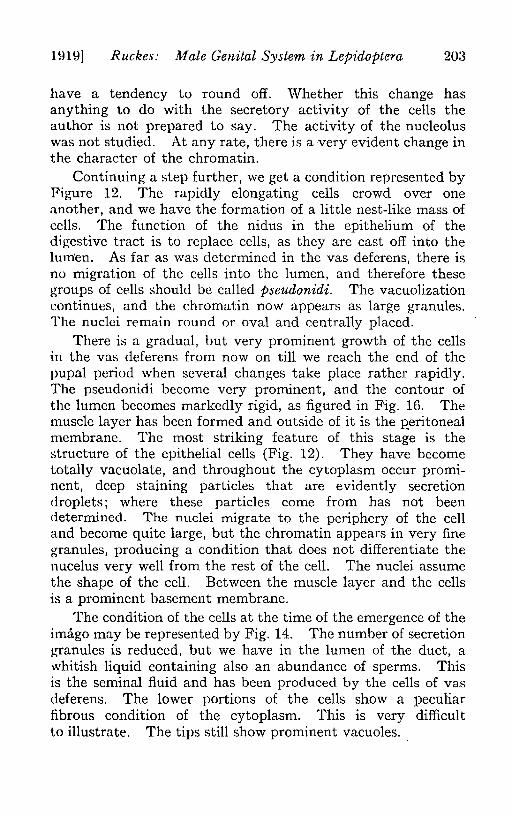
un!:}] Ruckes: Male Genital System in Lepidoptera 203
have a tendency to round off. Whether this change hasanything to do with the secretory activity of the cells theauthor is not prepared to say. The activity of the nucleoluswas not studied. At any rate, there is a very evident change inthe character of the chromatin.
Continuing a step further, we get a condition represented byFigure 12. The rapidly elongating cells crowd over oneanother, and we have the formation of a little nest-like mass ofcells. The function of the nidus in the epithelium of thedigestive tract is to replace cells, as they are cast off into thelumen. As far as was determined in the vas deferens, there isno migration of the cells into the lumen, and therefore thesegroups of cells should be called pseudonidi. The vacuolizationcontinues, and the chromatin now appears as large granules.The nuclei remain round or oval and centrally placed.
There is a gradual, but very prominent growth of the cellsin the vas deferens from now on till we reach the end of thepupal period when several changes take place rather rapidly.The pseudonidi become very prominent, and the contour ofthe lumen becomes markedly rigid, as figured in Fig. 16. Themuscle layer has been formed and outside of it is the p"eritonealmembrane. The most striking feature of this stage is thestructure of the epithelial cells (Fig. 12). They have becometotally vacuolate, and throughout the cytoplasm occur promi-nent, deep staining particles that are evidently secretiondroplets; where these particles come from has not beendetermined. The nuclei migrate to the periphery of the celland become quite large, but the chromatin appears in very finegranules, producing a condition that does not differentiate thenucelus very well from the rest of the cell. The nuclei assumethe shape of the cell. Between the muscle layer and the cellsis a prominent basement membrane.
The condition of the cells at the time of the emergence of theimago may be represented by Fig. 14. The number of secretiongranules is reduced, but we have in the lumen of the duct, awhitish liquid containing also an abundance of sperms. Thisis the seminal fluid and has been produced by the cells of vasdeferens. The lower portions of the cells show a peculiarfibrous condition of the cytoplasm. This is very difficultto illustrate. The tips still show prominent vacuoles.

2~ Annals {!:ntomotogicalSociety oj America [Vol.XII,
After copulation, all the secretion droplets have passed fromthe cytoplasm and this becomes still more fibrous in its nature.The vacuoles persis~ at the tips. (Fig. 15).
" ,
The above describep metamorphosis takes place in theupper two-fifths of the efferent duct. The lower two-fifthsis somewhat simila.r, though the lumen remains somewhatlarger, and the pseudonidi are more prominent. The middlefifth of the tube remains as it was in its larval, or early pupalcondition, that of a thin, uniform duct, with cells cuboidal andwithout pseudonidi. Secretion takes place in the upper portionand in the lower portion of the vas deferens, but not in themiddle part.
B. THE SEMINAL VESICLES.
These organs may be considered as simply distensions of thevas~ deferentia; their epithelia have the same growth tendency,i. e., to form pseudonidi. However, the development of thepseudonidi is not carried as far as in the case of the efferentduct~. At the cephalic and caudal ends of the vesicles, theepithelium lose? its irregular character and assumes a strictcolumnar condition, where it graduates at one end into theepithelium of the accessory glands and at the other into that ofthe ejaculatory puct. The vesicles are chambers ~or theretention of the semen and are not centers of secretion. Thislatt~r process is localized in the vasa deferentia.
Nussbaum (1882) originally considered the vesicles, as wellas the accessory glands; ej\lculatory duct and penis, asderivatives of the ectoderm. Others, who also have madeextensive studies of the reprodtictive system, believe, thevesi.<;:lesto be of mesodermal origin. At any rate, they are sosimi,lar to the vasa defer~ntia, and sufficiently unlike the otherorgans, to be called distentions of the former, and thereforeof a :J:llesoderm\lln~ture. '
Kochev:p.ikow(1891) found in the seminal vesicles of thehon~~ be~,.t~o la¥ers Q:E m~sc1e, an inn'eI;"circular one and ~no,ut~r10n,git\1dinal. :tI~could find none. <;Inth€:vasa deferentia.In the S~turniidre" there is,<;Ine-layer of mus91e,this is circulaI;",all<l~~)acql1tii1~atiolil<;lfthat found qn the v.<;t~Q.eferens(~i?:..~7).
UH9] Ruckes: Male Genital System in LePidoptera 20;')
C. TIlE ACCESSORY GLANDS.
In the accessory glands, the cells do not become con-spicuously glandular, as might be expected from the terminologyof these organs.
The cells remain columnar in character and do not formpseudonidi. They do not become vacuolate, in the same sensethat the cells of the vas deferens become vacuolate. Thecytoplasm presents a very spongy appearance, that is presentin very young cells of the glands. At first this condition mighteasily be taken for a general vacuolization. Comparisons withother types of cells show that it is quite different, however.Many authors believe that there is a secretion of these organs,and that the secretion causes the formation of spermatophores.This may be true. Secretion is not as evident as in the cells ofthe glandular part of the vas deferens, and proceeds in a differ-ent manner.
The accessory glands differ from the organs· previouslydiscussed, in the possession of a single longitudinal layer ofmuscle. This in early stages of the development of the repro-ductive system may be continuous with the muscle of the upperend of the ejaculatory duct. This has not been definitelydetermined.
Koschevnikow (1891) found three layers of muscle in theglands of Apis; an inner longitudinal layer which may notextend the entire length of the tubes, a central circular and anouter longitudinal.
In some insects the accessory glands may actually beectodermal in origin. Many authors do not believe so, however.If they were ectodermal, there would be a tendency toward theformation of an intima, which, as far as has been determined,does not exist in them.
D. THE EJACULATORY DUCT.
As stated previously, the ejaculatory duct consists of twoparts, at least it is so considered by several authors, prominentamong whom is Schroeder. The upper part is divided into twotubes, the ductus eJaculatorius duPlex (of Schroeder), whichare short and connect at their cephalic end with the seminalvesicles. These tubes are probably simply caudal prolonga-
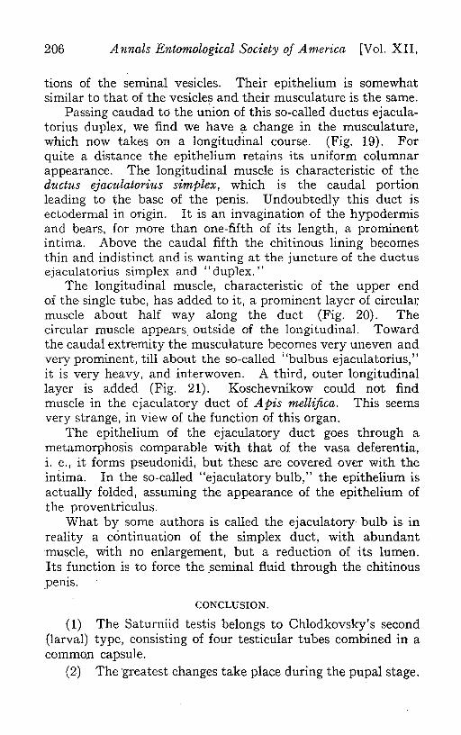
206 Annals Entomological Society oj America [Vol. XII,
tions of the seminal vesicles. Their epithelium is somewhatsimilar to that of the vesicles and their musculature is the same.
Passing caudad to the union of this so-called ductus ejacula-torius duplex, we find we have ?-change in the musculature,which now takes on a longitudinal course. (Fig. 19). Forquite a distance the epithelium retains its uniform columnarappearance. The longitudinal muscle is characteristic of theductus ejaculatorius simplex, which is the caudal portio'nleading to the base of the penis. Undoubtedly this duct isectodermal in origin. It is an invagination of the hypodermisand bears, for more than one-fifth of its length, a prominentintima. Above the caudal fifth the chitinous lining becomesthin and indistinct and is wanting at the juncture of the ductusejaculatorius simplex and "duplex."
The longitudinal muscle, characteristic of the upper endof the· single tube, has added to it, a prominent layer of circulal:muscle about half way along the duct (Fig. 20). Thecircular muscle appears. outside of the longitudinal. Towardthe caudal extremity the musculature becomes very uneven andvery prominent, till about the so-called "bulbus ejaculatorius,"it is very heavy, and interwoven. A third, outer longitudinallayer is added (Fig. 21). Koschevnikow could not findmuscle in the ejaculatory duct of Apis mellifica. This seemsvery strange, in view of the function of this organ.
The epithelium of the ejaculatory duct goes through ametamorphosis comparable with that of the vasa deferentia,i. e., it forms pseudonidi, but these are covered over with theintima. In the so-called "ejaculatory bulb," the epithelium isactually folded, assuming the appearance of the epithelium ofthe proventriculus.
What by some authors is called the ejaculatory· bulb is. inreality a continuation of the simplex duct, with abundantmuscle, with no enlargement, but a reduction of its lumen.Its function is to force the .seminal fluid through the chitinous'pems.
CONCLUSION.
(1) The Saturniid testis belongs to Chlodkovsky's second(larval) type, consisting of four testicular tubes combined in acommon capsule.
(2) The greatest changes take place during the pupal stage.

1919] R-uckes: il1ale Genital System in Lepidoptera 207
(3) The inner coat, of the testis and not the outer one, isfatty in nature.
(4) Verson's cell is a modified follicular epithelium, givingrise to the spermatocyst capsules.
(5) The cells of the vas deferens are the source of theseminal fluid, the accessory glands and seminal vesicles arestorehouses.
(G) The accessory glands have a longitudinal layer ofmuscle; the seminal vesicles a circular layer, the upper end ofthe ejaculatory duct has longitudinal muscles, the lower portion'has added a circular and an outer longitudinal layer as well.
(i) There is a definite and complicated innervation of theinternal genital organs. .
BIBLIOGRAPHY.
1. Berlese, A. 1909. G1i Insetti. Milan.2. Bessels, E. 1857. Studien uber die Entwicklung der Sexualdriisen bei dl'n
Lepidoptcren. Zeit. f. Wiss. Zool. Vol. XVII.3. Blatter, P. 1892. Sur l'histologie des organes annexes de l'appareil male chez
la Periplaneta orientalis. Comptes Rendus CXV. 1332-1334.4. Blatter, P. IS97. Etude sur la structure histologiquc des glandes annexes de
l'appareil male de I'hydrophile. Arch. Anat. Micro. Vol. I. 384-910.5. Bordas, L. 1900. Recherches sur les organes reproducteurs males des Cole-
opteres. Ann. Sc. Nat. Zool. Vol. 2. 283.-448.H. Cholodkovsky, N. N. 1880. Uber die Haden der Schmetterlinge. Zool. Anz.
Vol. III. 115-117, 214-215. .7. Cholodkovsky, N. N. 1884. Vber die Roden der Lepidopteren. Zool. Anz.
Vol. VII. 504-568-S. Cholodkovsky, N. N. 1892. Zur Kenntnis uer Mannerlichen Gcschlecht-
sorganc der Dipteren. Zool. Anz. XV. 178-180.!l. Cholodkovsky, N. N. 1905. Ube'!' den Bau des Dipterenhodens. Zeit. f. wiss:
Zool. Vol. XXXII. 389-410.10. Cholodkovsky, N. N. 1913. Zur Kenntniss der Trichopteren uod Lepidoptereo
Rodeos. Zool. Aoz. Vol. XLII.II. Demokldoff, K. 1902. Zur Kenntniss des Baues des Insectenhodens. Zool.
Anz. Vol. XX\". 575-578.12. Gruenberg, K. 1903. Untersuchung uber die Keirn und Nahrzellen in der
Haden und O\"arien der Lepidopteren. Zeit. f. wiss. Zool. Vol. LXXIV.327-395.
J:l. Hasse, E. 1887. Die Vorfahren der Insecten. Abhand. Gesell. Isis. Dresden.Vol. XI.
14. Herold, M. J. D. 1815. Entwicklungsgeschichtc der Schmettcrlinge, aoat-omisch u. physiologisch bcarbeitet. Cassel. w. Marburg.
Hi. Henneguy, F. M. 1904. Les Insectes. Paris.Hi. Kellogg, V. L. 1907. Sex differentiation in larval insects. BioI. Bull. Vol.
XXII.17. Koschevnikow, G. 1891. Zur Anztomie der mii.nnlicheo Geschlechtsorgane der
Honigbiene. Zool. Anz. Vol. XIV.1~. Landois, H. 1863. Uber die Vcrbinduog der Hoden mit dem Ruckengefass bei
der Insektl'n. Zeit. f. Wiss. Zool. Vol. XIII. 316-18.

208 Annals Entomological Society of America. [Vol. XII,
19. Lecaillon, A. 1902. Sur Ie testicule cl'Anurida maritima. Bull. Soc. Ent.France. 64-67.
20. Lecaillon, A. 1902. Sur la disposition, la structure et Ie fonctionnement del'appareil reproducteur mille des Collemboles. Bull. Soc. Philom. Vol. IV.99-103. .
21. Lowne, B. T. 1895. Anatomy of the Blow Fly. Vol. II. p. 660. London.22. Malpighl, M. 1669. Dissertatio epistolica de Bombyce, Societati regiae
Londini ad scientiam naturalem promovendam institutae dicata. London.23. Meves, F. 1907. tJber Centralk6rper in Mannerlichen Geschlechtszellen von
Schmetterlingen. Anat. Anz. Vol. XIV.24. Nusbaum, J. 1882,. Zur Entwicklungsgeschichte del' Ausfuhrungsgange clcr
Sexualdr1lsen bei del' Insecten. Zool. Anz. Vol. V. 637-643.25. Packard, A. S. 1898. Textbook of Entomology. New York.26. Peterson, W. 1900. Beitrage zur Morpho10gie del' Lepidopteren. Mem. Acad.
. St. Petersbourg. Vol. IX.'27. Peterson, W. 1904. Die Morphologie del' Generations-organs cler Schmetter-
linge und ihre Bedeutung fur die Artbilding. Mem. Acad. St. Petersbourg.Vol. XVI.
28. Peterson, A. 1912. Anatomy of the Tomato Worm Larva Protoprace car-olilla. Ann. Ent. Soc. Amer. Vol. V. 246-49.
29. Riley, W. A. 1915. Laboratory Outlines in Entomological Histology.30. Saint-George, LaValette. 1897. Zur Samen und Eibildung beim Seiden-
spinnen (B. mori). Arch. Mikr. Anat. Vol. I.31. Schneider, A. 18~3. tJber die Entwicklung del' Geschlechtsorgane del' Insek-
ten. Zool. Beitrage v. A. Schneider. Vol. I.32. Schroeder,' Chr. 1912. Handbuch del' Entomologie. Jena.33. Severin, H. H. P. and H. C. M. 1908. Internal organs of reproduction of thc
male saw-fly. Cimbex americana Leach. Ann. Ent. Soc. Amer. Vol. T.196-206.
34. DeSintey, R. 1900. Homologation du testicule chez les Phasmes. Bull. Soc.Ento. de France. Vol.
35. DeSintey, R. 1901. Recherches sur la biologie des Phasmes. La Cellule, 1901.36. Spichardt, C. 1886. Beitrag zur Entwickelung der mannlichen genitalen und
ihrer Ausfiihrgange bei Lepidopteren. Vel'. d. Nat. Vereins zu Bonn. ,13]ahrg.37. Stitz, H. 1903. Zum Genitalapparat der Lepidopteren. Zool. Anz. Vol:
XXVII.38. Tichomiroff, A. 1880. Bau del' Sexualdr1lsen und Entwicklung del' Sexual-
producte bei Bombyx m'ori. Zool. Anz. Vol. III.39. Tichomiroff, A. 1898. Zur Anatomie des Insectenhodens. Zool. Anz. Vol.
XXI. .40. Toyama, N. K. 1894. On the sperm'atogenesis of the silk worm. Bull. ColI.
Agri. Imp. Univ. Japan. Vol. II.41. Verson, E. 1889. Zur·Spermatogenesis. Zool. Anz. Vol. XII.42. Verson, E. 1889, La Spermatogenesi nel Bombyx mori. Padova.43. Verson, E. F. and Bisson, E. 1895. Development postembryonnaire des
organes sexual accessoires chez Ie male du Bombyx mori. Arch. Ital. dcBioI. Vol. XXIV.
44. Verson, E. 1899. Sullufficio della cellula gigante nei follicoli testicolaridegli insetti. Atti. 1st. Veneto Vol. LVII.
45. Verson, E. 1904. ,Zur Entwicklungsgeschichte del' mannlichen Geschlects-anhange bei Insecten. Zool. Anz. Vol. XXVII.
46. Verson, E. 1911. tJber die Versonsche'ZelIe der Autoren in den Hodenfacherndel' Lepidopteren. Zool. Anz. Vol. XXXVIII.
47. Zlck, Karl. 1911. Beitrage zur Kenntniss postembryonalen Elltwicklungs-geschichte del' Genitalorgane bei Lepidopteren. Zeit. f. wiss. Zoot. Vol.XCVIII.

1919] Ruckes: Male Genital System in LePidoptera 209
EXPLANATION OF PLATES
Fig. 1.
Fig. 3.
Fig. 2.
Fig. 4.Fig. 5.Fig. 6.Fig. 7.
PLATE X.A, B, C, D. Chlodkovsky's four types of Lepidopterous testes.
A, HePialusj B, Saturnia; C, Lycaenaj D, Pieris. All diagramatic.E, adult genital system in Samia cecropia, semi-diagramatic. X 8.
Sagittal section of testis, showing chambers, hilum, and two coats.Diagramatic.
Detail of hilum of testis. Note the continuity of the basement mem-brane of the chamber with that of the epithelium of the vas deferens.Camera lucida drawing.
PLATE XI.The relation of the nervous and reproductive systems in an early pupal
stage of Philosamia cynthia.Detail of coats of testis of Callosamia promethia.Detail of coats of testis of Philosamia cynthia.Detail of apical region of a chamber in thc testis of Pieris rapte, showing
Verson's cell giving rise to spermatocyst capsules. The black parti-cles in the cytoplasm of the apical cell are portions of the parentnucleus, (probably formed by amitosis) migrating to the fibrousprolongations to become the nuclei of the spermatocyst capsule.
Fig. S. Adult (sperm stage) spermatocyst.
PLATE XII.Figs. 9-15. Growth of the epithelium of vas deferens from larval (or early pupal)
stage (9) to the lat~ pupa, (13) when the cytoplasm contains secretiondroplets, to the adult stage (15), when the secretion droplets disappearand the tips of the cells vacuolize. .
Fig. 16. Trans-section of vas deferens, showing general contour of epithelium.Fig. 17. Epithelium of upper end of seminal vesicle.·Fig. 18. Epithelium of accessory glands. . ". .'j
Fig. 19. A, Trans-section of ejaculatory duct, near its union with the "ductusejaculatorius duplex." B, EpitUel~um of same. \
Fig. 20. Trans-section of ejaculatory ducfj;·.midway along its length. Notethe addition of circular muscle.
Fig. 21. Trans-section of ejaculatory duct near ejaculatory "bulb." Note theaddition of muscles and the presence of an intima.
Each scale line is equivalent to 100 micra.
FOR FIG. 4.7, 8, 9, IO-Abdominal nerves.
A-Anterior.P-Posterior.S-Sympathetic.
ABBREn ATraNS.Ac.-Apical cell eVerson's cell)- Spc.-Spermatocyst capsule (follicular
Follicular epithelium. epithelium).Ag.-Accessory glands. Spg.-Spermatogonia.Be.-Ejaculatory"bulb." Spn.-Nucleus of spermatocyst capsule.Bm.-Basement membrane. Spv.-Vestigial spiracle.Cc.-Capsular coat. Sv.-Seminal vesicles.Cm.-Circular muscles. T.-Testis.Cp.-Caudal plexus. Tm.-Tracheal membrane.Ded.-"Duetus ejaculatorius duplex." Tr.-Trache:e.Des.-Ductus ejaculatorious simplex. Ttc.- Testicular tube coat.Epi.-Epithelium (of genital ducts). Vd.-Vas deferens.Fd.-Fat droplets. Vp.-Vesicular plexus.I.-Intima.Lm.-Longitudinal muscles.Lvd.-Lumen of vas deferens.P.-Penis.Pg.-Pigment granules.Sd.-Secretion droplets.Sp.--Spiracle.

ANNALS E;. S. A.
1
"
H erberl Ruckes.
VOL. XII, PLATE X.
--1
VBCJCij)(((iV.

ANNALS E. S. A. \'OL. XII. PLATE Xl.
'0'
JI."I..,.,.-1I! •..(.~~
II rrbrrl Ruck.s.
7

ANNALS :e. s. A. VOL. XII. PLATE XII.
19
....•--Lm
18
-em17
IJ erberl Ruckes.