26-1 chapter 26 the urinary system kidneys, ureters, urinary bladder & urethra urine flows from...
TRANSCRIPT

26-1
Chapter 26The Urinary System
• Kidneys, ureters, urinary bladder & urethra
• Urine flows from each kidney, down its ureter to the bladder and to the outside via the urethra
• Filter the blood and return most of water and solutes to the bloodstream

26-2
Overview of Kidney Functions• Regulation of blood composition
– Na+, K+, Ca+2, Cl- and phosphate ions
• Regulation of blood pH, osmolarity & glucose• Regulation of blood volume
– conserving or eliminating water
• Regulation of blood pressure– secreting the enzyme renin– adjusting renal resistance
• Release of erythropoietin & calcitriol• Excretion of wastes & foreign substances

26-3
External Anatomy of Kidney• Paired kidney-bean-shaped
organ• 4-5 in length, 2-3 in wide,
1 in thick• Found just above the waist
between the peritoneum & posterior wall of abdomen– retroperitoneal along with
adrenal glands & ureters
• Protected by 11th & 12th ribs with right kidney lower
• Between T12 & L3

26-4
Internal Anatomy of the Kidneys
• Parenchyma of kidney– renal cortex = superficial layer of kidney– renal medulla
• inner portion consisting of 8-18 cone-shaped renal pyramids separated by renal columns
• renal papilla point toward center of kidney
• Drainage system fills renal sinus cavity– cuplike structure (minor calyces) collect urine
from the papillary ducts of the papilla– minor & major calyces empty into the renal pelvis
which empties into the ureter

Internal Anatomy of Kidney

26-6
The Nephron• Kidney has over 1 million nephrons composed of a corpuscle
and tubule• Renal corpuscle = site of plasma filtration
– glomerulus is capillaries where filtration occurs– glomerular (Bowman’s) capsule is double-walled epithelial cup that
collects filtrate
• Renal tubule– proximal convoluted tubule– loop of Henle dips down into medulla– distal convoluted tubule
• Collecting ducts and papillary ducts drain urine to the renal pelvis and ureter

26-7
Number of Nephrons• Remains constant from birth
– any increase in size of kidney is size increase of individual nephrons
• If injured, no replacement occurs
• Dysfunction is not evident until function declines by 25% of normal (other nephrons handle the extra work)
• Removal of one kidney causes enlargement of the remaining until it can filter at 80% of normal rate of 2 kidneys

26-8
Overview of Renal Physiology
• Nephrons and collecting ducts perform 3 basic processes– glomerular filtration
• a portion of the blood plasma is filtered into the kidney
– tubular reabsorption• water & useful substances are reabsorbed into the blood
– tubular secretion• wastes are removed from the blood & secreted into urine

Glomerular Filtration• Blood pressure produces glomerular filtrate
• Filtration fraction is 20% of plasma
• 48 Gallons/dayfiltrate reabsorbedto 500-1000mL. urine
• Filtering capacityenhanced by:
– thinness of membrane
& large surface area of
glomerular capillaries
– glomerular capillary BP is high
due to small size of efferent arteriole

Net Filtration Pressure
• NFP = total pressure that promotes filtration
• NFP = GBHP - (CHP + BCOP) = 10mm Hg

Glomerular Filtration Rate• Amount of filtrate formed in all renal corpuscles of
both kidneys / minute– average adult male rate is 125 mL/min
• Homeostasis requires GFR that is constant– too high & useful substances are lost due to the speed of
fluid passage through nephron– too low and sufficient waste products may not be
removed from the body
• Changes in net filtration pressure affects GFR– filtration stops if GBHP drops to 45mm Hg– functions normally with mean arterial pressures 80-180

Hormonal Regulation of GFR• Atrial natriuretic peptide (ANP) increases
GFR– stretching of the atria that occurs with an
increase in blood volume causes hormonal release
• relaxes glomerular mesangial cells increasing capillary surface area and increasing GFR
• Angiotensin II reduces GFR– potent vasoconstrictor that narrows both
afferent & efferent arterioles reducing GFR

Tubular Reabsorption & Secretion• Normal GFR is so high that volume of filtrate in
capsular space in half an hour is greater than the total plasma volume
• Nephron must reabsorb 99% of the filtrate– PCT with their microvilli do most of work with rest of
nephron doing just the fine-tuning • solutes reabsorbed by active & passive processes
• water follows by osmosis
• small proteins by pinocytosis
• Important function of nephron is tubular secretion– transfer of materials from blood into tubular fluid
• helps control blood pH because of secretion of H+
• helps eliminate certain substances (NH4+, creatinine, K+)
26-14
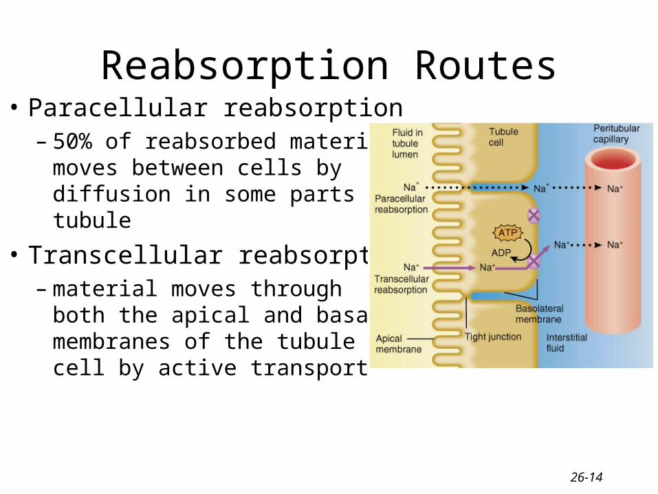
Reabsorption Routes• Paracellular reabsorption
– 50% of reabsorbed materialmoves between cells bydiffusion in some parts oftubule
• Transcellular reabsorption– material moves through
both the apical and basalmembranes of the tubulecell by active transport

26-15
Transport Mechanisms• Apical and basolateral membranes of tubule cells have
different types of transport proteins
• Reabsorption of Na+ is important– several transport systems exist to reabsorb Na+
– Na+/K+ ATPase pumps sodium from tubule cell cytosol through the basolateral membrane only
• Water is only reabsorbed by osmosis– obligatory water reabsorption occurs when water is “obliged” to
follow the solutes being reabsorbed
– facultative water reabsorption occurs in collecting duct under the control of antidiuretic hormone

26-16
Reabsorption in the PCT• Na+ channels help reabsorb
materials from the tubular filtrate
• Glucose, amino acids, water-soluble vitamins and other nutrients are completely reabsorbed in the first half of the proximal convoluted tubule
Reabsorption of Nutrients

Reabsorption of Bicarbonate, Na+ & H+ Ions• Na+ channels reabsorb Na+
and secrete H+– PCT cells produce the H+ &
release bicarbonate ion to the peritubular capillaries
– important buffering system
• For every H+ secreted into the tubular fluid, one filtered bicarbonate eventually returns to the blood

Passive Reabsorption in the 2nd Half of PCT• Electrochemical gradients
causes passive reabsorption of other solutes
• Cl-, K+, Ca+2, Mg+2 and urea passively diffuse into the peritubular capillaries
• Promotes osmosis in PCT (especially permeable due to aquaporin-1 channels

26-19
Secretion of NH3 & NH4+ in PCT• Ammonia (NH3) is a poisonous waste product of
protein deamination in the liver– most is converted to urea which is less toxic
• Both ammonia & urea are filtered at the glomerus & secreted in the PCT– PCT cells deaminate amino/acids’s in a process that
generates both NH3 and new bicarbonate ion.• Bicarbonate diffuses into the bloodstream
– during acidosis more bicarbonate is generated

26-20
Channels (Symporters) in the Loop of Henle
• Thick limb of loop of Henle has Na+ K- Cl- symporters that reabsorb these ions
• K+ leaks through K+ channels back into the tubular fluid leaving the interstitial fluid and blood with a negative charge
• Cations passively move to the vasa recta

26-21
Reabsorption in the DCT
• Removal of Na+ and Cl- continues in the DCT by means of Na+ Cl- symporters
• Na+ and Cl- then reabsorbed into peritubular capillaries
• DCT is major site where parathyroid hormone stimulates reabsorption of Ca+2– DCT is not very permeable to water

26-22
Reabsorption & Secretion in the Collecting Duct
• By end of DCT, 95% of solutes & water have been reabsorbed and returned to the bloodstream
• Cells in the collecting duct make the final adjustments– cells reabsorb Na+ and secrete K+– cells reabsorb K+ & bicarbonate ions and
secrete H+

26-23
Secretion of H+ and Absorption of Bicarbonate by Collecting Duct Cells• Proton pumps (H+ATPases) secrete H+
into tubular fluid– can secrete against a concentration gradient
so urine can be 1000 times more acidic than blood
• Cl-/HCO3- antiporters move bicarbonate ions into the blood– intercalated cells help regulate pH of body
fluids
• Urine is buffered by HPO4 2- and ammonia, both of which combine irreversibly with H+ and are excreted

26-24
Production of Dilute or Concentrated Urine
• Homeostasis of body fluids despite variable fluid intake
• Kidneys regulate water loss in urine
• ADH controls whether dilute or concentrated urine is formed– if lacking, urine contains high ratio of water to
solutes

Summary
• H2O Reabsorption – PCT---65%– loop---15%– DCT----10-15%– collecting duct--- 5-
10% with ADH
• Dilute urine has not had enough water removed, although sufficient ions have been reabsorbed.

Anatomy of Ureters
• 10 to 12 in long• Varies in diameter from 1-10 mm• Extends from renal pelvis to
bladder• Retroperitoneal• Enters posterior wall of bladder• Physiological valve only
– bladder wall compresses arterial opening as it expands during filling
– flow results from peristalsis, gravity & hydrostatic pressure

Location of Urinary Bladder
• Posterior to pubic symphysis
• In females is anterior to vagina & inferior to uterus
• In males lies anterior to rectum

Anatomy of Urinary Bladder
• Hollow, distensible muscular organ with capacity of 700 - 800 mL
• Trigone is smooth flat area bordered by 2 ureteral openings and one urethral opening

Micturition Reflex• Micturition or urination (voiding)• Stretch receptors signal spinal cord and brain
– when volume exceeds 200-400 mL
• Impulses sent to micturition center in sacral spinal cord (S2 and S3) & reflex is triggered– parasympathetic fibers cause detrusor muscle to contract,
external & internal sphincter muscles to relax
• Filling causes a sensation of fullness that initiates a desire to urinate before the reflex actually occurs– conscious control of external sphincter
– cerebral cortex can initiate micturition or delay its occurrence for a limited period of time

26-30
Urinary Incontinence
• Lack of voluntary control over micturition– normal in 2 or 3 year olds because neurons to
sphincter muscle is not developed
• Stress incontinence in adults– caused by increases in abdominal pressure that
result in leaking of urine from the bladder• coughing, sneezing, laughing, exercising, walking
– injury to the nerves, loss of bladder flexibility, or damage to the sphincter, pregnancy

26-31
Homework: Chapter 26
• A2, A3, B1, B4, B6, C6, C10, C11, D2, E1