khaleel alyahya [email protected]. urinary system kidney ureters urinary bladder urethra
TRANSCRIPT

Urinary System
Kidney Ureters Urinary Bladder Urethra

Functions
Maintain the purity & constancy of internal fluids.
Regulate blood volume.
Produce rennin to help regulate blood pressure.
Produce erythropoietin (hormone) to stimulate RBC
production.
Produce urine.
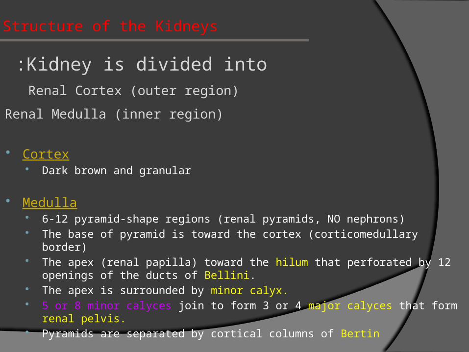
Structure of the Kidneys
Kidney is divided into :Renal Cortex (outer region)
Renal Medulla (inner region)
Cortex Dark brown and granular
Medulla 6-12 pyramid-shape regions (renal pyramids, NO nephrons) The base of pyramid is toward the cortex (corticomedullary border) The apex (renal papilla) toward the hilum that perforated by 12 openings of
the ducts of Bellini. The apex is surrounded by minor calyx. 5 or 8 minor calyces join to form 3 or 4 major calyces that form renal pelvis. Pyramids are separated by cortical columns of Bertin
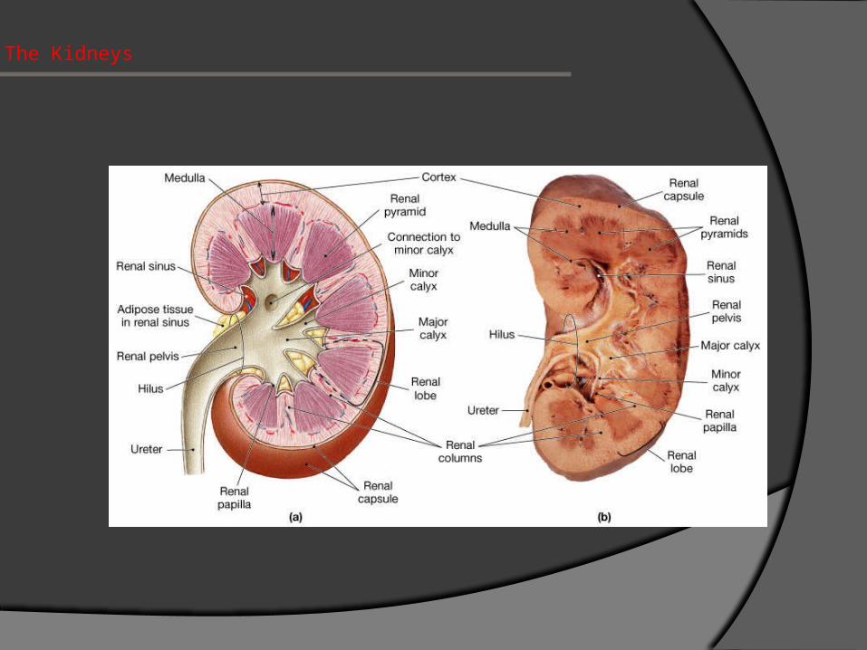
The Kidneys
Cortical Arch
Renal corpuscles
Convoluted tubules
Medullary rays (cortical continuation of pyramids)
Uriniferous Tubule
Is the functional unit of the kidney.
Is formed of:
1 -Nephron
2-Collecting tubule (duct)
They are densely packed.
They are separated by thin stroma and basal lamina
Corpuscles
Convoluted tubules (cortical labyrinth)
Medullary rays (cortical continuation of pyramids)

Nephron There are 2 types:
Cortical nephrons Juxtamedullary nephrons
Nephron is formed of: Renal corpuscle
(cortical & juxtamedullary). Proximal tubule. Henle’s loop. Distal tubule
Note: Collecting tubule (duct) is NOT a part of the nephron!
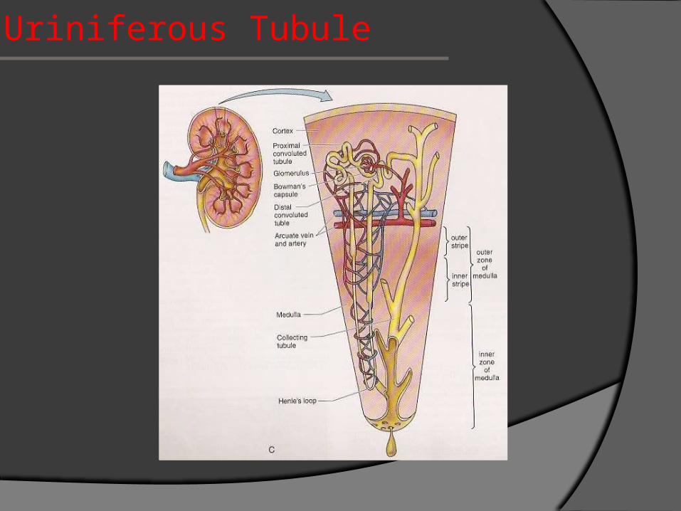
Uriniferous Tubule

Uriniferous Tubule

Renal Corpuscle Two poles:
vascular urinary
Glomerulus Bowman’s capsule
parietal layer urinary or glomerular space visceral layer or podocytes
Mesangial cells intraglomerular extraglomerular
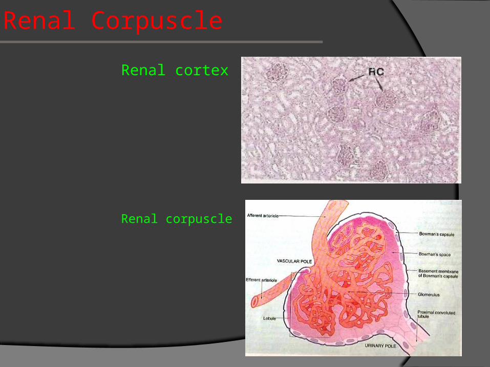
Renal Corpuscle
Renal cortex
Renal corpuscle
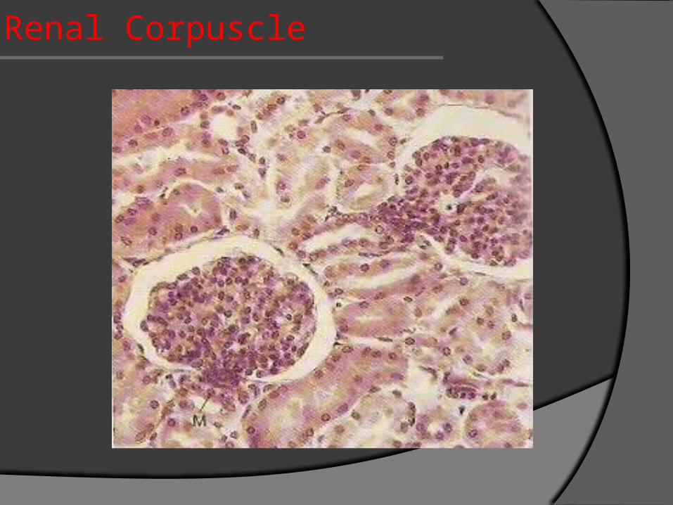
Renal Corpuscle

Renal Corpuscle
Renal Corpuscle

Renal Corpuscle

Renal Corpuscle

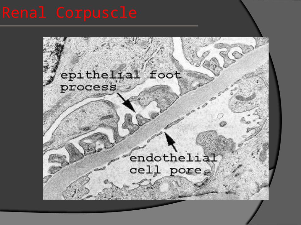
Filtration Barrier
Endothelial wall of the capillaries
Basal lamina
Visceral layer of Bowman’s capsule (podocytes) Podocytes have primary (major) processes and secondary
(minor) processes (pedicles).
Between pedicles (on the surface of capillaries) there are
filtration slits that have slit diaphragm

Filtration Barrier

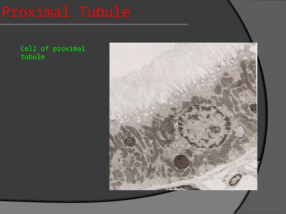
Proximal Tubule
It has 2 regions: Pars convoluta
proximal convoluted tubule Pars recta
descending thick limb of Henle’s loop
It is composed of simple cuboidal epithilum with acidophilic cytoplasm. The cells have striated or brush border and lateral interdigitations. They have well-defined basal lamina.
Proximal tubules are long and highly convoluted so most of cortical tubular sections are proximal tubules.
Proximal Tubule
Cell of proximal tubule
Thin limb of Henle’s Loop
It has 3 regions: Descending thin limb Henle’s loop Ascending thin limb
Note: It is longer in juxtamedullary nephron than in cortical nephron It is composed of simple squamous epithelium

Renal Medulla

Distal Tubule
It has 3 regions: Ascending thick limb of Henle’s loop
low cuboidal epithelium Macula densa
1st part, tall & narrow cells Pars convoluta
distal convoluted tubule formed of low cuboidal epithelium
Distal tubules drain into collecting tubules
Aldosterone hormone increase the active reabsorbtion of sodium from the lumen of tubule into interstitium

Distal Tubule
Cell of distal tubule
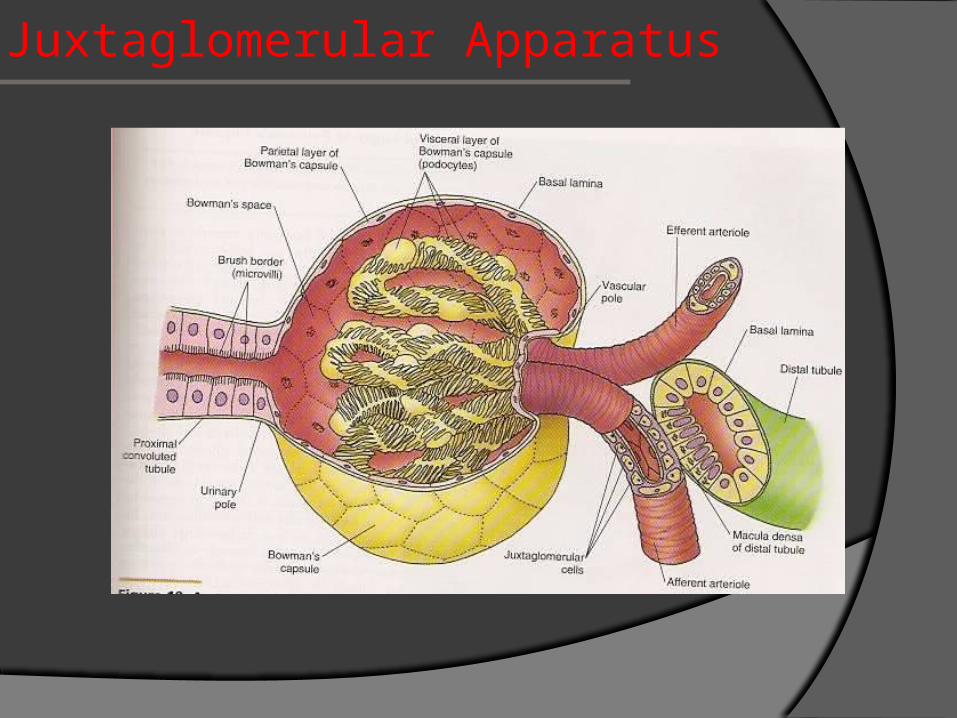
Juxtaglomerular Apparatus
It has 3 components:
The macula densa of distal tubule
Juxtaglomerular cells of afferent glomerular arteriole
modified smooth muscle of tunica media
they secrete renin, angiotensin-converting enzyme and
angiotensin
The extraglomerular mesangial cells
Juxtaglomerular Apparatus

Juxtaglomerular Apparatus
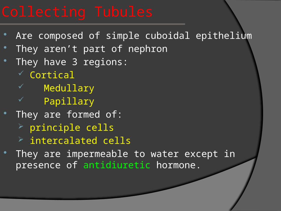
Collecting Tubules Are composed of simple cuboidal epithelium They aren’t part of nephron They have 3 regions:
Cortical Medullary Papillary
They are formed of: principle cells intercalated cells
They are impermeable to water except in presence of antidiuretic hormone.
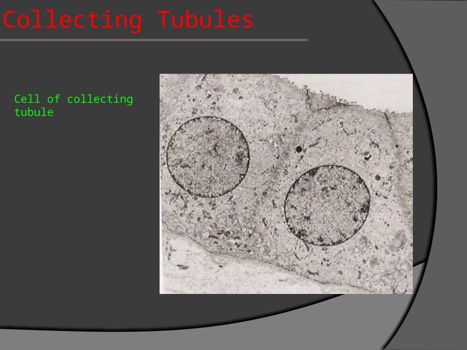
Collecting Tubules
Cell of collecting tubule
Renal Interstitium
It is a very flimsy, scant amount of CT contains. Fibroblasts. Macrophages. Interstitial cells
their nuclei are elongated and they contain lipid droplets they secrete medullipin I, which is converted in the liver
into medullipin II, that lowers blood pressure
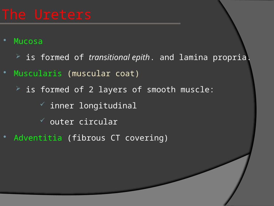
The Ureters
Mucosa
is formed of transitional epith. and lamina propria.
Muscularis (muscular coat)
is formed of 2 layers of smooth muscle:
inner longitudinal
outer circular
Adventitia (fibrous CT covering)

The Ureters

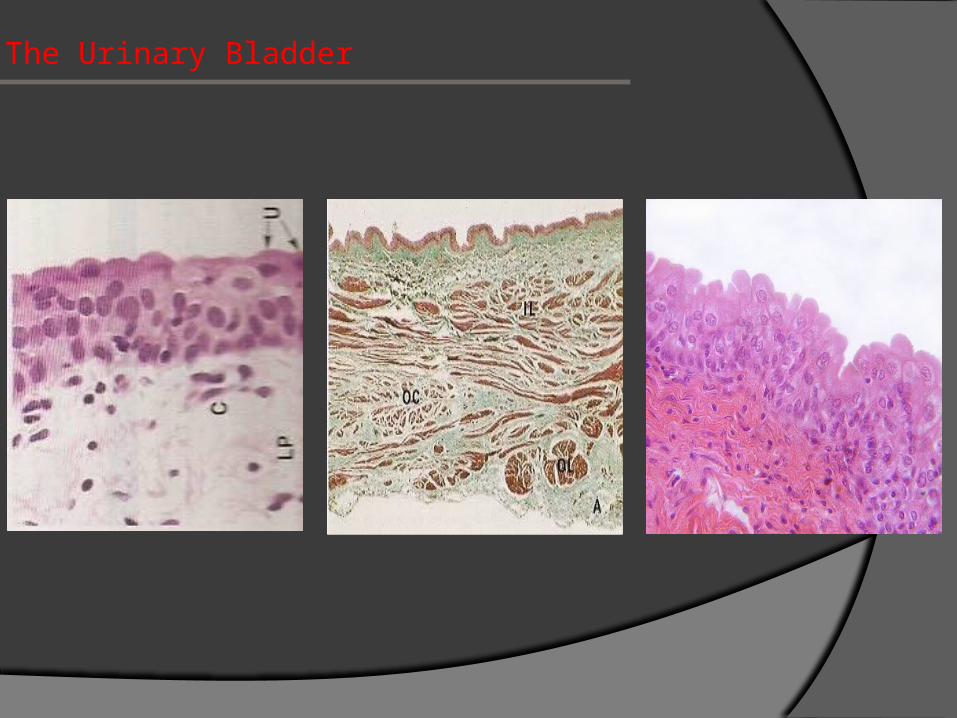
The Urinary Bladder
It has the same structure as the ureter EXCEPT:
The dome-shaped cells of transitional epithelium have
plaques (rigid, thickened regions of plasmalemma)
It has 3 layers of smooth muscle, inner and outer
longitudinal and middle circular
Its outer covering is serosa
The Urinary Bladder

THAT’S ALL FOLKS!
Questions?