28-1 chapter 28 the reproductive systems sexual reproduction produces new individuals –germ cells...
TRANSCRIPT

28-1
Chapter 28The Reproductive Systems
• Sexual reproduction produces new individuals– germ cells called gametes (sperm & 2nd oocyte) – fertilization produces one cell with one set of
chromosomes from each parent
• Gonads produce gametes & secrete sex hormones
• Reproductive systems– gonads, ducts, glands & supporting structures

28-2
Chromosomes in Somatic Cells & Gametes• Somatic cells (diploid cells)
– 23 pairs of chromosomes for a total of 46• each pair is homologous since contain similar genes
in same order• one member of each pair is from each parent
– 22 autosomes & 1 pair of sex chromosomes• sex chromosomes are either X or Y• females have two X chromosomes• males have an X and a smaller Y chromosome
• Gametes (haploid cells)– single set of chromosomes for a total of 23– produced by special type of division: meiosis
28-3
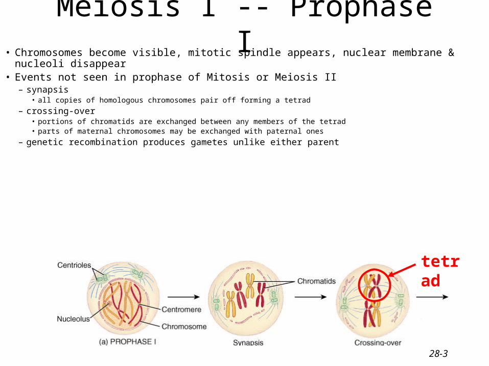
Meiosis I -- Prophase I• Chromosomes become visible, mitotic spindle appears, nuclear membrane & nucleoli disappear• Events not seen in prophase of Mitosis or Meiosis II
– synapsis• all copies of homologous chromosomes pair off forming a tetrad
– crossing-over• portions of chromatids are exchanged between any members of the tetrad• parts of maternal chromosomes may be exchanged with paternal ones
– genetic recombination produces gametes unlike either parent
tetrad
28-4
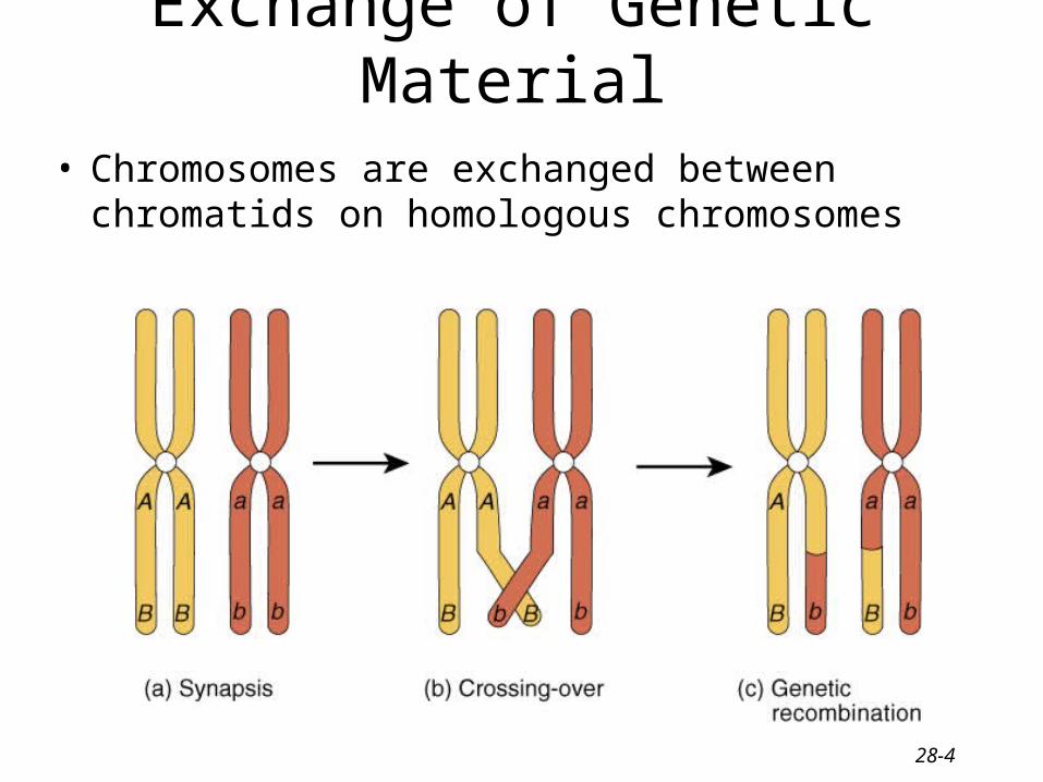
Exchange of Genetic Material
• Chromosomes are exchanged between chromatids on homologous chromosomes

28-5
Meiosis I -- Metaphase I, Anaphase I & Telophase I• In metaphase I, homologous pairs of chromosomes line up along metaphase
plate with attached microtubules• In anaphase I, each set of homologous chromatids held together by a
centromere are pulled to opposite ends of the dividing cell• Telophase I and cytokinesis are similar to mitotic division• Result is 2 cells with haploid number of chromosomes

28-6
Meiosis II• Consists of 4 phases : prophase II, metaphase II,
anaphase II and telophase II• Similar steps in this cellular process as in mitosis
– centromeres split– sister chromatids separate and move toward opposite
poles of the cell
• Each of the daughter cells produced by meiosis I divides during meiosis II and the net result is 4 genetically unique haploid cells or gametes.

28-7
Male Reproductive System• Gonads, ducts, sex glands & supporting structures• Semen contains sperm plus glandular secretions

28-8
Scrotum• Sac of loose skin, fascia & smooth muscle
divided into two pouches by septum
• Skin contains dartos muscle causes wrinkling
• Temperature regulation of testes– sperm survival requires 3 degrees lower
temperature than core body temperature– cremaster muscle in spermatic cord
• elevates testes on exposure to cold & during arousal
• warmth reverses the process

28-9
Testes• Paired oval glands
measuring 2 in. by 1in. • Surrounded by dense
white capsule called tunica albuginea– septa form 200 - 300
compartments called lobules
• Each is filled with 2 or 3 seminiferous tubules where sperm are formed
28-10
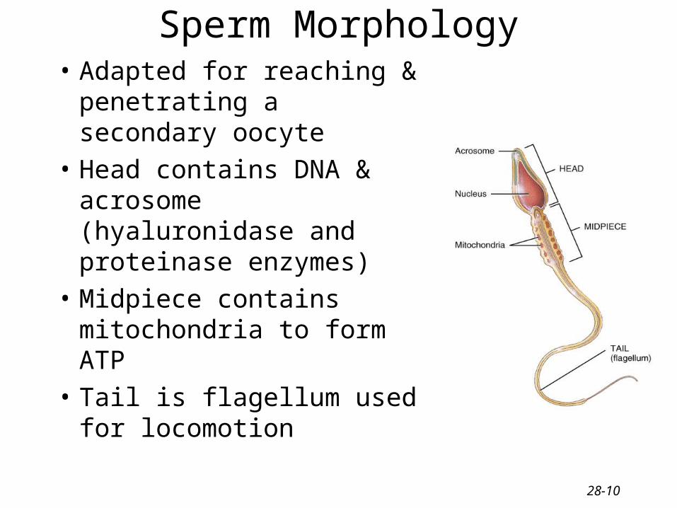
Sperm Morphology• Adapted for reaching &
penetrating a secondary oocyte
• Head contains DNA & acrosome (hyaluronidase and proteinase enzymes)
• Midpiece contains mitochondria to form ATP
• Tail is flagellum used for locomotion

28-11
Hormonal Control of Spermatogenesis• Puberty
– hypothalamus increases its stimulation of anterior pituitary with releasing hormones
– anterior pituitary increases secretion LH & FSH
• LH stimulates Leydig cells to secrete testosterone
• FSH stimulates spermatogenesis– with testosterone, stimulates sertoli cells to secrete
androgen-binding protein (keeps hormones levels high)– testosterone stimulates final steps spermatogenesis

28-12
Pathway of Sperm Flow through the Ducts of the Testis
• Seminiferous tubules • Straight tubules• Rete testis• Efferent ducts• Ductus epididymis• Ductus (vas) deferens

28-13
Epididymis• Comma-shaped organ, 1.5in long along
posterior border of each testis
• Head, body and tail region
• Multiple efferent ducts become a single ductus epididymis in the head region– 20 foot tube if uncoiled
• Tail region continues as ductus deferens

28-14
Epididymis
• Site of sperm maturation– motility increases over 2 week period
• Storage for 1-2 months
• Propels sperm onward

28-15
Ductus (Vas) Deferens• Pathway of 18 inch muscular tube
– ascends along posterior border of epididymis– passes up through spermatic cord and inguinal ligament– reaches posterior surface of urinary bladder– empties into prostatic urethra with seminal vesicle
• Lined with pseudostratified columnar epithelium & covered with heavy coating of muscle – convey sperm along through peristaltic contractions– stored sperm remain viable for several months

28-16
Urethra• 8 inch long passageway for urine & semen• Prostatic urethra (1 inch long)• Membranous urethra (passes through UG diaphragm )• Penile (spongy) urethra (through corpus spongiosum)

28-17
Accessory Sex Glands

28-18
Seminal Vesicles• Pair of pouchlike organs
found posterior to the base of bladder
• Alkaline, viscous fluid– neutralizes vaginal acid &
male urethra
– fructose for ATP production
– prostaglandins stimulate sperm motility & viability
– clotting proteins for coagulation of semen Posterior View

28-19
Prostate Gland• Single organ the size of
chestnut found inferior to bladder
• Secretes milky, pH 6.5 fluid that increases sperm motility and viability– citric acid for ATP
production & enzymes for seminal liquefaction
• Many duct openings• Enlarges with age

28-20
Bulbourethral or Cowper’s Gland
• Paired, pea-sized gland within the UG diaphragm
• Secretes alkaline mucous into spongy urethra
• Neutralizes acids and lubricates

28-21
Penis• Passageway for
semen & urine
• Body composed of three erectile tissue masses filled with blood sinuses
• Composed of bulb, crura, body & glans penis

28-22
Glans Penis
• Enlarged distal end of corpus spongiosum
• External urethral orifice is small slit
• Covered by loosely fitting prepuce or foreskin

28-23
Female Reproductive System• Ovaries produce 2nd oocytes & hormones• Uterine tubes transport fertilized ova• Uterus where fetal development occurs• Vagina & external genitalia constitute the vulva• Mammary glands produce milk

28-24
Follicular Stages• Stages of follicular development
– primordial – primary– secondary– graafian – ovulation
• Corpus luteum is ovulation wound – fills in with hormone secreting cells
• Corpus albicans is white scar left after corpus luteum is not needed

28-25
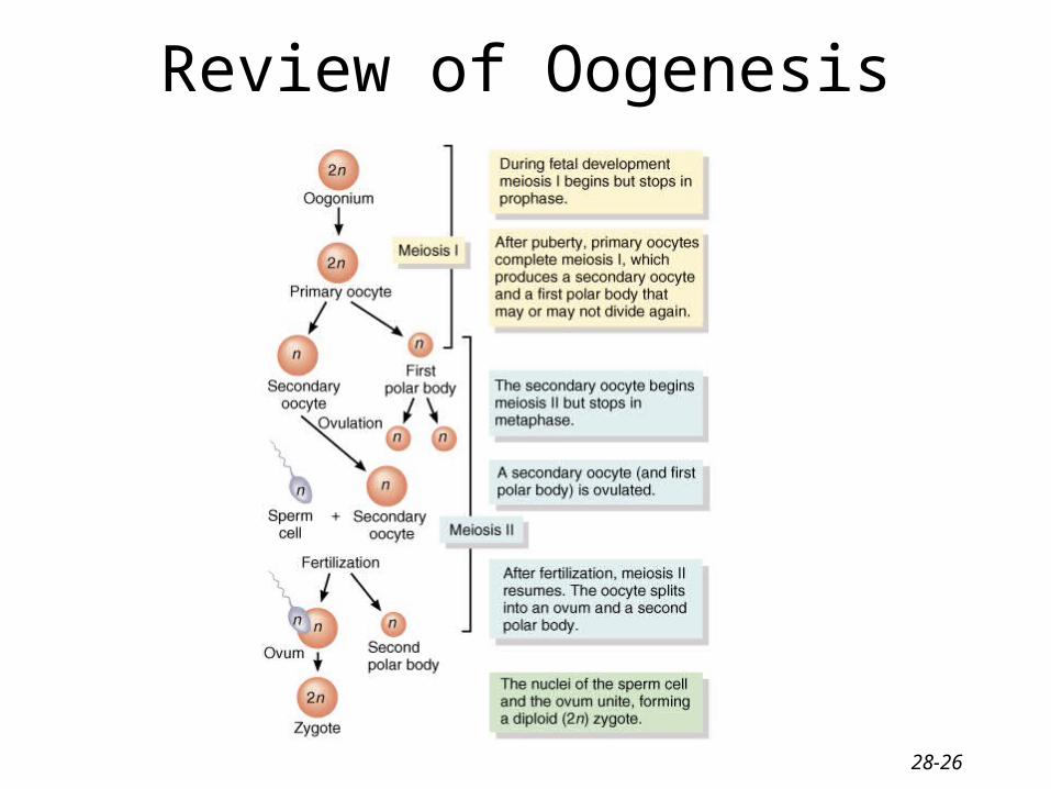
Life History of Oogonia• Germ cells from yolk sac migrate to ovary & become
oogonia• As a fetus, oogonia divide to produce millions by mitosis
but most degenerate (atresia)• Some develop into primary oocytes & stop in prophase
stage of meiosis I– 200,000 to 2 million present at birth– 40,000 remain at puberty but only 400 mature during a woman’s
life
• Each month, hormones cause meiosis I to resume in several follicles so that meiosis II is reached by ovulation
• Penetration by the sperm causes the final stages of meiosis to occur
28-26
Review of Oogenesis

28-27
Uterine or Fallopian Tubes• Narrow, 4 inch tube
extends from ovary to uterus– infundibulum is open,
funnel-shaped portion near the ovary
• fimbriae are moving finger-like processes
– ampulla is central region of tube
– isthmus is narrowest portion joins uterus

28-28
Histology & Function of Uterine Tube• Histology = 3 Layers
– mucosa = ciliated columnar epithelium with secretory cells provide nutrients & cilia move along ovum
– muscularis = circular & longitudinal smooth muscle• peristalsis helps move ovum down to the uterus
– serosa = outer serous membrane
• Function -- events occurring in the uterine tube– fimbriae sweep oocyte into tube, cilia & peristalsis move
it along, sperm reaches oocyte in ampulla, fertilization occurs within 24 hours after ovulation & zygote reaches uterus about 7 days after ovulation

28-29
Lining of the Uterine Tubes

28-30
Anatomy of the Uterus• Site of menstruation
& development of fetus
• Description– 3 inches long by 2 in.
wide and 1 in. thick– subdivided into fundus,
body, isthmus & cervix– interiorly contains uterine cavity accessed by cervical
canal (internal & external os)

28-31
Vagina• Passageway for birth, menstrual flow & intercourse• Description
– 4 inch long fibromuscular organ ending at cervix• mucosal layer
– stratified squamous epithelium & areolar connective tissue– large stores of glycogen breakdown to produce acidic pH
• muscularis layer is smooth muscle allows considerable stretch• adventitia is loose connective tissue that binds it to other organs
– lies between urinary bladder and rectum– orifice partially closed with membrane (hymen)

28-32
Vulva (pudendum)• Mons pubis -- fatty pad over the pubic symphysis• Labia majora & minora -- folds of skin encircling vestibule
where find urethral and vaginal openings• Clitoris -- small mass of erectile tissue• Bulb of vestibule -- masses of erectile tissue just deep to the
labia on either side of the vaginal orifice

28-33
Female Reproductive Cycle• Controlled by monthly hormone cycle of
anterior pituitary, hypothalamus & ovary
• Monthly cycle of changes in ovary and uterus
• Ovarian cycle– changes in ovary during & after maturation of
oocyte
• Uterine cycle– preparation of uterus to receive fertilized ovum– if implantation does not occur, the stratum
functionalis is shed during menstruation

28-34
Phases of Female Reproductive Cycle
28-35
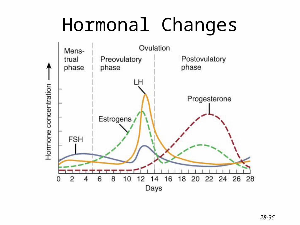
Hormonal Changes

28-36
Menstrual Phase• Menstruation lasts for 5 days• First day is considered beginning of 28 day cycle• In ovary
– 20 follicles that began to develop 6 days before are now beginning to secrete estrogen
– fluid is filling the antrum from granulosa cells
• In uterus– declining levels of progesterone caused spiral arteries to constrict --
glandular tissue dies– stratum functionalis layer is sloughed off along with 50 to 150 ml of
blood

28-37
Preovulatory Phase• Lasts from day 6 to 13 (most variable timeline)
• In the ovary (follicular phase)– follicular secretion of estrogen & inhibin has slowed the
secretion of FSH – dominant follicles survives to day 6– by day 14, graafian follicle has enlarged & bulges at
surface– increasing estrogen levels trigger the secretion of LH
• In the uterus (proliferative phase)– increasing estrogen levels have repaired & thickened the
stratum functionalis to 4-10 mm in thickness
28-38
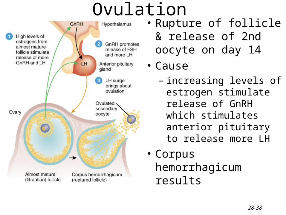
Ovulation• Rupture of follicle &
release of 2nd oocyte on day 14
• Cause– increasing levels of
estrogen stimulate release of GnRH which stimulates anterior pituitary to release more LH
• Corpus hemorrhagicum results

28-39
Signs of Ovulation
• Increase in basal body temperature
• Changes in cervical mucus
• Cervix softens
• Mittelschmerz---pain

28-40
Postovulatory Phase• Most constant timeline = lasts 14 days
• In the ovary (luteal phase)– if fertilization did not occur, corpus albicans is formed
• as hormone levels drop, secretion of GnRH, FSH & LH rise
– if fertilization did occur, developing embryo secretes human chorionic gonadotropin (hCG) which maintains health of corpus luteum & its hormone secretions
• In the uterus (secretory phase)– hormones from corpus luteum promote thickening of
endometrium to 12-18 mm• formation of more endometrial glands & vascularization
– if no fertilization occurs, menstrual phase will begin

28-41
Homework: Chapter
• B5, B8, B10, B12, B13, C1, C14