3d printing dinosaur bones - rsna€¦ · 3d printing dinosaur bones ... through education,...
TRANSCRIPT

A L S O I N S I D E :
RSNA’s Educational Programs Evident Worldwide
Quantitative Imaging Accelerates Clinical Research
ICD-10 Deadline Extended to 2015
Professionalism Critical for Residents/Fellows
Advance Registration and Housing Open May 1See Page 23
3D Printing Dinosaur Bones
May 2014 Volume 24, Number 5
Radi
olog
y 20
14:2
70;3
;864
-871

Interested in developing a protocol for your Imaging Clinical Trial? Now accepting applications for the RSNA 2015 Clinical Trials Methodology Workshop!
Learn more and apply at RSNA.org/CT2015
RES155.2
Learning objectives
Acquire the tools and expertise to develop a protocol and become a funded principal investigator for imaging clinical trials. Topics include:
O Principles of clinical study design
O Statistical methods for imaging studies
O Practicalities of running a clinical trial
O Sponsorship and economics of imaging trials
O Regulatory processes
This 6-1/2 day workshop is intended for M.D. and Ph.D. investigators who are faculty members or fellows in radiology, radiation oncology or nuclear medicine departments.
January 10-16, 2015 | Scottsdale, Arizona
Prerequisites:
Applicant must be familiar with basic concepts and techniques of statistics and study design.
Candidate’s department must commit to providing financial support for transportation and hotel (onsite stay required).
Application deadline:
June 15, 2014 (acceptance based on competitive selection process)
This live activity has been approved for AMA PRA Category 1 Credit™.
For more information contact Fiona Miller 1-630-590-7741 or [email protected]
RES155.2 Clinical Trials Journal Ad FIN.indd 1 14-03-24 9:34 AM

RSNA MISSIONThe RSNA promotes excellence in patient care and healthcare delivery through education, research and technologic innovation.
Follow us for exclusive news, annual meeting offers and more!
Access the RSNA News tablet edition on the App Store and Google play.
UP FRONT1 First Impression
1 Centennial Snapshots
4 My Turn
RADIOLOGY’S FUTURE 15 R&E Foundation Donors
NEWS YOU CAN USE 16 The Value of Membership
17 Education and Funding Opportunities
18 Technology Forum
19 Journal Highlights
20 Residents & Fellows Corner
21 Radiology in Public Focus
23 Annual Meeting Watch
24 RSNA.org
FEATURES
Impact of RSNA’s International Programs Felt Worldwide
ICD-10 Deadline Extension Offers Additional Preparation Time
3D Printing Creates Physical Models of 3D Images
Professionalism Skills Critical for Residents/Fellows
Quantitative, Clinical Sciences Intersect to Aid Research
5
7
9
11
13
EDITOR
David M. Hovsepian, M.D.
R&E FOUNDATION CONTRIBUTING EDITOR
R. Gilbert Jost, M.D.
EXECUTIVE EDITOR
Lynn Tefft Hoff
MANAGING EDITOR
Beth Burmahl
EDITORIAL ADVISORS
Mark G. Watson Executive Director
Roberta E. Arnold, M.A., M.H.P.E. Assistant Executive Director Publications and Communications
Marijo Millette Director: Public Information and Communications
EDITORIAL BOARD
David M. Hovsepian, M.D. Chair
Kavita Garg, M.D.Bruce G. Haffty, M.D.Bonnie N. Joe, M.D., Ph.D.Edward Y. Lee, M.D., M.P.H.Laurie A. Loevner, M.D.Tirath Y. Patel, M.D.Barry A. Siegel, M.D.Gary J. Whitman, M.D.Vahid Yaghmai, M.D.Mary C. Mahoney, M.D.
Board Liaison
GRAPHIC DESIGNERS
Ken EjkaErick Jurado
CONTRIBUTING WRITERS
Mike BassettFelicia Dechter, M.A.Elizabeth GardnerMary Henderson Paul LaTour
2014 RSNA BOARD OF DIRECTORS
Richard L. Baron, M.D. Chairman
Richard L. Ehman, M.D. Liaison for Science
Vijay M. Rao, M.D. Liaison for Information Technology and Annual Meeting
Valerie P. Jackson, M.D. Liaison for Education
James P. Borgstede, M.D. Liaison for International Affairs
Mary C. Mahoney, M.D. Liaison for Publications and Communications
N. Reed Dunnick, M.D. President
Ronald L. Arenson, M.D. President-elect/Secretary-Treasurer
Interested in developing a protocol for your Imaging Clinical Trial? Now accepting applications for the RSNA 2015 Clinical Trials Methodology Workshop!
Learn more and apply at RSNA.org/CT2015
RES155.2
Learning objectives
Acquire the tools and expertise to develop a protocol and become a funded principal investigator for imaging clinical trials. Topics include:
O Principles of clinical study design
O Statistical methods for imaging studies
O Practicalities of running a clinical trial
O Sponsorship and economics of imaging trials
O Regulatory processes
This 6-1/2 day workshop is intended for M.D. and Ph.D. investigators who are faculty members or fellows in radiology, radiation oncology or nuclear medicine departments.
January 10-16, 2015 | Scottsdale, Arizona
Prerequisites:
Applicant must be familiar with basic concepts and techniques of statistics and study design.
Candidate’s department must commit to providing financial support for transportation and hotel (onsite stay required).
Application deadline:
June 15, 2014 (acceptance based on competitive selection process)
This live activity has been approved for AMA PRA Category 1 Credit™.
For more information contact Fiona Miller 1-630-590-7741 or [email protected]
RES155.2 Clinical Trials Journal Ad FIN.indd 1 14-03-24 9:34 AM
MAY 2014 • VOLUME 24, NUMBER 5

1 RSNA News | May 2014
During this year as RSNA celebrates its 100th Scientific Assembly and Annual Meeting, RSNA News will take a look back at milestones in the Society’s history.
CENTENNIAL SNAPSHOTS
1948: Medical Physicists Granted RSNA MembershipAllowing full RSNA membership for medical physicists was one of many ways in which the Society responded to the public’s growing fear of radiation. A physics committee developed a sep-arate physics program that ran in parallel with presentations of medical radiology papers at the RSNA annual meetings and scheduled physics-based refresher courses.
1983: First Official Printed Meeting Program Mailed with Radiology
In his welcome remarks to meeting attendees, printed on the first page, 1983 RSNA President Richard G. Lester, M.D., noted that the number of papers
being presented had nearly doubled in less than a decade, “due to the proliferation of technologic advances in the radiologic field and the continuing importance of the RSNA meeting as a forum for scientific exchange.”
1994: RSNA Link DebutsA Learning Center exhibit at RSNA 1994 showcased RSNA’s foray onto the World Wide Web. The website offered general information about the Society and its annual meeting and education- and research-related projects. Site visi-tors could also reg-ister for the meeting, study meeting programs and view tables of contents for Radiology and RadioGraphics.
2005: Patient-centered Radiology Focus BeginsThe Society began develop-ing annual meeting ses-sions and other presenta-tions offering guidance in patient-centered practice and in 2012 launched the Radiology Cares™: The Art of Patient-Cen-tered Practice campaign, which encourages radiologists and other imaging professionals to take a pledge committing to more meaningful engagement in the patient experience.
2006: Radiology Unveils the “New Gray”
SCFR Awards Honors 2010 RSNA President Hedvig Hricak, M.D., Ph.D., Dr. h.c., was awarded the Dr. Jean-A Vézina Innovation and Excel-lence prize of the Société Canadienne-Française de Radiologie (SCFR) for her contributions to oncologic imaging, for both gynecological and prostatic cancers. Dr. Hricak is chair of the Department of Radiology at Memorial Sloan-Kettering Cancer Center in New York, a professor of radiology at Cornell University Medical College and an attending radiologist at Memorial Hospital in New York.
In the January 2006 debut issue, then-Radiology Editor Anthony V. Proto, M.D., noted, “As long as I can remember, Radi-ology has been known as the ‘Gray Journal’ … the new cover retains
some gray, while additional color has been introduced throughout the jour-nal pages along with what we believe to be a more appealing dis-play of … each published article.” The “New Gray” was not merely about appearance, however, as journal content was reorganized and new features were added as well.
The Albert-Jutras prize was awarded to Laurent A. Garel, M.D., a pediatric radiologist at the Sainte-Justine University Hospital Centre in Montreal. Dr. Garel is a pioneer in interventional pediatric radiology and is a member of the RSNA Pediatrics Subcommittee of the Education
Exhibits Committee. Patricia Noël, M.D., a radiologist at the Centre Hospitalier Universitaire de Québec, received the Ber-nadette-Nogrady prize. André Boisjoly, a radiologist at the Cité-de-la-Santé de Laval in Montreal, was named winner of the Personnalité prize.
2008: myPortfolio DebutsDeveloped by RSNA in collaboration with the education commit-tee of the Association of Program Directors in Radiology (APDR), the Web-based portfolio enables residents and their program directors to document their training activities and development as required by the Accreditation Council for Gradu-ate Medical Education (ACGME).
GarelHricak Noël Boisjoly
FIRST IMPRESSION

May 2014 | RSNA News 2
RSNA 2014 PLENARY LECTURERS ANNOUNCED
NEW HORIZONS LECTUREJonathan M. Rubin, M.D., Ph.D.Ann Arbor, Mich.ANNUAL ORATION IN DIAGNOSTIC RADIOLOGYDavid C. Levin, M.D.PhiladelphiaANNUAL ORATION IN RADIATION ONCOLOGYLawrence B. Marks, M.D.Chapel Hill, N.C.SPECIAL LECTUREFrancis S. Collins, M.D., Ph.D.Bethesda, Md.
SIR Bestows 2014 Gold MedalsThe Society of Interventional Radiology (SIR) recently presented a gold medal to 2001 RSNA Honorary Member Lenny K. Tan, M.D., at its annual meeting in San Diego. Dr. Tan is an emeritus consultant in the diagnostic imaging department at National University Hos-pital, Singapore. Michael D. Dake, M.D., and Matthew A. Mauro, M.D., also received the SIR gold medal. Dr. Dake is the Thelma and Henry Doelger Professor in Stanford Uni-versity’s Department of Cardiovascular Surgery and the medical director of Stanford’s catheterization and angi-ography laboratories. Dr. Dake is a member of RSNA’s Public Information Advisors Network. Dr. Mauro is the Ernest H. Wood Distinguished Pro-fessor of Radiology, Surgery and Biomedical Engineering and chair of the Department of Radiology at the Univer-sity of North Carolina Medical Center. Dr. Mauro has served on numerous RSNA committees and was most recently the chair of the Scientific Program Committee. DakeTan Mauro
The RSNA Board of Directors has announced the distinguished individuals who will deliver plenary lectures at the 100th Scientific Assembly and Annual Meeting:
RSNA Members Earn Advanced Quality CertificatesFour physicians recently received the first Advanced Level Quality Certificates from RSNA. Earning the certificate requires suc-cessful completion of Quality Essentials Certificate Courses in four domains—Quality Improvement in Your Practice, Staff and Patient Safety, Customer Satisfaction, and Radiologist Performance Improvement—and acceptance of a quality storyboard abstract for exhibit at an RSNA annual meeting.
NAME INSTITUTION QUALITY STORYBOARD ABSTRACT
Bradley N. Delman, M.D.Mount Sinai Medical Center, New York
Radiation Accumulation Database (RAD): An Automated Method for Summation of Patients’ Radiation Doses
Dana H. Smetherman, M.D., M.P.H.Ochsner Clinic Foundation, New Orleans Screening Mammography: Achieving Same Day Access
Marc H. Willis, D.O.Baylor College of Medicine, Houston
Musculoskeletal MRI Performance Improvement of Image Quality
Marc A. Bernstein, M.D. Swedish American Hospital, Rockford, Ill.
Steps for Decreasing Fluoroscopy Time for PICC Line Placement
To learn more about the RSNA Quality Improvement Certificate Program, including the Advanced Level Quality Certificate program, go to RSNA.org/QICP.
LevinRubin Marks Collins

3 RSNA News | May 2014
Vanderbilt Names Carr Endowed ChairJ. Jeffrey Carr, M.D., was named the endowed Cornelius Vanderbilt chair in Radiology and Radiological Sciences at Vanderbilt University in Nashville. He is also professor of clinical biomedical informatics at Vanderbilt. Dr. Carr joined Vanderbilt this past summer after 24 years of service at Wake Forest University. Dr. Carr, who specializes in non invasive cardiovascular CT and MR imaging, is a founding member and current president of the Society of Cardiovascular Computed Tomography (SCCT).
Set Up Group Billing for 2014
Min is Director of New Cardiovascular Imaging InstituteJames K. Min, M.D., an expert in cardiovascular imaging, has been appointed director of the new Dalio Institute of Cardiovascular Imaging. Dr. Min is an attending physician at New York-Presbyterian Hospital and a full-time faculty member in the Department of Radiology at Weill Cornell Medical College.
Medical College of Wisconsin Names Mathews Radiology Chair Vincent P. Mathews, M.D., has been named professor and chair of the Department of Radiology at the Medical College of Wisconsin in Mil-waukee. Dr. Mathews also will hold the James E. Youker Endowed Chair in Radiology. Dr. Mathews previously served as professor of radiology (neuroradiol-ogy) at the Indiana University School of Medicine in Indianapolis and as president and chief executive officer of Northwest Radiology Network. Dr. Mathews received a 1990 Mallinckrodt Medical Inc./RSNA Research Fellow Grant from the RSNA R&E Foundation and has served on numerous RSNA committees and currently serves on the RSNA Patient Centered Radiology Steering Committee and the Public Information Advisors Network. He is a reviewer for RadioGraphics.
Funded by a $20 million gift from the Dalio Foundation, New York-Presbyterian Hospital and Weill Cornell Medical College created the Dalio Institute of Cardiovascular Imaging to help reduce the burden of cardiovascular disease. The institute will combine research, clinical care and education to uncover new answers about preventing heart disease.
Practices or academic institutions with large numbers of RSNA members can take advan-tage of group billing to receive just one invoice during the next membership renewal cycle. To set up this option, contact the RSNA Mem-bership Department at [email protected] or 1-877-776-2636 (630-571-7873 outside the U.S. and Canada).
Filipe Veloso Gomes, M.B.Ch.B., (center), was the winner in RSNA’s iPad give-a-way during the European Congress of Radiology (ECR) meeting held in March in Vienna. Dr. Gomes, a 2013 participant in RSNA’s IRIYA program, is pictured with RSNA staff members at the RSNA booth at ECR 2014.
Carr
Min
Mathews
FIRST IMPRESSION

Radiology is International and so is RSNARSNA collaborates with other international, continental, regional, and national societies. A goal of RSNA is to enhance excellence in patient care and health care delivery through education, research, and technologic innovation as a worldwide leader in radiology. To that end, in 2012 RSNA consolidated its many inter-national efforts under a new portfolio that includes activities in developed and developing countries. RSNA’s international efforts include an International Advisory Committee with Regional Committees spanning the world. RSNA also has strong relationships with radiology societies worldwide. In 2014, RSNA will collaborate by providing speakers or meeting with leadership of regional societies such as the International Society of Radiology, European Society of Radiology, Intera-merican College of Radiology, and Asian Oceanian Society of Radiology. RSNA will also participate in major radiology society meetings in Argentina, Australia, Brazil, Canada, China, France, Japan, Korea, Mexico, and South Africa. The RSNA annual meeting includes unique “Country Pres-ents” sessions which invite two countries each year to present research and educational material on a topic of their choice. RSNA 2014 will feature “Korea Presents” and “Canada Pres-ents.” RSNA also supports efforts in developing countries includ-ing programs under the auspices of its Committee on Interna-tional Radiology Education (CIRE). These endeavors include International visiting professorships, Introduction to Research for International Young Academics (IRIYA), and the Derek Harwood-Nash International Fellowship. Support of efforts in developing countries highlights RSNA’s values of integrity, excellence, professionalism, leadership, innovation, service, and volunteerism. Such efforts extend RSNA’s presence into areas
Get more of this month’s news with the RSNA News Tablet edition, available for download through the App Store and Google Play. As part of this month’s cover story, we feature a video interview with Zbigniew Starosolski, Ph.D, discussing his RSNA 2013 research on 3D printing and a Podcast discussion of the Radiology research, “Reviving the Dinosaur: Virtual Reconstruction and Three-dimensional Printing of a Dinosaur Verte-bra,” with Radiology Editor Herbert Y. Kressel, M.D., and authors. Access the RSNA News tablet edition on the App Store at itunes.apple.com/us/app/rsna-news/id444083170?mt=8 and Google Play at https://play.google.com/store/apps/details?id=air.org.rsna.rsnanews&hl=en.
THIS MONTH IN THE RSNA NEWS TABLET
of the world which otherwise would not have access to these materials. Bridging RSNA’s work with developed and developing coun-tries is the International Trends session at the annual meeting, which addresses current significant topics affecting radiolo-gists and their patients. This session highlights RSNA’s role as a convener of thought leaders in radiology throughout the world in conjunction with the International Advisory Committee. The 2013 session addressed radiology education in developing nations, and 2014 will address radiation safety regulations and their impact on patient care. All of these endeavors allow RSNA to enhance its world-wide affiliations and international perspectives while afford-ing radiologists worldwide the benefit of RSNA’s expertise in research, education. I am proud to lead these international efforts.
May 2014 | RSNA News 4
My Turn
James P. Borgstede, M.D., is RSNA Board Liaison for International Affairs and chair of the Research & Education (R&E) Foundation. Dr. Borgstede is a professor of radiology and vice-chair of professional services, clinical operations and quality at the University of Colorado, Denver. Dr. Borgstede is presi-dent of the American Board of Radiology and president-elect of the International Society of Radiology.
Read “Impact of RSNA’s Interna-tional Programs Felt Worldwide,” on Page 5.
RSNA NewsMay 2014 • Volume 24, Number 5 Published monthly by the Radiological Society of North America, Inc. 820 Jorie Blvd., Oak Brook, IL 60523-2251. Printed in the USA.
Postmaster: Send address correction “changes” to: RSNA News, 820 Jorie Blvd., Oak Brook, IL 60523-2251Non-member subscription rate is $20 per year; $10 of active members’ dues is allo-cated to a subscription of RSNA News.
Contents of RSNA News copy-righted ©2014, RSNA. RSNA is a registered trademark of the Radiological Society of North America, Inc.
LETTERS TO THE [email protected] 1-630-571-7837 fax
[email protected] 1-888-600-0064 1-630-590-7770
REPRINTS AND [email protected] 1-630-571-7829 1-630-590-7724 fax
[email protected] Jim Drew, Director 1-630-571-7819
RSNA MEMBERSHIP1-877-rsna-mem

5 RSNA News | May 2014
Impact of RSNA’s International Programs Felt Worldwide
“ Interactive relationships are the most important things we can offer in our education programs.”Teresita L. Angtuaco, M.D.
RSNA’s international educational programs not only profoundly affect their many participants but also create a ripple effect that touches hundreds, if not thousands, of aspiring and practicing radiologists and patients in far reaches of the globe.Some of those experiences were recounted for the first time during the RSNA 2013 refresher course, “RSNA Educational Programs Around the World: An International Forum,” featuring present-ers from Egypt, South Africa, Nigeria and Thailand. “We’ve been offering these programs for a long time, but this was the first time we heard directly from the participants,” said Teresita L. Angtuaco, M.D., a professor of radiology and director of the Division of Imaging at the University of Arkansas for Medical Sciences and chair of RSNA’s Commit-tee on International Radiology Education (CIRE). “It was very gratifying to hear what they had to say and to know that the time and money RSNA spends is going toward something really important.” RSNA established the International Visiting Pro-fessor (IVP) program in 1986, the Derek Harwood-Nash (DHN) International Fellowship in 1998 and the Introduction to Research for Interna-tional Young Academics (IRIYA) in 2000. The pro-grams continue to evolve to accommodate RSNA’s ever-growing international membership.
IVP, DHN International Fellowship Change LivesThe DHN International Fellowship brings promis-ing international scholars to study at North Ameri-can institutions, while the IVP annually sends pro-fessors to lecture at national radiology society meet-ings and visit radiology residency training programs at selected host institutions in developing nations. Participants say the life-changing experiences put their careers on a new trajectory. For Savvas Andronikou, M.B.B.Ch., Ph.D., of South Africa, his 2007 DHN International Fellow-ship at Columbia Medical Center, New York City, helped establish him as an internationally recog-nized pediatric radiologist. “When I came back to South Africa, I was made chair of my department and it definitely made a big difference to the committee that I had international experience and added knowledge,” said Dr. Andron-ikou, now a professor and research coordinator in the radiology department of the University of the Witwatersrand in Johannesburg. “Without the fel-lowship, I wouldn’t have made half the American contacts I have now and I wouldn’t have been awarded honorary membership in the U.S. Society for Pediatric Radiology.”
Since learning about diffusion tensor imaging (DTI) in New York, Dr. Andronikou has established research relating to white matter disease in pediatric HIV, co-authored papers on DTI, collaborated with American researchers and supervised dozens of doctoral and masters’ projects. He also has helped bring imaging services and radiology education to developing countries as chairman of the outreach committee of the World Federation of Pediatric Imaging. Omolola M. Atalabi, M.B.B.S., M.B.A., of Nigeria, described how her 2007 DHN International Fellowship at the Children’s Hospital of Boston set off a cascade of career-defining events, including serving as secretary of the Asso-ciation of Radiologists of West Africa Nigeria Chapter that hosted the IVP program in 2008, co-authoring 41 journal articles and receiving a 2010 RSNA Research & Education Foundation seed grant to study ultrasound in assessing the renal status of Nigerian children with malaria. Dr. Atalabi now serves as senior lecturer and radiologist at the University of Ibadan in Nigeria. “Dr. Atalabi has become an authority and mentor to younger women in her country,” Dr. Angtuaco said. As the Executive Director of the Breast Cancer Foundation of Egypt, Norran H. Said, M.D., took back life-changing lessons after her 2011 DHN Inter-national Fellowship at Brigham and Women’s Hospital, Harvard University,
Angtuaco Mullan Said
FEATURE

May 2014 | RSNA News 6
Boston. While Egypt has a nationwide breast screening program, many women don’t return for further evaluation due to cultural factors and lack of awareness, according to Dr. Said. “At Brigham’s, I was able to follow up my interpretations with radiologic pathologic correlation, which increased my sensitivity and specificity,” Dr. Said observed. “Discussing these findings in multidisciplinary meetings was a privilege that I took home. My generous professors gave me a variety of teaching cases that were well perceived by my colleagues.” Having spent early mornings in a Kenyan wild game park and afternoons in a busy African teaching hospital last summer, Kathirkamanthan Shanmuganathan, M.D., M.B.B.S., a profes-sor at the University of Maryland School of Medicine, can attest to the once-in-a-lifetime experience the IVP program offers. He, Harris L. Cohen, M.D., and Leanne L. Seeger, M.D., spent 18 days participating in the annual scientific conference of the Kenyan Association of Radiology and visiting two hospitals in Nairobi. IVP teams have traveled to 43 developing nations to lecture at radiology meetings and work one-on-one with radiology resi-dents in local hospitals. “Having these exchanges is so useful,” Dr. Shanmuganathan said. “You see disease there that you don’t see in America. Because a large number of patients in Kenya come to the hospital when disease is in late stages, nearly all the scans are positive.” The visiting professors established a new “whole body” CT protocol to evaluate the polytrauma patients and encouraged the more than 50 radiology residents who attended their lectures to consider subspecialty training. One resident, Karen Onyambu, M.D., called the IVP visit to her hospital “an eye-opener” and will be a DHN fellow this fall at the University of Maryland School of Medicine. Dr. Angtuaco visited Thailand with the IVP program for the second time in 2010, when the group was asked to help improve the country’s seven radiology residency programs. “They wanted to know how the U.S. operated their residencies and had all seven of their program directors in attendance,” she said. Based on the visiting professors’ recommendations and a large-scale study, the group is planning to increase resident train-ing in specialized imaging, such as color Doppler ultrasound, musculoskeletal MR imaging, and medical IT, said Chamaree Chuapetcharasopon, M.D., of the Royal College of Radiologists of Thailand.
“The IVP program has brought new knowledge and ideas to us,” Dr. Chuapetcharasopon, a CIRE committee member, added. “Our residents are like children—excited about everything—and our junior faculty gained not only additional knowledge but also suggestions on how to teach.”
IRIYA: Launching Academic CareersThe IRIYA program helps young radiology professionals galva-nize careers in teaching and research, offering 15 scholars/young academics the chance to attend specialized courses, small group discussions, dinner receptions and networking opportunities dur-ing RSNA’s annual meeting. “The selection committee tries to select half the participants from developing nations and half from the developed world,” said Brian F. Mullan, M.D., professor of radiology at University of Iowa Hospitals & Clinics, Iowa City, and a CIRE subcommittee chair. “They spend about half their time with American residents and half in programs that are specialized for them.” Some of the best interactions are the informal gatherings and question-and-answer periods with IRIYA faculty and residents from around the world, Dr. Mullan said. “We’ve had discussions about compensation and internal motivation and balancing work and family life that lead to interesting exchanges between the American and international students,” he said. A 2012 PubMed survey indicated that 85 young radiology professionals who participated in the program between 2005 and 2009 have authored 1,113 articles.
One-on-One Interaction is Key to SuccessIn addition to enhancing relations between RSNA and radiology associations around the world, RSNA’s international programs provide education and support at various stages of a radiologist’s career. Despite their unique individual goals, the programs share an element that is key to their success—one-on-one interaction between students and teachers. “Interactive relationships are the most important things we can offer in our education programs and something we should continue to support,” Dr. Angtuaco said. q
Radiologists in RSNA’s international education programs have participated in life-changing experiences over the years. Top left: Members of the 2010 International Visiting Professors program reunited at RSNA 2010 (from left): Teresita L. Angtuaco, M.D., chair of RSNA’s Committee on International Radiol-ogy Education (CIRE), Chamaree Chuapetcharasopon, M.D., and Bayne Selby, M.D.; Top right: The 2013 Keyna IVP team (from left) Harris Cohen, M.D., Kathirkamanthan Shanmuganathan, M.D, M.B.B.S., and Leanne L. Seeger, M.D. Bottom right: 2007 Derek Harwood Nash scholars Omolola Atalabi, M.D., (far right) and Savvas Andronikou, M.D., (second from right) attended an outreach forum in London with pediatric radiologists Nicole Wieselthaler, M.B.B.Ch., of South Africa, (far left) and Dorothy Bulas, M.D., of the U.S.
WEB EXTRASFor more information on RSNA’s international education programs,
fellowships and grants, go to RSNA.org/International

7 RSNA News | May 2014
ICD-10 Deadline Extension Offers Additional Preparation TimeThe latest extension of the ICD-10 deadline buys radiology—and all of healthcare—at least one more year to prepare for its transition to the most radical change in medical billing in decades.
As healthcare was scrambling to comply with the Oct. 1, 2014 deadline, Congress approved a bill on March 31 that will delay implementation of the new set of diagnosis and procedure codes under ICD-10 until at least Oct. 1, 2015. This is the sec-ond time the deadline has been pushed back since ICD-10 was adopted by the U.S. Department of Health and Human Services in January 2009. The ICD-10 provision was included in the “Protecting Access to Medicare Act of 2014,” the so-called “doc-fix bill” that also suspends Medicare’s sustainable growth rate (SGR) formula that would have cut the physician reimbursement rate this year by nearly 24 percent. Congress faced a March 31 deadline to pass the legislation that averts the payment cut and further delays Medicare cuts to physicians until April 1, 2015. The diagnostic coding system was implemented by the World Health Organization (WHO) in 1993 to replace ICD-9, developed by the WHO in the 1970s. The U.S. is one of the few coun-tries that have not yet adopted ICD-10. The looming conversion is mandated by Congress under the auspices of the U.S. Department of Health and Human Services and administered by the Centers for Medicare & Medicaid Services (CMS). In lauding passage of the “doc-fix” bill, the American College of Radiology (ACR) supported its various provisions, including the plan to “delay implementation of controversial ICD-10 provider payment codes as ACR works to prepare radiology providers for the transition to this new system.” The American Health Information Manage-ment Association (AHIMA) opposed the ICD-10 extension but said in a statement the organization will work to clarify questions raised by the delay and continue to work with government officials to implement ICD-10. CMS has estimated that another one-year delay of ICD-10 would likely cost the industry an addi-tional $1 billion to $6.6 billion on top of the costs already incurred from the previous one-year delay. Some coders who have invested considerable time and energy preparing for the October 2014 deadline disagreed with the extension, but urged healthcare organizations to take full advantage of the extra year to get fully prepared for the new set of ICD-10 diagnosis and procedure codes—five times larger than ICD-9.
“Nevertheless, I anticipate that organizations that were already actively pre-paring will continue to do so, although they might slow down their timetables,” said Melody Mulaik, president of Coding Strategies, Atlanta, which offers ICD-10 training and consulting support to specialty physician practices. “Those
entities that had not begun preparing will delay their preparations by yet another year and will most likely be in the same position next year.” Radiology leaders are urging healthcare professionals to take full advantage of the much-needed extra year of
preparation. Before the extension was granted, ACR Chief Executive Officer William T. Thorwarth Jr., M.D., expressed concern about healthcare organiza-tions not prepared for the October 2014 transition, saying, “This is not some-thing you can tune up for in the final six to eight weeks.”
ICD-10 Testing to ContinuePayers, providers and claims clearinghouses who have been working together to test various facets of the new system are likely to continue the process, especially considering CMS estimates that providers can expect ICD-10 testing to take up to 19 months. “While that will be up to each individual payer, it is a reasonable expectation that payers and entities will use the additional time to perform more end-to-end testing to ensure a successful implementation,” Mulaik said. Format testing ensures that the bill gets through to the payer, without being returned for having invalid codes. Content testing—which determines whether the claim gets paid and at what level—is more expensive, complicated and time-consuming. It usually involves taking a paid claim that has been coded in ICD-9, seeing if it can be recoded in ICD-10 using the existing documentation, and sending it to the payer. If the new version is denied or sent back for a revision, or results in changed reimbursement (even though CMS and other payers have promised that the change will be revenue neutral), providers and payers have a chance to adjust their procedures and software accordingly. “End-to-end” testing is a combination of format and content testing. While CMS had not originally planned to do official end-to-end testing, problems with Healthcare.gov and requests from provider groups persuaded CMS to rethink its position.
Thorwarth Mulaik Engle
FEATURE

May 2014 | RSNA News 8
MAKING THE TRANSITION TO ICD-10Although healthcare professionals have another year to comply, experts stress the need to make the ICD-10 transition a priority long before the October 2015 deadline. Tips include:
Work with software vendors. Check with vendors of your billing system, electronic health records (EHRs) or any soft-ware that contributes data used for coding and billing, advises the American Health Information Management Association (AHIMA). Fields should exist in templates for all the pieces of information needed to code for ICD-10—for example, what type of contrast agent is used in a study. The fields are usually there, notes AHIMA, but not always in a logical spot.
Insist on complete documentation from the referring physician. Detail is the distinguishing fea-ture of ICD-10. For example, if a patient has a broken finger, the system has codes for which hand, which finger, which part of the finger, what type of fracture and perhaps even the cause of the injury. A diagnosis of localized pelvic pain needs to note the precise area and type of pain. The radiologist can deduce some information from the study itself, but probably not enough to get paid under ICD-10. “Radiologists will have to be more diligent about asking for information from referring physicians,” says radiology coding expert Renée Engle of Management Services Network of Locust Grove, Ga.
Wean yourself from “unspecified” codes. Radiologists who try to make up for inadequate diagnosis documentation by using “unspecified” codes, as they often do for ICD-9, may not get paid at all. ICD-10 is so much more specific that its unspecified codes are rarely appropriate and payers will return the claim for more details, says Melody Mulaik, president of Coding Strategies in Atlanta. “Payers and CMS have said that they’ll quickly identify where unspecified codes aren’t appropriate,” she says.
Determine where your own documentation needs to improve. Radiologists will need to supply higher levels of detail in their reports. The more detailed their interpretations, the more accurately we can code, says AHIMA
For training resources, visit ahima.org/education/onlineed/Programs/ICD1.
Maintaining Progress is CriticalWhen ICD-10 is finally implemented, the reimbursement process will be considerably more time-consuming for all involved. Refer-ring physicians will need to provide more detailed information, radiologists will need to use greater detail in their reports, coders will have to master the new system and all providers should brace for a payment slowdown of unpredictable magnitude and duration. And although the new deadline is more than a year away, experts suggest focus-ing on finances well before October 2015 arrives. Radiology coding expert Renée Engle recommends prac-tices secure a line of credit large enough to see them through a minimum of 30 days of expenses, though she says some consul-tants are recommending 90 to 180 days. Her company, Manage-
ment Services Network, Locust Grove, Ga., handles billing for radiology practices in 27 states. “My biggest concern is that ICD-10 has never been imple-mented in our current environment,” Engle says, noting that while it’s been an international coding standard for years and is widely used for research and planning, other countries don’t use it to determine payment. “We’re talking about thousands of carriers across the country implementing a new system.” To that end, the American Academy of Professional Coders (AAPC) acknowledged that the extension offers an opportunity for healthcare professionals to get better prepared, and its website urged its members to “Keep calm and code on.” “The postponement allows improvement of anatomical knowl-edge, review and adjustment of documentation quality and clini-cian education, and adjustment of coding and billing procedures,” AAPC said in a statement. q
EDITOR’S NOTE The June issue of RSNA News will feature a full report on the ramifications of the “Protecting Access to Medicare Act of 2014.”
WEB EXTRASAccess Centers for
Medicare & Medicaid Services (CMS) ICD-10 resources at cms.gov/Medicare/Coding/ICD10/ProviderResources.html.

9 RSNA News | May 2014
3D Printing Creates Physical Models of 3D ImagesAn emerging technology, 3D printing is rapidly expanding from manufacturing into a variety of medical-related applications. Physicians—especially radiologists who work with 3D datasets and surgeons planning treatments—can readily appreciate its potential. 3D printing has even made its way into paleontology, where researchers can now create accurate copies of fossilized bones.
At RSNA 2013, researchers demonstrated how 3D printing was used to aid orthopedic surgeons in pre-operative planning. In addition, the surgeons showed the 3D models to their patients to help them better understand their condition and the plan for treatment. “Say you have a 2D model on your screen, and after printing you have the same model but can hold it in your hand—it is not the same experi-ence,” said researcher Zbigniew Starosolski, Ph.D., of Texas Children’s Hospital in Houston. “It is much easier to analyze 3D data when you can hold it in your hand.” In his research, “3D Printing in the Pre-operative Assessment of Subtalar Coalitions Compared to 2D and 3D CT Datasets,” Dr. Starosolski focused on CT images from 12 patients ages 8 to 17 who had been previously diagnosed with subtalar coalition using traditional 2D CT imaging. The images were used to segment the calcaneus and talus for 3D printing. To compare the findings from the 3D models and the 2D images, researchers estimated the area of coalition in four ways: a radiologist reading con-ventional CT data; curved multiplanar reconstruc-tions; an orthopedic surgeon visually examining the 3D print; and breaking apart the 3D print and physically measuring the area. Coalition areas were classified as greater than or less than 50 percent, or as needing further assess-ment. Using Cohen’s Kappa statistic, researchers estimated the correlation between the methods after two experts reviewed the anonymous data sets. The Kappa statistic indicated substantial agreement (0.64) between manual reads and the 3D print and moderate agreement among the manual reads (0.4). The research did have drawbacks, notably a lim-ited number of cases. “If we expanded the cohorts for the study, I wouldn’t expect to have drastically different results,” Dr. Starosolski said. He added that while the 3D model would aid in a surgeon’s decision-making process regarding potential surgery, the model cannot be solely relied upon for the deci-sion. More recently, Dr. Starosolski has been extending the technique to other surgical planning purposes, including cranial distraction and congenital heart defect repair.
For patients, who often do not understand the technical terms or the images they are shown, the 3D model can serve as a valuable educational tool, Dr. Starosolski said. It pro-vides a strong visual element that helps the layperson understand the object and its scale. “On the screen, objects are often rendered in a variety of ways,” he said. “For the brain, it can be difficult to assimilate it all. But when you have a real object in your hand, you can understand it all very quickly.”
CT, 3D Printing Reproduce Dinosaur BonesIn Germany, data from CT scans coupled with 3D printing proved valuable in identi-fying fossilized dinosaur bones and for pro-viding accurate copies that could be shared among paleontologists. In a study published in the March 2014 issue of Radiology, researchers at Charité Campus Mitte in Berlin, Germany, were able to separate fossilized bone from surround-ing sediment to produce a 3D model of the image itself. “This process is much faster than conven-tional preparations for paleontologists,” said Ahi Sema Issever, M.D., of Charité’s Depart-ment of Radiology and lead author of the Radiology study. “Another benefit is you don’t harm the original object, which can be a real challenge for museum workers who are studying rare objects. Often, their painstaking work can take weeks or months and there is always the danger of damaging an object irreversibly.”
Issever
Starosolski
“ It is much easier to analyze 3D data when you can hold it in your hand."Zbigniew Starosolski, Ph.D.
FEATURE

May 2014 | RSNA News 10
Dr. Issever and researchers from the nearby Museum für Naturkunde, a major natural history museum in Berlin and the 3D Laboratory of the Technische Universiat, applied the method to fos-sils. During a World War II bombing raid, the museum collapsed and fossils stored in the base-ment were scattered among the rubble and some of the labeling was destroyed. In many cases the fossils had been identified using similar but not identical labeling systems, making it a difficult and time-consuming process for paleontologists to recreate. Working with the museum’s Oliver Wings, Dr. Rer. Net., Dr. Issever and her team studied an unidentified fossil using a 320-slice CT scanner, which allowed for a clear image of the fossil despite the plaster cast surrounding it as a protection from damage while in storage. The researchers compared the CT scan with the old excavation drawings and traced the fossil’s ori-gin to a major archeological dig in Germany from 1910 to 1927. Originally, Dr. Wings believed the fossil was from an excavation in Africa because of its labeling. “On the excavation drawing, however, it looked exactly the way we scanned it,” Dr. Issever said. “We were able to clearly show that it had been mislabeled.”
3D Printing Creates Exact Fossil ReplicaResearchers then went a step further, collaborating with Ben Jastram at the 3D Laboratory of the Tech-nische Universiat, by utilizing a 3D printer to create an identical replica of the fossil. Digital models of the objects can now be transferred rapidly among researchers, and endless numbers of exact copies can be produced and distributed, greatly advancing scientific exchange, Dr. Issever said. The technol-ogy also potentially enables a global interchange of unique fossils with museums, schools and other settings. Dr. Issever noted that the findings come at a time when advances in technology and cheaper availabil-ity of 3D printers are making them more common as research tools. “What we wanted to show is that you can actu-ally prepare an object, a fossil, virtually and you can make a 3D print and visualize the fracture lines and take measurements without risk of damaging the original object,” she said. “And you can copy the dataset onto a DVD and send it to a colleague on a different continent who can make their own 3D print; it is much easier to exchange data in this way.” q
Along with expanding into medical-related applications, 3D printing is even making its way into paleontology, where researchers are using data from CT scans with 3D printers to make accurate copies of fossilized bones. Top left: Virtual 3D reconstructions of fossilized vertebral body af-ter surrounding sediment matrix and protective plaster have been digitally removed through segmentation of CT dataset; Top right: A digitized image of an original excavation field map from Halberstadt, 1923. Inset is magnification of fossilized vertebra; Bottom left: An illustration of a Plateosaurus skeleton; Bottom right: ON THE COVER—A 3D print of a vertebral body.(Radiology 2014:270;3;864-871)
WEB EXTRASAccess the Radiology
study, “Reviving the Dinosaur: Virtual Reconstruction and Three-dimensional Printing of a Dinosaur Vertebra,” by Ahi Sema Issever, M.D., at pubs.rsna.org/doi/full/10.1148/radiol.13130666.
View a Radiology Podcast discussion of Dr. Issever’s research at pubs.rsna.org/page/radiology/podcasts.
To view a video interview of Zbigniew Starosolski, Ph.D., discussing his RSNA 2013 research on 3D printing, go to RSNA.org/News.
11 RSNA News | May 2014
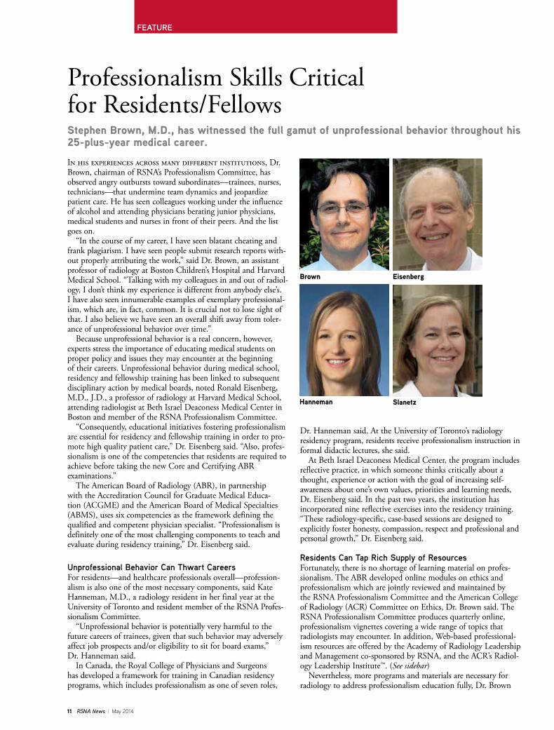
Professionalism Skills Critical for Residents/FellowsStephen Brown, M.D., has witnessed the full gamut of unprofessional behavior throughout his 25-plus-year medical career.
In his experiences across many different institutions, Dr. Brown, chairman of RSNA’s Professionalism Committee, has observed angry outbursts toward subordinates—trainees, nurses, technicians—that undermine team dynamics and jeopardize patient care. He has seen colleagues working under the influence of alcohol and attending physicians berating junior physicians, medical students and nurses in front of their peers. And the list goes on. “In the course of my career, I have seen blatant cheating and frank plagiarism. I have seen people submit research reports with-out properly attributing the work,” said Dr. Brown, an assistant professor of radiology at Boston Children’s Hospital and Harvard Medical School. “Talking with my colleagues in and out of radiol-ogy, I don’t think my experience is different from anybody else’s. I have also seen innumerable examples of exemplary professional-ism, which are, in fact, common. It is crucial not to lose sight of that. I also believe we have seen an overall shift away from toler-ance of unprofessional behavior over time.” Because unprofessional behavior is a real concern, however, experts stress the importance of educating medical students on proper policy and issues they may encounter at the beginning of their careers. Unprofessional behavior during medical school, residency and fellowship training has been linked to subsequent disciplinary action by medical boards, noted Ronald Eisenberg, M.D., J.D., a professor of radiology at Harvard Medical School, attending radiologist at Beth Israel Deaconess Medical Center in Boston and member of the RSNA Professionalism Committee. “Consequently, educational initiatives fostering professionalism are essential for residency and fellowship training in order to pro-mote high quality patient care,” Dr. Eisenberg said. “Also, profes-sionalism is one of the competencies that residents are required to achieve before taking the new Core and Certifying ABR examinations.” The American Board of Radiology (ABR), in partnership with the Accreditation Council for Graduate Medical Educa-tion (ACGME) and the American Board of Medical Specialties (ABMS), uses six competencies as the framework defining the qualified and competent physician specialist. “Professionalism is definitely one of the most challenging components to teach and evaluate during residency training,” Dr. Eisenberg said.
Unprofessional Behavior Can Thwart CareersFor residents—and healthcare professionals overall—profession-alism is also one of the most necessary components, said Kate Hanneman, M.D., a radiology resident in her final year at the University of Toronto and resident member of the RSNA Profes-sionalism Committee. “Unprofessional behavior is potentially very harmful to the future careers of trainees, given that such behavior may adversely affect job prospects and/or eligibility to sit for board exams,” Dr. Hanneman said. In Canada, the Royal College of Physicians and Surgeons has developed a framework for training in Canadian residency programs, which includes professionalism as one of seven roles,
Dr. Hanneman said. At the University of Toronto’s radiology residency program, residents receive professionalism instruction in formal didactic lectures, she said. At Beth Israel Deaconess Medical Center, the program includes reflective practice, in which someone thinks critically about a thought, experience or action with the goal of increasing self-awareness about one’s own values, priorities and learning needs, Dr. Eisenberg said. In the past two years, the institution has incorporated nine reflective exercises into the residency training. “These radiology-specific, case-based sessions are designed to explicitly foster honesty, compassion, respect and professional and personal growth,” Dr. Eisenberg said.
Residents Can Tap Rich Supply of ResourcesFortunately, there is no shortage of learning material on profes-sionalism. The ABR developed online modules on ethics and professionalism which are jointly reviewed and maintained by the RSNA Professionalism Committee and the American College of Radiology (ACR) Committee on Ethics, Dr. Brown said. The RSNA Professionalism Committee produces quarterly online, professionalism vignettes covering a wide range of topics that radiologists may encounter. In addition, Web-based professional-ism resources are offered by the Academy of Radiology Leadership and Management co-sponsored by RSNA, and the ACR’s Radiol-ogy Leadership Institute™. (See sidebar) Nevertheless, more programs and materials are necessary for radiology to address professionalism education fully, Dr. Brown
Brown
Hanneman Slanetz
Eisenberg
FEATURE

May 2014 | RSNA News 12
said. “Strategies to address the issue of impaired and/or incompetent colleagues begin with education to help people identify such behaviors and understand how they undermine care,” he said. “They also include establishing clear, upfront expectations of conduct, facile mechanisms for reporting such behavior and transparent understandings of how such conduct will be managed.”
Patients Deserve Professional BehaviorDisruptive or unprofessional behavior by any healthcare provider creates a suboptimal work environ-ment—and more importantly—places patient safety at risk, said Priscilla Slanetz, M.D., M.P.H., pro-gram director of the Radiology Residency Program at Beth Israel Deaconess Medical Center. “All physicians must remember that they serve as role models for those around them,” Dr. Slanetz said. “Along with reminding each other about being good role models, taking time to reflect on topics related to professionalism can be helpful in keeping professionalism at the forefront. In our residency training program, we hold dedicated sessions four-to-six times each year to discuss a variety of topics related to professional practice.” Although infrequent, there may be times when a radiologist encounters a colleague who is impaired or no longer possesses an up-to-date skill set, Dr. Slanetz said. A person’s response must be tailored to the situation at hand, she stressed. “If a patient’s safety is at risk, it is imperative to act immediately to prevent any harm from occur-ring,” Dr. Slanetz said. “However, in most cases, it is reasonable to speak to your colleague in a private setting. Many times, he or she will be receptive to your feedback and will seek assistance to remedy the situation.” Personal accountability and apologies for harmful errors are key to promoting safe, patient-centered care, Dr. Brown said. A sincere demonstration of warmth, understanding and concern—and immedi-ate availability—can go a long way toward alleviating potential problems, Dr. Eisenberg added. “It is important to treat the patient honestly as a mature, intelligent human being, just as you or a member of your family would want to be treated,” Dr. Eisenberg said. q
RSNA RESEARCH & EDUCATION FOUNDATION-FUNDED PROJECTSName
Stephen Brown, M.D.
Priscilla Slanetz, M.D., M.P.H.
Priscilla Slanetz, M.D., M.P.H. & Ronald L. Eisenberg M.D., J.D.
Kate Hanneman, M.D.
Grant
2011 GE Healthcare/RSNA Education Scholar Grant/$150,000
1995 RSNA Research Fellow Grant/$50,000
2012 RSNA Education Scholar Grant/$150,000
2013 RSNA Research Resident Grant/$30,000
Study
“ Program to Enhance Relational and Communica-tion Skills for Radiologists (PERCS-Radiology)”
“ Spiral CTA and MRA in Preoperative Assessment of Abdominal Aortic Aneurysms”
“ Development of a Peer Observation Teaching Program to Enhance Radiology Resident Teaching Skills”
“ Quantification of Diffuse Myocardial Iron Overload Related Interstitial Fibrosis with Cardiac Magnetic Resonance Imaging in Patients with Transfusion-Dependent Anemias”
Developed by the American Board of Radiology Foundation (ABRF), online modules on ethics and professionalism (accessible at RSNA.org/Profes-sionalism) are designed to educate physicians and physicists about the attributes and nuances of ethics and professionalism that are essential to diagnostic radiology, radiation oncology and medical physics. Each module is self-guided and includes self-testing features to help the reader assess his or her comprehension and application of the principles and practices described in the module. The modules were developed through educational grants from RSNA, the Academy of Radiology Research, the American Association of Physicists in Medicine, the ABR, the American College of Radiology, the American Radium Society and the American Society for Radiation Oncology.
WEB EXTRASAccess RSNA’s professionalism
resources, at RSNA.org/Professionalism. Highlights include: • A series of professionalism vignettes covering a wide range of topics including, ”Sexual Harassment,” ”Medical Trainees and Medical Training,” and the newest vignette, ”Conducting Research with Industry.”
• Online modules on ethics and professionalism developed by the American Board of Radiology Foundation; users can also access the modules from RSNA’s Science & Education page at RSNA.org/Science_and_Education.aspx.
Sponsored by five participating radiology education societies including RSNA, the Academy of Radiology Leadership and Management offers career-enhancing opportunities. For more information and to access the complete course catalog, go to Radleaders.org/catalog.cfm.
Access ACR’s Radiology Leadership Institute™ at Radiologyleaders.org.

13 RSNA News | May 2014
“ Validation is the key to accelerating the transfer of innovative imaging methods developed in research to the clinics.”Sonia Pujol, Ph.D.
Quantitative Imaging Accelerates Clinical ResearchTechnological breakthroughs in medical imaging hardware and increasingly sophisticated image processing software are hastening the progress of quantitative imaging as a clinical research tool—a particularly critical application for cancer therapy, experts say.
“Across all modalities—from nuclear medicine to ultrasound, CT and MR imaging, medical imag-ing provides a rich amount of quantitative data,” said Katarzyna J. Macura, M.D., Ph.D., associate professor of radiology, urology and oncology at The Johns Hopkins University in Baltimore. “Radiolo-gists perform some type of quantitative imaging in their everyday practice, from measuring abnor-malities to assessing blood flow, tissue perfusion or diffusion and tracer uptake. This quantitative approach to imaging data allows more objectivity and facilitates comparisons between different states of the same disease or different disease processes.” As defined by RSNA’s Quantitative Imaging Bio-markers Alliance (QIBA), quantitative imaging is “the extraction of quantifiable features from medical images for the assessment of normal or the severity, degree of change, or status of a disease, injury, or chronic condition relative to normal.” Quantitative imaging provides a noninvasive method for characterizing therapy response, said Sonia Pujol, Ph.D., a researcher at Brigham and Women’s Hospital, Harvard Medical School in Bos-ton and director of training of the National Alliance for Medical Imaging Computing (NAMIC) and the Neuroimaging Analysis Center. Dr. Pujol, along with Dr. Mac-ura and Ron Kikinis, M.D., a professor of radiology at Brigham and Women’s Hospital, Harvard Medical School and principal investigator of the 3D slicer, presented a workshop on quantitative imaging at RSNA 2013. Dr. Pujol said quantitative imag-ing data can potentially be used to give radiologists complementary information for interpreting images. “Biomarkers—as measurable characteristics used to indicate a biological state and where imaging can contribute quantifiable data—are used in oncologic clinical trials, for example, as surrogate end-points to evaluate treatment response, and especially to detect early failure of potentially toxic treatments or to predict patient outcome,” said Macura who received a 2003 Toshiba America Medical Systems/RSNA Research Resident Grant. “Most radiologists are familiar with the World Health Organization (WHO) and Response Evalua-tion Criteria in Solid Tumors (RECIST) criteria that
were introduced to enable meaningful comparison from one exam to another and from one clinical trial to another,” Dr. Pujol said. “Continuous advances are being made in imaging technology and post-processing software tools, and the intersection of quantitative and clinical sciences offers great promise to help improve outcome prediction and tumor response to therapy.” Quantitative imaging has the potential to provide useful clinical information in situations where disease progress is difficult to assess, Dr. Pujol said. “For example, small changes in slowly evolving pathologies such as meningiomas, can be clinically significant but difficult to detect in longitudinal MR imaging scans. Quantitative imaging methods that combine input from medical experts with advanced image analysis tools can aid clinicians in treatment decisions.”
Radiologists May Be Hesitant to Adopt Quantitative ImagingStill, quantitative imaging has a ways to go to reach its full potential. For example, one study shows that radiologists may be somewhat resistant to using quantitative imaging. A study by Richard Abramson, M.D., and col-leagues published in the July 2012 issue of Magnetic Resonance Imaging analyzed MR imaging and CT reports from two randomly selected weekdays at a mixed academic-community practice for the presence of quantitative descriptors. They found that while 44 percent of all reports contained at least one quantitative metric, just 2 percent contained an advanced quantitative metric.
SullivanMacura Pujol
FEATURE

May 2014 | RSNA News 14
Quantitative imaging is an active field of research, and its trans-lation into routine clinical radiology practice has been hindered by many factors, including a lack of standardization, Dr. Pujol said. “There’s a need to validate quantitative imaging biomarkers by evaluating the variability among methods and comparing different methods with outcomes,” Dr. Pujol said. “Similar problems exist with diffusion tensor imaging (DTI) tractography, and we are try-ing to evaluate current tractography techniques and define stan-dards to ascertain quality features for surgical guidance through the DTI Challenge.” (See sidebar) There are also workflow issues, she explained. “Quantitative imaging methods can be time consuming and require the integra-tion of additional post-processing tools to the clinical workflow,” Dr. Pujol said. Consortia such as the National Cancer Institute’s (NCI) Quan-titative Imaging Network (QIN) and QIBA are trying to improve the value of quantitative imaging by working on the challenges associated with standardizing and validating quantitative imaging biomarkers and post-processing techniques, Dr. Pujol said. “Vali-dation is the key to accelerating the transfer of innovative imaging methods developed in research to the clinics.”
RSNA Workshop Gives Radiologists Practical ExperienceThe RSNA workshop taught by Drs. Pujol, Macura and Kiki-nis—“Quantitative Medical Imaging for Clinical Research and Practice”—is designed to provide an introduction to quantitative imaging through a series of case studies involving different organs and multiple imaging modalities, Dr. Pujol said. The course combines the presentation of quantitative imaging biomarkers for diagnosis as well as clinical trial outcome measures with hands-on sessions on the basics of quantitative measure-ments. “The hands-on aspect of the course is intended to give radiolo-gists practical experience on the latest methods developed in medical research, as well as an opportunity to give feedback on quantitative imaging tools still in early development,” Dr. Pujol said.
“This combination of practical experience and the opportunity to influence how quantitative imaging tools are deployed makes this work-shop an invaluable experience for radiologists interested in improving the value of imag-ing in clinical practice,” said RSNA Science Advisor Daniel Sullivan, M.D., a professor in the Department of Radiology at Duke University and chair of the QIBA Steering Committee. The course also provides pre-senters with the opportunity to give radiologists practical expe-rience in quantitative image analysis using 3D Slicer—a multi-platform open-source software package for medical image computing and 3D visu-alization supported by a multi-institution effort and several large-scale grants funded by the National Institutes of Health. As a clinical research tool, 3D Slicer brings clinicians and research scientists together in the prototyping, development and evaluation of novel image analysis tools, Dr. Pujol said. “For example, 3D Slicer is currently used by several NCI Quantitative Imaging Network sites in a variety of clinical research applications, such as pharmacokinetic analysis of prostate DCE MR imaging and PET/CT analysis for therapy response assessment in head and neck cancer,” she said. Such hands-on courses and tutorials—including the RSNA quantitative imaging course—can hopefully accelerate the transfer of novel post-processing tools to clinical researchers, Dr. Pujol said. q
WEB EXTRASTo access an abstract of the
study, “Quantitative metrics in clinical radiology reporting: a snapshot perspective from a single mixed academic-community practice,” by Richard G. Abramson, M.D., and colleagues, go to mrijournal.com/article/S0730-725X%2812%2900202-0/abstract.
For more information on RSNA’s Quantitative Imaging Biomarker Alliance (QIBA) and to access the QIBA newsletter, go to rsna.org/QIBA.
For more information on National Cancer Institute’s Quantitative Imaging Network, go to imaging.cancer.gov/programsandresources/specializedinitiatives/qin.
For more information on the DTI Tractography Challenge, go to dti-challenge.org.
For more information on 3D Slicer, go to slicer.org.
Post-processing of a recurrent/residual ana-plastic olygoastrocytoma case within the 3D Slicer software. Right, top: Co-registered T2-weighted im-age, (middle) T1-weighted image and (bottom) Mean Diffusity map with seg-mented edema (light blue) and tumor regions (yellow, pink). Left: 3D surface models of tumor and edema reconstructed from the segmented regions.
Read more about QIBA in RSNA.org on Page 24.
Imag
e co
urts
ey o
f Son
ia P
ujol
, Ph.
D., a
nd A
lexa
ndra
Gol
by M
.D.

15 RSNA News | May 2014
[SECTION TITLE]
Individual DonorsDonors who give $1,500 or more per year qualify for the RSNA Presidents Circle. Their names are shown in bold face.
Visionary Donor ProgramIndividuals recognized for cumulative lifetime donations
PLATINUM VISIONARY ($25,000)David C. Levin, M.D.
BRONZE VISIONARY ($5,000)Daniel J. Wunder, M.D.
$5,000David C. Levin, M.D.$1,500 – $2,499Betty & O. Wayne Houser, M.D.Mary Lou & Brent J. Wagner, M.D.$500 – $1,499Dean A. GenthWilliam J. Glucksman, M.D.Ingrid E. & Stephen R. Thomas, Ph.D.Gary W. Swenson, M.D.Richard D. White, M.D.Daniel J. Wunder, M.D.$251 - $499Paul M. Aitchison, M.D.Rukhsana Begum, M.D.Ghina A. Birjawi, M.D.Jacques Blanca, M.D., Ph.D.Robert A. Breit, M.D.Pamela J. Brethauer, M.D.Erin McBride Cannington & Royce Cannington, M.D.
Kara L. Carlson, M.D.Ophelia B. Chang, M.D.Danny Chang, M.D.Karen & Brian L. Dunkley, M.D.Carlos E. Encarnacion, M.D.Nazih N. Farah, M.D.Parham Farid, M.D.T. Chen Fong, M.D., F.R.C.P.(C)Dominique Fournier, M.D.Sidney I. Green, M.D.Ben H. Harmon, M.D.Jennifer A. Harvey, M.D.Sue Heenan, M.B.B.Chir. & Kevin Lessey
Stanley M. Hicks, M.D.Stephanie P. Holz, M.D. & Timothy D. Holz
Sachiko & Zenichiro Hombo, M.D.Cristiana Iabichino, M.D.Haruo Isoda, M.D., Ph.D.Won-Hee Jee, M.D.Anthony J. Jennings, M.D.Carl E. Johnson, M.D.David M. Johnson, M.D.Reinhard Kubale, M.D., Ph.D.Stephen Kwong, M.D.Timothy J. Lach, M.D.William G. Lees, M.B.B.S.Melissa S. Liebling, M.D.
Cynthia & Paolo P. Lim, M.D.John P. Limbacher II, M.D.Martha & Peter J. Littrup, M.D.Giovanni Magistretti, M.D.Mary C. Mahoney, M.D.Erik J. Maurer, M.D.Stephanie & Timothy L. McGhee, M.D.Carol & Gordon McLennan, M.D.John M. Milbourn, M.D.Pietro Milone, M.D.David E. Moody, M.D.Miriam L. Neuman, M.D., M.P.H. & Henry Hammond
Njideka H. Nyako, M.B.B.S.Howard K. O’Neil, M.D.Jagdish M. Patel, M.D.Thurman N. Polchow, M.D.Gayatri & Gautham P. Reddy, M.D.Cathy & Kenneth V. Robbins, M.D.Decio Roveda, M.DGuy Saint-Georges, M.D.Alan M. Saunders, M.D.Barbara G. & Donald F. Schomer, M.D.Reinhard W. Schulte, M.D.George P. Shaughness, M.D.Ellen Shaw de Paredes, M.D.Jose Daniel Sierra, M.D.Joseph Spigel, M.D.Raymond L. Thomas, M.D.Michael D. Tripp, M.D.Muhammad A. Zaheer, M.D.$250 or lessM. Julie Armada, M.D.Deborah Arnold, M.D. & Philip ArnoldDana Tell & Nami R. Azar, M.D.Joseph T. Azok, M.D.Joe J. Barfett, M.D.Richard L. Barger Jr., M.D.Lia Bartella, M.D.Prabhakar Battu, M.D.Tanushree Bhattacharjya, M.D. & Shantanu Bhattacharjya
Carlo Biagini, M.D.Alexandre Bialowas, M.D.Katherine R. Birchard, M.D.David Boshell, M.B.B.S.Erin Yourtz & Lawrence D. Bub, M.D.Michael J. Callahan, M.D.Felipe D. Castro, M.D.Irene E. Chen, M.D. & Steven Yang
Jonathan R. Cogley, M.D.Matthew L. Cooper, M.D., M.S.Daniel L. Croteau, M.D.Angeles Cruz Diaz, M.D. & Roberto Martinez
Pedro Augusto N. Daltro, M.D.David De Bruin, M.D.Anne P. Dunne, M.D.Eli F. Dweck, M.D.Stacie & Nathan D. Elfrink, M.D.Roxana Fernandez-Castillo, M.D.James Ferretti, D.O.Kathleen C. Finzel, M.D.Michelle & Nicholas C. Fraley, M.D.Diogo Galheigo De Oliveira E Silva, M.D.Angelica Ramirez & Victor M. Garcia-Gallegos, M.D., Ph.D.
Ayca Gazelle, M.D. & G. Scott Gazelle, M.D., M.P.H., Ph.D.
Fernando Gil, M.D.Anna Krasnicua & Michael S. Girard, M.D.
Dana A. Gray, M.D.Travis Haskins, B.A., M.S.Nobuyuki Hayakawa, M.D.Eddie A. Hernandez Cardenas, M.D.Cheryl W. Hightower, M.D.Michael T. Hirleman, M.D.Mary G. Hochman, M.D.Uchenna J. Ikokwu, M.B.B.Ch. & Chibeze Ikokwu
Kimia K. Kani, M.D.Giedre KavaliauskieneYasushi Kawata, M.D.Kevin G. King, M.D.Alexander W. Korutz, M.D.Joseph G. Koza, M.D.Kenneth K. Lindell, M.D.Maria E. Lioni, M.D.Marina Z. & John W. Little, M.D.Flavio Loureiro, M.D.Deborah Mabin, M.B.Ch.B.Vasantha & Mahadevappa Mahesh, M.S., Ph.D.
Satkurunathan Maheshwaran, M.D.Peter M. Malek, M.D.David A. May, M.D.Sarah J. Menashe, M.D.Simone Pimenta & Paulo Sergio R. Mendlovitz, M.D.
Laurent Milot, M.D., M.Sc.Lex A. Mitchell, M.D.Walter J. Montanera, M.D.Kambiz Motamedi, M.D.Matthew H. Nett, M.D.Marc G. Ossip, M.D.Tobias Penzkofer, M.D.Irina Trofimenko, M.D. & Kirill Petrov, M.D.
Marc J. Pinchouck, M.D.William W. Qiu, M.D.Stephanie A. Reddick, M.D.Brijesh Reddy, M.D.Marcia M. & Jeffrey L. Rosengarten, M.D.
Shawn Del & Jeffrey T. Seabourn, M.D.Richard J. Silberstein, M.D.Frances M. Murphy, M.D., M.P.H. & James G. Smirniotopoulos, M.D.
Lyda Grimaldo & Josue Solis Ugalde, M.D.
Eric R. Sover, D.O.Malgorzata Stusinska, M.D., Ph.D. & Slawomir Stusinski
Thitiporn Suracharoenchaikun, M.D.Carol & Lawrence C. Swayne, M.D.Doug B. Tait, M.D.Renate Tewaag, M.D.Jay W. Thacker, M.D.Irina Trofimenko, M.D. & Kirill Petrov, M.D.
Sylvia M. Trumble, M.D.Daniel V. Tufariello, M.D.Toshiyuki Unno, M.D.Marion van Vliet, M.D.Teodor Vasile, M.D.Rommel G. Villacorta, M.D.Lilian Wang, M.D.Brent D. Weinberg, M.D., Ph.D.Frank J. Welte, M.D.Karen M. Wieseler-Stone, M.D.Alecia N. & Kyle M. Williamson, M.D.Stanley Yang, M.D.Hooman Yarmohammadi, M.D.
RESEARCH & EDUCATION FOUNDATION DONORSThe R&E Foundation thanks the following donors for gifts made February 5 – February 18, 2014.
The RSNA R&E Foundation provides the research and development that keeps radiology in the forefront of medicine. Support your future—donate today at RSNA.org/donate.
RADIOLOGY’S FUTURE

May 2014 | RSNA News 16
Your donations in action – $150,000 Investment Results in $1 Million to Combat Hepatocellular Carcinoma
2011-2013 RSNA Research Scholar Grant recipient David A. Woodrum, M.D., Ph.D., assistant professor of radiology at the Mayo Clinic, Rochester, Minn., has been awarded a 5-year, $1 million National Institutes of Health R01 grant for his project, “Regulation of Molecular Thermal Ablative Resistance in Hepatocellular Carcinoma (HCC).” “HCC is a major global burden of morbidity and mortality and its incidence in the U.S. has tripled over the past 30 years,” Dr. Woodrum said. “Locoregional thermal ablative therapies are important treatment options for early-mid stage HCC, achiev-ing short-term outcomes similar to surgery with less morbidity. However, high tumor recurrence rates after treatment of larger HCCs (up to 75 percent at 5 years) limit their applicability and overall survival remains poor for these patients,” Dr. Woodrum said. “We will use a combination of cellular and molecular methods, novel imaging techniques and in vitro, in vivo and patient-based approaches to systematically investigate the mechanistic role of the novel heat stress-induced MET/EGFR-PI3K-AKT axis in HCC thermal resistance, recurrence and tumor progression.”
The Value of Membership
Select RSNA Refresher Courses Now On SaleFor a limited time, RSNA is offering discount pricing on selected refresher courses from past annual meetings. One of the most popular collections this year, the Pulmonary Collection, is available at a 25 percent discount until October 31, 2014. The discount price is $90 for members and $130 for nonmembers. The three CDs included in the collection, “Guidelines for Thoracic Imaging,” “High-Resolution CT: A Pattern-based Approach,” and “Obstructive Lung Disease,” are a comprehensive study of CT imaging of the lungs, from the features of chronic obstructive conditions to evaluation of the patient at risk of pulmonary embolism. A CME test included with each course provides physicians the chance to earn CME credit by completing the test and sending it to RSNA. A score of 80 percent or higher is required. The Pulmonary Collection offers 4.25 AMA PRA Category 1 Credits™. To purchase the collection at the discounted rate, go to the RSNA Education Center catalog at RSNA.org/purchase.
RSNA Members Attend the World’s Premier Medical Meeting For FreeYour RSNA membership entitles you to free advance registration for RSNA 2014— an $825 value—as well as early hotel reservations and free SAMs. Register by November 7 to receive the discounted registration fee and full conference materials mailed to you in advance. Interna-tional visitors must register by October 24 to receive these materials in advance. After November 7, registrations will be processed at the increased fee and conference materials must be picked up at McCormick Place. Not an RSNA member? Join the RSNA community and attend the most important medical meeting in the world—for free. Apply or renew at RSNA.org/Membership. For more information, contact [email protected] or 1-877-RSNA-MEM (1-877-776-2636) or 1-630-571-7873 outside the U.S. and Canada.

17 RSNA News | May 2014
Education and Funding Opportunities
More information and application/nomination forms for these programs are available at rsna.org/Grant_Writing_and_Research_Development_Programs_.aspx. Questions can be directed to Rachel Nelson at 1-630-590-7741 or [email protected].
RSNA Clinical Trials Methodology WorkshopOver the course of this 6½-day workshop, each trainee will be expected to develop a protocol for a clinical study, ready to include in an application for external funding. Participants will learn how to develop protocols for the clinical evaluation of imaging modalities. A dynamic and experienced faculty will cover topics including:
January 10-16, 2015 Scottsdale/Ariz. Applications due June 15, 2014
• Principles of clinical study design • Statistical methods for imaging studies • Design and conduct of multi-institutional studies • Sponsorship and economics of imaging trials • Regulatory processes Applicants will undergo a competitive selection process for course entrance. Once admitted, trainees will participate in advance preparation, didactic sessions, one-on-one mentoring, small group discussions, self-study and individual protocol development. Familiarity with basic concepts and techniques of statistics and study design is required of all applicants.
RSNA Derek Harwood-Nash International FellowshipApplications Due July 1, 2014 for 2015 Program
The Derek Harwood-Nash Fellowship program supports international scholars pursuing a career in academic radiology to study at North American institutions. Accepted participants will receive a stipend of up to $10,000 from RSNA to be used toward travel, living expenses and educational materials for the 6- to 12-week fellowship period.
The application for this program is available at RSNA.org/DHN. For more information e-mail [email protected].
Sponsored by RSNA, the American Roentgen Ray Society (ARRS) and Association of University Radiologists (AUR), the Introduction to Academic Radiology program:
• Exposes second-year residents to academic radiology • Demonstrates the importance of research in diagnostic radiology • Illustrates the excitement of research careers • Introduces residents to successful clinical radiology researchers Successful applicants will be assigned to either a seminar held November 30–December 4, 2014, during the RSNA Scientific Assembly in Chicago, or the ARRS Scientific Meeting in Toronto, Canada, April 19-24, 2015. More information and the nomination form for this program are available at RSNA.org/ITAR.
RSNA/AUR/ARRS Introduction to Academic Radiology Program
Applications due July 15, 2014
Online application and additional information can be found at RSNA.org/CT2015.
NEWS YOU CAN USE

May 2014 | RSNA News 18
MAY 4–7 Radiology Business Managers Association (RBMA), 2014 Radiology Summit, The Westin Charlotte, North Carolina • www.rbma.org* Visit RSNA at Booth 603MAY 13-17Society for Pediatric Radiology (SPR), 57th Annual Meeting, JW Marriott Hotel, Washington, D.C. • www.pedrad.orgMAY 15-17Society for Imaging Informatics in Medicine (SIIM), Annual Meeting, Long Beach, California • www.siim2014.orgMAY 17-22American Society of Neuroradiology (ASNR) 52st Annual Meeting and the Foundation of the ASNR Symposium 2014, Montreal Convention Center, Montreal, Quebec, Canada • www.asnr.org/2014
MAY 22-25Sociedad Española de Radiología Médica/Spanish Society of Medical Radiology (SERAM), 32 Congreso Nacional/32nd National Congress, Palace of Congresses and Exhibitions, Oviedo, Spain • www.seram2014.comMAY 26-2826th Congress of the European Federation of Societies for Ultrasound in Medicine and Biology (EFSUMB), Israel Society for Diagnostic Ultrasound in Medicine (ISDUM), David Intercontinental Hotel, Tel Aviv, Israel • www.euroson2014.org
MAY 28-30The Russian National Congress of Radiologists, Radiology 2014, Crocus Expo International Exhibition Centre, Moscow • www.radiology-congress.ruMAY 28-31The German Radiological Society (DRG) and the Austrian Society of Radiology (ORG), The 95th German Radiology Congress and the 7th Joint Congress of the Austrian Society of Radiology and the German Radiological Society, Congress Centrumr Hamburg, Germany • www.drg.deMAY 30-JUNE 3American Society of Clinical Oncology (ASCO), 50th Annual Meeting, McCormick Place, Chicago• www.asco.orgJUNE 2-6European Society of Pediatric Radiology (ESPR), 51th Annual Meeting and 37th Postgraduate Course, Grand Hotel Krasnapolsky, Amsterdam, The Netherlands • www.ESPR2014.orgJUNE 7-11Society of Nuclear Medicine and Molecular Imaging (SNMMI), Annual Meeting, St. Louis, Missouri • www.snmmi.org
Medical Meetings May-June 2014
FIND MORE EVENTS AT RSNA.org/calendar.aspx.
RSNA Advanced Course in Grant Writing Applications due July 1, 2014
Applications are now being accepted for this course designed to assist participants—generally junior faculty members in radiology, radiation oncology or nuclear medicine programs—prepare and
submit a National Institutes of Health, National Sciences Foundation or equiva-lent grant application. The course, to be held at RSNA Headquarters in Oak Brook, Ill., will consist of four two-day sessions: October 10-11, 2014; January 30-31, 2015; March 13-14, 2015; and May 1-2, 2015. For more information and to download an application, go to RSNA.org/AGW.
“Imaging Forward” Campaign UnveiledThe Medical Imaging & Technology Alliance (MITA) has launched Imaging Forward, a campaign to highlight groundbreaking innovation in medical imaging technologies and the impact on patient care and healthcare delivery.
Technology Forum
Imaging Forward is intended to drive the conversation about medical imaging’s role in advancing quality healthcare, improving patient outcomes, and reducing costs. Such emphasis is necessary, says MITA, as Congress and the Centers for Medicare & Medicaid Services (CMS) have acted to cut Medicare imaging reimbursements multiple times since 2006. Further cuts will only continue to impede access to life-saving medical imaging services and undercut the benefits of early detection and treatment, according to MITA. More information about Imaging Forward can be found at medicalimaging.org/imagingforward.

19 RSNA News | May 2014
[SECTION TITLE]
The following are highlights from current issues of RSNA’s two peer-reviewed journals.
State of the Art: Dual-Energy CT of the AbdomenIntensive research devoted to exploiting the unique and powerful opportunities of dual-energy CT has led to protocol modifications for radiation dose reduction, improved diagnostic performance for detection and characterization of dis-eases and image quality optimization. In a State-of-the-Art article in the May issue of Radiology (RSNA.org/Radiology), Daniele Marin, M.D., Duke University Medical Center, Durham, N.C., and colleagues discuss the basic principles, instrumentation and design, examples of current clinical applica-tions in the abdomen and pelvis and future opportunities of dual-energy CT (DECT). DECT is gradually changing the way CT is practiced today, according to the authors. By interrogating the unique characteristics of different materials at different X-ray energies, DECT provides quantitative information about tissue composition, overcoming the limitations of attenuation-based conventional single energy CT imaging. “In the future, streamlined strategies to improve workflow efficiency (e.g., development vendor-independent dual energy post-processing workstation seamlessly integrated into PACS), decreased costs, and reimbursement by third-party payers, will expedite widespread adoption of this new technology into clinical practice,” the authors write.
Adult Bile Duct Strictures: Role of MR Imaging and MR Cholangiopancreatography in CharacterizationBile duct strictures in adults are secondary to a wide spectrum of benign and malignant pathologic conditions. Awareness of the various causes of bile duct strictures in adults and familiarity with their appearances at MR imag-ing–MR cholangiopancreatography are important for accu-rate diagnosis and optimal patient management. In an article in the May-June issue of RadioGraphics (RSNA.org/RadioGraphics), Venkata S. Katabathina, M.D., of the University of Texas Health Science Center at San Antonio, and colleagues review the spectrum of bile duct strictures in adult patients and discuss the MR imaging and MR cholangiopancreatographic findings, with emphasis on differentiation between benign and malignant strictures. Contrast-enhanced MR imaging with MR cholangio-pancreatography can be helpful in identifying bile duct strictures in adult patients and in differentiating between benign and malignant strictures, according to the authors. “Although biopsy is necessary for distinguishing malig-nant from benign strictures, certain MR imaging findings of the narrowed segment may favor a malignant cause,” the authors write.
Read the Invited Commentary by Andrew J. Taylor, M.D., of the University of Minnesota, Minneapolis.
This article meets the criteria for AMA PRA Category 1 Credit™. SA-CME is available online only.
This article meets the criteria for AMA PRA Category 1 Credit™. SA-CME is available online only.
Effectiveness of VNC im-aging in clinical practice. Contrast-enhanced dual-energy CT image. Note on VNC image the accurate subtraction of highly concentrated iodinated contrast material from the collecting system (curved arrow) with preservation of the pelvic renal stone (arrow). (Radiology 2014;271;2:327–342) ©RSNA, 2014. All rights re-served. Printed with permission.
Primary sclerosing cholangitis in a 63-year-old man. Maximum intensity projection image from a 3D RARE MR cholangiopancrea-tographic study shows multiple strictures of the common bile duct interspersed with focal dilatations, creating a beaded appearance (arrows), along with multiple discontinuous strictures of the intra-hepatic bile ducts involving both hepatic lobes (arrowheads). (RadioGraphics 2014;34;565-585) ©RSNA, 2014. All rights reserved. Printed with permission.
NEWS YOU CAN USE
Journal Highlights

May 2014 | RSNA News 20
Stroke Edition of Radiology Select Offered at Reduced Rate in MayIn recognition of Stroke Awareness month, Radiology Select, Volume 2: Stroke, is available at 50 percent off the standard price for the online education edi-tion during May to members and non-members. Radiology Select is a continuing series of selected Radiology articles that highlight developments in imaging science, tech-niques and clinical practice. Each volume focuses on a particular topic important in the field and is supplemented by commentar-ies, author interviews, podcasts and educational opportunities. Radiology Select is a great way to stay at the forefront of radiol-ogy while maintaining the necessary certifications.Volume 2: Stroke features 30 articles that cover: • CT applications including dual-energy imaging, perfusion
imaging and thrombus detection • Techniques for risk stratification, diagnosis and
therapeutic monitoring in the internal carotid artery • “Mismatch” MR imaging techniques • Observations on neonatal intracranial hemorrhage and
cerebral artery flow in pediatric sickle cell disease The Online Educational Edition includes four SAMs tests with an opportunity to earn eight SAM credits and 12 CME credits. This enduring material can be applied towards the ABR self-assessment requirement.
Don’t Miss Out: Keep Your RSNA Profile Up-to-DateResidents and fellows who will be moving to a new position in the coming months are encouraged to update their contact information with RSNA.Log in at myRSNA.org and click Account Tools in the left-hand sidebar to update your personal information. Having a current street and personal email addresses on file with RSNA means you will continue to receive: • Subscriptions to Radiology, RadioGraphics and RSNA News • Special membership rates for members transitioning from residency or fellowship to practice • E-mail news bulletins, including RSNA Insider and RSNA Weekly • Annual meeting announcements RSNA members transitioning to practice after residency or fellowship pay just $100 their first year and $200 their second year. Full dues are not required until the third year. If you have ques-tions or wish to update your information by phone, call 1-877-RSNA-MEM (776-2636) or 1-630-571-7873 (outside the U.S. or Canada).
Residents & Fellows Corner
Listen to Radiology Editor Her-bert Y. Kressel, M.D., deputy edi-tors and authors discuss the fol-lowing articles in the April issue of Radiology at pubs.rsna.org/page/radiology/podcasts:
“Digital Mammography Screening: Association between Detection Rate and Nuclear Grade of Ductal Carcinoma in Situ,” Stefanie Weigel, M.D., and colleagues.
“Mapping the Effect of the Apolipoprotein E Genotype on 4-Year Atrophy Rates in an Alzheimer Disease–related Brain Network,” Christopher A. Hostage, M.D., and colleagues.
“Cardiac Arrythmias: Multimodal Assessment Integrating Body Surface ECG Mapping into Cardiac Imaging,” Hubert Cochet, M.D., and colleagues.
Nominate Radiology Articles for the 2014 Margulis AwardThe Nominating Committee for the Margulis Award for Scientific Excellence is accepting nominations from readers for Radiology articles published between July 2013 and June 2014. The main selection criteria are scientific qual-ity and originality. Please send your nomination, including the article citation and a brief note highlighting the reasons for the nomination, to Pamela Lepkowski, assistant to the editor, [email protected]. The deadline for nomina-tions is June 10, 2014.

21 RSNA News | May 2014
[SECTION TITLE][SECTION TITLE]NEWS YOU CAN USE
New on RadiologyInfo.orgVisit RadiologyInfo.org to read the latest disease/condition topics posted to the site, including:• Cystic Fibrosis:
Radiologyinfo.org/en/info.cfm?pg=cysticfibrosis.
Press releases were sent to the medical news media for the following articles appearing in recent issues of Radiology.
Carotid Artery Plaque Morphology and Composition in Relation to Incident Cardiovascular Events: The Multi-Ethnic Study of Atherosclerosis (MESA)
RadiologyInfo.org Strategic Planning Sessions Lead to Change
Identification of vulnerable plaque characteristics with MR imaging aids in cardiovascular disease prediction and improves the reclassification of baseline cardiovascu lar risk, new research shows. In the Multi-Ethnic Study of Atheroscle-rosis, Anna E. H. Zavodni, M.D., of the University of Toronto, Canada, and col-leagues evaluated 946 participants using MR imaging and ultrasonog raphy. Researchers used MR imaging to define carotid plaque composition and remodeling index (wall area divided by the sum of wall area and lumen area), while ultrasound was used to assess carotid wall thickness. Incident of cardiovas-cular events were ascertained for an average of 5.5 years. Multivariable Cox propor tional hazards models, C statistics, and net reclas-sification improvement for event prediction were determined. Cardiovascular events occurred in 59 of the patients. Abnormal thickening of the carotid artery wall and the presence of a lipid core and calcium in the internal carotid artery on MR imaging were significant predic-tors of subsequent events. A lipid core was present in almost half of the patients who had an event, compared with only 17.8 per-cent of those who did not have an event.
Be on the lookout for some exciting changes coming to Radi-ologyInfo.org, RSNA and ACR’s jointly-sponsored public infor-mation website. In February, the RadiologyInfo.org Strategic Planning Task Force met with a consultant who facilitated sessions to aid in developing a strategic plan. The RSNA-ACR Public Informa-tion Website Committee adopted a draft of the plan during its February meeting. With new goals and strategies now in place, changes will soon be implemented to better address the radiology informa-tion needs of patients, caretakers, radiologists, referring physi-cians and others.
“The combination of MR imaging remodeling index and pres-ence of lipid core resulted in improve ment of the net reclassifica-tion index compared with traditional risk factors for cardiovascu-lar disease events,” according to researchers.
Transverse gadolinium-enhanced T1-weighted MR images obtained superior to the carotid artery bifurcation in a 72-year-old man. L = ICA lumen. (a) Low-signal-intensity calcium (arrow) and lipid core (arrowheads) can be seen. (b) Note contouring of the ICA. The outer adventitial wall (red), lipid core (blue), calcification (green) and vessel lumen (purple) are visible. (Radiology 2014;271;3:InPress) ©RSNA, 2014. All rights reserved. Printed with permission.
NEWS YOU CAN USE
Radiology in Public Focus

May 2014 | RSNA News 22
MAY PUBLIC INFORMATION ACTIVITIES FOCUS ON STROKE
MR Imaging of the Substantia Nigra at 7 T Enables Diagnosis of Parkinson Disease
MR imaging at 7 T reveals a three-layered organization of the substantia nigra (SN) in both the in-vivo and ex-vivo brainstem. The 3D multi-echo susceptibility-weighted images can be used to accurately differenti-ate healthy patients from those with Parkinson’s disease, new research shows. Mirco Cosottini, M.D., of the University of Pisa, Italy, and col-leagues described SN anatomy ex vivo on a gross brain specimen using highly resolved proton-density (spin-echo proton density) and gradient-recalled-echo (GRE) images, and in vivo in eight healthy subjects (mean age, 40.1 years) using GRE 3D multi echo susceptibility-weighted images. After training on appearance of SN in eight healthy subjects, SN anat omy was evaluated twice by two blinded observers in 13 healthy sub-jects (mean age, 54.7 years) and in 17 patients with Parkinson’s disease (mean age, 56.9 years). The abnormal architecture of the SN allowed researchers to discriminate between Parkinson’s patients and healthy subjects with a sensitivity and specificity of 100 percent and 96.2 percent respectively. Intraob server agreement (k = 1) and interobserver agreement (k = 0.932) were excellent, results showed. “The identification of a radiologic marker of the altered SN by using 7-T MR imaging allows an imag ing-based early diagnosis of Parkinson’s disease,” according to researchers. “A larger sample of patients would be desirable, but the high diag nostic accuracy of the 7-T MR imaging in Parkinson’s patients enrolled in this study appears to be a promising result for a future broader clinical application.”
Images show axial spin-echo proton density of the substantia nigra (SN) (on the right) and gradient-re-called echo (GRE) (on the left) of the SN at level I of an ex vivo brain sample in a 67-year-old woman. There is a triple-layered organization of the SN comparable to that showed in the in vivo images. Ventrally a low-signal-intensity layer (b) is attributable to the pars reticulata of the SN. In the middle part of the SN, a hyperintense band (c) corresponds to the ventral component of the pars compacta of the SN. The lateral part of this layer shows a high-signal-intensity spot (c1) corresponding to the oval shape hyperintensity of the in vivo three-dimensional multiecho susceptibility-weighted images that resemble the nigrosome formation. The dorsal hypointense layer visible on both spin-echo and GRE images (d) is referred to the dorsal component of the pars compacta of the SN. a = crus cerebri, e = brachjum conjunctivum, f = medial lemniscus, g = lateral lemniscus, h = central tegmental tract. (Radiology 2014;271;3:InPress) ©RSNA, 2014. All rights reserved. Printed with permission.
In recognition of American Stroke Month in May, RSNA is distributing public service announce-ments (PSAs) focusing on stroke imaging, inter-ventional treatments for stroke and the importance of receiving stroke treatment quickly. The “60-Second Checkup” audio program focusing on carotid artery screening to detect stroke risk will be distributed to nearly 100 radio stations across the U.S.
Media Coverage of RSNAIn February, 595 RSNA-related news stories were tracked in the media. These stories reached an esti-mated 275 million people. Coverage included The New York Times, The Globe and Mail, Philly.com, The Providence Journal, Sci-enceDaily, Auntminnie.com, Healthfinder.gov, Applied Radiology and Medical News Today.
RADIOLOGY.RSNA.ORG
MARCH 2014 VOLUME 270 NUMBER 3RADLAX 270(3) 635–940
3A This Month in Radiology
638 Communications
642 Reviews and Commentary
699 Original Research
929 Diagnosis Please

23 RSNA News | May 2014
Hotel Deposits RequiredA deposit equal to a one night stay including tax will be charged by the hotel for each room reserved. Reservations may be secured with a major credit card at the time of booking. The credit card must be valid through December 2014 and will be charged by the hotel approximately two weeks before the annual meeting. If the credit card is declined, the reservation may be cancelled by the hotel. Registrants can also send a check, money order or wire transfer (payable to RSNA) for the hotel deposit. (Attendees are responsible for all wire transfer fees) New this year, simplified and penalty-free cancel and change policies up to 72 hours prior to arrival making it easier than ever before to book a room through RSNA.
International VisitorsInternational Letters Available —Act Now for VisaPersonalized letters of invitation to RSNA 2014 are available by request during online registration. In addition, the International Visitors section of RSNA.org/Visas includes important information about the visa application process. Visa applicants are advised to apply as soon as they decide to travel to the U.S. and at least three to four months in advance of their travel date. International visitors are advised to begin the visa process now.
2 FAX (24 hours) 1-888-772-1888 1-301-694-5124
3 TELEPHONE (Mon.-Fri. 8 a.m. – 5 p.m. CT) 1-800-650-7018 1-847-996-5862
4 MAIL Experient/RSNA 2014 P.O Box 4088 Frederick, MD 21705 USA
1 INTERNET (fastest way) Go to RSNA.org/register
RSNA 2014 RegistrationHOW TO REGISTER There are four ways to register for RSNA 2014:
Registration FeesBY NOV. 7 VIRTUAL COMBO
$ 0 $ 100 $ 100 RSNA/AAPM Member
0 100 100 RSNA/AAPM Member Presenter0 0 0 RSNA Member-in-Training, RSNA Student Member
and Non-Member Student0 300 300 Non-Member Presenter
180 300 480 Non-Member Resident/Trainee
180 300 480 Radiology Support Personnel
825 300 1,125 Non-Member Radiologist, Physicist or Physician825 300 1,125 Hospital or Facility Executive, Commercial Research
and Development Personnel, Healthcare Consultant and Industry Personnel
325 300 625 One-day registration to view only the Technical Exhibits
Benefits of reserving your room through the RSNA Housing System include:1. Lowest rates. Eighty-four hotels in the heart of the
city offering a wide range of options and price points and the lowest rates possible.
2. Free transportation. Free Metra train service to the Randolph Street Station as well as shuttle bus service between all 84 hotels and McCormick Place.
3. Flexible terms (New this year). Unlike online travel agencies that require prepaid stays or have restric-tive penalties, RSNA has established flexible booking terms up to 72 hours prior to arrival.
4. Customer Service. RSNA is your advocate if a dispute or problem arises and is available to assist with hous-ing questions or concerns.
5. Supporting RSNA. By booking through the RSNA Housing System, you are supporting the Society and creating cost benefits that are passed on to attendees. By avoiding attrition fees, RSNA is able to negotiate better deals on room rates.
6. Easy booking. The RSNA website offers one-click booking while providing a wide range of choices at different price points. Another advantage: No need to scour the Internet to find the best rates.
News About RSNA 2014Advance Registration and Housing Open May 1RSNA 2014 advance registration and housing open May 1 for RSNA and AAPM members. General registration and housing open June 4. Advance Registration and Housing information is available at RSNA.org/Attendees.aspx.
Register by November 7 to receive the discounted registration fee and full conference materials mailed to you in advance. International visitors must register by October 24 to receive these materials in advance. Registrations received after November 7 will be processed at the increased fee and conference materials must be obtained at the McCormick Place Convention Center.
For more information about registering for RSNA 2014, visit RSNA.org/Annual_Meeting.aspx e-mail [email protected], or call 1-800-650-7018.
NEWS YOU CAN USE
Annual Meeting Watch
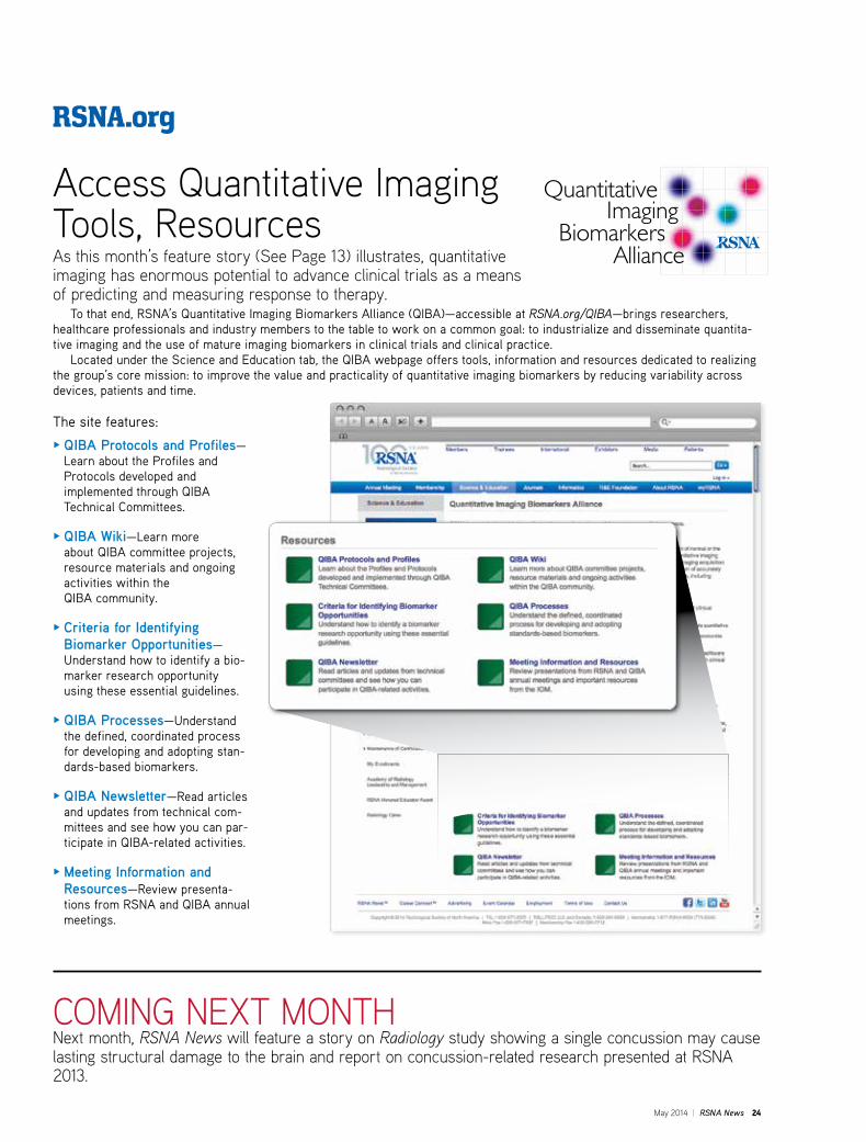
May 2014 | RSNA News 24
RSNA.org
COMING NEXT MONTHNext month, RSNA News will feature a story on Radiology study showing a single concussion may cause lasting structural damage to the brain and report on concussion-related research presented at RSNA 2013.
Access Quantitative Imaging Tools, ResourcesAs this month’s feature story (See Page 13) illustrates, quantitative imaging has enormous potential to advance clinical trials as a means of predicting and measuring response to therapy. To that end, RSNA’s Quantitative Imaging Biomarkers Alliance (QIBA)—accessible at RSNA.org/QIBA—brings researchers, healthcare professionals and industry members to the table to work on a common goal: to industrialize and disseminate quantita-tive imaging and the use of mature imaging biomarkers in clinical trials and clinical practice. Located under the Science and Education tab, the QIBA webpage offers tools, information and resources dedicated to realizing the group’s core mission: to improve the value and practicality of quantitative imaging biomarkers by reducing variability across devices, patients and time.
The site features:
O QIBA Protocols and Profiles—Learn about the Profiles and Protocols developed and implemented through QIBA Technical Committees.
O QIBA Wiki—Learn more about QIBA committee projects, resource materials and ongoing activities within the QIBA community.
O Criteria for Identifying Biomarker Opportunities—Understand how to identify a bio-marker research opportunity using these essential guidelines.
O QIBA Processes—Understand the defined, coordinated process for developing and adopting stan-dards-based biomarkers.
O QIBA Newsletter—Read articles and updates from technical com-mittees and see how you can par-ticipate in QIBA-related activities.
O Meeting Information and Resources—Review presenta-tions from RSNA and QIBA annual meetings.

This activity has been approved for AMA PRA Category 1 Credit™
100th S C IEN T IF IC A S SEMBLY A ND A NNU A L ME E T ING
The Celebration of a
The finest education and never-before-seen science
Groundbreaking technology from across the globe
An interactive historic showcase of RSNA and radiology
RSNA.org/registerGENERAL REGISTRATION
OPENS JUNE 4MEMBER REGISTRATION OPENS MAY 1
M c C O R M I C K P L A C E , C H I C A G O
RSNA 2014 Pubs Ad.indd 1 3/24/14 9:24 AM