document
TRANSCRIPT
NATURE MEDICINE • VOLUME 5 • NUMBER 1 • JANUARY 1999 97
ARTICLES
Transplantation of embryonic nigral tissue ameliorates func-tional deficiencies in Parkinson disease1,2. The main practicalconstraints of neural grafting are the shortage of human donortissue and the poor survival of dopaminergic neurons graftedinto patients, which is estimated at 5–10% (refs. 3,4). The re-quired amount of human tissue could be considerably reducedif the neuronal survival was augmented. Studies in rats indicatethat most implanted embryonic neurons die within 1 week oftransplantation5,6, and that most of this cell death is apoptotic6.Modified peptides, such as acetyl-tyrosinyl-valyl-alanyl-as-partyl-chloro-methylketone (Ac-YVAD-cmk), that specifically in-hibit proteases of the caspase family7 effectively blockapoptosis in a plethora of experimental paradigms, such asgrowth factor withdrawal8, excitotoxicity9, axotomy10, cerebralischemia11 and brain trauma12. Here we examined the effects ofcaspase inhibition by Ac-YVAD-cmk on cell death immediatelyafter donor tissue preparation and on long-term graft survival.Treatment of the embryonic nigral cell suspension with Ac-YVAD-cmk mitigated DNA fragmentation and reduced apopto-sis in transplants. It also increased survival of dopaminergicneurons grafted to hemiparkinsonian rats, and thereby sub-stantially improved functional recovery.
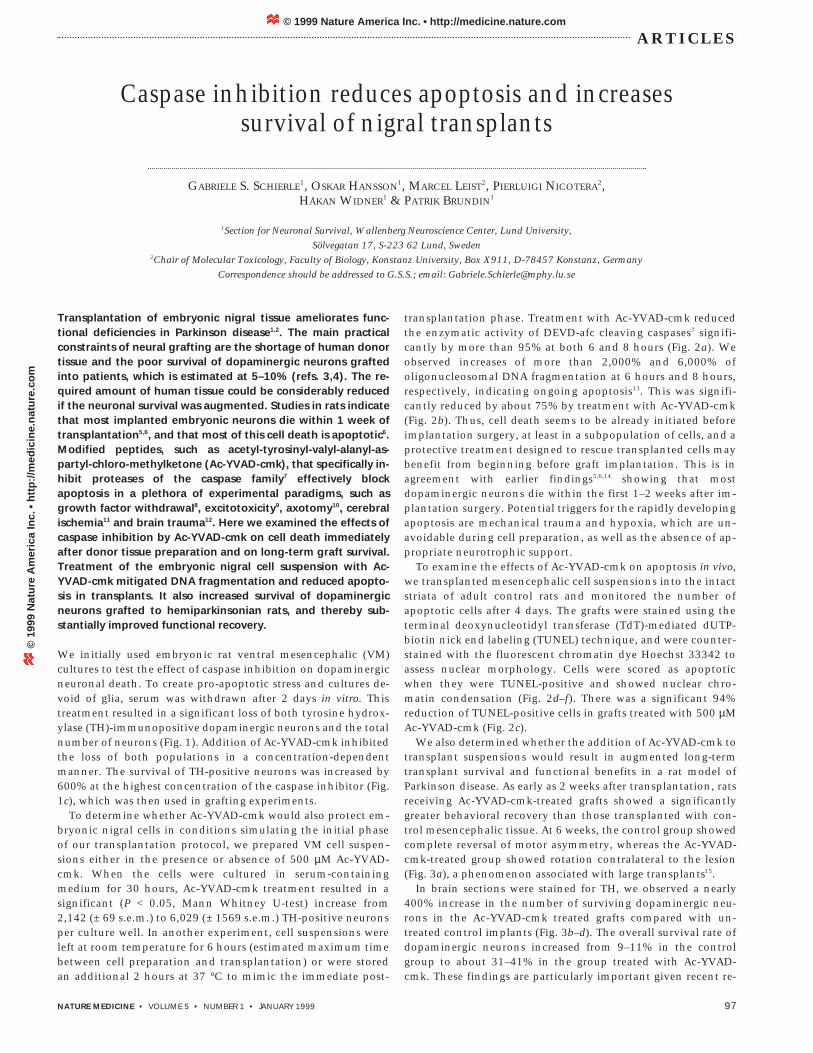
We initially used embryonic rat ventral mesencephalic (VM)cultures to test the effect of caspase inhibition on dopaminergicneuronal death. To create pro-apoptotic stress and cultures de-void of glia, serum was withdrawn after 2 days in vitro. Thistreatment resulted in a significant loss of both tyrosine hydrox-ylase (TH)-immunopositive dopaminergic neurons and the totalnumber of neurons (Fig. 1). Addition of Ac-YVAD-cmk inhibitedthe loss of both populations in a concentration-dependentmanner. The survival of TH-positive neurons was increased by600% at the highest concentration of the caspase inhibitor (Fig.1c), which was then used in grafting experiments.
To determine whether Ac-YVAD-cmk would also protect em-bryonic nigral cells in conditions simulating the initial phaseof our transplantation protocol, we prepared VM cell suspen-sions either in the presence or absence of 500 µM Ac-YVAD-cmk. When the cells were cultured in serum-containingmedium for 30 hours, Ac-YVAD-cmk treatment resulted in asignificant (P < 0.05, Mann Whitney U-test) increase from2,142 (± 69 s.e.m.) to 6,029 (± 1569 s.e.m.) TH-positive neuronsper culture well. In another experiment, cell suspensions wereleft at room temperature for 6 hours (estimated maximum timebetween cell preparation and transplantation) or were storedan additional 2 hours at 37 °C to mimic the immediate post-
transplantation phase. Treatment with Ac-YVAD-cmk reducedthe enzymatic activity of DEVD-afc cleaving caspases7 signifi-cantly by more than 95% at both 6 and 8 hours (Fig. 2a). Weobserved increases of more than 2,000% and 6,000% ofoligonucleosomal DNA fragmentation at 6 hours and 8 hours,respectively, indicating ongoing apoptosis13. This was signifi-cantly reduced by about 75% by treatment with Ac-YVAD-cmk(Fig. 2b). Thus, cell death seems to be already initiated beforeimplantation surgery, at least in a subpopulation of cells, and aprotective treatment designed to rescue transplanted cells maybenefit from beginning before graft implantation. This is inagreement with earlier findings5,6,14 showing that mostdopaminergic neurons die within the first 1–2 weeks after im-plantation surgery. Potential triggers for the rapidly developingapoptosis are mechanical trauma and hypoxia, which are un-avoidable during cell preparation, as well as the absence of ap-propriate neurotrophic support.
To examine the effects of Ac-YVAD-cmk on apoptosis in vivo,we transplanted mesencephalic cell suspensions into the intactstriata of adult control rats and monitored the number ofapoptotic cells after 4 days. The grafts were stained using theterminal deoxynucleotidyl transferase (TdT)-mediated dUTP-biotin nick end labeling (TUNEL) technique, and were counter-stained with the fluorescent chromatin dye Hoechst 33342 toassess nuclear morphology. Cells were scored as apoptoticwhen they were TUNEL-positive and showed nuclear chro-matin condensation (Fig. 2d–f). There was a significant 94%reduction of TUNEL-positive cells in grafts treated with 500 µM Ac-YVAD-cmk (Fig. 2c).
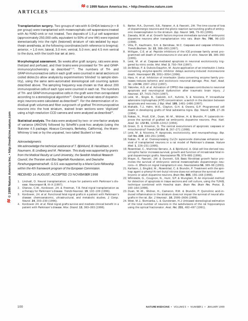
We also determined whether the addition of Ac-YVAD-cmk totransplant suspensions would result in augmented long-termtransplant survival and functional benefits in a rat model ofParkinson disease. As early as 2 weeks after transplantation, ratsreceiving Ac-YVAD-cmk-treated grafts showed a significantlygreater behavioral recovery than those transplanted with con-trol mesencephalic tissue. At 6 weeks, the control group showedcomplete reversal of motor asymmetry, whereas the Ac-YVAD-cmk-treated group showed rotation contralateral to the lesion(Fig. 3a), a phenomenon associated with large transplants15.
In brain sections were stained for TH, we observed a nearly400% increase in the number of surviving dopaminergic neu-rons in the Ac-YVAD-cmk treated grafts compared with un-treated control implants (Fig. 3b–d). The overall survival rate ofdopaminergic neurons increased from 9–11% in the controlgroup to about 31–41% in the group treated with Ac-YVAD-cmk. These findings are particularly important given recent re-
Caspase inhibition reduces apoptosis and increasessurvival of nigral transplants
GABRIELE S. SCHIERLE1, OSKAR HANSSON1, MARCEL LEIST2, PIERLUIGI NICOTERA2,HÅKAN WIDNER1 & PATRIK BRUNDIN1
1Section for Neuronal Survival, Wallenberg Neuroscience Center, Lund University,Sölvegatan 17, S-223 62 Lund, Sweden
2Chair of Molecular Toxicology, Faculty of Biology, Konstanz University, Box X911, D-78457 Konstanz, GermanyCorrespondence should be addressed to G.S.S.; email: [email protected]
© 1999 Nature America Inc. • http://medicine.nature.com©
199
9 N
atu
re A
mer
ica
Inc.
• h
ttp
://m
edic
ine.
nat
ure
.co
m
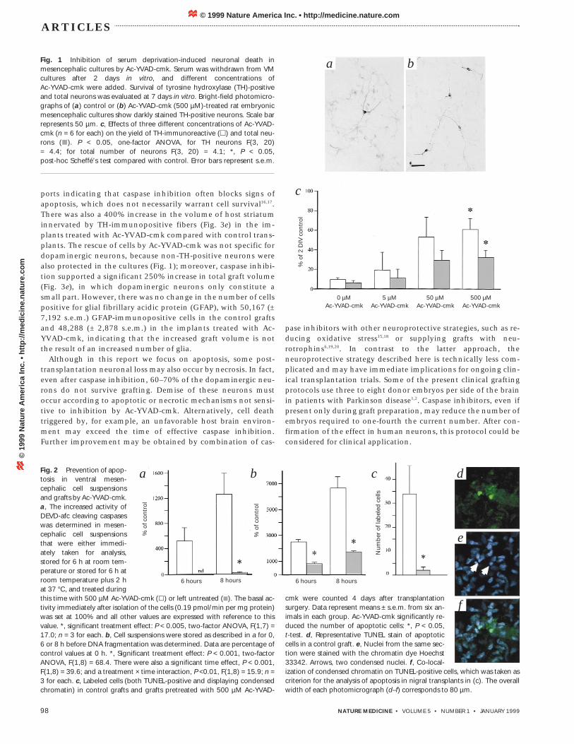
cmk were counted 4 days after transplantationsurgery. Data represent means ± s.e.m. from six an-imals in each group. Ac-YVAD-cmk significantly re-duced the number of apoptotic cells: *, P < 0.05,t-test. d, Representative TUNEL stain of apoptoticcells in a control graft. e, Nuclei from the same sec-tion were stained with the chromatin dye Hoechst33342. Arrows, two condensed nuclei. f, Co-local-ization of condensed chromatin on TUNEL-positive cells, which was taken ascriterion for the analysis of apoptosis in nigral transplants in (c). The overallwidth of each photomicrograph (d–f) corresponds to 80 µm.
98 NATURE MEDICINE • VOLUME 5 • NUMBER 1 • JANUARY 1999
ARTICLES
ports indicating that caspase inhibition often blocks signs ofapoptosis, which does not necessarily warrant cell survival16,17.There was also a 400% increase in the volume of host striatuminnervated by TH-immunopositive fibers (Fig. 3e) in the im-plants treated with Ac-YVAD-cmk compared with control trans-plants. The rescue of cells by Ac-YVAD-cmk was not specific fordopaminergic neurons, because non-TH-positive neurons werealso protected in the cultures (Fig. 1); moreover, caspase inhibi-tion supported a significant 250% increase in total graft volume(Fig. 3e), in which dopaminergic neurons only constitute asmall part. However, there was no change in the number of cellspositive for glial fibrillary acidic protein (GFAP), with 50,167 (±7,192 s.e.m.) GFAP-immunopositive cells in the control graftsand 48,288 (± 2,878 s.e.m.) in the implants treated with Ac-YVAD-cmk, indicating that the increased graft volume is notthe result of an increased number of glia.
Although in this report we focus on apoptosis, some post-transplantation neuronal loss may also occur by necrosis. In fact,even after caspase inhibition, 60–70% of the dopaminergic neu-rons do not survive grafting. Demise of these neurons mustoccur according to apoptotic or necrotic mechanisms not sensi-tive to inhibition by Ac-YVAD-cmk. Alternatively, cell deathtriggered by, for example, an unfavorable host brain environ-ment may exceed the time of effective caspase inhibition.Further improvement may be obtained by combination of cas-
pase inhibitors with other neuroprotective strategies, such as re-ducing oxidative stress15,18 or supplying grafts with neu-rotrophins6,19,20. In contrast to the latter approach, theneuroprotective strategy described here is technically less com-plicated and may have immediate implications for ongoing clin-ical transplantation trials. Some of the present clinical graftingprotocols use three to eight donor embryos per side of the brainin patients with Parkinson disease1,2. Caspase inhibitors, even ifpresent only during graft preparation, may reduce the number ofembryos required to one-fourth the current number. After con-firmation of the effect in human neurons, this protocol could beconsidered for clinical application.
Fig. 1 Inhibition of serum deprivation-induced neuronal death inmesencephalic cultures by Ac-YVAD-cmk. Serum was withdrawn from VMcultures after 2 days in vitro, and different concentrations of Ac-YVAD-cmk were added. Survival of tyrosine hydroxylase (TH)-positiveand total neurons was evaluated at 7 days in vitro. Bright-field photomicro-graphs of (a) control or (b) Ac-YVAD-cmk (500 µM)-treated rat embryonicmesencephalic cultures show darkly stained TH-positive neurons. Scale barrepresents 50 µm. c, Effects of three different concentrations of Ac-YVAD-cmk (n = 6 for each) on the yield of TH-immunoreactive (m) and total neu-rons (L). P < 0.05, one-factor ANOVA, for TH neurons F(3, 20) = 4.4; for total number of neurons F(3, 20) = 4.1; *, P < 0.05, post-hoc Scheffé’s test compared with control. Error bars represent s.e.m.
Fig. 2 Prevention of apop-tosis in ventral mesen-cephalic cell suspensionsand grafts by Ac-YVAD-cmk.a, The increased activity ofDEVD-afc cleaving caspaseswas determined in mesen-cephalic cell suspensionsthat were either immedi-ately taken for analysis,stored for 6 h at room tem-perature or stored for 6 h atroom temperature plus 2 hat 37 °C, and treated duringthis time with 500 µM Ac-YVAD-cmk (l) or left untreated (K). The basal ac-tivity immediately after isolation of the cells (0.19 pmol/min per mg protein)was set at 100% and all other values are expressed with reference to thisvalue. *, significant treatment effect: P < 0.005, two-factor ANOVA, F(1,7) =17.0; n = 3 for each. b, Cell suspensions were stored as described in a for 0,6 or 8 h before DNA fragmentation was determined. Data are percentage ofcontrol values at 0 h. *, Significant treatment effect: P < 0.001, two-factorANOVA, F(1,8) = 68.4. There were also a significant time effect, P < 0.001,F(1,8) = 39.6; and a treatment × time interaction, P <0.01, F(1,8) = 15.9; n =3 for each. c, Labeled cells (both TUNEL-positive and displaying condensedchromatin) in control grafts and grafts pretreated with 500 µM Ac-YVAD-
% o
f 2 D
IV c
ontr
ol0 µM
Ac-YVAD-cmk5 µM
Ac-YVAD-cmk50 µM
Ac-YVAD-cmk500 µM
Ac-YVAD-cmk
f
e
dc
c
a b
a b
6 hours 6 hours8 hours 8 hours
% o
f con
trol
% o
f con
trol
Num
ber
of l
abel
ed c
ells
© 1999 Nature America Inc. • http://medicine.nature.com©
199
9 N
atu
re A
mer
ica
Inc.
• h
ttp
://m
edic
ine.
nat
ure
.co
m

Fig. 3 Reversal of amphetamine-in-duced motor asymmetry by neural graft-ing, and histological evidence forincreased survival of dopaminergic neu-rons in nigral grafts treated with Ac-YVAD-cmk. (a) Net ipsilateral rotationscores for control groups and groupstreated with 500 µM Ac-YVAD-cmk (n = 8for each group). The rats were tested be-fore transplantation (3 weeks after unilat-eral 6-hydroxydopamine lesion), whenthey showed considerable rotationalasymmetry towards the side of the lesion.At 2 and 6 weeks after transplantation,both groups showed reversal of motorasymmetry compared with their pre-transplantation rotation, and this recov-ery was significantly greater in theAc-YVAD-cmk-treated group (treatment effect, P < 0.01, two-factorANOVA, F(1,14) = 15.0; P < 0.05, t-test between groups at 2 and 6 weeks).Error bars represent s.e.m. b and c, Tyrosine hydroxylase (TH) immuno-staining of representative coronal sections through the striatum of a hostbrain receiving a control graft (b) or a graft treated with 500 µM Ac-YVAD-cmk (c). The surviving nigral transplants are located in the center of thestriatum. The graft treated with Ac-YVAD-cmk has increased volume, moreTH-immunopositive dopaminergic neurons and more extensive TH-positiveinnervation of the host striatum. Scale bar represents 1 mm. d and e, mean
NATURE MEDICINE • VOLUME 5 • NUMBER 1 • JANUARY 1999 99
ARTICLES
MethodsMesencephalic cell cultures. We prepared primary neuronal cultures fromSprague-Dawley rat ventral mesencephalon (VM) at embryonic day 14 asdescribed21. For each experiment, cells from 24–30 rat embryos were‘pooled’ and the cells were plated at a density of 100,000 cells per cm2. Intotal, six independent experiments were done. After 2 days in vitro, themedium was changed to serum-free medium21 containing 0, 5, 50 or 500µM Ac-YVAD-cmk (Calbiochem, La Jolla, California) plus solvent (0.02%methanol). After 7 days in vitro, cultures were fixed with 4% paraformalde-hyde and stained with TH-rabbit antiserum (Pel-Freez, Brown Deer,Wisconsin) using 3,3’-diaminobenzidine as chromogen21. Three to six cul-ture wells were scored for each experimental group.
Cell counting and data analysis. Neurons stained positive for TH as wellas the total number of neurons were counted on coded slides (to allowanalysis by experimenters ‘blinded’ to sample identity) using an OlympusC.A.S.T. Grid® system (version 1.10; Olympus, Albertslund, Denmark)composed of an Olympus BX50 microscope and an X-Y-Z step motor stagerun by a computer. The area of the culture well was delineated and acounting frame was randomly placed in the well to mark the first area tobe sampled. The frame was then systematically moved through the well.The total number of neurons and TH-positive neurons per well were ex-trapolated from the data. All results are expressed as a percentage of thesurviving neurons compared to cultures fixed at 2 days in vitro during thesame experiment.
Mesencephalic cell suspensions. Cell suspensions from the VM of ratembryos at embryonic day 14 were prepared as described21. The caspase in-hibitor Ac-YVAD-cmk (500 µM) was added at the last step of the tissue dis-section (that is, the enzymatic and mechanical dissociation of the cells) andremained until the experiment ended. Some of these cell suspensions werecultured as described above in the presence of serum but were fixed at 30hours, whereas others were analyzed for caspase activity and DNA fragmentation.
Caspase activity and DNA fragmentation. VM suspensions with or with-out 500 µM Ac-YVAD-cmk were prepared as described above. Initial cellnumber and viability (trypan blue dye exclusion; >98% viable cells) were as-sessed, and 1 x 10⁵ cells were either incubated in 1 ml at room temperatureeither for 6 h or 8 h (6 h at room temperature, followed by 2 h at 37 °C) or
immediately processed as described in the next step. Then, cells werewashed with cold Hank’s balanced salt solution (HBSS; Life Technologies)supplemented with a proteinase inhibitor ‘cocktail’ (Complete , Mini;Boehringer), pelleted and immediately frozen in liquid nitrogen. Three dif-ferent cell suspensions were prepared for each experimental group. DEVD-afc cleavage, which is specific for caspase-3 related protease activity7, wasdetermined fluorimetrically in neuronal cell homogenates exactly as de-scribed9. DNA-fragmentation was detected using a commercially availablekit (Cell Death Detection ELISA; Boehringer). Oligonucleosomal fragments,which are specifically detected in apoptotic cells13, were analyzed accordingto the supplier’s instructions with every sample containing the DNA equiva-lent from about 10,000 neurons. The data are expressed as percent of con-trol values obtained from cell suspensions analyzed immediately aftermechanical tissue dissociation.
Apoptosis detection in neural grafts. Two groups of normal, non-lesionedrats (n = 6 for each) received intrastriatal transplants with or without the ad-dition of 500 µM Ac-YVAD-cmk. Four days after transplantation surgery, theanimals were perfused transcardially with 4% paraformaldehyde, and coro-nal cryostat sections 15 µm in thickness were cut, mounted and post-fixedin 4% paraformaldehyde for 15 min. Five serial sections containing the cen-tral graft region were chosen for each rat by two experienced independentobservers ‘blinded’ to sample identity. Apoptotic cells were detected in thetransplants by a combination of two methods as described22: TUNEL stain-ing using a commercially available kit (Apoptag; Oncor, Gaithersburg,Maryland); and fluorescent chromatin staining and morphological assess-ment of nuclear changes (Hoechst 33342; Molecular Probes, Eugene,Oregon). Cells that were both TUNEL-positive and had condensed chro-matin (Fig. 2 d–f) were scored manually using a fluorescence microscope(Olympus BX60) by an experimenter ‘blinded’ to sample identity.
Unilateral 6-hydroxydopamine (6-OHDA) lesion and motor asymmetrytest. Female Sprague-Dawley rats were subjected to unilateral 6-OHDA(Sigma) lesions of the ascending mesostriatal dopaminergic pathway asdescribed21. The efficiency of the 6-OHDA lesion was assessed, and ratswith a net rotational amphetamine-induced asymmetry of at least six fullturns per minute were selected for transplantation surgery. For statisticalanalysis, data for each rat at 2 and 6 weeks were expressed as a percent ofthe pre-transplantation score.
a b c
d e
Pre 2 weeks 6 weeks
Time after transplantation
Net
ipsi
late
ral t
urns
pre
min
Num
ber
of n
euro
ns (
×10
3 )
TH-positive neurons Graft size Fiber outgrowth
Vol
ume
(mm
3 )
number of TH-positive neurons, graft volume and re-innervated area are in-dicated for control transplants and transplants treated with 500 µM Ac-YVAD-cmk. *, P < 0.05, t-test. Error bars represent s.e.m.
© 1999 Nature America Inc. • http://medicine.nature.com©
199
9 N
atu
re A
mer
ica
Inc.
• h
ttp
://m
edic
ine.
nat
ure
.co
m
100 NATURE MEDICINE • VOLUME 5 • NUMBER 1 • JANUARY 1999
ARTICLES
Transplantation surgery. Two groups of rats with 6-OHDA lesions (n = 8per group) were transplanted with mesencephalic cell suspensions treatedwith Ac-YVAD-cmk or not treated. Two deposits of 1.5 µl cell suspension(approximately 250,000 cells, equivalent to 50% of one VM) were injectedstereotaxically into the right (lesioned) striatum of rats sedated by equi-thesin anesthesia, at the following coordinates (with reference to bregma):anterior, + 1.0 mm; lateral, 3.0 mm; ventral, 5.0 mm; and 4.5 mm ventralto the dura, with the tooth-bar set at zero.
Morphological assessment. Six weeks after graft surgery, rats were anes-thetized and perfused, and their brains were processed for TH- and GFAP-immunocytochemistry as described21,23. The numbers of TH- andGFAP-immunoreactive cells in each graft were counted in serial sections oncoded slides (to allow analysis by experimenters ‘blinded’ to sample iden-tity), using the same semi-automated stereological cell counting systemdescribed above. The sampling frequency was chosen so that about 200immunopositive cells of each type were counted in each rat. The numbersof TH- and GFAP-immunopositive cells in the graft were then extrapolatedaccording to a stereological algorithm24. Overall survival rates of dopamin-ergic neurons were calculated as described15. For the determination of in-dividual graft volumes and fiber outgrowth of grafted TH-immunopositiveneurons into the host striatum, stained brain sections were ‘digitized’using a high-resolution CCD camera and were analyzed as described18.
Statistical analysis. The data were analyzed by two- or one-factor analysisof variance (ANOVA) followed by Scheffé’s post-hoc analysis (using theStatview 4.5 package; Abacus Concepts, Berkeley, California), the Mann-Whitney U test or by the unpaired, two-tailed Student’s t-test.
AcknowledgmentsWe acknowledge the technical assistance of T. Björklund, B. Haraldsson, H.Naumann, B. Lindberg and M. Pettersson. This study was supported by grantsfrom the Medical Faculty at Lund University, the Swedish Medical ResearchCouncil, the Thorsten and Elsa Segerfalk Foundation, and DeutscheForschungsgemeinschaft. G.S.S. was supported by a Marie Curie Fellowshipwithin the 4th framework program of the European Commission.
RECEIVED 16 AUGUST; ACCEPTED 23 NOVEMBER 1998
1. Lindvall, O. Neural transplantation: a hope for patients with Parkinson’s dis-ease. Neuroreport 8, III–X (1997).
2. Olanow, C.W., Kordower, J.H. & Freeman, T.B. Fetal nigral transplantation asa therapy for Parkinson’s disease. Trends Neurosci. 19, 102–109 (1996).
3. Kordower, J.H. et al. Functional fetal nigral grafts in a patient with Parkinson’sdisease: chemoanatomic, ultrastructural, and metabolic studies. J. Comp.Neurol. 24, 203–230 (1996).
4. Kordower J.H. et al. Fetal nigral grafts survive and mediate clinical benefit in apatient with Parkinson’s disease. Mov. Disord. 13, 383–393 (1998).
5. Barker, R.A., Dunnett, S.B., Faissner, A. & Fawcett, J.W. The time course of lossof dopaminergic neurons and the gliotic reaction surrounding grafts of embry-onic mesencephalon to the striatum. Exp. Neurol. 141, 79–93 (1996).
6. Zawada, W.M. et al. Growth factors improve immediate survival of embryonicdopamine neurons after transplantation into rats. Brain Res. 786, 96–103(1998).
7. Villa, P., Kaufmann, S.H. & Earnshaw, W.C. Caspases and caspase inhibitors.Trends Biochem. Sci. 22, 388–393 (1997).
8. Milligan, C.E. et al. Peptide inhibitors of the ICE protease family arrest pro-grammed cell death of motoneurons in vivo and in vitro. Neuron 15, 385–393(1995).
9. Leist, M. et al. Caspase-mediated apoptosis in neuronal excitotoxicity trig-gered by nitric oxide. Mol. Med. 3, 750–764 (1997).
10. de Bilbao, F. & Dubois-Dauphin, M. Acute application of an interleukin-1 beta-converting enzyme-specific inhibitor delays axotomy-induced motoneuronedeath. Neuroreport 25, 3051–3054 (1996).
11. Hara, H. et al. Inhibition of interleukin 1beta converting enzyme family pro-teases reduces ischemic and excitotoxic neuronal damage. Proc. Natl. Acad.Sci. USA 94, 2007–2012 (1997).
12. Yakovlev, A.G. et al. Activation of CPP32-like caspases contributes to neuronalapoptosis and neurological dysfunction after traumatic brain injury. J.Neurosci. 17, 7415–7424 (1997).
13. Leist, M., Single, B., Castoldi, A.F., Kuhnle, S. & Nicotera, P. Intracellularadenosine triphosphate (ATP) concentration: a switch in the decision betweenapoptosis and necrosis. J. Exp. Med. 185, 1481–1486 (1997).
14. Mahalik, T.J., Hahn, W.E., Clayton, G.H. & Owens, G.P. Programmed celldeath in developing grafts of fetal substantia nigra. Exp. Neurol. 129, 27–36(1994).
15. Nakao, N., Frodl, E.M., Duan, W.-M., Widner, H. & Brundin, P. Lazaroids im-prove the survival of grafted rat embryonic dopamine neurons. Proc. Natl.Acad. Sci. USA 91, 12408–12412 (1994).
16. Green, D. & Kroemer, G. The central executioners of apoptosis: caspases ormitochondria? Trends Cell Biol. 8, 267–271 (1998).
17. Leist, M. & Nicotera, P. Apoptosis, excitotoxicity, and neuropathology. Exp.Cell Res. 239, 183–201 (1998).
18. Nakao, N. et al. Overexpressing Cu/Zn superoxide dismutase enhances sur-vival of transplanted neurons in a rat model of Parkinson’s disease. NatureMed. 1, 226–231 (1995).
19. Rosenblad, C., Martinez-Serrano, A. & Björklund, A. Glial cell line-derived neu-rotrophic factor increases survival, growth and function of intrastriatal fetal ni-gral dopaminergic grafts. Neuroscience 75, 979–985 (1996).
20. Mayer, E., Fawcett, J.W. & Dunnett, S.B. Basic fibroblast growth factor pro-motes the survival of embryonic ventral mesencephalic dopaminergic neu-rons—II. Effects on nigral transplants in vivo. Neuroscience 56, 389–98 (1993).
21. Karlsson, J., Emgård, M., Rosenblad, C. & Brundin, P. Treatment with the spin-trap agent α-phenyl-N-tert-butyl nitrone does not enhance the survival of em-bryonic or adult dopamine neurons. Brain Res. 805, 155–168 (1998).
22. Whiteside, G., Cougnon, N., Hunt, S.P. & Munglani, R. An improved methodfor detection of apoptosis in tissue sections and cell culture, using the TUNELtechnique combined with Hoechst stain. Brain Res. Brain Res. Protoc. 2,160–164 (1998).
23. Duan, W.-M., Widner, H., Cameron, R.M. & Brundin, P. Quinolinic acid-in-duced inflammation in the striatum does not impair the survival of neural allo-grafts in the rat. Eur. J. Neurosci . 10, 2595–2606 (1998).
24. West, M.J., Slomianka, L. & Gundersen, H.J. Unbiased stereological estimationof the total number of neurons in the subdivisions of the rat hippocampususing the optical fractionator. Anat. Rec. 231, 482–497 (1991).
© 1999 Nature America Inc. • http://medicine.nature.com©
199
9 N
atu
re A
mer
ica
Inc.
• h
ttp
://m
edic
ine.
nat
ure
.co
m