88 peritoneal and omental abnormalities
TRANSCRIPT

88 Peritoneal and Omental Abnormalities
CLINICAL IMAGAGINGAN ATLAS OF DIFFERENTIAL DAIGNOSIS
EISENBERG
DR. Muhammad Bin Zulfiqar PGR-FCPS III SIMS/SHL

• Fig GI 88-1 Peritoneal carcinomatosis. Hematogenous dissemination of malignant melanoma causes multiple nodules in the peritoneal space, including the omentum (arrows), retroperitoneal spaces, and the subcutaneous fat layer of the abdomen.193

• Fig GI 88-2 Tuberculous peritonitis. Large amount of ascites with even peritoneal thickening (arrowhead) and diffuse omental infiltration (arrow) without associated lymphadenopathy. The initial clinical impression was carcinomatosis.193

Fig GI 88-3 Malignant peritoneal mesothelioma. Diffuse platelike mass in the greater omentum (arrows), massive ascites, and peritoneal thickening.193
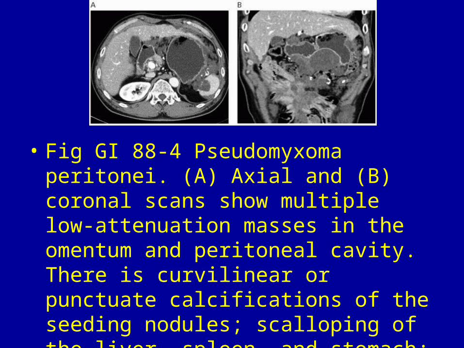
• Fig GI 88-4 Pseudomyxoma peritonei. (A) Axial and (B) coronal scans show multiple low-attenuation masses in the omentum and peritoneal cavity. There is curvilinear or punctuate calcifications of the seeding nodules; scalloping of the liver, spleen, and stomach; and small bowel adhesions from mesenteric infiltration.193

• Fig GI 88-5 Lymphomatosis. Innumerable seeding nodules in the peritoneal cavity and omentum (white arrow) with evidence of ascites. Multiple enlarged lymph nodes with conglomeration (black arrows) are seen in the retroperitoneal spaces.193

Fig GI 88-7 Sarcoidosis. Diffuse soft-tissue thickening involving the mesentery, omentum, and parietal peritoneum.194

• Fig GI 88-8 GIST. Large, heterogenous, omentum-based mass.195

Fig GI 88-9 Malignant fibrous histiocytoma. Large, heterogeneous soft-tissue peritoneal mass.195
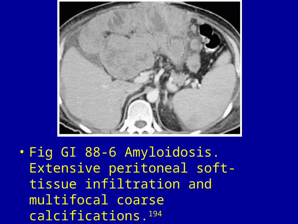
• Fig GI 88-6 Amyloidosis. Extensive peritoneal soft-tissue infiltration and multifocal coarse calcifications.194

• Fig GI 88-10 Primary serous papillary carcinoma. (A) Omental cake in the left lower quadrant displaces adjacent loops of large and small bowel. (B) In this lower image, there is lace-like omental infiltration (large arrow) with irregular nodular thickening of the peritoneum (small arrow). The mesenteric fat is normal and there is no lymphadenopathy or ascites.196

• Fig GI 88-11 Metastasis. Large lobulated mass (arrows) in the left upper quadrant of the abdomen representing an exophytic carcinoma extending directly from the greater curvature of the stomach.193

• Fig GI 88-12 Metastasis. Large, lobulated, heterogeneous mass in the mid-abdomen, inferior to the stomach. The thickened peritoneum (arrow) adjacent to the mass is suggestive of a malignant lesion, which in this case was an ovarian carcinoma.193

Fig GI 88-13 Lymphangioma. Coronal contrast scan shows a lobulated cystic mass in the greater omentum inferior to the gastric antrum.193

Fig GI 88-14 Paragonimiasis. Multifocal ill-defined cystic lesions and several nodules (arrow) in the omentum on the right side of the abdomen.193

• Fig GI 88-15 Omental infarction. Localized fatty infiltration and congestion with a secondary mass (arrow) in the right lower aspect of the anterior abdomen.193

• Fig GI 88-16 Foreign-body granuloma. Large, well-circumscribed mass with dense calcification in the anterior mid-abdomen, an appearance suggestive of a foreign-body granuloma or organizing hematoma. After contrast injection, the mass showed no enhancement. The patient had a palpable mass for 10 years that developed soon after a Caesarian section.193

• Fig GI 88-17 Ventral hernia. Sagittal scan shows herniation of omental fat through a defect (arrow) in the anterior abdominal wall. Focal ill-defined lesions with increased attenuation (arrowheads) in the omental fat adjacent to the abdominal wall defect are suggestive of omental fat infarction secondary to vascular compromise.193
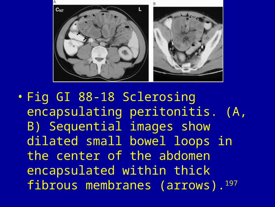
• Fig GI 88-18 Sclerosing encapsulating peritonitis. (A, B) Sequential images show dilated small bowel loops in the center of the abdomen encapsulated within thick fibrous membranes (arrows).197

• Fig GI 88-19 Inflammatory pseudotumor. The huge mass suggests a peritoneal malignancy.194