9/18/2012 chapter 11ems.jbpub.com/henry/emt/docs/ppt_lectures/chapter_011.pdf · 9/18/2012 3 7...
TRANSCRIPT

9/18/2012
1
Chapter 11
Respiratory Emergencies
2
Learning Objectives
List structures & functions of the respiratory system
State signs/symptoms of patient with breathing difficulty
Describe emergency medical care of patient with breathing difficulty
Describe emergency medical care of patient with respiratory distress
3
Learning Objectives
Establish relationship between airway management & patient with breathing difficulty
List signs of adequate air exchange
Recognize need for medical direction to assist in emergency medical care of patient with breathing difficulty
Copyright © 2013 by Jones & Bartlett Learning, LLC, an Ascend Learning Company

9/18/2012
2
4
Learning Objectives
State generic name, medication forms, dose, administration, action, indications, contraindications for prescribed inhaler
Defend treatment regimens for various respiratory emergencies
Explain rationale for administering an inhaler
5
Learning Objectives
Distinguish among emergency medical care of infant, child, adult patient with breathing difficulty
Differentiate between upper airway obstruction, lower airway disease in infant, child patient
6
Introduction
Respiratory emergencies Life-threatening situations
Effective management• Maintain open airway
• Ensure adequate ventilation
• Oxygenation
Asthma• You will learn to assist with administration of self -
administered metered dose inhaler (MDI)
Copyright © 2013 by Jones & Bartlett Learning, LLC, an Ascend Learning Company

9/18/2012
3
7
Anatomy & Physiology
Provides basis for understanding assessment, treatment of illness/injury Respiratory system delivers O2 to blood for body
tissues
8
Anatomy & Physiology
9
Anatomy & Physiology
Airway Begins at nose/mouth as air enters/passes
through nasopharynx/oropharynx
Air continues through upper airways, past epiglottis
Down trachea, into 2 main bronchi, that direct air into right & left lungs
Copyright © 2013 by Jones & Bartlett Learning, LLC, an Ascend Learning Company
9/18/2012
4
10
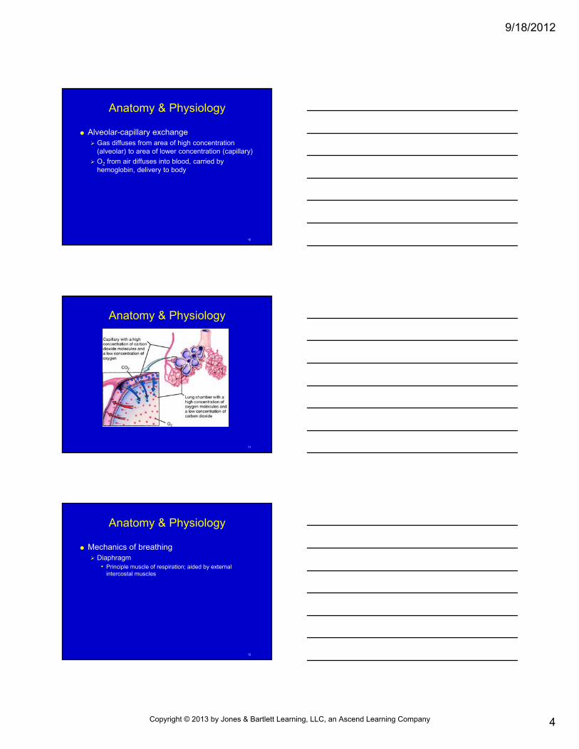
Anatomy & Physiology
Alveolar-capillary exchange Gas diffuses from area of high concentration
(alveolar) to area of lower concentration (capillary)
O2 from air diffuses into blood, carried by hemoglobin, delivery to body
11
Anatomy & Physiology
12
Anatomy & Physiology
Mechanics of breathing Diaphragm
• Principle muscle of respiration; aided by external intercostal muscles
Copyright © 2013 by Jones & Bartlett Learning, LLC, an Ascend Learning Company

9/18/2012
5
13
Anatomy & Physiology
Central nervous system controls Normal breathing is voluntary
Affected by CO2 & O2 levels in blood; adjusts rate/depth of breathing
Tidal volume
Minute volume
Monitor chest rise/fall, and rate of breathing
14
Pathophysiology
Airway can be obstructed at multiple points: In unconscious patients, tongue may fall back,
block oropharynx, producing snoring sound
Epiglottis swollen from infection
15
Pathophysiology
Airway can be obstructed at multiple points: Inadequate breathing
• Too shallow or respiratory rate is to slow
Average adult: 12 breaths/min × 500 mL/breath = 6000 mL/min
Inadequate oxygenation• Fluid, pus, other material present at alveoli level
• Fluid collecting in alveoli or between alveoli and capillaries impairs diffusion
• Increase oxygenation; if inadequate ventilation, give positive-pressure ventilation
Copyright © 2013 by Jones & Bartlett Learning, LLC, an Ascend Learning Company

9/18/2012
6
16
Anatomic Considerations—Infants & Children
Airway differs from adult Internal airway diameter smaller at all levels
Tongue large in relation to airway
Narrowest part of airway - cricoid ring
17
Anatomic Considerations—Infants & Children
Airway differs from adult Larynx/trachea cartilage softer
Chest wall softer
Head positioning to open airway:• Infants: place head in sniffing position
• Toddlers/small children: extend neck slightly
Small obstructions, swelling, mucus - possible airflow blockage
Infants – obligate nose breathers
18
Assessing Patient with Difficulty Breathing
Scene size-up Make sure scene is safe
If trauma is contributing factor, consider MOI
Be alert for:• Toxic environment
• Poisonous gases
• Hazmat
Use BSI
Copyright © 2013 by Jones & Bartlett Learning, LLC, an Ascend Learning Company

9/18/2012
7
19
Assessing Patient with Difficulty Breathing
Initial (primary) assessment On approach, what is your general impression
• Is there obvious threat to life?
• Is patient unconscious?
• What position is patient found in?
• Does patient speak in complete sentences, or struggling to catch their breath and speaking in short sentences?
20
Assessing Patient with Difficulty Breathing
Initial (primary) assessment Patient position
• 1st clue to seriousness of problem
• Sitting bolt upright for unrestricted expansion of diaphragm/chest wall
• Tripod position
• Transport in position of comfort
21
Assessing Patient with Difficulty Breathing
Copyright © 2013 by Jones & Bartlett Learning, LLC, an Ascend Learning Company

9/18/2012
8
22
Assessing Patient with Difficulty Breathing
Initial (primary) assessment Mental status
• Assess according to Alert, verbal, painful, unresponsive (AVPU)
• Alterations observed as result of hypoxia:
Restlessness
Agitation/anxiety
Lethargy/sleepiness
Complete unresponsiveness
• Administer high-concentration supplemental O2
• Observe adequacy of breathing/ventilation
23
Assessing Patient with Difficulty Breathing
Initial (primary) assessment Airway
• Inability to speak - sign of severe airway obstruction
If foreign body airway obstruction (FBAO) suspected - initiate basic life support (BLS) maneuvers
If another cause suspected - initiate rapid transport/positive pressure ventilatin (PPV)
Use age-appropriate techniques/equipment
If suspected neck/head trauma – c-spine precautions
Rapid transport if obstruction not relieved with basic maneuvers
Consider advanced life support (ALS) intercept
24
Assessing Patient with Difficulty Breathing
Initial (primary) assessment Breathing
• Determine if positive-pressure ventilation needed
Slow, irregular breathing
Shallow breathing
Lower breath sounds
Seesaw breathing
Lower level of consciousness (LOC)
Severe hypoxia signs
• Rely on assessment
Copyright © 2013 by Jones & Bartlett Learning, LLC, an Ascend Learning Company

9/18/2012
9
25
Assessing Patient with Difficulty Breathing
Initial (primary) assessment Breathing
• Rate & depth of respiration
Increased rate/depth of breathing during respiratory distress
• Observe for:
Accessory muscle use in neck, between ribs, or below rib cage
Excessive movement of abdomen
26
Assessing Patient with Difficulty Breathing
27
Assessing Patient with Difficulty Breathing
Initial primary assessment Assisted ventilation
• Some respiratory efforts but inadequate ventilation
• If inadequate ventilation but respiratory rate more than12/min
• If slow, shallow breathing
• Administer supplemental 02 whenever assisted ventilation is required
Copyright © 2013 by Jones & Bartlett Learning, LLC, an Ascend Learning Company

9/18/2012
10
28
Assessing Patient with Difficulty Breathing
Initial (primary) assessment Circulation
• During respiratory distress/other serious problems, HR increases/decreases as hypoxia progresses
Early: increased HR
• Severe respiratory distress
Cool, pale, sweaty skin
Cyanosis
Consider need for rapid transport/ALS intercept
29
Assessing Patient with Difficulty Breathing
Focused (secondary) assessment Obtain SAMPLE history
Ask OPQRST questions
30
Assessing Patient with Difficulty Breathing
Focused (secondary) assessment Physical examination
• If responsive patient with no trauma
• Reassess mental status/skin condition
• Check head, neck, and chest
• Check for JVD
• Check for crepitation
Copyright © 2013 by Jones & Bartlett Learning, LLC, an Ascend Learning Company

9/18/2012
11
31
Assessing Patient with Difficulty Breathing
Focused (secondary) assessment Baseline vital signs
• Record, paying particular attention to respiratory rate/pulse
• Reevaluate need for positive-pressure ventilation
32
Emergency Medical Care
Shortness of breath High-concentration supplemental O2
If inadequate breathing – assist with PPV
Priority patient for early transport• Seated position/position of comfort
Reduce unnecessary physical exertion
33
Emergency Medical Care
Prescribed inhalers Understand general pharmacologic principles
Medication name• Identify name of prescribed inhaler, communicate
information to medical direction, or compare with standing orders
Copyright © 2013 by Jones & Bartlett Learning, LLC, an Ascend Learning Company
9/18/2012
12
34
Emergency Medical Care
Prescribed inhalers Actions & side effects
• Dilate bronchioles
• Contain B-agonists
• Inhaled drug administered directly to bronchioles focuses effects on respiratory tree; minimizes other side effects
35
Emergency Medical Care
Prescribed inhalers Indications
• Signs/symptoms of respiratory distress
• Handheld MDI prescribed by physician
• Specifically authorized by medical direction
36
Emergency Medical Care
Prescribed inhalers Contraindications
• Unable to use device
• AMS
• Inadequate ventilation
• Unable to inhale medication
• Taken maximum prescribed dose before arrival
Copyright © 2013 by Jones & Bartlett Learning, LLC, an Ascend Learning Company

9/18/2012
13
37
Emergency Medical Care
Prescribed inhalers Medication form
Dosage
Administration
38
Administering a Prescribed Inhaler
Obtain order
Check expiration date & doses taken
Shake inhaler vigorously
Administer inhaler
Replace O2
Repeat per medical direction
39
Skill 11-1Medication Administration via Nebulizer
Check for allergies; obtain order from medical direction
Check medication 3 times for: Correct medication
Correct dose
Correct patient
Expiration date
Loss of clarity, particulate matter
Copyright © 2013 by Jones & Bartlett Learning, LLC, an Ascend Learning Company

9/18/2012
14
40
Skill 11-1Medication Administration via Nebulizer
Pour contents of unit dose into nebulizer chamber
Screw top back on nebulizer
41
Skill 11-1Medication Administration via Nebulizer
Remove O2 delivery device from patient
In adult, attach nebulizer O2 tubing to regulator
42
Skill 11-1Medication Administration via Nebulizer
In young child, hold mouthpiece at opening of patient’s mouth
Monitor patient & medication
Copyright © 2013 by Jones & Bartlett Learning, LLC, an Ascend Learning Company

9/18/2012
15
43
Skill 11-1Medication Administration via Nebulizer
When complete, reattach O2 device
Reevaluate patient
44
Emergency Medical Care
Ongoing assessment/reassessment Assess vital signs, repeat secondary (focused)
assessment
Document time medication administered, findings from reassessment on prehospital report
45
Emergency Medical Care
Infants & children Asthma - common condition in children
In very young children - inflammation/constriction of bronchioles
Inhaler therapy in children similar to adults
Copyright © 2013 by Jones & Bartlett Learning, LLC, an Ascend Learning Company

9/18/2012
16
46
Emergency Medical Care
47
Emergency Medical Care
Infants & children Distinguish between lower/upper respiratory
disease
Rib cage softer, more moveable in children & retractions may be more evident
Cyanosis considered danger sign; condition can deteriorate rapidly
48
Conditions that Cause Respiratory Emergencies
Respiratory emergencies may have new illness or complications of chronic respiratory condition: Asthma
Emphysema
Chronic bronchitis
Heart failure
Croup
Epiglottis
Pneumonia
Pneumothorax
Hyperventilation syndrome
Copyright © 2013 by Jones & Bartlett Learning, LLC, an Ascend Learning Company

9/18/2012
17
49
Conditions that Cause Respiratory Emergencies
Chronic Obstructive Pulmonary Disease (COPD) Includes chronic bronchitis/emphysema
Primary complaint - shortness of breath
Bronchoconstriction
50
Conditions that Cause Respiratory Emergencies
COPD Chronic bronchitis
• Chronic productive cough present more than 3 mo/yr for more than 2 yrs
51
Conditions that Cause Respiratory Emergencies
Copyright © 2013 by Jones & Bartlett Learning, LLC, an Ascend Learning Company

9/18/2012
18
52
Conditions that Cause Respiratory Emergencies
COPD Emphysema
• Disease caused by destruction of alveoli
• Less lung surface in which O2 can diffuse into blood
• Muscle portion of bronchioles within lung damaged
53
Conditions that Cause Respiratory Emergencies
54
Conditions that Cause Respiratory Emergencies
Copyright © 2013 by Jones & Bartlett Learning, LLC, an Ascend Learning Company

9/18/2012
19
55
Conditions that Cause Respiratory Emergencies
COPD Respiratory failure
• Respiratory system becomes ineffective
Subject to infections that aggravate patient condition
56
Conditions that Cause Respiratory Emergencies
Asthma Obstructive respiratory disease
Caused by constriction of lower airways
Triggered by stress, infection, or allergy
Treatment
57
Conditions that Cause Respiratory Emergencies
Asthma Acute asthma attack
• Shortness of breath
• Patient assumes upright posture
• Uses accessory muscles to increase ventilation
• Patient - flushed and breathes forcefully
• Wheezing & prolonged expirations audible without stethoscope
Copyright © 2013 by Jones & Bartlett Learning, LLC, an Ascend Learning Company

9/18/2012
20
58
Conditions that Cause Respiratory Emergencies
Asthma Severe asthma attack
• Patient becomes exhausted & produces little airflow
• No wheezing
• Difficulty speaking
• ↓Breath sounds
Assist ventilations during rapid transport
59
Conditions that Cause Respiratory Emergencies
Pneumonia Inflammation of alveolar spaces caused by various
infecting organisms/aspiration of gastric contents into tracheobronchial tree
Signs/symptoms• Fever
• Cough with productive sputum
• Difficulty breathing
• Chills
• Headache
• Pain that increases with breathing/coughing
60
Conditions that Cause Respiratory Emergencies
Pulmonary embolism Blood clots released from leg veins after surgery/
patients taking birth control
Fat emboli released from long-bone fractures, as clot releases, travels up through vena cava, into right atrium, right ventricle, into pulmonary artery
Copyright © 2013 by Jones & Bartlett Learning, LLC, an Ascend Learning Company

9/18/2012
21
61
Conditions that Cause Respiratory Emergencies
Pulmonary embolism Signs
• Difficulty breathing
• Chest pain that increases with breathing
• Coughing up bloody sputum
• Calf tenderness
• Hypoxia, including cyanosis
• AMS
• Recent history of surgery, prolonged bed rest, recent travel, use of oral contraceptives, phlebitis
62
Conditions that Cause Respiratory Emergencies
Pulmonary embolism Physical findings
• Often normal
• Rapid pulse
• Shock
• Signs of right-sided heart failure
Treatment• High-concentration O2
• Treat for shock
63
Conditions that Cause Respiratory Emergencies
Hyperventilation syndrome Feel as if cannot breathe, may begin to voluntarily
increase rate and depth of breathing• Often accompanied by anxiety
Increased minute ventilation decreases amount of CO2 in blood; changes acidity of blood
Chief complaint – shortness of breath
Treatment• Supplemental O2
• Calm reassurance
Copyright © 2013 by Jones & Bartlett Learning, LLC, an Ascend Learning Company

9/18/2012
22
64
Conditions that Cause Respiratory Emergencies
Spontaneous pneumothorax Part of lung ruptures
• Allows air to exit lung, enter space between pleural lining of chest cavity and outer covering of lung
65
Conditions that Cause Respiratory Emergencies
Spontaneous pneumothorax Monitor for progression of simple pneumothorax to
tension pneumothorax• Absence of breath sounds on one side
• Distended neck veins
• Hypotension
• Tracheal deviation
66
Conditions that Cause Respiratory Emergencies
Croup & epiglottitis Upper respiratory problem - primarily in children
• Croup
Viral infection– Causes swelling/narrowing of upper airway
• Epiglottitis
Bacterial infection– Causes swelling of epiglottis
Copyright © 2013 by Jones & Bartlett Learning, LLC, an Ascend Learning Company

9/18/2012
23
67
Conditions that Cause Respiratory Emergencies
Croup & epiglottitis Signs
• Fever
• Dyspnea
• Coughing
Treatment• Administer humidified O2
• Position of comfort
• Positive-pressure ventilation
• Stridor/crowing
• Increased work of breathing
• Tripod position
68
Conditions that Cause Respiratory Emergencies
Pertussis (“whooping cough”) Highly contagious bacterial infection
Transmitted by direct contact with mucous• Symptoms
High-pitched, whooping cough
Fever
Signs of hypoxia
• Treatment
Supplemental O2 via nonrebreather mask
69
Summary
Patients with respiratory emergencies typically have difficulty breathing, inadequate breathing, or respiratory arrest
Primary management includes: Airway management
Positive-pressure ventilation
Administration of supplemental O2
Positioning
Assisting patients in administration of prescribed inhalers
Copyright © 2013 by Jones & Bartlett Learning, LLC, an Ascend Learning Company
9/18/2012
24
70
Summary
Signs and symptoms of difficulty breathing include: Dyspnea
Restlessness
Increased HR
Increased or decreased RR
Shallow or irregular breathing
Abdominal breathing
Noisy breathing
Crowing/stridor
Audible wheezing
Gurgling
Snoring
Inability to speak
Pale/cyanotic skin
Coughing
Tripod position
71
Summary
Signs of increased work of breathing include accessory muscle use, retractions, nasal flaring
Patients with dyspnea often sit bolt upright, supported by their hands in tripod position
72
Summary
Management of airway may include: Clearing obstruction of upper airway
Suctioning
Manual maneuvers to open airway
Adjuncts to maintain patent airway
Patients with respiratory distress should receive supplemental O2
Copyright © 2013 by Jones & Bartlett Learning, LLC, an Ascend Learning Company
9/18/2012
25
73
Summary
Patient with signs of inadequate breathing should receive high-concentration supplemental O2, positive-pressure ventilation when needed
Patients with asthma/COPD may carry MDIs. You may have to assist these patients in administering medication.
74
Questions?
Copyright © 2013 by Jones & Bartlett Learning, LLC, an Ascend Learning Company