a biphasic and brain-region selective down-regulation of
TRANSCRIPT

A Biphasic and Brain-Region Selective Down-Regulationof Cyclic Adenosine Monophosphate ConcentrationsSupports Object Recognition in the RatMaıte Hotte1,2, Francois Dauphin1, Thomas Freret1, Michel Boulouard1, Guenaelle Levallet1,3*
1 Universite de Caen Basse–Normandie, Groupe Memoire et Plasticite comportementale (GMPc), EA4259, IFR 146, Caen, France, 2 Universite de Rouen, NeoVasc, EA 4309,
IFRMP23, IHURBM, Rouen, France, 3 CHU de Caen, Service d’Anatomie Pathologie, Caen, France
Abstract
Background: We aimed to further understand the relationship between cAMP concentration and mnesic performance.
Methods and Findings: Rats were injected with milrinone (PDE3 inhibitor, 0.3 mg/kg, i.p.), rolipram (PDE4 inhibitor, 0.3 mg/kg, i.p.) and/or the selective 5-HT4R agonist RS 67333 (1 mg/kg, i.p.) before testing in the object recognition paradigm.Cyclic AMP concentrations were measured in brain structures linked to episodic-like memory (i.e. hippocampus, prefrontaland perirhinal cortices) before or after either the sample or the testing phase. Except in the hippocampus of rolipramtreated-rats, all treatment increased cAMP levels in each brain sub-region studied before the sample phase. After the samplephase, cAMP levels were significantly increased in hippocampus (1.8 fold), prefrontal (1.3 fold) and perirhinal (1.3 fold)cortices from controls rat while decreased in prefrontal cortex (,0.83 to 0.62 fold) from drug-treated rats (except formilrinone+RS 67333 treatment). After the testing phase, cAMP concentrations were still increased in both the hippocampus(2.76 fold) and the perirhinal cortex (2.1 fold) from controls animals. Minor increase were reported in hippocampus andperirhinal cortex from both rolipram (respectively, 1.44 fold and 1.70 fold) and milrinone (respectively 1.46 fold and 1.56fold)-treated rat. Following the paradigm, cAMP levels were significantly lower in the hippocampus, prefrontal andperirhinal cortices from drug-treated rat when compared to controls animals, however, only drug-treated rats spent longertime exploring the novel object during the testing phase (inter-phase interval of 4 h).
Conclusions: Our results strongly suggest that a ‘‘pre-sample’’ early increase in cAMP levels followed by a specific loweringof cAMP concentrations in each brain sub-region linked to the object recognition paradigm support learning efficacy after amiddle-term delay.
Citation: Hotte M, Dauphin F, Freret T, Boulouard M, Levallet G (2012) A Biphasic and Brain-Region Selective Down-Regulation of Cyclic AdenosineMonophosphate Concentrations Supports Object Recognition in the Rat. PLoS ONE 7(2): e32244. doi:10.1371/journal.pone.0032244
Editor: Nicoletta Landsberger, University of Insubria, Italy
Received September 2, 2011; Accepted January 23, 2012; Published February 16, 2012
Copyright: � 2012 Hotte et al. This is an open-access article distributed under the terms of the Creative Commons Attribution License, which permitsunrestricted use, distribution, and reproduction in any medium, provided the original author and source are credited.
Funding: The authors have no support or funding to report.
Competing Interests: The authors have declared that no competing interests exist.
* E-mail: [email protected]
Introduction
Most modern theories of learning and memory postulate that
memory processes require cyclic adenosine monophosphate
(cAMP) synthesis [1]; however, there is little evidence concerning
the mechanisms by which memory affects adenylyl cyclase activity
(cAMP synthesis) and/or phosphodiesterase (PDE) activity (cAMP
degradation). Literature reports indicate that activation of the
cAMP-PKA pathway cascade by memory processes triggers
activation of transcription factors such as CREB [2], leading to
neural processes that underlie learning and memory [1,3–5].
Consequently, several studies argue that artificial cAMP-PKA
cascade activation through intra-hippocampal infusion of 8Br-
cAMP, adenylyl cyclase or PKA activation improves memory
performance [4,6–9] whereas pharmacological inhibition of PKA
disrupts hippocampal long term potentiation and hippocampus-
based long-term memory [6,10,11]. Memory efficiency seems,
however, to require a restricted or selective cAMP production;
high cAMP levels do not necessarily improve memory. Indeed,
studies on flies and mice show that increases in adenylyl cyclase
activity can result in memory deficits [12,13]. Similarly, increasing
PKA activity impairs prefrontal cortex-dependent memory in mice
and expression of a constitutively active isoform of the G-protein
subunit Gas impairs mice behavioural performance in a fear-
conditioning task [14]. These works clearly demonstrate the
complexity of cAMP-dependent responses.
Mnesic mechanisms may be investigated through the use of an
object recognition memory task, a one-phase task based on
spontaneous activity and the natural preference that rodents
display to explore a novel object rather than a familiar one [15].
With this paradigm, memory performances were demonstrated to
be enhanced by the activation of serotonin 5-HT4 receptors (5-
HT4R) [16–19], receptors that have been also demonstrated to be
implicated in short- and long-term memory processes in laboratory
animals [20–30] (for review see [31]). Activation of 5-HT4R,
positively coupled to adenylyl cyclase, induce increases in cAMP
concentrations that can be regulated by activation of cAMP
phosphodiesterases (PDE) isoforms from families 1, 2, 3, and 4
PLoS ONE | www.plosone.org 1 February 2012 | Volume 7 | Issue 2 | e32244
brought to you by COREView metadata, citation and similar papers at core.ac.uk
provided by PubMed Central

[18]. Involvement of PDE4 inhibitors in working and reference
memory [5,32–34] has already been well investigated through the
use of rolipram, a selective PDE4 inhibitor. In fact, several studies
have already reported a positive effect of PDE4 inhibition on
spatial memory [3,5,33–37], inhibitory avoidance learning
[5,33,38], contextual fear conditioning [4,39], and object
recognition [40–42]. The PDE4 isoenzymes are encoded by four
independent genes (Pde4a to Pde4d), which generate more than 25
splice variants [43,44]. Each splice variant exhibits unique
properties leading to specific control of cAMP levels [45,46].
Few studies deal with the involvement of each PDE4 isoforms in
memory performance as few studies have also investigated the
effect of others cAMP-PDE families on memory performance,
especially in the object recognition task [42,47]. Thus, from the
literature, little is known about the behavioural consequence and
especially memory performance following PDE4 [18], PDE3 [48]
or PDE2 [47] inhibition although these PDE families accounts for
a major part of the total cAMP-PDE-hydrolysing activities in the
hippocampus, the prefrontal and perirhinal cortices, brain
structures involved in recognition memory [18].
A way to enhance cAMP signaling and consequently influence
the pathways involved in object recognition (episodic-like)
memory, is to stimulate 5-HT4Rs and/or inhibit PDE enzymes,
especially PDE3 and 4 [18]. Here, we further characterize the
respective role of PDE3 and PDE4 in the processes of recognition
memory and assayed the relationship between cAMP concentra-
tions and mnesic performance. With this aim, we injected rats
before the acquisition phase, with milrinone (a selective PDE3
inhibitor currently used in heart failure studies [49], with a half-life
of 1.5–2.3 h [50,51], 0.3 mg/kg), or rolipram (a selective PDE4
inhibitor with good brain penetration and a relatively short half-
life of 1–3 h [52], 0.3 mg/kg) both alone and in combination or
not with the selective 5-HT4R agonist RS 67333 (1 mg/kg)
[17,18]. The half-life of RS 67333 in the rat is not reported
in the literature, but the data from behavioral studies [17,18]
suggest that this is comparable (1–3 hours) to those of the two PDE
inhibitors.
Before or after either the sample or the testing phase, cAMP
concentrations were measured in the hippocampus, prefrontal and
perirhinal cortices. We show that a ‘‘pre-sample’’ early increase in
cAMP levels followed by a specific lowering of cAMP concentra-
tions in each brain sub-region linked to the object recognition
paradigm support learning efficacy after a middle-term delay.
Following the different treatments and at the end of the testing
trial, we also examined in these brain sub-regions i) the PDE
activities to validate efficiency of PDE3 or PDE4 inhibition by
their respective inhibitor and ii) the phosphoprotein phosphatase
(PP) type 2 activities since cAMP concentrations have been shown
to be transiently up- or down-regulated by PP2A activation in
various cell types [53–55]. In fact, as cAMP-PDE limit excessive
cAMP production by catalysing its hydrolysis; PP1 and PP2
(accounting for more than 90% of total phosphatase activity in
brain [56]) limit PDE-induced excessive catabolism of cAMP by
reversing PP2A phosphorylation of phosphorylated PPE3B [57] or
particulate PDE4 activities [58]. We also demonstrate here, that
milrinone alters type PP2 activities in anatomical structures linked
to object recognition memory in rat.
Materials and Methods
1. SubjectsA total of 172 adult male Sprague-Dawley rats (300–350 g,
Rene Janvier, France) were used in these experiments. Rats were
housed in groups of three in a temperature controlled room under
a 12L:12D cycle (lights on at 8:00 pm), with food and water
provided ad libitum. All procedures were performed in conformity
with National (JO 887–848) and European (86/609/EEC)
legislations on animal experimentation. Behavioural procedures
received approval from the Ethics Committee for Animal
Experimentation of Normandy (Approval number 1009-01).
2. Behavioural experiments2.1 Apparatus. The apparatus consisted of an open-box
(1006100660 cm) made of wood with the inside painted in black.
The objects to be discriminated were made of plastic, or glass (all
5 cm height) and were available in four copies. The objects were
fixed (Patafix) on the floor in the box, to ensure that they could not
be displaced by the rats.
2.2 Handling and habituation. Rats were handled daily for
one week prior to the study and then habituated to the apparatus
and the test room. The first two days, rats were put together as a
group of 3 to explore the empty arena for 10 min. On the third
day, rats were put individually in the empty box for 3 min and the
next two days, in the presence of an object that will not be used for
the experimental task. Testing began on day 6.
2.3 Object recognition task. Animals were tested in the
object recognition task as described previously [15,18]. The test
session consisted of two phases with a duration of 3 min each on
day 6. During the sample phase, each rat was placed in the box
with two identical objects (placed close to the corners). After a
delay of 4 h, during which the animal returned to its cage and
both objects were replaced (one by its identical copy, the other by
a new object in the same locations), the rat was returned to the box
(testing phase). From rat to rat, the role (familiar or new object) as
well as the relative position of the two objects were
counterbalanced and randomly permuted.
The number of animals in each group was: saline-treated
(n = 32), RS 67333 1 mg/kg (n = 32), milrinone 0.3 mg/kg
(n = 27), milrinone+RS 67333 (n = 27), rolipram 0.3 mg/kg
(n = 27), rolipram+RS 67333 (n = 27).
3. Drugs and drug administrationIn all experiments, each rat was given an i.p injection of either
saline (NaCl 0.9%) or RS 67333 (1 mg/kg) 30 minutes prior to the
sample phase as previously described [18]. We have not tested
other steps of memorization or lower doses of RS 67333 because i)
RS6733-induced enhancement was reported only for the acqui-
sition phase of information processing and ii) doses of 0.001 or
0.01 mg/kg were ineffective to enhance recognition memory
[16,17]. Milrinone (PDE3 inhibitor) or rolipram (PDE4 inhibitor)
was injected each at the dose of 0.3 mg/kg i.p. 45 min prior to the
sample phase. Higher doses of PDE inhibitors were not tested
since at high dosage, milrinone can have vasodilatory and
arrhythmogenic effects [59] and rolipram can have sedative side-
effects [60,61]. Efficiency of PDE3 or PDE4 inhibition was
confirmed by specific PDE3 or 4 activity measures in hippocam-
pus, prefrontal and perirhinal cortices at the end of the
behavioural task. Moreover, since object recognition performance
can only be determined if the animals show sufficient exploration
[62], we concomitantly evaluated the exploration levels of the
animals. In our experiment, a dose of 0.3 mg/kg rolipram or
0.3 mg/kg milrinone, given 45 min before the sample phase,
resulted in a substantial decrease in locomotor activity but not in
exploratory behaviour. A total of 148 rats were used to perform
the object recognition task (tested animals), and 24 rats received
the different injections, without being subjected to the behavioural
task (untested animals).
Biphasic cAMP Regulation and Recognition Memory
PLoS ONE | www.plosone.org 2 February 2012 | Volume 7 | Issue 2 | e32244

4. Biochemical analysis4.1 Cyclic AMP measurement. Cyclic AMP extraction was
performed according to a procedure adapted from Rodriguez
[63]. Immediately after the testing phase or at the corresponding
delay following the different injections (for untested animals), rats
were subjected to euthanasia by decapitation without prior
anesthesia. Intact brains were dissected on ice into prefrontal
cortex, perirhinal cortex and hippocampus, taken systematically in
this order and in less than 3 min following euthanasia. Brain sub-
regions were rinsed with cold saline and dried. Each sub-region
was homogenized in cold 100% ethanol in an ice bath and the
homogenate centrifuged at 20,000 g for 15 min. The supernatant
was recovered and, the pellet resuspended in 1 ml of 2:1
ethanol:water solution and centrifuged as before. The combined
supernatants were evaporated to dryness in a 60uC bath under a
stream of nitrogen gas. The final residue was dissolved in 0.5 ml of
assay buffer (0.05 M sodium acetate, pH 5.8, containing sodium
azide). Cyclic AMP levels were determined with a radioimmuno-
assay (Amersham). This assay measures the competitive binding of3H-labeled cAMP to a cAMP-specific antibody.
4.2 Preparation of rat brain membranes and soluble
fractions. Sub-cellular fractionation of the brain regions was
also performed immediately after euthanasia as detailed previously
[18]. Briefly, each cerebral tissue was placed, immediately upon
isolation, into ice-cold homogenization buffer (20 mMTris-HCl
pH 7.2, 1 mM EDTA, 250 mM sucrose, supplemented with
0.1 mMphenylmethanesulfonyl fluoride, 2 mMbenzamidin, and a
mixture of antiproteases (antipain, aprotinin, leupeptin, pepstatin
A) at a final concentration of 1 microg/ml), homogenized by
several passages through 25-G needle. Homogenates were
centrifuged at 1,000 g, 4uC for 5 min and the supernatants
decanted and centrifuged at 100,000 g, 4uC for 1 h. Each
supernatant (soluble fraction) and the respective pellet
(particulate fraction, re-suspended in ice-cold complete
homogenization buffer) were then stored at 220uC. Protein
content of each fraction was determined by the method of
Bradford with BSA as a standard [64]. Purity of each subcellular
fraction was assayed by both lactate deshydrogenase (soluble
activity) and alkaline phosphatase (membrane-associated activities)
as already reported [18].
4.3 PDE Assay. Phosphodiesterase activities were assayed
according to the two-step modified procedure of Thompson and
Applemann [65] as already described [18]. To discriminate PDE2,
PDE3 or PDE4 activities from other PDE activities, protein from
each sample were incubated either in the absence (total PDE
activities) or in the presence of specific inhibitors of each family:
20 mM erythro-9-(2-hydroxy-3-nonyl)-adenine (EHNA), 20 mM
milrinone or 10 mM rolipram for PDE2, PDE3 and PDE4,
respectively, according to their respective IC50 described
elsewhere [18]. Differences between total and selective inhibitor-
insensitive PDE activities were considered as corresponding PDE
activities.
4.4 Phosphatase Assay. Total PP2 activities in subcellular
fractions of the different rat brain structures were determined by
Serine/Threonine Phosphatase Assay (Promega, Charboniere-les-
Bains, France) which used a specific substrate for PP2. Assays were
conducted according to the manufacturer’s procedure. Free
phosphate was then quantified by a colorimetric method.
4.5 SDS-PAGE Western Blot Analysis. Subcellular fraction
protein from hippocampus, prefrontal and perirhinal cortices were
boiled for 5 min and separated by 8% SDS-PAGE. The proteins
were transferred onto a nitrocellulose membrane (1 h at 100 V
and 4uC). Western blotting was then performed using an affinity-
purified goat polyclonal antibody raised against a peptide that
maps near the C-terminus of the human PDE4D (Santa Cruz
Biotechnology). Immunoblotting with antibody that was pre-
incubated with an excess of the peptide used for immunization
(Santa Cruz Biotechnology) was performed as a negative control,
following the instructions of the supplier. Immunoreactive bands
were detected using a donkey anti-goat IgG-horseradish
peroxidase (HRP) complex and an enhanced chemiluminescence
(ECL) Advance Western Blotting Detection Kit (Amersham
Biosciences). For b-actin detection, the blots were stripped in a
stripping buffer that contained 62.5 mM Tris-HCl (pH 6.7) 2%
SDS, and 100 mM b-mercaptoethanol at 58uC for 30 min, and
reprobed for actin with monoclonal mouse anti-actin antibody and
goat anti-mouse IgG-HRP (Calbiochem). The immunoblots were
scanned on the ProXPRESS Proteomic Imaging System (Perkin
Elmer Life Science, Boston, MA) and analyzed with the TotalLab
Image Analysis software (Nonlinear Dynamics Ltd., Newcastle,
UK).
5. Data scoring and analysis5.1 Behavioural analysis. The experimenter sat in front of
the box. Total time spent exploring each object in both the sample
and the testing phases were recorded. Exploration of an object was
defined as follows: directing the nose to the object at a distance
,2 cm. Overall exploration times across phases were analyzed by
a two-way ANOVA (phase and treatment as factors) with repeated
measures. For testing phase data, exploration of each object was
analyzed using a two-way repeated-measurements ANOVA with
object and treatment as factors. When appropriates, post-hoc
testing was performed using Fisher’s least significant difference
(LSD) test. We calculated discrimination indexes as D1, which is
the difference in time spent exploring the two objects in testing
trial (i.e. time with novel object minus time with familiar object);
and D2, the discrimination ratio, which is the difference in
exploration time (D1) expressed as a ratio of the total time spent
exploring the two objects in the testing trail (e.g. novel-familiar/
novel+familiar). This ratio makes it possible to adjust for individual
or group differences in the total amount of exploration time.
Comparisons were made using one-way ANOVA with treatment
as factor and post-hoc testing was performed using Fisher’s least
significant difference (LSD) test.
Locomotor activity was measured during the test session
through videotaping. The arena was divided into 9 squares
(32632 cm). During each phase, the number of entries in each
square was measured. Analysis was performed using two-way
repeated-measurements ANOVA with entry and treatment as
factors, followed by Fisher’s LSD test when necessary.
5.2 Biochemical analysis. After construction of a standard
curve, cAMP levels were determined directly from the counts (in
duplicate for each brain region of each animal) in nanomoles per
milligram of tissue wet weight. PDE activities (in triplicates) were
expressed in pmol of cAMP hydrolyzed per min and mg of
protein. PP2 activities were expressed as nmol of phosphate
released per min.
Statistical differences were determined through non-parametric
tests adapted to small size data (Friedman and Kruskal-Wallis,
followed by a post-hoc Mann-Whitney U-test; Sigma Stat software
SPSS Inc, Chicago, IL).
Results
Hippocampus, prefrontal and perirhinal cortices exhibitdifferent patterns of particulate PDE4D isoforms
Cyclic AMP-PDE was assayed in subcellular fraction from
hippocampus, prefrontal and perirhinal cortices. Here, we
Biphasic cAMP Regulation and Recognition Memory
PLoS ONE | www.plosone.org 3 February 2012 | Volume 7 | Issue 2 | e32244

confirmed our previous work [18] reporting that the total cAMP-
PDE-hydrolysing activities of the particulate fraction from
hippocampus, prefrontal and perirhinal cortices are mainly
composed by PDE3 (38,2%, 34,8% and 43,4% respectively in
hippocampus, prefrontal and perirhinal cortices) and PDE4
(26,7%, 43,4% and 21,4% respectively in hippocampus, prefrontal
and perirhinal cortices) (data not shown). Since each PDE4D
isoform plays specific roles on the cAMP concentration feedback
[45,46], we furthermore characterized by western blotting the
pattern of PDE4D isoforms present in these brain sub-region
(Fig. 1). We demonstrated that PDE4D protein expression
differed according to the subcellular fraction and the brain sub-
region. In the particulate fraction from prefrontal cortex, the
presence of nine immunoreactive proteins suggests that all nine
PDE4D isoforms (i.e. PDE4D1 to PDE4D9) are expressed
whereas particulate fraction of hippocampus did not exhibit
PDE4D6 and of perirhinal cortex neither particulate PDE4D8/9
nor particulate PDE4D3. Nevertheless, in the particulate fraction
from hippocampus, prefrontal or perirhinal cortices, both the short
PDE4D1 and the long PDE4D4 isoforms are the mainly PDE4D
isoforms expressed. Finally, PDE4D1, PDE4D2, PDE4D4 and
PDE4D6 were the isoforms revealed in the soluble fraction from
prefrontal or perirhinal cortices. A similar panel of PDE4D
isoform was revealed in the soluble fraction from the hippocam-
pus, except that no immunoreactive band matches with PDE4D2.
As reflected by the densitometric analyses (right panel of Fig. 1),
the patterns of the putative PDE4D isoforms did not display any
significant structure-related differences.
RS 67333 enhances particulate PDE3 activity from theperirhinal cortex in rat
To identify which cAMP-PDE family support the rolipram-
insensitive PDE activities increased in the perirhinal cortex following
the selective activation of 5-HT4R (RS 67333; [18]), rats were
injected with a saline solution or RS 67333 (1 mg/kg, i.p.) before the
object recognition paradigm (inter-phase interval of 4 h). Immedi-
ately after the testing phase, rats were subjected to euthanasia and
PDE activities were assayed. As shown in Fig. 2, RS 67333 elevated
PDE3 activities by 71% (P,0.01) in the perirhinal cortex lightening
this family as a key regulator of cAMP concentration in this structure
linked to object recognition. No variation was measured in the
supernatant fraction (data not shown).
5-HT4 receptor stimulation, PDE3- or PDE4-inhibitionimprove familiar object recognition after a 4-h delay inrat
Rats were then injected with milrinone (PDE3 inhibitor,
0.3 mg/kg, i.p.), rolipram (PDE4 inhibitor, 0.3 mg/kg, i.p.)
and/or the selective 5-HT4R agonist RS 67333 (1 mg/kg, i.p.)
before the object recognition paradigm sample phase.
We first validate the efficiency of the treatments with PDE
inhibitors; rats were immediately subjected to euthanasia after the
testing phase, and PDE activities from the hippocampus,
prefrontal and perirhinal cortices, were assessed. We especially
measured PDE3 activity for milrinone-treated animals (Fig. 3)
and PDE4 activity for rolipram-treated animals (Fig. 4). Con-
cerning the measurements of particulate PDE3 activities in
milrinone-treated rats (Fig. 3), we showed that PDE3 activity
was inhibited in the hippocampus (230%, P,0.01) and prefrontal
cortex (263%, P,0.001), but not in the perirhinal cortex, when
compared to saline-treated rats. However, pre-treatment of rats by
milrinone before RS 67333 prevented the RS 67333-induced
increase in particulate PDE3 in the perirhinal cortex. Finally,
milrinone did not affect significantly cAMP-PDE activities
supported by other families than PDE3. As illustrated in Fig. 4,
in rolipram-treated rats, particulate PDE4 activity was lower than
in saline-treated rats, in the hippocampus (260%, P,0.05) and
the prefrontal (242%, P,0.05) cortex, while tend to be lower in
the perirhinal cortex (226%). Similar decrease when compared to
RS 67333-treated group was also observed for rolipram+RS
67333-treated animals in the prefrontal cortex (242%, P,0.05)
and, despite no significant, in the perirhinal cortex (247%).
As shown in Table 1, all rats spent a similar total time exploring
both objects during either the sample or the testing phase after a 4-
h delay (P.0.05). Comparison of locomotor activities revealed an
overall significant effect of treatment during the sample phase (F (5,
112) = 9.25, P,0.001). Post hoc analyses showed that rolipram- or
rolipram+RS 67333-treated rats had a smaller number of entries
compared to saline-treated (respectively P,0.001, P,0.01), RS
67333-treated (P,0.001), milrinone-treated (respectively P,0.001,
P,0.05), milrinone+RS 67333- treated animals (respectively
P,0.001, P,0.01) (Table 2). Finally, we found that milrinone-
treated animals explore less than RS 67333-treated ones (P,0.01).
Analysis of the testing phase revealed no significant treatment effect
on the exploratory behaviour.
The repeated-measures ANOVA revealed i) for saline-treated
rats, no significant difference of novel object exploration time
(Fig. 5); ii) for drug-treated rats, both an overall significant effect
of time spent exploring each object (F(1, 112 = 109.3, P,0.001)
and an interaction between time exploring each object and
treatment (F 5, 122) = 5.4, P,0.001). Post hoc analyses showed
that all drug-treated rats significantly spent more time exploring
the novel object, when compared to saline-treated rats (RS 67333-
treated, P,0.001, rolipram-treated, P,0.001; rolipram+RS
67333-treated, P,0.01, P,0.001; milrinone-treated, P,0.001;
milrinone+RS 67333-treated, P,0.05) (Fig. 5). This result is also
confirmed by analysis of discrimination indexes (Table 3).
ANOVA performed on D1 and D2 showed a significant treatment
effect [for both D1 and D2: (F(5, 112) = 4.5, P,0.001)]. Post-hoc
analysis revealed that all treated animals had a greater
discrimination index (D1) compared to saline-treated animals
(RS 67333-treated (P,0.001), rolipram-treated (P,0.001), roli-
pram+RS 67333 treated (p,0.01), milrinone-treated (P,0.01)
milrinone+RS 67333- treated animals (P,0.05). These results are
also confirmed by post-hoc analysis of the discrimination ratio
(D2), compared to saline-treated rats (RS 67333-treated
(P,0.001), rolipram-treated (P,0.001), rolipram+RS 67333
treated (p,0.05) milrinone-treated (P,0.01) milrinone+RS
67333- treated animals (P,0.05).
Familiar object recognition is associated with a ‘‘pre-sample’’ early increase in cAMP levels in hippocampus,prefrontal and perirhinal cortices
To further characterize the cellular mechanisms involved after a
4 h-delay, rats were subjected to euthanasia before or after the sample
or the testing phase of the paradigm, and cAMP was measured in the
anatomical structures linked to the object recognition task (i.e.
hippocampus, prefrontal and perirhinal cortices) (Fig. 6).
As illustrated in Fig. 6, cAMP concentrations measured before
the sample phase (white bars), demonstrate, except in the
hippocampus of rolipram treated-rats, the efficiency of RS
67333 (1 mg/kg, i.p.), rolipram (0.3 mg/kg, i.p.) and milrinone
(0.3 mg/kg, i.p., data not illustrated for more readability) to
increase cAMP levels in the three brain structures studied, i.e.
hippocampus (,1.3 fold), prefrontal (,1.5 fold) and perirhinal
cortices (,1.6 fold) when compared to saline-treated rats (Mann-
Whitney test versus saline group, P,0.05).
Biphasic cAMP Regulation and Recognition Memory
PLoS ONE | www.plosone.org 4 February 2012 | Volume 7 | Issue 2 | e32244

RS 67333, milrinone and rolipram treatments prevent thesample phase-induced increase in cAMP levels in the ratcentral nervous system
Analysis of the cAMP concentrations reveal that, in saline-treated rats, the sample phase induces an increase in cAMPlevels in all brain regions studied (Fig. 6, grey bars versuswhite bars; ,1.8 fold, ,1.3 fold and ,1.3 fold for the
hippocampus, prefrontal and perirhinal cortices, respectively;
Mann-Whitney test, P,0.05). Such a sample phase-induced
increase could not be observed for the drug-treated animals; a
tendency to a decreased level of cAMP was even noticed in the
prefrontal cortex of these animals (,0.86 to 0.62 fold according to
drug treatment, Fig. 6) except those treated with milrin-
one+RS67333.
Figure 1. Expression of PDE4D proteins in the particulate and soluble fractions of rat hippocampus, prefrontal and perirhinalcortices. Particulate and soluble fractions from the rat hippocampus, the prefrontal cortex and perirhinal cortex were isolated and proteins extractedas described in Materials and Methods section. The left panel shows representative immunoblots of particulate (25 mg) and soluble (25 mg) proteinfractions probed with goat polyclonal human anti-PDE4D antibody in the hippocampus, prefrontal and perirhinal cortices. Arrowheads indicate themolecular weights of the immunoreactive proteins. The right panel shows quantification; the intensities of the immunoreactive bands in theparticulate and soluble fractions from hippocampus, prefrontal and perirhinal cortices were determined and normalized to those of actin. Thedensitometry values are the mean 6 SEM (n = 3).doi:10.1371/journal.pone.0032244.g001
Biphasic cAMP Regulation and Recognition Memory
PLoS ONE | www.plosone.org 5 February 2012 | Volume 7 | Issue 2 | e32244

Drug treatments lower cAMP levels in hippocampus,prefrontal cortex and perirhinal cortex after the objectrecognition test
After the testing phase (black bars, Fig. 6) when compared with
the situation before the testing phase (hatched bar), cAMP
concentrations in saline –treated rats were once more increased
in both the hippocampus (2.76 fold, P,0.05) and the perirhinal
cortex (2.10 fold, P,0.05) but not in the prefrontal cortex.
Nevertheless cAMP concentrations in the prefrontal cortex from
saline-treated animals still tend to be higher than before the
sample phase (P.0.05). Thus, between the beginning and the end
of the paradigm, we reported a 4.1, 1.3 and 3.2 fold increase in
cAMP concentrations, respectively in the hippocampus, prefrontal
cortex and perirhinal cortex from the saline group (Mann-Whitney
test, respectively P,0.05; P.0.05 and P,0.05).
Figure 3. Milrinone (0.3 mg/kg, i.p.) specifically inhibits PDE3activities in hippocampus, prefrontal cortex and perirhinalcortex from rats. Rats were injected with the PDE3 inhibitor(milrinone, 0.3 mg/kg, i.p.) and then with saline or the 5-HT4 receptoragonist (RS 67333, 1 mg/kg, i.p.), respectively 45 minutes and30 minutes before the sample phase of the object recognition task.Immediately after the testing phase, both particulate and solublefractions from the hippocampus, the prefrontal cortex and perirhinalcortex were isolated and particulate fraction was assayed for milrinone(20 mM)-sensitive PDE activities. Milrinone-sensitive and –insensitivePDE activities were expressed as pmolcAMPhydrolysed/min/mg protein.Results are means 6 SEM of four independent subcellular fractionationsperformed in triplicate. Within each subcellular compartment,* indicated significant differences of PDE activity as compared withother treatment within a type of PDE activity (PDE3 or other PDE)(*, P,0.05, **, P,0.01, ***, P,0.01, ANOVA followed by Fisher’s LSDtest).
Figure 2. Effect of RS 67333 (1 mg/kg, i.p.) on PDE activities inthe perirhinal cortex from rats performing the object recogni-tion task with a 4-h delay. Rats were injected with saline or the 5-HT4 receptor agonist RS 67333 (1 mg/kg, i.p.), 30 minutes beforeexposure to the sample trial of he object recognition task. Immediatelyafter the testing trial, particulate fractions from the hippocampus,prefrontal and perirhinal cortices were isolated and assayed for EHNA-(20 mM), milrinone- (20 mM) and rolipram- (10 mM) sensitive PDEactivities, respective inhibitors of PDE2, PDE3 and PDE4 families. PDEinhibitor-sensitive and –insensitive PDE activities were each expressedas pmolcAMPhydrolysed/min/mg protein. Results are means 6 SEM offour independent subcellular fractionations performed in triplicate.Within each subcellular compartment, * indicates a significantdifference of PDE activity as compared with saline treatment within afamily of PDE activity (PDE2, 3, 4 or other PDE) (**, P,0.01, ANOVAfollowed by Fisher’s LSD test).doi:10.1371/journal.pone.0032244.g002
Biphasic cAMP Regulation and Recognition Memory
PLoS ONE | www.plosone.org 6 February 2012 | Volume 7 | Issue 2 | e32244

In the drug-treated animals, minor increases in cAMP levels
(lower than in the saline-treated group) were also reported after the
testing phase. Both rolipram and milrinone alone or administered
before RS 67333 induced an almost 1.4 fold increase in cAMP
concentrations in the hippocampus between the beginning and the
end of the paradigm (P,0.05). Similarly, in the perirhinal cortex,
a 1.6 fold increase of cAMP levels in the rolipram group (Fig. 6,
P,0.05) and a 1.5 fold increase in the milrinone group (P,0.05)
were reported after the testing phase. Finally, in the prefrontal
cortex from drug-treated rats, cAMP levels still tend to decrease
(,28 to 222% according to drug treatment).
At the end of the paradigm, cAMP concentrations from rats
injected with a PDE inhibitor alone or in combination with RS
67333 were markedly lower when compared to saline-treated rats
in the hippocampus (254 to 261% according to the treatment,
P,0.05), the prefrontal (213 to 231% according to the
treatment) or perirhinal cortices (213 to 235% according to the
treatment) (Mann-Whitney test versus saline group, P,0.05).
Milrinone altered PP2 activities in anatomical structureslinked to object recognition memory in rat
We assessed PP2 activities in both subcellular compartments
(soluble and particulate) of the hippocampus, prefrontal and
perirhinal cortices since excessive cAMP catabolism is limited by
reversing phosphorylation of particulate PDE4 activities [53–55].
PP2 activity was measured in both the soluble and particulate
fraction (Table 4); PP2 activity was however mainly present in the
soluble fraction (68.162.1% to 75.361.7% of total PP2 activity
Figure 4. Rolipram (0.3 mg/kg, i.p.) specifically inhibits PDE4activities in hippocampus, prefrontal cortex and perirhinalcortex from rats. Rats were injected with the PDE4 inhibitor (rolipram,0.3 mg/kg), and then with saline or the 5-HT4 receptor agonist (RS67333, 1 mg/kg), respectively 45 minutes and 30 minutes before thesample phase of the object recognition task. Immediately after thetesting phase, both particulate and soluble fractions from thehippocampus, the prefrontal cortex and perirhinal cortex were isolatedand the particulate fraction was assayed for rolipram (10 mM)-sensitivePDE activities. Rolipram-sensitive and –insensitive PDE activities wereexpressed as pmolcAMPhydrolysed/min/mg protein. Results are means6 SEM of four independent subcellular fractionations performed intriplicate. Within each subcellular compartment, * indicated significantdifferences of PDE activity as compared with other treatment within atype of PDE activity (PDE4 or other PDE) (*, P,0.05, **, P,0.01, ANOVAfollowed by Fisher’s LSD test).doi:10.1371/journal.pone.0032244.g004
Table 1. Time of exploration of objects measured during thesample and the testing trails in the object recognition task.
Time exploring objects(in s, mean ±SEM)
Sample Testing
Saline (n = 32) 31.762.9 29.462.4
RS 67333 (1 mg/kg, i.p.) (n = 32) 33.163.1 34.662.5
Rolipram (0.3 mg/kg, i.p.) (n = 27) 29.863.5 32.662.4
Rolipram (0.3 mg/kg, i.p.)+RS 67333(1 mg/kg, i.p.) (n = 27)
32.763.9 36.963.4
Milrinone (0.3 mg/kg, i.p.) (n = 27) 29.863.2 31.763.3
Milrinone (0.3 mg/kg, i.p.)+RS 67333(1 mg/kg, i.p.) (n = 27)
27.362.7 26.762.4
doi:10.1371/journal.pone.0032244.t001
Biphasic cAMP Regulation and Recognition Memory
PLoS ONE | www.plosone.org 7 February 2012 | Volume 7 | Issue 2 | e32244
according to the structure considered). Milrinone significantly
altered PP2 activities in the soluble fraction of the hippocampus
(Fig. 7a) and perirhinal cortex (Fig. 7c) as well as the soluble and
particulate fractions of the prefrontal cortex (Fig. 7b). In fact, a
slight decrease was evidenced in the soluble fraction of the
hippocampus from RS 67333-treated rats (220%, P,0.01,
Fig. 7a). Thus, in milrinone-treated rats, soluble PP2 activities
decrease (244% when compared to saline rats, P,0.001) was
significantly strengthened (P,0.01) when rats were also injected
with RS 67333 (PP2 activities was diminished by 270% when
compared to saline rats, P,0.001). Similar observations were done
in the prefrontal cortex of milrinone-treated rats (Fig. 7b), in
which PP2 activities decrease in the soluble fraction (253% when
compared to saline rats, P,0.001) was significantly strengthened
(P,0.01) when rats were also injected with RS 67333 (PP2
activities was diminished by 279% when compared to saline rats,
P,0.001). PP2 activities of the particulate compartment were
altered in milrinone-treated rat (279%, P,0.001) but this
decrease was not strengthened in milrinone+RS 67333 treated
animals. Finally, milrinone affected soluble PP2 activities in the
perirhinal cortex, when administrated alone or in association with
RS 67333 (275% when compared to saline-treated rats, P,0.001;
Fig. 7c).
Such modifications of PP2 activities could not be evidenced in
rolipram-treated animals (data not shown).
Discussion
Object recognition memory in rodents and primates is thought
to be mediated, at least in part, by interactions between the
perirhinal cortex, prefrontal cortex and hippocampus [66–70].
Interestingly, 5-HT4R, known to be involved in learning and
memory [31], including acquisition of information [18] are widely
distributed among these brain structures [71–73]. Since 5-HT4R
stimulation induces an increase in cAMP that has been associated
to memory processes [6,8,9,11], we first hypothesized that the
drug-induced increase in cAMP might support the improvement
of object recognition memory performance. Data presented here
strongly suggest that the ‘‘pre-sample’’ early increase in cAMP
levels followed by a specific lowering of cAMP concentrations in
each brain sub-region involved to the object recognition paradigm
improve learning efficacy after a middle-term delay.
We first confirmed the major role of PDE3 and PDE4 in the
control of cAMP levels in the anatomical structures linked to the
object recognition task [18]. Indeed, we reported that both the
stimulation of cAMP production (RS 67333) and the inhibition of
its hydrolysis (milrinone, PDE3 inhibitor or rolipram, PDE4
inhibitor) in the rat, improve familiar object recognition after a 4-h
delay. Besides, we observed similar effects of PDE3 and PDE4
inhibitors. In line with previous studies, we reported a higher
Table 2. Locomotor activity measured during the sample andthe testing trails in the object recognition task.
Total number of entries(mean ±SEM)
Sample Testing
Saline (n = 32) 50.862.1 48.963.0
RS 67333 (1 mg/kg, i.p.) (n = 32) 54.961.4 51.862.6
Rolipram (0.3 mg/kg, i.p.) (n = 27) 36.262.6 49.963.3
Rolipram (0.3 mg/kg, i.p.)+RS 67333(1 mg/kg, i.p.) (n = 27)
37.663.4 53.363.3
Milrinone (0.3 mg/kg, i.p.) (n = 27) 46.262.6 46.763.2
Milrinone (0.3 mg/kg, i.p.)+RS 67333(1 mg/kg, i.p.) (n = 27)
51.862.9 49.862.5
doi:10.1371/journal.pone.0032244.t002
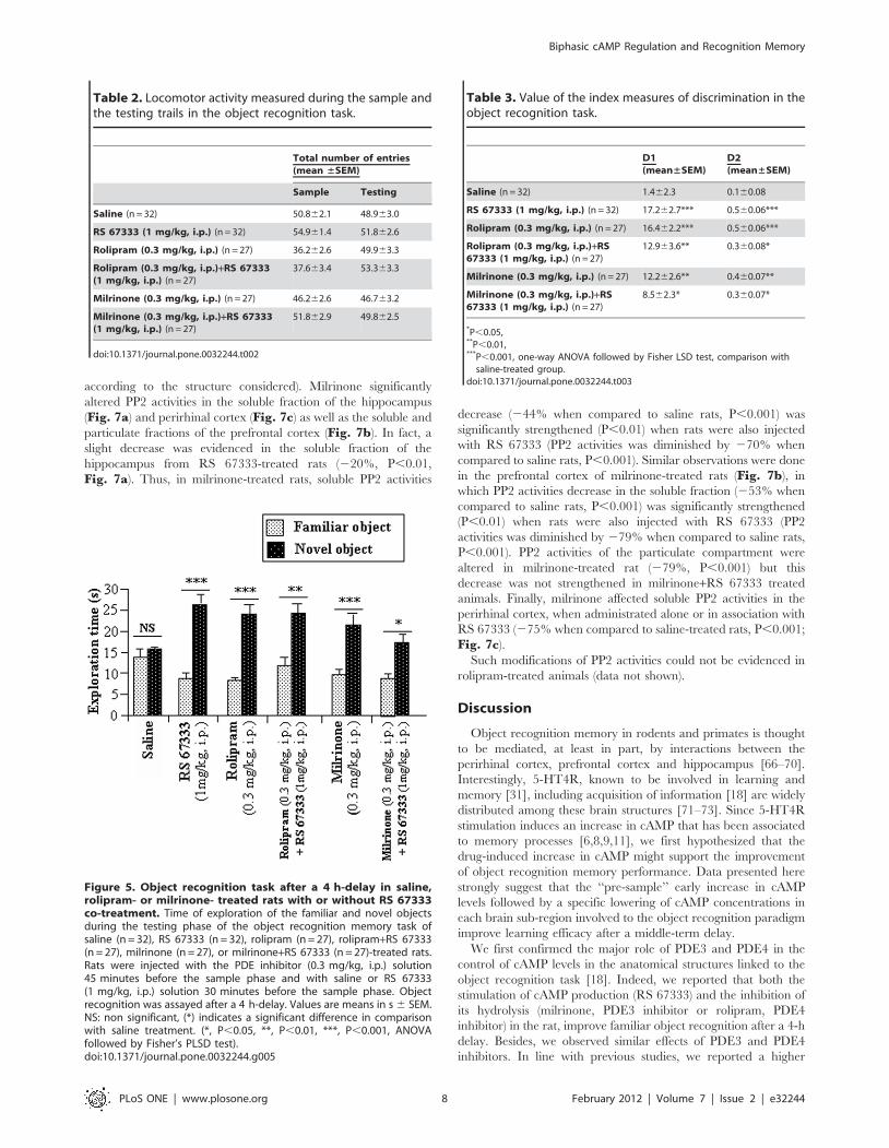
Figure 5. Object recognition task after a 4 h-delay in saline,rolipram- or milrinone- treated rats with or without RS 67333co-treatment. Time of exploration of the familiar and novel objectsduring the testing phase of the object recognition memory task ofsaline (n = 32), RS 67333 (n = 32), rolipram (n = 27), rolipram+RS 67333(n = 27), milrinone (n = 27), or milrinone+RS 67333 (n = 27)-treated rats.Rats were injected with the PDE inhibitor (0.3 mg/kg, i.p.) solution45 minutes before the sample phase and with saline or RS 67333(1 mg/kg, i.p.) solution 30 minutes before the sample phase. Objectrecognition was assayed after a 4 h-delay. Values are means in s 6 SEM.NS: non significant, (*) indicates a significant difference in comparisonwith saline treatment. (*, P,0.05, **, P,0.01, ***, P,0.001, ANOVAfollowed by Fisher’s PLSD test).doi:10.1371/journal.pone.0032244.g005
Table 3. Value of the index measures of discrimination in theobject recognition task.
D1(mean±SEM)
D2(mean±SEM)
Saline (n = 32) 1.462.3 0.160.08
RS 67333 (1 mg/kg, i.p.) (n = 32) 17.262.7*** 0.560.06***
Rolipram (0.3 mg/kg, i.p.) (n = 27) 16.462.2*** 0.560.06***
Rolipram (0.3 mg/kg, i.p.)+RS67333 (1 mg/kg, i.p.) (n = 27)
12.963.6** 0.360.08*
Milrinone (0.3 mg/kg, i.p.) (n = 27) 12.262.6** 0.460.07**
Milrinone (0.3 mg/kg, i.p.)+RS67333 (1 mg/kg, i.p.) (n = 27)
8.562.3* 0.360.07*
*P,0.05,**P,0.01,***P,0.001, one-way ANOVA followed by Fisher LSD test, comparison with
saline-treated group.doi:10.1371/journal.pone.0032244.t003
Biphasic cAMP Regulation and Recognition Memory
PLoS ONE | www.plosone.org 8 February 2012 | Volume 7 | Issue 2 | e32244

Figure 6. Rolipram and/or RS 67333 induce a biphasic modulation of cAMP concentrations in the hippocampus, prefrontal andperirhinal cortices of rats performing an object recognition task with a 4-h delay. Rats were injected with the inhibitor of PDE4 (rolipram,0.3 mg/kg, i.p.) and then with saline or the 5-HT4 receptor agonist (RS 67333, 1 mg/kg, i.p.), respectively 45 minutes and 30 minutes before to thesample phase of the object recognition task. Rats were euthanized before or after the sample phase, or before or after the testing phase. Cyclic AMPwas extracted from the hippocampus, prefrontal and perirhinal cortices and then assayed. Cyclic AMP was expressed as pmolcAMP/mg of weighttissue. Results were means 6 SEM of three independent extractions performed in duplicate. (0) indicated significant differences in comparison withother steps of the paradigm in each brain sub-region, Mann-Whitney test, P,0.05. (*) indicated a significant difference in comparison with salinetreatment in each brain sub-region. (0,*, P,0.05).doi:10.1371/journal.pone.0032244.g006
Biphasic cAMP Regulation and Recognition Memory
PLoS ONE | www.plosone.org 9 February 2012 | Volume 7 | Issue 2 | e32244

sensitivity of PDE3- than PDE4-regulated cAMP pools to PKA
activation, associated to a lower efficacy of PDE3 to hydrolyze
cAMP [74–77]. We hypothesized this familiar object recognition
improvement to be linked to the early cAMP levels increase
measured before the sample phase in the hippocampus, prefrontal
and perirhinal cortices from these animals. Early cAMP
activations in the prefrontal cortex have already been described
to be beneficial for working memory under conditions that require
hippocampal-prefrontal cortex interactions [14,78]. Moreover, the
observation that the drug-induced early increase in cAMP in the
brain sub-regions improves the mnesic trace is consistent with data
reporting that activation of the cAMP-PKA pathway cascade
improves memory processes [2,4,6,8,9] unlike inhibition of PKA
[6,10,11,79]. Indeed, according to the model of Frankland and
Bontempi, experience is both initially encoded in hippocampal
and cortical networks. Subsequent reactivation of the hippocampal
network reinstates activity in different cortical networks. This
coordinated replay across hippocampal–cortical networks leads to
gradual strengthening of cortico-cortical connections, progressive-
ly disengaging the memory trace from the hippocampus [80].
Thus, the higher cAMP levels induced by drug treatments could
support a better acquisition of the mnesic trace that will, in turn,
benefit to the animal during the testing phase.
We also observed that drug treatments induce a lowering, and/
or a reduction of the awaited increases, in cAMP levels in all
brains regions studied after the testing phase, when compared to
saline injected animals. Hence, cAMP concentrations were
systematically lower in both the hippocampus and perirhinal
cortex from animals that have increased behavioural performances
than in the saline-treated rats that exhibit poor object recognition
performances. Appearing at a first glance as a discrepancy from
data from the literature, this situation can easily be reconciled by
recent studies suggesting that memory requires a restricted, or
selective cAMP production rather than a large and widespread
increase in cAMP levels [28,81]. Indeed, Kelly and co-workers
(2008) observed an impairment of memory consolidation and/or
retrieval in a fear-conditioning task in mice that express a
constitutively active isoform of the G-protein subunit Gas in the
forebrain [81]. Perez-Garcia and Meneses, (2008) also demon-
strate that the hippocampal production of cAMP was higher in
untrained rats than in rats subjected to a behavioral task [28]. In
this respect, low cAMP levels might be optimal to convert
temporary memory during acquisition to long-term memory (4 h-
delay) while high cAMP levels might disturb such a conversion of
short-term memory to long-term memory, resulting in low
performances in controls animals.
These few elements point out the complexity of cAMP-
dependent responses and the putative interactions between
behaviour- and drug-induced effects on cellular signaling. By
rapidly degrading cAMP from selected compartments, PDEs can
fix the boundaries for cAMP diffusion, shape the intracellular
gradients of the second messenger and thereby modulate defined
sets of PKA-mediated intracellular events. Hence, PDE alteration
may affect cAMP compartmentalization, leading to untargeted
cAMP signals, aberrant phosphorylation of target proteins and
thus contribute to dysfunction. We report here that hippocampus,
prefrontal and perirhinal cortices exhibit different patterns of
particulate PDE4D isoforms. One can therefore hypothesize
differential implications of PDE isoforms, keys mediators of
memory and learning processes in the limitation of cAMP increase
[36,37,82,83]. For example, PDE4D8 found only in the
particulate fraction from the hippocampus or the prefrontal
cortex, has been shown to be responsible for controlling local
cAMP concentrations and PKA activity in the vicinity of b1
adrenergic receptors [46]. PDE4D3 that we only described in the
particulate fraction from the hippocampus or the prefrontal
cortex, was reported to bind to muscle- specific A-kinase
anchoring protein (mAKAP), which in turns controls perinuclear
AMP levels and recruits the MAP kinases MEK5 and ERK5 [84].
Hence, each PDE isoform plays a critical role in the specificity of
cAMP-signaling, effectively creating cyclic nucleotide microdo-
mains and/or cAMP gradients that can be sensed by the cell
[46,85–88]. Accordingly, some PDE4D isoforms precisely regulate
the coupling between GPCR and the Gs protein. A desensitization
of 5-HT4R, through an over-stimulation by RS67333 and/or an
alteration of PDE, could thus lead to a lowering of cAMP
concentrations in brain regions expressing 5-HT4R. Indeed, PKA
activated after a 5-HT4R stimulation, will phosphorylate 5-HT4R
leading to cell membrane recruitment of GRK2, which in turn will
phosphorylate i) the associated GPCR [89] inhibiting its coupling
with Gs, and ii) PDE4, which locally attenuates the PKA activity
by lowering local cAMP levels [90–92].
Effects induced by 5-HT4R stimulation are time-limited since
PKA phosphorylation of 5-HT4R, GRK2 or PDE can be reversed
by phosphatase (PP) activity [58]. PP1 and PP2, compartmental-
ized inside the mammalian cell [93,94], account for the major
phosphatase activities [56]; PP2 is however more particularly
investigated because of its ability to dephosphorylate many
signaling proteins [93]. Since PP2A is activated by cAMP level
increases [53–55], we suggested that phosphatase activity may
have been raised in the brain sub-region structures from drug-
treated rats, but it is not what we found here. While inhibition of
PDE4 activity failed to alter PP2 activity, milrinone administration
induced an alteration of PP2 activity, especially in the supernatant
fractions from the brain regions investigated; such an alteration
could in turn alter dephosphorylation of the 5-HT4R, prevent the
efficiency of coupling between this receptor and the Gs protein,
and thus lead to a lowered cAMP production. Such a functional
contrast following selective inhibition of PDE3 and PDE4 has been
already observed in many cell types [95–97]. Interestingly, the cell
Table 4. Distribution of the total PP2 activities between particulate and soluble fractions from rat hippocampus, prefrontal cortexand perirhinal cortex.
Total PP2 activities (nmol of phosphate released/min) (% of the total PP2 activities)
Hippocampus (n = 4) Prefrontal cortex (n = 4) Perirhinal cortex (n = 4)
Particulate fraction 15.8361.18 = 24.761.7% 2.5860.24 = 32.062.1% 3.3860.39 = 27.861.7%
Soluble fraction 48.0760.9111 = 75.361.7% 5.4760.27111 = 68.162.1% 8.7360.20111 = 72.261.7%
Values are means 6 SEM.111P,0.001 (ANOVA followed by Fisher’s LSD test): different from the corresponding particulate fraction.doi:10.1371/journal.pone.0032244.t004
Biphasic cAMP Regulation and Recognition Memory
PLoS ONE | www.plosone.org 10 February 2012 | Volume 7 | Issue 2 | e32244

membrane recruited PDE4 also desensitizes the switched coupling
of the b2AR to activation of Gi induced by the PKA-mediated
phosphorylation [98], defining thus an appropriate coupling of the
GPCR [99]. Hence, in our opinion, drugs injected before the
sample phase rapidly increase cAMP levels leading to the
uncoupling of 5-HT4R. However, during the test phase, further
5-HT4R stimulation does not raise cAMP level, probably because
of 5-HT4R uncoupling. 5-HT4R uncoupling could thus be an
adaptative mechanism to reduce cAMP levels in the presence of an
excessive stimulation of 5-HT4R or an absence of PDE3 or 4
activities thus avoiding an excessive accumulation of cAMP. If
such threshold of cAMP level exists and is reached by either the
stimulation of 5-HT4R or inhibition of PDE3 or PDE4 alone, thus
no further improvement of memory performance could be
induced by the pharmacological treatments by the combination
of RS 67 333 and PDE inhibitor.
Finally, early increases in cAMP levels followed by an
immediate drop in cAMP concentrations have already been well
described in cell differentiation, particularly in Sertoli cells [58].
Indeed, before the cAMP increase, stimulation of Sertoli cells by
gonadotropin leads to an activation of the ERK pathway, while
following the peak of cAMP, gonadotropin activates the PKA
pathway. Interestingly, ERK pathway could prolong activation of
the cAMP signaling system in cells by having both short and long
term effects on PDE4D activity by respectively inactivating long
PDE4D isoform (the ones to exhibits a site that allows
phosphorylation by ERK) and altering PDE4D mRNA stability
(for review [99]). Hence, by analogy to differentiation mechanisms,
another hypothesis is that ‘‘cellular learning’’ may result from the
crossing of a milestone, resulting in the subsequent activation of
alternative intracellular signalling pathways. Increases in cAMP
levels but also their subsequent declines account in mnesic
performance improvement. The part of ERK pathway in these
processes should be addressed in furthers works.
Our results show that a ‘‘pre-sample’’ early increase in cAMP
levels followed by both a ‘‘post-sample’’ lowering of cAMP
concentrations in the prefrontal cortex and a ‘‘post-test’’ lowering
of cAMP concentrations in the hippocampus and perirhinal cortex
support improved learning efficacy after a middle-term delay. If
cAMP triggers a temporally defined cellular response, a major
question that should be addressed in future works is to clarify how
such a functionally ubiquitous signaling pathway may be involved
in memory formation.
Acknowledgments
The authors thank Dr. S. Carreau (Laboratoire Œstrogenes et Reproduc-
tion, EA 2608, INRA USC 2006, University of Caen) and Dr. P. Barbey
(LAMARE) for giving us access to the ultracentrifuge and the radioactivity
laboratory, respectively, as well as Pr. B. Cox and Dr M. Dacher
(Uniformed Service University, Bethesda, MD, USA) for english editing of
the manuscript.
Author Contributions
Conceived and designed the experiments: MH GL FD MB. Performed the
experiments: MH GL. Analyzed the data: MH TF FD MB GL.
Contributed reagents/materials/analysis tools: MH TF GL. Wrote the
paper: TF FD MB GL MH. Statistical analysis: TF.
Figure 7. Effect of milrinone (0.3 mg/kg) on PP2 activities inhippocampus, prefrontal and perirhinal cortices from ratsperforming the object recognition task with a 4-h delay. Ratswere injected with the inhibitor of PDE3 (milrinone, 0.3 mg/kg)45 minutes before exposure then with saline or the 5-HT4 receptoragonist (RS 67333, 1 mg/kg), 30 minutes before exposure to the sampletrial of the object recognition task. Immediately after the testing trial,both particulate (white bar) and soluble (black bar) fractions from thehippocampus (a), the prefrontal cortex (b) and perirhinal cortex (c)were isolated and were assayed for PP2 activity. PP2 activities werepmol of phosphate released by min and mg protein. Results are means
6 SEM of four independent subcellular fractionations performed intriplicate. Within each subcellular compartment, # indicated significantdifferences of PP2 activity as compared with other treatment (#,P,0.05, ##, P,0.01, ###, P,0.001, ANOVA followed by Fisher’sLSD test).doi:10.1371/journal.pone.0032244.g007
Biphasic cAMP Regulation and Recognition Memory
PLoS ONE | www.plosone.org 11 February 2012 | Volume 7 | Issue 2 | e32244

References
1. Monti B, Berteotti C, Contestabile A (2006) Subchronic rolipram delivery
activates hippocampal CREB and arc, enhances retention and slows downextinction of conditioned fear. Neuropsychopharmacology 31: 278–286.
2. Hoyer D, Pompe B, Friedrich H, Zwiener U, Baranowski R, et al. (2004)
Autonomic Information Flow during awakeness, sleep, and multiple organ
dysfunction syndrome assessed by mutual information function of heart ratefluctuations. Conf Proc IEEE Eng Med Biol Soc 1: 628–630.
3. Bach ME, Barad M, Son H, Zhuo M, Lu YF, et al. (1999) Age-related defects in
spatial memory are correlated with defects in the late phase of hippocampallong-term potentiation in vitro and are attenuated by drugs that enhance the
cAMP signaling pathway. Proc Natl Acad Sci U S A 96: 5280–5285.
4. Barad M, Bourtchouladze R, Winder DG, Golan H, Kandel E (1998) Rolipram,a type IV-specific phosphodiesterase inhibitor, facilitates the establishment of
long-lasting long-term potentiation and improves memory. Proc Natl AcadSci U S A 95: 15020–15025.
5. Zhang HT, O’Donnell JM (2000) Effects of rolipram on scopolamine-induced
impairment of working and reference memory in the radial-arm maze tests in
rats. Psychopharmacology (Berl) 150: 311–316.
6. Bernabeu R, Cammarota M, Izquierdo I, Medina JH (1997) Involvement ofhippocampal AMPA glutamate receptor changes and the cAMP/protein kinase
A/CREB-P signalling pathway in memory consolidation of an avoidance task inrats. Braz J Med Biol Res 30: 961–965.
7. Bernabeu R, Schmitz P, Faillace MP, Izquierdo I, Medina JH (1996)
Hippocampal cGMP and cAMP are differentially involved in memoryprocessing of inhibitory avoidance learning. Neuroreport 7: 585–588.
8. Prickaerts J, de Vente J, Honig W, Steinbusch HW, Blokland A (2002) cGMP,
but not cAMP, in rat hippocampus is involved in early stages of object memoryconsolidation. Eur J Pharmacol 436: 83–87.
9. Rutten K, Prickaerts J, Hendrix M, van der Staay FJ, Sik A, et al. (2007) Time-
dependent involvement of cAMP and cGMP in consolidation of object memory:
Studies using selective phosphodiesterase type 2, 4 and 5 inhibitors.Eur J Pharmacol 558: 107–112.
10. Bourtchouladze R, Abel T, Berman N, Gordon R, Lapidus K, et al. (1998)
Different training procedures recruit either one or two critical periods forcontextual memory consolidation, each of which requires protein synthesis and
PKA. Learn Mem 5: 365–374.
11. Taylor JR, Birnbaum S, Ubriani R, Arnsten AF (1999) Activation of cAMP-dependent protein kinase A in prefrontal cortex impairs working memory
performance. J Neurosci 19: RC23.
12. Connolly DA, Hockley WE, Pratt MW (1996) A developmental evaluation of
frequency memory for actions presented in lists, scripts, and stories. Memory 4:243–263.
13. Pineda VV, Athos JI, Wang H, Celver J, Ippolito D, et al. (2004) Removal of
G(ialpha1) constraints on adenylyl cyclase in the hippocampus enhances LTPand impairs memory formation. Neuron 41: 153–163.
14. Ramos BP, Birnbaum SG, Lindenmayer I, Newton SS, Duman RS, et al. (2003)
Dysregulation of protein kinase a signaling in the aged prefrontal cortex: newstrategy for treating age-related cognitive decline. Neuron 40: 835–845.
15. Ennaceur A, Delacour J (1988) A new one-trial test for neurobiological studies of
memory in rats. 1: Behavioral data. Behav Brain Res 31: 47–59.
16. Lamirault L, Guillou C, Thal C, Simon H (2003) Combined treatment withgalanthaminium bromide, a new cholinesterase inhibitor, and RS 67333, a
partial agonist of 5-HT4 receptors, enhances place and object recognition in
young adult and old rats. Prog Neuropsychopharmacol Biol Psychiatry 27:185–195.
17. Lamirault L, Simon H (2001) Enhancement of place and object recognition
memory in young adult and old rats by RS 67333, a partial agonist of 5-HT4receptors. Neuropharmacology 41: 844–853.
18. Levallet G, Hotte M, Boulouard M, Dauphin F (2009) Increased particulate
phosphodiesterase 4 in the prefrontal cortex supports 5-HT4 receptor-inducedimprovement of object recognition memory in the rat. Psychopharmacology
(Berl) 202: 125–139.
19. Moser PC, Bergis OE, Jegham S, Lochead A, Duconseille E, et al. (2002)SL65.0155, a novel 5-hydroxytryptamine(4) receptor partial agonist with potent
cognition-enhancing properties. J Pharmacol Exp Ther 302: 731–741.
20. Fontana DJ, Daniels SE, Wong EH, Clark RD, Eglen RM (1997) The effects of
novel, selective 5-hydroxytryptamine (5-HT)4 receptor ligands in rat spatialnavigation. Neuropharmacology 36: 689–696.
21. Galeotti N, Ghelardini C, Bartolini A (1998) Role of 5-HT4 receptors in the
mouse passive avoidance test. J Pharmacol Exp Ther 286: 1115–1121.
22. Lelong V, Dauphin F, Boulouard M (2001) RS 67333 and D-cycloserineaccelerate learning acquisition in the rat. Neuropharmacology 41: 517–522.
23. Letty S, Child R, Dumuis A, Pantaloni A, Bockaert J, et al. (1997) 5-HT4
receptors improve social olfactory memory in the rat. Neuropharmacology 36:681–687.
24. Marchetti E, Dumuis A, Bockaert J, Soumireu-Mourat B, Roman FS (2000)
Differential modulation of the 5-HT(4) receptor agonists and antagonist on ratlearning and memory. Neuropharmacology 39: 2017–2027.
25. Marchetti E, Jacquet M, Jeltsch H, Migliorati M, Nivet E, et al. (2008) Complete
recovery of olfactory associative learning by activation of 5-HT4 receptors afterdentate granule cell damage in rats. Neurobiol Learn Mem 90: 185–191.
26. Meneses A (2007) Stimulation of 5-HT1A, 5-HT1B, 5-HT2A/2C, 5-HT3 and
5-HT4 receptors or 5-HT uptake inhibition: short- and long-term memory.Behav Brain Res 184: 81–90.
27. Orsetti M, Dellarole A, Ferri S, Ghi P (2003) Acquisition, retention, and recall of
memory after injection of RS67333, a 5-HT(4) receptor agonist, into the nucleus
basalis magnocellularis of the rat. Learn Mem 10: 420–426.28. Perez-Garcia G, Meneses A (2008) Ex vivo study of 5-HT(1A) and 5-HT(7)
receptor agonists and antagonists on cAMP accumulation during memory
formation and amnesia. Behav Brain Res 195: 139–146.
29. Restivo L, Roman F, Dumuis A, Bockaert J, Marchetti E, et al. (2008) Thepromnesic effect of G-protein-coupled 5-HT4 receptors activation is mediated
by a potentiation of learning-induced spine growth in the mouse hippocampus.Neuropsychopharmacology 33: 2427–2434.
30. Terry AV, Jr., Buccafusco JJ, Jackson WJ, Prendergast MA, Fontana DJ, et al.
(1998) Enhanced delayed matching performance in younger and older macaquesadministered the 5-HT4 receptor agonist, RS 17017. Psychopharmacology
(Berl) 135: 407–415.
31. King MV, Marsden CA, Fone KC (2008) A role for the 5-HT(1A), 5-HT4 and5-HT6 receptors in learning and memory. Trends Pharmacol Sci 29: 482–492.
32. Egawa T, Mishima K, Matsumoto Y, Iwasaki K, Fujiwara M (1997) Rolipram
and its optical isomers, phosphodiesterase 4 inhibitors, attenuated the
scopolamine-induced impairments of learning and memory in rats.Jpn J Pharmacol 75: 275–281.
33. Zhang HT, Crissman AM, Dorairaj NR, Chandler LJ, O’Donnell JM (2000)
Inhibition of cyclic AMP phosphodiesterase (PDE4) reverses memory deficitsassociated with NMDA receptor antagonism. Neuropsychopharmacology 23:
198–204.
34. Zhang HT, Zhao Y, Huang Y, Dorairaj NR, Chandler LJ, et al. (2004)Inhibition of the phosphodiesterase 4 (PDE4) enzyme reverses memory deficits
produced by infusion of the MEK inhibitor U0126 into the CA1 subregion of
the rat hippocampus. Neuropsychopharmacology 29: 1432–1439.35. Rutten K, Wallace TL, Works M, Prickaerts J, Blokland A, et al. (2011)
Enhanced long-term depression and impaired reversal learning in phosphodi-
esterase 4B-knockout (PDE4B(2/2)) mice. Neuropharmacology 61: 138–147.
36. Burgin AB, Magnusson OT, Singh J, Witte P, Staker BL, et al. (2010) Design ofphosphodiesterase 4D (PDE4D) allosteric modulators for enhancing cognition
with improved safety. Nat Biotechnol 28: 63–70.
37. Li YF, Cheng YF, Huang Y, Conti M, Wilson SP, et al. (2011)Phosphodiesterase-4D knock-out and RNA interference-mediated knock-down
enhance memory and increase hippocampal neurogenesis via increased cAMPsignaling. J Neurosci 31: 172–183.
38. Randt CT, Judge ME, Bonnet KA, Quartermain D (1982) Brain cyclic AMP
and memory in mice. Pharmacol Biochem Behav 17: 677–680.
39. Gong B, Vitolo OV, Trinchese F, Liu S, Shelanski M, et al. (2004) Persistentimprovement in synaptic and cognitive functions in an Alzheimer mouse model
after rolipram treatment. J Clin Invest 114: 1624–1634.
40. Bourtchouladze R, Lidge R, Catapano R, Stanley J, Gossweiler S, et al. (2003) A
mouse model of Rubinstein-Taybi syndrome: defective long-term memory isameliorated by inhibitors of phosphodiesterase 4. Proc Natl Acad Sci U S A 100:
10518–10522.
41. Rutten K, Prickaerts J, Blokland A (2006) Rolipram reverses scopolamine-induced and time-dependent memory deficits in object recognition by different
mechanisms of action. Neurobiol Learn Mem 85: 132–138.
42. Rutten K, Van Donkelaar EL, Ferrington L, Blokland A, Bollen E, et al. (2009)Phosphodiesterase inhibitors enhance object memory independent of cerebral
blood flow and glucose utilization in rats. Neuropsychopharmacology 34:1914–1925.
43. Houslay MD (2001) PDE4 cAMP-specific phosphodiesterases. Prog Nucleic
Acid Res Mol Biol 69: 249–315.
44. Zhang HT (2009) Cyclic AMP-specific phosphodiesterase-4 as a target for thedevelopment of antidepressant drugs. Curr Pharm Des 15: 1688–1698.
45. Hoffmann R, Baillie GS, MacKenzie SJ, Yarwood SJ, Houslay MD (1999) The
MAP kinase ERK2 inhibits the cyclic AMP-specific phosphodiesterase
HSPDE4D3 by phosphorylating it at Ser579. EMBO J 18: 893–903.46. Richter W, Day P, Agrawal R, Bruss MD, Granier S, et al. (2008) Signaling
from beta1- and beta2-adrenergic receptors is defined by differential interactions
with PDE4. EMBO J 27: 384–393.
47. van Donkelaar EL, Rutten K, Blokland A, Akkerman S, Steinbusch HW, et al.(2008) Phosphodiesterase 2 and 5 inhibition attenuates the object memory deficit
induced by acute tryptophan depletion. Eur J Pharmacol 600: 98–104.
48. Zhao J, Harada N, Kurihara H, Nakagata N, Okajima K (2010) Cilostazolimproves cognitive function in mice by increasing the production of insulin-like
growth factor-I in the hippocampus. Neuropharmacology 58: 774–783.
49. O’Donnell JM, Frith S (1999) Behavioral effects of family-selective inhibitors ofcyclic nucleotide phosphodiesterases. Pharmacol Biochem Behav 63: 185–192.
50. Edelson J, Stroshane R, Benziger DP, Cody R, Benotti J, et al. (1986)
Pharmacokinetics of the bipyridines amrinone and milrinone. Circulation 73:III145–152.
51. Lindsay CA, Barton P, Lawless S, Kitchen L, Zorka A, et al. (1998)
Pharmacokinetics and pharmacodynamics of milrinone lactate in pediatric
patients with septic shock. J Pediatr 132: 329–334.
Biphasic cAMP Regulation and Recognition Memory
PLoS ONE | www.plosone.org 12 February 2012 | Volume 7 | Issue 2 | e32244

52. Krause W, Kuhne G (1988) Pharmacokinetics of rolipram in the rhesus and
cynomolgus monkeys, the rat and the rabbit. Studies on species differences.Xenobiotica 18: 561–571.
53. Feschenko MS, Stevenson E, Nairn AC, Sweadner KJ (2002) A novel cAMP-
stimulated pathway in protein phosphatase 2A activation. J Pharmacol Exp Ther302: 111–118.
54. Moon EY, Lerner A (2003) PDE4 inhibitors activate a mitochondrial apoptoticpathway in chronic lymphocytic leukemia cells that is regulated by protein
phosphatase 2A. Blood 101: 4122–4130.
55. Pullar CE, Grahn JC, Liu W, Isseroff RR (2006) Beta2-adrenergic receptoractivation delays wound healing. FASEB J 20: 76–86.
56. Oliver CJ, Shenolikar S (1998) Physiologic importance of protein phosphataseinhibitors. Front Biosci 3: D961–972.
57. Resjo S, Oknianska A, Zolnierowicz S, Manganiello V, Degerman E (1999)Phosphorylation and activation of phosphodiesterase type 3B (PDE3B) in
adipocytes in response to serine/threonine phosphatase inhibitors: deactivation
of PDE3B in vitro by protein phosphatase type 2A. Biochem J 341(Pt 3):839–845.
58. Levallet G, Levallet J, Bonnamy PJ (2008) FSH-induced phosphoproteinphosphatase 2A-mediated deactivation of particulate phosphodiesterase-4
activities is abolished after alteration in proteoglycan synthesis in immature rat
Sertoli cells. J Endocrinol 197: 45–54.59. Desjardins S, Cauchy MJ (1995) Comparative cardiac effects of milrinone and
sodium nitroprusside in conscious rats. Drug Chem Toxicol 18: 43–59.60. Griebel G, Misslin R, Vogel E, Bourguignon JJ (1991) Behavioral effects of
rolipram and structurally related compounds in mice: behavioral sedation ofcAMP phosphodiesterase inhibitors. Pharmacol Biochem Behav 39: 321–323.
61. Silvestre JS, Fernandez AG, Palacios JM (1999) Effects of rolipram on the
elevated plus-maze test in rats: a preliminary study. J Psychopharmacol 13:274–277.
62. Sik A, van Nieuwehuyzen P, Prickaerts J, Blokland A (2003) Performance ofdifferent mouse strains in an object recognition task. Behav Brain Res 147:
49–54.
63. Rodriguez R, Molino B, Weiss HR, Scholz PM (2004) Negative metabolic andcoronary flow effects of decreases in cAMP and increases in cGMP in control
and renal hypertensive rabbit hearts. J Appl Physiol 97: 439–445.64. Bradford MM (1976) A rapid and sensitive method for the quantitation of
microgram quantities of protein utilizing the principle of protein-dye binding.Anal Biochem 72: 248–254.
65. Thompson WJ, Appleman MM (1971) Multiple cyclic nucleotide phosphodi-
esterase activities from rat brain. Biochemistry 10: 311–316.66. Bussey TJ, Duck J, Muir JL, Aggleton JP (2000) Distinct patterns of behavioural
impairments resulting from fornix transection or neurotoxic lesions of theperirhinal and postrhinal cortices in the rat. Behav Brain Res 111: 187–202.
67. Ennaceur A, Neave N, Aggleton JP (1997) Spontaneous object recognition and
object location memory in rats: the effects of lesions in the cingulate cortices, themedial prefrontal cortex, the cingulum bundle and the fornix. Exp Brain Res
113: 509–519.68. Hotte M, Thuault S, Lachaise F, Dineley KT, Hemmings HC, et al. (2006) D1
receptor modulation of memory retrieval performance is associated with changesin pCREB and pDARPP-32 in rat prefrontal cortex. Behav Brain Res 171:
127–133.
69. Parker A, Gaffan D (1998) Interaction of frontal and perirhinal cortices in visualobject recognition memory in monkeys. Eur J Neurosci 10: 3044–3057.
70. Warburton EC, Brown MW (2010) Findings from animals concerning wheninteractions between perirhinal cortex, hippocampus and medial prefrontal
cortex are necessary for recognition memory. Neuropsychologia 48: 2262–2272.
71. Bonaventure P, Hall H, Gommeren W, Cras P, Langlois X, et al. (2000)Mapping of serotonin 5-HT(4) receptor mRNA and ligand binding sites in the
post-mortem human brain. Synapse 36: 35–46.72. Eglen RM, Bonhaus DW, Johnson LG, Leung E, Clark RD (1995)
Pharmacological characterization of two novel and potent 5-HT4 receptor
agonists, RS 67333 and RS 67506, in vitro and in vivo. Br J Pharmacol 115:1387–1392.
73. Vilaro MT, Cortes R, Gerald C, Branchek TA, Palacios JM, et al. (1996)Localization of 5-HT4 receptor mRNA in rat brain by in situ hybridization
histochemistry. Brain Res Mol Brain Res 43: 356–360.74. Chini CC, Grande JP, Chini EN, Dousa TP (1997) Compartmentalization of
cAMP signaling in mesangial cells by phosphodiesterase isozymes PDE3 and
PDE4. Regulation of superoxidation and mitogenesis. J Biol Chem 272:9854–9859.
75. Dousa TP (1998) Signaling role of PDE isozymes in pathobiology of glomerularmesangial cells. Studies in vitro and in vivo. Cell Biochem Biophys 29: 19–34.
76. Dousa TP (1999) Cyclic-39,59-nucleotide phosphodiesterase isozymes in cell
biology and pathophysiology of the kidney. Kidney Int 55: 29–62.77. Matousovic K, Grande JP, Chini CC, Chini EN, Dousa TP (1995) Inhibitors of
cyclic nucleotide phosphodiesterase isozymes type-III and type-IV suppress
mitogenesis of rat mesangial cells. J Clin Invest 96: 401–410.78. Aujla H, Beninger RJ (2001) Hippocampal-prefrontocortical circuits: PKA
inhibition in the prefrontal cortex impairs delayed nonmatching in the radialmaze in rats. Behav Neurosci 115: 1204–1211.
79. Abel T, Nguyen PV, Barad M, Deuel TA, Kandel ER, et al. (1997) Genetic
demonstration of a role for PKA in the late phase of LTP and in hippocampus-based long-term memory. Cell 88: 615–626.
80. Frankland PW, Bontempi B (2005) The organization of recent and remotememories. Nat Rev Neurosci 6: 119–130.
81. Kelly MP, Cheung YF, Favilla C, Siegel SJ, Kanes SJ, et al. (2008) Constitutiveactivation of the G-protein subunit Galphas within forebrain neurons causes
PKA-dependent alterations in fear conditioning and cortical Arc mRNA
expression. Learn Mem 15: 75–83.82. Giorgi M, Modica A, Pompili A, Pacitti C, Gasbarri A (2004) The induction of
cyclic nucleotide phosphodiesterase 4 gene (PDE4D) impairs memory in a watermaze task. Behav Brain Res 154: 99–106.
83. Rutten K, Basile JL, Prickaerts J, Blokland A, Vivian JA (2008) Selective PDE
inhibitors rolipram and sildenafil improve object retrieval performance in adultcynomolgus macaques. Psychopharmacology (Berl) 196: 643–648.
84. Dodge-Kafka KL, Bauman A, Mayer N, Henson E, Heredia L, et al. (2010)cAMP-stimulated protein phosphatase 2A activity associated with muscle A
kinase-anchoring protein (mAKAP) signaling complexes inhibits the phosphor-ylation and activity of the cAMP-specific phosphodiesterase PDE4D3. J Biol
Chem 285: 11078–11086.
85. Besheer J, Jensen HC, Bevins RA (1999) Dopamine antagonism in a novel-object recognition and a novel-object place conditioning preparation with rats.
Behav Brain Res 103: 35–44.86. Fischmeister R, Castro LR, Abi-Gerges A, Rochais F, Jurevicius J, et al. (2006)
Compartmentation of cyclic nucleotide signaling in the heart: the role of cyclic
nucleotide phosphodiesterases. Circ Res 99: 816–828.87. Xiang Y, Naro F, Zoudilova M, Jin SL, Conti M, et al. (2005) Phosphodiesterase
4D is required for beta2 adrenoceptor subtype-specific signaling in cardiacmyocytes. Proc Natl Acad Sci U S A 102: 909–914.
88. Zaccolo M, Pozzan T (2002) Discrete microdomains with high concentration ofcAMP in stimulated rat neonatal cardiac myocytes. Science 295: 1711–1715.
89. Cong M, Perry SJ, Lin FT, Fraser ID, Hu LA, et al. (2001) Regulation of
membrane targeting of the G protein-coupled receptor kinase 2 by proteinkinase A and its anchoring protein AKAP79. J Biol Chem 276: 15192–15199.
90. Baillie GS, Sood A, McPhee I, Gall I, Perry SJ, et al. (2003) beta-Arrestin-mediated PDE4 cAMP phosphodiesterase recruitment regulates beta-adreno-
ceptor switching from Gs to Gi. Proc Natl Acad Sci U S A 100: 940–945.
91. Li X, Huston E, Lynch MJ, Houslay MD, Baillie GS (2006) Phosphodiesterase-4influences the PKA phosphorylation status and membrane translocation of G-
protein receptor kinase 2 (GRK2) in HEK-293beta2 cells and cardiac myocytes.Biochem J 394: 427–435.
92. Xin W, Tran TM, Richter W, Clark RB, Rich TC (2008) Roles of GRK andPDE4 activities in the regulation of beta2 adrenergic signaling. J Gen Physiol
131: 349–364.
93. Janssens V, Goris J (2001) Protein phosphatase 2A: a highly regulated family ofserine/threonine phosphatases implicated in cell growth and signalling.
Biochem J 353: 417–439.94. Sim AT, Ludowyke RI, Verrills NM (2006) Mast cell function: regulation of
degranulation by serine/threonine phosphatases. Pharmacol Ther 112:
425–439.95. Huang Z, Ducharme Y, Macdonald D, Robichaud A (2001) The next
generation of PDE4 inhibitors. Curr Opin Chem Biol 5: 432–438.96. Lugnier C (2006) Cyclic nucleotide phosphodiesterase (PDE) superfamily: a new
target for the development of specific therapeutic agents. Pharmacol Ther 109:
366–398.97. Maurice DH, Palmer D, Tilley DG, Dunkerley HA, Netherton SJ, et al. (2003)
Cyclic nucleotide phosphodiesterase activity, expression, and targeting in cells ofthe cardiovascular system. Mol Pharmacol 64: 533–546.
98. Zamah AM, Delahunty M, Luttrell LM, Lefkowitz RJ (2002) Protein kinase A-mediated phosphorylation of the beta 2-adrenergic receptor regulates its
coupling to Gs and Gi. Demonstration in a reconstituted system. J Biol Chem
277: 31249–31256.99. Houslay MD, Baillie GS (2003) The role of ERK2 docking and phosphorylation
of PDE4 cAMP phosphodiesterase isoforms in mediating cross-talk between thecAMP and ERK signalling pathways. Biochem Soc Trans 31: 1186–1190.
Biphasic cAMP Regulation and Recognition Memory
PLoS ONE | www.plosone.org 13 February 2012 | Volume 7 | Issue 2 | e32244