a cassette holder to be used in pinning hips
TRANSCRIPT
A CASSETTE HOLDER TO BE USED IN PINNING HIPS Charles C. Voorhis, M.D., F.A.C.S., Kent, Ohio
EDITOR'S NOTE
AORN IOURNAL has been given the privi- lege of reprinting this very valuable article. Although the article was originally published in a magazine read mainly by surgeons, we feel that the simple instructions given herein are of value to operating room nurses. This holder may be the answer to your problems of positioning patients properly for hip mi l - ings and, at the same time, provides ready access to the X-ray equipment without up- setting the sterile jield.
Open reduction and fixation of hip frac- tures and other procedures on hips requiring roentgenographic visualization during the operation have resulted in the development of many mechanical aids. This report con- cerns one such device which has been used at Robinson Memorial Portage County Hos- pital, Ravenna, Ohio, for 15 years. No claim is made for its originality. However, I have used many different methods of roentgen- ographic control since 1939, and the cassette holder described herein, constructed from easily obtainable materials, is the least com- plicated and most satisfactory of any of them. Although I have employed this wooden cassette holder only during the operative fixation of hip fractures, it may also be of
value during other operative procedures on the hip.
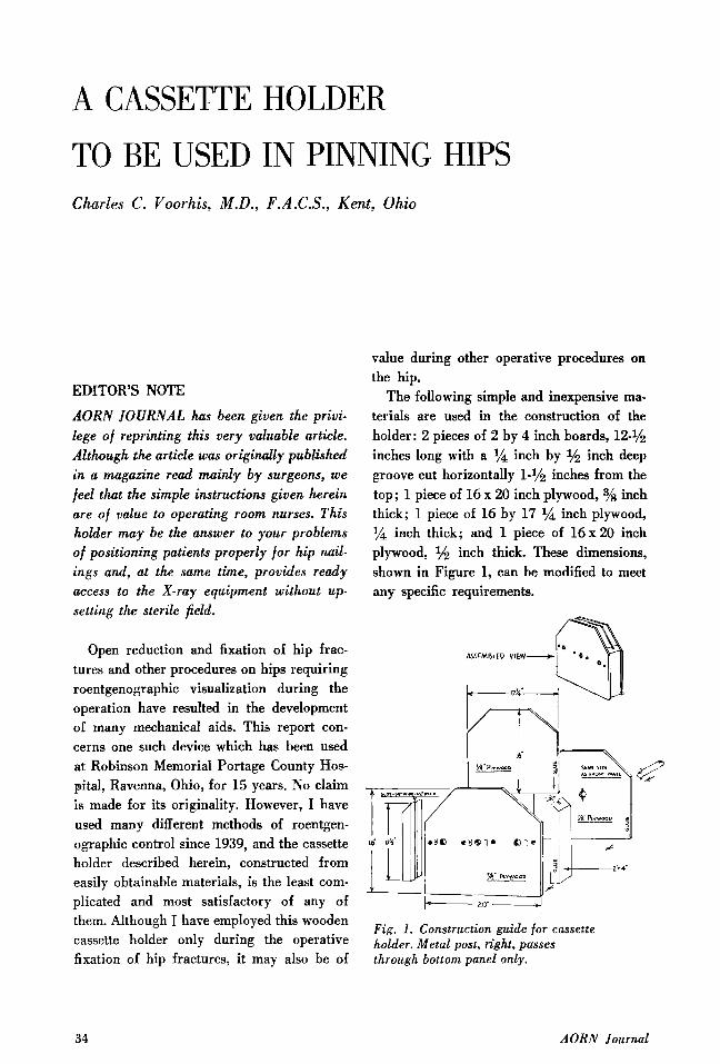
The following simple and inexpensive ma- terials are used in the construction of the holder: 2 pieces of 2 by 4 inch boards, 12-1/2 inches long with a 1/4 inch by inch deep groove cut horizontally 1-1/2 inches from the top; 1 piece of 16 x 20 inch plywood, 3/s inch thick; 1 piece of 16 by 17 l/a inch plywood, l/a inch thick; and 1 piece of 1 6 x 2 0 inch plywood, 1h inch thick. These dimensions, shown in Figure 1, can be modified to meet any specific requirements.
Fig. 1. Construction guide for cassette holder. Metal post, right, passes through bottom panel only.
34 AORN Journal
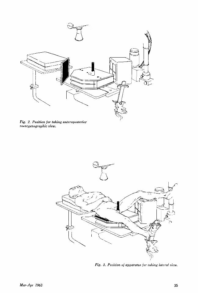
Fig. 2. Position for taking anteroposterior roentgenographic view.
Mar-Apr 1963
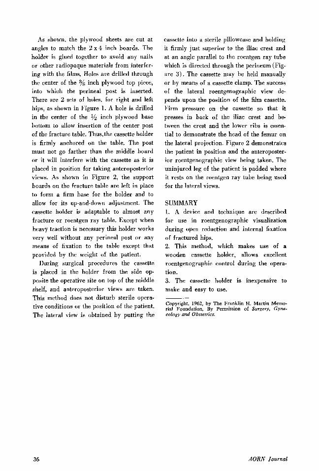
Fig. 3. Position of apparatus for taking Lateral view.
35
As shown, the plywood sheets are cut at angles to match the 2 x 4 inch boards. The holder is glued together to avoid any nails or other radiopaque materials from interfer- ing with the films. Holes are drilled through the center of the 3/s inch plywood top piece, into which the perineal post is inserted. There are 2 sets of holes, for right and left hips, as shown in Figure 1. A hole is drilled in the center of the % inch plywood base bottom to allow insertion of the center post of the fracture table. Thus,the cassette holder is firmly anchored on the table. The post must not go farther than the middle board or it will interfere with the cassette as it is placed in position for taking anteroposterior views. As shown in Figure 2, the support boards on the fracture table are left in place to form a firm base for the holder and to allow for its up-and-down adjustment. The cassette holder is adaptable to almost any fracture or roentgen ray table. Except when heavy traction is necessary this holder works very well without any perineal post or any means of fixation to the table except that provided by the weight of the patient.
During surgical procedures the cassette is placed in the holder from the side op- posite the operative site on top of the middle shelf, and anteroposterior views are taken. This method does not disturb sterile opera- tive conditions or the position of the patient. The lateral view is obtained by putting the
cassette into a sterile pillowcase and holding it firmly just superior to the iliac crest and at an angle parallel to the roentgen ray tube which is directed through the perineum (Fig- ure 3) . The cassette may be held manually or by means of a cassette clamp. The success of the lateral roentgenographic view de- pends upon the position of the film cassette. Firm pressure on the cassette so that it presses in back of the iliac crest and be- tween the crest and the lower ribs is essen- tial to demonstrate the head of the femur on the lateral projection. Figure 2 demonstrates the patient in position and the anteroposter- ior roentgenographic view being taken. The uninjured leg of the patient is padded where it rests on the roentgen ray tube being used for the lateral views.
SUMMARY 1. A device and technique are described for use in roentgenographic visualization during open reduction and internal fixation of fractured hips. 2. This method, which makes use of a wooden cassette holder, allows excellent roentgenographic control during the opera- tion. 3. The cassette holder is inexpensive to make and easy to use.
Copyright, 1962, by The Franklin H. Martin Memo- rial Foundation, By Permission of Surgery, Gyno- cology and Obstetrics.
36 AORN Journal