a cd8 t cell/indoleamine 2,3-dioxygenase axis is required for … · 2016-06-07 · chen, md,...
TRANSCRIPT

TitleA CD8 T Cell/Indoleamine 2,3-Dioxygenase Axis Is Required forMesenchymal Stem Cell Suppression of Human Systemic LupusErythematosus
Author(s) Wang, D; Feng, X; Lu, L; Konkel, JE; Zhang, H; Chen, Z; Li, X;Gao, X; Lu, L; Shi, S
Citation Arthritis & Rheumatology, 2014, v. 66, p. 2234-2245
Issued Date 2014
URL http://hdl.handle.net/10722/212092
Rights Creative Commons: Attribution 3.0 Hong Kong License

ARTHRITIS & RHEUMATOLOGYVol. 66, No. 8, August 2014, pp 2234–2245DOI 10.1002/art.38674© 2014 The Authors. Arthritis & Rheumatology is published by Wiley Periodicals, Inc. on behalf of theAmerican College of Rheumatology. This is an open access article under the terms of the CreativeCommons Attribution NonCommercial License, which permits use, distribution, and reproduction in anymedium, provided the original work is properly cited and is not used for commercial purposes.
A CD8 T Cell/Indoleamine 2,3-Dioxygenase Axis IsRequired for Mesenchymal Stem Cell Suppression of
Human Systemic Lupus Erythematosus
Dandan Wang,1 Xuebing Feng,1 Lin Lu,1 Joanne E. Konkel,2 Huayong Zhang,1 Zhiyong Chen,1
Xia Li,1 Xiang Gao,3 Liwei Lu,4 Songtao Shi,5 Wanjun Chen,2 and Lingyun Sun1
Objective. Allogeneic mesenchymal stem cells(MSCs) exhibit therapeutic effects in human auto-immune diseases such as systemic lupus erythematosus(SLE), but the underlying mechanisms remain largelyunknown. The aim of this study was to investigate howallogeneic MSCs mediate immunosuppression in lupuspatients.
Methods. The effects of allogeneic umbilical cord–derived MSCs (UC-MSCs) on inhibition of T cell pro-liferation were determined. MSC functional moleculeswere stimulated with peripheral blood mononuclearcells from healthy controls and SLE patients and exam-ined by real-time polymerase chain reaction. CD4� andCD8� T cells were purified using microbeads to stim-ulate MSCs in order to determine cytokine expressionby MSCs and to further determine which cell subset(s)
or which molecule(s) is involved in inhibition of MSC–mediated T cell proliferation. The related signalingpathways were assessed. We determined levels of serumcytokines in lupus patients before and after UC-MSCtransplantation.
Results. Allogeneic UC-MSCs suppressed T cellproliferation in lupus patients by secreting largeamounts of indoleamine 2,3-dioxygenase (IDO). Wefurther found that interferon-� (IFN�), which is pro-duced predominantly by lupus CD8� T cells, is the keyfactor that enhances IDO activity in allogeneic MSCsand that it is associated with IFNGR1/JAK-2/STATsignaling pathways. Intriguingly, bone marrow–derivedMSCs from patients with active lupus demonstrateddefective IDO production in response to IFN� andallogeneic CD8� T cell stimulation. After allogeneicUC-MSC transplantation, serum IDO activity increasedin lupus patients.
Conclusion. We found a previously unrecognizedCD8� T cell/IFN�/IDO axis that mediates the thera-peutic effects of allogeneic MSCs in lupus patients.
Mesenchymal stem cells (MSCs) are non-hematopoietic stem cells (non-HSCs) that can supportthe function of HSCs in bone marrow (BM). MSCs havebeen shown to possess regenerative properties andunique immunoregulatory functions that make them anattractive option for cellular therapy in patients withautoimmune diseases and chronic inflammation (1). Wehave previously shown that allogeneic BM- and umbili-cal cord (UC)–derived MSC transplantation is a safeand effective treatment of active systemic lupus ery-thematosus (SLE) (2,3) and other autoimmune diseases,such as systemic sclerosis (4), Sjögren’s syndrome (5),and myositis (6). Conversely, autologous MSCs fromlupus patients cannot offer therapeutic benefits due tointrinsic abnormal functions (7–9). However, the mech-
ClinicalTrials.gov identifier: NCT01741857.Supported by grants from the Chinese Major International
(Regional) Joint Research Project (grant 81120108021), the NationalNatural Science Foundation of China (grant 81273304), and theJiangsu Province 333 Talent Program of China (to Dr. Sun), and theNIH (Intramural Research Program, National Institute of Dental andCraniofacial Research award to Drs. Konkel and W. Chen).
1Dandan Wang, MD, PhD, Xuebing Feng, MD, PhD, Lin Lu,MD, Huayong Zhang, MD, Zhiyong Chen, MD, PhD, Xia Li, MD,PhD, Lingyun Sun, MD, PhD: The Affiliated Drum Tower Hospital ofNanjing University Medical School, Nanjing, China; 2Joanne E. Kon-kel, PhD, Wanjun Chen, MD: National Institute of Dental andCraniofacial Research, NIH, Bethesda, Maryland; 3Xiang Gao, PhD:Nanjing University, Nanjing, China; 4Liwei Lu, PhD: University ofHong Kong, Hong Kong, China; 5Songtao Shi, PhD: University ofSouthern California, Los Angeles.
Address correspondence to Lingyun Sun, MD, PhD, Depart-ment of Rheumatology and Immunology, The Affiliated Drum TowerHospital of Nanjing University Medical School, 321 Zhongshan Road,Nanjing 210008, China (e-mail: [email protected]); or to WanjunChen, MD, National Institute of Dental and Craniofacial Research,NIH, Mucosal Immunology Section, Building 30, Room 304, Bethesda,MD 20892 (e-mail: [email protected]).
Submitted for publication October 27, 2013; accepted inrevised form April 15, 2014.
The copyright line for this article was changed on October 15,2014 after original online publication.
2234

anisms by which allogeneic MSC transplantation ame-liorates SLE remain largely unknown.
It is now clear that MSCs exert immunoregula-tory properties on various immune cells. This includessuppression of T cell proliferation, regulation of den-dritic cell (DC) maturation and function, modulation ofB cell proliferation and terminal differentiation, andregulation of natural killer cells and macrophage func-tion (10–12). Many factors are involved in MSC immu-nomodulation, including but not limited to, productionof transforming growth factor � (TGF�), hepatocytegrowth factor (HGF), prostaglandin E2 (PGE2),interleukin-10 (IL-10), indolamine 2,3-dioxygenase(IDO), nitric oxide (NO), heme oxygenase 1 (HO-1),and HLA–G (13–16). IDO, which is mainly produced byDCs and macrophages, is an enzyme that degrades theessential amino acid tryptophan and participates inimmune tolerance (17,18). In 2004, a study demon-strated that human MSCs could secrete IDO in vitro inthe presence of mixed lymphocyte reaction. The IDOthat was secreted by MSCs mediated inhibition of nor-mal T cell proliferation (19). However, other studieshave demonstrated that IDO plays a dispensable role inhuman MSC suppression of T cell proliferation and haveinstead suggested that HLA–G and IL-10 have a cell-contact–dependent role (20). In animal studies, it hasbeen suggested that NO rather than IDO is involved inimmunomodulation by MSCs (21). Importantly, theprecise mechanisms responsible for the regulatory ef-fects of MSCs in lupus patients remain unknown.
In this study, we determined that high levels ofinterferon-� (IFN�), produced predominantly by CD8�T cells in lupus patients, are a key factor involved in thestimulation of allogeneic UC-MSCs to produce IDO,which can then inhibit the proliferation of T cells fromlupus patients. Thus, we uncovered a previously unrec-ognized CD8� T cell/IFN�/IDO axis that mediates thetherapeutic benefit of allogeneic MSCs in lupus.
PATIENTS AND METHODS
Lupus patients and healthy subjects. Seventy-nineSLE patients and 89 healthy subjects were included in thisstudy. Informed consent was obtained from each subject forthe collection of peripheral blood or BM. Clinical study ofUC-MSC transplantation among lupus patients was registeredwith ClinicalTrials.gov (identifier: NCT01741857). Six patientsunderwent UC-MSC transplantation as previously described(3). This study was approved by the Ethics Committee at TheAffiliated Drum Tower Hospital of Nanjing University Medi-cal School and was conducted in accordance with the 1989Declaration of Helsinki.
Antibodies and reagents. The following antibodies (tohumans) were used in this study: fluorescein isothiocyanate(FITC)–conjugated anti-human CD3 (OKT3), anti-CD4(11830), anti–HLA–DR (L203), phycoerythrin (PE)–conjugated anti-human CD4 (11830), allophycocyanin (APC)–conjugated anti-human CD8 (RPA-T8), CD25 (M-A251), andthe respective isotype-matched control antibodies (mouseIgG1 and mouse IgG2a) (all from BD Biosciences); andFITC–conjugated anti-human CD34 (4H11), CD44 (IM7),PE-conjugated anti-human CD45 (HI30), CD29 (TS2/16),CD166 (3A6), CD138 (DL-101), FoxP3 (150D/14), PE–Cy7–conjugated FoxP3 (PCH101), APC-conjugated anti-humanCD4 (RPA-T4), CD19 (HIB19), PE–Cy7–conjugated anti-human IFN� (4S.B3), purified anti-human CD3 (OKT3),CD28 (CD28.2), CD40 (5C3) (no azide and low endotoxin) (allfrom eBioscience). Recombinant human TGF�1 and anti-human TGF� antibody were both from R&D Systems. Recom-binant human IL-2, IL-4, IL-10, IL-6, tumor necrosis factor �,IFN�, IL-1�, IFN�, and IFN� were from PeproTech.1-methyl-DL-tryptophan was from Sigma-Aldrich. F(ab�)2 frag-ment goat anti-human IgM was from Jackson Immuno-Research. Purified anti-human IFN� (NIB42) and mouse IgG1isotype (no azide and low endotoxin) were from BioLegend.Human HGF, total IgG, and IgM enzyme-linked immunosor-bent assay (ELISA) kits were from eBioscience. The humanTGF�1 ELISA kit was from BioLegend. Cell isolation kitswere from Miltenyi Biotec.
Human MSC isolation and purification. Human MSCswere isolated from the UC and BM of lupus patients andhealthy subjects. Information on the purification and identifi-cation of MSCs is available upon request from the correspond-ing author.
Isolation and culture of T cells. Peripheral bloodmononuclear cells (PBMCs) were isolated from patients withactive lupus and healthy controls. CD4� and CD8� T cellsubsets were purified by positive isolation using microbeads(Miltenyi Biotec). CD4�CD25� and CD4�CD25� T cellsubsets were purified using a human CD4�CD25� regulatoryT cell isolation kit (Miltenyi Biotec). CD4� T cells werepurified using negative isolation, then CD25 T cells werepurified using positive isolation. The purified CD4� or CD8�T cell subsets were cocultured with or without pre-platedallogeneic human MSCs (4:1) in the presence of solubleanti-human CD3 (2 �g/ml) and anti-human CD28 (2 �g/ml)antibodies, and a non-CD4/CD8 T cell subset was used as acontrol. After 48 hours, nonadherent cells were removed andsupernatants were collected for measurement of cytokines(IFN� and TGF�1) by ELISA (BioLegend). The adherentMSCs were washed 3 times with phosphate buffered saline(PBS) and lysed with TRIzol (Takara) for real-time polymer-ase chain reaction (PCR) analysis. In some experiments, aTranswell system (0.4 �m pore size; Millipore) was used toblock cell–cell contact.
Treg cell differentiation in vitro. CD4�CD25� T cellswere cultured with soluble anti-CD3 (2 �g/ml) and anti-CD28(2 �g/ml) antibodies, with the addition of recombinant humanTGF�1 (10 ng/ml) and IL-2 (100 IU/ml) to induce Treg cellconversion (22,23). In some cultures, allogeneic human MSCsor human lung fibroblast (HLF) cells were initially included.Cells were cultured for 5 or 6 days and collected for measure-ment of Treg cells.
A CD8 T CELL/IDO AXIS IN MSC IMMUNOTHERAPY IN SLE 2235

T cell proliferation assay. For the carboxyfluoresceinsuccinimidyl ester (CFSE)–labeling assay, 106 cells/ml ofPBMCs or purified T cells were incubated with 3 �moles/literof CFSE in PBS/0.5% bovine serum albumin (BSA) at 37°C for15 minutes. Cells were washed 3 times with fresh, ice-coldcomplete 1640 medium and resuspended in complete 1640medium for further culture. After the cells were cultured forseveral days as indicated, cells were harvested to examine theCFSE-negative cells using flow cytometry.
Flow cytometric analysis. PBMCs or purified T cellswere resuspended in PBS containing 1% BSA and 0.1%sodium azide. For the staining of surface antigens, cells wereincubated with FITC-conjugated, PE-conjugated, or APC-conjugated monoclonal antibodies or their negative controlantibodies for 30 minutes on ice as indicated. Intracellularstaining of FoxP3 and IFN� was performed as describedpreviously (3,24).
Real-time quantitative PCR. Complementary DNA(cDNA) was synthesized from TRIzol-isolated total RNAusing a SuperScript III First Strand Synthesis SuperMix forquantitative reverse transcription–PCR (Takara). For real-time PCR experiments, reactions containing SYBR Premix EXTaq (Takara), ROX Reference Dye (50�; Takara), cDNA,and gene primers were run on a StepOnePlus real-time PCRsystem and analyzed using StepOne Software, version 2.1(Applied Biosystems). Gene primers are available upon re-quest from the corresponding author. Relative gene quantifi-cation was calculated by the 2���Ct method and then normal-ized to the level of GAPDH (25).
Western blot analysis and ELISA. We used antibodiesrecognizing human STAT-1, STAT-3, STAT-5, Akt, I�B, ERKand their phosphorylation forms, p52, p65, and GAPDH(1:1,000; Cell Signaling Technology) to examine the concen-trations of proteins in MSCs lysates. The concentration of IDOprotein in MSCs (1:400 dilution; Epitomics Technology) wasalso determined (methods are available upon request from thecorresponding author).
We detected amounts of HGF, TGF�1, and IFN� inthe conditioned media and/or human serum using ELISA kits(eBioscience or BioLegend) according to the manufacturer’sinstructions.
High-performance liquid chromatography. Kynure-nine and tryptophan concentrations were analyzed by high-performance liquid chromatography as reported (26) (methodsare available upon request from the corresponding author).
Statistical analysis. We used a t-test for statisticalanalysis of parametric data and the Mann-Whitney test foranalysis of nonparametric data. One-way analysis of variancewas used when there were �2 groups, followed by the Bonfer-roni test. Statistical analyses were performed with SPSS ver-sion 16.0 and GraphPad Prism version 4.3 software packages.Data are presented as the mean � SEM. P values less than 0.05were considered significant.
RESULTS
Allogeneic UC-MSC inhibition of the prolifera-tion of T cells from lupus patients. MSCs have beenreported to inhibit T cell proliferation in healthy subjects
(27,28), but whether this can occur in patients with lupusremains largely unknown. We first investigated whetherallogeneic UC-MSCs regulated T cell proliferative re-sponses in lupus patients. We found that UC-MSCssignificantly inhibited the proliferation of anti-CD3 andanti-CD28–activated CD4� T lymphocytes from bothhealthy controls and lupus patients (Figure 1A). Theallogeneic normal human fibroblasts (HLF cells) thatserved as controls, however, exhibited no suppression ofT cell proliferation (Figure 1A). To determine whichsubset of CD4�T cells was inhibited by UC-MSCs, weseparated CD4�CD25� (responder) and CD4�CD25�(predominantly regulatory) T cells from the peripheralCD4� T cells of patients and found that UC-MSCsefficiently inhibited CD4�CD25� T cell proliferation(Figure 1B), while they promoted CD4�CD25� Tregcell proliferation and maintained their survival in vitro(Figures 1C and D).
To study whether UC-MSC–mediated suppres-sion was due to conversion of induced CD4�CD25�Treg cells (23), we stimulated CD4�CD25� T cells withanti-CD3/CD28 (2 �g/ml), TGF� (10 ng/ml), and IL-2(100 IU/ml) in the presence and absence of UC-MSCs orHLF cells. As expected, fewer induced Treg cells weredifferentiated in lupus CD4�CD25� T cells as com-pared to healthy control T cells (Figure 1E). Surpris-ingly, UC-MSCs failed to enhance and actually inhibitedthe conversion to induced Treg cells both in healthycontrols and in lupus patients (Figure 1E), whereas HLFcells had no effect. Thus, UC-MSCs blocked T cellreceptor (TCR)–driven lupus CD4� T cell prolifera-tion, which was not attributable to induced Treg cellconversion.
Role of IDO in UC-MSC–mediated inhibition oflupus T cell proliferation. We next investigated theunderlying molecular mechanisms by which UC-MSCssuppressed lupus T cell proliferation. We hypothesizedthat UC-MSCs expressed or secreted molecules/factorsin lupus patients that in turn inhibited T cell prolifera-tion. To assess this, we cultured UC-MSCs with PBMCsisolated from patients with active SLE or healthy con-trols, in the absence or presence of soluble anti-CD3 andanti-CD28 antibodies (both 1 �g/ml). After 48 hours,PBMCs were removed through extensive washing, andthe molecules and cytokines produced by UC-MSCswere determined. We examined HGF, TGF�1, andIDO, since they have all been reported to influenceMSC-mediated T cell proliferation (19,28). Althoughunstimulated lupus PBMCs increased HGF andTGF�1 mRNA levels and protein levels in UC-MSCs
2236 WANG ET AL

as compared to untreated or normal PBMC–treatedUC-MSCs, TCR-stimulated lupus PBMCs failed to fur-ther up-regulate the aforementioned cytokines (Supple-mentary Figure 1, available on the Arthritis & Rheuma-tology web site at http://onlinelibrary.wiley.com/doi/10.1002/art.38674/abstract). Strikingly, however, TCR-stimulated lupus PBMCs drove UC-MSCs to produceextremely high levels of mRNA for IDO (�200-fold)(Figure 2A). We also saw enhanced IDO enzyme activ-ity (Figure 2B) and larger amounts of protein (Figure2C) as compared to untreated or healthy PBMC–treatedUC-MSCs.
The increase in IDO levels by UC-MSCs inresponse to lupus PBMCs prompted us to determinewhether IDO was involved in UC-MSC–mediated sup-pression of lupus T cell proliferation. For this, we added1-methyl-DL-tryptophan the inhibitor of IDO enzymeactivity, to the cocultures of UC-MSCs and TCR-stimulated lupus PBMCs that had been prelabeled with
CFSE. While the IDO inhibitor itself had no effect onTCR-driven T cell proliferation, it completely reversedthe suppression of lupus T cell proliferation mediated byUC-MSCs (Figure 2D).
We next determined whether IDO-mediated in-hibition of cell proliferation by UC-MSCs was specific toT cells. We examined effects of IDO on UC-MSC–mediated B cell suppression. PBMCs from lupus pa-tients were cocultured with allogeneic UC-MSCs, and Bcells were stimulated with anti-human CD40 and anti-human IgM (F[ab�]2). We found that UC-MSCs inhib-ited B cell differentiation (as determined by CD138staining), proliferation (as determined by CFSE label-ing), and IgG production (as determined by ELISA)(Supplementary Figure 2, available on the Arthritis &Rheumatology web site at http://onlinelibrary.wiley.com/doi/10.1002/art.38674/abstract). However, inclusion of1-methyl-DL-tryptophan failed to reverse the inhibitionof B cells by UC-MSCs (Supplementary Figure 2). Thus,
CFSE
SS
C
C D
1.0
1.2
1.4
1.6
1.8
2.0 **
Prol
ifera
tion
Inde
x (P
I)
0
20
40
60
80*
Abso
lute
num
ber o
f cel
ls (1
03)
HC iTreg HC iTreg+ MSC HC iTreg + HLF
SLE iTreg SLE iTreg+ MSC SLE iTreg+ HLF
CFSE
Foxp
3
E
0
10
20
30
40
50
60
70
**
**
55.7% 14.4% 46.8%
37.3% 2.65% 10.7%
CD4+CD25+T
CD4+CD25+T+MSC
CD4+CD25+T
CD4+CD25+T+MSC
CD4+CD25+ T
1.4tion
CD4+CD25+ T+MSC
62.39% 25.95% 52.17%52.17%
47.37%47.37% 12.5% 38.9%38.9%
SLE T cell SLE T cell + MSC SLE T cell + HLF
HC T cell HC T cell + MSC HC T cell + HLF
HC T cell
HC T cell+
MSC
HC T cell+
HLF
SLE T ce
ll
SLE T ce
ll+MSC
SLE T ce
ll+HLF
0
20
40
60
80
*** ***
*** ***
Perc
enta
ge o
f pro
lifer
atio
n (%
)
CFSE
67.17%67.17% 29.25%29.25% 60.07%60.07%
44.77%44.77% 10.05%10.05% 38.34%38.34%
HC Tresp HC Tresp+ MSC HC Tresp + HLF
SLE Tresp SLE Tresp+ MSC SLE Tresp+ HLF
CFSEHC Tres
p
HC Tresp+
MSC
HC Tresp+
HLF
SLE re
sp
SLE Tres
p+MSC
SLE re
sp+HLF
0
20
40
60
80
*** ****** ***
Perc
enta
ge o
f pro
lifer
atio
n (%
)A B
Cou
nt
Cou
nt
CD
25+F
oxp3
+/C
D4+
T c
ells
(%)
HC SLE
iTregiTreg+MSCiTreg+HLF
Figure 1. Umbilical cord–derived mesenchymal stem cells (UC-MSCs) inhibit lupus T cell proliferation, but not via conversion to induced Tregcells. A, UC-MSCs inhibit proliferation of T cells from patients with systemic lupus erythematosus (SLE) and healthy controls (HCs). B, UC-MSCsinhibit proliferation of CD4�CD25� responder T (Tresp) cells from patients with lupus patients and healthy controls. In A and B, ��� P 0.001by one-way analysis of variance (ANOVA) followed by Bonferroni test. C and D, UC-MSCs promote, rather than inhibit, proliferation ofCD4�CD25� Treg cells from lupus patients (C) and increase the absolute number of CD4�CD25� Treg cells (D). Symbols represent individualsubjects; horizontal lines show the mean (n 6 per group in C and D). �� P 0.01; � P 0.05, by t-test. E, UC-MSCs markedly inhibit, ratherthan induce, differentiation of Treg cells from lupus patients and healthy controls. �� P 0.01 by one-way ANOVA followed by Bonferroni test.Bars in A, B, and E show the mean � SEM (n 4 per group). HLF human lung fibroblast; CFSE carboxyfluorescein succinimidyl ester; iTreg induced Treg cells.
A CD8 T CELL/IDO AXIS IN MSC IMMUNOTHERAPY IN SLE 2237

we found that IDO plays a key role in MSC-mediatedsuppression of T cells from lupus patients, although thesame is not true for B cells.
We also wanted to determine whether the en-hanced proliferation of Treg cells by UC-MSCs wasdependent upon IDO. UC-MSCs were cocultured invitro with Treg cells from healthy controls or patientswith active lupus. We found that healthy Treg cells failedto induce IDO in UC-MSCs, but lupus Treg cellsinduced some IDO gene expression in UC-MSCs (Sup-plementary Figure 2F). In a coculture of lupus PBMCsand UC-MSCs, the addition of the TGF� kinase inhib-itor SB431542 inhibited the proliferation ofCD4�FoxP3� Treg cells. Interestingly, the IDO inhib-itor, 1-methyl-DL-tryptophan, had no effect on Treg cellproliferation (Supplementary Figure 2G); thus, the ef-fect of MSCs on Treg cells is not IDO dependent.
Lupus CD8� T cell–derived IFN� promotion ofIDO activity in UC-MSCs. To investigate which cellsubset(s) in lupus PBMCs stimulates UC-MSC produc-
tion of IDO, we first purified CD4� T cells from thePBMCs of healthy controls and lupus patients. CD4� Tcells were cocultured with UC-MSCs for 48 hours in thepresence of anti-CD3 and anti-CD28 antibodies. Non-CD4� cells were used as controls. Unexpectedly, wefound that non-CD4� cells from lupus patients droveUC-MSCs to increase expression of mRNA for IDO andexhibited higher levels of IDO enzymatic activity whencompared to CD4� T cells from lupus patients (Supple-mentary Figure 3, available on the Arthritis & Rheuma-tology web site at http://onlinelibrary.wiley.com/doi/10.1002/art.38674/abstract).
To identify which cell type(s) in non-CD4� cellswas responsible for the IDO production of UC-MSCs,we separated CD4� and CD8� T cells and the non-CD4/CD8 cell population (B cells, monocytes, and othercells) from both lupus and healthy PBMCs and repeatedthe experiment. We determined that CD8� T cells fromlupus patients induced the highest levels of mRNA forIDO and its enzymatic activity (Figures 3A and B).
MSC
sPBMC+MSC
hPBMC+MSCMSC
sPBMC+MSC
hPBMC+MSC0
1
2
3
4
5
100
200
300
400
anti-CD3/CD28
** **
Relat
ive IDO
gene
exp
ress
ion
MSC
sPBMC+MSC
nPBMC+MSCMSC
sPBMC+MSC
nPBMC+MSC0
5
10
15
20
25
anti-CD3/CD28
**
***
Supe
rnat
ant K
ynur
enine
(μM)
A B
71.98%71.98% 68.42%68.42% 32.95%32.95% 63.35%63.35%
PBMC PBMC+DL- MT PBMC+MSC PBMC+MSC+DL-MT
CFSE
Coun
t
0
10
20
30
40
50
60
70
80
90 ** **
D
24 hours
IDOGAPDH
48 hours
C
Perc
enta
ge o
f pro
lifer
atio
n (%
)PBM
C
PBMC+D
L-M
T
PBMC+M
SC
PBMC+M
SC+DL-
MT
No sti
mula
tion
SLE P
BMC
HC PBM
CNo
stim
ulatio
n
SLE P
BMC
HC PBM
C
Figure 2. Indoleamine 2,3-dioxygenase (IDO) is key to UC-MSC–mediated inhibition of lupus T cell proliferation. A, T cell receptor–treatedperipheral blood mononuclear cells from patients with active SLE (sPBMC) stimulate significant up-regulation of IDO gene expression byUC-MSCs. B, Levels of kynurenine in supernatant are increased in the presence of PBMCs from SLE patients. C, IDO protein levels produced byUC-MSCs are increased in the presence of PBMCs from SLE patients, as shown by Western blot analysis. Results are representative of 3 separateexperiments. D, Treatment with 1-methyl-DL-tryptophan (DL-MT) (0.4 �M) significantly blocks UC-MSC–mediated inhibition of CD4� T cellproliferation. Bars in A, B, and D show the mean � SEM (n 5 per group in A, 5 per group in B, and 4 per group in D). � P 0.05; �� P 0.01, by one-way ANOVA followed by Bonferroni test. hPBMC healthy control PBMCs (see Figure 1 for other definitions).
2238 WANG ET AL
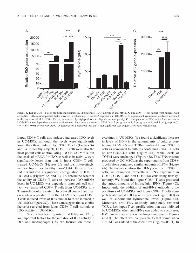
Lupus CD4� T cells also induced increased IDO levelsin UC-MSCs, although the levels were significantlylower than those induced by CD8� T cells (Figures 3Aand B). In healthy subjects, CD8� T cells were also themost potent cells at stimulating IDO in UC-MSCs, butthe levels of mRNA for IDO, as well as its activity, weresignificantly lower than that in lupus CD8� T cell–treated UC-MSCs (Figures 3A and B). Interestingly,neither lupus nor healthy non-CD4/CD8 cells fromPMBCs induced a significant up-regulation of IDO inUC-MSCs (Figures 3A and B). To determine whetherthe ability of CD8� T cells to increase IDO mRNAlevels in UC-MSCs was dependent upon cell–cell con-tact, we separated CD8� T cells from UC-MSCs in aTranswell coculture system. In cell–cell contact cultures,even when separated from the UC-MSCs, lupus CD8�T cells induced levels of IDO similar to those induced inUC-MSCs (Figure 3C). These data suggest that a solublefactor(s) secreted from lupus CD8� T cells stimulatesIDO activity in UC-MSCs.
Since it has been reported that IFN� and TGF�are important factors for the initiation of IDO activity inDCs and macrophages (18), we focused on these 2
cytokines in UC-MSCs. We found a significant increasein levels of IFN� in the supernatants of cultures con-taining UC-MSCs and TCR-stimulated lupus CD8� Tcells as compared to cultures containing CD4� T cellsor non-CD4/CD8 cells (Figure 4A), while levels ofTGF�1 were unchanged (Figure 4B). This IFN� was notproduced by UC-MSCs, as the supernatants from CD8�T cells alone contained similar amounts of IFN� (Figure4A). To further confirm that IFN� was from CD8� Tcells, we examined intracellular IFN� expression inCD4�, CD8�, and non-CD4/CD8 cells using flow cy-tometry. We found that lupus CD8� T cells producedthe largest amounts of intracellular IFN� (Figure 4C).Importantly, the addition of anti-IFN� antibody to thecocultures of UC-MSCs and lupus CD8� T cells com-pletely abrogated IDO gene expression (Figure 4D) aswell as supernatant kynurenine levels (Figure 4E).Moreover, anti-IFN� antibody completely restoredTCR-driven lupus T cell proliferation that was inhibitedby UC-MSCs; when anti-IFN� was added to cocultures,IDO enzyme activity was no longer increased (Figures4F–H). The effect was comparable to that found when1-DL-MT was added to the cocultures (Figures 4F–H). In
Contro
l
HC CD4
HC CD8
HC non-C
D4/CD8
SLE C
D4
SLE C
D8
SLE no
n-CD4/C
D80
1020304050
5000
10000
15000
20000 *** ***
***
***R
elat
ive IDO
gen
e ex
pres
sion
Contro
l
HC CD4
HC CD8
HC non-C
D4/CD8
SLE C
D4
SLE C
D8
SLE no
n-CD4/C
D80
50
100
150 ****** ***
Kynu
reni
ne (μ
M)
Contro
lCD8
CD8 tran
swell
0
10000
20000
30000***
n.s.
***
Rel
ativ
e IDO
gen
e ex
pres
sion
A B C
Figure 3. Lupus CD8� T cells promote indoleamine 2,3-dioxygenase (IDO) activity in UC-MSCs. A, The CD8� T cell subset from patients withactive SLE is the most important factor involved in enhancing IDO mRNA expression in UC-MSCs. B, Supernatant kynurenine levels are increasedin the presence of SLE CD8� T cells, as assessed by high-performance liquid chromatography. C, Up-regulation of IDO mRNA expression inUC-MSCs is not dependent upon cell–cell contact. Bars show the mean � SEM (n 7 per group in A, 7 per group in B, and 4 per group in C).��� P 0.001 by one-way ANOVA followed by Bonferroni test. NS not significant (see Figure 1 for other definitions).
A CD8 T CELL/IDO AXIS IN MSC IMMUNOTHERAPY IN SLE 2239

contrast, neutralization of TGF� with anti-TGF� anti-body failed to significantly decrease IDO activity and/orrestore T cell proliferation in the same UC-MSCs andlupus PBMC cocultures (Figure 4F–I). These data col-lectively indicate that the IFN� that is secreted by lupusCD8� T cells stimulates IDO activity in UC-MSCs andthat enhanced levels of IDO then cause inhibition of Tcell proliferation.
Association of JAK/STAT pathways with en-hanced levels of IDO activity in UC-MSCs. We nextsought to elucidate the signaling pathways by whichlupus CD8� T cells induced IDO production in UC-
MSCs. In DCs, the initiation of IDO activity is mostlydependent upon noncanonical NF-�B signaling path-ways (29). On the other hand, JAK/STAT signalingactivation is involved in IFN�-induced immune re-sponses (30). How CD8� T cells induce IDO in UC-MSCs is unknown. In vitro stimulation by lupus CD8� Tcells resulted in a significant increase in IFNGR1 but notIFNGR2 in UC-MSCs (Figure 5A and SupplementaryFigure 4A, available on the Arthritis & Rheumatology website at http://onlinelibrary.wiley.com/doi/10.1002/art.38674/abstract). In addition, downstream JAK-2 (al-though not JAK-1) gene expression in UC-MSCs was
CD4CD8
non-C
D4/CD8
CD4CD8
non-C
D4/CD8
Contro
l0
50
100
150
200
250
without MSC with MSC
******
**
Sup
erna
tant
IFN
-γ (p
g/m
l)
CD4CD8
non-C
D4/CD8
CD4CD8
non-C
D4/CD8
Contro
l10
12
14
16
18
20
22
with MSCwithout MSC
Sup
erna
tant
TG
F-β
1 (p
g/m
l)
CD4-FITCCD4-FITC CD8-APC
HC
SLE
IFN
--
γP
EC
y7
MSCCon
trol γ
anti-I
FN
IgG is
otype
0
5000
10000
15000
20000
SLE CD8+T cell+MSC
**
Rel
ativ
e IDO
gen
e ex
pres
sion
MSCCon
trol γ
anti-I
FN
IgG is
otype
0
10
20
30
SLE CD8 +T cell+MSC
*** ***
Kynu
reni
ne (μ
M)
PBMC
PBMC+MSC γ
anti-I
FN β
anti-T
GF
IgG is
otype
1-DL-M
T0
50
100
150 ****** ***
Kynu
reni
ne/T
rypt
opha
n (μ
M/μ
M)
PBMC
PBMC+MSC γ
anti-I
FN β
anti-T
GF
IgG is
otype
1-DL-M
T0
20
40
60
80 **
****
Perc
enta
ge o
f pro
lifer
atio
n (%
)
A B C
D E F G
CD4CD8
non-C
D4/CD8CD4
CD8
non-C
D4/CD8
0
20
40
60
80
SLEHC
******
Perc
enta
ge o
f IFN
-γ+
cells
(%)
11%
42%
43%
77%
9%
14%
CD4+T cells CD8+T cells Non-CD4/CD8 cells
PBMC
PBMC+MSC
anti-I
FN
anti-T
GF
IgG iso
type
1-DL-M
T0
10
20
30
40
**
*
*
μM
)
PBMC
PBMC+MSC
anti-I
FNγ
anti-T
GFβ
IgG iso
type
1-DL-M
T0
5
10
15
20
μM
) ******
***
H I
PBMC+MSC PBMC+MSC
Kyn
uren
ine
(
Tryp
toph
an (
PBMC+MSC PBMC+MSC
γ β
Figure 4. Lupus CD8� T cell–derived interferon-� (IFN�) promotes indoleamine 2,3-dioxygenase (IDO) production. A and B, Levels of IFN� insupernatant are increased in the presence of UC-MSCs and SLE CD8� T cells (A), while no change in levels of transforming growth factor �(TGF�) is observed (B). Symbols represent individual subjects; horizontal lines show the mean (n 7 per group). C, CD8� T cells derived frompatients with SLE produce much higher levels of intracellular IFN� as compared to other cell subsets. D, Recombinant anti-human IFN� antibodysignificantly abrogates SLE CD� T cell–mediated IDO mRNA expression in UC-MSCs. E, Kynurenine levels in supernatant decrease in thepresence of anti-human IFN� antibody. F–H, In cocultured lupus peripheral blood mononuclear cells (PBMCs) and UC-MSCs, anti-human IFN�antibodies (10 �g/ml) significantly inhibit kynurenine levels in supernatant (F), while the level of tryptophan is increased (G), and the ratio ofkynurenine to tryptophan is decreased (H) (effects similar to those observed when 1-methyl-DL-tryptophan [1-DL-MT] is added). I, In the coculturesystem described above, UC-MSC–mediated inhibition of CD4� T cell proliferation is abrogated by the addition of anti-human IFN� antibody. Barsin C–I show the mean � SEM (n 7 per group in C, 8 per group in D, 8 per group in E, 5 per group in F–H, and 5 per group in I). � P 0.05;�� P 0.01; ��� P 0.001, by one-way ANOVA followed by Bonferroni test. PE phycoerythrin; FITC fluorescein isothiocyanate; APC allophycocyanin (see Figure 1 for other definitions). Color figure can be viewed in the online issue, which is available at http://onlinelibrary.wiley.com/doi/10.1002/art.38674/abstract.
2240 WANG ET AL

significantly up-regulated in the presence of lupusCD8� T cells, an up-regulation which was similar to thatseen when cells were stimulated with 20 ng/ml of recom-binant human IFN� (Figure 5B and SupplementaryFigure 4B). Furthermore, STAT-1 gene expression wasalso increased in these settings (Figure 5C). Westernblot analysis confirmed the activation of STAT-1,STAT-3, and STAT-5 signaling pathways following co-culture of lupus CD8� T cells with UC-MSCs (Figure5D). We also examined NF-�B, ERK, and Akt pathwaysin UC-MSCs, but found no obvious activation in thepresence of lupus CD8� T cells (Supplementary Figure
4C). Thus, IFNGR1/JAK-2/STAT signaling pathwaysare associated with the IDO activity in UC-MSCs that isstimulated by lupus CD8� T cells.
Defective IDO activity in lupus BM-MSCs. Theprofound IDO production by allogeneic UC-MSCs inresponse to lupus CD8� T cells prompted us to explorewhether MSCs in patients with active lupus had defec-tive IDO activity. We therefore isolated MSCs from theBM of patients with active lupus and healthy subjectsand cultured the cells in vitro. The expanded BM-MSCswere stimulated with IFN�, and UC-MSCs were used ascontrols. We found that lupus BM-MSCs had signifi-
γ0
1
2
3
4
5
6
7
***
***A
n0
5
10
15
***
0
10
20
30
40
**
**B C D
0
5000
10000
15000
20000***
0
0
00
50
200
n.s.
μ
F G H
Contro
l α
IFN- β
IFN- γ
IFN- β
TGF-
α
TNF- IL-
6 βIL-
10
20000
40000
60000
80000UCHC BMSLE BM
******
E
0
20
40
60
80
100n.s.
*****
Rela
tive IFNGR1
gen
e ex
pres
sion
Rela
tive JAK2
gen
e ex
pres
sion
Rela
tive STAT1
gene
exp
ress
ion
Rela
tive IDO
gen
e ex
pres
sion
Kynu
reni
ne (
M)
Perc
enta
ge o
f pro
lifera
tion
(%)
MSC
CD4CD8
non
CD4/CD8
IFN- γ
MSC
CD4CD8
non
CD4/CD8
IFN- γ
MSC
CD4CD8
non
CD4/CD8
IFN-
MSC
SLE C
D8SLE
CD4
SLE n
on-C
D4/CD8
HC CD8
HC CD4
HC non
-CD4/
CD8
γ
IFN-
Rela
tive IDO
gen
e ex
pres
sion
Coun
t
CFSE
PBMC PBMC+HC MSC PBMC+SLE MSC
p-Stat1
t-Stat1p-Stat3
t-Stat3
p-Stat5
t-Stat5
GAPDH
UC
HC BM
SLE B
M UC
HC BM
SLE B
MPBMC
PBMC+HC MSC
PBMC+SLE MSC
***
Figure 5. Defective indoleamine 2,3-dioxygenase (IDO) production in lupus bone marrow–derived MSCs (BM-MSCs). A–C, Expression ofIFNGR1 (A), JAK-2 (B), and STAT-1 (C) was examined by real-time quantitative polymerase chain reaction. D, STAT-1, STAT-3, STAT-5, andtheir phosphorylated forms were assessed by Western blot analysis after treatment with UC-MSCs alone, UC-MSCs with different cell subsets(CD4�, CD8�, and non-CD4/CD8 T cells), or recombinant human interferon-� (IFN�). E, IDO mRNA expression in UC-MSCs, BM-MSCs fromhealthy controls, and BM-MSCs from SLE patients was examined after stimulation with different cytokines for 48 hours. F and G, Peripheral bloodCD8� T cells derived from patients with lupus were purified and used to stimulate UC-MSCs and BM-MSCs from healthy controls or lupus patients.Forty-eight hours later, IDO gene expression (F) and kynurenine enzyme activity (G) were determined. H, The ability of BM-MSCs from healthycontrols and lupus patients to inhibit CD4� T cell proliferation was compared. Bars in A–C and E–H show the mean � SEM (n 7 per group inA–D, 6 per group in E, 3 per group in F and G, and 4 per group in H). �� P 0.01; ��� P 0.001, by one-way ANOVA. TGF� transforminggrowth factor �; IL-6 interleukin-6; PBMC peripheral blood mononuclear cell; NS not significant (see Figure 1 for other definitions).
A CD8 T CELL/IDO AXIS IN MSC IMMUNOTHERAPY IN SLE 2241

cantly lower IDO mRNA levels compared to healthyBM-MSCs or UC-MSCs, in response to IFN� stimula-tion (Figure 5E). When cocultured with allogeneic TCR-activated lupus CD8� T cells, lupus BM-MSCs alsoexhibited a profound defect in IDO secretion and activ-ity compared to normal BM-MSCs (Figures 5F and G).Importantly, when cocultured with allogeneic lupus PB-MCs, lupus BM-MSCs had a reduced ability to suppressT cell proliferation as compared to normal BM-MSCs(Figure 5H). These data demonstrate a decrease in IDOlevels in MSCs derived from lupus patients in responseto IFN� and CD8� T cells.
Increased circulating IDO activity after UC-MSC transplantation in lupus patients. Since IFN� andCD8� T cells trigger allogeneic MSCs to produce IDOand inhibit lupus T cell proliferation, we assessedwhether IFN� and CD8� T cells were increased in lupuspatients. We therefore analyzed a clinical index in lupuspatients in vivo. First, we compared circulating levels ofIFN�, CD4�, and CD8� T cell subsets in the peripheralblood of lupus patients and healthy controls. We foundthat lupus patients had a significantly higher frequencyand total number of peripheral CD8� T cells andincreased levels of circulating IFN� (Figures 6A–C).Moreover, there was an increased frequency and abso-lute number of IFN��CD8� T cells from lupus patients
compared to those from healthy controls (Figures 6Dand E). These data suggest that the lupus microenviron-ment can initiate the function of allogeneic MSCs invivo. Importantly, we observed that serum IDO activity(as evidenced by kynurenine concentrations) was signif-icantly increased in 6 lupus patients 1 month afterintravenous UC-MSC transplantation (Figure 6F). Se-rum tryptophan levels, however, did not change (Figure6G), while the ratio of kynurenine to tryptophan wasmarkedly increased (Figure 6H). Furthermore, in an-other clinical study, we found that percentages of peri-pheral blood CD3�CD4� T cells decreased after UC-MSC transplantation in patients (Supplementary Figure5, available on the Arthritis & Rheumatology web site athttp://onlinelibrary.wiley.com/doi/10.1002/art.38674/abstract). These data are consistent with our in vitrodata and suggest that IDO plays a pivotal role inallogeneic MSC treatment in lupus patients.
DISCUSSION
The molecules that mediate MSC inhibition oflupus inflammatory cells remain incompletely under-stood. Herein we show that UC-MSC–produced IDOwas critical for the inhibition of T cells and that IFN�produced by lupus CD8� T cells was the main factor
Normal SLE0
20
40
60
80
CD
8+ CD
4- /CD
3+ T ce
lls (%
)
Normal SLE0
1000
2000
3000
4000
**
CD
3+ CD
8+ CD
4- T c
ell n
umbe
r (μ l
)
SLE 0
20
40
60
80 *
Seru
m IF
N -γ
(pg/
ml)
Normal SLE0
20
40
60
80
100
CD
3+ CD
8+ CD
4- IFN
γ+ T c
ell (
%)
Normal SLE0
500
1000
1500**
CD
3+ CD
8+ CD
4- IFN
γ+
T ce
ll nu
mbe
r (μ l
)
A B C D
E F
Before After0
50
100
150
200
250 **
Kynu
renin
e/Tr
ypto
phan
(nM
/ μM
)
Normal
Before After0
2
4
6
8
Kynu
reni
ne (μ
M)
*
Before After0
20
40
60
80
Tryp
toph
an (μ
M)
G H
*** ***
Figure 6. Serum indoleamine 2,3-dioxygenase (IDO) activity increases after UC-MSC transplantation in lupus patients. Peripheral bloodmononuclear cells were isolated from lupus patients and healthy controls, and levels of CD8� T cell subsets and interferon-� (IFN�) were compared.A–C, The percentage (A) and total number (B) of CD8�CD4�/CD3� T cells, as well as serum levels of IFN� (C) are significantly increased in SLEpatients. D and E, The percentage (D) and total number (E) of intracellular IFN��CD8� T cells are also increased in SLE patients. F–H, Serumkynurenine levels are increased (F), tryptophan levels remain unchanged (G), and the ratio of kynurenine to tryptophan is increased (H) 1 monthafter UC-MSC transplantation in lupus patients. � P 0.05; �� P 0.01; ��� P 0.001. Symbols in A–E represent individual subjects;horizontal lines show the mean. See Figure 1 for other definitions.
2242 WANG ET AL

driving IDO induction by MSCs. We have highlighted anovel mechanism by which allogeneic MSCs regulatelupus T cells in the disease microenvironment.
SLE is a typical autoimmune disease, character-ized by abnormal T and B cell functions. Recently, adefect in Treg cell number and function was reported inpatients with active lupus, which correlated with diseaseonset and progression (31). Current immunosuppressivedrugs used to treat lupus inhibit T and B lymphocytes invivo indiscriminately, which may increase drug-relatedadverse events, such as infection (32,33). New biologicdrugs that target B cells, such as anti-CD20 monoclonalantibody (rituximab) and anti-BAFF monoclonal anti-body (belimumab), have shown satisfactory clinical effi-cacy in patients with refractory disease, but treatment-related adverse events occurred after long-termapplication and followup (34,35). MSCs, however, mayselectively inhibit activated lymphocytes and have there-fore been proposed as an alternative treatment optionfor patients with lupus and other autoimmune diseases.The activity of MSCs is affected by the microenviron-ment into which they are transferred. As such, establish-ing how MSCs act within a diseased environment and,more specifically, how they mediate immune tolerancein lupus patients, is pivotal to improving our understand-ing of MSC transplantation and identifying the patientsin whom an MSC transplant would provide the mostclinical benefit. Our findings reveal a CD8� T cell/IFN�/IDO axis, by which allogeneic MSCs inhibit T cellproliferation.
IFN signaling pathways are activated in lupuspatients and are tightly correlated with disease activity(36). It has been reported that circulating IFN� isincreased in lupus patients and can facilitate B cellactivation and antibody production (37,38). Previously,it was reported that the high levels of IFN� in lupuspatients were mainly produced by DCs (39) or naturalkiller cells (40). Not only have we identified CD8� Tcells as dominant cellular sources of IFN� in lupuspatients, but we have also importantly discovered thatCD8� T cells are the major stimulus for induction ofIDO in UC-MSCs. Furthermore, we observed signifi-cantly elevated levels of circulating IFN� produced byCD8� T cells in lupus patients. More research on lupusis needed to determine whether circulating IFN� levelsare positively correlated with allogeneic MSC treatmentefficacy. In the present study, we also found that lupusTreg cells enhanced UC-MSCs–mediated IDO produc-tions, but IDO was not involved in Treg proliferation.This increase in IDO expression by UC-MSCs cocul-tured with lupus Treg cells might be due to a slight
increase in IFN� production by these lupus Treg cells(Wang D, et al: unpublished observations), although thisneeds to be further confirmed in future studies.
Our previous studies showed that BM-MSCsfrom lupus patients functioned abnormally (7,41) andthat autologous BM-MSC infusion had no significanteffect on animal models of lupus (42). In this study, wefound that BM-MSCs from patients with active SLEwere much less responsive to recombinant IFN� orallogeneic CD8� T cell stimulation and, importantly,failed to inhibit allogeneic T cell proliferation. Thisdefect in suppressing T cell proliferation is at least partlyattributed to their reduced ability to produce IDO inresponse to IFN� and/or lupus CD8� T cells. Thesefindings explain why activated T cells were elevated inlupus patients and why autologous lupus BM-MSCtransplantation was less effective at treating lupus pa-tients. Many patients have received allogeneic MSCtransplantation in our facility, and we have shown a goodclinical safety profile as well as treatment efficacy (43).However, because the outcome of treatment with theinfused allogeneic cells is unknown, long-term clinicalsafety needs further investigation.
We recently examined the characteristics of BM-MSCs from SLE patients and healthy controls. Our datashowed that there were no significant differences in thesurface phenotype of CD29, CD44, CD105, CD14,CD34, CD45, and HLA–DR cells (41). We suggest thatthe decreased IDO from SLE-derived MSCs may be aresult of some intrinsic factors that occur in lupusdisease progression, but which do not necessarily causechanges in the surface phenotype of MSCs. Moreover,IFN� treatment in vitro had no effect on MSC surfaceHLA–DR, CD80, and CD86 expression, although itenhanced IDO activity in a dose-dependent manner(Wang D, et al: unpublished observations). Further-more, a study has demonstrated that BM-MSCs fromyoung (NZB � NZW)F1 lupus mice (5–6 weeks old)efficiently reduced severity of lupus in an MRL/lprmouse model (42). However, BM-MSCs from old(NZB � NZW)F1 mice (26–27 weeks old) failed toameliorate disease, indicating that BM-MSCs lose theirsuppressive capabilities as the disease progresses. Lossof regulatory activity by BM-MSCs is likely caused bythe inflammatory milieu present in the host, which couldwell mediate epigenetic changes and in consequenceyield changes in protein expression in BM-MSCs fromlupus patients.
Taken together, our findings reveal novel mech-anistic insight into how UC-MSC–mediated immuno-suppression occurs in lupus patients. Additionally, our
A CD8 T CELL/IDO AXIS IN MSC IMMUNOTHERAPY IN SLE 2243

data suggest that allogeneic MSCs are more appropriatefor clinical transplantation in lupus patients, althoughautologous MSCs are not.
ACKNOWLEDGMENTS
We thank Drs. DaWei Xiao and MingWei Yang(Department of Pharmacy and Phase I Clinical Trial Base, TheAffiliated Drum Tower Hospital of Nanjing University Medi-cal School) for technical assistance with high-performanceliquid chromatography. We also thank the patients and healthyvolunteers for their cooperation and for consenting to partic-ipate in this study.
AUTHOR CONTRIBUTIONS
All authors were involved in drafting the article or revising itcritically for important intellectual content, and all authors approvedthe final version to be published. Dr. Sun had full access to all of thedata in the study and takes responsibility for the integrity of the dataand the accuracy of the data analysis.Study conception and design. Wang, Feng, Gao, Lu, Shi, W. Chen,Sun.Acquisition of data. Wang, Feng, Lu, Konkel, Zhang, Z. Chen, Li.Analysis and interpretation of data. Wang, W. Chen, Sun.
REFERENCES
1. Le Blanc K, Mougiakakos D. Multipotent mesenchymal stromalcells and the innate immune system. Nat Rev Immunol 2012;12:383–96.
2. Liang J, Zhang H, Hua B, Wang H, Lu L, Shi S, et al. Allogenicmesenchymal stem cells transplantation in refractory systemiclupus erythematosus: a pilot clinical study [Ann Rheum Dis2011;70:237]. Ann Rheum Dis 2010;69:1423–9.
3. Sun L, Wang D, Liang J, Zhang H, Feng X, Wang H, et al.Umbilical cord mesenchymal stem cell transplantation in severeand refractory systemic lupus erythematosus. Arthritis Rheum2010;62:2467–75.
4. Akiyama K, Chen C, Wang D, Xu X, Qu C, Yamaza T, et al.Mesenchymal-stem-cell-induced immunoregulation involves FAS-ligand-/FAS-mediated T cell apoptosis. Cell Stem Cell 2012;10:544–55.
5. Xu J, Wang D, Liu D, Fan Z, Zhang H, Liu O, et al. Allogeneicmesenchymal stem cell treatment alleviates experimental andclinical Sjögren syndrome. Blood 2012;120:3142–51.
6. Wang D, Zhang H, Cao M, Tang Y, Liang J, Feng X, et al. Efficacyof allogeneic mesenchymal stem cell transplantation in patientswith drug-resistant polymyositis and dermatomyositis. Ann RheumDis 2011;70:1285–8.
7. Li X, Liu L, Meng D, Wang D, Zhang J, Shi D, et al. Enhancedapoptosis and senescence of bone-marrow-derived mesenchymalstem cells in patients with systemic lupus erythematosus. StemCells Dev 2012;21:2387–94.
8. Carrion F, Nova E, Ruiz C, Diaz F, Inostroza C, Rojo D, et al.Autologous mesenchymal stem cell treatment increased T regula-tory cells with no effect on disease activity in two systemic lupuserythematosus patients. Lupus 2010;19:317–22.
9. Nie Y, Lau C, Lie A, Chan G, Mok M. Defective phenotype ofmesenchymal stem cells in patients with systemic lupus erythem-atosus. Lupus 2010;19:850–9.
10. English K, French A, Wood KJ. Mesenchymal stromal cells:
facilitators of successful transplantation? Cell Stem Cell 2010;7:431–42.
11. Keating A. Mesenchymal stromal cells: new directions. Cell StemCell 2012;10:709–16.
12. Nauta AJ, Fibbe WE. Immunomodulatory properties of mesen-chymal stromal cells. Blood 2007;110:3499–506.
13. Yagi H, Soto-Gutierrez A, Parekkadan B, Kitagawa Y, TompkinsRG, Kobayashi N, et al. Mesenchymal stem cells: mechanisms ofimmunomodulation and homing. Cell Transplant 2010;9:667–79.
14. Sato K, Ozaki K, Oh I, Meguro A, Hatanaka K, Nagai T, et al.Nitric oxide plays a critical role in suppression of T-cell prolifer-ation by mesenchymal stem cells. Blood 2007;109:228–34.
15. Chabannes D, Hill M, Merieau E, Rossignol J, Brion R, SoulillouJP, et al. A role for heme oxygenase-1 in the immunosuppressiveeffect of adult rat and human mesenchymal stem cells. Blood2007;110:3691–4.
16. Selmani Z, Naji A, Gaiffe E, Obert L, Tiberghien P, Rouas-FreissN, et al. HLA-G is a crucial immunosuppressive molecule secretedby adult human mesenchymal stem cells. Transplantation 2009;87:S62–6.
17. Mellor AL, Munn DH. IDO expression by dendritic cells: toler-ance and tryptophan catabolism. Nat Rev Immunol 2004;4:762–74.
18. Pallotta MT, Orabona C, Volpi C, Vacca C, Belladonna ML,Bianchi R, et al. Indoleamine 2,3-dioxygenase is a signalingprotein in long-term tolerance by dendritic cells. Nat Immunol2011;12:870–8.
19. Meisel R, Zibert A, Laryea M, Gobel U, Daubener W, Dilloo D.Human bone marrow stromal cells inhibit allogeneic T-cell re-sponses by indoleamine 2,3-dioxygenase-mediated tryptophan de-gradation. Blood 2004;103:4619–21.
20. Selmani Z, Naji A, Zidi I, Favier B, Gaiffe E, Obert L, et al.Human leukocyte antigen-G5 secretion by human mesenchymalstem cells is required to suppress T lymphocyte and natural killerfunction and to induce CD4�CD25highFOXP3� regulatory T cells.Stem Cells 2008;26:212–22.
21. Ren G, Zhang L, Zhao X, Xu G, Zhang Y, Roberts AI, et al.Mesenchymal stem cell-mediated immunosuppression occurs viaconcerted action of chemokines and nitric oxide. Cell Stem Cell2008;2:141–50.
22. Qian X, Wang K, Wang X, Zheng SG, Lu L. Generation of humanregulatory T cells de novo with suppressive function preventxenogeneic graft versus host disease. Int Immunopharmacol 2011;11:630–7.
23. Chen W, Jin W, Hardegen N, Lei KJ, Li L, Marinos N, et al.Conversion of peripheral CD4�CD25� naive T cells toCD4�CD25� regulatory T cells by TGF-� induction of transcrip-tion factor Foxp3. J Exp Med 2003;198:1875–86.
24. Sun L, Akiyama K, Zhang H, Yamaza T, Hou Y, Zhao S, et al.Mesenchymal stem cell transplantation reverses multiorgan dys-function in systemic lupus erythematosus mice and humans. StemCells 2009;27:1421–32.
25. Jasperson LK, Bucher C, Panoskaltsis-Mortari A, Mellor AL,Munn DH, Blazar BR, et al. Inducing the tryptophan catabolicpathway, indoleamine 2,3-dioxygenase (IDO), for suppression ofgraft-versus-host disease (GVHD) lethality. Blood 2009;114:5062–70.
26. Widner B, Werner ER, Schennach H, Wachter H, Fuchs D.Simultaneous measurement of serum tryptophan and kynurenineby HPLC. Clin Chem 1997;43:2424–6.
27. Glennie S, Soeiro I, Dyson PJ, Lam EW, Dazzi F. Bone marrowmesenchymal stem cells induce division arrest anergy of activatedT cells. Blood 2005;105:2821–7.
28. Aggarwal S, Pittenger MF. Human mesenchymal stem cells mod-ulate allogeneic immune cell responses. Blood 2005;105:1815–22.
29. Puccetti P, Grohmann U. IDO and regulatory T cells: a role forreverse signalling and non-canonical NF-�B activation. Nat RevImmunol 2007;7:817–23.
2244 WANG ET AL

30. Hu X, Ivashkiv LB. Cross-regulation of signaling pathways byinterferon-�: implications for immune responses and autoimmunediseases. Immunity 2009;31:539–50.
31. Scheinecker C, Bonelli M, Smolen JS. Pathogenetic aspects ofsystemic lupus erythematosus with an emphasis on regulatory Tcells. J Autoimmun 2010;35:269–75.
32. Crispin JC, Kyttaris VC, Terhorst C, Tsokos GC. T cells astherapeutic targets in SLE. Nat Rev Rheumatol 2010;6:317–25.
33. Sanz I, Lee FE. B cells as therapeutic targets in SLE. Nat RevRheumatol 2010;6:326–37.
34. Navarra SV, Guzman RM, Gallacher AE, Hall S, Levy RA,Jimenez RE, et al, for the BLISS-52 Study Group. Efficacy andsafety of belimumab in patients with active systemic lupus ery-thematosus: a randomised, placebo-controlled, phase 3 trial. Lan-cet 2011;377:721–31.
35. Merrill JT, Neuwelt CM, Wallace DJ, Shanahan JC, Latinis KM,Oates JC, et al. Efficacy and safety of rituximab in moderately-to-severely active systemic lupus erythematosus: the randomized,double-blind, phase II/III Systemic Lupus Erythematosus Evalua-tion of Rituximab trial. Arthritis Rheum 2010;62:222–33.
36. Hayashi T. Therapeutic strategies for SLE involving cytokines:mechanism-oriented therapies especially IFN-� targeting genetherapy. J Biomed Biotechnol 2010;2010:461641.
37. Harigai M, Kawamoto M, Hara M, Kubota T, Kamatani N,Miyasaka N. Excessive production of IFN-gamma in patients withsystemic lupus erythematosus and its contribution to induction of
B lymphocyte stimulator/B cell-activating factor/TNF ligand su-perfamily-13B. J Immunol 2008;181:2211–9.
38. Karonitsch T, Feierl E, Steiner CW, Dalwigk K, Korb A, Binder N,et al. Activation of the interferon-� signaling pathway in systemiclupus erythematosus peripheral blood mononuclear cells. ArthritisRheum 2009;60:1463–71.
39. Teichmann LL, Ols ML, Kashgarian M, Reizis B, Kaplan DH,Shlomchik MJ. Dendritic cells in lupus are not required foractivation of T and B cells but promote their expansion, resultingin tissue damage. Immunity 2010;33:967–78.
40. Hervier B, Beziat V, Haroche J, Mathian A, Lebon P, Ghillani-Dalbin P, et al. Phenotype and function of natural killer cells insystemic lupus erythematosus: excess interferon-� production inpatients with active disease. Arthritis Rheum 2011;63:1698–706.
41. Sun LY, Zhang HY, Feng XB, Hou YY, Lu LW, Fan LM.Abnormality of bone marrow-derived mesenchymal stem cells inpatients with systemic lupus erythematosus. Lupus 2007;16:121–8.
42. Gu F, Molano I, Ruiz P, Sun L, Gilkeson GS. Differential effect ofallogeneic versus syngeneic mesenchymal stem cell transplantationin MRL/lpr and (NZB/NZW)F1 mice. Clin Immunol 2012;145:142–52.
43. Wang D, Zhang H, Liang J, Li X, Feng X, Wang H, et al.Allogeneic mesenchymal stem cell transplantation in severe andrefractory systemic lupus erythematosus: 4 years experience. CellTransplant 2013;22:2267–77.
A CD8 T CELL/IDO AXIS IN MSC IMMUNOTHERAPY IN SLE 2245