a common variant of prok1 (v67i) acts as a genetic modifier in
TRANSCRIPT
International Journal of
Molecular Sciences
Article
A Common Variant of PROK1 (V67I) Acts asa Genetic Modifier in Early Human Pregnancythrough Down-Regulation of Gene Expression
Mei-Tsz Su 1, Jyun-Yuan Huang 1, Hui-Ling Tsai 1, Yi-Chi Chen 2 and Pao-Lin Kuo 1,*1 Department of Obstetrics and Gynecology, National Cheng Kung University Hospital, College of Medicine,
National Cheng Kung University, Tainan 704, Taiwan; [email protected] (M.-T.S.);[email protected] (J.-Y.H.); [email protected] (H.-L.T.)
2 Department of Economics, National Cheng Kung University, Tainan 704, Taiwan; [email protected]* Correspondence: [email protected]; Tel.:+886-6-235-3535 (ext. 5222); Fax: +886-6-276-6185
Academic Editor: Constantinos StathopoulosReceived: 7 December 2015; Accepted: 16 January 2016; Published: 27 January 2016
Abstract: PROK1-V67I has been shown to play a role as a modifier gene in the PROK1-PROKR systemof human early pregnancy. To explore the related modifier mechanism of PROK1-V67I, we carriedout a comparison study at the gene expression level and the cell function alternation of V67I, and itswild-type (WT), in transiently-transfected cells. We, respectively, performed quantitative RT-PCRand ELISA assays to evaluate the protein and/or transcript level of V67I and WT in HTR-8/SV neo,JAR, Ishikawa, and HEK293 cells. Transiently V67I- or WT-transfected HTR-8/SV neo and HEK293cells were used to investigate cell function alternations. The transcript and protein expressionswere down-regulated in all cell lines, ranging from 20% to 70%, compared with WT. There wereno significant differences in the ligand activities of V67I and WT with regard to cell proliferation,cell invasion, calcium influx, and tubal formation. Both PROK1 alleles promoted cell invasion andintracellular calcium mobilization, whereas they had no significant effects on cell proliferation andtubal formation. In conclusion, the biological effects of PROK1-V67I on cell functions are similar tothose of WT, and the common variant of V67I may act as a modifier in the PROK1-PROKR systemthrough down-regulation of PROK1 expression. This study may provide a general mechanism thatthe common variant of V67I, modifying the disease severity of PROK1-related pathophysiologies.
Keywords: modifier gene; Prokineticin 1 (PROK1; EG-VEGF); recurrent miscarriage; calcium influx;cell invasion
1. Introduction
Prokineticin 1 (PROK1), also known as endocrine gland-derived vascular endothelial growthfactor (EG-VEGF), is a small, secreted peptide that belongs to the prokineticin family [1]. PROK1 isa tissue-specific proangiogenetic mitogen and chemotactic factor, and it acts through activation of twocognate G-protein-coupled receptors (GPCRs), prokineticin receptor 1 (PROKR1) and prokineticinreceptor 2 (PROKR2). The expression of PROK1 is predominantly in the steroidogenic glands, such asovary, testis, adrenal cortex, and placenta [2–4], and has been shown to have a wide range of functionsincluding angiogenesis, modulation of inflammatory responses, and regulation of hematopoiesis) [5,6].
In recent years, PROK1 has been shown to play a role in female reproduction and humanpregnancy. The temporal and spatial expression of PROK1, and its receptors in ovary and earlygestational tissue, highlights their functions in follicular maturation, luteal angiogensis, embryoimplantation, and uterine receptivity [1,7,8]. The dynamic expression and regulation profile of thePROK1-PROKR system through human pregnancy also suggests its regulatory role in the process of
Int. J. Mol. Sci. 2016, 17, 162; doi:10.3390/ijms17020162 www.mdpi.com/journal/ijms

Int. J. Mol. Sci. 2016, 17, 162 2 of 13
placental development and initiation of parturition [9–11]. Previous publication had shown that PROK1inhibits and controls trophoblast invasion in human pregnancy [12], whereas a recent prospectivestudy suggested PROK1 may facilitate an embryo endowing with adequate implantation potential [13].Moreover, aberrant expression or activity of PROK1 and its receptors are also reported to be associatedwith several gestational complication, and most were related to inadequate or inappropriate trophoblastinvasion, such as gestational trophoblastic diseases, recurrent pregnancy losses, preeclampsia, andintrauterine fetal growth restriction [11,14–16].
A common polymorphism of PROK1, V67I (c.199 G>A, rs7514102), located in the first nucleotide ofexon 3, is a non-synonymous substitution resulting in amino acid change from valine (V) to isoleucine (I).The frequency of G to A transition varies among different ethnicities, and ranges from 43% to 64%in the general population (43% in Caucasians, 54% in Han Chinese, 64% in Japanese, and 46% inNigerians, with this data taken from http://www.ncbi.nlm.nih.gov/pubmed/). The evolution of V67Iis highly conserved, and the change of amino acid from V to I seems neutral. However, the impact ofthis genetic variant in the PROK1 system and its clinical relevance have not yet been explored.
We recently reported the PROK1 variant (V67I) act as a genetic modifier in human earlypregnancy [17]. Previous studies of recurrent pregnancy loss (RPL) showed that women carryingPROKR1 and PROKR2 variants (I379V and V331M) have less susceptibilities for RPL risk, and thattrophoblastic cell function alternation by enhancing cell invasiveness may provide the protectionfrom recurrent abortion [17,18]. In contrast, the protection effect of the PROKR1/2 variant may beattenuated if the woman also carries PROK1-V67I [17]. Although genetic association research showedevidence of these effects, the underlying mechanism of the common variant of PROK1 (V67I) withregard to modifying RPL risk remains unclear. In order to better understand the critical role of PROK1in physiological and pathological pregnancy, this study aimed to explore the modification role ofPROK1-V67I and compare various cell functions to those of its wild-type (WT) in several associatedcell lines. The results of this study may provide a general mechanism of a common variant’s effect onPROK1-related disorders.
2. Results
2.1. V67I Is a Common PROK1 Variant in the General Population
We analyzed the coding regions of PROK1 using the Sanger sequence in 142 RPL womenand 149 normal controls in a previous report [17]. The allele and genotype frequencies ofPROK1-V67I variant (c.199 G>A, rs7514102) and wild-type (WT) showed no significant differencesbetween RPL and control groups, and the pooled data are 51%, 21% and 28% for GG, GA, andAA genotypes. The highly-conserved V67I variant of PROK1 is located in the first nucleotideof exon 3, and the G to A transition changes the amino acid from Valine (V) to Isoleucine (I)(Figure 1). We further compared the population diversity of V67I using HapMap data from NCBI(http://www.ncbi.nlm.nih.gov/pubmed/). In general, the frequency of the G to A transition is around50%, ranging from 39% to 64%, and varies among different populations (Table 1).
Table 1. Population diversity of PROK1 wild-type and V67I (c.199 G>A, rs7514102) variant.
Population Group/Sample Count Genotype Frequency Allele Frequency
GG GA AA G A
HapMap-CEU European/118 0.339 0.458 0.203 0.568 0.432HapMap-HCB Asian/90 0.200 0.511 0.289 0.456 0.544HapMap-JPT Asian/90 0.089 0.533 0.378 0.356 0.644HapMap-YRISub-Saharan African/120 0.319 0.450 0.233 0.542 0.458
Reference data of HapMap population diversity was derived from NCBI (updated 2015.6.24); GTC: Valine (V);ATC: Isoleucine (I); CEU: Utah Residents with Northern and Western European Ancestry; HCB: Han Chinese inBeijing, China; JPT: Japanese in Tokyo, Japan; YRI: Yoruba in Ibadan, Nigeria.
Int. J. Mol. Sci. 2016, 17, 162 3 of 13Int. J. Mol. Sci. 2016, 17, 162 3 of 13
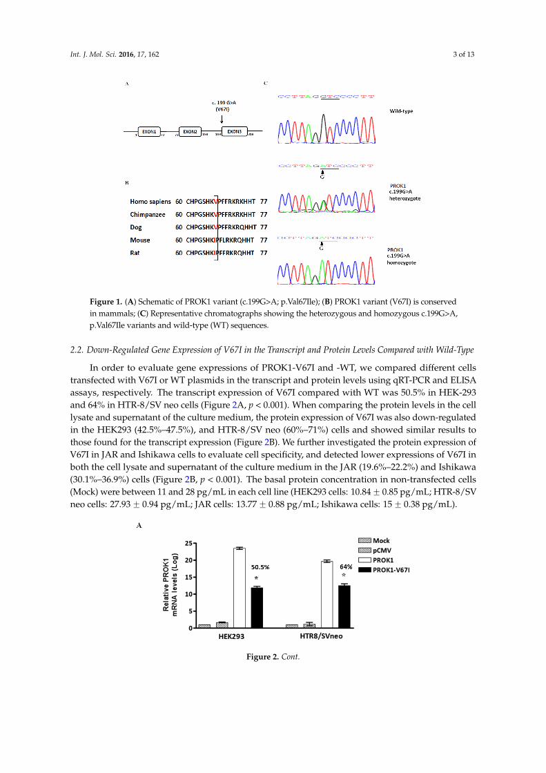
Figure 1. (A) Schematic of PROK1 variant (c.199G>A; p.Val67Ile); (B) PROK1 variant (V67I) is conserved in mammals; (C) Representative chromatographs showing the heterozygous and homozygous c.199G>A, p.Val67Ile variants and wild-type (WT) sequences.
2.2. Down-Regulated Gene Expression of V67I in the Transcript and Protein Levels Compared with Wild-Type
In order to evaluate gene expressions of PROK1-V67I and -WT, we compared different cells transfected with V67I or WT plasmids in the transcript and protein levels using qRT-PCR and ELISA assays, respectively. The transcript expression of V67I compared with WT was 50.5% in HEK-293 and 64% in HTR-8/SV neo cells (Figure 2A, p < 0.001). When comparing the protein levels in the cell lysate and supernatant of the culture medium, the protein expression of V67I was also down-regulated in the HEK293 (42.5%–47.5%), and HTR-8/SV neo (60%–71%) cells and showed similar results to those found for the transcript expression (Figure 2B). We further investigated the protein expression of V67I in JAR and Ishikawa cells to evaluate cell specificity, and detected lower expressions of V67I in both the cell lysate and supernatant of the culture medium in the JAR (19.6%–22.2%) and Ishikawa (30.1%–36.9%) cells (Figure 2B, p < 0.001). The basal protein concentration in non-transfected cells (Mock) were between 11 and 28 pg/mL in each cell line (HEK293 cells: 10.84 ± 0.85 pg/mL; HTR-8/SV neo cells: 27.93 ± 0.94 pg/mL; JAR cells: 13.77 ± 0.88 pg/mL; Ishikawa cells: 15 ± 0.38 pg/mL).
Figure 2. Cont.
Figure 1. (A) Schematic of PROK1 variant (c.199G>A; p.Val67Ile); (B) PROK1 variant (V67I) is conservedin mammals; (C) Representative chromatographs showing the heterozygous and homozygous c.199G>A,p.Val67Ile variants and wild-type (WT) sequences.
2.2. Down-Regulated Gene Expression of V67I in the Transcript and Protein Levels Compared with Wild-Type
In order to evaluate gene expressions of PROK1-V67I and -WT, we compared different cellstransfected with V67I or WT plasmids in the transcript and protein levels using qRT-PCR and ELISAassays, respectively. The transcript expression of V67I compared with WT was 50.5% in HEK-293and 64% in HTR-8/SV neo cells (Figure 2A, p < 0.001). When comparing the protein levels in the celllysate and supernatant of the culture medium, the protein expression of V67I was also down-regulatedin the HEK293 (42.5%–47.5%), and HTR-8/SV neo (60%–71%) cells and showed similar results tothose found for the transcript expression (Figure 2B). We further investigated the protein expression ofV67I in JAR and Ishikawa cells to evaluate cell specificity, and detected lower expressions of V67I inboth the cell lysate and supernatant of the culture medium in the JAR (19.6%–22.2%) and Ishikawa(30.1%–36.9%) cells (Figure 2B, p < 0.001). The basal protein concentration in non-transfected cells(Mock) were between 11 and 28 pg/mL in each cell line (HEK293 cells: 10.84 ˘ 0.85 pg/mL; HTR-8/SVneo cells: 27.93 ˘ 0.94 pg/mL; JAR cells: 13.77 ˘ 0.88 pg/mL; Ishikawa cells: 15 ˘ 0.38 pg/mL).
Int. J. Mol. Sci. 2016, 17, 162 3 of 13
Figure 1. (A) Schematic of PROK1 variant (c.199G>A; p.Val67Ile); (B) PROK1 variant (V67I) is conserved in mammals; (C) Representative chromatographs showing the heterozygous and homozygous c.199G>A, p.Val67Ile variants and wild-type (WT) sequences.
2.2. Down-Regulated Gene Expression of V67I in the Transcript and Protein Levels Compared with Wild-Type
In order to evaluate gene expressions of PROK1-V67I and -WT, we compared different cells transfected with V67I or WT plasmids in the transcript and protein levels using qRT-PCR and ELISA assays, respectively. The transcript expression of V67I compared with WT was 50.5% in HEK-293 and 64% in HTR-8/SV neo cells (Figure 2A, p < 0.001). When comparing the protein levels in the cell lysate and supernatant of the culture medium, the protein expression of V67I was also down-regulated in the HEK293 (42.5%–47.5%), and HTR-8/SV neo (60%–71%) cells and showed similar results to those found for the transcript expression (Figure 2B). We further investigated the protein expression of V67I in JAR and Ishikawa cells to evaluate cell specificity, and detected lower expressions of V67I in both the cell lysate and supernatant of the culture medium in the JAR (19.6%–22.2%) and Ishikawa (30.1%–36.9%) cells (Figure 2B, p < 0.001). The basal protein concentration in non-transfected cells (Mock) were between 11 and 28 pg/mL in each cell line (HEK293 cells: 10.84 ± 0.85 pg/mL; HTR-8/SV neo cells: 27.93 ± 0.94 pg/mL; JAR cells: 13.77 ± 0.88 pg/mL; Ishikawa cells: 15 ± 0.38 pg/mL).
Figure 2. Cont. Figure 2. Cont.

Int. J. Mol. Sci. 2016, 17, 162 4 of 13Int. J. Mol. Sci. 2016, 17, 162 4 of 13
Figure 2. Decreased gene expression of PROK1 variant (V67I) compared with wild-type (WT) in different cell lines. Cells were transiently transfected with either WT or variant PROK1 construct for 48 h. (A) Quantitative RT-PCR analysis showed decreased transcript level of V67I compared with WT in cell lines (HEK293 cells: 50.5%; HTR-8/SV neo cells: 64%); (B) ELISA analysis showed consistently decreased protein concentrations in the cell lysate and supernatant of the culture medium among different cell lines (HEK293 cells: 42.5%–47.5%; HTR-8/SV neo cells: 60%–71%; JAR cells: 19.6%–22.2%; and Ishikawa cells: 30.1%–36.9%). The comparison of V67I and WT is shown in percentages. Data are presented as means ± SEM. * p < 0.001 compared with the corresponding control (WT). Mock: cells without transfecting any vectors; pCMV: cells with transfecting empty control vectors.
2.3. PROK1 Wild-Type and Variant (V67I) Have No Significantly Different Effects on Cell Proliferation and Tubal formation
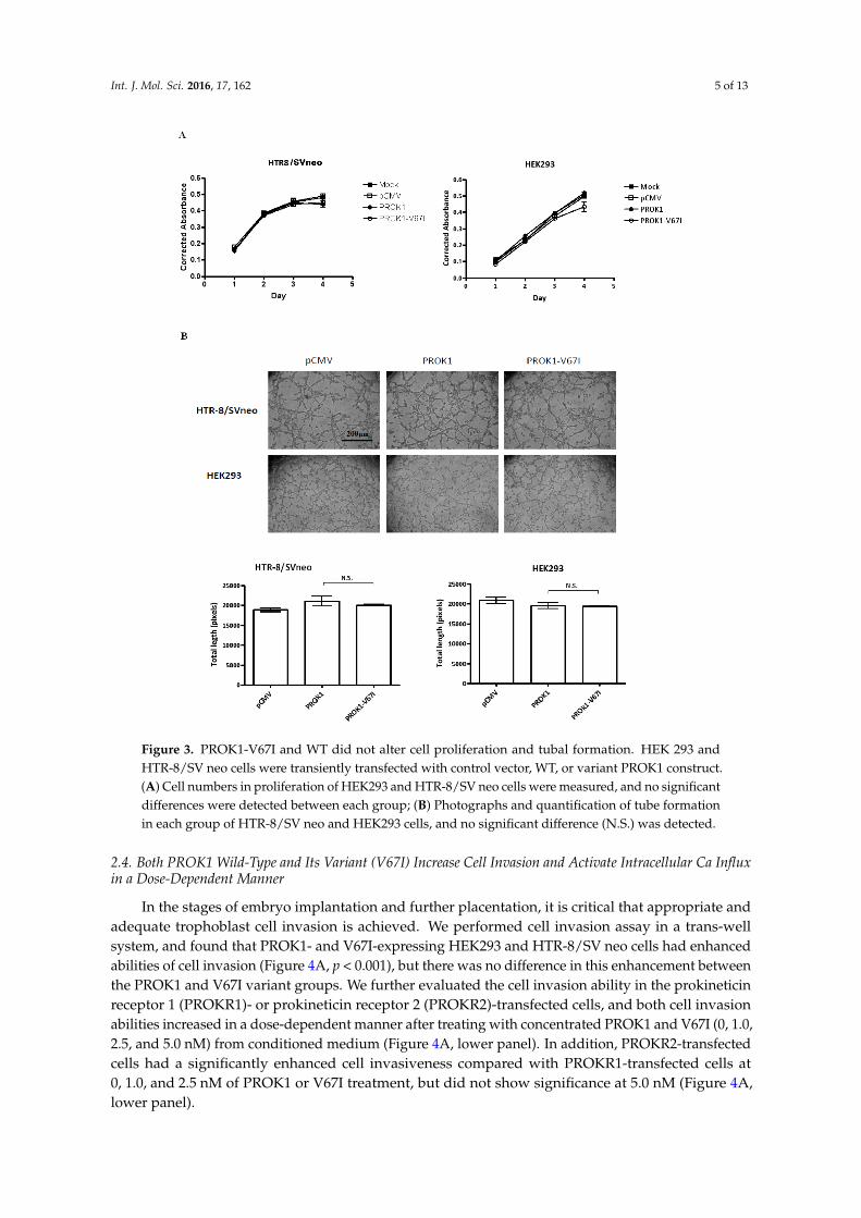
Cell proliferation and angiogenesis are critical in the stages of implantation, embryogenesis, and placentation. We examined if PROK1 and its V67I variant altered the abilities of cell proliferation and tube organization, following their individual ectopic expression in cells. When comparing an empty control vector, variant, and wild-type PROK1, the cell numbers of transfected HEK293 and HTR-8/SV neo cells were not significantly different after 1 to 4 days of cell culture, based on the results of a cell viability assay (Figure 3A). To evaluate the angiogenic ability of PROK1 WT and its variant (V67I), we measured capillary tube formation of PROK1- or V67I-transfected cells by calculating branching length between two nodes at different time intervals (4–6 h). After 4 h incubation on the Matrigel, HEK-293 and HTR-8/SV neo cells rapidly reorganized and subsequently formed tube-like structures on Matrigel. Average tubal length was measured in each group, and there was no stimulatory effect on tube formation in PROK1- and V67I-transfected HEK-293 and HTR-8/SV neo cells compared with those of the empty control vector group (Figure 3B). In addition, both PROK1 and V67I groups behaved similarly, without any effects on tube formation in HEK 293 or HTR-8/SV neo cells. The results, thus, showed that PROK1 and V67I did not have any effects on cell proliferation and tubal organization in either cell line.
Figure 2. Decreased gene expression of PROK1 variant (V67I) compared with wild-type (WT) indifferent cell lines. Cells were transiently transfected with either WT or variant PROK1 construct for48 h. (A) Quantitative RT-PCR analysis showed decreased transcript level of V67I compared with WTin cell lines (HEK293 cells: 50.5%; HTR-8/SV neo cells: 64%); (B) ELISA analysis showed consistentlydecreased protein concentrations in the cell lysate and supernatant of the culture medium amongdifferent cell lines (HEK293 cells: 42.5%–47.5%; HTR-8/SV neo cells: 60%–71%; JAR cells: 19.6%–22.2%;and Ishikawa cells: 30.1%–36.9%). The comparison of V67I and WT is shown in percentages. Data arepresented as means ˘ SEM. * p < 0.001 compared with the corresponding control (WT). Mock: cellswithout transfecting any vectors; pCMV: cells with transfecting empty control vectors.
2.3. PROK1 Wild-Type and Variant (V67I) Have No Significantly Different Effects on Cell Proliferation andTubal formation
Cell proliferation and angiogenesis are critical in the stages of implantation, embryogenesis, andplacentation. We examined if PROK1 and its V67I variant altered the abilities of cell proliferation andtube organization, following their individual ectopic expression in cells. When comparing an emptycontrol vector, variant, and wild-type PROK1, the cell numbers of transfected HEK293 and HTR-8/SVneo cells were not significantly different after 1 to 4 days of cell culture, based on the results of a cellviability assay (Figure 3A). To evaluate the angiogenic ability of PROK1 WT and its variant (V67I),we measured capillary tube formation of PROK1- or V67I-transfected cells by calculating branchinglength between two nodes at different time intervals (4–6 h). After 4 h incubation on the Matrigel,HEK-293 and HTR-8/SV neo cells rapidly reorganized and subsequently formed tube-like structureson Matrigel. Average tubal length was measured in each group, and there was no stimulatory effect ontube formation in PROK1- and V67I-transfected HEK-293 and HTR-8/SV neo cells compared withthose of the empty control vector group (Figure 3B). In addition, both PROK1 and V67I groups behavedsimilarly, without any effects on tube formation in HEK 293 or HTR-8/SV neo cells. The results, thus,showed that PROK1 and V67I did not have any effects on cell proliferation and tubal organization ineither cell line.
Int. J. Mol. Sci. 2016, 17, 162 5 of 13Int. J. Mol. Sci. 2016, 17, 162 5 of 13
Figure 3. PROK1-V67I and WT did not alter cell proliferation and tubal formation. HEK 293 and HTR-8/SV neo cells were transiently transfected with control vector, WT, or variant PROK1 construct. (A) Cell numbers in proliferation of HEK293 and HTR-8/SV neo cells were measured, and no significant differences were detected between each group; (B) Photographs and quantification of tube formation in each group of HTR-8/SV neo and HEK293 cells, and no significant difference (N.S.) was detected.
2.4. Both PROK1 Wild-Type and Its Variant (V67I) Increase Cell Invasion and Activate Intracellular Ca Influx in a Dose-Dependent Manner
In the stages of embryo implantation and further placentation, it is critical that appropriate and adequate trophoblast cell invasion is achieved. We performed cell invasion assay in a trans-well system, and found that PROK1- and V67I-expressing HEK293 and HTR-8/SV neo cells had enhanced abilities of cell invasion (Figure 4A, p < 0.001), but there was no difference in this enhancement between the PROK1 and V67I variant groups. We further evaluated the cell invasion ability in the prokineticin receptor 1 (PROKR1)- or prokineticin receptor 2 (PROKR2)-transfected cells, and both cell invasion abilities increased in a dose-dependent manner after treating with concentrated PROK1 and V67I (0, 1.0, 2.5, and 5.0 nM) from conditioned medium (Figure 4A, lower panel). In addition, PROKR2-transfected cells had a significantly enhanced cell invasiveness compared with PROKR1-transfected cells at 0, 1.0, and 2.5 nM of PROK1 or V67I treatment, but did not show significance at 5.0 nM (Figure 4A, lower panel).
Figure 3. PROK1-V67I and WT did not alter cell proliferation and tubal formation. HEK 293 andHTR-8/SV neo cells were transiently transfected with control vector, WT, or variant PROK1 construct.(A) Cell numbers in proliferation of HEK293 and HTR-8/SV neo cells were measured, and no significantdifferences were detected between each group; (B) Photographs and quantification of tube formationin each group of HTR-8/SV neo and HEK293 cells, and no significant difference (N.S.) was detected.
2.4. Both PROK1 Wild-Type and Its Variant (V67I) Increase Cell Invasion and Activate Intracellular Ca Influxin a Dose-Dependent Manner
In the stages of embryo implantation and further placentation, it is critical that appropriate andadequate trophoblast cell invasion is achieved. We performed cell invasion assay in a trans-wellsystem, and found that PROK1- and V67I-expressing HEK293 and HTR-8/SV neo cells had enhancedabilities of cell invasion (Figure 4A, p < 0.001), but there was no difference in this enhancement betweenthe PROK1 and V67I variant groups. We further evaluated the cell invasion ability in the prokineticinreceptor 1 (PROKR1)- or prokineticin receptor 2 (PROKR2)-transfected cells, and both cell invasionabilities increased in a dose-dependent manner after treating with concentrated PROK1 and V67I (0, 1.0,2.5, and 5.0 nM) from conditioned medium (Figure 4A, lower panel). In addition, PROKR2-transfectedcells had a significantly enhanced cell invasiveness compared with PROKR1-transfected cells at0, 1.0, and 2.5 nM of PROK1 or V67I treatment, but did not show significance at 5.0 nM (Figure 4A,lower panel).

Int. J. Mol. Sci. 2016, 17, 162 6 of 13Int. J. Mol. Sci. 2016, 17, 162 6 of 13
Figure 4. PROK1-V67I and -WT (A) enhanced cell invasion ability and (B) altered intracellular calcium influx in a dose-dependent manner. PROKR2-transfected cells had increased abilities of inducing calcium release and cell invasiveness compared with PROKR1-transfected cells. HEK 293 and HTR-8/SV neo cells were transiently transfected with control vector, WT or variant PROK1 construct. Photographs and quantification of invaded cells stained with Giemsa’s azur eosin methylene blue solution are shown in the upper and middle panels in A. HEK293 and HTR-8/SV neo cells were transiently transfected with either PROKR1 or PROKR2 plasmid and treated with various concentrations of PROK1 WT or V67I condition medium (0, 1.0, 2.5, 5.0 nM). Invaded cells (A, lower panel) and intracellular cell influx alternation (B, upper and lower panels) were measured. * p < 0.05; ** p < 0.01; *** p < 0.001 compared between PROKR1- and PROKR2-transfected cells, and groups of different concentration. PROK1 recombinant protein (Biovision, Milpitas, CA, USA) was used as a standard control to validate protein function of PROK1 (WT) and PROK1-V67I in condition medium (CM). N.S.: no significant difference.
Figure 4. PROK1-V67I and -WT (A) enhanced cell invasion ability and (B) altered intracellularcalcium influx in a dose-dependent manner. PROKR2-transfected cells had increased abilities ofinducing calcium release and cell invasiveness compared with PROKR1-transfected cells. HEK 293and HTR-8/SV neo cells were transiently transfected with control vector, WT or variant PROK1construct. Photographs and quantification of invaded cells stained with Giemsa’s azur eosin methyleneblue solution are shown in the upper and middle panels in A. HEK293 and HTR-8/SV neo cellswere transiently transfected with either PROKR1 or PROKR2 plasmid and treated with variousconcentrations of PROK1 WT or V67I condition medium (0, 1.0, 2.5, 5.0 nM). Invaded cells (A, lowerpanel) and intracellular cell influx alternation (B, upper and lower panels) were measured. * p < 0.05;** p < 0.01; *** p < 0.001 compared between PROKR1- and PROKR2-transfected cells, and groupsof different concentration. PROK1 recombinant protein (Biovision, Milpitas, CA, USA) was used asa standard control to validate protein function of PROK1 (WT) and PROK1-V67I in condition medium(CM). N.S.: no significant difference.
Int. J. Mol. Sci. 2016, 17, 162 7 of 13
Calcium signaling is a critical function index that is used to evaluate G protein-coupled receptors(GPCRs) and their ligand activities. We evaluated PROK1-induced intracellular calcium mobilizationby using a fluorescence-based assay in the prokineticin receptor 1 (PROKR1)- or prokineticin receptor2 (PROKR2)-overexpressed cells. In this assay, HEK293 and HTR-8/SV neo cells were initiallytransfected with PKOKR1 or PROKR2 plasmid to enhance the amplitude of calcium signaling beforeinvestigation. The intracellular calcium influx was stimulated, but no significant differences were seenin the PROKR1- or PROKR2-transfected cells after treating with 5 nM PROK1 or V67I from conditionedmedium (Figure 4B, upper panel). Although the induced calcium influx showed a dose-dependentresponse after treating with PROK1 or V67I conditioned medium (0.5, 1.0, 2.5, 5.0 nM), there was nodifference in the stimulation effect between PROK1 and V67I (Figure 4B, lower panel). By comparingPROKR1- and PROKR2-transfected cells on calcium signaling, PROKR2-transfected cells had a strongersignaling under concentration 0.5, 1.0, and 2.5 nM of PROK1 or V67I treatment, but did not showsignificance at 5.0 nM (Figure 4B, upper and lower panel).
3. Discussion
In a previous genetic association study of recurrent pregnancy loss (RPL), a PROK1 variant (V67I)was shown to be a genetic modifier gene in early human pregnancy [17]. We evaluated the functionaleffect of this common variant and compared it to PROK1-WT in order to investigate the possiblemechanism of genetic modification in this study. We demonstrated that the conserved non-synonymousvariant of PROK1 (V67I) has similar cell functions with regard to enhancing trophoblast cell invasionand stimulating intracellular calcium signaling compared with its wild-type. However, the geneexpression of V67I is down-regulated in both transcript and protein levels across all associatedcell lineages.
The biological interaction of PROK1 and PROKR1/2 is a ligand-receptor relationship. The impactof an amino acid change in PROK1 on the protein activity and the binding affinity between its receptorsremain unclear, and no PROK1 variants have been reported before. We identified PROK1-V67I to be theonly non-synonymous variant in the coding regions of PROK1 using Sanger sequence in 291 subjects(142 RPL patients and 149 normal controls) [17]. The non-synonymous variant of PROK1-V67I is socommon that it is possessed by nearly 50% of the general population, appears to have no visible effecton the fitness of individuals and is, therefore, usually considered as a neutral variant. However, severalstudies have demonstrated that a common non-synonymous variant may have an association with orfunctional significance for the pathophysiology of a disease [19–21]. From an evolutionary perspective,the common non-synonymous variants usually have modest structural effects, and some of themare simply functionally neutral, while others are deleterious [22]. Moreover, some variant sites thatare predicted to be deleterious might have advantages under unique conditions of developmental orenvironmental changes [22]. During the early stages of placental development, the coordination of cellproliferation, differentiation, and invasion of trophoblasts is subtle and fine-tuned in the feto-maternalinterface. The lower protein production of V67I seems to have a negative effect on the functionof PROK1, which implies less cell invasion ability and attenuated downstream calcium signaling.However, whether the down-regulated PROK1 expression is beneficial or deleterious is an issue thatrequires more research.
The PROK1 level during pregnancy is dynamic, both in placental tissue and serum collected frompregnant women. Placental PROK1 expression gradually rises after embryo implantation and peaks at8–11 weeks of gestation, subsequently decreasing until the end of the first trimester, and then this levelis maintained throughout the remaining gestation [4,7,23]. Plasma PROK1 concentration also graduallyincreases during the first trimester (~200 pg/mL), and then decreased to ~70–80 pg/mL in the secondand third trimesters [23]. Adequate trophoblast invasion and proper placental development ensuresthe embryo or fetus has sufficient nutrient and oxygen, and PROK1 is regarded as playing a criticalrole in regulating trophoblast invasion and placental development [13,23]. Excessive trophoblastinvasion could result in placenta accreta and invasive moles, whereas poor trophoblast invasion of
Int. J. Mol. Sci. 2016, 17, 162 8 of 13
maternal vessels could contribute to preeclampsia and intrauterine fetal growth restriction [10,13,23,24].As a result, the differential down-regulated PROK1 expression of V67I in different cell lineagespresented in this study could be an underlying cause for a pathological condition, or a normalregulatory mechanism of the PROK1 system in human pregnancy. We, therefore, speculated thatwomen carrying different PROK1 genotypes may have varied susceptibility to different pregnancycomplications. Moreover, PROK1 was shown to be up-regulated in several types of cancers, such ascolorectal cancer, pancreatic cancer, prostate cancer, and ovarian cancer [25–30], and was regardedas a poor prognostic marker and survival factor. These PROK1-related malignancies were closelyassociated with advanced clinical stage, histological grade, and distant metastasis, which may probablythrough regulating peritumoral angiogenesis or/and strengthened cancer cell invasiveness [25–30].The PROK1-overexpressing cells in the present study could be a mimicking condition of malignancy,and the increased cell invasion ability suggested the potential role of PROK1 in cancer biology.Nevertheless, more studies are required to elucidate the role of PROK1 WT and V67I in both humanpregnancy and cancers, and the complex interaction between PROK1 and its receptors in variouspathophysiologies of human clinical situations.
Disease severity could be influenced by different genetic backgrounds, and genetic modifiersare known to alter the outcomes in various human diseases or animal models of disease, such asmuscular dystrophy (MD), epidermolysis bullosa (EB), and some motor neuron diseases [31–35]. Sincegenetic modifiers act in a non-Mendelian manner to alter the phenotype in question, the comparativelysmall effect of V67I in the general population is highly consistent with its role as a modifier in earlyhuman pregnancy. From our previous data, PROKR1 and PROKR2 variants (I379V and V331M)may protect women from RPL through enhanced trophoblast invasiveness, thus facilitating embryoimplantation [18]. In women carrying wild-types of PROKR1 and PROKR2, the genotypes of PROK1WT or V67I do not have significant effects on RPL risk. In contrast, women carrying V67I will lose theprotective effect with regard to RPL risk if they also carry PROKR1 or PROKR2 variants, indicatingthat PROK1-V67I modifies RPL risk in specific populations [17]. This could be explained by the resultsof the cell functional assays carried out in the present study. These showed that the effects of V67Ion cell function are not different from those of its wild-type, although its gene expression efficiencyis only 20%–70% that of the latter (depending on cell types). The ability of trophoblast invasion ispromoted by PROK1 and V67I, and both facilitate invasiveness in a dose-response manner. Therefore,PROK1-V67I may reduce PROKRs’ protective effect of promoting cell invasiveness by decreasingPROK1 protein production.
4. Experimental Section
4.1. Subjects
The present study was approved by the Institutional Review Board of National Cheng KungUniversity Hospital (#HR-96-39) (Tainan, Taiwan), and informed consents were obtained from allpatients and controls. The clinical and molecular details of the RPL subjects examined in the currentwork were reported in a previous study [17,18]. In this study we compared the V67I allele andgenotype frequencies of our population, Han Chinese individuals in Taiwan, with those of otherHapMap Populations (Table 1).
4.2. Cell Cultures and Treatments
The human HTR-8/SVneo trophoblast cell line was a gift from Dr. Charles Graham (Queen’sUniversity, Kingston, ON, Canada). HTR-8/SVneo cells were grown in RPMI 1640 medium(Invitrogen, Grand Island, NY, USA) supplemented with 10% fetal bovine serum (FBS) and 100 IU/mLpenicillin-streptomycin. The human embryonic kidney cell line (HEK293) were grown in DulbeccoModified Eagle’s Medium (DMEM) (Invitrogen) supplemented with 10% FBS (Invitrogen) and 1%penicillin-streptomycin solution. JAR cells were derived from human placental choriocarcinoma,

Int. J. Mol. Sci. 2016, 17, 162 9 of 13
which were purchased from Bioresource Collection and Research Center (Taiwan, Taiwan), andgrown in RPMI 1640 medium with 10 mM HEPES, 1 mM sodium pyruvate, 10% FBS and1% penicillin-streptomycin solution. Ishikawa cells were grown in Minimum Essential Media(MEM) (Invitrogen) supplemented with 1% non-essential amino acids (NEAA) + 5% FBS and 1%penicillin-streptomycin solution. These cells were cultured in a 5% CO2 humidified incubator at 37 ˝C.After confluent growth, the attached cells were trypsinized and either cryopreserved or subculturedfor further use.
4.3. Generation of Variant PROK1 Expressing Plasmids and Transfection Experiments
The variant sequence of PROK1 (V67I) was introduced into the WT Myc-DDK-tagged cDNAin a pCMV6-Entry vector (Origene Technologies, Inc., Rockville, MD, USA), which encodes theentire coding regions of human cDNA (GenBank NM_032414.2) of PROK1, using Stratagene’s QuikChange II Site-directed Mutagenesis Kit (La Jolla, CA, USA). A vector control, pCMV6 empty vector,was constructed from pCMV6-PROK1 plasmid using sgfI and MluI digestion and the T4 ligationmethod. All constructs were verified by nucleotide sequencing. pCMV6 and PROK1 constructs werepropagated in JM109 Escherichia coli. Transfection efficiency was tested on HEK293 (5 ˆ 105 cells) andHTR8/SVneo (3 ˆ 105 cells) using 1 µg of pCMV-EGFP (Clontech Laboratories, Inc., Palo, Alto, CA,USA) expressing plasmid and 3 µL of TurboFect in six-well plates for 24 and 48 h. The transfectionefficiency is 90% ˘ 10% in the two cell lines. The established constructs were transfected into HEK293and HTR-8/SVneo cell lines by Turbofect for 24 h (Fermentas/Thermo Scientific, Waltham, MA, USA),and their expressions were confirmed by Western blotting. The PROKR1 and PROKR2 plasmids wereconstructed as reported in a previous study [18].
4.4. Measurements of PROK1 Gene Expression
4.4.1. Quantitative Real-Time PCR (qRT-PCR) Analysis
We extracted total RNA from scraped cells using the Trizol reagents (Invitrogen, Carlsbad, CA,USA) according to the manufacturer’s instructions. The extracted RNA was spectrophotometricallyquantified, and its quality was assessed by measuring the absorbance ratios at 260/280 and 260/230nm using a GeneQuantTM Pro Spectrophotometer (GE Healthcare Biosciences, Piscataway, NJ, USA).Two micrograms of total RNA was reverse-transcribed using a SuperScript® Reverse Transcriptionkit (Invitrogen, Carlsbad, CA, USA) RNaseOut, dNTPs and random primers according to themanufacturer’s protocols. The qPCR reactions were carried out in an Applied Biosystems StepOne Plussystem (Applied Biosystems, Foster City, CA, USA). The primers used were the PROK1 sense primer,51-CATGCTCCTCCTAGTAACTG-31, the PROK1 antisense primer, 51-TTTCCTGAAGAAGGGGAC-31,and the internal reference, GAPDH sense primer, 51-ACAGTTGCCATGTAGACC-31, and the GAPDHantisense primer, 51-TTTTTGGTTGAGCACAGG-31, which amplifies fragments of 190 bp for PROK1cDNA and 225 bp for GAPDH cDNA (Sigma-Aldrich, Bornem, Belgium). A 10-µL reaction mixturecontaining cDNA, specific primers and Fast SYBR Green® Master Mix (Applied Biosystems, Carlsbad,CA, USA) was used in the PCR. Reactions were performed at 95 ˝C for 20 s, followed by 40 cycles of95 ˝C for 3 s and 60 ˝C for 30 s. To calculate the relative expression for each gene, the 2´∆∆Ct methodwas used to relate the Ct values of PROK1 expression in each sample to the Ct values of GAPDH.
4.4.2. Immunoassay (ELISA)
HEK293, HTR-8/SVneo, JAR, and Ishikawa cells (3 ˆ 105) were transfected with pCMV vector,PROK1 wild type or V67I plasmid for 48 h. Cell culture supernatant and cell lysate (cells werelysed with RIPA buffer) were harvested for an assay using an ELISA kit to assess the PROK1concentration following the manufacturer’s instructions (R and D Systems, Minneapolis, MN, USA).Briefly, supernatant or cell lysate were applied to the microplate coated with the capture antibodyfor 2 h at room temperature. Detection antibody conjugated to streptavidin-horseradish peroxidase

Int. J. Mol. Sci. 2016, 17, 162 10 of 13
was then applied, followed by the color development solution (tetramethylbezidine substrate) for20 min. Color development was terminated by addition of sulfuric acid, and the optical density wasdetermined at 450 nm by a microplate reader (SpectraMa ˆ 340PC384, Molecular Devices, Sunnyvale,CA, USA). A PROK1 standard (Biovision, Milpitas, CA, USA) was used for control, and calculation ofthe results was performed using computer software capable of generating a four parameter logistic(4-PL) curve-fit.
4.5. Recombinant PROK1 and V67I by Concentrating Secreted Proteins
HEK293 cells (5 ˆ 105) were transfected in six-well plates with PROK1-WT or PROK1-V67Iplasmid in DMEM with 1% FBS for 48 h. We then collected the culture medium to concentrate secretedprotein using a centrifugal filter device, Amicon® Ultra4-3000 MWCO (Merck Millipore, Tullagreen,Carrigtwohill, Co., Cork, Ireland), under 3500 rpm for 40 min. We calculated the concentrated proteinlevel using the ELISA method, as described above. The concentrated protein medium (conditionedmedium (CM)) was stored at ´80 ˝C, and its protein activity (WT and V67I) was compared withthat of a PROK1 standard (Biovision, Milpitas, CA, USA) and tested for various cell functions,as described below.
4.6. Cell Proliferation Assay
The effect of PROK1 variant (V67I) on cell proliferation was assessed when compared with that ofPROK1 wild-type and pCMV6 control. Cell lines were grown at 2 ˆ 103 cells/well in a 96-well plate inDMEM or RPMI medium containing 10% FBS for 24 h. Cell proliferation was determined after one,two, three, and four days. Cell numbers were determined with PrestoBlue™ cell viability reagent(Invitrogen, Carlsbad, CA, USA). After adding 100 µL of cell proliferation reagent in each well for6 h at 37 ˝C in a CO2 incubator, the absorbance at 570 nm (with 600 nm as the reference wavelengthfor normalization), reflecting the number of viable cells, was measured with a microplate reader(SpectraMax 340PC384, Molecular Devices, Sunnyvale, CA, USA). All the treatments were carried outin quadruplicate, and each experiment was carried out at least three times.
4.7. Tube Formation Assay
The effects of PROK1 wild-type and variant (V67I) on tube organization were assessed by growingtransfected cells (HEK293 and HTR-8/SVneo) on Matrigel. Approximately 80 µL ice-cold Matrigel(BD Biosciences, San Diego, CA, USA) was layered into each well of a 96-well plate. The Matrigelwas allowed to completely solidify at 37 ˝C for 1 h. The transfected HEK293 (5 ˆ 104 cells/well) andHTR-8/SVneo (3.5 ˆ 104 cells/well) cells were added and incubated at 37 ˝C in an atmosphere ofhumidified 95% air/5% CO2 for 4 and 6 h, respectively. Observations were made under an invertedphotomicroscope to document the developmental stages. Tubal formation was assessed by measuringtubal length in four quadrants of high power fields in each well. Each assay was done in triplicateand each experiment was repeated at least three times. Quantification was measured with Image Jsoftware (Image J 1.47 h, Wayne Rasband, National Institute of Mental Health, Bethesda, MD, USA).
4.8. Cell Invasion Assay
To investigate the effects of PROK1 and its variant (V67I) on cell invasion, the transfected HEK293(1.5 ˆ 105) or HTR-8/SVneo (1 ˆ 104) cells were trypsinized and re-suspended in serum-free mediumand placed in the upper chamber coated with Matrigel (1 mg/mL at 37 ˝C for 1 h; BD Biosciences,San Diego, CA, USA) in trans-well plates (millicell cell culture insert; 8 µm pore size; Milipore, MA,USA) with or without PROK1 or V67I conditioned medium (1.0, 2.5, 5.0 nM). DMEM containing 10%FBS was placed in the lower chamber. The cells were incubated for 20 h in a humidified atmospherewith 95% air and 5% CO2 at 37 ˝C. Invaded cells on the bottom side of the membrane were fixedwith 100% cold methanol and stained with Giesmsa’s azur eosin methylene blue solution (Merck,
Int. J. Mol. Sci. 2016, 17, 162 11 of 13
Darmatadt, Germany) and counted from a minimum of four high-power fields per insert under lightmicroscopy. The data were expressed as the averages of three independent experiments.
4.9. Intracellular Calcium Influx Assay
Twenty-four hours after transfection, cells were harvested from plates using EDTA-trypsin, andwashed with HBSS-based buffer (20 mM HEPES, 1 mM MgSO4, 3.3 mM Na2CO3, 1.3 mM CaCl2,2.5 mM probenecid, pH 7.4) supplemented with 0.1% bovine serum album. Cells were loaded with4 µM calcium indicator Fluo-4 AM (Molecular Probes, Eugene, OR, USA) for 1 h at 37 ˝C. After washingtwice, the cells were re-suspended to a concentration of 1 ˆ 106 cells/mL. The green fluorescenceemission of Fluo-4 was analyzed using FACSCalibur flow cytometry (BD Immunocytometry System),as described previously [18]. Following the establishment of a green fluorescence Ca2+ baseline, theindicated level of concentrated PROK1, V67I or PROK1 standard (Biovision, Milpitas, CA, USA) (0.5, 1,2.5, or 5 nM) was added to the cell suspension to detect fluctuations in the green fluorescence.
4.10. Statistical Analysis
All values of the experimental assays were expressed as means ˘ SEM. Differences between thegroups were compared using the unpaired two-tailed t-test or one-way ANOVA, and a p value of lessthan 0.05 was considered statistically significant.
5. Conclusions
We evaluated the function and possible regulatory mechanism of a common variant of PROK1as a genetic modifier in early human pregnancy. Although PROK1-V67I has a similar functionaleffect to that of WT on cell behavior, gene expression at the transcript and protein levels is impaired.The results of this work provide an explanation for how a common, innocuous PROK1 variant could actepistatically with its receptor genes to interfere with the outcome of early human pregnancy. However,more research is needed to assess whether the clinical impact of the decreased protein productionof PROK1-V67I is positive or negative. Considering the important role of PROK1 in pregnancy andthe high prevalence of V67I in the general population, the findings of the present study may providea common mechanism with regard to modulate the risk of various PROK1-related diseases.
Acknowledgments: This work was supported by grants from the National Science Council of the Republic ofChina (NSC- 99-2314-B-006-026-MY2 and NSC-101-2314-B-006-039-MY3).
Author Contributions: Study design, execution and manuscript drafting: Mei-Tsz Su; Study execution:Jyun-Yuan Huang, Hui-Ling Tsai; Statistical analysis and data interpretation: Yi-Chi Chen; Critical discussion andcorrespondence: Pao-Lin Kuo.
Conflicts of Interest: All authors have no conflicts of interest.
References
1. Ferrara, N.; LeCouter, J.; Lin, R.; Peale, F. EG-VEGF and Bv8: A novel family of tissue-restricted angiogenicfactors. Biochim. Biophys. Acta 2004, 1654, 69–78. [CrossRef] [PubMed]
2. Ferrara, N.; Frantz, G.; LeCouter, J.; Dillard-Telm, L.; Pham, T.; Draksharapu, A.; Giordano, T.; Peale, F.Differential Expression of the Angiogenic Factor Genes Vascular Endothelial Growth Factor (VEGF) andEndocrine Gland-Derived VEGF in Normal and Polycystic Human Ovaries. Am. J. Pathol. 2003, 162,1881–1893. [CrossRef]
3. Fraser, H.M.; Bell, J.; Wilson, H.; Taylor, P.D.; Morgan, K.; Anderson, R.A.; Duncan, W.C. Localization andQuantification of Cyclic Changes in the Expression of Endocrine Gland Vascular Endothelial Growth Factorin the Human Corpus Luteum. J. Clin. Endocrinol. Metab. 2005, 90, 427–434. [CrossRef] [PubMed]
4. Hoffmann, P.; Feige, J.J.; Alfaidy, N. Expression and Oxygen Regulation of Endocrine Gland-Derived VascularEndothelial Growth Factor/Prokineticin-1 and Its Receptors in Human Placenta during Early Pregnancy.Endocrinology 2006, 147, 1675–1684. [CrossRef] [PubMed]

Int. J. Mol. Sci. 2016, 17, 162 12 of 13
5. LeCouter, J.; Kowalski, J.; Foster, J.; Hass, P.; Zhang, Z.; Dillard-Telm, L.; Frantz, G.; Rangell, L.; DeGuzman, L.;Keller, G.A.; et al. Identification of an angiogenic mitogen selective for endocrine gland endothelium. Nature2001, 412, 877–884. [CrossRef] [PubMed]
6. Ngan, E.S.; Lee, K.Y.; Yeung, W.S.; Ngan, H.Y.; Ng, E.H.; Ho, P.C. Endocrine Gland-Derived VascularEndothelial Growth Factor Is Expressed in Human Peri-implantation Endometrium, But Not in EndometrialCarcinoma. Endocrinology 2006, 147, 88–95. [CrossRef] [PubMed]
7. Evans, J.; Catalano, R.D.; Morgan, K.; Critchley, H.O.; Millar, R.P.; Jabbour, H.N. Prokineticin 1 Signaling andGene Regulation in Early Human Pregnancy. Endocrinology 2008, 149, 2877–2887. [CrossRef] [PubMed]
8. Haouzi, D.; Mahmoud, K.; Fourar, M.; Bendhaou, K.; Dechaud, H.; De Vos, J.; Rème1, T.; Dewailly, D.;Hamamah, S. Identification of new biomarkers of human endometrial receptivity in the natural cycle.Hum. Reprod. 2009, 24, 198–205. [CrossRef] [PubMed]
9. Denison, F.C.; Battersby, S.; King, A.E.; Szuber, M.; Jabbour, H.N. Prokineticin-1: A Novel Mediator of theInflammatory Response in Third-Trimester Human Placenta. Endocrinology 2008, 149, 3470–3477. [CrossRef][PubMed]
10. Brouillet, S.; Hoffmann, P.; Feige, J.J.; Alfaidy, N. EG-VEGF: A key endocrine factor in placental development.Trends Endocrinol. Metab. 2012, 23, 501–508. [CrossRef] [PubMed]
11. Gorowiec, M.R.; Catalano, R.D.; Norman, J.E.; Denison, F.C.; Jabbour, H.N. Prokineticin 1 InducesInflammatory Response in Human Myometrium: A Potential Role in Initiating Term and Preterm Parturition.Am. J. Pathol. 2011, 179, 2709–2719. [CrossRef] [PubMed]
12. Shaw, J.L.; Denison, F.C.; Evans, J.; Durno, K.; Williams, A.R.; Entrican, G.; Critchley, H.O.D.; Jabbour, H.N.;Horne, A.W. Evidence of prokineticin dysregulation in fallopian tube from women with ectopic pregnancy.Fertil. Steril. 2010, 94, 1601–1608. [CrossRef] [PubMed]
13. Hoffmann, P.; Saoudi, Y.; Benharouga, M.; Graham, C.H.; Schaal, J.P.; Mazouni, C.; Feige, J.J.; Alfaidy, N.Role of EG-VEGF in human placentation: Physiological and pathological implications. Cell. Mol. Life Sci.2009, 13, 2224–2235. [CrossRef] [PubMed]
14. Alfaidy, N.; Hoffmann, P.; Gillois, P.; Gueniffey, A.; Lebayle, C.; Garçin, H.; Thomas-Cadi, C.; Bessonnat, J.;Coutton, C.; Villaret, L.; et al. PROK1 level in the follicular microenvironment: A new non-invasive predictivebiomarker of embryo implantation. J. Clin. Endocrinol. Metab. 2015. [CrossRef] [PubMed]
15. Alfaidy, N.; Hoffmann, P.; Boufettal, H.; Samouh, N.; Aboussaouira, T.; Benharouga, M.; Feige, J.J.; Brouillet, S.The Multiple Roles of EG-VEGF/PROK1 in Normal and Pathological Placental Angiogenesis. Biomed. Res. Int.2014, 2014, 451906. [CrossRef] [PubMed]
16. Su, M.T.; Lin, S.H.; Lee, I.W.; Chen, Y.C.; Hsu, C.C.; Pan, H.A.; Kuo, P.L. Polymorphisms of endocrinegland-derived vascular endothelial growth factor gene and its receptor genes are associated with recurrentpregnancy loss. Hum. Reprod. 2010, 25, 2923–2930. [CrossRef] [PubMed]
17. Su, M.T.; Lin, S.H.; Chen, Y.C.; Kuo, P.L. Gene-gene interactions and risk of recurrent miscarriages in carriersof endocrine gland–derived vascular endothelial growth factor and prokineticin receptor polymorphisms.Fertil. Steril. 2014, 102, 1071–1077. [CrossRef] [PubMed]
18. Su, M.T.; Lin, S.H.; Chen, Y.C.; Wu, L.W.; Kuo, P.L. Prokineticin receptor variants (PKR1-I379V andPKR2-V331M) are protective genotypes in human early pregnancy. Reproduction 2013, 146, 63–73. [CrossRef][PubMed]
19. Andreasen, L.; Nielsen, J.B.; Christophersen, I.E.; Holst, A.G.; Sajadieh, A.; Tveit, A.; Haunsø, S.;Svendsen, J.H.; Schmitt, N.; Olesen, M.S. Genetic Modifier of the QTc Interval Associated With Early-OnsetAtrial Fibrillation. Can. J. Cardiol. 2013, 29, 1234–1240.
20. Flanigan, K.M.; Ceco, E.; Lamar, K.M.; Kaminoh, Y.; Dunn, D.M.; Mendell, J.R.; King, W.M.; Pestronk, A.;Florence, J.M.; Mathews, K.D.; et al. LTBP4 genotype predicts age of ambulatory loss in duchenne musculardystrophy. Ann. Neurol. 2013, 73, 481–488. [CrossRef] [PubMed]
21. Barry, E.L.; Poole, E.M.; Baron, J.A.; Makar, K.W.; Mott, L.A.; Sandler, R.S.; Ahnen, D.J.; Bresalier, R.S.;McKeown-Eyssen, G.E.; Ulrich, C.M. CYP2C9 Variants Increase Risk of Colorectal Adenoma Recurrenceand Modify Associations with Smoking but Not Aspirin Treatment. Cancer Causes Control 2013, 24, 47–54.[CrossRef] [PubMed]
22. Solomon, O.; Bazak, L.; Levanon, E.Y.; Amariglio, N.; Unger, R.; Rechavi, G.; Eyal, E. Characterizing offunctional human coding RNA editing from evolutionary, structural, and dynamic perspectives. Proteins2014, 82, 3117–3131. [CrossRef] [PubMed]
Int. J. Mol. Sci. 2016, 17, 162 13 of 13
23. Brouillet, S.; Murthi, P.; Hoffmann, P.; Salomon, A.; Sergent, F.; de Mazancourt, P.; Dakouane-Giudicelli, M.;Dieudonné, M.N.; Rozenberg, P.; Vaimanet, D.; et al. EG-VEGF controls placental growth and survival innormal and pathological pregnancies: Case of fetal growth restriction (FGR). Cell. Mol. Life Sci. 2013, 70,511–525. [CrossRef] [PubMed]
24. Kaufmann, P.; Black, S.; Huppertz, B. Endovascular Trophoblast Invasion: Implications for the Pathogenesisof Intrauterine Growth Retardation and Preeclampsia. Biol. Reprod. 2003, 69, 1–7. [CrossRef] [PubMed]
25. Balu, S.; Pirtea, L.; Gaje, P.; Cîmpean, A.M.; Raica, M. The immunohistochemical expression of endocrinegland-derived-VEGF (EG-VEGF) as a prognostic marker in ovarian cancer. Rom. J. Morphol. Embryol. 2012,53, 479–483. [PubMed]
26. Goi, T.; Nakazawa, T.; Hirono, Y.; Yamaguchi, A. Prokineticin 1 expression in gastrointestinal tumors.Anticancer Res. 2013, 33, 5311–5315. [PubMed]
27. Ren, L.; Guo, X.; Shao, X.; Li, H.; Yao, H. Endocrine gland-derived vascular endothelial growth factormodulates proliferation, apoptosis and migration in pancreatic cancer cells. Mol. Med. Rep. 2015, 11,4279–4284. [CrossRef] [PubMed]
28. Torres, C.; Linares, A.; Alejandre, M.J.; Palomino-Morales, R.J.; Caba, O.; Prados, J.; Aránega, A.; Delgado, J.R.;Irigoyen, A.; Martínez-Galánet, J.; et al. Prognosis Relevance of Serum Cytokines in Pancreatic Cancer.BioMed Res. Int. 2015, 2015, 518284. [CrossRef] [PubMed]
29. Nakazawa, T.; Goi, T.; Hirono, Y.; Yamaguchi, A. Prokineticin 1 protein expression is a useful new prognosticfactor for human sporadic colorectal cancer. Ann. Surg. Oncol. 2015, 22, 1496–1503. [CrossRef] [PubMed]
30. Pasquali, D.; Rossi, V.; Staibano, S.; de Rosa, G.; Chieffi, P.; Prezioso, D.; Mirone, V.; Mascolo, M.;Tramontano, D.; Bellastella, A.; et al. The endocrine-gland-derived vascular endothelial growthfactor (EG-VEGF)/Prokineticin 1 and 2 and receptor expression in human prostate: Up-regulation ofEG-VEGF/Prokineticin 1 with malignancy. Endocrinology 2006, 147, 4245–4251. [CrossRef] [PubMed]
31. Swaggart, K.A.; Demonbreun, A.R.; Vo, A.H.; Swanson, K.E.; Kim, E.Y.; Fahrenbach, J.P.; Holley-Cuthrell, J.;Eskin, A.; Chen, Z.; Squire, K.; et al. Annexin A6 modifies muscular dystrophy by mediating sarcolemmalrepair. Proc. Natl. Acad. Sci. USA 2014, 111, 6004–6009. [CrossRef] [PubMed]
32. Sproule, T.J.; Bubier, J.A.; Grandi, F.C.; Sun, V.Z.; Philip, V.M.; McPhee, C.G.; Adkins, E.B.; Sundberg, J.P.;Roopenian, D.C. Molecular Identification of Collagen 17a1 as a Major Genetic Modifier of Laminin Gamma2 Mutation-Induced Junctional Epidermolysis Bullosa in Mice. PLoS Genet. 2014, 10, e1004068. [CrossRef][PubMed]
33. Cooper-Knock, J.; Shaw, P.J.; Kirby, J. The widening spectrum of C9ORF72-related disease;genotype/phenotype correlations and potential modifiers of clinical phenotype. Acta Neuropathol. 2014, 127,333–345. [CrossRef] [PubMed]
34. Van Blitterswijk, M.; Mullen, B.; Heckman, M.G.; Baker, M.C.; DeJesus-Hernandez, M.; Brown, P.H.;Murray, M.E.; Hsiung, G.H.R.; Stewartc, H.; Karydasd, A.M.; et al. Ataxin-2 as potential disease modifier inC9ORF72 expansion carriers. Neurobiol. Aging 2014, 35, 2421.e13–2421.e17. [CrossRef] [PubMed]
35. Heydemann, A.; Huber, J.M.; Demonbreun, A.; Hadhazy, M.; McNally, E.M. Genetic background influencesmuscular dystrophy. Neuromuscul. Disord. 2005, 15, 601–609. [CrossRef] [PubMed]
© 2016 by the authors; licensee MDPI, Basel, Switzerland. This article is an open accessarticle distributed under the terms and conditions of the Creative Commons by Attribution(CC-BY) license (http://creativecommons.org/licenses/by/4.0/).