a comparative study of clinical, radiological and
TRANSCRIPT

Mrinal et al. European Journal of Pharmaceutical and Medical Research
www.ejpmr.com
363
A COMPARATIVE STUDY OF CLINICAL, RADIOLOGICAL AND LABORATORY
PARAMETERS OF COMMUNITY ACQUIRED LRTI OF LESS THAN 2 WEEKS
DURATION AND ASSESSING THE RESPONSE TO STANDARD TREATMENT
GUIDELINES AMONG HIV-INFECTED AND HIV-NON INFECTED ADULTS
ATTENDING A TERTIARY CARE CENTRE IN KOLKATA
1*Dr. Mrinal Baidya,
2Dr. Debajyoti Majumdar,
3Dr. Nayan Brahme,
4Dr. Manab Kumar Ghosh, 5. Dr. Arindam
Naskar, 6Dr. Sudeshna Mallik and
7Dr. Bibhuti Saha
1,2,3
MBBS, MD(Tropical Medicine), School of Tropical Medicine, Kolkata. 4,5,6
MBBS, MD(Tropical Medicine), Assistant Professor, Dept of Tropical Medicine, School of Tropical Medicine,
Kolkata. 7Head and Professor, Dept of Tropical Medicine, School of Tropical Medicine, Kolkata.
Article Received on 28/01/2018 Article Revised on 18/02/2018 Article Accepted on 09/03/2018
MATERIALS AND METHODS
1) STUDY AREAS: OPD of School of Tropical
Medicine, Kolkata/ Inpatient Department of Carmichael
Hospital for Tropical Diseases, Kolkata.
2) STUDY POPULATION: HIV- infected & HIV-non
infected adults with community acquired LRTI of less
than 2 weeks duration attending OPD or are admitted at
CHTD, School of Tropical Medicine.
3)STUDY PERIOD: July-2013 to June-2014.
4) SAMPLE SIZE: 50 (25 in each group)
5) SAMPLE DESIGN: SELECTION OF THE
PATIENT: Adult patients presenting with fever, cough
with respiratory distress for less than two weeks duration
will be selected. All the patient will be referred to
ICTC.HIV antibody non-reactive people will comprise
the HIV non-infected group and HIV antibody reactive
people will comprise HIV infected group. Both the
groups will be screened and finally recruited for the
study using, following inclusion and exclusion criteria
after taking informed consent.
a) Inclusion Criteria
HIV non-infected adults of both sexes with fever, cough,
chest pain, breathlessness etc.
HIV infected adults of both sexes with fever, cough,
chest pain, dyspnoea etc.
b) Exclusion criteria Infant, children and adolescents (<18years). HIV
infected and non-infected adults with cough, fever,
dyspnoea for more than 2 weeks duration. History of
hospitalisation within last three months
SJIF Impact Factor 4.897
Research Article
ISSN 2394-3211
EJPMR
EUROPEAN JOURNAL OF PHARMACEUTICAL
AND MEDICAL RESEARCH www.ejpmr.com
ejpmr, 2018,5(4), 363-376
Corresponding Author: Dr. Mrinal Baidya
MBBS, MD(Tropical Medicine), School of Tropical Medicine, Kolkata.
ABSTRACT RTI are a substantial cause of morbidity and mortality in young children and the elderly. Every year RTI in young
children is responsible for an estimated 3.9 million deaths worldwide. Acute respiratory infections (ARI) may
cause inflammations anywhere in the respiratory tract from nose to alveoli with a wide range of combinations of
symptoms and signs. In India, during the year 2011, about 26.3 million cases ARI were reported. During 2011
about 2492 people died of ARI and 2770 died of pneumonia. Pneumonia affects approximately 450 million people
globally per year, (seven per cent of population) and results in about 4million deaths mostly in the third world
countries. A reasonable number of LRTI patients require in-patient treatment. Our study revealed increased age is a
significant risk factor for morbidity in LRTI. Our study also showed LRTI in HIV-R group is more common in
younger age group as compared to HIV-NR group in which it is more common after 50 years of age with male
preponderance in both the groups. It also proved that the severity criteria given by BTS guideline i.e. CURB-65 is
pretty useful in a set up like us. It helps to recognize the severely ill patients early. CD4 level has a good predictive
value in determination of causative organisms, outcome and prognosis in the HIV reactive group. The most
common single organism found in sputum culture of patients of LRTI in HIV-R group is Staphylococcus aureus
whereas in HIV-NR group it is Klebsiella pneumoniae.
KEYWORDS: LRTI, HIV, NON HIV.

Mrinal et al. European Journal of Pharmaceutical and Medical Research
www.ejpmr.com
364
6) STUDY DESIGN- DESCRIPTIVE, CROSS
SECTIONAL COMPARATIVE STUDY which
includes.
METHODS
Every patient selected by the inclusion criteria had
undergone the following
• Informed consent of the patient
• Detailed medical history
• Physical examination
• ICTC screening
• Routine investigations
(a) Complete haemogram (b) sputum for Gram stain,
culture and sensitivity (c) sputum for AFB (d) sputum for
fungal stain, culture (e) Blood culture (f) fasting and post
prandial blood sugar (g) urea, creatinine (h) LFT (i) chest
X-RAY (j) ECG.
Others as deemed necessary e.g. routine urine & stool
examination, Mantoux test, lipid profile, serum ANA, RF
and ACE estimation, throat swab culture, CT chest and
guided FNAC, USG whole abdomen, Bone Marrow
aspiration, abdominal CT.
7)PARAMETERS TO BE STUDIED PARAMETERS SPECIFIC FOR OBJECTIVE 1-
Clinical parameters include specific medical history
along with findings on physical examinations of the
patients, laboratory investigations including radiological
findings etc.
PARAMETERS SPECIFIC FOR OBJECTIVE 2- All
clinical findings, microbiological and pathological
examination findings.
PARAMETERS SPECIFIC FOR OBJECTIVE 3-
Progress of clinical findings, biochemical,
microbiological, radiological findings with treatment.
8)STUDY TOOLS
Pre-designed and pre-tested schedule for data collection.
Prescription used by the patient.
Other allied instruments required during physical
examinations
Consent forms in Bengali, Hindi and English languages.
9) STUDY TECHNIQUES All patients after selection
were asked for the history and examined thoroughly, and
then the sputum was collected and processed. Blood was
collected for culture and sensitivity, complete blood
count,and other relevant investigations. Patients were
directed to go to our radiology department for chest
radiograph.
Sputum collection and processing Sputum was collected at early morning of admitted
patients. For ODP cases patients were sent to department
of bacteriology for giving sample. Patients were asked to
deeply cough out the sample. If they were unable to do
that, a sputum induction by nebulisation with hypertonic
saline was undertaken. The sample was collected in a
wide neck screw cap sterile container and transported to
bacteriology department. Then the quality of the sputum
was assessed by Bartlett’s grading system. If the sample
had epithelial cell more than 10/ low power field, then it
was discarded and the patient were asked to produce
another sputum sample. If the sample was not acceptable
the patient were excluded from our study.
The gram stained sputum were examined under
microscope to detect the pus cell count and predominant
bacteria. All acceptable samples were further processed
for culture. The rest of the sputum was liquefied by
freshly prepared solution of 1% dithioerythrotol (1:1)
and incubated at 37ºC for 30 min. Then it was serially
diluted with normal saline. Samples having 10ˉ4, 10ˉ,
10ˉ, 10ˉ dilutions were inoculated in MacConkey, Blood
and Chocolate agar and incubated for 48 hours. 2 days
later media was examined for growth and if colony count
≥ 10 then further identification were undertaken by
standard bacteriological methods.
Blood culture
For collection of blood culture after proper cleansing of
venepuncture site with povidone iodine 10 ml of blood
was drawn and inoculated in blood culture bottle. It was
then incubated for 2 days and if growth of some
organism were detected then subsequently subcultured
on Blood agar and MacConkey agar.
10) PLAN FOR ANALYSIS OF DATA
Adult patients attending OPD or admitted at CHTD,
School of Tropical Medicine, Kolkata with history of
fever, cough, and respiratory distress with less than 2
weeks duration selected as HIV non-infected and HIV
infected groups based on ICTC screening and also
considering their inclusion and exclusion criteria.
Interviewing the patient for proper medical history.
Clinical examination.
Scrutiny of biochemical, radiological, microbiological
examinations.
management as per standard guideline.
RESULTS AND ANALYSIS
For this study a total of 50 patients who attended the
Tropical Medicine OPD or admitted to the Carmichael
Hospital for Tropical Diseases were eligible as their
clinical presentation fulfilled the inclusion criteria.

Mrinal et al. European Journal of Pharmaceutical and Medical Research
www.ejpmr.com
365
A)AGE AND SEX AND OTHER DEMOGRAPHIC
FACTORS,
Table 1: Age distribution of HIV-R and HIV-NR
patients (n=50).
Age
(in yrs)
HIV-R
(% out of n)
HIV-NR
(% out of n)
18 to 30 9(18%) 4(8%)
31 to 40 6(12%) 3(6%)
41 to 50 7(14%) 1(2%)
51 to 60 3(6%) 7(14%)
61 to 70 0 1(2%)
71 to 80 0 5(10%)
80 and above 0 4(8%)
There were 50 patients in our study. Among them 25
patients were HIV infected and 25 patients were HIV
non- infected. Most common HIV infected was in the
age group of 18 to 30 yrs and HIV-NR in the age group
of 51 to 60 yrs. The range of age was 19 to 55 in HIV-R
and 19 to 85 in HIV-NR patients.
Figure 1: Bar-diagram showing age distribution of
HIV-R and HIV-NR patients.
Table 2: Showing patient profile of HIV-R and HIV-
NR patients (n=50).
HIV-R
(% out of n)
HIV-NR
(%out of n)
a)Sex
Male 17(68%) 14(56%)
Female 8(32%) 11(44%)
b)Residence
Urban 10(40%) 16(64%)
Rural 15(60%) 9(36%)
c)Place of treatment
Indoor 17(68%) 13 (52%)
OPD 8(32%) 12(48%)
From the above table we can see that, among the HIV
infected patients 68% are male and 32% are
female.Among the HIV Non-infected patients 56% are
male and 44% are female.Major portion of our study
population in HIV- Reactive patients were urban people
40% and rural people 60%. And in HIV-Non Reactive
patients urban people were 64% and rural people were
36%.68% of the patients were admitted and 32% were
treated in OPD in HIV-R group. 52% of the patients
were admitted and 48% were treated in OPD in HIV-NR
group. Most of the admitted patients were severely ill
and fulfilled CURB-65 criteria.
Figure 2a: Bar-diagram showing sex distribution of
HIV-R and HIV-NR patients.
Figure 2b: Bar-diagram showing residence
distribution of HIV-R and HIV-NR patients.
Figure 2c: Bar-diagram showing place of treatment
distribution of HIV-R and HIV-NR patients.
Mrinal et al. European Journal of Pharmaceutical and Medical Research
www.ejpmr.com
366
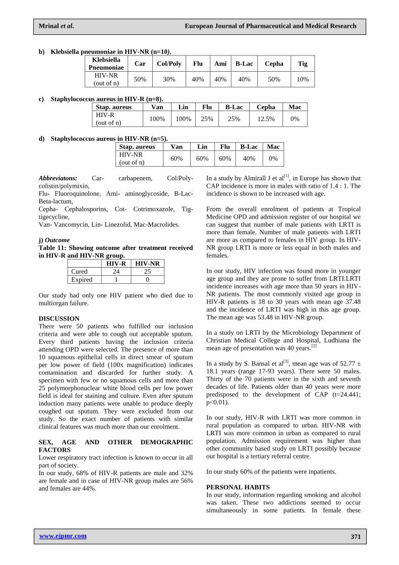
B) ASSOCIATED DISEASES
Table 3: Showing associated co-morbid illness in
HIV-R patients and HIV-NR patients.
Comorbidity HIV-R(%) HIV-NR(%)
COPD 5(20%) 11(44%)
Asthma 1(4%) 2(8%)
Past history of TB 2(8%) 3(12%)
Diabetes 6(24%) 8(32%)
High B.P 3(12%) 10(40%)
Chronic Liver disease 4(16%) 3(12%)
The most commonly associated condition with LRTI in
our study was COPD which was present in 20% HIV-R
patients and 44% in HIV-NR patients. The next common
disease prevalent by decreasing order is diabetes,
hypertension, CLD,past H/O of TB and asthma.
Figure 3: Bar-diagram showing associated co-morbid
illness distribution of HIV-R and HIV-NR patients.
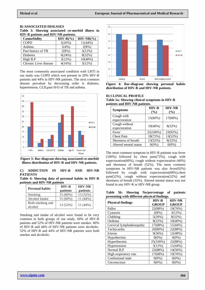
C) ADDICTION IN HIV-R AND HIV-NR
PATIENTS
Table 4: Showing data of personal habits in HIV-R
patients and HIV-NR patients
Personal habits HIV-R
patients HIV-NR
patients Smoking 15 (60%) 13 (52%) Alcohol Intake 15 (60%) 11 (44%) Both smoking and
alcohol 13 (52%) 11 (44%)
Smoking and intake of alcohol were found to be very
common in both groups of our study. 60% of HIV-R
patients and 52% of HIV-NR patients were smoker. 60%
of HIV-R and 44% of HIV-NR patients were alcoholic.
52% of HIV-R and 44% of HIV-NR patients were both
smoker and alcoholic.
Figure 4: Bar-diagram showing personal habits
distribution of HIV-R and HIV-NR patients.
D) CLINICAL PROFILE
Table 5a: Showing clinical symptoms in HIV-R
patients and HIV-NR patients.
Symptoms HIV-R
(%)
HIV-NR
(%)
Cough with
expectoration 15(60%) 17(68%)
Cough without
expectoration 10(40%) 8(32%)
Fever 25(100%) 23(92%)
Chest Pain 18(72%) 13(52%)
Shortness of breath 13(52%) 8(32%)
Altered mental status 0(0%) 0(0%)
The most common symptom in HIV-R patients was fever
(100%) followed by chest pain(72%), cough with
expectoration(60%), cough without expectoration (40%)
and shortness of breath (52%). The most common
symptoms in HIV-NR patients was also fever(92%)
followed by cough with expectoration(68%),chest
pain(52%), cough without expectoration(32%) and
shortness of breath (32%). Altered mental status was not
found in any HIV-R or HIV-NR group.
Table 5b: Showing No/percentage of patients
presenting with different physical findings.
Physical findings HIV-R
GROUP
HIV-NR
GROUP
Pallor 22(88%) 19(76%)
Cyanosis 2(8%) 3(12%)
Clubbing 5(20%) 8(32%)
Oedema 8(32%) 10(40%)
Cervical lymphadenopathy 17(68%) 11(44%)
Tachycardia 20(80%) 22(88%)
Icterus 9(36%) 12(48%)
Hypothermia 0(0%) 0(0%)
Hyperthermia 25(100%) 22(88%)
Hypertension 3(12%) 11(44%)
Normal B.P 22(88%) 14(56%)
High respiratory rate 17(68%) 19(76%)
Confusional state 0(0%) 0(0%)
Unconscious 0(0%) 0(0%)

Mrinal et al. European Journal of Pharmaceutical and Medical Research
www.ejpmr.com
367
Hyperthermia(94%) was the most common overall
general survey finding followed by pallor(82%).
80% of HIV-R group had tachycardia and high
respiratory rate(68%).Around 17 HIV-R patients had
cervical lymph node palpable. High blood pressure was
recorded in 3 patients. Clubbing(16%),oedema(56%)
were present in HIV-R group.
In HIV-NR patients, the most common symptom was
hyperthermia (88%) and tachycardia (88%). Next
common was pallor (76%).High respiratory rate was
detected in 19 patients. Oedema (40%), clubbing (28%)
and high blood pressure (40%). Cervical
lymphadenopathy was present in 44% of patients in
HIV-NR group.
Table 6a: Showing respiratory system examination
findings in HIV-R patients and HIV-NR patients.
Trachea HIV-R
(%)
HIV-NR
(%)
Central 23(92%) 23(92%)
Deviated 2 (8%) 2(8%)
Movement of chest
Normal 17(68%) 9(36%)
Diminished 7(28%) 2(8%)
Indrawing 1(4%) 11(44%)
Percussion note
Dull 6(24%) 3(12%)
Breath sounds
B/L VBS 19(76%) 20(80%)
Diminished VBS 6(24%) 3(12%)
Bronchial 1(4%) 1(4%)
Rhonchi 20(80%) 18(72%)
Crepitations 18(72%) 20(80%)
Complications
Pleural Effusion 3(12%) 3(12%)
Empyema 1(4%) 0(0%)
Table 6b: Showing No/percentage of patients with
various respiratory clinical findings.
HIV-R HIV-NR
Shifting of Trachea 2(8%) 2(8%)
Restricted movement of chest 7(28%) 2(8%)
Impaired/ dull percussion note 6(24%) 3(12%)
Hyperresonant percussion note 0(0%) 1(4%)
Normal VBS 19(76%) 20(80%)
Diminished VBS 6(24%) 5(20%)
Bronchial breath sound 1(4%) 1(4%)
Increased vocal fremitus
/resonance 1(4%) 1(4%)
Decreased vocal fremitus
/resonance 6(24%) 3(12%)
Adventitious sound
Crackles
Wheeze
Pleural rub
18(72%)
16(64%)
12(48%)
0(0%)
20(80%)
15(60%)
12(48%)
0(0%)
Chest indrawing 1(4%) 11(44%)
On examination of respiratory system of HIV infected
patients-92% had trachea central.Deviated trachea was
seen in 8% of the patients.68% of the study group had
normal movements of the chest wall. Excessive work by
accessory muscles of respiration (indrawing) was evident
in 4% patients. Diminished movement found in 7
patients. Percussion note was dull in 6 patients. Most of
the patients had bilateral vesicular breath sound(76%). 6
patients had diminished vesicular breath sound and 1
patient had bronchial breath sound. Among the added
respiratory sounds, crepitation was heard in 18 patients.
Rhonchi or wheeze was presents in 20 patients.
Parapneumonic pleural effusion developed in 3 patients.
Empyema was found in 1 HIV-R patients.
On examination of the respiratory system in HIV-NR
patients-92% had trachea central.Nearly less than half of
the study group had normal movements of the chest wall
(36%). Excessive work by accessory muscles of
respiration (indrawing) was evident in 11 patients,all of
whom were diagnosed cases of COPD. Diminished
movement was found in 2 patients. Percussion note was
dull in 3 patients. Most of the patients (80%) had
bilateral vesicular breath sound(20%). 3 patients had
diminished vesicular breath sound and 1 patient had
bronchial breath sound with increased vocal resonance.
Among the added respiratory sounds, crepitation was
heard in 20 patients. Rhonchi or wheeze was present in
18 patients. Parapneumonic pleural effusion developed in
3 patients. Empyema was also not detected in HIV-NR
patients.
E) LABORATORY EVALUATION
Table 7a: Showing Hb and TLC level in HIV-R and
HIV-NR.
Hemoglobin level
l(gm/dl) HIV-R HIV-NR
<8 7(28%) 3(12%)
8.1-10 12(48%) 13(52%)
10.1-12 5(20%) 3(12%)
>12.1 1(4%) 6(24%)
TLC(cells/cu.mm)
<8000 16(64%) 3(12%)
8001-10000 1(4%) 1(4%)
10,001-12000 3(12%) 9(36%)
12001-14000 3(12%) 6(24%)
14,001-16000 1(4%) 4(16%)
>16001 1(4%) 2(8%)
From the above table we can see that 48% of patients in
HIV-R group had Hb level between 8.1-10 mg/dl and
52% of patients in HIV-NR group had Hb level between
8.1-10 mg/dl.64% of patient in HIV-R group had TLC
<8000 cells/cu.mm and 36% of patients in HIV-NR
group had TLC >10000 cells/cu.mm.

Mrinal et al. European Journal of Pharmaceutical and Medical Research
www.ejpmr.com
368
Table 7b: Showing comparison of wheeze and raised
Eosinophil count in HIV-R and HIV-NR groups.
HIV-R HIV-NR
Wheeze 20 18
Raised Eosinophil count 7 12
In our study 7 out of 20 HIV-R patients with wheeze had
raised eosinophil count (>4%). In HIV-NR group 18
patients had wheeze out of which 12 had raised
eosinophil count.
Figure 5:Bar-diagram showing Hb level in both
groups.
Figure 6: Bar-diagram showing TLC level in both
groups.
F) CD4 level
Table 8a: Showing result of CD4 count in HIV(R)
patients.
CD4 count No of pts Percentage
0-50 5 20%
51-100 2 8%
101-150 7 28%
151-200 1 4%
>200 10 40%
The above table shows that 40% of the HIV-R patients
had CD4 count above 200 cells/μl.
Table 8b: Showing microorganisms found in sputum
at different CD4 levels of HIV-R patients.
CD4
count Microorganisms
0-50 Pseudomonas, K. pneumoniae, E.coli,
Acinetobacter
51-100 K.oxytoca, K.pneumoniae,
Acinetobacter, Pseudomonas, E.coli,
A.fumigatus, A.niger
101-150 Klebsiella, Pseudomonas,
Acinetobacter, Candida spp. 151-200 S.aureus, S.pyogenes, S.pneumoniae 201-250 S.aureus, S.pneumoniae, S.pyogenes,
The above table shows that Gram negative bacteria are
more common in CD4 count less than 150 cells/μl as
compared to Gram positive bacteria which are more
common in CD4 count more than 150 cells/μl.
G) Sputum Culture
Table 9a: Showing comparison of sputum culture in
HIV-R and HIV-NR patients.
Organism HIV-R HIV-NR
Single organism 20(80%) 21(84%)
Mixed organism 3(12%) 3(12%)
Other organism 3(12%) 2(8%)
The above table shows result of sputum culture which
were acceptable considered for this study. On gram
staining 20 sample(80%) had shown single organism in
HIV-R patients. In HIV-NR patients it is 21
sample(84%). Mixed organism were found to be 3% in
each group.
Figure 7: Bar-diagram showing comparison of
sputum culture in HIV-R and HIV-NR patients.

Mrinal et al. European Journal of Pharmaceutical and Medical Research
www.ejpmr.com
369
Table 9b: Showing number of patients showing
culture of specific bacteria in HIV-R and HIV-NR
patients
Single organism HIV-R HIV-
NR
S.aureus 6(24%) 4(16%)
S.pneumoniae 2(8%) 2(8%)
K.pneumoniae 4(16%) 7(28%)
S.pyogenes 5(20%) 3(12%)
Pseudomonas aeruginosa 3(12%) 3(12%)
K.oxytoca 0(0%) 2(8%)
E.coli 0(0%) 1(4%)
Mixed organism
K.pneumoniae+s.aureus 1(4%) 1(4%)
S.pyogenes+s.aureus 1(4%) 0(0%)
Pseudomonas spp +
Acinobacter spp 1(4%) 0(0%)
S.pneumoniae+K.pneumoniae 0(0%) 1(4%)
K.pneumoniae+pseudomonas 0(0%) 1(4%)
The above table shows Gram positive cocci
(staphylococcus aureus) were more commonly isolated
in direct smear in HIV-R patients which was 6 in
number(24%), next common was streptococcus
pyogenes(20%). Klebsiella spp were found in 16% of the
HIV-R patients.12% patients were found to be
Pseudomonas spp.
On the other hand 28% of the HIV-R patients had
Klebsiella spp. Next common was Staphylococcus
aureus (16%). Pseudomonas spp was found in 12% of
the patients. Klebsiella oxytoca 2% and E. coli found in
1% of patients.
Figure 8a: Bar-diagram showing comparison of
sputum culture specific bacteria in HIV-NR patients.
Figure 8b: Bar-diagram showing comparison of
sputum culture specific bacteria in HIV-R patients.
H) Radiological Findings
Table10a: Showing comparison of Radiological
Parameters in HIV-R and HIV-NR patients.
Chest radiograph No of HIV-R
patients
No of HIV-
NR patients
Normal 3(12%) 3(12%)
Patchy consolidation 17(68%) 16(64%)
Pleural effusion 2(8%) 3(12%)
Calcification 0(0%) 1(4%)
Mediastinal
lymphadenopathy 0(0%) 0(0%)
Bilobar patchy
consolidation 0(0%) 1(4%)
Hilar
lymphadenopathy 2(8%) 2(8%)
Table 10b: Correlation of CD4 count with Chest X
ray Findings (n=25)
<200(%
out of n)
200-350
(% out of n)
>350
(% out of n)
Patchy
consolidation 9(36%) 4(16%) 4(16%)
Pleural
effusion 2(8%) 0 0
Hilar L.N. 2(8%) 0 0
Normal 1(4%) 0 2(8%)

Mrinal et al. European Journal of Pharmaceutical and Medical Research
www.ejpmr.com
370
Table 10c: Growth of different micro organisms in different Radiological findings.
Radiological findings HIV-R / Growth of organism HIV-NR / Growth of organism
Patchy Consolidation
S.aureus, S.pyogenes,
S.pneumoniae, K.pneumoniae,
Pseudomonas
S.aureus, S.pyogenes,
S.pneumoniae, K.pneumoniae,
E.coli, Pseudomonas, K.oxytoca
Hilar lymphadenopathy Pseudomonas, K.pneumoniae,
S.pyogenes K.pneumoniae, S.pneumoniae
Pleural effusion K.pneumoniae, S.pneumoniae,
S.aureus
K.pneumoniae, S.pneumoniae,
Pseudomonas
Calcification None Pseudomonas
Bilobar patchy Consolidation S.aureus, S.pyogenes,
K.pneumoniae, Pseudomonas S.pyogenes, S.aureus
Normal x-ray K.pneumoniae, S.pyogenes K.oxytoca, K.pneumoniae,
S.aureus
A chest radiograph was undertaken routinely in all
patients. The commonest chest x-ray finding in HIV-R
was patchy consolidation (68%). Pleural effusion was
seen in 8% and also hilar lymphadenopathy was
8%.Pleural tap was done in 3 patients. There was no
calcification, no mediastinal lymphadenopathy or
bilateral patchy consolidation in HIV-R patients. The
patients showing patchy consolidation ultimately found
to be culture positive. Normal chest x-ray finding was
seen in 3 patients which were also sputum culture
positive.
Patchy consolidation in chest X ray was observed in
36,16 and 16% of patients in CD4 range of less than 200,
200 to 350 and more than 350 cells respectively.
In our 25 HIV-NR patients the commonest chest x-ray
finding was also patchy consolidation (64%). Among
them, parapneumonic pleural effusion was seen in 12%
of the patients. Hilar lympthadenopathy was seen in only
2 patients. Calcification was seen in only 1 patient.
Bilobar patchy consolidation was seen in only 4% of the
patients. No mediastinal lymphadenopathy was seen in
HIV-NR patients. Normal chest x-ray finding was seen
in 12% of the patients but they were also sputum culture
positive showing growth of organism.
Figure 9a: Bar-diagram showing comparison of
Radiological Parameters in HIV-R and HIV-NR
patients.
Figure 9a: Bar-diagram showing CD4 count
relationship with Chest X ray in HIV-R patient.
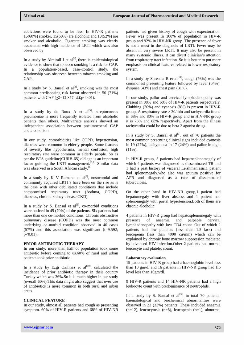
I ) Anti-microbial susceptibility pattern of isolated micro-organisms
a) Klebsiella pneumoniae in HIV-R (n=6)
Klebsiella
pneumoniae Car Flu Ami B-Lac Cepha
HIV-R
(out of n) 100% 83.33% 16.66% 16.66% 33.33%
Mrinal et al. European Journal of Pharmaceutical and Medical Research
www.ejpmr.com
371
b) Klebsiella pneumoniae in HIV-NR (n=10).
Klebsiella
Pneumoniae Car Col/Poly Flu Ami B-Lac Cepha Tig
HIV-NR
(out of n) 50% 30% 40% 40% 40% 50% 10%
c) Staphylococcus aureus in HIV-R (n=8).
Stap. aureus Van Lin Flu B-Lac Cepha Mac
HIV-R
(out of n) 100% 100% 25% 25% 12.5% 0%
d) Staphylococcus aureus in HIV-NR (n=5).
Stap. aureus Van Lin Flu B-Lac Mac
HIV-NR
(out of n) 60% 60% 60% 40% 0%
Abbreviatons: Car- carbapenem, Col/Poly-
colistin/polymixin,
Flu- Fluoroquinolone, Ami- aminoglycoside, B-Lac-
Beta-lactum,
Cepha- Cephalosporins, Cot- Cotrimoxazole, Tig-
tigecycline,
Van- Vancomycin, Lin- Linezolid, Mac-Macrolides.
j) Outcome
Table 11: Showing outcome after treatment received
in HIV-R and HIV-NR group.
HIV-R HIV-NR
Cured 24 25
Expired 1 0
Our study had only one HIV patient who died due to
multiorgan failure.
DISCUSSION
There were 50 patients who fulfilled our inclusion
criteria and were able to cough out acceptable sputum.
Every third patients having the inclusion criteria
attending OPD were selected. The presence of more than
10 squamous epithelial cells in direct smear of sputum
per low power of field (100x magnification) indicates
contamination and discarded for further study. A
specimen with few or no squamous cells and more than
25 polymorphonuclear white blood cells per low power
field is ideal for staining and culture. Even after sputum
induction many patients were unable to produce deeply
coughed out sputum. They were excluded from our
study. So the exact number of patients with similar
clinical features was much more than our enrolment.
SEX, AGE AND OTHER DEMOGRAPHIC
FACTORS
Lower respiratory tract infection is known to occur in all
part of society.
In our study, 68% of HIV-R patients are male and 32%
are female and in case of HIV-NR group males are 56%
and females are 44%.
In a study by Almirall J et al[1]
, in Europe has shown that
CAP incidence is more in males with ratio of 1.4 : 1. The
incidence is shown to be increased with age.
From the overall enrolment of patients at Tropical
Medicine OPD and admission register of our hospital we
can suggest that number of male patients with LRTI is
more than female. Number of male patients with LRTI
are more as compared to females in HIV group. In HIV-
NR group LRTI is more or less equal in both males and
females.
In our study, HIV infection was found more in younger
age group and they are prone to suffer from LRTI.LRTI
incidence increases with age more than 50 years in HIV-
NR patients. The most commonly visited age group in
HIV-R patients is 18 to 30 years with mean age 37.48
and the incidence of LRTI was high in this age group.
The mean age was 53.48 in HIV-NR group.
In a study on LRTI by the Microbiology Department of
Christian Medical College and Hospital, Ludhiana the
mean age of presentation was 40 years.[2]
In a study by S. Bansal et al[3]
, mean age was of 52.77 ±
18.1 years (range 17-93 years). There were 50 males.
Thirty of the 70 patients were in the sixth and seventh
decades of life. Patients older than 40 years were more
predisposed to the development of CAP (t=24.441;
p<0.01).
In our study, HIV-R with LRTI was more common in
rural population as compared to urban. HIV-NR with
LRTI was more common in urban as compared to rural
population. Admission requirement was higher than
other community based study on LRTI possibly because
our hospital is a tertiary referral centre.
In our study 60% of the patients were inpatients.
PERSONAL HABITS
In our study, information regarding smoking and alcohol
was taken. These two addictions seemed to occur
simultaneously in some patients. In female these
Mrinal et al. European Journal of Pharmaceutical and Medical Research
www.ejpmr.com
372
addictions were found to be less. In HIV-R patients
15(60%) smoker, 15(60%) are alcoholic and 13(52%) are
smoker and alcoholic. Cigarette smoking was clearly
associated with high incidence of LRTI which was also
observed by
In a study by Almirall J et al[4]
, there is epidemiological
evidence to show that tobacco smoking is a risk for CAP.
In a population-based, case–control study, the
relationship was observed between tobacco smoking and
CAP.
In a study by S. Bansal et al[3]
, smoking was the most
common predisposing risk factor observed in 50 (71%)
patients with CAP (χ2=12.857; d.f,p<0.01).
In a study by de Roux A et al[5]
, streptococcus
pneumoniae is more frequently isolated from alcoholic
patients than others. Multivariate analysis showed an
independent association between pneumococcal CAP
and alcoholism.
In our study, comorbidities like COPD, hypertension,
diabetes were common in elderly people. Some features
of severity like hypothermia, mental confusion, high
respiratory rate were common in elderly people. So as
per the BTS guideline(CURB-65) old age is an important
factor guiding the LRTI management.[6,7]
Similar data
was observed in a South African study.[8]
In a study by K V Ramana et al[9]
, nosocomial and
community acquired LRTI’s have been on the rise as is
the case with other debilitated conditions that include
compromised respiratory tract (Asthma, COPD),
diabetes, chronic kidney disease CKD).
In a study by S. Bansal et al[3]
, co-morbid conditions
were noticed in 49 (70%) of the patients. Six patients had
more than one co-morbid conditions. Chronic obstructive
pulmonary disease (COPD) was the most common
underlying co-morbid condition observed in 40 cases
(57%) and this association was significant (t=9.592;
p<0.01).
PRIOR ANTIBIOTIC THERAPY
In our study, more than half of population took some
antibiotic before coming to us.60% of rural and urban
patients took prior antibiotic.
In a study by Ezgi Ozilmaz et al[10]
, calculated the
incidence of prior antibiotic therapy in their country
Turkey which was 36%.So it is much higher in our study
(overall 60%).This data might also suggest that over use
of antibiotics is more common in both rural and urban
areas.
CLINICAL FEATURE In our study, almost all patients had cough as presenting
symptom. 60% of HIV-R patients and 68% of HIV-NR
patients had given history of cough with expectoration.
Fever was present in 100% of population in HIV-R
group and 92% in HIV-NR group. The presence of fever
is not a must in the diagnosis of LRTI. Fever may be
absent in very severe LRTI. It may also be present in
many systemic illness. It can divert clinician’s attention
from respiratory tract infection. So it is better to put more
emphasis on clinical features related to lower respiratory
system.
In a study by Shrestha R et al[11]
, cough (76%) was the
commonest presenting feature followed by fever (64%);
dyspnea (43%) and chest pain (31%).
In our study, pallor and cervical lymphadenopathy was
present in 88% and 68% of HIV-R patients respectively.
Clubbing (20%) and cyanosis (8%) is present in HIV-R
group. A respiratory rate > 30/min and tachycardia found
in 68% and 80% in HIV-R group and in HIV-NR group
it is 76% and 88% respectively. Apart from the illness
tachycardia could be due to beta 2 agonist drugs.
In a study by S. Bansal et al[3]
, out of 70 patients the
most common presenting clinical signs included cyanosis
in 19 (27%), tachypnoea in 17 (24%) and pallor in eight
(11%).
In HIV-R group, 5 patients had hepatosplenomegaly of
which 4 patients was diagnosed as disseminated TB and
1 had a past history of visceral Leishmaniasis.1 patient
had splenomegaly,who also was sputum positive for
AFB and diagnosed as a case of disseminated
tuberculosis.
On the other hand in HIV-NR group,1 patient had
hepatomegaly with liver abscess and 1 patient had
splenomegaly with portal hypertension.Both of them are
chronic alcoholic.
4 patients in HIV-R group had hepatosplenomegaly with
presence of anaemia and palpable cervical
lymphadenopathy with low CD4 count. Out of which 2
patients had low platelets (less than 1.5 lacs) and
leucopenia (less than 4000 cu/mm) which can be
explained by chronic bone marrow suppression mediated
by advanced HIV infection.Other 2 patients had normal
leucocyte and platelet count.
Laboratory evaluation 19 patients in HIV-R group had a haemoglobin level less
than 10 gm/dl and 16 patients in HIV-NR group had Hb
level less than 10gm/dl.
9 HIV-R patients and 14 HIV-NR patients had a high
leukocyte count with predominance of neutrophils.
In a study by S. Bansal et al[3]
, in total 70 patients-
haematological and biochemical abnormalities were
observed in 23 (33%) patients. These included anaemia
(n=12), leucocytosis (n=8), leucopenia (n=1), abnormal

Mrinal et al. European Journal of Pharmaceutical and Medical Research
www.ejpmr.com
373
liver function (n=13) and abnormal renal function (n=7)
tests. Eleven patients had more than one abnormality.
LRTI produces neutrophilic leukocytosis(TLC more than
10,000). 84% of the patients had leukocytosis in HIV-
NR group and 32% in HIV-R group. Out of the 20
patients with wheeze,7 patients had high eosinophil
count(more than 4) in HIV-R group and out of 18
patients with wheeze in HIV-NR group 12 patients had
high eosinophil count.Leucopenia was found in 32% of
the patients in HIV-R patients and 8% of HIV-NR
group.Rest of the patients had lymphocytic leucocytosis.
Growth of mixed organisms in the sputum with low CD4
count in HIV-R group are usually sensitive to
cephalosporin,glycopeptides (vancomycin),polypeptide
antibiotics (colistin,polymixin-B),fluoroquinolones
(levofloxacin,ciprofloxacin) and oxazolidinone
(linezolid).
Sputum culture showed growth of bacteria in all the 25
patients in HIV-NR group and 23 patients in HIV-R
group.
Sputum culture showed growth of microorganism in
almost all the patients and blood culture was positive in
one patient of HIV-R group only (Staph.aureus). So
bacteremia is not common in LRTI patients.
Both of the group showed growth of micro organism in
sputum culture.On Gram staining in HIV-R group 13
patients had Gram positive bacteria and 7 patients had
Gram negative bacteria. In HIV-NR group it was 9 and
15 respectively. Growth was detected in 92% of the
patient’s sputum on conventional sputum culture in HIV-
R group and 100% in HIV-NR group.
Single organism was found in 80% in HIV-R group and
84% in HIV-NR group. Mixed organism was detected in
12% in each group. Detection of multiple organisms is
important. Patients with mixed flora had a severe course
of illness. The assumed clinical diagnosis of resistant
bacteria could be due to infection caused by mixed
organisms. In this way finding of a mixed flora can
reduce the use of unnecessary broad spectrum antibiotics
and the cost of treatment.
In a study by Ezgi Ozilmaz et al[10]
percentage of
multiple organism was around 10%.
The most commonly detected single organism is
Staphylococcus aureus (24%) in HIV -R patients.
Klebsiella pneumoniae (28%) is the most common single
organism in HIV-NR patients.
In a study by Mustaq Ahmed et al[12]
, Egbe et al[13]
and
Akingbade[14]
have also reported the same.
Klebsiella pneumoniae to be the most common isolate
recovered from patients with LRTIs. This was the
findings in all groups like diabetics, smokers, alcoholics,
patients with COPD, etc.All the patients having
Staphylococcus aureus(24%) required admission.
In a study by Baik I et al[15]
, among the bacteria
Staphylococcus aureus (24%) and Streptococcus
pyogenes(20%) was commonly cultured organism in
HIV-R patients.
In a study by Hirschtick et al[16]
, atypical pyogenic
bacteria may also be the causative agent, particularly in
patients with advanced HIV disease. For example,
Klebsiella pneumoniae, other members of the
Enterobacteriaceae family and Pseudomonas aeruginosa
were present in 13, 10 and 8% of cases, respectively, of
confirmed pneumonia in this US cohort study.
In a study by Bekele Afessa et al[17]
, P. aeruginosa, the
Enterobacteriaceae family and Staphylococcus aureus
were the cause of 25, 9 and 10% of community-acquired
pneumonia, respectively.
One HIV-R patients with very low CD4 count (base
line=34 cells/μl) with right sided encysted empyema had
sputum growth of Klebsiella pneumonia.His direct smear
of sputum also showed plenty of pseudohyphae (candida)
and pus cells. Sputum for acid fast bacilli was negative.
In our study it has been found that in very low CD4
count we usually found sputum growth of Gram negative
bacteria like Klebsiella,Pseudomonas,E.coli,
acinetobacter etc.All these patients were severely ill.
In a study by Bekele Afessa et al[17]
, pseudomonal
pneumonia is becoming a common pulmonary
complication, especially in patients with low leukocyte
and CD4 lymphocyte counts. Compared with
pneumococcal pneumonia, pseudomonal pneumonia is
associated with a lower incidence of bacteremia and a
longer hospital stay. Despite the low CD4 lymphocyte
and leukocyte counts associated with pseudomonal
pneumonia, the mortality rate is only 19%.
In a study by C Feldman et al[18]
, pneumonia is most
common when the CD4+ count falls below 200 cells/μl.
The organisms responsible for CAP in HIV-seropositive
patients are the same as in HIV-seronegative cases. The
most common bacterial causes of pneumonia are S.
pneumonia and H. influenzae.
In our study, pleural tap was done in 3 patients in each
group of which all showed bacterial growth in the culture
of pleural fluid.It indicates that empyema fluid, when
present is a better bacteriological sample than sputum.
In a study by S. Bansal et al[3]
, by pleural fluid specimens
were available from seven (10%) patients. Pleural fluid
from two patients grew Staphylococcus aureus.

Mrinal et al. European Journal of Pharmaceutical and Medical Research
www.ejpmr.com
374
12% of patients in each group had normal finding in
chest X ray. Patchy consolidation was found in 60% of
HIV-R patients and 64% of HIV-NR patients. In both the
groups presenting with consolidation sputum showed
growth of bacteria or fungus.Sputum culture detected
growth in 92% in HIV-R group and 100% in HIV-NR
group. So chance of getting organism in sputum is high
in patients showing consolidation in chest x-ray.
One patient in the HIV-R group had a very low CD4
count (base line CD4 27 cells/μl) showed ill defined non
homogeneous opacity in right mid and lower zone in
chest X ray.Her sputum showed growth of mixed
organism- Pseudomonas,Acinetobactor and Aspergillus
fumigatus. She was anaemic,icteric, had bilateral pedal
edema with hepatomegaly with high serum ferritin and
triglyceride level. Her CMV DNA PCR was also
positive. She was diagnosed as a case of
Hemophagocytic syndrome.
One HIV-R COPD patient presenting with high
fever,cough with expectoration,anemia(Hb-7.1 g/dl) with
low CD4 count (CD4 86 cells/μl) showed growth of
Klebsiella pneumoniae in sputum sensitive to beta
lactum,quinolones,and third generation
cephalosporin.Chest X ray showed bilateral non
homogenous opacity in lower lung fields with increased
broncho vascular markings in upper and mid zones of
both lungs.USG showed bilateral renal parenchymal
disease(Creatinine- 2.88 mg/dl).
One HIV-R patient came to us with low CD4 count with
history of cough with expectoration and fever suggestive
of LRTI showed growth of mixed organism -Klebsiella
pneumoniae, Streptococcus pyogenes and
Staphylococcus aureus. Patient was chronic
alcoholic,smoker with past history of treatment for
EPTB. His Chest x-ray showed left sided
pleuropericardial effusion and USG showed mild
hepatomegaly with presence of significant retro
peritoneal lymph node.The patient needed inpatient
treatment and was cured by giving inj. Meropenem and
inj. Linezolid.
One HIV-R patient showed growth of Streptococcus
pneumoniae in sputum but Staphylococcus aureus in
blood culture.Chest x-ray showed right mid and lower
zone consolidation.
2 HIV-R patients did not show growth of any organism
in sputum.The cause could be prior antibiotic therapy,
viral pneumonia, infections by anaerobes or atypical
organisms that were difficult to grow by conventional
aerobic bacteriological culture like Mycoplasma,
Chlamydophilla, Legionella etc.
In the study of CMC, Ludhiana in 2006 used serology for
Mycoplasma and Chlamydophilla for all patients. They
detected the occurrence of atypical pneumoniae around
34%.
Dey AB et al from AIIMS, New Delhi in 2000
suggested that the prevalence of Mycoplasma was
around 35%.[19]
In HIV-R groups 4 patients had fungal growth in sputum.
Among them Aspergillus fumigatus was detected in 2
patients. 1 patient had candida spp. 1 patient presented
with high grade fever, cough with expectoration with
growth of Aspergillus niger and pseudomonas in sputum
culture.The patient was admitted and treated successfully
with inj. Piperacillin tazobactum intravenously.
In a study by VV Shailaja et al[20]
, among the 27 fungal
isolates from HIV reactive patients, 9 were pathogenic
(12.83%), 6/9 were Candida albicans, 2/9 were
Cryptococcus neoformans and one was Aspergillus
niger. The rest of the 18 isolates were non albicans
Candida spp. that were considered as colonizers of the
oropharynx.
In our study,klebsiella pneumonia in HIV-NR group is
usually sensitive to carbapenems,Colistin,polymyxin-B
& fluoroquinolones/aminoglycosides in some cases but
resistant to beta-lactums and third generation
cephalosporins.Klebsiella pneumonia in HIV-R group is
usually sensitive to carbapenems and fluoroquinolones
and resistant to third generation cephalosporins,beta
lactums,cotrimoxazole and in some cases to
aminoglycosides also.
In our study,staphylococcus aureus in HIV-R and HIV-
NR group is usually sensitive to vancomycin and
linezolid and resistant to aminoglycosides, beta-
lactum,third generation
cephalosporin,macrolides,cotrimoxazole and
fluoroquinolones but was found to be sensitive to
levofloxacin in HIV-NR group.
In a study by K V Ramana et al[9]
, imipenem and
amikacin was found to show greater activity against
gram negative bacteria isolates whereas linezolid,
amikacin, ciprofloxacin, ofloxacin and cotrimoxazole
were effective against gram positive bacteria isolates.
In our study only one patient in HIV-R group expired
even after taking full treatment for LRTI.This patient
was on treatment for sputum positive DTB with Cat-1
ATD.He was also undergoing treatment for COPD and
DM. He also developed skin rash and oro-genital ulcer
during treatment. Rest of the patients got cured in both
HIV-R and HIV-NR group.
In a study by S. Bansal et al[3]
, out of the 21 patients
hospitalised 8 patients died.2 patients out of 8 had COPD
and one had pulmonary embolism.
None of the patients in my study were vaccinated with
pneumococcal or influenza vaccine.

Mrinal et al. European Journal of Pharmaceutical and Medical Research
www.ejpmr.com
375
CONCLUSIONS
At the end of my study of clinical, radiological and
laboratory parameters of HIV infected and HIV non
infected adults, it is concluded that following points
should be considered while dealing with a case of LRTI
• A reasonable number of LRTI patients require in-
patient treatment.
• Increased age is a significant risk factor for
morbidity in LRTI.
• LRTI in HIV-R group is more common in younger
age group as compared to HIV-NR group in which it
is more common after 50 years of age.
• Males are more commonly affected with LRTI in
both groups.
• LRTI is common among diabetics, smokers and
alcoholics in both groups.
• COPD is an important association for LRTI in both
groups, more commonly in HIV-NR group and a
risk factor for mortality in HIV-R group.
• Past history of tuberculosis is also an important
association for LRTI in both groups.
• Use of antibiotic before coming to hospital is a very
common practice in both rural and urban population.
Cough and fever are the most common presentations
in both groups.
• Chest pain and shortness of breath is more common
in HIV-R group.
• High BP associated with LRTI is more common in
HIV-NR group.
• The severity criteria given by BTS guideline i.e.
CURB-65 is pretty useful in a set up like us. It helps
to recognize the severely ill patients early.
• Crepitations are commonly present in both the
groups.
• Anemia and cervical lymphadenopathy are more
common in HIV-R group.
• Clubbing is more common in HIV-NR group.
• Pleural effusion is a common finding in both groups.
• Leucopenia was more common in HIV-R group
whereas, leucocytosis was more common in HIV-
NR group.
• Most of the HIV-R patients had CD4 count less than
350 cells/μl.
• Gram negative bacteria is more commonly found in
CD4 less than 150 cells/μl..
• Gram positive bacteria is more commonly found in
CD4 more than 150 cells/μl..
• Single or mixed organism causing LRTI does not
depend on immune status of the patient.
• The most common single organism found in sputum
culture of patients of LRTI in HIV-R group is
Staphylococcus aureus and in HIV-NR group is
Klebsiella pneumoniae.
• Klebsiella pneumonia in HIV-NR group is usually
sensitive to carbapenems,Colistin,polymyxin-B &
fluoroquinolones/aminoglycosides in some cases but
resistant to beta-lactums and third generation
cephalosporins.
• Klebsiella pneumonia in HIV-R group is usually
sensitive to carbapenems and fluoroquinolones and
resistant to third generation cephalosporins,beta
lactums,cotrimoxazole and in some cases to
aminoglycosides also.
• Staphylococcus aureus in HIV-R and HIV-NR group
is usually sensitive to vancomycin and linezolid and
resistant to aminoglycosides, beta-lactum,third
generation cephalosporin, macrolides, cotrimoxazole
and fluoroquinolones but was found to be sensitive
to levofloxacin in HIV-NR group.
• Fungal respiratory infections are more common in
HIV-R group as compared to HIV-NR group.
• Patchy consolidation is the commonest radiological
finding in both the groups.
• Pleural effusion is more common in HIV-NR group.
• Normal radiological finding is found equally in both
groups.
• Hilar lymphadenopathy was more common in HIV-
R group.
• Consolidation is more common in CD4 count <200
cells/μl.
• Gram negative bacteria like Pseudomonas,
Klebsiella Pneumoniae,E.coli, Klebsiella Oxytoca,
Acinetobacter are usually found in HIV- Infected
patients with CD4 Count <150 cells/μl.
• Gram positive bacteria like Staphylococcus aureus,
S.Pyogenes, S.pneumoniae are usually found in
sputum of HIV-Infected patients with CD4 count
>150 cells/μl.
• The demerit of our study was small number of
patients included in the study, sensitivity pattern was
not done against all available antibiotics and
minimum inhibitory concentration of the tested
antibiotics could not be performed due to lack of
facilities.
Conflicts of interest
All contributing authors declare no conflicts of interest.
REFERENCES
1. Almirall J, Bolíbar I, Vidal J, et al. Epidemiology of
communityacquired pneumonia in adults: A
population-based study. Eur Respir J, 2000; 15:
757- 763.
2. Bacteriological Profile,Serology and Antibiotic
Sensitivity Pattern of Micro-organisms from
Community Acquired Pneumonia. Aroma Oberoi,
Aruna Agarwal. JK Science, Vol.8; No.2, April-June
2006.
3. S. Bansal, S. Kashyap1, L.S. Pal and A. Goel2
Clinical and Bacteriological Profile of Community
Acquired Pneumonia in Shimla, Himachal Pradesh.
4. Almirall J, González CA, Balanzó X, et al.
Proportion of communityacquired pneumonia cases
attributable to tobacco smoking. Chest, 1999; 116:
375-379.
5. De Roux A, Cavalcanti M, Marcos MA, et al.
Impact of Alcohol Abuse in the Etiology and
Severity of Community-Acquired Pneumonia.
Chest, 2006; 129: 1219-1225.

Mrinal et al. European Journal of Pharmaceutical and Medical Research
www.ejpmr.com
376
6. Defining community acquired pneumonia severity
on presentation to hospital: An international
derivation validation study by Lim WS, van der
Eerden MM, Laing R, et al. Thorax, 2003; 58:
377-382.
7. BTS guidlines for the management community
acquired pneumonia in adults. Thorax, 2001; 56(4):
IVI-IV64.
8. Pneumonia in elderly. Feldman C. Clin Chest Med,
1999 Sep; 20(3): 563-73.
9. K V Ramana*, Anand Kalaskar, Mohan Rao,
Sanjeev D Rao Department of Microbiology,
Prathima Institute of Medical Sciences, Karimnagar,
Andhrapradesh, India.Aetiology and Antimicrobial
Susceptibility Patterns of Lower Respiratory Tract
Infections (LRTI’s) in a Rural Tertiary Care
Teaching Hospital at Karimnagar, South India.
10. Major Bacteria of Community - Acquired
Respiratory Tract Infections in Turkey. Ezgi
Ozilmaz, Kamruddin Ahmed. et al. Jpn. J. Infect.
Dis, 2005; 58: 50-52.
11. Shrestha R, Paudel N, Barakoti B,Dhungana D,
Sharma P2: Etiology and clinical profile of
inpatients with Community acquired pneumonia in
Manipal Teaching hospital, Pokhara, Nepal.
12. Mustaq Ahmed S, Jakribettu RP,Meletath SK,Arya
B,Shakir VPA. Lower Respiratory Tract
Infection(LRTIs):an insight into the prevalence and
the antibiogram of the gram negative
respiratory,bacterial agents.J Clin Diag Res, 2013;
1-4.
13. Egbe CA,Ndiok were C,Omoregie R.Microbiology
of Lower Respiratory Tract Infections in Benin
City,Nigeria Malaysian J Med Sci, 2011; 18(2):
27-31.AA
14. Akingbade OA, Ogiogwa JI, Okerentugba PO,
Innocent-Adiele HC et al.Prevalence and antibiotic
susceptibility pattern of bacterial agents involved in
Lower Respiratory Tract Infections in Abeokuta,
Ogun state,Nigeria.Report and opinion, 2012; 4(5):
25-30.
15. A prospective study of age and lifestyle factors in
relation to community acquired pneumonia in US
men and women by Baik 1, Curhan GC rimn EB, et
al. Arch Intern Med, 2000; 160: 3082- 3088.
16. Hirschtick R, Glassroth J, Jordan M, et al. Bacterial
pneumonia in persons infected with the human
immunodeficiency virus. N Engl J Med, 1995; 333:
845–851.
17. Afessa B, Green B. Bacterial pneumonia in
hospitalized patients with HIV infection. Chest,
2000; 117: 1017–1022.
18. C Feldman, A J Brink, G A Richards, G Maartens, E
D Bateman Management of Community-Acquired
Pneumonia in Adults.
19. Clearence of particles from the human
tracheobronchial tree by Camner P:. Clin Sci, 1980;
59: 79-84.
20. VV Shailaja, LA Pai, DR Mathur, V Lakshmi
Prevalence of bacterial and fungal agents causing
lower respiratory tract infections in patients with
human immunodeficiency virus infection.