a computer-based approach for developing linamarase
TRANSCRIPT

The Nelson Mandela AFrican Institution of Science and Technology
NM-AIST Repository https://dspace.mm-aist.ac.tz
Materials, Energy, Water and Environmental Sciences Research Articles [MEWES]
2020-04-18
A computer-based approach for
developing linamarase inhibitory agents
Paul, Lucas
Walter de Gruyter GmbH
https://doi.org/10.1515/psr-2019-0098
Provided with love from The Nelson Mandela African Institution of Science and Technology

Auto
mat
ically
gene
rate
dro
ugh
PDFb
yPro
ofCh
eckf
rom
Rive
rVal
leyT
echn
olog
iesL
td
DE GRUYTER Physical Sciences Reviews. 2020; 20190098
Lucas Paul1,6 / Celestin N. Mudogo2,7 / Kelvin M. Mtei3 / Revocatus L. Machunda3 / Fidele Ntie-Kang4,5,8
A computer-based approach for developinglinamarase inhibitory agents1 TheDepartment ofMaterials and Energy Science& Engineering, TheNelsonMandela African Institution of Science and Tech-nology, P.O. Box 447 Arusha, Tanzania, E-mail: [email protected], [email protected]
2 Biochemistry andMolecularbiology, University of Hamburg Institute of Biochemistry andMolecularbiology, Hamburg, Ger-many, E-mail: [email protected]
3 TheDepartment ofWater and Environmental Science and Engineering, TheNelsonMandela African Institution of Scienceand Technology, P.O. Box 447 Arusha, Tanzania, E-mail: [email protected], [email protected]
4 Department of Pharmaceutical Chemistry,Martin-Luther University Halle-Wittenberg,Wolfgang-Langenbeck Str. 4, Halle(Saale) 06120, Germany, E-mail: [email protected]
5 Department of Informatics and Chemistry, University of Chemistry and Technology Prague, Technická 5, Prague 6, Dejvice 16628, Czech Republic, E-mail: [email protected]
6 Department of Chemistry, Dar es SalaamUniversity College of Education, P.O. Box 2329, 255 Dar es Salaam, Tanzania, E-mail:[email protected], [email protected]
7 Department of Basic Sciences, School ofMedicine, University of Kinshasa, Kinshasa, Congo (Democratic Republic of the), E-mail: [email protected]
8 Department of Chemistry, University of Buea, P. O. Box 63 Buea, Cameroon, E-mail: [email protected]
Abstract:Cassava is a strategic crop, especially for developing countries. However, the presence of cyanogenic com-pounds in cassava products limits the proper nutrients utilization. Due to the poor availability of structurediscovery and elucidation in the Protein Data Bank is limiting the full understanding of the enzyme, how toinhibit it and applications in different fields. There is a need to solve the three-dimensional structure (3-D) oflinamarase from cassava. The structural elucidation will allow the development of a competitive inhibitor andvarious industrial applications of the enzyme. The goal of this review is to summarize and present the available3-D modeling structure of linamarase enzyme using different computational strategies. This approach couldhelp in determining the structure of linamarase and later guide the structure elucidation in silico and experi-mentally.Keywords: linamarase, cassava, structural determination, computational strategiesDOI: 10.1515/psr-2019-0098
1 Introduction
Cassava (Manihot esculenta Crantz) (Euphorbiaceae), also known as mandioca, yucca, tapioca or manioc. It is theleading supplier of energy ranked after rice and corn [1]. It is the most grown crop in the tropics and subtropicsregions. The tuber is the primary source of carbohydrate while leaves provide protein as a vegetable. About105 countries grow cassava as a strategical crop against famine since it can sustain and produces in droughtand poor soil, can stay within the farm and be harvested at the time of demand [2]. The roots are the primarysource of carbohydrate while leaves provide vitamins, minerals, and protein, as well as a vegetable that isavailable throughout the year. The leaves have high crude protein, and the amino acids, which are well balancedand its amount is beyond the minimal amount recommended by the Food and Agriculture Organization [2].Leaves also have various minerals like iron, zinc, manganese, magnesium and calcium, vitamin B1, B2, C andcarotenoids [2, 3], The combination of cassava roots and leaves can provide a meal with almost all essentialdietary needs.
All body parts of the cassava except seeds contain cyanogenic glucosides compounds known as linamarinand lotaustralin. Linamarase hydrolyses these compounds to hydrogen cyanide as the main product [4]. Thereare about 5,000 varieties of cassava; all of them are known to contain cyanogenic glucosides which range from10 to 500 mg HCN/kg. Based on the amount of hydrogen cyanide (HCN) released, cassavas are classified intothree groups: group one those with greater than 100 mg HCN/kg are called very bitter and very toxic, group
Lucas Paul is the corresponding author.© 2020Walter de Gruyter GmbH, Berlin/Boston.
1

Auto
mat
ically
gene
rate
dro
ugh
PDFb
yPro
ofCh
eckf
rom
Rive
rVal
leyT
echn
olog
iesL
td
Paul et al. DE GRUYTER
two those with between 50 to 100 mg HCN/kg are regarded as moderate bitter and moderate toxic while thosewith less than 50 mg HCN/kg are sweet cassava [5].
Cyanogenic glucosides are mainly composed of Linamarin (95%) and Lotaustralin (5%) [6], enzymatic hy-drolysis of cyanogenic glucosides by linamarase is initiated by any physical damage of cassava tissue this allowsinteraction between enzyme from cell wall which is physically separated from substrate found in cell vacuole soare not compartmentalized [7]. Interaction between enzymes and substrate (mainly Linamarin) start by the re-lease of glucose and acetone cyanohydrin at pH > 4 and temperature >35 °C is converted to hydrogen cyanide[8], Figure 1. The presence of hydroxynitrile lyase (HNL) helps to complete the reaction of acetone cyanohydrinto cyanide. This enzyme is highly available in cassava leaves and very little in the roots [9]. The study by [10]has reported that processed cassava flours contain high levels of acetone cyanohydrin but little linamarin orHCN, this is due to the little amount of HNL in the roots, which brings about this accumulation.
Figure 1: A complete enzymatic reaction between linamarase and linamarin.
Linamarin is a chemically stable compound, soluble in water and resists boiling in acid. Acetone cyanohydrin isalso soluble in water and has a boiling point of 82 °C. But, HCN is a volatile compound, which evaporates only at27 °C so volatilize at ambient temperature. So for effective processing techniques to be useful, we should reducecyanogens to a safe level. The methods should maximize cassava tissue rupture to ensure effective enzyme-substrate interaction, to release acetone cyanohydrin and finally, volatile cyanide [11, 12].
The potential concentration of HCN determines the toxicity of the cyanogenic product consumed. If inad-equately processed food is ingested, the HCN concentration is expected to be high within the body. For thetoxicity of cyanogens depends on the following factors:
a. Unsufficient processing of plant which causes linamarin or HCN to remain in the food.
b. When raw cassava is consumed or insufficiently processed cassava product.
HCN, when released continuously until when low pH value from the stomach, deactivates the enzyme (lina-marase). Cyanide ingested into the body always follows the metabolic pathway of detoxification, wherebyrhodanese works by converting it to thiocyanide which later excreted in the urine [13]. When HCN ingestedin the body gets absorbed quickly into the blood and combines with all forms of iron (methemoglobin andhemoglobin) which are present in erythrocytes [14]. The body eliminates the toxic cyanide by using the en-zyme rhodanese, which contains an active disulfide group. It works by reacting with thiosulphate and cyanide,which converts cyanide to excretable thiocyanate, for this process to be complete sulfur donors that usually isprovided by dietary sulfur amino acids are highly required [15, 16]. HCN binds to the Fe3+/Fe2+ present incytochrome and inactivates its activity.
This HCN inhibits the oxygen uptake and then causes glucose and lactic acid accumulation and deficiencyof Adenosine triphosphate (ATP)/adenosine diphosphate (ADP), which brings the body to anaerobic insteadof aerobic respiration [17]. HCN in the body inhibits many enzymatic reactions; if they contain iron, copperor molybdenum and its effect is highly and immediately appreciated in the respiratory system and heart. Theamount recommended for cassava products, should not exceed 10 mg HCN/kg [4]. Any consumption of cas-sava products beyond the recommended amount can cause the following health problems vomiting, nausea,diarrhoea, dizziness, headache, stomach pains and sometimes death.
Linamarase is among the β-glucosidase belonging to the GH1 family which can convert glucosyl group froma glycoside (nonreducing) or carbohydrate by hydrolysis resulting in water or by transglycosylation gives alco-hol. It has the (β/α)8 barrel structure, the properties of acid-base catalysis and the nucleophilic are contributedby the two carboxylic acid residues at β-strands 4 and 7 [18]. One most crucial property of linamarase which
2
Auto
mat
ically
gene
rate
dro
ugh
PDFb
yPro
ofCh
eckf
rom
Rive
rVal
leyT
echn
olog
iesL
td
DE GRUYTER Paul et al.
differentiate it from other GH1 family is the ability to effectively catalyze the transglycosylation using primary,secondary and tertiary alcohol as acceptors [19]. However, cannot synthesize oligosaccharides and glycosidesby reverse hydrolysis [20].
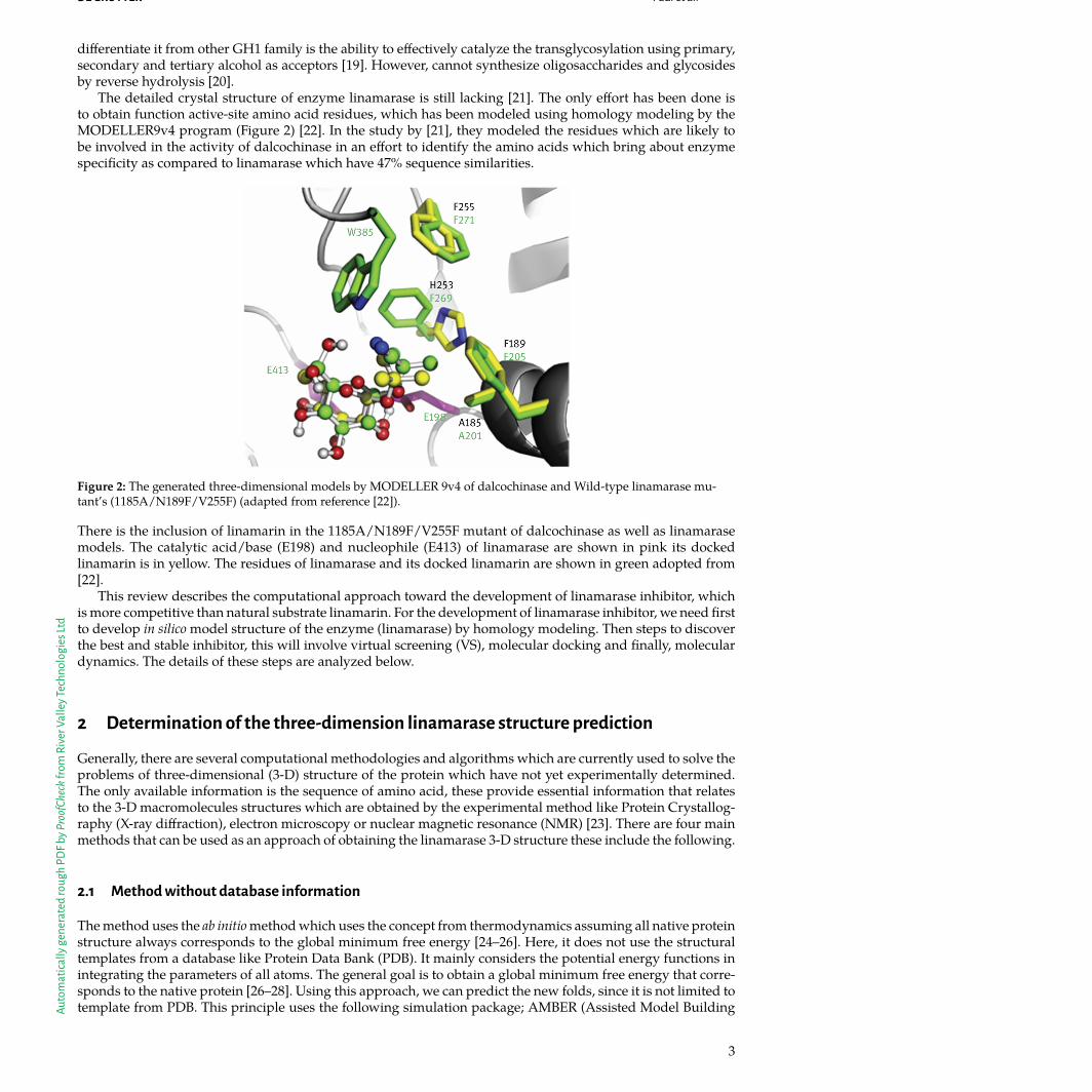
The detailed crystal structure of enzyme linamarase is still lacking [21]. The only effort has been done isto obtain function active-site amino acid residues, which has been modeled using homology modeling by theMODELLER9v4 program (Figure 2) [22]. In the study by [21], they modeled the residues which are likely tobe involved in the activity of dalcochinase in an effort to identify the amino acids which bring about enzymespecificity as compared to linamarase which have 47% sequence similarities.
Figure 2: The generated three-dimensional models by MODELLER 9v4 of dalcochinase and Wild-type linamarase mu-tant’s (1185A/N189F/V255F) (adapted from reference [22]).
There is the inclusion of linamarin in the 1185A/N189F/V255F mutant of dalcochinase as well as linamarasemodels. The catalytic acid/base (E198) and nucleophile (E413) of linamarase are shown in pink its dockedlinamarin is in yellow. The residues of linamarase and its docked linamarin are shown in green adopted from[22].
This review describes the computational approach toward the development of linamarase inhibitor, whichis more competitive than natural substrate linamarin. For the development of linamarase inhibitor, we need firstto develop in silico model structure of the enzyme (linamarase) by homology modeling. Then steps to discoverthe best and stable inhibitor, this will involve virtual screening (VS), molecular docking and finally, moleculardynamics. The details of these steps are analyzed below.
2 Determination of the three-dimension linamarase structure prediction
Generally, there are several computational methodologies and algorithms which are currently used to solve theproblems of three-dimensional (3-D) structure of the protein which have not yet experimentally determined.The only available information is the sequence of amino acid, these provide essential information that relatesto the 3-D macromolecules structures which are obtained by the experimental method like Protein Crystallog-raphy (X-ray diffraction), electron microscopy or nuclear magnetic resonance (NMR) [23]. There are four mainmethods that can be used as an approach of obtaining the linamarase 3-D structure these include the following.
2.1 Method without database information
The method uses the ab initio method which uses the concept from thermodynamics assuming all native proteinstructure always corresponds to the global minimum free energy [24–26]. Here, it does not use the structuraltemplates from a database like Protein Data Bank (PDB). It mainly considers the potential energy functions inintegrating the parameters of all atoms. The general goal is to obtain a global minimum free energy that corre-sponds to the native protein [26–28]. Using this approach, we can predict the new folds, since it is not limited totemplate from PDB. This principle uses the following simulation package; AMBER (Assisted Model Building
3

Auto
mat
ically
gene
rate
dro
ugh
PDFb
yPro
ofCh
eckf
rom
Rive
rVal
leyT
echn
olog
iesL
td
Paul et al. DE GRUYTER
with Energy Refinement) [29, 30], CHARMM (Chemistry at HARvard Molecular Mechanics) [31], UNRES [32],GROMACS (Groningen Machine for Chemical Simulation) [33], TINKER (Software tools for Molecular Design)[34].
2.2 Methods with database information
The starting point here is the 3-D protein structure is obtained from the database, and it compares the fragmentsof the target sequences to that of known protein. The short amino acid sub-sequence of the target structureagainst the known protein’s structure fragments [35]. The newly discovered protein structure will be composedof a similar structure of motifs like the known protein. Therefore, this method is based on fragments of aminoacid sequence with a different motif which, when combined, they form the 3-D protein structure [26]. Thehomology fragments are used for finding the structures which are achieved through scoring functions andalgorithm optimization to get the structure with the lowest potential energy [36]. The fragment-based approachalways needs to look for a criterion that exists between the fragments so that the final fragment will have ahigh chance of being inserted at the final structure predicted as summarized in illustration (Figure 3) [37].This method is similar to ab initio when it finds polypeptide structures with the lowest energy, but the maindifference is that it uses the database to predict the structure of polypeptide [38].
Figure 3: Schematic representation of the method based on fragments (adapted from reference [23]).
2.3 Fold recognition and threading methods
The method involves using the amino acid sequence and evaluates how well it fits the 3-D structure of the pro-tein of the known. This approach is used because structures are more evolutionary preserved than the sequence[35, 39–41]. The sequential order is followed in placing the target amino acid sequences and is governed by twoprocedures; first searching the correct replacement between the target sequence versus the model which is inthe space of possible sequence-structure alignment. The threads tried to find the templates with a similar foldthat have or don’t have direct evolutionary relations (analogue).
2.4 Comparative modeling method and sequence alignment strategies
This method use target protein’s amino acid sequences to align against know protein’s amino acid sequence(used as a template). The information of a known protein is experimentally determined and deposited in PDB[42]. If there are high similarities between the two amino acid sequences, then the structural information of theknown (template) can be used to modal the target protein of interest [43, 44]. The homology protein with fullinformation obtained experimentally are the ones to be used to model the target protein, and their amino acidresidue is similar as they occupy the same position in the homology protein and have similar physic-chemicalproperties. Currently, comparative modeling is highly used because it is useful in protein structure prediction,which has more impact in the field of drug discovery [45]. The sequence alignment can either be pairwise whichis the sequence–sequence comparison or multiple sequence comparison. The first approach uses the targetsequence to compare with sequences in the database independently [46]. It uses methods like FASTA [47, 48],PSI-BLAST [49] and BLAST [50], while multiple sequence comparison allows multiple sequence alignments
4

Auto
mat
ically
gene
rate
dro
ugh
PDFb
yPro
ofCh
eckf
rom
Rive
rVal
leyT
echn
olog
iesL
td
DE GRUYTER Paul et al.
whereby the sensitivity of the search is maximized [51–53]. The methods used here include CLUSTALW [54],PSI-BLAST [49] and T-COFFEE [55].
In the study by [21]identify the specific residues with similarities but have different catalytic properties. Soeight amino acid residues in the glycine binding pocket of dalcochinase were replaced with respective residuesof linamarase. Since the crystal structure of both enzymes is unavailable, then homology modeling of dalcochi-nase which has 47% amino acid sequence identity with linamarase, were performed by using ClustalW 2.0with similar procedures as reported by [54]. The 3-D structure of wild-type dalcochinase was obtained us-ing the template with a 45% identity to dalcochinase from Maize β-glucosidase 1 (Figure 4) with its substrateDIMBOA-β-D-glucoside with PDB code 1E56A as also reported by [56].
Figure 4: The three-dimensional structure of Maize β-glucosidase (from N terminus dark blue to C terminus dark red)(adapted from reference [56]).
The structure was built using the Sybyl 7.2 molecular modeling package, the overall structure of the modelwas checked by the PROCHECK, ProSA, Verify-3D and WHATIF programs [57–60]. Whereby PROCHECKshowed 97% of the residues in the homology were located in the most favorable regions of the Ramachandranplot, whereby only 1.5% was in the rejected region when compared with the template with 98.5% and 0%.PROCHECK was also used to obtain G-factor, which was 0.22 above −0.5 which shows the model was reliableas compared to 0.34 of a template.
The ProSA Z-scores is explained well by its application to check the errors of the 3-D models. It can beused to determine errors of the experimentally and theoretically determined structures. It uses the coordinatesthen the structure’s energy is evaluated by using a distance-based pair potential as well as the potential thatrelates to solvent exposure. The Z-scores validates the quality of the model and measures the total energy ofthe structure with regards to energy distribution derived from random conformations. The obtained Z-scorewhich appears outside a range property of native proteins verifies erroneous structure [58]. In general positivevalue correspond to problematic or erroneous parts of a model. For example the model showedProSA Z-scoreof −8.15 while template −9.68 this is within the acceptable range [58]. The compatibility of the residues with thesurrounding environment was done by verify-3D which scored above 0.2 (91.2%) shows reliable as comparedwith 93.9% for the template. WHATIF program managed to bring the confidence of the packing quality whichscored above −5.0 and the picture of the 3-D model of the active site pocket of dalcochinase was generated byusing PyMOL version 0.99.
Another study by [22] used the report of single mutation brought by replacing eight amino acid residues inthe dalcochinase’s binding pocket using residues from linamarase. The mutants namely 1185A, N189F, V255F
5

Auto
mat
ically
gene
rate
dro
ugh
PDFb
yPro
ofCh
eckf
rom
Rive
rVal
leyT
echn
olog
iesL
td
Paul et al. DE GRUYTER
which have been identified to contribute to the hydrolytic and transglycosylation specificity of dalcochinase.The 1185A and V255F mutants have a low contribution to natural (Dal-Glc) substrate while all three mutantshave more significant transglycosylation activities by using primary and secondary alcohol as acceptors, butnone of these mutants demonstrated linamarase activities of transglycosylation; glucose to tertial alcohol andhydrolyzing linamarin. So in the standing study by [22], it brought an intention of further mutating the residuesof dalcochinase in order to attain the linamarase specificities (transglycosylation and hydrolysis reaction). Sothe 3-D model of the previously reported mutants (1185A, N189F, V255F) of dalcochinase [61] and wild-typelinamarase [18] were created using MODELLER9v4 [62], using the template of cyanogenic β-glucosidase fromTrifolium repens L with (PDB code 1CBG), which have a similarity of 60% to dalcochinase and 51% to wild-typelinamarase [56]. The model’s quality was checked using PROCHECK, ProSA and Verify-3D programs [58, 59]where the active site was defined as 15 Å which is at the center of residues E182 and E396 of dalcochinase.
3 Identification and validation of linamarase inhibitors
The approach of identifying the inhibitor of a specific enzyme uses the same approach of drug discovery whichtargets to obtain a small molecule, known as the entity that can preferentially interact with the valid target. Thetarget which has identified to cause or have a link with the disease or biological effect and need to be inhibited[63].
3.1 Virtual Screening for the identification of linamarase inhibitors
This is described as the step by step approach of searching novel compounds that referred to as hit and ledwith potential biological effects is achieved by filtering and narrowing down until the lead is obtained as analternative to the natural ligand. Depending on the intended application the databases for VS consist of up toabout 10 million compounds and they can be obtained from compound libraries that are provided from com-mercial venders, public and commercial databases. The application of VS depends mainly on the availability ofthe validated structural target (3-D structure). VS is categorized into either structural-based virtual screening(SBVS) or ligand-based Virtual Screening (LBVS).
3.1.1 Structure-based virtual screening
This approach is used in identifying the best ligand through searching to the chemical library for identifying itsinteraction with drug target, and it uses the 3-D structure of the protein which obtained either experimentallyusing X-ray crystallography, NMR or computational modeling [64]. Where the candidate is docked then rankedbased on the binding affinity to the binding site [65].
3.1.2 Ligand-based virtual screening
The approach uses information obtained from the known ligand rather than structural protein for led identifi-cation as well as optimization, and it typically applied when there is no 3-D structure of the protein. It dependson the pharmacophores and relies on the knowledge of the ligand that will bind the active site of the biologicaltarget. The primary goal is to come up with the structure which retains the physicochemical properties. Theapproach is based on the principle that structurally similar molecules will always have similar properties [66].
3.2 Molecular docking
A method is an essential tool in the field of drug discovery and design. The main objective is to predict thebest ligand’s conformation in a target binding site/protein of known 3-D structure [67]. It concentrates muchon either accuracy of the structure or correct prediction activity. The algorithms and scoring function allowsthe evaluation of the interaction of compounds and potential targets. It starts from simple, then advances to itscomplicated stage of the scoring function. It depends mainly on electrostatic and van der Waals of the interactionof solvation or entropic effects.
6

Auto
mat
ically
gene
rate
dro
ugh
PDFb
yPro
ofCh
eckf
rom
Rive
rVal
leyT
echn
olog
iesL
td
DE GRUYTER Paul et al.
There are basic ways of representing the protein and ligands during the docking process these ensure evalu-ation of their methods used, which include; atomic, surface, and grid [68]. Atomic representation is mainly forevaluating pair-wise atomic interaction which brings the complexity so it uses potential energy function [69].Surface-based is used to minimize the angles of the opposite or different interacting molecules [70, 71]. Rigidbody access the energy contribution of receptor specifically on grid points which are used in ligand scoringand stores electrostatic and van der Waals (potentials) [72].
3.2.1 Searching methods for ligands
These are methods that allow molecular flexibility by focusing on the algorithms which treat ligands flexiblebut in a few cases, protein. These methods are divided into (a) systematic approach, (b) random or stochasticmethod and (c) shape matching.
3.2.1.1 Systematic searchingA method is used for flexible ligand docking whereby all number of possible conformations for ligand bindingto the active site is measured, visiting the degrees of freedom of ligand. It can be considered in three approaches;Exhaustive, incremental/fragment, and assembling of conformation. The exhaustive is done by rotating allbonds of the ligand at a specific time allocated. They generate good conformation. To avoid the exhaustivesearch, the screening is done initially for different poses, filtered and then optimized. It uses Glide [73, 74] andFRED [75] for the sampling method. The fragment method is used to avoid the combinatorial explosion. So thealgorithm uses the fragments which may be generated by three steps.
(i) Core fragment selection (ii) Core fragment ligand placement (iii) Incremental ligand placement.By incrementally growing of the ligand into the binding site at a specific time, it is covalently linked, the pro-
grams used include Dock [76], LUDI [77], FlexX [77], ADAM [78] and eHiTs [79]. The conformational ensemblemethods search for the pre-generated ligand confirmation with the libraries. The binding mode is comparedby ranking them by considering the scoring energy. Programs used include FLOG [80], DOCK 3.5 [81], PhDock[82], MS-DOCK [83], MDock [84, 85] and Q-Dock [86].
3.2.1.2 Random search (stochastic algorithms)The algorithms consider the conformation of ligands at the active site, whether individually or populated. Itexamines the translational/rotational space and conformational space of ligand. The approach includes;
The first is the Monte Carlo method (MC); the algorithms allows the generation of random conformationwith translation and rotation at an initial stage of ligands docking at the active site. The new configuration isgenerated after scoring of the initial one and the probability of being accepted is achieved by considering theBoltzmann probability function,
𝑃 ∼ 𝑒𝑥𝑝 [− (𝐸1 − 𝐸0)𝑘𝐵𝑇 ] (1)
where E0 and E1 are the energy scores of the ligand before and after the random change.Respectively, kB is the Boltzmann constant, and T is the absolute temperature of the system. MC uses pro-
grams like DockVision [87], ICM [88], Prodock [89] and MCDOCK [90].The second approach is genetic/evolutionary algorithms, and this uses the approach of biological com-
petition and population dynamics. The varying parameters are included in the chromosome and randomlyvaried. The result produced is evaluated by its fitness. The chromosomes that produce optimal characteristicsare crossed to produce the next generation. This uses GOLD [91, 92], AutoDock [93], DIVALI [94], DARWIN[95], MolDock [96], PSI-DOCK [97], FLIPDock [98], Lead finder [99] and EADock [100].
The third approach is Tabu search algorithms, and this considers the Tabu (those rejected conformation).It operates by making random changes on available conformations then each change is ranked. The Tabu isdetermined, and their changes that have the lower value are going to be accepted even if it was in tabu otherwisethe non-tabu change is accepted. The program uses PRO_LEADS [101] and PSI-DOCK [97].
3.2.1.3 Shape matchingThis method is used at the initial stage of docking, and it is the simplest approach. It places the ligand inthe position that its molecular surface is in complementary with the surface of the binding site of the protein
7

Auto
mat
ically
gene
rate
dro
ugh
PDFb
yPro
ofCh
eckf
rom
Rive
rVal
leyT
echn
olog
iesL
td
Paul et al. DE GRUYTER
involves translation and rotational which allow different orientations of ligands at the binding site. So, it mainlylooks at which the ligands will be easily placed at the binding site as quickly as possible. Programs used includeDOCK [102], FRED [103], FLOG [104], LigandFit [105], Surflex [106] MS-Dock [67] and MDock [107, 108].
3.2.2 Scoring functions
For determining the accuracy of the docking algorithm, it is important to consider the scoring function. So itis the fundamental element for the protein-ligand algorithm. It looks at the reliability and efficiency of anyalgorithm. The scoring functions can be grouped into three categories: Force field, empirical and knowledge-based scoring functions.
3.2.2.1 Force field scoring functionIt uses the molecular mechanics’ force field to consider the interaction between the ligand and receptor. Basi-cally, the Van der Waals energies, the bond bending/stretching/torsional energies, are used at this strategy.The program which can be used here include AMBER [109] or CHARMM [110, 111]. The effect of water forthe force field is accounted for by the inclusion of distance-dependent dielectric constant 𝜀(rij), which uses aprogram like DOCK [112] and implements eq. (2) below.
𝐸 = ∑𝑖
∑𝑗
⎛⎜⎜⎝
𝐴𝑖𝑗
𝑟12𝑖𝑗−
𝐵𝑖𝑗
𝑟6𝑖𝑗+
𝑞𝑖𝑞𝑗
𝜀 (𝑟𝑖𝑗) 𝑟𝑖𝑗
⎞⎟⎟⎠
(2)
where rij stands for the distance between protein atom i and ligand atom j, Aij and Bij are the VDW parame-ters, and qi and qj are the atomic charges. 𝜀(rij) is usually set to 4rij, reflecting the screening effect of water onelectrostatic interactions.
3.2.2.2 Empirical scoring functionThis method is useful for reproducing the experimental data by summation of the binding energy of ligand to areceptor such as VDW, electrostatic, hydrogen bonding, desolvation, entropy and hydrophobicity representedby eq. (3) below:
Δ𝐺 = ∑𝑖
𝑊𝑖 ⋅ Δ𝐺𝑖 (3)
where {∆Gi} is for individual empirical energy and {Wi} produced by the binding affinity after training the com-plex ligand-protein from the known 3-D structures. So, this method mainly depends on the information fromthe crystal structure of different protein-ligand complex whose binding affinities are known. It uses programslike Glide Score [74, 113], LUDI [114–117], SCORE [118–120] X-SCORE [121], ChemScore [122, 123], SFscore[124, 125].
3.2.2.3 Knowledge-based scoring functionThese are scoring functions which aim at reproducing the experimental structures of protein-ligand complexes.It uses the potential of mean force which is given by inverse Boltzmann eq. (4).
𝑤 (𝑟) = −𝑘𝐵𝑇𝐼𝑛 [𝜌 (𝑟) /𝜌∗ (𝑟)] (4)
where kB is the Boltzmann constant, T is the absolute temperature of the system, 𝜌(r) is the number densityof the protein-ligand atom pair at distance r in the training set, and 𝜌∗ (𝑟) is the pair density in a referencestate where the interatomic interactions are zero. Different from the field and empirical scoring function havea right balance between the accuracy and speed because the potentials from eq. (4) above are obtained from alarger number of structures rather than generating the known affinity, it uses a programs like PMF [126, 127],DrugScore [128], SMOG [129].
8

Auto
mat
ically
gene
rate
dro
ugh
PDFb
yPro
ofCh
eckf
rom
Rive
rVal
leyT
echn
olog
iesL
td
DE GRUYTER Paul et al.
3.2.2.4 Consensus scoring functionThe approach is not a specific type of scoring function; it combines and balances the scoring information byremoving/minimizing errors of each scoring function. So it makes the true binder to be distinguished from theothers [130, 131]. Programs used include X-Score and MultiScore [132, 133].
3.2.2.5 Clustering and entropy-based scoring methodsThis method also used to improve the performance of different scoring functions. It includes the entropic ef-fect by taking the ligand binding modes then divided into various clusters [134, 135] The impact of entropiccontribution in each of the clusters is considered, and it uses AutoDock [135, 136].
A case study for docking was reported by [21], after homology modeling and identification of the struc-ture of Thai rosewood dalcochinase which have high similarity to linamarase. The natural substrate (Dal-Glc)of dalcochinase were docked into the active site pocket of dalcochinase obtained using homology modeling,the docking program used was GOLD 3.1 [137]. Maize β-glucosidase was used by docking with DIMBOA-β-D-glucoside, which achieved a fitness score of 69.15, and when as compared with the fitness of the score fordocking Dal-Glc into the dalcochinase it gave score 61.41. The interaction of the substrate positioned it by stack-ing π–π interaction between the phenyl ring of substrate and indole ring of the conserved Trp368. The distancebetween amino acid residues and Dal-Glc (substrate) specifically in the binding pocket of dalcochinase modelwas predicted by molecular docking as summarized in Table 1.
Table 1: The binding pocket of dalcochinase model showing the distances between Dal-Glc and amino acid residues ob-tained by molecular docking, (adapted from reference [21]).
Dal-Glc position Amino acid position Distance (Å)
Sugar ringO2 Y325-HH 2.49O3 N181-HD22 2.89O4 W453-He1 1.48O6 W445-He1 2.16H at O2 E396-Oe1 1.72CMethylene H235-He1 2.34Methylene W368-Hz3 2.51Ring-ARing-B π–π (3.52–4.09)Ring-COH W368-He1 2.35
In different studies [22] and [21], the authors considered the single mutation, whereby residues Ile185, Asn189,and Val255 in dalcochinase binding pocket were replaced by Ala201, Phe205, and Phe271 from linamaraseconsider Figure 2. This affected dalcochinase by increasing transglycosylation, mainly primary and secondaryalcohol. However, this replacement of single mutation did not change dalcochinase activities to be similar tothat of linamarase specifically transglycosylation to tertiary alcohol.
To bring dalcochinase specificity to linamarase multiple mutations of the identified key amino acid residues.The homology models of the 1185A/N189F/V255F which considered a triple mutation of the respective en-zyme were obtained and docked to linamarin which its structure was driven from PubChem (CID 11128) usingthe AutoDock version 4.2 [138]. The analysis of the docked conformation was analyzed by Accelrys DsVisual-izer 3.0 consider Figure 2.
The results of docking of these multiple mutations show that for both enzymes when were docked withlinamarin the covalent bond glycosidic and the C1 atom are proposed to be at the same location.
4 Conclusion
The enzymatic reaction of linamarase is associated with application in biotechnology like transglycosylationand hydrolysis by using acceptors. The most important and which has more concern is in cassava which thehydrolysis of cyanogenic glucoside and ultimately leads to the release of toxic product hydrogen cyanide. Toget the insight of linamarase’s potential and how can be used to different productive reaction, there is a needto elucidate the structure experimentally. However, due to the improvement of computational approaches
9

Auto
mat
ically
gene
rate
dro
ugh
PDFb
yPro
ofCh
eckf
rom
Rive
rVal
leyT
echn
olog
iesL
td
Paul et al. DE GRUYTER
in carrying out structural modeling, this work has reviewed and analyzed the approaches to be used predict thestructure of linamarase. Homology modeling is mainly used then other techniques can be applied to identifythe competitive inhibitor against the natural substrate and inhibits the enzymatic reaction. These are mainlymolecular docking and molecular dynamics. In homology modeling of dalcochinase, it has proved that theenzyme shares 47% amino acid sequence with dalcochinase, which its structure is now available. So it can beused as a template to generate the 3-D structure of linamarase.
Regardless, of these similarities the enzymes they have the different catalytic ability as linamarase worksby hydrolyzing the natural substrate linamarin and dalcochinase the substrate dalcochinin-8-O-β-D-glucoside,but not the reverse. Their distinct also is that dalcochinase can catalyze the transglycosylation of primary andsecondary alcohol but not tertiary while linamarse can work effectively on both. Additionally, linamarase cannotsynthesize oligosaccharides and glycosides by reverse hydrolysis while dalcochinase can do.
Therefore, regardless of the availability of amino acid sequences similar to linamarase, there is a great needto find out the crystal structure experimentally. This will unlock more scientific understanding, research, andapplication of linamarase especially in food processing like products with glycosides residues retained duringthe processing.
Acknowledgements
The authors are indebted to the Nelson Mandela African Institutional of Science and Technology (NM-AIST)through African Development Bank (AfDB) project for financial support. FNK acknowledges a return fellow-ship and equipment subsidy from the Alexander von Humboldt foundation, Germany.
References
[1] AndamaM, Oloya B. Effectiveness of traditional processing techniques in reducing cyanide levels in selected cassava varieties in ZomboDistrict, Uganda. Int J Food Sci Biotechnol. 2017;2:121–5.
[2]Montagnac JA, Davis CR, Tanumihardjo SA. Processing techniques to reduce toxicity and antinutrients of Cassava for use as a staple food.Compr Rev Food Sci Food Saf. 2009;8:17–27.
[3] Burns AE, Bradbury JH, Cavagnaro TR, GleadowRM. Journal of food composition and analysis total cyanide content of cassava food prod-ucts in Australia. J Food Compos Anal. 2012;25:79–82.
[4] Nicolau AI. Safety of fermented cassava products. In: Prakash V,Martín-Belloso O, Keener L, Astley S, Braun S,McMahonH, LelieveldH,editors. Regulating safety of traditional and ethnic foods. London, Oxford, Boston, NewYork und SanDiego: Academic Press, 2016:319–35.
[5] AttahDaniel BE, Ebisike K, Adeeyinwo CE, Ojumu TV, Olusunle SO, AdewoyeOO. Towards arresting the harmful effect of cyanogenic po-tential of cassava toman in the environment. Int J Eng Sci. 2013;2:100–4.
[6] Nartey F.Manihot Esculemta (Cassava): cyanogenesis, ultrastructure and seed germination. Australia: Copenhagen,Munksgaard, 1978.[7]Mcmahon JM, WhiteWL, Sayre RT. Review article: cyanogenesis in cassava (manihot esculenta crantz). J Exp Bot. 1995;46:731–41.[8]MkpongOE, YanH, ChismG, Sayre RT. Purification, characterization, and localization of linamarase in cassava. Plant Physiol.
1990;93:176–81.[9]WhiteWL, Arias-GarzonDI,McMahon JM, Sayre RT. Cyanogenesis in cassava: the role of hydroxynitrile lyase in root cyanide production.
Plant Physiol. 1998;116:1219–25.[10] Tylleskar T, BaneaM, Bikangi N, Cooke RD, Poulter NH, RoslingH. Cassava cyanogens and konzo, an uppermotoneuron disease found in
Africa. Lancet. 1992;339:208–11.[11] Cooke RD. An enzymatic assay for the total cyanide content of cassava (manihot esculenta crantz). J Sci Food Agric. 1978;29:345–52.[12] Brien GM, Taylor AJ, Poulter NH. Improved enzymic assay for cyanogens in fresh and processed cassava. J Sci Food Agric. 1991;56:277–89.[13] Frankenberg L. Enzyme therapy in cyanide poisoning: effect of rhodanese and sulfur compounds. Arch Toxicol. 1980;45:315–23.[14] Food Standards Australia NewZealand, Cassava and bamboo shoots; a human health risk assessment. 2005.[15] Cardoso AP, ErnestoM, Cliff J, Egan SV, Bradbury JH. Cyanogenic potential of cassava flour: field trial inMozambique of a simple kit. Int J
Food Sci Nutr. 1998;49:93–9.[16] RoslingH.Measuring effects in humans of dietary cyanide exposure from cassava. International Society for Horticultural Science (ISHS),
Nov. 1994.[17] Solomonson LP. Cyanide as ametabolic inhibitor. Cyanide Biol. 1981;2013:11–28.[18] Keresztessy Z, Kiss L, HughesMA. Investigation of the active site of the cyanogenic β-D-glucosidase (linamarase) fromManihot escu-
lenta Crantz (cassava). II. Identification of Glu-198 as an active site carboxylate groupwith acid catalytic function. Arch BiochemBiophys.1994;315:323–30.
[19] Svasti J, Phongsak T, Sarnthima R. Transglucosylation of tertiary alcohols using cassava β-glucosidase. BiochemBiophys Res Commun.2003;305:470–5.
[20] Srisomsap C, Subhasitanont P, Techasakul S, Surarit R, Svasti J. Synthesis of homo- and hetero-oligosaccharides by Thai rosewood β-glucosidase. Biotechnol Lett. 1999;21:947–51.
10

Auto
mat
ically
gene
rate
dro
ugh
PDFb
yPro
ofCh
eckf
rom
Rive
rVal
leyT
echn
olog
iesL
td
DE GRUYTER Paul et al.
[21] Kongsaeree PT, RatananikomK, Choengpanya K, TongtubtimaN, Sujiwattanarat P, Porncharoennop C, et al. Substrate specificity inhydrolysis and transglucosylation by family 1 β-glucosidases from cassava and Thai rosewood. JMol Catal B Enzym. 2010;67:257–65.
[22] TongtubtimN, Thenchartanan P, RatananikomK, Choengpanya K, Svasti J, Kongsaeree PT.Multiplemutations in the aglycone bindingpocket to convert the substrate specificity of dalcochinase to linamarase. BiochemBiophys Res Commun. 2018;504:647–53.
[23] DornM, SilvaMB, Buriol LS, Lamb LC. Three-dimensional protein structure prediction:methods and computational strategies. ComputBiol Chem. 2014;53:251–76.
[24] Anfinsen CB, Haber E, SelaM,White FH. The kinetics of formation of native ribonuclease during oxidation of the reduced polypeptidechain. Proc Natl Acad Sci USA. 1961;47:1309–14.
[25] Anfinsen CB. Principles that govern protein folding Publication;. Science. 1973;181:223–30.[26] BüssowKA. Protein structure prediction. Concepts and applications. Anal Bioanal Chem. 2006;386:1579–80.[27] Bujnicki JM. Protein-structure prediction by recombination of fragments. ChemBioChem. 2006;7:19–27 1579–80.[28] Osguthorpe DJ. Ab initio protein folding. Curr Opin Struct Biol. 2000;10:146–52.[29] Case DA, Cheatham III TE, Darden T, GohlkeH, Luo R,Merz Jr KM, et al. The Amber biomolecular simulation programs. J Comput Chem.
2005;26:1668–88.[30] PearlmanDA, Case DA, Caldwell JW, RossWS, Cheatham III TE, DeBolt S, et al. AMBER, a package of computer programs for applying
molecularmechanics, normalmode analysis, molecular dynamics and free energy calculations to simulate the structural and energeticproperties ofmolecules. Comput Phys Commun. 1995;91:1–41.
[31] Best RB, Zhu X, Shim J, Lopes PE,Mittal J, FeigM, et al. Optimization of the additive CHARMMall-atomprotein force field targeting im-proved sampling of the backboneϕ,ψ and side-chain χ 1 and χ 2 dihedral Angles. J ChemTheory Comput. 2012;8:3257–3273.
[32] Liwo A, Kaźmierkiewicz R, Czaplewski C, GrothM, Ołdziej S,Wawak RJ, et al. United-residue force field for off-lattice protein-structuresimulations: III. Origin of backbone hydrogen-bonding cooperativity in united-residue potentials. J Comput Chem. 1998;19:259–76.
[33] Pronk S, Páll S, Schulz R, Larsson P, Bjelkmar P, Apostolov R, et al. GROMACS 4.5: A high-throughput and highly parallel open sourcemolecular simulation toolkit. Bioinformatics. 2013;29:845–54.
[34] Kundrot CE, Ponder JW, Richards FM. Algorithms for calculating excluded volume and its derivatives as a function ofmolecular confor-mation and their use in energyminimization. J Comput Chem. 1991;12:402–9.
[35] Floudas CA, FungHK,McAllister SR,MönnigmannM, Rajgaria R. Advances in protein structure prediction and de novo protein design: areview. ChemEng Sci. 2006;61:966–88.
[36] Simons KT, Kooperberg C, Huang E, Baker D. Assembly of protein tertiary structures from fragments with similar local sequences usingsimulated annealing and Bayesian scoring functions. JMol Biol. 1997;268:209–25.
[37] Zhang Y, Skolnick J. SPICKER: A clustering approach to identify near-native protein folds. J Comput Chem. 2004;25:865–71.[38]Moult J, Fidelis K, Kryshtafovych A, Schwede T, Tramontano A. Critical assessment ofmethods of protein structure prediction (CASP)—
RoundXII. Proteins Struct Funct Bioinforma. 2018;86:7–15.[39] Finkelstein AV, Ptitsyn OB.Why do globular proteins fit the limited set of folding patterns? Prog BiophysMol Biol. 1987;50:171–90.[40] LevittM, Chothia C. Structural patterns in globular proteins. Nature. 1976;261:552–8.[41] Setubal JC,Meidanis J. Introduction to computationalmolecular biology. Boston: PWS Pub, 1997.[42] BermanHM, Battistuz T, Bhat TN, BluhmWF, Bourne PE, et al. The protein data bank. Acta Crystallogr Sect D Biol Crystallogr.
2002;58:899–907.[43] Bajorath J, StenkampR, Aruffo A. Knowledge-basedmodel building of proteins: concepts and examples. Protein Sci. 1993;2:1798–810.[44] SternbergMJ, Thornton JM, Blundell TL, Sibanda BL. Knowledge-based prediction of protein structures and the design of novel
molecules. Nat Int J Sci. 1987;326:347–52.[45] Kopp J, Schwede T. Automated protein structure homologymodeling: a progress report. Pharmacogenomics. 2004;5:405–16.[46] Apostolico A, Giancarlo R. Sequence alignment inmolecular biology. J Comput Biol. 1998;5:173–96.[47] PearsonWR, LipmanDJ. Improved tools for biological sequence comparison. Proc Natl Acad Sci USA. 1988;85:2444–8.[48] LipmanDJ, PearsonWR. Rapid and sensitive protein similarity searches. Science (80-.). 1985;227:1435–41.[49] Altschul SF,Madden TL, Schäffer AA, Zhang J, Zhang Z,MillerW, et al. Gapped BLAST and PSI-BLAST: a new generation of protein
database search programs. Nucleic Acids Res. 1997;25:3389–402.[50] Altschup SF, GishW, Pennsylvania T, Park U. Basic local alignmen. JMol Biol. 1990;215:403–10.[51] Notredame C. Recent progresses inmultiple sequence alignment: a survey. Pharmacogenomics. 2002;3:131–44.[52] Notredame C. Recent evolutions ofmultiple sequence alignment algorithms. PLoS Comput Biol. 2007;3:1405–8.[53] Thompson JD, Plewniak F, PochO. A comprehensive comparison ofmultiple sequence alignment programs. Nucleic Acids Res.
1999;27:2682–90.[54] Thompson JD, Higgins DG. CLUSTALW: improving the sensitivity of progressivemultiple sequence alignment through sequence
weighting, position-specific gap penalties andweightmatrix choice. Nucleic Acids Res. 1994;22:4673–80.[55] Notredame C, Higgins DG, Heringa J. T-Coffee: a novelmethod for fast and accuratemultiple sequence alignment. JMol Biol.
2000;302:205–17.[56] CzjzekM, CicekM, Zamboni V, BevanDR, Henrissat B, Esen A. Themechanism of substrate (aglycone) specificity in β-glucosidases is
revealed by crystal structures ofmutantmaize β-glucosidase-DIMBOA, -DIMBOAGIc, and -dhurrin complexes. Proc Natl Acad Sci USA.2000;97:13555–60.
[57] Laskowski RA,MacArthurMW,Moss DS, Thornton JM. PROCHECK: a program to check the stereochemical quality of protein structures. JAppl Crystallogr. 1993;26:283–91.
[58]WiedersteinM, SipplMJ. ProSA-web: interactive web service for the recognition of errors in three-dimensional structures of proteins.Nucleic Acids Res. 2007;35:407–10.
[59] Kresge JS, Leonowicz CT, RothME, VartuliWJ, Beck JC. Assessment of proteinmodels with three-dimensional profiles. Nature.1992;359:710–13.
[60] Vriend G.WHAT IF: amolecularmodeling and drug design program. JMol Graph. 1990;8:52–6.
11

Auto
mat
ically
gene
rate
dro
ugh
PDFb
yPro
ofCh
eckf
rom
Rive
rVal
leyT
echn
olog
iesL
td
Paul et al. DE GRUYTER
[61] Ketudat Cairns JR, Champattanachai V, Srisomsap C,Wittman-Liebold B, Thiede B, Svasti J. Sequence and expression of Thai Rosewoodbeta-glucosidase/beta-fucosidase, a family 1 glycosyl hydrolase glycoprotein. J Biochem. 2000;128:999–1008.
[62] EswarN,Webb B,Marti-RenomMA,MadhusudanMS, EramianD, ShenM, et al. Comparative protein structuremodeling usingMod-eller. Curr Protoc BioinformaticsCurr. 2006;15:5.6.1–5.6.30.
[63] Lavecchia A, Giovanni C. Virtual screening strategies in drug discovery: a critical review. CurrMed Chem. 2013;20:2839–60.[64] Li Q, Shah S. Structure-based virtual screening.MethodsMol Biol. 2017;1558:111–24.[65] Dror O, Shulman-peleg A, Nussinov R,WolfsonHJ. Predictingmolecular interactions. CurrMed Chem. Curr. 2004;11:71–90.[66] Hamza A, Wei NN, Zhan CG. Ligand-based virtual screening approach using a new scoring function. J Chem InfModel. 2012;52:963–74.[67] Kukol A. Molecular docking. In: Kahl G, editor(s). The dictionary of genomics, transcriptomics and proteomics, 5th ed. Vol. 443.
Mannheim:Wiley-Blackwell, 2015:1–1.978-3-527-32852-9.[68] Halperin I, Ma B,WolfsonH, Nussinov R. Principles of docking: an overview of search algorithms and a guide to scoring functions. Pro-
teins Struct Funct Genet. 2002;47:409–43.[69] Taylor JS, Burnett RM. DARWIN: A program for docking flexiblemolecules. Proteins Struct Funct Genet. 2000;41:173–91.[70] Norel R, Lin SL,WolfsonHJ, Nussinov R. Shape complementarity at protein–protein interfaces. Biopolymers. 1994;34:933–40.[71] Norel R, Petrey D,WolfsonHJ, Nussinov R. Examination of shape complementarity in docking of unbound proteins. Proteins.
1999;36:307–17.[72] Goodford PJ. A computational procedure for determining energetically favorable binding sites on biologically importantmacro-
molecules. J Med Chem. 1985;28:849–57.[73] Friesner RA, Banks JL, Murphy RB, Halgren TA, Klicic JJ, Mainz DT, et al. Glide: A new approach for rapid, accurate docking and scoring. 1.
Method and assessment of docking accuracy. JMed Chem. 2004;47:1739–49.[74] Halgren TA,Murphy RB, Friesner RA, BeardHS, Frye LL, PollardWT, et al. Glide: A new approach for rapid, accurate docking and scoring.
2. Enrichment factors in database screening. JMed Chem. 2004;47:1750–9.[75]McGannMR, AlmondHR, Nicholls A, Grant JA, Brown FK. Gaussian docking functions. Biopolymers. 2003;68:76–90.[76] Ewing TJ, Kuntz ID. Critical evaluation of search algorithms for automatedmolecular docking and database screening. J Comput Chem.
1997;18:1175–89.[77] BohmHJ. The computer program LUDI: a newmethod for the de novo design of enzyme inhibitors. J Comput AidedMol Des. 1992;6:61–
78.[78]MizutaniMY, TomiokaN, Itai A. Rational automatic searchmethod for stable dockingmodels of protein and ligand. JMol Biol.
1994;243:310–26.[79] Zsoldos Z, Reid D, Simon A, Sadjad BS, Johnson AP. eHiTS: An innovative approach to the docking and scoring function problems. Curr
Protein Pept Sci. 2006;7:421–35.[80]MillerMD, Kearsley SK, UnderwoodDJ, Sheridan RP. FLOG: a system to select ‘quasi-flexible’ ligands complementary to a receptor of
known three-dimensional structure. J Comput AidedMol Des. 1994;8:153–74.[81] Lorber DM, Shoichet BK. Flexible ligand docking using conformational ensembles Despite important successes. Protein Sci. 1998;7:938–
50.[82] Joseph-McCarthy D, Thomas BE, BelmarshM,Moustakas D, Alvarez JC. Pharmacophore-basedmolecular docking to account for ligand
flexibility. Proteins Struct Funct Genet. 2003;51:172–88.[83] SautonN, Lagorce D, Villoutreix BO,MitevaMA.MS-DOCK: accuratemultiple conformation generator and rigid docking protocol for
multi-step virtual ligand screening. BMCBioinformatics. 2008;9:1–12.[84] Di Costanzo L, Jr LV, ChristiansonDW. Ensemble docking ofmultiple protein structures: considering protein structural variations in
molecular docking. Proteins. 2006;642:637–42.[85] Huang SY, Zou X. Efficientmolecular docking of NMR structures: application toHIV-1 protease. Protein Sci. 2006;16:43–51.[86] BrylinskiM, Skolnick J. Q-dock: low-resolution flexible ligand dockingwith pocket-specific threading restraints. J Comput Chem.
2008;29:1574–88.[87] Thomsen R, ChristensenMH.MolDock: a new technique for high-accuracymolecular docking. JMed Chem. 2006;49:3315–21.[88] Benigni R, Bossa C.Mechanisms of chemical carcinogenicity andmutagenicity: a reviewwith implications for predictive toxicology.
ChemRev. 2011;111:2507–36.[89]McGannM. FRED andHYBRID docking performance on standardized datasets. J Comput AidedMol Des. 2012;26:897–906.[90] Chen R, Li L,Weng Z. ZDOCK: an initial-stage protein-docking algorithm. Proteins Struct Funct Genet. 2003;52:80–7.[91] KitchenDB, DecornezH, Furr JR, Bajorath J. Docking and scoring in virtual screening for drug discovery:methods and applications. Nat
Rev DrugDiscov. 2004;3:935–49.[92] BrooijmansN, Kuntz ID.Molecular recognition and docking algorithms. Annu Rev Biophys Biomol Struct. 2003;32:335–73.[93] Rishton GM. Reactive compounds and in vitro false positives in HTS. DrugDiscov Today. 1997;2:382–4.[94]Moitessier N, Englebienne P, Lee D, Lawandi J, Corbeil CR. Towards the development of universal, fast and highly accurate docking/scor-
ingmethods: a longway to go. Br J Pharmacol. 2008;153:7–26.[95] Huang SY, Grinter SZ, Zou X. Scoring functions and their evaluationmethods for protein-ligand docking: recent advances and future
directions. Phys Chem ChemPhys. 2010;12:12899–908.[96] Krovat E, Steindl T, Langer T. Recent advances in docking and scoring. Curr Comput Aided-DrugDes. 2006;1:93–102.[97] Jain AN. Scoring functions for protein-ligand docking. Curr Protein Pept Sci. 2006;7:407–20.[98] Seiler KP, George GA, HappMP, BodycombeNE, Carrinski HA, Norton S, et al. ChemBank: a small-molecule screening and cheminfor-
matics resource database. Nucleic Acids Res. 2008;36:351–9.[99]Meng EC, Shoichet BK, Kuntz ID. Automated dockingwith grid-based energy evaluation. J Comput Chem. 1992;13:505–24.[100] Goodsell DS, Olson AJ. Automated docking of substrates to proteins by simulated annealing. Proteins Struct Funct Bioinforma.
1990;8:195–202.[101] Shoichet BK, Leach AR, Kuntz ID. Ligand solvation inmolecular docking. Proteins. 1999;34:4–16.
12

Auto
mat
ically
gene
rate
dro
ugh
PDFb
yPro
ofCh
eckf
rom
Rive
rVal
leyT
echn
olog
iesL
td
DE GRUYTER Paul et al.
[102] Song CM, Lim SJ, Tong JC. Recent advances in computer-aided drug design. Brief Bioinform. 2009;10:579–91.[103] Knox C, LawW, Jewison T, Liu P, Ly S, Frokis A, et al. DrugBank 3.0: A comprehensive resource for ‘Omics’ research on drugs. Nucleic
Acids Res. 2011;39:1035–41.[104] Gaulton A, Bellis LJ, Bento AP, Chambers J, DaviesM, Hersey A, et al. ChEMBL: a large-scale bioactivity database for drug discovery.
Nucleic Acids Res. 2012;40:1100–7.[105] Del Rio A, Barbosa AJ, Caporuscio F,Mangiatordi GF. CoCoCo: a free suite ofmulticonformational chemical databases for high-
throughput virtual screening purposes.Mol Biosyst. 2010;6:2122–8.[106] Bissantz C, Folkers G, RognanD. Protein-based virtual screening of chemical databases. 1. Evaluation of different docking/scoring com-
binations. JMed Chem. 2000;43:4759–67.[107] Baell JB. Observations on screening-based research and some concerning trends in the literature. FutureMed Chem. 2010;2:1529–46.[108] Lagorce D,Maupetit J, Baell J, Sperandio O, Tufféry P,MitevaMA, et al. The FAF-Drugs2 server: amultistep engine to prepare electronic
chemical compound collections. Bioinformatics. 2011;27:2018–20.[109] SeifertMH. Robust optimization of scoring functions for a target class. J Comput AidedMol Des. 2009;23:633–44.[110] Charifson PS, Corkery JJ, MurckoMA,WaltersWP. Consensus scoring: amethod for obtaining improved hit rates fromdocking
databases of three-dimensional structures into proteins. JMed Chem. 1999;42:5100–9.[111] FeherM. Consensus scoring for protein-ligand interactions. DrugDiscov Today. 2006;11:421–8.[112] Raub S, Steffen A, Kämper A,Marian CM. AIScore - Chemically diverse empirical scoring function employing quantum chemical binding
energies of hydrogen-bonded complexes. J Chem InfModel. 2008;48:1492–510.[113]Warren GL, Andrews CW, Capelli AM, ClarkeM, LaLonde J, LambertMH, et al. A critical assessment of docking programs and scoring
functions. JMed Chem. 2006;49:5912–31.[114]Willett P, Barnard JM, Downs GM. Chemical similarity searching. J Chem Inf Comput Sci. 1998;38:983–96.[115]MaX, Jia J, Zhu F, Xue Y, Li Z, Chen Y. Comparative analysis ofmachine learningmethods in ligand-based virtual screening of large
compound libraries. Comb ChemHigh Throughput Screen. 2009;12:344–57.[116] BöhmHJ. The development of a simple empirical scoring function to estimate the binding constant for a protein-ligand complex of
known three-dimensional structure. J Comput AidedMol Des. 1994;8:243–56.[117] BoehmHJ. Prediction of binding constants of protein ligands: a fastmethod for the prioritization of hits obtained fromde novo design
or 3D database search programs. J Comput AidedMol Des. 1998;12:309–23.[118]Melville JL, Burke EK, Hirst JD.Machine learning in virtual screening. Comb ChemHigh Throughput Screen. 2009;12:332–43.[119]Wang R, Liu L, Lai L, Tang Y. SCORE: a new empiricalmethod for estimating the binding affinity of a protein-ligand complex. JMol
Model. 1998;4:379–94.[120] Tao P, Lai L. Protein ligand docking based on empiricalmethod for binding affinity estimation. J Comput AidedMol Des. 2001;15:429–
46.[121]Wang R, Lai L,Wang S. Further development and validation of empirical scoring functions for structure-based binding affinity predic-
tion. J Comput AidedMol Des. 2002;16:11–26.[122] Eckert H, Vogt I, Bajorath J. Mapping algorithms formolecular similarity analysis and ligand-based virtual screening: design of Dyna-
MAD and comparisonwithMAD andDMC. J Chem InfModel. 2006;46:1623–34.[123] EldridgeMD,Murray CW, Auton TR, Paolini GV,Mee RP. Empirical scoring functions: I. The development of a fast empirical scoring
function to estimate the binding affinity of ligands in receptor complexes. J Comput AidedMol Des. 1997;11:425–45.[124] Barreiro G, Guimarães CR, Tubert-Brohman I, Lyons TM, Tirado-Rives J, JorgensenWL. Search for non-nucleoside inhibitors of HIV-1
reverse transcriptase using chemical similarity,molecular docking, andMM-GB/SA scoring. J Chem InfModel. 2007;47:2416–28.[125] RareyM, Kramer B, Lengauer T, Klebe G. A fast flexible dockingmethod using an incremental construction algorithm. JMol Biol.
1996;261:470–89.[126]Muegge I. A knowledge-based scoring function for protein-ligand interactions: probing the reference state. Perspect DrugDiscov Des.
2000;20:99–114.[127]Muegge I, Martin YC. A general and fast scoring function for protein-ligand interactions: a simplified potential approach. JMed Chem.
1999;42:791–804.[128] GohlkeH, HendlichM, Klebe G. Knowledge-based scoring function to predict protein-ligand interactions. JMol Biol. 2000;295:337–56.[129] DeWitte RS, Ishchenko AV, Shakhnovich EI. SMoG: de novo designmethod based on simple, fast, and accurate free energy estimates. 2.
Case studies inmolecular design. J AmChemSoc. 1997;119:4608–17.[130] Smith RD, Hu L, Falkner JA, BensonML, Nerothin JP, CarlsonHA. Exploring protein-ligand recognitionwith bindingMOAD. JMol Graph
Model. 2006;24:414–25.[131] Okuno Y, TamonA, Yabuuchi H, Niijima S,Minowa Y, Tonomura K, et al. GLIDA: GPCR - Ligand database for chemical genomics drug
discovery - Database and tools update. Nucleic Acids Res. 2008;36:907–12.[132]Mcgaughey GB, Sheridan RP, Bayly CI, Culberson JC, Kreatsoulas C, Lindsley S, et al. Comparison of topological, shape, and docking
methods in virtual screening. J Chem InfModelJ . 2007;47:1504–19.[133] Krejsa CM, HorvathD, Rogalski SL, Penzotti JE, Mao B, Barbosa F, et al. Predicting ADME properties and side effects: the BioPrint ap-
proach. Curr Opin DrugDiscov Devel. 2003;6:470–80.[134] Lin X, Huang XP, Chen G,Whaley R, Peng S,Wang Y, et al. Life beyond kinases: structure-based discovery of sorafenib as nanomolar
antagonist of 5-HT receptors. J Med Chem. 2012;55:5749–59.[135] Corbeil CR, Englebienne P,Moitessier N. Docking ligands into flexible and solvatedmacromolecules. 1. Development and validation of
FITTED 1.0. J Chem InfModel. 2007;47:435–49.[136]Wang R, Fang X, Lu Y, Yang CY,Wang S. The PDBbind database:methodologies and updates. JMed Chem. 2005;48:4111–19.[137] VerdonkML, Cole JC, HartshornMJ,Murray CW, Taylor RD. Improved protein-ligand docking using GOLD. Proteins. 2003;52:609–23.[138]Morris GM, Huey R, LindstromW, SannerMF, BelewRK, Goodsell DS, et al. AutoDock4 and AutoDockTools4: automated dockingwith
selective receptor flexibility. J Comput Chem. 2009;30:2785–91.
13