a conserved role for notch signaling in priming the...
TRANSCRIPT

RESEARCH ARTICLE
A conserved role for Notch signaling in priming the cellularresponse to Shh through ciliary localisation of the key Shhtransducer SmoMagdalena Stasiulewicz1, Shona D. Gray1, Ioanna Mastromina1, Joana C. Silva1, Mia Bjorklund1,Philip A. Seymour1,2, David Booth1, Calum Thompson1, Richard J. Green1, Emma A. Hall1,3, Palle Serup1,2 andJ. Kim Dale1,*
ABSTRACTNotochord-derived Sonic Hedgehog (Shh) is essential fordorsoventral patterning of the overlying neural tube. Increasingconcentration and duration of Shh signal induces progenitors toacquire progressively more ventral fates. We show that Notchsignalling augments the response of neuroepithelial cells to Shh,leading to the induction of higher expression levels of the Shh targetgene Ptch1 and subsequently induction of more ventral cell fates.Furthermore, we demonstrate that activated Notch1 leads topronounced accumulation of Smoothened (Smo) within primary ciliaand elevated levels of full-length Gli3. Finally, we show that Notchactivity promotes longer primary cilia both in vitro and in vivo.Strikingly, these Notch-regulated effects are Shh independent. Thesedata identify Notch signalling as a novel modulator of Shh signallingthat acts mechanistically via regulation of ciliary localisation of keycomponents of its transduction machinery.
KEY WORDS: Notch, Shh, Embryo, Chick, Mouse, Cilia, Notochord,Floor plate, P3 progenitors
INTRODUCTIONThe notochord is the source of a signal, Sonic Hedgehog (Shh), thatpatterns the dorsoventral aspect of the neural tube (Echelard et al.,1993; Krauss et al., 1993; Marti et al., 1995). The response of neuralprogenitors is dependent on both the concentration and duration ofShh signalling to which they are exposed (reviewed by Cohen et al.,2013; Briscoe and Novitch, 2008; Briscoe and Thérond, 2013). Thisleads to the induction and spatial distribution of distincttranscription factors in different progenitor pools along the dorsal-ventral axis of the neural tube. The most ventral populations arefloor plate and p3 progenitors (which will give rise to V3interneurons). In the absence of Shh signalling, these cell typesdo not develop. Floor plate induction initially requires exposure to ahigh burst of Shh but full floor plate maturation requires that thesecells then attenuate their response to Shh (Ribes et al., 2010). By
contrast, maintenance of Shh signalling is required for the fulldifferentiation of the p3 progenitors.
It is well established that Shh signal transduction requires primarycilia (Huangfu et al., 2003; reviewed by Sasai and Briscoe, 2012).In the absence of Shh ligand, the transmembrane receptor Patched1(Ptch1) is located in the base of the cilia and represses the pathwayby binding and inhibiting the ciliary localisation of Smoothened(Smo) (Taipale et al., 2002; Rohatgi et al., 2009, 2007; Sasai andBriscoe, 2012; Chen et al., 2011; Milenkovic et al., 2009; Stamatakiet al., 2005; Briscoe and Thérond, 2013). Activation of the pathwayis achieved by binding of Shh to Ptch1, which releases the inhibitionon Smo, allowing it to translocate into the cilia. Smo preventsproteolytic cleavage of the Gli transcription factors, so that full-length Gli translocates to the nucleus to activate target genetranscription. It is possible that other signalling pathways mayinteract with the Shh pathway by regulating ciliary translocation ofthese key transduction components.
The Notch pathway plays a key role in various aspects of patterningand cell fate choice during neurogenesis (Henrique et al., 1995; LouviandArtavanis-Tsakonas, 2006; Pierfelice et al., 2011; Hori et al., 2013;Guruharsha et al., 2012), such as balancing numbers of progenitor cellswith that of differentiating neurons through lateral inhibition/specification (Henrique et al., 1995; Pierfelice et al., 2011) andregulating binary cell fate choice of progenitors as theydifferentiate intodifferent neuronal subtypes (Okigawa et al., 2014). Both receptor andligands are membrane-bound proteins so the pathway is activated bycell-cell communication. Upon activation by the Delta/Serrate ligandsin adjacent cells, the Notch receptor undergoes a number of proteolyticcleavage events, the last of which is mediated by a γ-secretase enzymecomplex that cleaves the intracellular domain of Notch (NICD). NICDtranslocates to the nucleus where it binds to the obligate transcriptionfactor of the pathway, RBPJ, and creates a dual binding interface for themastermind-like (Maml) family of proteins that are essentialcomponents of the transcriptional activation complex. This ternarycomplex is essential for transcriptional activation ofNotch target genes.
Recent reports have demonstrated enrichment of Notch signallingcomponents in primary cilia and that aberrations in ciliogenesisimpact on activation of the Notch pathway (Leitch et al., 2014;Ezratty et al., 2011). Despite extensive studies on the role of Shh andNotch pathways in patterning and development of the centralnervous system, and the intriguing fact that primary cilia mediateefficient signalling for both pathways (at least in some contexts),nothing is known about the potential crosstalk between thesepathways during establishment of the dorsoventral pattern ofprogenitor domains across the neural tube.
Here, we report a novel role for Notch in augmenting the responseof neural progenitors to Shh in the chick and mouse neural tube, andReceived 14 April 2015; Accepted 11 May 2015
1Division of Cell and Developmental Biology, College of Life Sciences, University ofDundee, Dow Street, Dundee DD1 5EH, Scotland, UK. 2The Danish Stem CellCenter, Faculty of Health Sciences, University of Copenhagen, Blegdamsvej 3B,CopenhagenDK-2200, Denmark. 3MRCHumanGenetics, Institute for Genetics andMolecular Medicine, University of Edinburgh, Edinburgh EH4 2XU, UK.
*Author for correspondence ( [email protected])
This is an Open Access article distributed under the terms of the Creative Commons AttributionLicense (http://creativecommons.org/licenses/by/3.0), which permits unrestricted use,distribution and reproduction in any medium provided that the original work is properly attributed.
2291
© 2015. Published by The Company of Biologists Ltd | Development (2015) 142, 2291-2303 doi:10.1242/dev.125237
DEVELO
PM
ENT

provide insight into the establishment of floor plate and P3 identity.Using gain- and loss-of-function assays, we show that Notch isrequired for cells to acquire themost ventral cell fate in response toShhbut that attenuation of Notch signalling is equally important for thesecells to fully differentiate as floor plate. Strikingly, we show thatNotch activation promotes Shh-independent accumulation of Smowithin cilia, leads to elevated levels of full-length Gli3 and formationof longer cilia. Together, the data suggest Notch acts mechanisticallyto prime neural progenitor cells to respond to Shh through changingciliary architecture and localisation of Smo to the cilia.
RESULTSNotch activation mirrors Shh target gene expression in floorplate and P3 domainsWe previously showed that the Notch target gene cHairy2 isexpressed in Hensen’s node and axial tissues as they leave the nodein HH stage 5-11 chick embryos, where it is co-expressed with theearliest floor plate marker cFoxa2 (Gray and Dale, 2010). Asdevelopment proceeds cHairy2 expression in the ventral neural tubemirrors that of the Shh target Ptch1, both spatially and temporally:expression is high in the floor plate and P3 progenitor domains inthe caudal neuraxis, whereas, in more anterior developmentallymature regions, expression is downregulated in the floor plate butmaintained in the adjacent P3 progenitor domain (Fig. 1A-C′; Ribeset al., 2010). Immunohistochemistry for the cleaved activated formof the Notch1 receptor, NICD, in mouse and chick, reveals that theprofile of NICD production coincides with cHairy2 expression inthe floor plate and P3 domain, in addition to the previously reportedNICD activity in progenitors lining the lumen of the neural tube(Fig. 1D-E′). The Notch target Hes1, orthologue of cHairy2, is alsoexpressed in the mouse ventral neural tube (data not shown; Sasaiet al., 1992; Jouve et al., 2000). Thus, NICD production andcHairy2/Hes1 expression occur at the right time and place to play arole in floor plate development.
Shh induces cHairy2 expression in I-LNP in a Notch-dependent mannerTo examine whether cHairy2 transcription is Shh dependent wemicrodissected intermediate lateral neural plate (I-LNP) explants,which would never normally express cHairy2, from HH stage 6chick embryos and cultured them in the presence/absence ofrecombinant Shh protein (ShhN). I-LNP alone did not expresscHairy2 (n=0/3; 2 LNPs per explant; Fig. 2A). However, exposureto 4 nM ShhN (the concentration required to induce floor platemarkers; Ericson et al., 1996) induced cHairy2 (n=12/13; 2 LNPsper explant; Fig. 2B). Using the γ-secretase inhibitor DAPT (Daleet al., 2003; Morohashi et al., 2006) to inhibit Notch signalling, wefound that 50 µMDAPT inhibited Shh induction of cHairy2 (n=9/9;2 LNPs per explant; Fig. 2C). Thus, Shh induces cHairy2 in theneuroepithelium in a Notch-dependent manner. This suggests thatShh-dependent onset of cHairy2 expression is part of the responseof these midline cells to becoming floor plate.
Notch inhibition prevents notochord induction of Foxa2To address whether Notch plays a role in Shh-mediated floor plateinduction bynotochord,wemicro-dissectedHHstage6 chick I-LNPsand cultured themwith aHH stage 6 notochord fromGFP-expressingchickembryos. Explants co-cultured inDAPT showedno cNetrin1orFoxa2 expression (n=12/14 and n=5/5, respectively; Fig. 2F,L)compared with controls (n=5/5; Fig. 2E; n=16/16; Fig. 2K).Surprisingly, DAPT did not affect induction of the motor neuronmarker Islet1 (Isl1) (controls n=5/5; DAPT n=5/5; Fig. 2H-I).As expected, cHairy2was completely lost in floor plate andHensen’snode explants following DAPT treatment (controls n=21/22;n=20/22: treated n=25/25; n=16/18, respectively; supplementarymaterial Fig. S1A-D). I-LNPs alone failed to express cNetrin1, Isl1 orFoxa2 (n=18/18; n=32/32, respectively; Fig. 2D,G,J). These datasuggest that Notch is required for notochord-mediated induction offloor plate but not motor neurons.
Fig. 1. Notch activation mirrors Shh target gene expression in floor plate and P3 domains. (A-C′) Sections showing cHairy2 (A,A′) and Ptch1(B,B′) expression in the same neural tube, analysed by fluorescent in situ hybridisation. Scale bars: 30 µm. (D-E′) Transverse sections of chick (D,D′) and mouse(E,E′) embryos showing the profile of NICD by immunohistochemistry. Scale bars: 20 µm in D; 50 µm in E; 30 µm in E′). (A′-D′) Sections through caudal, lumbarregions of the neuraxis. (A-E) Sections through more developmentally mature, brachial regions of the neuraxis. (C,C′) Merged images of cHairy2 and Ptch1mRNA expression. cHairy2 is also expressed in more the dorsal neural tube (Broom et al., 2012).
2292
RESEARCH ARTICLE Development (2015) 142, 2291-2303 doi:10.1242/dev.125237
DEVELO
PM
ENT
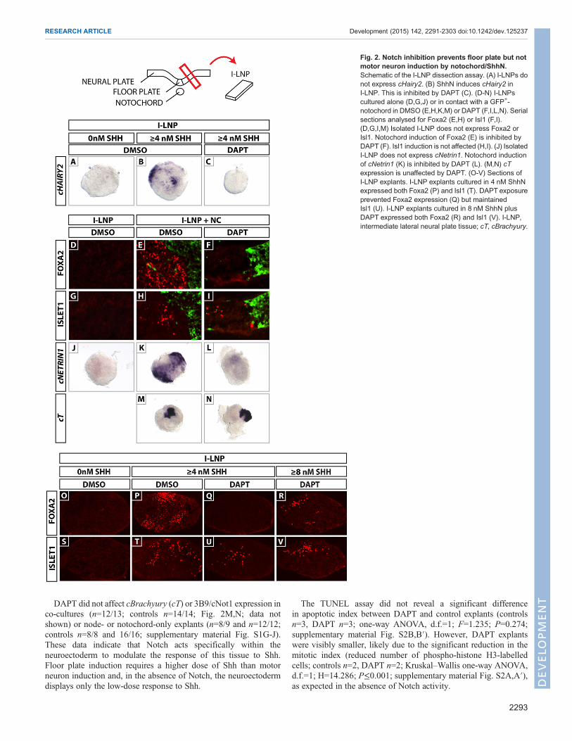
DAPT did not affect cBrachyury (cT) or 3B9/cNot1 expression inco-cultures (n=12/13; controls n=14/14; Fig. 2M,N; data notshown) or node- or notochord-only explants (n=8/9 and n=12/12;controls n=8/8 and 16/16; supplementary material Fig. S1G-J).These data indicate that Notch acts specifically within theneuroectoderm to modulate the response of this tissue to Shh.Floor plate induction requires a higher dose of Shh than motorneuron induction and, in the absence of Notch, the neuroectodermdisplays only the low-dose response to Shh.
The TUNEL assay did not reveal a significant differencein apoptotic index between DAPT and control explants (controlsn=3, DAPT n=3; one-way ANOVA, d.f.=1; F=1.235; P=0.274;supplementary material Fig. S2B,B′). However, DAPT explantswere visibly smaller, likely due to the significant reduction in themitotic index (reduced number of phospho-histone H3-labelledcells; controls n=2, DAPT n=2; Kruskal–Wallis one-way ANOVA,d.f.=1; H=14.286; P≤0.001; supplementary material Fig. S2A,A′),as expected in the absence of Notch activity.
Fig. 2. Notch inhibition prevents floor plate but notmotor neuron induction by notochord/ShhN.Schematic of the I-LNP dissection assay. (A) I-LNPs donot express cHairy2. (B) ShhN induces cHairy2 inI-LNP. This is inhibited by DAPT (C). (D-N) I-LNPscultured alone (D,G,J) or in contact with a GFP+-notochord in DMSO (E,H,K,M) or DAPT (F,I,L,N). Serialsections analysed for Foxa2 (E,H) or Isl1 (F,I).(D,G,I,M) Isolated I-LNP does not express Foxa2 orIsl1. Notochord induction of Foxa2 (E) is inhibited byDAPT (F). Isl1 induction is not affected (H,I). (J) IsolatedI-LNP does not express cNetrin1. Notochord inductionof cNetrin1 (K) is inhibited by DAPT (L). (M,N) cTexpression is unaffected by DAPT. (O-V) Sections ofI-LNP explants. I-LNP explants cultured in 4 nM ShhNexpressed both Foxa2 (P) and Isl1 (T). DAPT exposureprevented Foxa2 expression (Q) but maintainedIsl1 (U). I-LNP explants cultured in 8 nM ShhN plusDAPT expressed both Foxa2 (R) and Isl1 (V). I-LNP,intermediate lateral neural plate tissue; cT, cBrachyury.
2293
RESEARCH ARTICLE Development (2015) 142, 2291-2303 doi:10.1242/dev.125237
DEVELO
PM
ENT

Notch modifies sensitivity to ShhThe finding that Notch inhibition blocks floor plate induction couldbe due to lower Shh production by notochord or to a higherconcentration of Shh required by the neuroectodermal cells. Todistinguish between these possibilities, we repeated the assay butsubstituted notochord with varying concentrations of ShhN proteinin the presence/absence of DAPT. Controls cultured in 4 nM ShhNexpressed both Foxa2 (n=12/12) and Isl1 (n=13/13; Fig. 2P,T).DAPT abrogated Foxa2 expression (n=16/16) but Isl1 persisted(n=17/20; Fig. 2Q,U). These data recapitulate the results observedwith the notochord assay. I-LNP explants cultured in 8 nM ShhNplus DAPT expressed both Foxa2 (n=11/15) and Isl1 (n=15/15;Fig. 2R,V). As an excess of ShhN rescued floor plate induction,these data imply Notch acts in neural cells to lower their thresholdresponse to Shh and thereby specify the cell fate they acquire.
cHairy2 misexpression leads to dorsal expansion of P3 andearly floor plate markersTo test whether Notch modifies the threshold concentration of Shhperceived via induction of Shh itself, we electroporated the caudalneural tube of HH stage 10 embryos with pCIG-NICD [pCAAGsvector encoding both a constitutively active form of Notch (Notchintracellular domain, NICD, normallyonly released following ligand-activated γ-secretase cleavage) andGFP, separated by an IRES] or theNotch target cHairy2 [pCIG-cHairy2], and analysed Shh expressionby immunohistochemistry. We observed by in situ hybridisation andqRT-PCR that NICD misexpression induces ectopic cHairy2expression in the neural tube (n=5, 75/93 sections; supplementary
material Fig. S3; data not shown). However, neither NICD norcHairy2 electroporation altered the endogenous expression profile ofShh (n=5, n=3 embryos, respectively; Fig. 3A-C′). To ensure this wasnot due to cells having lost competence to acquire floor platecharacteristics, we electroporated the open neural plate withpCIG-NICD at HH stage 6, cultured the embryos for 6 h andisolated GFP-positive I-LNP explants, then cultured these for 36 h;again, we saw no Shh induction (n=6; supplementary material Fig.S3). Thus, Notch signalling does not induce Shh expression.
We tested the hypothesis that cHairy2 misexpression in moredorsal regions may induce the differentiation of more ventralcharacteristics by changing the sensitivity of those cells to theendogenous Shh morphogen gradient. cHairy2 electroporation ledto a dorsal expansion of the domains of Foxa2+ cells and Nkx2.2+
cells and a concomitant reduction of the domain of Olig2+ cells (n=2for each marker pCIG; n=6 for each marker cHairy2; Fig. 3D-L″).Moreover, within the motor neuron progenitor domain, cellsthat downregulated Olig2 cell-autonomously upregulated Nkx2.2,indicating a change of fate from a motor neuron to a p3 progenitor(Fig. 3J-L″, n=6). Specification of ventral cell types is progressive:midline cells, which constitute the presumptive floor plate, initiallyexpress markers common to p3 progenitors, i.e. Foxa2 and Nkx2.2.Nkx2.2 becomes downregulated, and late FP markers, includingShh and Arx, are induced (Ribes et al., 2010). We used doubleimmunohistochemistry to determine whether ectopic activation ofFoxa2 by misexpression of cHairy2 is indicative of floor plate orp3 identity. We observed that the predominant response wasupregulation of Nkx2.2 (n=2 embryos, 90/159 sections), with half
Fig. 3. cHairy2misexpression dorsally expands P3 and early floor plate domains. (A-L′) Sections of HH17 chick neural tube 24 h after electroporation withpCIG (A,A′,D,D′,E,E′,F,F′), pCIG-cHairy2 (B-C′,G-L′) or 48 h after pCIG-NICD electroporation (C,C′) analysed by immunohistochemistry for GFP (A-L′).Samples were also analysed for Shh (A-B′), Foxa2 (D,D′), Nkx2.2 (E,E′,G-G″) or Olig2 (F,F′) or double immunohistochemistry for Foxa2 and Nkx2.2 (H-I″) orOlig2 and Nkx2.2 (J-L″). (G″-L″) Magnified regions of interest are shown in G′-L′. Arrowheads in J″-L″ indicate three cells analysed for GFP, Nkx2.2 and Olig2.Scale bar: 30 µm.
2294
RESEARCH ARTICLE Development (2015) 142, 2291-2303 doi:10.1242/dev.125237
DEVELO
PM
ENT

those sections co-expressing Foxa2 (47 sections; Fig. 3H-I′). Bycontrast, definitive early floor plate fate (Foxa2+ only) was inducedless robustly (Fig. 3H-I″).In a complementary approach, we electroporated a dominant-
negative form of cHairy2 (lacking the WRPW domain; Broomet al., 2012) and observed downregulation of Nkx2.2 in the P3domain where cHairy2 is endogenously expressed at this stage(n=2; supplementary material Fig. S4). These findings imply thatreducing Notch activity can increase the threshold concentration atwhich neural cells respond to Shh and thereby modify the extent ofthe expression domains of distinct dorsoventral markers induced byShh. In particular, Notch activity, mediated by cHairy2, promotesacquisition of P3 identity and, to a lesser extent, early floor plateidentity in response to Shh.
Prolonged Notch activity/cHairy2 expression in ventralmidline cells prevents floor plate maturation and promotesP3 identityPtch1mRNA is only transiently expressed by floor plate as these cellsattenuate their response to Shh to acquire full floor plate fate, incontrast to P3 progenitors that require sustained Shh signalling andmaintain Ptch1 expression (Ribes et al., 2010). cHairy2 expressionmirrors that of Ptch1 in these domains. We tested the hypothesis thatcHairy2 too must be extinguished in ventral midline cells for them toacquire full floor plate fate. Embryos electroporated with cHairy2 inthe ventral midline at HH10 and harvested at HH17 displayed a cell-autonomous exclusion of the mature floor plate marker ARX intargeted cells (n=3 embryos, Fig. 4A-B′) with a concomitantupregulation of Nkx2.2 in some cases, indicating a fate change toP3 identity (Fig. 4A-B′). These data demonstrate that cHairy2 canmodulate the response of cells to Shh and imply that cHairy2 isnecessary for the acquisition of ventral cell fate in response to high
Shh signal concentration but it also needs to be downregulated forfloor plate cells to fully mature and differentiate. We next investigatedwhether loss of Notch activity is also necessary for full acquisition offloor plate fate. To achieve this, we used a conditional mouse line inwhich NICD is persistently expressed in the floor plate [tamoxifen-inducible mER;Cre;mER recombinase driver line under the control ofthe Foxa2 promoter (Foxa2mcm) crossed with a Rosa26LSL-NICD line(Murtaugh et al., 2003; Park et al., 2008)]. The Rosa26LSL-NICD strainpermits conditional expression of NICD in cells expressing Crerecombinase. Strikingly, in these E9.5 and E10.5 embryos, weobserved a cell-autonomous upregulation of Nkx2.2 in lineagelabelled ventral midline cells (Fig. 4C-E,G-L′) concomitant with adownregulation of both Foxa2 and Arx (n=6 embryos; Fig. 4G-L′).These results phenocopy cHairy2 electroporation in the chick floorplate. They suggest that elevated Notch activity is sufficient tomaintain competence to respond to Shh in the ventral midline,supporting a role for Notch in the induction/maintenance of the p3fate. Strikingly, qRT-PCR for Ptch1 in caudal neural tissue isolatedfrom E9.5Foxa2mcm; Rosa26LSL-NICD embryos revealed Ptch1mRNAlevels were doubled in the neural tube of these embryos (Fig. 4F;n=13) when compared with wild-type siblings, demonstrating thatNotch activity augments the cellular response to Shh.
Notch modifies the cell fate choice of neural progenitors inresponse to Shh in vivoTo determine whether Notch is necessary for neural tube patterningin response to Shh, we analysed the same progenitor markers inpresenilin (Psen) Psen1−/−; Psen2−/− and Rbpj−/− embryos.Presenilins 1 and 2 are the key components of the γ-secretaseenzyme complex that cleaves NICD. Thus, Psen1−/−; Psen2−/−
embryos lack all Notch signalling, whereas Rbpj−/− embryos lackthe obligate transcription factor required for Notch signalling. Both
Fig. 4. Maintained Notch activity/cHairy2 expression prevents floor plate maturation and promotes P3 identity. (A-B′) Sections of HH17 neural tube 24 hafter pCIG-cHairy2 electroporation into the ventral midline (cH2-GFP). Scale bar: 15 µm. (C-E) Sections of E9.5 or (G-L′) E10.5 Foxa2mcm; Rosa26LSL-NICD
embryos. Scale bar: 30 µm. Sections analysed for GFP, Arx and Nkx2.2 (A-B′,G-I′) or GFP and Nkx2.2 (C-E) or GFP, Foxa2 and Nkx2.2 (J-L′). (F) qRT-PCRanalysis of Ptch1 mRNA levels in E9.5 Foxa2mcm; Rosa26LSL-NICD caudal neural tube compared with wild types. Bars represent mean values (plus s.e.m.) ofrelative expression levels from wild-type (0.913±0.078, n=3) and mutant embryos (1.918±0.239, n=9) normalised against β-actin.
2295
RESEARCH ARTICLE Development (2015) 142, 2291-2303 doi:10.1242/dev.125237
DEVELO
PM
ENT

phenotypes are embryonic lethal at E9.5 (Oka et al., 1995; Donovielet al., 1999). In both lines, the combined expression domains ofFoxa2 and Nkx2.2 in the ventral neural tube were dramaticallyreduced at E9 (n=6 heterozygous controls and n=3 Psen1−/−;Psen2−/−; n=3 heterozygotes and n=3 Rbpj−/−; supplementarymaterial Fig. S5; data not shown). In Rbpj−/− embryos, cell countsshowed the domain of the caudal neural tube occupied by Foxa2+/Nkx2.2+ cells to be significantly lower than in controls (4% versus7% in heterozygotes; P<0.001). To examine mutant embryos thatsurvive beyond E9.5, we analysed mice in which the transcriptionalactivity of Notch is blocked only in ventral midline and P3progenitors using Foxa2T2AiCre-induced expression of a dominant-negative Mastermind-like1 eGFP fusion protein from a targetedRosa26 locus (Rosa26dnMaml1; Fig. 5; High et al., 2008; Tu et al.,2005; Horn et al., 2012; Maillard et al., 2008). Dominant-negativeMAMl1 eGFP fusion protein is a potent and specific inhibitor of allfour mammalian Notch receptors in vivo. Foxa2T2AiCre;Rosa26dnMaml1 embryos analysed at E9 (n=3) revealed that, withinthe combined Foxa2+, Nkx2.2+ or Foxa2+/Nkx2.2+ domain, mostcells were Foxa2+/Nkx2.2+, indicative of an immature FP and/or P3cell type (n=5; Fig. 5A,B), when compared with controls(Foxa2T2AiCre; Rosa26RYFP stage-matched embryos in whichthe Cre-recombined cells are normally Notch sensitive; data notshown). Quantification of these effects revealed a significantlyhigher proportion of Foxa2+/Nkx2.2+ cells and a significantly lowerproportion of P3 (Nkx2.2+ only) cells (Fig. 5I; χ2=50.52, d.f.=1,n=2351 cells, P=1.068×10⁻¹¹). The number of Foxa2+-only cells isrelatively low in both controls and mutants at this developmentalstage. E10.5 Foxa2T2AiCre; Rosa26dnMaml1 embryos also showed asignificantly different distribution of these three cell types whencompared with controls, in particular maintaining a higherproportion of Foxa2+/Nkx2.2+ cells and a lower proportion of P3(Nkx2.2+ only) and early floor plate cells (Foxa2+ only) (Fig. 5C,D;χ2=145.66, d.f.=1, n=2462 cells, P≤2.2×10⁻¹⁶). At E11.5, asignificantly different distribution of cells in these three categorieswas maintained (Fig. 5I; χ2=83.44, d.f.=1, n=3151 cells,P≤2.2×10⁻¹⁶); while the proportion of double-positive cells wassimilar to controls (Fig. 5E,F,I), these embryos exhibited asignificantly higher proportion of P3 and lower proportion of floorplate cells compared with controls (Fig. 5I). Analysis of Arx and Shhexpression at E10.5 reveals that this small population of early floorplate cells go on to express late floor plate markers in the absence ofNotch (Fig. 5G,H; data not shown). These data suggest that in theabsence of Notch signaling in the ventral progenitors, the adoption ofP3 or floor plate fates is delayed. Eventually, this resolves such thatsignificantly fewer cells give the high dose response.
Notch activity regulates localisation of Smo to the primarycilia in a Shh-independent mannerCiliary localisation of Smo is crucial for activation of the Shh pathway(Huangfu et al., 2003; reviewed by Sasai and Briscoe, 2012). To testwhether regulation of Smo localisationmight be amechanism throughwhichNotch activitymodulates the efficacyof Shh signalling, we usedthe Shh-responsive NIH-3T3 primary fibroblast cell line. Double-labelling with antibodies to Smo and to acetylated tubulin (whichmarks stable microtubules found in cilia) revealed a highly significantaccumulation of Smo in the cilia of NIH-3T3 cells upon addition ofShhN (Fig. 6B,G; χ2=61.19, d.f.=1, n=157 cells, P=5.16×10⁻¹⁵)whereas, in the absence of ShhN, cilia are largely devoid of Smo(Fig. 6A,G) (Rohatgi et al., 2007). Strikingly, activation of Notch, bytransfecting NIH-3T3 cells with NICD-pCIG, dramatically andsignificantly augmented the number of cells showing Smo
Fig. 5. Loss of Notch in the ventral midline in Foxa2T2AiCre;Rosa26LSL-dnMaml1 mice modifies cell fate. (A-H) Sections of spinal cordat lumbar level of E9 (A,B), E10.5 (C,D,G,H) or E11.5 (E,F) Foxa2T2AiCre;Rosa26LSL-dnMaml1mutants. Scale bar: 30 µm. (A-F) Sections analysed for Foxa2and Nkx2.2. (G,H) Sections analysed for Arx. (I) Changes in proportions of cellspositive for either Foxa2 (red, early floor plate), Nkx2.2 (green, P3 progenitor) orFoxa2+/Nkx2.2+ (orange, midline cells that may become floor plate or P3) withinthe ventral neural tube of E9, E10.5 or E11.5 Foxa2T2AiCre; Rosa26LSL-dnMaml1
mutants. The number/location of Foxa2+/Nkx2.2+ cells (white dots in A-D)changes in the mutant at E9 and E10.5. The table presents the results of astatistical comparison between wild-type and mutant embryos of the three celltypes using aChi-square test. The distribution of cellswithin these three cell typeswas statistically different in the mutant compared with wild type at all threedevelopmental stages. Chi squared values are in the top right of the table andP-values are at the bottom left of the table. All pairwise comparisons werehighly statistically significant. Sections counted at E9: wild type, n=40; mutant,n=16; at E10.5, wild type, n=25; mutant, n=16; at E11.5, wild type, n=32;mutant, n= 25.
2296
RESEARCH ARTICLE Development (2015) 142, 2291-2303 doi:10.1242/dev.125237
DEVELO
PM
ENT

localisation in cilia, in a Shh-independent manner (Fig. 6C,G;χ2=41.62, d.f.=1, n=139, P=1.11×10⁻¹⁰). Addition of ShhN to theNICD-pCIG-transfected cells further enhances the effect (Fig. 6D,G;χ2=6.56, d.f.=1, n=130, P=0.01041). To preclude the possibility of acell-type specific effect, we replicated the experiments in chickenDF-1fibroblast cells by transfecting cells with cHairy2-pCIG, to closelymimic the electroporation analysis performed in chick embryos. Weobserved a very similar phenotype, whereby cHairy2-pCIGtransfection dramatically increased the proportion of cells with SMOlocalisation in cilia, again in a Shh-independent manner (Fig. 6H;χ2=7.65, d.f.=1, n=116, P=0.00565). The further addition of ShhNaugmented this effect (Fig. 6H; χ2=9.36, d.f.=1, n=73, P=0.002212).
In the absence of Shh, Ptch1 inhibits Smo localisation in primarycilia.Wemeasured intensity of Ptch1 staining in cilia of NIH-3T3 cellsby double-labeling with antibodies to Ptch and Arl13b (a smallGTPase that localises to cilia; see Caspary et al., 2007), and saw adramatic reduction whenwe added ShhN, as expected (supplementarymaterial Fig. S6). To address whether Notch modulates this effect, wetreatedNIH-3T3 cells with 150 nMLY411575, a γ-secretase inhibitor,which abolished Hes1 mRNA expression, as expected (Fig. 6F;Ferjentsik et al., 2009). Remarkably, Notch inhibition led to a highlysignificant dampening of Ptch1 clearance from cilia in response toShhN (n=2 replicates; 1205 cells). These data are reflected in thesignificant post-hoc pairwise comparisons using Tukey’s HSD test
Fig. 6. Notch promotes cilia localisation of Smo in aShh-independentmanner. (A-D) NIH3T3 cells transfectedwith pCIG-GFP (A,B) or NICD-pCIG-GFP (C,D)in the presence (B,D) or absence (A,C) of Shh-N. green, pCIG-GFP or NICD-pCIG-GFP; red, cilia labelled with α-acetylated tubulin; white, Smo antibody staining.Scale bar: 8 µm. (E) Western blot showing levels of full-length Gli3 (Gli3FL) and partially proteolysed repressor form (Gli3R) in cells transfected with pCIG-GFP orNICD-pCIG-GFP in presence or absence of ShhN. (F) qRT-PCR analysis of Ptch1,Gli1 andHes1mRNA levels in NIH3T3 cells in the presence of DMSO, DMSO+Shh-Nor LY+Shh-N.Data shownare themean from twobiological replicates. (G,H)QuantificationofNIH3 T3 (G) orDF-1 cells (H) depicting theproportions of cellsthat showed no Smo in cilia (red) versus cells that showed Smo localization in cilia (green) under different conditions. Data show proportion of cells with or withoutSmo localised to theprimarycilium,andwere comparedusingaChi-square test.Cells transfectedwithNICD-pCIG-GFPhadsignificantly longercilia thanpCIG-GFPtransfected cells independent of ShhN (compare C,Dwith A,B). The table presents statistical analysis of effects on smo localisation to the cilia in 3T3 cells (table onthe left hand) andDF-1 cells (table on the right hand). All pairwise comparisonsmadewith 1 degreeof freedom.Significant differencesafterBonferroni correction arehighlighted in red. Chi squared values are in the top right of the table and P-values are at the bottom left of the table. Significant results are in red.
2297
RESEARCH ARTICLE Development (2015) 142, 2291-2303 doi:10.1242/dev.125237
DEVELO
PM
ENT
[F(2,1205)=176.5, P≤0.001; supplementary material Fig. S6]. Takentogether, these data demonstrate that Notch activity has a conserved,Shh-independent and significant effect upon localisation of keycomponents of the Shhpathwaywithin primary cilia, functioningat thelevel of the Ptch1/Smo interface to modulate ciliary localisation ofthese two signalling components.
Notch signalling regulates the level of full-length Gli3 in NIH-3T3 cellsFull activation of Smo is a two-step process and ciliary transportis the initial requirement (Rohatgi et al., 2009, 2007; Sasaiand Briscoe, 2012; Chen et al., 2011; Milenkovic et al., 2009).Within cilia, Smo can exist both in an inactive state and in thephosphorylated active form. The latter prevents proteolytic cleavageof the Gli transcription factors that then translocate to the nucleus toreplace the cleaved repressor form and activate transcription oftarget genes (Stamataki et al., 2005; Briscoe and Thérond, 2013).Addition of 4 nM ShhN to NIH-3T3 fibroblasts leads to a sharpreduction in levels of Gli3R (n=3; Fig. 6E; Humke et al., 2010;Niewiadomski et al., 2014). We did not observe a concomitantincrease in levels of full-length Gli3, probably due to the fact thatthis highly labile transcriptional activator undergoes rapid nucleartranslocation, phosphorylation and destabilization (Humke et al.,2010). By contrast, exposure to NICD-pCIG or Hes1-pCIG aloneled to an increase in the levels of full-length Gli3, in NIH-3T3fibroblasts (n=3 replicates; Fig. 6E; data not shown). This isa striking result, given the 20% transfection efficiency (analysedby flow cytometry; data not shown). The addition of both ShhN andNICD-pCIG did not change levels of full-length Gli3 from thatseen with NICD/Hes1 alone, although levels of Gli3R weredramatically reduced (n=3 replicates; Fig. 6E). These data revealthat concomitant with accumulating Smo protein in cilia, Notchsignalling elevates levels of full-length Gli3. We next determinedwhether loss of Notch would affect Shh target gene transcriptionin NIH-3T3 fibroblasts. qRT-PCR analysis revealed 150 nMLY411575 dramatically reduced ShhN-mediated induction of Ptc1andGli1 (n=2 replicates; Fig. 6F). Notably, ShhN also inducedHes1expression in a Notch-dependent fashion, supporting the I-LNP data(Fig. 2B). Together, these data support the hypothesis that Notchsignalling amplifies the cellular response to Shh.
Notch signalling regulates cilia length both in the ventralneural tube and in NIH-3T3 fibroblastsAn additional striking and unexpected effect of NICD in NIH-3T3cells is that cilia were significantly longer at the P≤0.001 level[F(2,1534)=128.557, P≤2.2×10⁻¹⁶; Fig. 6C,D compared with 6A,B; Fig. 7A]. In vivo, previous reports have shown the floor platecilia become significantly longer than P3 cilia (Cruz et al., 2010;Fig. 7). To assess whether the effects of Notch upon ventral neuraltube patterning may be associated with changes in cilia length, wemeasured cilia in the floor plate and lateral neural plate in both gain-and loss-of-function transgenic mouse models (Foxa2T2AiCre;Rosa26dnMaml1 and in Foxa2mcm; Rosa26LSL-NICD mice). UsingArl13b antibodies, we observed that floor plate cilia inFoxa2T2AiCre; Rosa26dnMaml1 embryos are significantly shorterthan in control litter mates at E9, E10.5 and E11.5 at the P<0.01level (E9 n=3 embryos, E10.5 n=3 embryos, E11.5 n=2 embryos;1354 cells counted; Fig. 7C-F; floor plate: P=0.003682,P<2.2×10⁻¹⁶, P<2.2×10⁻¹⁶, respectively). These data arereflected in the significant post-hoc pairwise comparisons usingthe Tukey’s HSD test in Table 1. By contrast, whenwe analyse cilialength in the floor plate and P3 domain in gain-of-function
Foxa2mcm; Rosa26LSL-NICD mice at E10.5, we observed P3 ciliawere significantly longer (0.4 µm more) than in control siblings(n=2 embryos; 249 cells; Fig. 7B; P≤2.2×10⁻¹⁶; Table 1). Floorplate cilia, however, were no different in length compared withcontrols (Fig. 7B; P=0.5624687; Table 1). These observationssuggest that the mechanism by which Notch modulates theresponse of cells to Shh might be through regulating both cilialength and localisation of Smo within the cilia.
DISCUSSIONPrimary cilia are important regulators of Shh signal transduction(Sasai and Briscoe, 2012). We provide in vitro and in vivo evidencesupporting a novel role for the Notch pathway in modulating ciliaryarchitecture and localisation of Smo to this cell appendage. Wepropose that this serves as a means to prime cells to respond to theShh morphogen. In vivo, this synergistic interaction plays a key partin dorsoventral patterning of the developing neural tube in bothchick and mouse embryos.
Intriguingly, NICD production occurs throughout the dorsoventralaxis of the neural tube, yet the Notch target cHairy2 expression isrestricted to the floor plate/P3 domain (and the roof plate), in contrastto the broader expression of other Hes homologues (Fior andHenrique, 2005). We propose this is probably due to cooperativeactivation of cHairy2 by Shh and Notch in the ventral midline. Onepossible explanation for how this synergy might operate is that Shhmight induce expression of Notch itself or a Notch ligand. Indeed,NICD electroporation in more dorsal regions drives ectopic cHairy2expression in the neural tube indicative of the fact that endogenouslevels of Notch signallingmay be higher in the floor plate. Serrate 1 isa potential candidate in this respect, as it is expressed in the ventralmidline of the chick and mouse neural tube at early developmentalstages (J.K.D., unpublished; le Roux et al., 2003).
Paradoxically, we have shown previously that DAPT treatment ofwhole embryos completely blocked cHairy2, but did not alter Foxa2expression (Gray and Dale, 2010), whereas we show here thatDAPT treatment of I-LNPs blocks Shh induction of both cHairy2and Foxa2. However, in the whole embryo assay, we demonstrated alarger notochord forms from node progenitors in the absence ofNotch. This increase in the number of Shh-producing cells andpotentially the levels of Shh was proposed to mediate the inductionof Foxa2 expression/floor plate characteristics in the absence ofNotch. In support of that idea, we show in this study that rescue ofFoxa2 in the absence of Notch can be achieved by simply increasingconcentration of Shh to which the I-LNP is exposed.
We use a wide variety of gain- and loss-of-function approaches inboth chicken andmouse to show that interactions betweenNotch andShh are crucial for dorsal-ventral cell fate specification in the neuraltube. Inhibiting Notch signalling impedes floor plate inductionwhereas ectopic Notch activation in more dorsal regions ventralisesthose cells. After floor plate induction, Notch signalling is normallydownregulated in the ventral midline, and forced maintenanceprevents floor plate maturation, resulting in the formation of more P3progenitors in both chick andmouse at the expense of floor plate. Thisnovel role forNotch is verydifferent from thewell-established role forNotch in maintaining neural progenitors and preventing theirdifferentiation. We provide multiple lines of evidence that theseeffects are due to Notch modulating progenitor cell interpretation ofthe Shh spatiotemporal gradient rather than directly regulating cellfate or differentiation: first the in vitro I-LNP assay shows Notchinhibition changes sensitivity but not competence of cells to respondto Shh; second the Foxa2T2AiCre; Rosa26dnMaml1 embryos show atemporal delay in competence to respond to Shh; third Notch
2298
RESEARCH ARTICLE Development (2015) 142, 2291-2303 doi:10.1242/dev.125237
DEVELO
PM
ENT

modulation of Smo/Ptch1 trafficking to cilia, levels of Gli3 and Shhtarget gene expression in vivo and in vitro directly demonstrates thatNotch affects progenitor cell interpretation/response to Shh.Although the dorsoventral patterning output of this Notch/Shhsynergistic interaction has not been previously reported, an intriguingparallel with our findings is that, in zebrafish lateral floor plateprogenitors, loss of Notch leads to loss of Hh response and initiation
of Kolmer–Agduhr interneuron differentiation (Huang et al., 2012),although the mechanism governing this synergistic interactionremains unknown.
We show part of this mechanism relies on the subcellularlocalisation of the key Shh signalling component Smo; NICDmisexpression in vitro dramatically augments the initial step ofciliaryaccumulationofSmo, in a Shh-independentmanner. This effect
Fig. 7. Notch regulates cilia length. (A,B) Quantitation of cilia length; box plots showmedian and range in NIH3t3 cells in presence/absence of NICD-pCIG-GFP(A), in floor plate and P3 domain of Foxa2mcm; Rosa26LSL-NICDmutants (NICD+) versus control littermates (B). (C,D) Cilia labelled with Arl13b antibody in the floorplate of Foxa2T2AiCre; Rosa26LSL-dnMaml1 mutant (D) versus wild-type littermate (C). Scale bar: 5 µm. (E,F) Quantitation of cilia length in floor plate and lateralneural plate of Foxa2T2AiCre; Rosa26LSL-dnMaml1mutants (E9 n=3, E10.5 n=3 and E11.5 n=2) versus controls (E9 n=5, E10.5 n=2 and E11.5 n=2). Box plots showmedian and range; analysed by ANOVA. There is a highly significant three-way interaction between cell type (floor plate and P3), developmental stage (E9, 10.5,11.5) and genotype (mutant/control) at the *P≤0.01 level [F(2,1354)=5.27, P=0.00525].
2299
RESEARCH ARTICLE Development (2015) 142, 2291-2303 doi:10.1242/dev.125237
DEVELO
PM
ENT

is phenocopied by cHairy2 and thus is likely to be a transcriptionalresponse. The identification of theNICD/cHairy2 target gene involvedin trafficking Smo to the cilia will require further investigation.In addition, NICD misexpression led to elevated levels of full-
length Gli3 in vitro. This might be due to Notch promoting inhibitionof the cleavage of full-length Gli to the repressor form directly orindirectly through increasing accumulation of Smo in cilia, and/or thismay be a transcriptional response, given that Gli2 and Gli3 havepreviously been reported to be direct targets of N1ICD/Rbpj (Li et al.,2012). As expected, exposure to ShhN dramatically lowers levels ofGli3R, both in the presence/absence of NICD, which is notphenocopied by exposure to NICD alone. These data suggestNICD-mediated ciliary accumulation of Smo and elevated levels offull-lengthGli3 are not sufficient to stimulate full activation of the Shhpathway. Rather, we propose that these events prime cells to respondefficiently and in a timely fashion when they become exposed to Shh.An additional unexpected and Shh-independent effect of NICD
activation within NIH-3T3 fibroblasts was the formation ofsignificantly longer primary cilia. This effect on ciliary length isalso evident in vivo in transgenic lines that serve to activate or inhibitNotch activity specifically in the ventralmidline. This effect is likely tobe transcriptionally regulated as it is observed in Foxa2T2AiCre;Rosa26dnMaml1 embryos. A recent report demonstrated thatsupernumerary cilia, resulting in more ciliary signaling surfaces,reduced Shh pathway transcriptional activation (Mahjoub andStearns, 2012). By contrast, within the neural tube of Arl13bmutants that form cilia half the length of normal cilia, there is a failureto induce dorsoventral markers that are characteristic of the highestlevels of Shh signalling, although expression of genes that requirelower levels of Shh signalling continues (Caspary et al., 2007). Thus,Shh signalling is sensitive to cilia length, number and architecture. Ithas previously been shown that cilia length changes within the ventralmidline of the developing neural tube during normal development(Cruz et al., 2010). Thus, floor plate cilia become almost double thelength of those in the adjacent lateral neural tube and this has beenassociated with Shh-dependent onset of Foxj1 expression within thefloor plate (Cruz et al., 2010). Indeed, Foxj1misexpression is sufficient(but not necessary) for longer cilia in the neural tube. In a variety ofdevelopmental contexts, Foxj1 has been associated with production ofmotile cilia that are considerably longer than primary cilia (Cruz et al.,2010;Blatt et al., 1999;Chen et al., 1998; Stubbs et al., 2008; Tichelaaret al., 1999;Yu et al., 2008).Aprevious report linkingNotch signallingto ciliary architecture came from zebrafish (Lopes et al., 2010).Kupffer’s vesicle is enriched with long motile cilia, the function ofwhich is key for onset of left-right asymmetry. Lopes et al. showedNotch signalling directly regulates ciliary length, and inefficiencies in
Notch signal transduction result in shorter cilia and aberrations in left-right asymmetry. One target of Notch in this regard is Foxj1 (Lopeset al., 2010). Thus, it is possible that Foxj1 mediates the NICD-dependent changes to ciliary architecture in the ventral neural tube.Intriguingly, Cruz et al. suggest Foxj1 attenuates the response to Shhand that this is cilia dependent. It is noteworthy, however, that thesubcellular localisation of Smo is not affected by Foxj1misexpression,indicating that NICD-mediated changes in cilia length and Smolocalisation might be differentially regulated (Cruz et al., 2010).Indeed,wedid not observe changes in cilia length following cHairy2orHes1 misexpression, indicating that this effect is mediated by adifferent set of Notch target effectors. Thus, our working modelproposes that NICD activity leads to both increased ciliary length andciliary localisation of Smo, by two independent mechanisms, whichtogether prime the cell for an accentuated response to the Shh ligand.
The delay in acquisition of ventral fates seen in neural progenitorsexpressing dnMAMl1-eGFP indicates a potential role for Notch inregulating the temporal response to Shh. It is well established thatthe spatiotemporal pattern of ventral marker induction in the neuraltube reflects changes in both concentration and duration of Shhsignalling over time. Our data invoke a model in which the role ofShh-dependent Notch activity is to prime cells to respond efficientlyand appropriately to Shh by increasing length of cilia andaccumulation of SMO within these structures, thereby facilitatingrapid Shh-triggered activation of SMO once ligand is received.
In conclusion, our findings, using both in vitro and in vivomodels,reveal a conserved and novel mechanism to refine and modulate theresponse repertoire and cell sensitivity to Shh during tissuedevelopment. This role for Notch may also affect a broad range ofother pathways that are reliant on ciliary localisation of signallingcomponents in a wide variety of developmental and disease contexts.
MATERIALS AND METHODSChick embryoWhite Leghorn Gallus gallus eggs (Henry Stewart, Lincolnshire, andWinter Farm, Royston, UK) or GFP-expressing embryos [Roslin Institute,Midlothian (McGrew et al., 2004)] were incubated at 38.5°C in a humidifiedincubator to yield embryos between Hamburger–Hamilton (HH) stages 5and 17, according to Hamburger and Hamilton, 1992.
Mouse embryosWild-type CD1 mouse (Mus musculus) embryos were obtained at E8.5-E11.5, fixed for immunohistochemistry or in situ hybridisation. Genotypingof Rosa26dnMaml1 (Horn et al., 2012), Foxa2mcm (Park et al., 2008),Rosa26LSL-NICD (Murtaugh et al., 2003), Psen1−/−; Psen2−/− (Donovielet al., 1999), Rbpj−/− (Oka et al., 1995) and Rosa26LSL-YFP (Srinivas et al.,2001) was carried out using PCR. Rosa26dnMaml1 and Rbpj−/− mice were
Table 1. Post-hoc pairwise comparisons using the Tukey’s HSD test to compare cilia length in floor plate and P3 domains in Foxa2T2AiCre;Rosa26LSL-dnMaml1 and Foxa2mcm; Rosa26LSL-NICD mutant embryos versus wild-type littermates
Difference incilia length (μm)
Lowerlimit (μm)
Upperlimit (μm)
AdjustedP-value
Rosa26LSL-dnMaml1 versus wild typeE9 P3 0.003687 −0.1207 0.128075 1E9 FP −0.15237 −0.27676 −0.02799 0.003682E10.5 P3 −0.14352 −0.23154 −0.05551 7.1×10⁻⁶
E10.5 FP −0.25416 −0.34218 −0.16615 2.2×10⁻¹⁶
E11.5 P3 −0.06368 −0.14389 0.016532 0.2811E11.5 FP −0.33704 −0.41725 −0.25683 2.2×10⁻¹⁶
Foxa2mcm; Rosa26LSL-NICD
versus wild typeE10.5 FP −0.11615 −0.34688 0.114577 0.5624687E10.5 P3 0.477011 0.329829 0.624193 2.2×10⁻¹⁶
2300
RESEARCH ARTICLE Development (2015) 142, 2291-2303 doi:10.1242/dev.125237
DEVELO
PM
ENT

kept as heterozygotes. Foxa2mcm; Rosa26LSL-NICD embryos were obtainedby crossing Foxa2mcm male with Rosa26LSL-NICD female. Pregnant femaleswere administered 8 mg of tamoxifen (Sigma, T5648) by oral gavage at E7.5and E8.5 dpc with embryos collected at E10.5 or at E6.5 and E7.5 dpc withembryos collected at E9.5.
Cell cultureNIH-3T3 mouse and DF1 chick fibroblasts were cultured in a humidifiedincubator andmaintained at 37°C in an atmosphere of 5%CO2, in Dulbecco’smodified Eagle’s medium (DMEM; Life Technologies) supplemented with10% heat-inactivated Newborn Calf Serum (NBCS; Life Technologies) orfoetal bovine serum (FBS, Sigma), respectively. Cells were transfected usingLipofectamine LTX with Plus Reagent Kit (Invitrogen) either with controlempty plasmid (PCIG; Megason and McMahon, 2002), PCIG-NICD orpCIG-cHairy2 (Dale et al., 2003) at final concentration of 1 μg/ml. Forciliated cell enrichment, transfected cells were maintained 24 h in DMEMsupplemented with 0.5% NBCS or 0.5% FBS for NIH3T3 and DF1 (whichenriches for cells in G1, when cilia formation predominantly occurs),respectively (Basten and Giles, 2013), before treating them with 4 nMrecombinant ShhN protein (University of Dundee, UK) for 12 h.
Explant cultureExplants isolated from HH stage 6 or 7 chick embryos were cultured incollagen (Placzek and Dale, 1999) in 100 μM γ-secretase inhibitor IX(DAPT) (Calbiochem) dissolved in dimethylsulphoxide (DMSO) (Sigma)or DMSO alone for 36 h unless stated otherwise. Notochord andintermediate lateral neural plate (I-LNP) co-culture explants employednotochord from a GFP-transgenic embryo to distinguish neural versusmesodermal expression of Foxa2 and Isl1. Where stated, I-LNP explantswere cultured in 1 nM or 4 nM recombinant Shh protein.
ElectroporationpCIG-NICD, pCIG-cHairy2, pCIG-dominant-negative cHairy2 (Broomet al., 2012) or pCIG vectors were introduced to the caudal neural plate ofHH10-12 embryos using standard in ovo electroporation (Dale et al., 2003;Briscoe et al., 2000). Embryos were cultured overnight before fixation.
In situ hybridisation and immunohistochemistryStandard in situ hybridisation methods were used (Henrique et al., 1995).Embryoswere fixed in 4%PFA in PBS at pH 7.2, on ice for 2 h. Cells growingoncoverslipswere fixed in4%PFA inPBS for 15 min. Forpatched1 antibody,cells were fixed with 2% PFA for 5 min followed by 5 min of ice-coldmethanol at−20°C. Antibody protocols have been described for Foxa2 (1:10;DSHB), 3B9, Nkx2.2 (1:10; DSHB), Isl1 (1:10; DSHB), Shh (1:10; DSHB),Olig2 (1:16,000; a generous gift from B. Novitch, UCLA, USA), aristaless(Arx, 1:1000; Jamel Chelly, IGBMC, Paris, France), Arl13b (1:3000;N295B/66, UC Davis/NIH NeuroMa, USA), Smo (1:3000; Abcam, ab38686), Ptch1(1:750; University of Dundee, UK), acetylated tubulin (1:1000; Sigma,T7451), mNICD (1:200; Cell Signaling, D3B8), cNICD (1:2000; Universityof Dundee), anti-GFP (1:1000; Life Technologies, A6455) (Ericson et al.,1996, 1997, 1992;Gibb et al., 2009; Yamada et al., 1991; Caspary et al., 2007;Cruz et al., 2010; Bone et al., 2014; Huppert et al., 2005).
Phospho-histone-H3 immunohistochemistry on explantsFixed tissue was proteinase K (Roche) treated and fixed (4% formaldehydein PBS, 2 mMEGTA, 0.1% glutaraldehyde (Sigma). Anti-phospho-histone-H3 antibody (Upstate) was added at a concentration of 10 μg/ml.
Marker gene analysis in mutant embryosCell counts for each marker were performed on sections and expressed as aproportion of the domain covered by the two markers. Heterozygotesprovided a characteristic template of neuronal patterning from whichcomparisons could be made with mutant littermates.
Western blottingWestern blot analysis has been described previously (Bone et al., 2014). Tenmicrograms of sample was loaded. Gli3 rabbit antibodies (a generous gift
from Susan Mackem, Center for Cancer Research, Frederick, MD, USA)and mouse anti-tubulin (Abcam) were diluted 1:1000 and 1:5000.
qRT-PCRTotal RNA was extracted from the caudal region (below forelimb) of E9.5Foxa2mcm; Rosa26LSL-NICD using a Qiagen Micro Plus kit. cDNA wassynthesised using SuperScript III reverse transcriptase (Life Technologies).qRT-PC was performed with Power SYBR Green Master Mix (LifeTechnologies) and reactions measured in a C1000 Thermal Cycler (Bio-Rad) under the following conditions: 95°C for 5 min, 40 cycles 95°C for15 s and 60°C for 1 min. Ptch1, Gli1 (Han et al., 2009), Hes1 (Li et al.,2012) and cHairy2 primers (F: 5′-CCGTACCCTGCAAGCCAGGTG-3′,R: 5′-GCCCATCA-GAGGCAAGCAGCA-3′) were described previouslyand normalised against β-actin (Ferjentsik et al., 2009; Ribes et al., 2010)using the Pfaffl equation (Pfaffl, 2001).
Image acquisition/analysisFluorescent signal was acquired using a compound microscope (LeicaDM5000 B), an Olympus IX70 deconvolution microscope or the ZeissLSM-710 confocal microscope. Image analysis was carried out indeconvolved pseudo-coloured images using Volocity software or the openaccess software Fiji.
Statistical analysisData analysis was conducted using the open source statistical software R (RCore Team, 2012). Differences in cilia length and PTC cilia intensity weretested using ANOVA on log transformed response data, with post-hocpairwise comparisons conducted using Tukey’s honest significant differencestest. Differences in Smo protein localisation in the cilia in addition to changesin the counts of categories of cells expressing different markers across theanterior-posterior axis of the Rosa26dnMaml1 or control mouse embryos wereevaluated using a chi-square test. Plots were generated using R studio, sigmaplot and Microsoft Office Excel. Phospho-histone-H3-labelled chromatin incontrol and treated explants was analysed by ANOVA.
AcknowledgementsWe are grateful to J.K.D.’s laboratory. Special thanks go to I. Jones andM. C. Jørgensen for experimental assistance, and to K. Storey and M. Maroto forcritical reading of the manuscript. We thank B. Novitch for communication prior tosubmission; J. Briscoe, O. Pourquie, M. Stavridis, R. Kageyama and J. Chelly forreagents; and Marianne Reilly and Nikoletta Patourgia for administrative support.
Competing interestsThe authors declare no competing or financial interests.
Author contributionsJ.K.D. developed the concepts/approach, performed some experiments andprepared the manuscript. M.S. and S.G. designed and performed the majority ofexperiments, all data analysis and preparation of figures. I.M. performed cell culture.D.B. performed statistical analyses. P.A.S. collected and genotyped Foxa2T2AiCre;Rosa26dnMaml1 embryos. M.S., I.M., P.A.S. and P.S. edited the manuscript. J.C.S.performed QRT-PCR. M.B. provided technical assistance. E.A.H. performed cellculture. C.T. performed cryosectioning. R.G. performed double in situ hybridisation.
FundingThis work was supported by a BBSRC studentship to M.S.; by a Cancer ResearchUK studentship to I.M.; by a MRC project grant to J.K.D.; and by a Wellcome TrustStrategic award [097945/Z/11/Z]. P.S. was funded by the Novo Nordisk Foundation.Deposited in PMC for immediate release.
Supplementary materialSupplementary material available online athttp://dev.biologists.org/lookup/suppl/doi:10.1242/dev.125237/-/DC1
ReferencesBasten, S. G. and Giles, R. H. (2013). Functional aspects of primary cilia in
signaling, cell cycle and tumorigenesis. Cilia 2, 6.Blatt, E. N., Yan, X. H., Wuerffel, M. K., Hamilos, D. L. and Brody, S. L. (1999).
Forkhead transcription factor HFH-4 expression is temporally related tociliogenesis. Am. J. Respir. Cell Mol. Biol. 21, 168-176.
2301
RESEARCH ARTICLE Development (2015) 142, 2291-2303 doi:10.1242/dev.125237
DEVELO
PM
ENT

Bone, R. A., Bailey, C. S. L., Wiedermann, G., Ferjentsik, Z., Appleton, P. L.,Murray, P. J., Maroto, M. and Dale, J. K. (2014). Spatiotemporal oscillations ofNotch1, Dll1 and NICD are coordinated across the mouse PSM. Development141, 4806-4816.
Briscoe, J. and Novitch, B. G. (2008). Regulatory pathways linking progenitorpatterning, cell fates and neurogenesis in the ventral neural tube. Philos. TransR. Soc. Lond. B Biol. Sci. 363, 57-70.
Briscoe, J. and Therond, P. P. (2013). The mechanisms of Hedgehog signallingand its roles in development and disease. Nat. Rev. Mol. Cell Biol. 14, 418-431.
Briscoe, J., Pierani, A., Jessell, T. M. and Ericson, J. (2000). A homeodomainprotein code specifies progenitor cell identity and neuronal fate in the ventralneural tube. Cell 101, 435-445.
Broom, E. R., Gilthorpe, J. D., Butts, T., Campo-Paysaa, F. andWingate, R. J. T.(2012). The roof plate boundary is a bi-directional organiser of dorsal neural tubeand choroid plexus development. Development 139, 4261-4270.
Caspary, T., Larkins, C. E. and Anderson, K. V. (2007). The graded response toSonic Hedgehog depends on cilia architecture. Dev. Cell 12, 767-778.
Chen, J., Knowles, H. J., Hebert, J. L. and Hackett, B. P. (1998). Mutation of themouse hepatocyte nuclear factor/forkhead homologue 4 gene results in anabsence of cilia and random left-right asymmetry. J. Clin. Invest. 102, 1077-1082.
Chen, Y., Yue, S., Xie, L., Pu, X.-h., Jin, T. and Cheng, S. Y. (2011). DualPhosphorylation of suppressor of fused (Sufu) by PKA and GSK3beta regulatesits stability and localization in the primary cilium. J. Biol. Chem. 286, 13502-13511.
Cohen, M., Briscoe, J. and Blassberg, R. (2013). Morphogen interpretation: thetranscriptional logic of neural tube patterning. Curr. Opin. Genet. Dev. 23,423-428.
Cruz, C., Ribes, V., Kutejova, E., Cayuso, J., Lawson, V., Norris, D., Stevens, J.,Davey, M., Blight, K., Bangs, F. et al. (2010). Foxj1 regulates floor plate ciliaarchitecture and modifies the response of cells to sonic hedgehog signalling.Development 137, 4271-4282.
Dale, J. K., Maroto, M., Dequeant, M.-L., Malapert, P., McGrew,M. and Pourquie,O. (2003). Periodic notch inhibition by lunatic fringe underlies the chicksegmentation clock. Nature 421, 275-278.
Donoviel, D. B., Hadjantonakis, A.-K., Ikeda, M., Zheng, H., Hyslop, P. S. G. andBernstein, A. (1999). Mice lacking both presenilin genes exhibit early embryonicpatterning defects. Genes Dev. 13, 2801-2810.
Echelard, Y., Epstein, D. J., St-Jacques, B., Shen, L., Mohler, J., McMahon, J. A.and McMahon, A. P. (1993). Sonic hedgehog, a member of a family of putativesignaling molecules, is implicated in the regulation of CNS polarity. Cell 75,1417-1430.
Ericson, J., Thor, S., Edlund, T., Jessell, T. M. and Yamada, T. (1992). Earlystages of motor neuron differentiation revealed by expression of homeobox geneIslet-1. Science 256, 1555-1560.
Ericson, J., Morton, S., Kawakami, A., Roelink, H. and Jessell, T. M. (1996). Twocritical periods of Sonic Hedgehog signaling required for the specification of motorneuron identity. Cell 87, 661-673.
Ericson, J., Rashbass, P., Schedl, A., Brenner-Morton, S., Kawakami, A., vanHeyningen, V., Jessell, T. M. and Briscoe, J. (1997). Pax6 controls progenitorcell identity and neuronal fate in response to graded Shh signaling. Cell 90,169-180.
Ezratty, E. J., Stokes, N., Chai, S., Shah, A. S., Williams, S. E. and Fuchs, E.(2011). A role for the primary cilium in Notch signaling and epidermaldifferentiation during skin development. Cell 145, 1129-1141.
Ferjentsik, Z., Hayashi, S., Dale, J. K., Bessho, Y., Herreman, A., De Strooper,B., del Monte, G., de la Pompa, J. K. and Maroto, M. (2009). Notch Is a CriticalComponent of the Mouse Somitogenesis Oscillator and Is Essential for theFormation of the Somites. PLoS Genet. 5, e1000662.
Fior, R. and Henrique, D. (2005). A novel hes5/hes6 circuitry of negative regulationcontrols Notch activity during neurogenesis. Dev. Biol. 281, 318-333.
Gibb, S., Zagorska, A., Melton, K., Tenin, G., Vacca, I., Trainor, P., Maroto, M.and Dale, J. K. (2009). Interfering with Wnt signalling alters the periodicity of thesegmentation clock. Dev. Biol. 330, 21-31.
Gray, S. D. and Dale, J. K. (2010). Notch signalling regulates the contribution ofprogenitor cells from the chick Hensen’s node to the floor plate and notochord.Development 137, 561-568.
Guruharsha, K. G., Kankel, M.W. and Artavanis-Tsakonas, S. (2012). The Notchsignalling system: recent insights into the complexity of a conserved pathway.Nat.Rev. Genet. 13, 654-666.
Hamburger, V. and Hamilton, H. L. (1992). A series of normal stages in thedevelopment of the chick embryo. Dev. Dyn. 195, 231-272.
Han, Y.-G., Kim, H. J., Dlugosz, A. A., Ellison, D. W., Gilbertson, R. J. andAlvarez-Buylla, A. (2009). Dual and opposing roles of primary cilia inmedulloblastoma development. Nat. Med. 15, 1062-1065.
Henrique, D., Adam, J., Myat, A., Chitnis, A., Lewis, J. and Ish-Horowicz, D.(1995). Expression of a Delta homologue in prospective neurons in the chick.Nature 375, 787-790.
High, F. A., Lu, M. M., Pear, W. S., Loomes, K. M., Kaestner, K. H. and Epstein,J. A. (2008). Endothelial expression of the Notch ligand Jagged1 is required forvascular smooth muscle development. Proc. Natl. Acad. Sci. USA 105,1955-1959.
Hori, K., Sen, A. and Artavanis-Tsakonas, S. (2013). Notch signaling at a glance.J. Cell Sci. 126, 2135-2140.
Horn, S., Kobberup, S., Jørgensen, M. C., Kalisz, M., Klein, T., Kageyama, R.,Gegg, M., Lickert, H., Lindner, J., Magnuson, M. A. et al. (2012). Mind bomb 1 isrequired for pancreatic β-cell formation. Proc. Natl. Acad. Sci. USA 109,7356-7361.
Huang, P., Xiong, F., Megason, S. G. and Schier, A. F. (2012). Attenuation ofNotch and Hedgehog signaling is required for fate specification in the spinal cord.PLoS Genet. 8, e1002762.
Huangfu, D., Liu, A., Rakeman, A. S., Murcia, N. S., Niswander, L. andAnderson, K. V. (2003). Hedgehog signalling in the mouse requires intraflagellartransport proteins. Nature 426, 83-87.
Humke, E. W., Dorn, K. V., Milenkovic, L., Scott, M. P. and Rohatgi, R. (2010).The output of Hedgehog signaling is controlled by the dynamic associationbetween Suppressor of Fused and the Gli proteins. Genes Dev. 24, 670-682.
Huppert, S. S., Ilagan, M. X. G., De Strooper, B. andKopan, R. (2005). Analysis ofNotch function in presomitic mesoderm suggests a gamma-secretase-independent role for presenilins in somite differentiation. Dev. Cell 8, 677-688.
Jouve, C., Palmeirim, I., Henrique, D., Beckers, J., Gossler, A., Ish-Horowicz, D.and Pourquie, O. (2000). Notch signalling is required for cyclic expression of thehairy-like gene HES1 in the presomitic mesoderm.Development 127, 1421-1429.
Krauss, S., Concordet, J.-P. and Ingham, P. W. (1993). A functionally conservedhomolog of the Drosophila segment polarity gene hh is expressed in tissues withpolarizing activity in zebrafish embryos. Cell 75, 1431-1444.
le Roux, I., Lewis, J. and Ish-Horowicz, D. (2003). Notch activity is required tomaintain floorplate identity and to control neurogenesis in the chick hindbrain andspinal cord. Int. J. Dev. Biol. 47, 263-272.
Leitch, C. C., Lodh, S., Prieto-Echague, V., Badano, J. L. and Zaghloul, N. A.(2014). Basal body proteins regulate Notch signaling through endosomaltrafficking. J. Cell Sci. 127, 2407-2419.
Li, Y., Hibbs, M. A., Gard, A. L., Shylo, N. A. and Yun, K. (2012). Genome-wideanalysis of N1ICD/RBPJ targets in vivo reveals direct transcriptional regulation ofWnt, SHH, and hippo pathway effectors by Notch1. Stem Cells 30, 741-752.
Lopes, S. S., Lourenco, R., Pacheco, L., Moreno, N., Kreiling, J. and Saude, L.(2010). Notch signalling regulates left-right asymmetry through ciliary lengthcontrol. Development 137, 3625-3632.
Louvi, A. and Artavanis-Tsakonas, S. (2006). Notch signalling in vertebrate neuraldevelopment. Nat. Rev. Neurosci. 7, 93-102.
Mahjoub, M. R. and Stearns, T. (2012). Supernumerary centrosomes nucleateextra cilia and compromise primary cilium signaling. Curr. Biol. 22, 1628-1634.
Maillard, I., Koch, U., Dumortier, A., Shestova, O., Xu, L., Sai, H., Pross, S. E.,Aster, J. C., Bhandoola, A., Radtke, F. et al. (2008). Canonical notch signaling isdispensable for the maintenance of adult hematopoietic stem cells.Cell Stem Cell2, 356-366.
Marti, E., Bumcrot, D. A., Takada, R. and McMahon, A. P. (1995). Requirement of19K form of Sonic hedgehog for induction of distinct ventral cell types in CNSexplants. Nature 375, 322-325.
McGrew, M. J., Sherman, A., Ellard, F. M., Lillico, S. G., Gilhooley, H. J.,Kingsman, A. J., Mitrophanous, K. A. and Sang, H. (2004). Efficient productionof germline transgenic chickens using lentiviral vectors. EMBO Rep. 5, 728-733.
Megason, S. G. and McMahon, A. P. (2002). A mitogen gradient of dorsal midlineWnts organizes growth in the CNS. Development 129, 2087-2098.
Milenkovic, L., Scott, M. P. and Rohatgi, R. (2009). Lateral transport ofSmoothened from the plasma membrane to the membrane of the cilium. J. CellBiol. 187, 365-374.
Morohashi, Y., Kan, T., Tominari, Y., Fuwa, H., Okamura, Y., Watanabe, N., Sato,C., Natsugari, H., Fukuyama, T., Iwatsubo, T. et al. (2006). C-terminal fragmentof presenilin is the molecular target of a dipeptidic gamma-secretase-specificinhibitor DAPT (N-[N-(3,5-difluorophenacetyl)-L-alanyl]-S-phenylglycine t-butylester). J. Biol. Chem. 281, 14670-14676.
Murtaugh, L. C., Stanger, B. Z., Kwan, K. M. and Melton, D. A. (2003). Notchsignaling controlsmultiple steps of pancreatic differentiation.Proc. Natl. Acad. Sci.USA 100, 14920-14925.
Niewiadomski, P., Kong, J. H., Ahrends, R., Ma, Y., Humke, E., Khan, S., Teruel,M. N., Novitch, B. G. and Rohatgi, R. (2014). Gli protein activity is controlled bymultisite phosphorylation in vertebrate Hedgehog signaling. Cell Rep. 6, 168-181.
Oka, C., Nakano, T., Wakeham, A., Pompa, J. L. de la, Mori, C., Sakai, T.,Kawaichi, M., Shiota, K., Mak, T. M. and Honjo, T. (1995). Disruption of themouse RBP-J kappa gene results in early embryonic death. Development 121,3291.
Okigawa, S., Mizoguchi, T., Okano, M., Tanaka, H., Isoda, M., Jiang, Y.-J.,Suster, M., Higashijima, S.-i., Kawakami, K. and Itoh, M. (2014). Differentcombinations of Notch ligands and receptors regulate V2 interneuron progenitorproliferation and V2a/V2b cell fate determination. Dev. Biol. 391, 196-206.
Park, E. J., Sun, X., Nichol, P., Saijoh, Y., Martin, J. F. and Moon, A. M. (2008).System for tamoxifen-inducible expression of cre-recombinase from the Foxa2locus in mice. Dev. Dyn. 237, 447-453.
Pfaffl, M.W. (2001). A newmathematical model for relative quantification in real-timeRT-PCR. Nucleic Acids Rec. 29, e45.
2302
RESEARCH ARTICLE Development (2015) 142, 2291-2303 doi:10.1242/dev.125237
DEVELO
PM
ENT
Pierfelice, T., Alberi, L. and Gaiano, N. (2011). Notch in the vertebrate nervous
system: an old dog with new tricks. Neuron 69, 840-855.Placzek, M. and Dale, K. (1999). Tissue recombinations in collagen gels. MethodsMol. Biol. 97, 293-304.
R Core Team (2012). R: A Language and Environment for Statistical Computing.Vienna, Austria: R Foundation for Statistical Computing. http://www.R-project.org/
Ribes, V., Balaskas, N., Sasai, N., Cruz, C., Dessaud, E., Cayuso, J., Tozer, S.,Yang, L. L., Novitch, B., Marti, E. et al. (2010). Distinct Sonic Hedgehog
signaling dynamics specify floor plate and ventral neuronal progenitors in the
vertebrate neural tube. Genes Dev. 24, 1186-1200.Rohatgi, R., Milenkovic, L. and Scott, M. P. (2007). Patched1 regulates hedgehog
signaling at the primary cilium. Science 317, 372-376.Rohatgi, R., Milenkovic, L., Corcoran, R. B. and Scott, M. P. (2009). Hedgehogsignal transduction by Smoothened: pharmacologic evidence for a 2-step
activation process. Proc. Natl. Acad. Sci. USA 106, 3196-3201.Sasai, N. and Briscoe, J. (2012). Primary cilia and graded Sonic Hedgehog
signaling. Wiley Interdiscip. Rev. Dev. Biol. 1, 753-772.Sasai, Y., Kageyama, R., Tagawa, Y., Shigemoto, R. and Nakanishi, S. (1992).Two mammalian helix-loop-helix factors structurally related to Drosophila hairy
and Enhancer of split. Genes Dev. 6, 2620-2634.
Srinivas, S., Watanabe, T., Lin, C.-S., William, C. M., Tanabe, Y., Jessell, T. M.and Costantini, F. (2001). Cre reporter strains produced by targeted insertion ofEYFP and ECFP into the ROSA26 locus. BMC Dev. Biol. 1, 4.
Stamataki, D., Ulloa, F., Tsoni, S. V., Mynett, A. and Briscoe, J. (2005). A gradientof Gli activity mediates graded Sonic Hedgehog signaling in the neural tube.Genes Dev. 19, 626-641.
Stubbs, J. L., Oishi, I., Izpisua Belmonte, J. C. and Kintner, C. (2008). Theforkhead protein Foxj1 specifies node-like cilia in Xenopus and zebrafishembryos. Nat. Genet. 40, 1454-1460.
Taipale, J., Cooper, M. K., Maiti, T. and Beachy, P. A. (2002). Patched actscatalytically to suppress the activity of Smoothened. Nature 418, 892-896.
Tichelaar, J. W., Lim, L., Costa, R. H. and Whitsett, J. A. (1999). HNF-3/forkheadhomologue-4 influences lung morphogenesis and respiratory epithelial celldifferentiation in vivo. Dev. Biol. 213, 405-417.
Tu, L., Fang, T. C., Artis, D., Shestova, O., Pross, S. E., Maillard, I. and Pear,W. S. (2005). Notch signaling is an important regulator of type 2 immunity. J. Exp.Med. 202, 1037-1042.
Yamada, T., Placzek, M., Tanaka, H., Dodd, J. and Jessell, T. M. (1991). Control ofcell pattern in the developing nervous system: polarizing activity of the floor plateand notochord. Cell 64, 635-647.
Yu, X., Ng, C. P., Habacher, H. and Roy, S. (2008). Foxj1 transcription factors aremaster regulators of the motile ciliogenic program. Nat. Genet. 40, 1445-1453.
2303
RESEARCH ARTICLE Development (2015) 142, 2291-2303 doi:10.1242/dev.125237
DEVELO
PM
ENT