a coumaroyl-ester-3-hydroxylase insertion mutant … synthesize a wide variety of natural products...
TRANSCRIPT
A coumaroyl-ester-3-hydroxylase Insertion MutantReveals the Existence of Nonredundant meta-Hydroxylation Pathways and Essential Roles for PhenolicPrecursors in Cell Expansion and Plant Growth1[W][OA]
Nawroz Abdulrazzak, Brigitte Pollet, Jurgen Ehlting, Kim Larsen, Carole Asnaghi, Sebastien Ronseau,Caroline Proux, Mathieu Erhardt, Virginie Seltzer, Jean-Pierre Renou, Pascaline Ullmann, Markus Pauly,Catherine Lapierre, and Daniele Werck-Reichhart*
Department of Plant Metabolic Responses (N.A., J.E., C.A., S.R., P.U., D.W.-R.) and DepartmentCell Biology (M.E., V.S.), Institute of Plant Molecular Biology Centre National de la RechercheScientifique-Unite Propre de Recherche 2357, Universite Louis Pasteur, 67000 Strasbourg, France;Laboratoire de Chimie Biologique-Unite Mixte de Recherche 206, Institut National de la RechercheAgronomique-Institut National Agronomique Paris-Grignon, 78850 Thiverval-Grignon, France(B.P., C.L.); Plant Cell Wall Group, Max Planck Institute for Molecular Plant Physiology, 14476Golm, Germany (K.L., M.P.); and Unite de Recherche Genomique Vegetale, 91057 Evry cedex,France (C.P., J.-P.R.)
Cytochromes P450 monooxygenases from the CYP98 family catalyze the meta-hydroxylation step in the phenylpropanoidbiosynthetic pathway. The ref8 Arabidopsis (Arabidopsis thaliana) mutant, with a point mutation in the CYP98A3 gene, waspreviously described to show developmental defects, changes in lignin composition, and lack of soluble sinapoyl esters. Weisolated a T-DNA insertion mutant in CYP98A3 and show that this mutation leads to a more drastic inhibition of plantdevelopment and inhibition of cell growth. Similar to the ref8 mutant, the insertion mutant has reduced lignin content, withstem lignin essentially made of p-hydroxyphenyl units and trace amounts of guaiacyl and syringyl units. However, its rootsdisplay an ectopic lignification and a substantial proportion of guaiacyl and syringyl units, suggesting the occurrence of analternative CYP98A3-independent meta-hydroxylation mechanism active mainly in the roots. Relative to the control, mutantplantlets produce very low amounts of sinapoyl esters, but accumulate flavonol glycosides. Reduced cell growth seemscorrelated with alterations in the abundance of cell wall polysaccharides, in particular decrease in crystalline cellulose, andprofound modifications in gene expression and homeostasis reminiscent of a stress response. CYP98A3 thus constitutesa critical bottleneck in the phenylpropanoid pathway and in the synthesis of compounds controlling plant development.CYP98A3 cosuppressed lines show a gradation of developmental defects and changes in lignin content (40% reduction) andstructure (prominent frequency of p-hydroxyphenyl units), but content in foliar sinapoyl esters is similar to the control. Thepurple coloration of their leaves is correlated to the accumulation of sinapoylated anthocyanins.
Plants synthesize a wide variety of natural productsbased on the phenylpropane skeleton derived fromPhe (Petersen et al., 1999). These compounds have anarray of important functions, which are not yet fully
evaluated or understood and involve a variety ofecological and physiological phenomena (Stafford,1990; Debeaujon et al., 2000). The derivatives ofhydroxycinnamic acids are essential intermediates inlignification and influence the physicochemical prop-erties of the cell walls (Boerjan et al., 2003). Sinapoylesters function to protect Arabidopsis (Arabidopsisthaliana) from UV irradiation (Landry et al., 1995;Booij-James et al., 2000) and caffeoyl and feruloylesters play a significant role as antioxidants (Graceand Logan, 2000; Mathew and Abraham, 2004). Hy-droxycinnamic acids are the precursors of a widerange of volatile compounds with most likely allelo-chemical functions (Dudareva et al., 2004). Other im-portant groups of natural products derived fromphenylpropane units are involved in plant responseto environmental biotic and abiotic stimuli. For exam-ple, stilbenes and isoflavones are important phyto-alexins in plants (Nicholson and Hammerschmidt,1992).
1 This work was supported by Genoplante (AF grant no. 2001024),the Human Frontier of Science (programme RG 280/1999M forisolation of the null cyp98A3 mutant), and the Region Ile de France(SESAME grant). N.A. was supported by the Centre Regional desOeuvres Universitaires et Scolaires for his Ph.D. thesis.
* Corresponding author; e-mail [email protected]; fax 33–3–90–24–19–21.
The author responsible for distribution of materials integral to thefindings presented in this article in accordance with the policydescribed in the Instructions for Authors (www.plantphysiol.org) is:Daniele Werck-Reichhart ([email protected]).
[W] The online version of this article contains Web-only data.[OA] Open Access articles can be viewed online without a sub-
scription.Article, publication date, and citation information can be found at
www.plantphysiol.org/cgi/doi/10.1104/pp.105.069690.
30 Plant Physiology, January 2006, Vol. 140, pp. 30–48, www.plantphysiol.org � 2005 American Society of Plant Biologists www.plantphysiol.orgon May 23, 2018 - Published by Downloaded from
Copyright © 2006 American Society of Plant Biologists. All rights reserved. www.plantphysiol.orgon May 23, 2018 - Published by Downloaded from
Copyright © 2006 American Society of Plant Biologists. All rights reserved. www.plantphysiol.orgon May 23, 2018 - Published by Downloaded from
Copyright © 2006 American Society of Plant Biologists. All rights reserved. www.plantphysiol.orgon May 23, 2018 - Published by Downloaded from
Copyright © 2006 American Society of Plant Biologists. All rights reserved. www.plantphysiol.orgon May 23, 2018 - Published by Downloaded from
Copyright © 2006 American Society of Plant Biologists. All rights reserved.

Lignin, which originates from the oxidative poly-merization of p-hydroxycinnamyl alcohols, is a majorstructural component of secondarily thickened cellwalls of tissues with conducting and/or mechanicalfunctions (Lewis and Yamamoto, 1990; Boerjan et al.,2003). Angiosperm lignin is essentially made of guaiacyl(G) and syringyl (S) units, together with weak or traceamount of p-hydroxyphenyl (H) units. Three hydrox-ylations are necessary to form these three units, whichare catalyzed by three different cytochrome P450monooxygenases: cinnamate 4-hydroxylase (C4H orCYP73), p-coumaroyl ester 3#-hydroxylase (C3#H orCYP98), and ferulate 5-hydroxylase (F5H or CYP84;Humphreys and Chapple, 2002). The 3-hydroxylationstep was only recently shown to involve the cyto-chrome P450 CYP98A3 in Arabidopsis (Schoch et al.,2001; Franke et al., 2002a, 2002b; Nair et al., 2002). Themeta-hydroxylation (or 3-hydroxylation) is not cata-lyzed on the free p-coumaric acid as anticipated, buton its conjugates with shikimic or quinic acids (Schochet al., 2001). CYP98A3, together with the associatedhydroxycinnamoyl-CoA:shikimate/quinate hydroxy-cinnamoyl transferase (HCT; Hoffmann et al., 2003,2004), controls a major branch point dispatching phe-nolic precursors between the synthesis of lignin mon-omers and that of flavonoids, stilbenes, and probablycoumarins (Fig. 1).
An ethyl methanesulfonate mutant of the CYP98A3gene has recently been described (Franke et al., 2002a,2002b). The ref8 mutation was a single missense basechange in the last one-third of the coding sequence,which was sufficient to result in a strong impact onplant development and in the inhibition of the forma-tion of sinapoyl esters and of the G and S lignin units.Here we describe isolation of a T-DNA insertion mu-tant and show that complete inactivation of CYP98A3has an even greater impact on plant development.Gene inactivation is associated with reduced cell ex-pansion, altered sugar composition of cell walls, anddecreased content in crystalline cellulose and pectins.
Changes in plant and cell wall composition are asso-ciated with strong perturbations in the expression ofgenes involved in energetic and stress metabolism,signaling cascades, and cell wall development. Inagreement with the results obtained on the ref8 mutant(Franke et al., 2002a), the lignification of the aerial partof this insertion mutant is deeply altered, with a re-duced lignin content essentially made of H units andonly trace amounts of G and S units. In contrast tostems, roots of the null mutant, however, display anectopic lignification phenotype and a substantial fre-quency of G and S lignin units. Similar to the ref8mutant, only trace amounts of sinapoyl malate (SM)are observed in the null mutant, which not only con-tains unusual p-coumaroyl derivatives, but also accu-mulates flavonol glycosides. To get further insight intothe role of CYP98A3 and plants less severely affectedin their development, we also produced cosuppressedplants. Surprisingly enough, while changes in lignifi-cation were similar to those observed in the null mu-tant, albeit less pronounced, our analyses revealed thatthe leaves of CYP98A3 cosuppressed plants overaccu-mulate sinapoylated anthocyanins, and that theirlevels of SM are similar to the control.
Together, these data indicate (1) an alternative meta-hydroxylation pathway leading to G and S units inroot lignin and, to a lesser extent, in the rachis; (2) crosstalk between hydroxycinnamic acid/lignin and flavo-noid pathways; and (3) a crucial role of phenolic pre-cursors for cell wall expansion and homeostasiscontrol during plant development.
RESULTS
cyp98A3 T-DNA Insertion and Cosuppression MutantsShow Drastic Impairments in Growth and Development
A cyp98A3 insertional mutant was isolated by PCRscreening of a T-DNA-mutagenized population in the
Figure 1. The phenylpropanoid pathway and modi-fication occurring as a result of CYP98A3 suppres-sion. The pathways activated in the cyp98A3 mutantsare shown in red. The pathways inactivated areshown in gray. The monolignol DCG has been de-scribed as a growth regulator (Binns et al., 1987;Tamagnone et al., 1998). Aromatic amino acids,including Phe, are synthesized in the plastids via theso-called shikimate pathway (Herrmann and Weaver,1999). The mode of transport of shikimate and Phefrom the plastids to the cytoplasm is not yet de-scribed. 4CL, 4-Hydroxy cinnamoyl-CoA ligase.
meta-Hydroxylation of Phenolic Compounds: Roles and Pathways
Plant Physiol. Vol. 140, 2006 31 www.plantphysiol.orgon May 23, 2018 - Published by Downloaded from
Copyright © 2006 American Society of Plant Biologists. All rights reserved.

accession Wassilewskija (Ws) 2 (Krysan et al., 1999) fora T-DNA insertion in the CYP98A3 locus (At2g40890),as described in ‘‘Materials and Methods.’’ A segrega-tion pattern of 3:1 in the progeny of the selfed T1 andT2 mutants, based on kanamycin resistance, and a 1:2:1segregation (wild-type:heterozygous:homozygous in-sertion) based on PCR and phenotypic inspection in-dicated that it contained a single T-DNA insertionlocus. Sequencing of the PCR-amplified flanking re-gions revealed that the T-DNA was inserted in anintron 623 nucleotides downstream of the ATG startcodon. Based on PCR amplifications, using a gene-specific primer on either side of the insertion andT-DNA left- and right-border primers, respectively, atleast two inverted T-DNAs are present at the insertionsite (data not shown). Therefore, a DNA insertion of12 kb or more in the intron was predicted, which isexpected to result in a complete loss of gene function.The total inactivation of the CYP98A3 gene in homo-zygous plants was confirmed by RNA-blot hybridiza-tion (Fig. 2C) and quantitative reverse-transcription(RT)-PCR (see below). No apparent phenotype wasdetected for heterozygous cyp98A3 plants. In contrast,visual examination of the progeny immediately in-dicated that homozygous plants were severely re-tarded in their growth (Fig. 2, A and B). In 15-d-old
seedlings, the cyp98A3 mutation resulted in hypoco-tyls with shortened length (5 6 1 mm instead of 31 64 mm for the wild type) and increased diameter (1,033671 mM instead of 711 6 55 mM for the wild type). Rootshad a swollen aspect (576 6 31 mM diameter instead of405 6 23 mM in the wild type) with increased initiationfrom the crown and showed reduced growth (13 63 mm length instead of 71 6 7 mm in the wild type) andgravitropism compared to the wild type (Fig. 2A).When transferred to soil, homozygous cyp98A3 plantsmaintained their dwarf phenotype with a rosette neverexceeding 1 to 1.5 cm in diameter (Fig. 2B). Plantsdeveloped a bushy miniature rosette of round leavesand growth of cyp98A3 plants was arrested latest at theonset of primary stem development, which occurredusually about 2 weeks later than in wild-type plants.Mutants only occasionally initiated bolting stems,which never exceeded 3 cm in height and never de-veloped fertile flowers. Despite this developmentalarrest, cyp98A3 plants can survive for months withoutgrowing or showing signs of senescence (data notshown). When grown on soil, homozygous mutantsalso showed a darker leaf coloration compared to wild-type and heterozygous plants.
The reddish-dark bushy appearance and reducedgrowth of the homozygous null mutants was alsoshared by plants where CYP98A3 overexpressionunder the control of a 35S promoter led to genecosuppression. Approximately 10% of primary trans-formants (ecotype Columbia [Col-0]) containing a cau-liflower mosaic virus (CaMV) 35S::CYP98A3 constructshowed a reduced growth phenotype (Fig. 3). Theseindependently transformed plants were arrested atdifferent stages of development. The cosuppression ofCYP98A3 in each primary transformant was con-firmed by RNA-blot hybridization (data not shown)and quantitative RT-PCR (see below). A gradation ofphenotypes was obtained, which appears to be corre-lated with the onset of the cosuppression (Fig. 3). Someplants arrested earlier in development did not bolt andshowed a very limited growth of the rosette (line IV).For others, bolting was delayed and growth of in-florescence stems was retarded (Fig. 3, B and D, lines Vto VIII). When plants bolted, inflorescences showedpurple stems and cauline leaves. If flowers developed,they were limp, in most cases male sterile, and onlyoccasionally produced viable seeds (Fig. 3, C–E). Afew seeds were recovered from primary transformantsdesignated as lines V, VI, and VII in Figure 3, andCYP98A3 expression was monitored throughout thelife cycle in T2 plants derived from each line (Fig. 3F).Cosuppression was preceded by a phase of CYP98A3overexpression for all plants (data not shown), andwas fully established only when plants were 5 to6 weeks old. CYP98A3 transcript levels were thenreduced to undetectable levels within the subsequent3 weeks (Fig. 3F). In fully cosuppressed plants, noCYP98A3 transcripts were detectable in RNA-blot anal-ysis, and no protein was detected based on protein-blot analyses using CYP98A3-directed polyclonal
Figure 2. Characterization of cyp98A3 insertion mutants. A cyp98A3insertion mutant was isolated by PCR screening of a T-DNA-mutagen-ized population in the Ws accession. A, Fifteen-day-old seedlingsgrown on vertical agar plates. Wild-type Ws plants are shown on theleft; homozygous cyp98A3 mutants on the right. B, Six-week-old nullcyp98A3 insertion plants (bottom) grown on soil compared to wild-typeWs plants (top). C, Fifteen micrograms of total RNA isolated from 15-d-old wild-type Ws and cyp98A3 insertion plants were used for RNA-blothybridizations using a CYP98A3 radiolabeled probe. No CYP98A3transcript is detectable in homozygous cyp98A3 insertion mutants.
Abdulrazzak et al.
32 Plant Physiol. Vol. 140, 2006 www.plantphysiol.orgon May 23, 2018 - Published by Downloaded from
Copyright © 2006 American Society of Plant Biologists. All rights reserved.

antibodies (data not shown). The timing and degree ofthe cosuppression observed in each cosuppressed linediffered slightly and seems to be correlated with theseverity of the phenotype, in particular with the onsetof developmental arrest.
cyp98A3 Mutants Display Modified Lignin in Stems
But Ectopic Lignin Deposition in Roots
Given the severe phenotype of the cyp98A3 insertionmutant, plants were compared to wild type both atthe same age and at a comparable stature. Lignin yieldand composition were determined using all aerialparts from insertion mutant plants (6 weeks old) andwild-type Ws plants (6 weeks old or same size as6-week-old mutants). Lignin determination by the stan-dard Klason gravimetric method could not be appliedto the dwarf null plants because this method is toosample demanding. Instead, the lignin structure wasanalyzed by thioacidolysis, which specifically gives riseto H, G, and S monomers from H, G, or S ligninarylglycerol lignin units involved in b-O-4 bonds(Lapierre et al., 1995). The total yield in lignin-derivedmonomers from dry stems was close to 180 mmol g21
for 6-week-old wild-type and heterozygous cyp98A3lines (Table I). These monomers were almost exclu-sively G and S, with a very low amount of H monomers(close to 1% of the [H1G1S] total). In contrast, thio-acidolysis of cyp98A3 insertion plants resulted in amuch lower yield of total monomers (1.5 mmol g21).In addition, approximately 95% of the thioacidolysismonomers were representative of H units, while Gand S monomers were found only in very low, butdetectable, amounts (Table I). For comparison purposes,juvenile wild-type plants with a size similar to that ofthe 6-week-old mutant were subjected to thioacidolysis.Relative to 6-week-old wild-type plants, these juvenileplants yielded 100 times less lignin-derived monomers,which is comparable to the cyp98A3 insertion mutant.However, the lignin from juvenile wild-type plants wasmainly composed of G and S subunits, whereas H unitscontributed only 3.5% of monomers, compared to 95%in the cyp98A3 mutant (Table I). Taken together, theseresults mirror the lignin alteration in the ref8 mutantcaused by a point mutation in CYP98A3 (Franke et al.,2002a). The drastically low thioacidolysis yield is in-dicative of very low lignin content. It is also very likelythe result of the high frequency of H units in the mutantlignin, since H units have a high tendency to be in-volved in carbon-carbon or biphenyl-ether interunitbonds that resist thioacidolysis (as also demonstratedby the lignin analyses of the cosuppressed plants). Incontrast to the results reported for the ref8 mutant(Franke et al., 2002a), we could detect weak, butquantifiable, amounts of G and S units in the lignin
Figure 3. Characterization of cosuppressed cyp98A3 lines. Arabidop-sis Col-0 plants were transformed with a 35S::CYP98A3 construct.Approximately 10% of primary transformants showed cosuppression ofCYP98A3. The phenotypes of selected transformants are shown in A toE (roman numbers identify individual transformants). A, Three-week-old T1 plants grown on vertical agar plates. Wild-type (Col-0) plants areshown on both sides. B to E, Different 10-week-old cosuppressed T1lines grown on soil (B). Moderately cosuppressed plants arrested atdifferent stages of bolting stem development (C–E). When plants bolted,inflorescences showed purple stems and cauline leaves; they werelimp, in most cases male sterile, and rarely produced viable seeds (Cand E). Viable seeds were collected from plants V, VI, and VII (B and D)and progeny derived from these lines was used for quantitative real-time RT-PCR. Plants grown for 4 to 10 weeks were used for total RNAisolation. As an internal standard, Actin II was coamplified with theCYP98A3 cDNA and DDCT values are given relative to the expressionlevel observed for wild-type (Col-0) plants at each time point (F).
Cosuppression was observed starting at 5 weeks for lines V and VI, andincreased over time in all lines until no transcripts were detectable at 10weeks.
meta-Hydroxylation of Phenolic Compounds: Roles and Pathways
Plant Physiol. Vol. 140, 2006 33 www.plantphysiol.orgon May 23, 2018 - Published by Downloaded from
Copyright © 2006 American Society of Plant Biologists. All rights reserved.

of the cyp98A3 null mutant (Table I). This importantobservation constitutes a first indication that the3-hydroxylation of phenolic compounds might pro-ceed not exclusively via CYP98A3, which is entirelysilenced in the null mutant.
Phloroglucinol staining was performed in an at-tempt to determine the tissue localization of lignin inthe insertion mutant. Cytological examination of theminiature inflorescence stems from cyp98A3 insertionplants was impossible due to the poor mechanicalproperties of the tissues and crushing of the sections.In contrast, the less severe phenotype of the rootspermitted this cytological analysis, and phloroglucinolstaining revealed an ectopic lignification phenotype.In addition to xylem cell and fibers, which stain both inwild-type and mutant plant roots, phloroglucinolstaining of pith and cortex cells was observed only inthe roots of cyp98A3 insertion mutants (Fig. 4, A andB). A similar ectopic lignification phenotype in rootshas been previously reported for various mutantsaffected in cell wall biogenesis and cell expansion(Cano-Delgado et al., 2000) and in plants treated withinhibitors of cellulose synthase (Cano-Delgado et al.,2003). More surprisingly, an ectopic lignification phe-notype was also observed in the rachis (stem, caulineleaves, and sepals) of cosuppressed transformants,which developed bolting stems (Fig. 4, C–E). Nophloroglucinol-reactive material was, however, de-tected in the rosette leaves of cosuppressed plants orin the hypocotyls and rosette leaves of the cyp98A3insertion plants. To clarify the nature of the phloro-glucinol-reactive material accumulated, we analyzedthe lignin content and structure in stems of cosup-pressed plants, as well as the structure of root ligninfrom 3-week-old cyp98A3 insertion plants. Inflores-cence stems of cosuppressed plants were harvestedfrom T2 plants derived from lines V, VI, and VII at anage of 13 weeks, when plant growth was fully arrestedand CYP98A3 transcripts were undetectable (Fig. 3F).Due to the limited number of T2 seeds/plants avail-able, material from all three lines was pooled. The
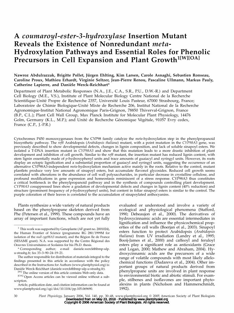
extract-free stems of cosuppressed plants were char-acterized by a 40% reduced Klason lignin content rel-ative to the corresponding Col-0 control (Table II).Upon thioacidolysis, the lignin from cosuppressedplants released H, G, and S monomers in much lowertotal yield compared to wild type (93% reduction),which is indicative of a high content in resistant
Table I. Lignin-derived monomers (H, G, S) after thioacidolysis of dry aerial parts from control Ws and null cyp98A3 mutant
The data represent the mean value (SE in brackets) of duplicate analyses. Stems/leaves from 6-week-old null plants (grown on soil) or 3-week-oldroots (plate grown) were collected and compared to samples either from similarly sized Ws plants or from similarly aged Ws plants.
SampleYield in (H 1 G 1 S)
Monomers
Molar Frequency of Thioacidolysis Monomers
H G S
mmol g21 dry sample %
Dry stems/leaves fromSix-week-old Ws 162 (8) 0.7 (0.02) 65.7 (0.3) 33.6 (0.2)Six-week-old heterozygous cyp98A3 187 (17) 1.3 (0.1) 67.1 (0.7) 31.6 (0.7)Ws of same size as below 1.2 (0.4) 3.2 (0.02) 82.1 (1.5) 14.7 (1.6)Six-week-old homozygous cyp98A3 1.5 (0.2) 94.6 (0.2) 2.2 (0.1) 3.2 (0.02)
Dry roots fromThree-week-old Ws 63 (0.5) 5.2 (0.1) 89.0 (0.2) 5.8 (0.1)Ws of same size as below 7.5 (0.02) 6.6 (0.7) 85.9 (0.2) 7.5 (0.4)Three-week-old homozygous cyp98A3 1.8 (0.1) 43.1 (9.8) 50.9 (9.9) 6.0 (0.1)
Figure 4. Ectopic lignification phenotypes of cyp98A3 insertion andcosuppressed plants. Plant material was stained with phloroglucinoland visualized using whole-mount bright-field microscopy. A, Rootsfrom 3-week-old wild-type (Ws) and cyp98A3 insertion plants (303 ).B to E, Different organs from 10-week-old T2 plants derived from thecosuppressed line VII (Fig. 3) were used for phloroglucinol staining.Roots (B, 303 ), inflorescence stems (C), cauline leaves (D), and flowers(E, each 103 ).
Abdulrazzak et al.
34 Plant Physiol. Vol. 140, 2006 www.plantphysiol.orgon May 23, 2018 - Published by Downloaded from
Copyright © 2006 American Society of Plant Biologists. All rights reserved.
interunit bonds. Similar to cyp98A3 insertion mutants,the cosuppressed plants are characterized by a strongdecrease in the relative amounts of S and G units instems and a dramatic increase in the proportion of Hunits, which constitute 74% of lignin thioacidolysismonomers compared to less than 1% in stems fromwild-type Col-0 plants (Table II). In addition, thedithioketal derivative of p-OH coumaraldehyde wasobtained in a low, but quantifiable, amount (about1.3% of the H main monomers), which indicates thatp-OH-coumaraldehyde end groups occur that couldcontribute to the positive phloroglucinol staining instems of cosuppressed plants.
The analyses of wild-type Ws roots confirmed that,compared to the corresponding stem lignin, roots con-tain relatively less S units and correlatively more Gand H units (Sibout et al., 2003), with H units contrib-uting 5% to 7% to the total lignin thioacidolysismonomers, depending on the developmental stage ofwild-type plants (Table I). Similar to stems, roots of thecyp98A3 insertion mutants provided a lower totalthioacidolysis yield and a much higher proportion ofH monomers (43%) compared to the wild-type roots.However, the frequency of G thioacidolysis monomersreleased by root lignin was not reduced to the sameextent as in the case of lignin derived from stems, andthe frequency of S monomers was comparable in wild-type and mutant roots (Table I). Indeed, G and S unitsin the root tissues of the cyp98A3 insertion mutant stillconstitute more than 56% of the thioacidolysis-derivedmonomers (wild-type roots contain about 94%; TableI). Together with the persistence of G and S lignin unitsin the mutant stem, this result further supports theoccurrence of an alternative 3-hydroxylation mecha-nism leading to the ectopic root lignin.
Insertion and Cosuppressed cyp98A3 Lines AccumulateFlavonoids and p-Coumaroyl Derivatives, But Sinapoyl
Esters Also Persist
To further characterize the impact of the cyp98A3mutation on phenolic metabolism, we analyzed solu-ble phenolics by liquid chromatography (LC)-massspectrometry (MS) of soluble phenolics. Given thesevere phenotype of the insertion mutant, analyseswere performed using 15-d-old seedlings when phe-notypic differences to wild-type Ws plants are lesspronounced. Despite this, the results obtained for
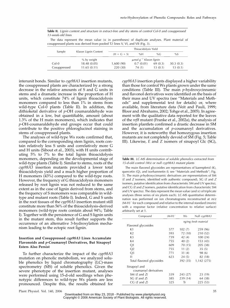
cyp98A3 insertion plants displayed a higher variabilitythan those for control Ws plants grown under the sameconditions (Table III). The main p-hydroxycinnamicand flavonol derivatives were identified on the basis oftheir mass and UV spectra (see ‘‘Materials and Meth-ods’’ and supplemental text for details) or, whereavailable, from literature data (Veit and Pauli, 1999;Bloor and Abrahams, 2002; Tohge et al., 2005). In agree-ment with the qualitative data reported for the leavesof the ref8 mutant (Franke et al., 2002a), the analysis ofinsertion plantlets confirmed a drastic decrease in SMand the accumulation of p-coumaroyl derivatives.However, it is noteworthy that homozygous insertionmutants are not completely devoid of SM (Fig. 5; TableIII). Likewise, E and Z isomers of sinapoyl Glc (SG),
Table II. Lignin content and structure in extract-free and dry stems of control Col-0 and cosuppressed13-week-old lines
The data represent the mean value (SE in parentheses) of duplicate analyses. Plant material ofcosuppressed plants was derived from pooled T2 lines V, VI, and VII (Fig. 3).
Sample Klason Lignin ContentThioacidolysis Yield
(H 1 G 1 S) %H %G %S
% by weight mmol g21 Klason lignin
Col-0 18.48 (0.05) 1,600 (90) 0.7 (0.01) 69 (0.3) 30.3 (0.3)Cosuppressed 11.65 (0.11) 220 (30) 74 (1) 13 (0.1) 13 (0.1)
Table III. LC-MS determination of soluble phenolics extracted from15-d-old control (Ws) or null cyp98A3 mutant plants
The main flavonol glycosides are representatives of kaempferol (K),quercetin (Q), and isorhamnetin (I; see ‘‘Materials and Methods’’; Fig.5). The main p-hydroxycinnamic derivatives are representatives of SM(E and Z isomers, identified with authentic compound), SG (E and Zisomers, putative identification from characteristic SM and UV spectra),and CG (E and Z isomers, putative identification from characteristic SMand UV spectra). The data represent the mean value (and SE) of triplicateanalyses (three series of six plants each). LC-MS quantitative determi-nation was performed on ion chromatograms reconstructed at m/z(M-H)2 for each compound and relative to the internal standard (morin)with a response factor (relative concentration to relative surface)arbitrarily set at 1.
Compound (M-H)2 Ws Null cyp98A3
ng/mg fresh material
Flavonol glycosidesK1 577 102 (7) 259 (96)K2 593 72 (10) 210 (52)K3 739 42 (4) 100 (24)K4 755 40 (2) 153 (43)Q1 609 70 (15) 205 (38)Q2 755 11 (2) 35 (5)Q3 771 31 (4) 98 (6)I1 623 24 (5) 82 (18)Total flavonol glycosides 392 (35) 1,142 (275)
Sinapoyl andcoumaroyl derivatives
SM (E and Z) 339 243 (27) 23 (19)SG (E and Z) 385 239 (14) 64 (38)CG (E and Z) 325 Tr 225 (53)
meta-Hydroxylation of Phenolic Compounds: Roles and Pathways
Plant Physiol. Vol. 140, 2006 35 www.plantphysiol.orgon May 23, 2018 - Published by Downloaded from
Copyright © 2006 American Society of Plant Biologists. All rights reserved.
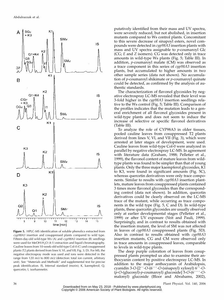
putatively identified from their mass and UV spectra,were severely reduced, but not abolished, in insertionmutants compared to Ws control plants. Concomitantto this severe decrease of sinapoyl esters, novel com-pounds were detected in cyp98A3 insertion plants withmass and UV spectra assignable to p-coumaroyl Glc(CG; E and Z isomers). CG was detected only in traceamounts in wild-type Ws plants (Fig. 5; Table III). Inaddition, p-coumaroyl malate (CM) was observed asa trace component in this series of cyp98A3 insertionplants, but accumulated to higher amounts in twoother sample series (data not shown). No accumula-tion of p-coumaroyl shikimate or p-coumaroyl quinatecould be detected, as confirmed by the analysis of au-thentic standards.
The characterization of flavonol glycosides by neg-ative electrospray LC-MS revealed that their level was3-fold higher in the cyp98A3 insertion seedlings rela-tive to the Ws control (Fig. 5; Table III). Comparison ofthe profiles indicates that the mutation leads to a gen-eral enrichment of all flavonol glycosides present inwild-type plants and does not seem to induce theincrease of selective or specific flavonol derivatives(Table III).
To analyze the role of CYP98A3 in older tissues,pooled cauline leaves from cosuppressed T2 plantsderived from lines V, VI, and VII (Fig. 3), which werearrested at later stages of development, were used.Cauline leaves from wild-type Col-0 were analyzed inparallel by negative electrospray LC-MS. In agreementwith literature data (Graham, 1988; Pelletier et al.,1999), the flavonol content of mature leaves from wild-type plants was found to be simpler than that of youngplants. Only the three major kaempferol glycosides, K1to K3, were found in significant amounts (Fig. 5C),whereas quercetin derivatives were only trace compo-nents. Similar to results with cyp98A3 insertion plant-lets, mature leaves from cosuppressed plants contained3 times more flavonol glycosides than the correspond-ing control (data not shown). In addition, quercetinderivatives could be clearly observed on the LC-MStrace of the mutant, while occurring as trace compo-nents in the wild type (Fig. 5, C and D). In wild-typeplants, these quercetin glycosides are usually observedonly at earlier developmental stages (Pelletier et al.,1999) or after UV exposure (Veit and Pauli, 1999).Surprisingly, and in contrast to results obtained withthe insertion mutant, the level of SM was not affectedin leaves of cyp98A3 cosuppressed plants (Fig. 5D).Also in contrast to results obtained with cyp98A3insertion mutants, CG and CM were observed onlyin trace amounts in cosuppressed leaves, comparableto levels in wild-type plants.
The deep purple coloration of leaves from cosup-pressed plants prompted us also to examine their an-thocyanin content by positive electrospray LC-MS. Inaddition to the major anthocyanin of Arabidopsis,cyanidin 3-O-[2##-O-(6###-O-(sinapoyl) xylosyl) 6##-O-(p-O-(glucosyl)-p-coumaroyl) glucoside] 5-O-(6####-O-malonyl) glucoside (Bloor and Abrahams, 2002),
Figure 5. HPLC-MS identification of soluble phenolics extracted fromcyp98A3 insertion and cosuppressed plants compared to wild type.Fifteen-day-old wild-type Ws (A) and cyp98A3 insertion (B) seedlingswere used for MeOH:H2O (4:1) extraction and liquid chromatography.Cauline leaves from 10-week-old wild-type Col-0 (C) and cosuppressedplants (T2 plants derived from lines V, VI, and VII; Fig. 3; D). For MS, thenegative electrospray mode was used and ions were detected in therange from 120 m/z to 800 m/z (detection: total ion current, arbitraryunit). See ‘‘Materials and Methods’’ and supplemental text for precisepeak identification. IS, Internal standard (morin); K, kaempferol; Q,quercetin; I, isorhamnetin.
Abdulrazzak et al.
36 Plant Physiol. Vol. 140, 2006 www.plantphysiol.orgon May 23, 2018 - Published by Downloaded from
Copyright © 2006 American Society of Plant Biologists. All rights reserved.

we observed a series of other, less abundant cyanidinderivatives in cosuppressed plants only (data notshown). These are, on the basis of their mass fragmen-tation pattern, identical to those recently identified byTohge et al. (2005), who reported the accumulation ofsuch cyanidin derivatives in the leaves of an Arabi-dopsis mutant overexpressing a MYB transcriptionfactor. All these acylated cyanidin glycosides, whichsurprisingly include substantial amounts of sinapoyl-ated derivatives, were below the detection level inleaves of wild-type plants.
In summary, the aerial parts of the cyp98A3 inser-tion mutant display drastically reduced, although notnegligible, levels of sinapoyl esters and S and G lig-nin units. It is accompanied by a strong increase inp-coumaroyl esters, H lignin units, and flavonoids.This confirms the central role of CYP98A3 as the C3#Hin the general phenylpropanoid pathway (Schoch et al.,2001; Franke et al., 2002a, 2002b; Nair et al., 2002).However, the persistence of detectable amounts of SMin cyp98A3 null plants and the presence of G and Slignin units mainly in their roots support the hypoth-esis of an alternative meta-hydroxylase pathway inde-pendent of CYP98A3. The occurrence of large amountsof SM and sinapoylated cyanidin glucosides in com-pletely cosuppressed cyp98A3 plants gives further sup-port to this hypothesis.
Impaired Plant Development of cyp98A3 Mutants Is
Accompanied by Reduced Cell Size and Alterationin Cell Wall Polysaccharide Composition
In an attempt to understand the observed inhibitionof plant growth, cell size and cell wall modificationswere investigated. A statistical analysis of the size ofepidermal cells in different leaves and of different leafareas indicated a significant 6-fold decrease of the cellsize in the cyp98A3 insertion mutant (Fig. 6A). Thisreduced cell size is not accompanied by modificationof the cell shape, and an estimation of the total numberof cells per leaf based on leaf and cell size (Fig. 6A)revealed no difference to wild-type plants. A sub-stantial, but less dramatic, reduction in cell size wasalso observed in cosuppressed plants (data not shown).
In addition to decreased cell size, an alteration in thecell wall polysaccharide composition was observed
Figure 6. Modifications in cell size and cell wall polysaccharides incyp98A3 insertion mutants. A, Reduced epidermal cell size and leafarea in 15-d-old cyp98A3 insertion mutants compared to wild type(Ws). Fully expanded leaves were stained with Hoyer’s solution andwere used for Normarski optics microscopy (left). Average cell size wasdetermined using 40 leaf areas from 10 plants. Mean cell sizes and SDsare shown as bar plots (right). Mean of total leaf area size of wild-typeand cyp98A3 insertion plants are shown on the far right. B, Cell wallmaterial was prepared from leaves of 5-week-old cyp98A3 insertionmutants (white bars), 2-week-old wild-type (Ws; gray bars), and5-week-old wild-type (black bars) plants. Dried cell wall material
was used for TFA hydrolysis (representing matrix polysaccharides) andthe resulting monosaccharide derivatives were quantified using gasGC-MS. UA content was determined using the metahydroxyl-biphenylassay (Blumenkrantz and Asboe-Hansen, 1973). Shown at the top areresults from three replicates; error bars indicate SDs. At the bottom,monosaccharide composition of the crystalline cellulose fraction basedon Seaman hydrolysis is shown. Dried cell wall material was hydro-lyzed using sulfuric acid and the released monosaccharides werequantified using GC-MS. All amounts are shown in micromoles permilligram of dry cell wall material. C, Roots from 2-week-old seedlingswere used for immunostaining using tubulin antibodies. Staining ofcortical microtubules in the root elongation zone of wild type (Ws) andthe cyp98A3 insertion mutant are shown.
meta-Hydroxylation of Phenolic Compounds: Roles and Pathways
Plant Physiol. Vol. 140, 2006 37 www.plantphysiol.orgon May 23, 2018 - Published by Downloaded from
Copyright © 2006 American Society of Plant Biologists. All rights reserved.

(Fig. 6B). Polysaccharide analysis of 5-week-old plantsrevealed a significant increase in Glc released by tri-fluoroacetic acid (TFA) hydrolysis representing amor-phous glucans (mainly amorphous cellulose). Aconcomitant decrease in the level of TFA-releaseduronic acids (UA; reflecting a decrease of pectic poly-saccharides) compared to wild-type plants of the sameage was observed (Fig. 6B). In addition, the amount ofGlc released by Seaman hydrolysis, reflecting crystal-line cellulose, was 50% lower in the cyp98A3 insertionmutant compared to mature Ws plants. It is interestingto note that the decrease in crystalline cellulose isaccompanied by an increase in amorphous cellulose incyp98A3, suggesting that the overall amount of cellu-lose is not altered in cell walls of the mutant, but ratherits state of crystallinity, and hence possibly the archi-tecture of the wall. However, at 5 weeks, wild-typeplants are significantly larger than cyp98A3 homozy-gous plants. To test whether the differences in thecomposition of the cell wall polysaccharides did notmerely reflect delayed development, wild-type plantswere grown for 2 weeks until they reached a sizecomparable to the size of the 5-week-old null cyp98A3.Certain changes in polysaccharide composition occurduring development of wild-type Ws plants. In ma-ture plants, an increase in UAs was observed, accom-panied by a slight increase in all neutral sugarsanalyzed with the exception of TFA-released Glc. Inaddition, a strong increase of crystalline cellulose wasobserved (Fig. 6B), which is likely due to the higherproportion of secondary walls present in the olderplants. When comparing cyp98A3 insertion mutants toboth developmental stages of wild-type plants, a re-duction of UAs, an increase in amorphous cellulose,and a significant reduction in Ara, reflecting lowerlevels of arabinogalactan proteins and/or pectin-derived arabinans, was apparent (Fig. 6B).
Cytoskeleton and, in particular, cortical microtu-bules, are known to be intimately associated with thebiogenesis of the cell wall and cellulose microfibrilelongation, as well as secondary cell wall depositionsand signaling of morphogenetic processes (Baskin,2001; Wasteneys, 2004). Immunostaining of corticalmicrotubules in elongating root cells did not revealany significant perturbation of the transversal orien-tation of the microtubules. However, the overall mi-crotubule structure was altered and appeared morepunctuated (Fig. 6C).
Impaired Development and Reduced Cell Size Is
Accompanied by Massive Changes in Gene Expression
The strong alterations in cell wall composition, cellsize, and in overall plant development observed in thecyp98A3 insertion mutant go beyond expectations andcannot be explained by modifications in phenylpro-panoid metabolism alone. Gene expression-profilinganalyses were thus performed to pinpoint the under-lying developmental defects. Given the severe pheno-type of the mutant, 15-d-old seedlings grown in vitro
were used to minimize the impact of differences inplant development on gene expression and to catcha picture of the phenomenon at its onset. Althougheven at this early stage pleiotropic and other indirecteffects cannot be excluded, they were expected to beless pronounced.
In a preliminary experiment, the expression ofselected genes upstream in the phenylpropanoidpathway was compared using quantitative real-timeRT-PCR analysis. Given that genes encoding phenyl-propanoid enzymes display diurnal changes in theirexpression (Rogers et al., 2005), we examined expres-sion levels throughout the 12-h light cycle in plantsgrown under a 12-h-light/12-h-dark regime. In wild-type plants, the transcript levels of all phenylpropa-noid genes were highest 4 h into the light period. Atthe end of the day period, they returned to the samelevels observed at 0 h (Fig. 7A). This variation in geneexpression is in agreement with studies by Rogers et al.(2005), and a comparable cycling of all transcriptsanalyzed was also observed in cyp98A3 insertion mu-tants, with the exception of a total inactivation ofCYP98A3 (Fig. 7A). While transcript levels of genesencoding Phe ammonia lyase (PAL1; At2g37040) andHCT (At5g48930; Fig. 7A) were comparable in cyp98A3mutants and wild type at all time points, an increasein C4H (CYP73A5; At2g30490) expression comparedto wild type was observed at the 2-, 4-, and 8-h timepoints in cyp98A3 insertion mutants (Fig. 7A). In10-week-old cosuppressed T2 plants derived fromthe primary transformants V, VI, and VI (Fig. 3), whichwere arrested in growth when analyzed, CYP98A3gene expression is suppressed to undetectable levels(Figs. 7B and 3F). This suppression was accompaniedby a decrease in the expression of PAL1, C4H, andHCT at all time points analyzed (Fig. 7B). However, inall cases, cycling during the daylight period was notaltered. Cosuppression of the CYP98A3 gene in adultplants, but not inactivation via a T-DNA insertion injuvenile plantlets, thus appears to result in a repressionof the general phenylpropanoid pathway. But theinactivation of CYP98A3 has no impact on the circa-dian cycle of phenylpropanoid gene expression. Thecyp98A3 insertion and wild-type Ws lines grown un-der the same conditions can thus be compared on alarger scale in juvenile plants.
Microarray analysis of gene expression was thusperformed using 15-d-old homozygous cyp98A3 in-sertion plants compared to wild-type Ws. Two bi-ological replicates were analyzed, with two technicalreplicates (dye swaps) each for a total of four micro-arrays. The CATMA Arabidopsis near-full genomearray was employed, which contains 24,576 gene-specific elements representing 21,371 annotated genes.Statistical analysis (see ‘‘Materials and Methods’’ fordetails) identified 1,944 elements representing 1,889annotated genes to be differentially expressed (ad-justed p [t test] , 0.05). Among these, 1,067 elements(1,038 genes) changed more than 2-fold, and 362elements representing 352 annotated genes changed
Abdulrazzak et al.
38 Plant Physiol. Vol. 140, 2006 www.plantphysiol.orgon May 23, 2018 - Published by Downloaded from
Copyright © 2006 American Society of Plant Biologists. All rights reserved.

more than 3-fold (94 genes overexpressed and 260genes repressed in the mutant).
Supplemental Table I lists normalized expressionratios and annotation details for all differentially ex-pressed genes; the complete dataset was deposited
into the ArrayExpress database (accession E-MEXP-346). Functional grouping of differentially expressedgenes using the Functional Category Database atMatDB (Schoof et al., 2002; Fig. 8A) indicates a pro-found modification of plant metabolism. As expectedin a mutant almost completely blocked in carbon uti-lization for lignin synthesis and with a strong pheno-type, perturbations in primary metabolism are muchmore severe than in mutants such as pal1pal2, wherelignin synthesis is only decreased to 30% (Rohde et al.,2004). Functional groups over-represented in the groupof down-regulated genes, which change more than3-fold, point to a strong repression of light- andoxygen-dependent energy production and carbon fix-ation, reflected by a decreased expression of plastidalgenes involved in photosynthesis and respiration (Fig.8A). Indeed, several genes encoding proteins involvedin the light-harvesting complex (e.g. chlorophylla/b-binding proteins At2g34430, At3g08940, andAt3g47470), in photosynthetic electron transport (e.g.the putative ferredoxin At1g10960 and proton gradi-ent regulation 5, At2g05620; Munekage et al., 2002),and in the Calvin cycle (e.g. Rubisco At5g38410 andthe putative Rib-5-P isomerase At3g04790) are stronglydown-regulated in the mutant compared to the wildtype (Supplemental Table I). In contrast, genes ex-pressed to higher levels in cyp98A3 insertion mutantscompared to wild type were over-represented in func-tional categories covering carbohydrate metabolismand include genes encoding enzymes in glycolysis (e.g.the glyceraldehyde 3-P dehydrogenase, At3g04120),the tricarboxylic acid cycle (e.g. the phosphoenolpyr-uvate carboxylase, At3g14940), and other anaerobicmetabolisms (alcohol dehydrogenase, At1g77120).Also, functional categories related to defense responseswere over-represented (Fig. 8A). Among these weregenes known to be transcriptionally induced by insectfeeding (the b-glucosidase BGL1, At1g52400; and thelipoxygenase LOX2, At3g45140; Stotz et al., 2000) orwounding/jasmonate (e.g. jasmonate response JR1,At3g16470; Leon et al., 1998). Finally, genes involvedin secondary metabolism are over-represented in thegroup of up-regulated genes (Fig. 8A). It is noteworthythat genes encoding enzymes involved in flavonoidand anthocyanin biosynthesis and transport are ex-pressed to higher levels in the cyp98A3 inser-tion mutant; These include chalcone isomerase(CHI, At3g55120), dihydroflavonol 4-reductase (DFR,At5g42800), flavanone 3-hydroxylase (F3H, At3g51240),and gluthation transferase (TT19, At5g17220; Weisshaarand Jenkins, 1998; Kitamura et al., 2004). In contrast, butin agreement with quantitative RT-PCR results (seeabove), among genes involved in monolignol biosynthe-sis apart from CYP98A3, only C4H is differentially ex-pressed, while the expression of all other genes of thephenylpropanoid pathway remains unaltered (Array-Express accession E-MEXP-346).
The higher expression of genes related to stressresponse, in particular mediated by jasmonate,prompted us to dissect the potential involvement of
Figure 7. Phenylpropanoid gene expression in cyp98A3 insertion andcosuppression mutants. A, Fifteen-day-old cyp98A3 insertion mutant(cyp98A3 null) and wild-type (Ws) plants were harvested at differenttime points after the onset of light (time 0 h) during a regular 12-h lightperiod as indicated (plants grown on agar plates under 12-h-light/12-h-dark cycle). Total RNA was used for quantitative real-time RT-PCR. Asan internal standard, Actin II was coamplified with the PAL1, C4H,HCT, and CYP98A3 cDNA and DDCT values are given relative to theexpression level of the respective gene observed for wild-type (Ws)plants at 0 h. B, Ten-week-old cosuppressed T2 Col-0 plants derivedfrom lines V, VI, and VII (Fig. 3) were used for quantitative real-timeRT-PCR. Plants were cultivated on soil under a 12-h-light/12-h-darkcycle. Expression levels of each gene are given relative to the respectivelevel observed for wild-type (Col-0) plants at 0 h.
meta-Hydroxylation of Phenolic Compounds: Roles and Pathways
Plant Physiol. Vol. 140, 2006 39 www.plantphysiol.orgon May 23, 2018 - Published by Downloaded from
Copyright © 2006 American Society of Plant Biologists. All rights reserved.

this plant hormone and to compare it to the impactof other hormones. Therefore, we retrieved from TheArabidopsis Information Resource (TAIR; Garcia-Hernandez et al., 2002) curator-annotated lists of genesthat are involved in the ‘‘biosynthesis of,’’ in the‘‘signal transduction mediated by,’’ and in the ‘‘re-sponse to’’ the plant hormones auxin, abscisic acid,brassinosteroid, cytokinin, ethylene, gibberellic acid,jasmonic acid, and salicylic acid. Within each of thesegroups of genes, the frequency of differentially ex-pressed genes was determined and compared to thefrequency of differentially expressed genes among allgenes represented on the array. Genes expressed tohigher levels in the cyp98A3 mutant were clearly over-represented in the functional group related to jasmo-nate signaling (Fig. 8B). Indeed, many genes involvedin the biosynthesis of jasmonate are significantly high-er expressed in the mutant compared to wild type(Ws); These include LOX2 (see above), LOX1, alleneoxide synthase (CYP74A, At5g42650), and allene oxidecyclase 4 (At1g13280).
In addition, genes related to the abscisic acid cas-cade are over-represented in the group of genes ex-pressed to higher levels in cyp98A3 insertion mutants(Fig. 8B). These include the gene responsive to de-hydration 22 (RD22I, At5g25610), which is stronglyinduced by abscisic acid and drought (Yamaguchi-Shinozaki and Shinozaki, 1993), and the Arg decar-boxylase 2 (ADC2, At4g34710) gene, which is stronglyactivated by abscisic acid, but also by methyl jasmo-nate and mechanical wounding, and is involved inpolyamine biosynthesis (Perez-Amador et al., 2002).Also expressed to higher levels in cyp98A3 insertionmutants were two homeodomain Leu-zipper tran-scription factors (ATHB5, At5g65310; and ATHB7,At2g46680) involved in abscisic acid-mediated sig-naling pathways related to drought and osmoticstress (Soderman et al., 1996) and seed germination(Johannesson et al., 2003), respectively.
None of the other plant hormones analyzed appearsto have a similar relevance for the group of genesexpressed to higher levels in the cyp98A3 insertionmutant. To a lesser extent, genes related to gibberellicacid and salicylic acid are more frequently found in thegroup of genes expressed to lower levels in cyp98A3insertion mutants than expected by chance (Fig. 8B).Among genes related to salicylic acid signaling andexpressed to lower levels in cyp98A3 insertion mu-tants, defective in induced resistance 1 (At5g48485)encodes a lipid transfer protein that, when mutated,results in specific loss of systemic acquired resistance(Maldonado et al., 2002).
Figure 8. Global changes in gene expression in cyp98A3 insertionmutants. Wild-type Ws and cyp98A3 insertion plants grown for 15 d onagar plates were used for total RNA isolation. Microarray analyses wereperformed using two biological replicates with two technical replicates(dye swaps) each, and the CATMA Arabidopsis near-full genome array.Upon normalization and statistical analysis, 1,889 annotated geneswere found to be differentially expressed (Bonferroni-adjusted p [t test],0.05). A, Functional grouping of differentially expressed genes thatdiffered in expression more than 3-fold between cyp98A3 insertionmutants and wild-type Ws using the Functional Category Database atMAtDB (Schoof et al., 2002; http://mips.gsf.de/proj/funcatDB). Thefrequency of up-regulated genes in each functional category (shown asdark-gray bars) was compared to the frequency of all genes representedon the microarray in the same functional category (shown as light-graybars). Only functional categories that are over-represented (p [hyper-geometric distribution] , 0.05) in the group of up-regulated genes areshown. At the bottom, results of the same analysis for 249 genes, whichwere expressed to more than 3-fold lower levels in cyp98A3 mutantsand which were annotated in the Functional Category Database, areshown. B, We retrieved from TAIR (Garcia-Hernandez et al., 2002;http://www.arabidopsis.org/tools/bulk/go) curator-annotated lists ofgenes that were placed into the GO terms ‘‘biosynthesis of,’’ ‘‘signaltransduction mediated by,’’ and ‘‘response to’’ the plant hormonesauxin (AUX), abscisic acid (ABA), brassinosteroid (BS), cytokinin (CYT),ethylene (ET), gibberellic acid (GA), jasmonic acid (JA), and salicylicacid (SA). Two asterisks indicate over-represented groups (p [hyper-geometric distribution] , 0.01), one asterisk indicates P , 0.05. Shownas stacked bar plots are the frequencies of genes in each group
expressed to higher levels in cyp98A3 mutants (on top of the verticalaxis, separately for genes changing more [black bars] or less [dark-graybars] than 2-fold), as well as for genes expressed to lower levels incyp98A3 insertion plants (below the vertical axis; medium-gray barsindicate less than 2-fold difference, light-gray bars indicate more than2-fold difference).
Abdulrazzak et al.
40 Plant Physiol. Vol. 140, 2006 www.plantphysiol.orgon May 23, 2018 - Published by Downloaded from
Copyright © 2006 American Society of Plant Biologists. All rights reserved.

Finally, in an attempt to explain the dramatic de-crease in cell growth and plant development observedin the mutant, we further explored the expression ofgenes involved in cell expansion and cell division. Thisanalysis revealed that no gene annotated in TAIR geneontology (GO) term ‘‘cell division’’ is differentiallyexpressed more than 2-fold. In addition, none ofthe genes involved in the core cell cycle machinery(Vandepoele et al., 2002) is differentially expressedbetween wild-type and cyp98A3 mutant plants. Incontrast, 12.5% of genes annotated in the GO term‘‘cell growth’’ are differentially expressed betweencyp98A3 insertion and wild-type plants. Among genesin this group, putative expansins, which mainly act inloosening cell walls for regulating cell expansion (Leeet al., 2001), form the largest group. Six of the 28expansin family members represented on the arrayused are differentially expressed, significantly morethan expected by chance. However, some expansinisoforms (EXP1, At1g69530; EXP10, At1g26770; andEXPR, At4g17030) are expressed to higher levels incyp98A3 insertion mutants, whereas others (EXP8,At2g40610; EXP3, At2g37640; and EXP11, At1g20190)are down-regulated compared to levels found in wild-type plants.
Genetic and Chemical Complementation
To ensure that the observed phenotype, in particularthe drastic growth and developmental inhibition of thenull mutant, was indeed the result of the CYP98A3gene inactivation, a genetic complementation was per-formed by transformation of heterozygous plants witha construct containing the CYP98A3 coding regionunder control of the CaMV 35S promoter. Transform-ants were identified based on the BASTA resistancemarker present in the construct, and homozygouscyp98A3 plants in the progeny of BASTA-resistantplants were identified using PCR (Fig. 9A). Homozy-gous cyp98A3 insertion plants containing the 35S::CYP98A3 constructs were morphologically indistin-guishable from wild-type plants (Fig. 9B). They dis-played the same phenotype, including growth anddevelopment, as wild-type and heterozygous plants.Based on phloroglucinol staining, no signs of ectopiclignification were detected in roots of complementedplants (data not shown).
The observed growth inhibition in cyp98A3 insertionplants may result either from a depletion of 3-hydrox-ylated products or from the accumulation of poten-tially toxic or bioactive upstream or side products (e.g.flavonoids that were reported to modulate auxin trans-port [Peer et al., 2004]). In an attempt to dissect thesehypotheses, chemical complementation tests wereperformed. CYP98A3 metabolizes with high efficiencyboth p-coumaroyl shikimate and p-coumaroyl quinate,both of which can be generated from p-coumaric acidby HCT. HCT is also expected to catalyze the conver-sion of the CYP98A3 product, caffeoyl shikimate/quinate, to caffeoyl-CoA (Hoffmann et al., 2003;
Niggeweg et al., 2004). Complementation was there-fore first attempted using the commercially availablecaffeic acid and caffeoyl quinate (chlorogenic acid).cyp98A3 insertion plants were grown on media con-taining these compounds, but no stimulation of plantgrowth was observed (data not shown). Given thatcaffeoyl shikimate is likely the major CYP98A3 prod-uct in vivo based on catalytic parameters (Schoch et al.,
Figure 9. Genetic and chemical complementation of the cyp98A3mutation. Heterozygous cyp98A3 insertion plants were transformedwith a CaMV 35S::CYP98A3 construct, and homozygous cyp98A3plants in the progeny of BASTA-resistant plants were identified usingPCR (A) and (i) a gene-specific (Tuc) and a T-DNA-specific (Lbnes)primer; (ii) two gene-specific primers (Tuc, P2) located on each site ofthe T-DNA insertion site; and (iii) two 35S::CYP98A3 construct-specificprimers (A3/Bar). Plant 1, Heterozygous, not transformed; plants 2 and3, heterozygous, transformed; plant 4, homozygous cyp98A3, trans-formed; plant 5, homozygous wild type, transformed. B, Impact of thegenetic complementation on 10-week-old plants: wild-type Ws (left)and homozygous cyp98A3 insertion mutant transformed with the35S::CYP98A3 construct (right). C, Chemical complementation, Ho-mozygous cyp98A3 insertion mutants grown on Murashige and Skoogmedium for 2 weeks were transferred to Murashige and Skoog mediumcontaining 90 mm caffeoyl shikimate and grown for an additional2 weeks (left). Control plants were transferred to nonsupplementedMurashige and Skoog medium (right).
meta-Hydroxylation of Phenolic Compounds: Roles and Pathways
Plant Physiol. Vol. 140, 2006 41 www.plantphysiol.orgon May 23, 2018 - Published by Downloaded from
Copyright © 2006 American Society of Plant Biologists. All rights reserved.

2001; M. Morant, G. Schoch, P. Ullmann, T. Ertuncx, D.Little, C.E. Olsen, M. Petersen, J. Negrel, D. Werck-Reichhart, unpublished data), we synthesized thiscompound enzymatically from p-coumaroyl shikimateusing recombinant CYP98A3 and purified the productcaffeoyl shikimate in sufficient amounts to attempt com-plementation. Exogenously applied caffeoyl shikimateat least partially restored plant growth (Fig. 9C).Growth inhibition is thus more likely to result fromthe depletion of an essential 3-hydroxylated productrather than from the accumulation of a toxic precursor.In the absence of a commercially available source ofradiolabeled compounds, we cannot exclude a differ-ential uptake of caffeic acid, caffeoyl quinate, andcaffeoyl shikimate, but it seems likely that chlorogenicacid or caffeic acid cannot be converted into this es-sential growth-promoting compound in cyp98A3 in-sertion mutants.
DISCUSSION
Analysis of cyp98A3 Mutants Reveals the Existenceof an Alternative meta-Hydroxylation Pathway ofPhenolic Compounds
The genes belonging to the CYP98 family recentlyemerged as the cytochrome P450 monooxygenases cat-alyzing the meta-hydroxylation of phenolic compoundsin several plant species. These enzymes catalyze themeta-hydroxylation of p-coumaric derivatives esteri-fied to shikimate, quinate, or phenyllactate (Schochet al., 2001; Matsuno et al., 2002). In Arabidopsis,CYP98A3 is required for the synthesis of lignin mono-mers and soluble sinapoyl esters (Franke et al., 2002a).A point mutation in the CYP98A3 gene was sufficientto completely suppress the production of G and Slignin monomers (as revealed by derivatization fol-lowed by reductive cleavage, nitrobenzene oxidation,and analytical pyrolysis) and to drastically reduce thelevel of soluble meta-hydroxylated phenolics in aerialparts of the ref8 mutant (Franke et al., 2002a, 2002b).It was thus expected that CYP98A3 was the sole3-hydroxylation enzyme of the phenylpropanoidpathway in Arabidopsis, and that the phenylpropa-noid pathway had evolved to exclusively channel the3-hydroxylation step through p-coumaroyl esters andthe highly conserved CYP98 enzymes (Schoch et al.,2001; Franke et al., 2002a, 2002b). Our present resultschallenge this hypothesis.
This analysis of Arabidopsis cyp98A3 insertion andcosuppressed lines confirms the essential role ofCYP98A3 in the synthesis of G and S lignin monomersin aerial parts of the plant and, to some extent, also inthe roots. It also confirms that, under normal growthand homeostasis conditions, CYP98A3 is the majorcontributor to the biosynthesis of soluble sinapoylderivatives as previously reported by Franke et al.(2002a). However, the cyp98A3 insertion mutant dis-plays a more dramatic phenotype compared to the ref8mutant (Franke et al., 2002b), which suggests that the
ref8 gene still had a low residual activity. In the T-DNAinsertion mutant analyzed here, the CYP98A3 gene istotally inactivated. It nevertheless contains weak, butquantifiable, amounts of G and S units in the lignin ofthe aerial parts of the plants. G and S units are evenmore frequent in the ectopic root lignin. Our results,therefore, suggest the occurrence of an alternative meta-hydroxylation pathway in Arabidopsis, which seemsto be more active in producing the ectopic ligninobserved on the roots. It is likely that the same pathwayis activated ectopically in the rachis of cosuppressedplants. The detection of SM in the seedlings of the in-sertion mutant further supports this hypothesis.
It is interesting to note that the alternative meta-hydroxylation pathway observed in cyp98A3 mutantsappears to be activated when the prevalent pathway isnot functional. However, this alternative pathwaycannot complement the defects observed in cyp98A3mutants (e.g. reduced growth, reduced lignin in stems,limp stems, and male sterility). It does not ensurenormal growth and appears to be active in tissues thatdo not normally lignify, as judged from the ectopicphloroglucinol staining observed in cyp98A3 insertionand cosuppressed plants. This alternative pathwayseems to be ectopically activated under conditions ofperturbed metabolism, but the nature and normalphysiological relevance, in specific tissues or at spe-cific stages of plant development, remains to be eluci-dated. Interestingly enough, the ectopic CYP98A3overexpression for complementation of the null mu-tant does not promote any ectopic lignification. It isworth mentioning that the analysis of precursor con-version by peltate glands isolated from two sweet basil(Ocimum basilicum) lines that differ in their ability toproduce eugenol also led Gang et al. (2002) to proposethe existence of an alternative meta-hydroxylationpathway. The line that produced methyl chavicolinstead of eugenol had a strongly decreased abilityto meta-hydroxylate p-coumaroyl esters, but anunaltered capability to convert p-coumaric acid intocaffeic acid. The alternate pathway operating in sweetbasil may, however, be different from that producingmeta-hydroxylated phenolics in the cyp98A3 mutantsfrom Arabidopsis.
Impact of CYP98A3 Gene Inactivation on Plant Growthand Metabolism
The ectopic activation of an alternative meta-hydrox-ylation pathway in cyp98A3 mutants is paralleled withstrong morphological changes and with an accumula-tion of flavonoids. The accumulation of flavonoids(flavonol glycosides and anthocyanins) in null andcosuppressed cyp98A3 mutants is not surprising, sincecreating a bottleneck at the level of CYP98A3 leads toan accumulation of p-coumaroyl-CoA precursors thatmay subsequently simply overflow into the flavonoidpathway (Fig. 1).
The severe modification in plant development andthe complete growth arrest observed in both cyp98A3
Abdulrazzak et al.
42 Plant Physiol. Vol. 140, 2006 www.plantphysiol.orgon May 23, 2018 - Published by Downloaded from
Copyright © 2006 American Society of Plant Biologists. All rights reserved.

insertion and cosuppressed mutants, however, is moresurprising. Given the reduced cell size, which is notaccompanied by a reduction of the total number ofcells per leaf, these morphological changes are likelydue to an inhibition of cellular growth. Consistent withthis hypothesis, our microarray analysis revealed nodifferences in transcript abundance of genes related tothe cell cycle, while several genes related to cell ex-pansion are expressed to different levels in wild-typeand cyp98A3 insertion plants. Our data suggest thatthreshold amounts of meta-hydroxylated lignin mono-mers or of other caffeoyl shikimate-derived com-pounds are required for normal organization ofthe cell wall and plant growth. This conclusion issupported by the fact that external application ofcaffeoyl shikimate complements, at least partially,the growth phenotype of the null mutant. Such ameta-hydroxylated derivative might be needed asessential building blocks of the primary cell wall ormight more indirectly promote cell wall modificationsand growth (e.g. via Ca21 chelation). Alternatively, itmay directly act as a growth regulator. Phenolic com-pounds have been reported to play a role in cell divi-sion and expansion, either directly or as componentsof a cytokinin-dependent signaling cascade (Lynnet al., 1987). A major role in this respect was attributedto dehydrodiconiferyl alcohol glucoside (DCG), whichwas shown to directly activate cell growth and di-vision in tobacco (Nicotiana tabacum) pith, leaves, orcell cultures (Binns et al., 1987; Tamagnone et al., 1998).DCG, as lignin G monomers, is expected to directlyderive from CYP98A3 products (Fig. 1). DCG accu-mulation during plant growth so far has not beendescribed in Arabidopsis. Its ability to restore growthof the null cyp98A3 mutant will be assessed.
Another possibility is that growth arrest simplyresults from global perturbations of plant metabolism.A block in lignin biosynthesis suppresses a majorcarbon sink and induces an accumulation of flavo-noids and possibly of indolic derivatives. The latter arestress-related metabolites. Many of the differences ingene expression observed in the cyp98A3 insertionmutant compared to wild type are reminiscent of astress response. These include a general down-regulation of photosynthesis/chloroplastic genes, in-dicating reduced energy and carbon assimilation, andelevated expression levels of genes related to defenseresponses with activation of the jasmonate and absci-sic acid signaling pathways. The observed ectopiclignification in roots of the cyp98A3 insertion mutantand in the rachis of cosuppressed plants can also beconsidered as indicative of a constitutive inductionof a stress response. Ectopic lignification, resultingfrom mutations in genes encoding cellulose synthase,a chitinase-like enzyme, or a vacuolar ATPase(Schumacher et al., 1999; Cano-Delgado et al., 2000,2003; Zhong et al., 2002), has been associated withactivation of jasmonate and ethylene cascades, withdefects in plant growth and cell wall biogenesis, andwith stress response. The existence of a link between
ectopic lignification and jasmonate cascade was fur-ther confirmed by the impact of chemical inhibition ofcellulose synthesis with isoxaben, which phenocopiesa mutation in cellulose synthase. Isoxaben treatmentwas reported to induce the jasmonate pathway as wellas ectopic lignification (Ellis et al., 2002; Cano-Delgadoet al., 2003). Conversely, the combined treatment ofwild-type seedlings with jasmonate and 1-aminocyclo-popane-carboxylic acid (an ethylene precursor) leadsto both reduced cellulose synthesis and ectopic ligni-fication (Cano-Delgado et al., 2003). These changeswere correlated with increased expression of the genesinvolved in stress response and with pathogen re-sistance (Ellis et al., 2002).
Our data indicate that the defect in the biosynthesisof an essential phenolic component leads to a similarphenotype, including elevated expression levels ofgenes related to jasmonate signaling, reduced growth,and activation of several stress/defense-related genes.Phenolics thus seem to be one of the elements thatrelate cell wall biosynthesis with growth promotionand stress signaling. No significant activation of theethylene signaling pathway was observed in our ex-periments, but expression profiling indicated highertranscript levels for genes related to abscisic acidsignaling in cyp98A3 mutants. Abscisic acid has beenimplicated as a signal transduction component inmainly abiotic stress responses, in particular responsesto salt, cold, and drought stress (Pastori and Foyer,2002, and refs. therein), but it has also long beenknown that abscisic acid inhibits plant growth and isan inducer of dormancy (Koornneef et al., 2002; Xiongand Zhu, 2003). Therefore, it appears plausible that theobserved higher levels of transcripts related to abscisicacid signaling may also be related to the observedgrowth phenotypes, including developmental arrest.In this context, it is noteworthy that cyp98A3 insertionmutants maintain in a state of developmental arrestand can survive for months without apparent signs ofsenescence.
What Is the Alternative meta-Hydroxylation Pathway?
The proposed alternative meta-hydroxylation path-way is not expressed during developmental stemlignification and cannot complement developmentallignification, because mutations in CYP98A3 lead to analmost complete lack of G and S lignin units in stem(Franke et al., 2002a; Table I). However, it is expressedin tissues of the cyp98A3 insertion mutant, whichnormally do not lignify. Together with the increasedproduction of soluble phenolics and stress-relatedphenylpropanoids, this suggests that the proposedalternative pathway is active under stress conditionsand that it is likely a defense-related pathway. Sucha hypothesis is supported by the observed activationof genes related to the jasmonate and abscisic acidcascades. The existence of divergent isoforms withseparate functions in developmental lignin biosynthe-sis and in stress-related production of phenolics has
meta-Hydroxylation of Phenolic Compounds: Roles and Pathways
Plant Physiol. Vol. 140, 2006 43 www.plantphysiol.orgon May 23, 2018 - Published by Downloaded from
Copyright © 2006 American Society of Plant Biologists. All rights reserved.

been proposed for some enzymes of the phenylpropa-noid pathway, e.g. for cinnamoyl-CoA reductase(Lauvergeat et al., 2001). However, it is unlikely thatan alternative, stress-related pathway uses the samesubstrates as CYP98A3, because plants with reducedenergy assimilation are not expected to accumulateshikimic or quinic acid in amounts sufficient for al-lowing their coupling to p-coumaric acid. The CoAester of p-coumaric acid or its precursors are, however,expected to accumulate when CYP98A3 and/or shiki-mate become limiting (Fig. 1), which could also lead tothe observed accumulation of flavonoids. Such an ac-cumulation of precursor substrates under stress con-ditions, or in mutant plants, may allow the activity ofan enzyme with low affinity for phenolics to becomephysiologically relevant. It is also possible that theexpression of a specific hydroxylase is activated ex-clusively under stress conditions. The most obviouscandidate genes encoding alternative 3-hydroxylaseswould be the other members of the CYP98 family. Thegenes most closely related to CYP98A3 in Arabidopsis,CYP98A8 and CYP98A9, do not catalyze the meta-hydroxylation of any free or conjugated phenolic sub-strates, including Glc and CoA derivatives (Schochet al., 2001; M. Morant, G. Schoch, P. Ullmann,T. Ertuncx, D. Little, C.E. Olsen, M. Petersen, J. Negrel,D. Werck-Reichhart, unpublished data). However, manyother enzymes were previously proposed to catalyzethe meta-hydroxylation of various phenolic precursors(Schoch et al., 2001, and refs. therein; Franke et al.,2002b, and refs. therein). The production of meta-hydroxylated units under stress conditions may relyon one or several of them.
MATERIALS AND METHODS
Isolation of the cyp98A3 Mutant
An insertion mutant for CYP98A3 was isolated by screening the Alpha
collection at the Arabidopsis Knockout Facility at the University of Wisconsin
Biotechnology Center (http://www.biotech.wisc.edu/Arabidopsis/Index2.asp;
Krysan et al., 1999). Two primers were selected to identify insertions in the
CYP98A3 coding sequences: 98A3F1 (5#-CATGAGCAGCAGCAACAAAA-
GGTAG-3#) and 98A3R2 (5#-AACATCCAGGTCAGGTACATGTATTTCAC-3#).
An insertion was identified in the first intron of CYP98A3, and plants
heterozygous for the insertion were found in pool 177. The T-DNA contained
left borders at both T-DNA junctions based on PCR and DNA sequence analysis.
Construction of the CYP98A3 Overexpression Vectorfor Genetic Complementation and Cosuppression
The CYP98A3 cDNA inserted in a pGEM-T vector was amplified using
specific primers with added attB recombination sites (Gateway Technology;
Karimi et al., 2002); sense primer 98A3b1 (5#-GGGGACAAGTTTGTA-
CAAAAAAGCAGGCTATGTCGTGGTTTCTAATAGCGGTG-3#) and anti-
sense primer 98A3b2 (5#-GGGGACCACTTTGTACAAGAAAGCTGGGTT-
TACATATCGTAAGGCACGCGTTT-3#). The PCR reaction was carried out
using as template 200 ng of the plasmid DNA, 10 mM of each primer, and High
Fidelity PCR master mix (Roche), according to the recommendations of the
manufacturer. After 3 min of heating at 96�C, 18 cycles of amplification were
carried out as follows: 1 min denaturation at 95�C, 1 min at 60�C, and 2-min
extension at 72�C. The reaction was completed by a 10-min extension at 72�C.
The PCR product was purified on a 1% agarose gel and cloned into the
pDONR201 donor vector (Invitrogen, Gateway Technology). The ligation mix
(20 mL) contained 300 ng of the PCR product, 300 ng of the pDONR201 vector,
and BP Clonase reaction buffer, the volume being adjusted to 16 mL with
sterile water, according to the instructions of the manufacturer. The reaction
was started with 4 mL of BP Clonase enzyme mix. It was incubated at 25�C for
18 h and stopped by 10-min incubation at 37�C with proteinase K (4 mg/20 mL
of ligation medium). Electrocompetent bacteria (DH5a) were transformed by
electroporation with 1 mL of the ligation medium. Plasmid DNA of positive
colonies selected on kanamycin (100 mg/mL) was prepared (NucleoSpin
Plasmid) and sequenced for selection of a clone corresponding to the expected
sequence. After extraction of the pDONR201 plasmid from the selected clone,
the insert was transferred to the destination expression vector pB7WG2
(Karimi et al., 2002) for insertion of the CYP98A3 cDNA downstream of the
CaMV 35S promoter. The reaction was carried out in a 20-mL mix containing
300 ng of pDONR201 with the CYP98A3 insert, 800 ng of pB7WG2, and LR
Clonase buffer, the volume being adjusted to 16 mL with sterile water. Four
microliters of LR Clonase enzyme mix were added before incubation at 25�C
for 18 h, according to the protocol recommended by the manufacturer. The
reaction was stopped by 10-min incubation at 37�C in the presence of
proteinase K (4 mg/20 mL of reaction medium). The plasmid DNA (Nucleo-
Spin Plasmid) prepared from the positive colonies selected on spectinomycin
(100 mg/mL) was verified by digestion and PCR.
Plant Growth and Transformation
Seeds from Arabidopsis (Arabidopsis thaliana) Ws, Col-0, and heterozygous
cyp98A3 mutants were kept at 4�C for 4 d and then washed for 5 min in 70%
ethanol. After 20-min incubation with gentle shaking in 20% bleach supple-
mented with 0.1% Triton X-100, they were washed four times in sterile
distilled water and germinated on Murashige and Skoog medium (Duchefa)
containing 0.8% Pastagar (Sigma-Aldrich), 1% Suc (Carlo Erba Reagenti), with
or without 50 mg/mL kanamycin (Duchefa). Plates were placed in a 21�C
growth chamber with a 16-h-light/8-h-dark photoperiod for 15 d.
For plant transformation (genetic complementation and cosuppression),
some cyp98A3 heterozygous mutant or wild-type seedlings were grown on
soil at 21�C under a 16-h-light/8-h-dark photoperiod. The pB7WG2 vector
was introduced into Agrobacterium tumefaciens LBA 4404 virG by electro-
poration, and stable transformation of the construct in A. tumefaciens was
confirmed by digestion of the plasmid isolated from the transformants and
PCR using T-DNA-specific primers. The plants were then transformed using
the floral-dip method (Clough and Bent, 1998).
Seeds from the T0 plants were collected and grown on soil at a density of
1,500 plants per 25 3 50-cm pot. Plants were sprayed with Basta (AgroEvo),
250 mg/L, 5, 14, and 21 d after germination. Transgenic plants resistant to the
herbicide were transferred into individual pots and seeds were collected as
above. For in vitro selection, seeds of the T1 plants were grown on Murashige
and Skoog medium supplemented with 0.8% Pastagar, 1% Suc, 10 mg/mL
Basta, and 500 mg/mL carbenicillin.
Circadian rhythm analysis was performed with plants grown under a 12-h-
light/12-h-dark cycle in vitro for 15 d for the insertion null mutants and on soil
for 10 weeks for the cosuppressed plants.
Analysis of Transgenic Plants by PCR
Segregation analysis and selection of transgenic plants were performed
using PCR. Approximately 100 mg of leaf tissue from T1 plants was bulked for
DNA isolation using the method described by Fulton et al. (1995). The PCR
mixture contained 600 ng of template, 20 pmol of primers, 200 mM dNTPs, 1 3
DyNazyme buffer (Finnzyme), 0.5 M betain (N,N,N-trimethyl-glycine; Sigma-
Aldrich), and 0.6 units of DNA polymerase (DyNazyme) in a total volume of
25 mL. The following primers were used for testing for the presence of the
T-DNA in the CYP98A3 gene (Fig. 2):
Tuc2 (CYP98A3 promoter region) 5#-AACATCCAGGTCAGGTACATGT-
ATTTCAC-3#; Lbnes (T-DNA left border) 5#-TTGCTTTCGCCTATAAATAC-
GACGGATCG-3#; and P2 (3# end of the CDS) 5#-GCCAAAGTATGCTCCT-
CCATGATAGCTCGA-3#. The primers used for testing for the presence of the
T-DNA construction in A. tumefaciens and in the complemented cyp98A3
mutants were 98A3 (CYP98A3 CDS) 5#-ACCGGAGGAGATTGACATGTC-3#and bar (Bar gene) 5#-TCAGTTCCAAACGTAAAACGG-3#.
RNA-Blot Hybridization
Total RNA was extracted from transgenic and wild-type plants using the
TRIzol method described by Chomczynski and Sacchi (1987). Total RNA
Abdulrazzak et al.
44 Plant Physiol. Vol. 140, 2006 www.plantphysiol.orgon May 23, 2018 - Published by Downloaded from
Copyright © 2006 American Society of Plant Biologists. All rights reserved.

(15 mg/lane) was electrophoresed in 1% agarose-formaldehyde gels and
transferred by capillary action onto Zeta-Probe GT genomic tested blotting
membranes (Bio-Rad). An 850-bp CYP98A3 probe was labeled with a-32P-
dCTP using the prime-a-gene labeling system (Promega). Prehybridization
and hybridization were performed according to instructions provided by the
membrane supplier. Equivalent loading of the RNA sample was confirmed by
ethidium bromide staining of ribosomal RNA.
Quantitative PCR
Approximately 2.5 mg of total RNA were first reverse transcribed into
cDNA. RNA was mixed with 0.5 mg oligo(dT) primers, dNTPs (0.83 mM final),
made up to 12 mL with sterile diethyl pyrocarbonate water. The mix was
incubated at 65�C for 5 min and kept on ice, before addition of first-strand
buffer, 10 mM dithiothreitol, and sterile water to obtain a final volume of 19 mL.
One microliter of SuperScript II (200 units/mL) was added, and the reaction
incubated 50 min at 42�C, and then 15 min at 70�C for inactivation of the
enzyme.
The cDNA resulting from RT was used for real-time quantitative PCR
(performed using the TaqMan system on Applied Biosystems GeneAmp 5700
sequence detection system). Couples of primers specific for the genes studied
were designed, based on sequences available in GenBank, and according to
the following criteria: a size of 20 to 30 nucleotides, a melting temperature
around 60�C, and an amplicon size of about 50 to 200 nucleotides. Primers
were chosen with the help of Primer Express software (Applied Biosystems;
see supplemental text).
For each quantitative PCR, 5 mL of a 5-fold dilution of the cDNA were
mixed with the primers, 0.3 mM each, and 13 mL of SYBR Green mix (Applied
Biosystems). The volume was adjusted to 25 mL with sterile water and the mix
maintained on ice. PCR was initiated with a hot start and a first step at 95�C for
10 min for denaturation, followed by 40 cycles of 15 s at 95�C, and 1 min at
60�C. A first step of sample standardization was performed by quantitative
PCR using a constitutively expressed gene for adjustment of cDNA amounts
in the PCR reaction. The reference gene used was Actin II. The adjusted cDNA
dilutions were used for quantification of gene expression. Each quantitative
PCR was repeated three times.
Determination of the Area of Epidermal Cells
Leaves were cleared in successive baths of aqueous ethanol (30%, 50%,
70%, 90%, 100%, 100%, and 100%, 30 min each) and left overnight in 100%
ethyl ethanol. They were then incubated for 2 h in Hoyer’s solution
(HCl:Arabic gum:glycerol:water; 100:7.5:5:60). The cells of the epidermal layer
were visualized using Nomarski optics (microscope Nikon E800, objective
403 in immersion of oil) and photographed with a digital camera (Sony
DXM1200). Average cell areas in different and comparable leaf zones in wild
type and mutants were analyzed using ImageJ software (developed at the
National Institutes of Health; available at rsb.info.nih.gov.ij).
Preparation of Cell Wall Material and Sugar Composition
Cell wall material was prepared from leaf material (2 or 5 weeks old). Plant
tissue was frozen in liquid nitrogen and macerated in 70% ethanol (aqueous)
using a Retschmill (Retsch). The ground tissue was pelleted by centrifugation
and the resulting pellet washed with chloroform:methanol (1:1, v/v). The
pellet was then washed twice with acetone and dried. The cell wall material
was hydrolyzed using 2 M TFA and the solubilized monosaccharides were
converted into their corresponding alditol acetates followed by gas chroma-
tography (GC)-MS analysis (Albersheim et al., 1967). The UA content was
determined using the metahydroxyl-biphenyl assay (Blumenkrantz and
Asboe-Hansen, 1973). The crystalline cellulose content was determined using
the method described by Updegraff (1969). In brief, cell wall material was
incubated with a mixture of acetic acid, nitric acid, and water (8:1:2; v/v) and
the supernatant was discarded. The resulting pellet was washed with water
until neutral pH and subsequently with acetone until the pellet was dry. The
insoluble pellet was dissolved in 72% sulfuric acid (aqueous; Selvendran et al.,
1979), shaking for 30 min at room temperature sonication for 15 min and
further shaking for 15 min. The sulfuric acid was diluted to 15% with water,
incubated for another 2 h, and centrifuged, after which the supernatant was
assayed for neutral monosaccharides by GC-MS analysis of their correspond-
ing alditol acetates.
Lignin Histochemistry
Inflorescence stems, leaves, and roots of 10-week-old cosuppressed plants
and roots of 3-week-old null seedlings were cleared in ethanol and sectioned
with a razor blade (sections cut to approximately 200-mm thickness). The stem
sections were stained with phloroglucinol-HCl (Wiesner reagent, 1% [w/v]
phloroglucinol in 6 M HCl; Adler et al., 1948) and observed under a micro-
scope.
LC-MS Analyses of Soluble Phenolics
Soluble phenolics from 15-d-old plate-grown seedlings were analyzed as
described by Goujon et al. (2003). Briefly, phenolics from whole control (Ws)
and homozygous or heterozygous cyp98A3 and control plants (six plants per
assay carefully weighted and three assays per line) were extracted by
a prolonged maceration in 2 mL of methanol:water (4:1; v/v) mixture in the
presence of morin hydrate (5 mg; Fluka) as internal standard. The extracts
were ultrafiltrated (0.45 mm; Millex) before injection (10 mL) into a HPLC-
PDA/electrospray ionization-MS system comprising a Finnigan LCQ-DECA
mass spectrometer (Thermofinnigan). HPLC was carried out on a HYPURITY
C18 column (Thermo-Hypersil; 4.6 mm 3 150 mm, particle size 5 mm) at
a flow rate of 1 mL/min. The elution was performed using a linear elution
gradient from 5% to 60% solvent B (CH3CN 1 HCOOH, 0.1%) in solvent A
(H2O 1 HCOOH, 0.1%) within 30 min. The mass spectral data were collected
in the negative mode with the ion trap mass spectrometer equipped with
a heated capillary electrospray interface. The sprayer needle voltage was set to
4 kV and the capillary was heated to 350�C. Product peaks were identified by
their mass spectrometric fragmentation pattern and relative to data reported
in the literature (Veit and Pauli, 1999; Bloor and Abrahams, 2002; Tohge et al.,
2005).
The quantitative evaluation of the main identified p-hydroxycinnammoyl
derivatives and flavonol glycosides was made from ion chromatograms
reconstructed at their most specific ion (deprotonated molecule most often)
relative to the morin internal standard and with a response factor arbitrarily
set at 1 as each flavonol glycoside was not available as an authentic
compound. The main flavonol glycosides identified and evaluated herein
were kaempferol and quercetin derivatives (Figs. 4 and 5), together with trace
amounts of isorhamnetin as detailed in supplemental text. Besides the
flavonol glycosides, SM, SG, CG, and CM could be identified from their
mass and UV spectra (see supplemental text).
The leaves of Col-0 and of the corresponding 10-week-old cosuppressed
plants grown on soil at 21�C under a 16–h-light/8-h-dark photoperiod were
also analyzed for their soluble phenolics by electrospray-MS run in the
negative mode for the analyses of flavonol and p-hydroxycinnamic deriva-
tives and in the positive mode to detect anthocyanins. In addition to the major
anthocyanin of Arabidopsis, cyanidin 3-O-[2##-O-(6###-O-(sinapoyl) xylosyl)
6##-O-(p-O-(glucosyl)-p-coumaroyl) glucoside] 5-O-(6####-O-malonyl) gluco-
side, identified by Bloor and Abrahams (2002), we could observe a series of
other cyanidin derivatives differing from the main one by their acylation
mode, as reported by Tohge et al. (2005) in the case of an Arabidopsis mutant
overexpressing a MYB transcription factor.
Lignin Analysis
Lignin content and structure of extract-free and dry stems of 10-week-old
control Col-0 and of the corresponding cosuppressed plants was determined
by the standard Klason method, as described by Dence (1992), and by
thioacidolysis, as described by Lapierre et al. (1995), respectively. Lignin
structure of dry stems and roots of 6-week-old control Ws and null homozy-
gous cyp98A3 plants was also investigated using thioacidolysis at an early step
of plant development for allowing a better comparison between the wild type
and the mutant, which has a very impaired growth and never develops more
than aborted inflorescence stems. Thioacidolysis reagent contained 2.5 mL of
BF3 etherate and 10 mL of ethanethiol, adjusted to a 100-mL volume with
dioxane. Each dried and ground sample (10–30 mg, analyzed in duplicate
experiments) was added to 10 mL of reagent and 1 mL of a docosane solution
(0.5 mg/mL in CH2Cl2 as GC internal standard) in a glass tube closed with
a Teflon-lined screwcap. Thioacidolysis was performed at 100�C for 4 h. The
cooled reaction mixture was diluted with 30 mL of water and its pH was
adjusted to 3 to 4 with aqueous NaHCO3. The reaction mixture was extracted
with CH2Cl2 (3 3 30 mL). Combined organic extracts were dried over Na2SO4
and then evaporated under reduced pressure at 40�C. The final residue was
meta-Hydroxylation of Phenolic Compounds: Roles and Pathways
Plant Physiol. Vol. 140, 2006 45 www.plantphysiol.orgon May 23, 2018 - Published by Downloaded from
Copyright © 2006 American Society of Plant Biologists. All rights reserved.

resolubilized in 1 mL of CH2Cl2 before silylation and GC-MS analysis as
previously described (Lapierre et al., 1995). The quantitative determination of
the main H, G, and S lignin-derived monomers, analyzed as their trimethyl-
silylated derivatives, was carried out from specific ion chromatograms
reconstructed at m/z 239 for the H monomers, 269 for G monomers, and 299
for S monomers after an appropriate calibration relative to the docosane
internal standard.
Chemical Complementation
Chlorogenic, p-coumaric, and caffeic acids were obtained from Sigma-
Aldrich. Caffeoyl shikimate was synthesized enzymatically from enzymati-
cally synthesized p-coumaroyl shikimate using recombinant CYP98A3
expressed in yeast (Saccharomyces cerevisiae; Schoch et al., 2001). The enzymatic
conversion was carried out at 27�C for 45 min in several vials, with a final
volume of 200 mL for each vial. The assay mixture contained 2 mM
p-coumaroyl shikimate, 100 pmol of CYP98A3, 0.9 mM NADPH, 0.1 M
potassium phosphate, pH 7.4, and a regeneration system (1 mM Glc 6-P and
1 unit Glc 6-P dehydrogenase). The reaction was stopped by acidification
with 12 mL of 4 M HCL. The reaction medium was then extracted three times
with 1 mL of ethyl acetate. The organic phases were evaporated under argon
and the identity of the product was checked by UV spectroscopy and by HPLC
analysis as described (Schoch et al., 2001).
To test chemical complementation, 2-week-old cyp98A3 mutant seedlings
were transferred to plates with Murashige and Skoog medium containing
90 mM of the tested compounds for 2 more weeks.
Transcriptome Studies
The microarray analysis has been performed using the CATMA array
containing 24,576 gene-specific tags from Arabidopsis (Crowe et al., 2003;
Hilson et al., 2004). The gene-specific tag amplicons were purified on
Multiscreen plates (Millipore) and resuspended in Tris-EDTA-dimethyl sulf-
oxide at 100 ng/mL. The purified probes were transferred to 1,536-well plates
with a Genesis workstation (Tecan) and spotted on UltraGAPS slides (Corn-
ing) using a Microgrid II (Genomic Solution). The current CATMA version
printed at the Unite de Recherche Genomique Vegetale consists of three
metablocks, each composed of 64 blocks of 144 spots. A block is a set of spots
printed with the same print tip, which is used three times to create three
metablocks. For transcriptome studies, total RNA from around 1 g of whole-
plant material from 15-d-old wild-type Ws and cyp98A3 null plants (grown on
Murashige and Skoog plates at 21�C with a 16-h-light/8-h-dark photoperiod)
were extracted using the TRIzol (Invitrogen) method as described by
Chomczynski and Sacchi (1987) followed by two ethanol precipitations,
then checked for RNA integrity with the Bioanalyzer from Agilent. Two
biological replicates with independent dye swaps were performed for each
comparison. cRNAs were produced from 2 mg of total RNA from each pool
with the Message Amp aRNA kit (Ambion). Then 5 mg of cRNAs were reverse
transcribed in the presence of 200 units of SuperScript II (Invitrogen), Cy3-
dUTP, and Cy5-dUTP (NEN) according to Puskas et al. (2002) for each slide.
Samples were combined, purified, and concentrated with YM30 Microcon
columns (Millipore). Slides were prehybridized for 1 h and hybridized
overnight at 42�C in 25% formamide, 53 CCS, 1% SDS. Slides were washed
at room temperature in 23 SSC 1 0.1% SDS for 4 min, 13 SSC for 4 min,
0.23 SSC for 4 min, 0.053 SSC for 1 min, and dried by centrifugation. Four
hybridizations (two dye swaps) were carried out. The arrays were scanned on
a GenePix 4000A scanner (Axon Instruments) and images were analyzed
using the GenePix Pro 3.0 software (Axon Instruments).
Statistical Analysis of Microarray Data
Statistical analysis was performed as described in Lurin et al. (2004) based
on two dye swaps, i.e. four arrays each containing the 24,576 gene-specific tags
and 384 controls. The controls were used for assessing the quality of the
hybridizations, but were not included in the statistical tests. For each array, the
raw data comprised the logarithm of median feature pixel intensity at
a wavelength of 635 nm (red) and 532 nm (green). No background was
subtracted. First, we excluded spots that were considered badly formed
features by manual inspection. Then we performed a global intensity-
dependent normalization using the LOESS procedure (Yang et al., 2002) to
correct the dye bias, thus generating four sets of log2 ratios comparing mutant
and control. Finally, on each block, the log-ratio median is subtracted from
each value of the log ratio of the block to correct for print-tip effects in each
metablock. To determine differentially expressed genes, we performed
a paired t test on the log ratios. The number of observations per spot varies
between 2 and 4 and is therefore inadequate for calculating a gene-specific
variance. For this reason, we assume that the variance of the log ratios is the
same for all genes and adjusted the parametric P-values by the Bonferroni
method, which controls the family wise error rate. Differentially expressed
genes (adjusted p [t test] , 0.05) between mutant and wild type were divided
into groups of up- and down-regulated genes (mutant/wild type). Upon
removal of duplicates and elements lacking a locus annotation (Atxgxxxxx),
these genes were used to search the Functional Category Database at MAtDB
(mips.gsf.de/proj/funcatDB; Schoof et al., 2002). The frequency of differen-
tially expressed genes that change more than 3-fold in each functional
category was compared to the frequency of all genes represented on the
microarray using a hypergeometric distribution and only categories that
contain more than three gene entries and are over-represented in the group of
differentially expressed genes (p [hyper] , 0.01) were retained. Using the
same statistical analysis, the frequency of differentially expressed genes that
have been placed in selected groups of GO categories was compared to the
frequency of all genes on the array in these GO terms. Curator-annotated
genes placed in the GO terms ‘‘involved in the metabolism of,’’ ‘‘involved in
the signaling mediated by,’’ or ‘‘involved in the response to’’ the hormones
auxin, abscisic acid, brassinosteroid, cytokinin, ethylene, gibberellic acid,
jasmonic acid, and salicylic acid, respectively, were retrieved from TAIR
database (Arabidopsis.org; Garcia-Hernandez et al., 2002). Gene lists related
to each hormone were combined and duplicate entries in each group were
removed prior to statistical analysis.
Immunofluorescence Visualization of the Microtubules
Whole 15-d-old seedlings were fixed in 1.5% (v/v) formaldehyde and 0.5%
(v/v) glutaraldehyde in PEMT buffer (50 mM PIPES, 2 mM EGTA, 2 mM
MgSO4, 0.05% [v/v] Triton X-100, pH 7.2) for 40 min, and rinsed in PEMT
buffer three times for 10 min. The samples were subsequently digested with
0.05% (w/v) Pectolyase Y-23 (Kikkoman) in PEM buffer (50 mM PIPES, 2 mM
EGTA, 2 mM MgSO4) with 0.4 M mannitol for 20 min, rinsed in PEM buffer
three times, treated with 220�C methanol for 10 min, and rehydrated in
phosphate-buffered saline (PBS; 136 mM NaCl, 6 mM Na2HPO4, 2.7 mM KCl,
1.5 mM KH2 PO4, pH 7.0) for 10 min. Autofluorescence caused by free
aldehydes from glutaraldehyde fixation was reduced with 1 mg/mL NaBH4 in
PBS for 20 min, followed by a treatment with 50 mM Gly in PBS (incubation
buffer) for 30 min. Seedlings were incubated with primary antibodies against
tubulin at 4�C overnight, rinsed in incubation buffer three times for 10 min, and
secondary antibodies were applied for 3 h at 37�C. After rinsing in PBS three
times, roots were cut off from the rest of seedlings and mounted in 0.1% (w/v)
paraphenylene diamine in 1:1 PBS:glycerol, pH 9. Cut cover glasses were used
to space slide and cover glasses so as to avoid crushing delicate root tips.
Antitubulin (product N357; Amersham) was used at a dilution of
1:1,000 for this study. Fluorescein isothiocyanate-conjugated anti-mouse
IgG (Silenus/Amrad Biotech), diluted 1:100, was used as a secondary anti-
body.
Immunofluorescence images were collected with an MRC-Bio-Rad 600
(Microscience Division) confocal laser-scanning microscope, coupled to a Zeiss
Axiovert IM-10 inverted microscope. Excitation at 488 nm with an argon ion
laser was used for fluorescein isothiocyanate fluorochromes. Images were
collected using a Plan Neofluar 1003 objective lens, N.A. 1.30, following
Kalman averaging of six full scans. Images were processed with image-
processing software programs including COMOS 7.0 (Microscience Division),
confocal assistant 4.02 (written by Todd Clark Brelje), and Adobe Photoshop
4.0 (Adobe Systems).
Sequence data from this article can be found in the GenBank/EMBL data
libraries under accession number NM_180006.
ACKNOWLEDGMENTS
We thank Frans Tax and Ken Feldmann (University of Arizona, Tucson,
AZ) for isolation of the cyp98A3 T-DNA insertion mutant and critical reading
of the manuscript. The technical support of Marta Ramel is gratefully
acknowledged. We sincerely thank Laurent Cezard for performing the
thioacidolysis experiments and the INRA for the LC-MS equipment.
Abdulrazzak et al.
46 Plant Physiol. Vol. 140, 2006 www.plantphysiol.orgon May 23, 2018 - Published by Downloaded from
Copyright © 2006 American Society of Plant Biologists. All rights reserved.

Received August 19, 2005; revised November 4, 2005; accepted November 5,
2005; published December 23, 2005.
LITERATURE CITED
Adler E, Bjorkquist KJ, Haggroth S (1948) Uber die Ursache der Far-
breaktionen des Holzes. Acta Chem Scand 2: 93–94
Albersheim P, Nevins DJ, English PD, Karr A (1967) A method for the
analysis of sugars in plant cell-wall polysaccharides by gas-liquid
chromatography. Carbohydr Res 5: 340–345
Baskin TI (2001) On the alignment of cellulose microfibrils by cortical
microtubules: a review and a model. Protoplasma 215: 150–171
Binns AN, Chen RH, Wood HN, Lynn DG (1987) Cell division promoting
activity of naturally occurring dehydrodiconiferyl glucosides: Do
cell wall components control cell division? Proc Natl Acad Sci USA 84:
980–984
Bloor SJ, Abrahams S (2002) The structure of the major anthocyanin in
Arabidopsis thaliana. Phytochemistry 59: 343–346
Blumenkrantz N, Asboe-Hansen G (1973) New methods for quantitative
determination or uronic acids. Anal Biochem 54: 484–489
Boerjan W, Ralph J, Baucher M (2003) Lignin biosynthesis. Annu Rev Plant
Biol 54: 519–546
Booij-James IS, Dube SK, Jansen MAK, Edelman M, Mattoo AK (2000)
Ultraviolet-B radiation impact light-mediated turnover of the photo-
system II reaction center heterodimer in Arabidopsis mutants altered in
phenolic metabolism. Plant Physiol 124: 1275–1283
Cano-Delgado A, Penfield S, Smith C, Catley M, Bevan M (2003) Reduced
cellulose synthesis invokes lignification and defense responses in
Arabidopsis thaliana. Plant J 34: 351–362
Cano-Delgado AI, Metzlaff K, Bevan MW (2000) The eli1 mutation reveals
a link between cell expansion and secondary cell wall formation in
Arabidopsis thaliana. Development 127: 3395–3405
Chomczynski P, Sacchi N (1987) Single step method of RNA isolation by
guanidinum thiocyanate-phenol-chloroform extraction. Anal Biochem
162: 156–159
Clough SJ, Bent AF (1998) Floral dip: a simplified method for Agrobacte-
rium-mediated transformation of Arabidopsis thaliana. Plant J 16: 735–743
Crowe ML, Serizet C, Thareau V, Aubourg S, Rouze P, Hilson P, Beynon J,
Weisbeek P, van Hummelen P, Reymond P, et al (2003) CATMA:
a complete Arabidopsis GST database. Nucleic Acids Res 31: 156–158
Debeaujon I, Leon-Kloosterziel KM, Koornneef M (2000) Influence of the
testa on seed dormancy, germination, and longevity in Arabidopsis.
Plant Physiol 122: 403–413
Dence CW (1992) The determination of lignin. In SY Lin, CW Dence, eds,
Methods in Lignin Chemistry. Springer-Verlag, New York, pp 33–61
Dudareva N, Pichersky E, Gershenzon J (2004) Biochemistry of plant
volatiles. Plant Physiol 135: 1893–1902
Ellis C, Karafyllidis I, Wasternack C, Turner JG (2002) The Arabidopsis
mutant cev1 links cell wall signaling to jasmonate and ethylene
responses. Plant Cell 14: 1557–1566
Franke R, Hemm MR, Denault JW, Ruegger MO, Humphreys JM,
Chapple C (2002a) Changes in secondary metabolism and deposi-
tion of an unusual lignin in the ref8 mutant of Arabidopsis. Plant J 30:
47–59
Franke R, Humphreys JM, Hemm MR, Denault JW, Ruegger MO,
Cusumano JC, Chapple C (2002b) The Arabidopsis REF8 gene encodes
the 3-hydroxylase of phenylpropanoid metabolism. Plant J 30: 33–45
Fulton TM, Chunwongse J, Tanksley SD (1995) Microprep protocol for
extraction of DNA from tomato and other herbaceous plants. Plant Mol
Biol 13: 207–209
Gang DR, Beuerle T, Ullmann P, Werck-Reichhart D, Pichersky E (2002)
Differential production of meta-hydroxylated phenylpropanoids in
sweet basil peltate glandular trichomes and leaves is controlled by the
activities of specific acyltransferases and hydroxylases. Plant Physiol
130: 1536–1544
Garcia-Hernandez M, Berardini TZ, Chen G, Crist D, Doyle A, Huala E,
Knee E, Lambrecht M, Miller N, Mueller LA, et al (2002) TAIR:
a resource for integrated Arabidopsis data. Funct Integr Genomics 2:
239–253
Goujon T, Minic Z, Amrani AE, Lerouxel O, Aletti E, Lapierre C, Joseleau
JP, Jouanin L (2003) AtBXL1, a novel higher plant (Arabidopsis thaliana)
putative beta-xylosidase gene, is involved in secondary cell wall
metabolism and plant development. Plant J 33: 677–690
Grace SC, Logan BA (2000) Energy dissipation and radical scavenging by
the plant phenylpropanoid pathway. Philos Trans R Soc Lond B Biol Sci
355: 1499–1510
Graham TL (1988) Flavonoids and flavonol glycoside metabolism in
Arabidopsis. Plant Physiol Biochem 36: 135–144
Herrmann KM, Weaver LM (1999) The shikimate pathway. Annu Rev Plant
Physiol Plant Mol Biol 50: 473–503
Hilson P, Allemeersch J, Altmann T, Aubourg S, Avon A, Beynon J,
Bhalerao RP, Bitton F, Caboche M, Cannoot B, et al (2004) Versatile
gene-specific sequence tags for Arabidopsis functional genomics:
transcript profiling and reverse genetics applications. Genome Res 14:
2176–2189
Hoffmann L, Besseau S, Geoffroy P, Ritzenthaler C, Meyer D, Lapierre C,
Pollet B, Legrand M (2004) Silencing of hydroxycinnamoyl-coenzyme A
shikimate/quinate hydroxycinnamoyltransferase affects phenylpropa-
noid biosynthesis. Plant Cell 16: 1446–1465
Hoffmann L, Maury S, Martz F, Geoffroy P, Legrand M (2003) Purification,
cloning and properties of an acyltransferase controlling shikimate and
quinate ester intermediates in phenylpropanoid metabolism. J Biol
Chem 278: 95–103
Humphreys JM, Chapple C (2002) Rewriting the lignin roadmap. Curr
Opin Plant Biol 5: 224–229
Johannesson H, Wang Y, Hanson J, Engstrom P (2003) The Arabidopsis
thaliana homeobox gene ATHB5 is a potential regulator of abscisic acid
responsiveness in developing seedlings. Plant Mol Biol 51: 719–729
Karimi M, Inze D, Depicker A (2002) GATEWAY vectors for Agrobacte-
rium-mediated plant transformation. Plant Sci 7: 193–195
Kitamura S, Shikazono N, Tanaka A (2004) TRANSPARENT TESTA 19 is
involved in the accumulation of both anthocyanins and proanthocya-
nidins in Arabidopsis. Plant J 37: 104–114
Koornneef M, Bentsink L, Hilhorst H (2002) Seed dormancy and germi-
nation. Curr Opin Plant Biol 5: 33–36
Krysan PJ, Young JC, Sussman MR (1999) T-DNA as an insertional
mutagen in Arabidopsis. Plant Cell 11: 2283–2290
Landry LG, Chapple CCS, Last R (1995) Arabidopsis mutants lacking
phenolic sunscreens exhibit enhanced ultraviolet-B injury and oxidative
damage. Plant Physiol 109: 1159–1166
Lapierre C, Pollet B, Rolando C (1995) New insight into the molecular
architecture of hardwood lignins chemical degradative methods. Res
Chem Intermediates 21: 397–412
Lauvergeat V, Lacomme C, Lacombe E, Lasserre E, Roby D, Grima-Pettenati J
(2001) Two cinnamoyl-CoA reductase (CCR) genes from Arabidopsis
thaliana are differentially expressed during development and in
response to infection with pathogenic bacteria. Phytochemistry 57:
1187–1195
Lee Y, Choi D, Kende H (2001) Expansins: ever-expanding numbers and
functions. Curr Opin Plant Biol 4: 527–532
Leon J, Rojo E, Titarenko E, Sanchez-Serrano JJ (1998) Jasmonic acid-
dependent and -independent wound signal transduction pathways are
differentially regulated by Ca21/calmodulin in Arabidopsis thaliana.
Mol Gen Genet 258: 412–419
Lewis NG, Yamamoto E (1990) Lignin: occurrence, biogenesis and bio-
degradation. Annu Rev Plant Physiol Plant Mol Biol 41: 455–496
Lurin C, Andres C, Aubourg S, Bellaoui M, Bitton F, Bruyere C, Caboche
M, Debast C, Gualberto J, Hoffmann B, et al (2004) Genome-wide
analysis of Arabidopsis pentatricopeptide repeat proteins reveals their
essential role in organelle biogenesis. Plant Cell 16: 2089–2103
Lynn DG, Chen RH, Manning KS, Wood HN (1987) The structural
characterization of endogenous factors from Vinca rosea crown gall
tumors that promote cell division of tobacco cells. Proc Natl Acad Sci
USA 84: 615–619
Maldonado AM, Doerner P, Dixon RA, Lamb CJ, Cameron RK (2002) A
putative lipid transfer protein involved in systemic resistance signalling
in Arabidopsis. Nature 419: 399–403
Mathew S, Abraham TE (2004) Ferulic acid: an antioxidant found naturally
in plant cell walls and feruloyl esterases involved in its release and their
applications. Crit Rev Biotechnol 24: 59–83
Matsuno M, Nagatsu A, Ogihara Y, Ellis BE, Mizukami H (2002) CYP98A6
from Lithospermum erythrorhizon encodes 4-coumaroyl-4#-hydroxyphe-
nyllactic acid 3-hydroxylase involved in rosmarinic acid biosynthesis.
FEBS Lett 514: 219–224
meta-Hydroxylation of Phenolic Compounds: Roles and Pathways
Plant Physiol. Vol. 140, 2006 47 www.plantphysiol.orgon May 23, 2018 - Published by Downloaded from
Copyright © 2006 American Society of Plant Biologists. All rights reserved.

Munekage Y, Hojo M, Meurer J, Endo T, Tasaka M, Shikanai T (2002)
PGR5 is involved in cyclic electron flow around photosystem I and is
essential for photoprotection in Arabidopsis. Cell 110: 361–371
Nair RB, Xia Q, Kartha CJ, Kurylo E, Hirji RN, Datla R, Selvaraj G (2002)
Arabidopsis CYP98A3 mediating aromatic 3-hydroxylation: develop-
mental regulation of the gene, and expression in yeast. Plant Physiol
130: 210–220
Nicholson RL, Hammerschmidt R (1992) Phenolic compounds and their
role in disease resistance. Annu Rev Phytopathol 30: 369–389
Niggeweg R, Michael AJ, Martin C (2004) Engineering plants with in-
creased levels of the antioxidant chlorogenic acid. Nat Biotechnol 22:
746–754
Pastori GM, Foyer CH (2002) Common components, networks, and path-
ways of cross-tolerance to stress: the central role of ‘‘redox’’ and abscisic
acid-mediated controls. Plant Physiol 129: 460–468
Peer WA, Bandyopadhyay A, Blakeslee JJ, Makam SN, Chen RJ, Masson
PH, Murphy AS (2004) Variation in expression and protein localization
of the PIN family of auxin efflux facilitator proteins in flavonoid
mutants with altered auxin transport in Arabidopsis thaliana. Plant Cell
16: 1898–1911
Pelletier MK, Burbulis IA, Winkel-Shirley B (1999) Disruption of
specific flavonoid genes enhances the accumulation of flavonoid en-
zymes and end-products in Arabidopsis seedlings. Plant Mol Biol 40:
45–54
Perez-Amador MA, Leon J, Green PJ, Carbonell J (2002) Induction of the
arginine decarboxylase ADC2 gene provides evidence for the involve-
ment of polyamines in the wound response in Arabidopsis. Plant
Physiol 130: 1454–1463
Petersen M, Strack D, Matern U (1999) Biosynthesis of phenylpropanoıds
and related compounds. In M Wink, ed, Biochemistry of Plant Second-
ary Metabolism, Annual Plant Reviews. CRC Press, Boca Raton, FL, pp
151–221
Puskas LG, Zvara A, Hackler L Jr, Van Hummelen P (2002) RNA
amplification results in reproducible microarray data with slight ratio
bias. Biotechniques 32: 1330–1334
Rogers LA, Dubos C, Cullis IF, Surman C, Poole M, Willment J, Mansfield
SD, Campbell MM (2005) Light, the circadian clock, and sugar percep-
tion in the control of lignin biosynthesis. J Exp Bot 56: 1651–1663
Rohde A, Morreel K, Ralph J, Goeminne G, Hostyn V, De Rycke R,
Kushnir S, Van Doorsselaere J, Joseleau JP, Vuylsteke M, et al (2004)
Molecular phenotyping of the pal1 and pal2 mutants of Arabidopsis
thaliana reveals far-reaching consequences on phenylpropanoid, amino
acid, and carbohydrate metabolism. Plant Cell 16: 2749–2771
Schoch G, Goepfert S, Morant M, Hehn A, Meyer D, Ullmann P, Werck-
Reichhart D (2001) CYP98A3 from Arabidopsis thaliana is a 3#-hydrox-
ylase of phenolic esters, a missing link in the phenylpropanoid pathway.
J Biol Chem 276: 36566–36574
Schoof H, Zaccaria P, Gundlach H, Lemcke K, Rudd S, Kolesov G, Arnold
R, Mewes HW, Mayer KF (2002) MIPS Arabidopsis thaliana database
(MAtDB): an integrated biological knowledge resource based on the first
complete plant genome. Nucleic Acids Res 30: 91–93
Schumacher K, Vafeados D, McCarthy M, Sze H, Wilkins T, Chory J
(1999) The Arabidopsis det3 mutant reveals a central role for the
vacuolar H(1)-ATPase in plant growth and development. Genes Dev
13: 3259–3270
Selvendran RR, March JF, Ring SG (1979) Determination of aldoses and
uronic acid content of vegetable fiber. Anal Biochem 96: 282–292
Sibout R, Eudes A, Pollet B, Goujon T, Mila I, Granier F, Seguin A,
Lapierre C, Jouanin L (2003) Expression pattern of two paralogs
encoding cinnamyl alcohol dehydrogenases in Arabidopsis: isolation
and characterization of the corresponding mutants. Plant Physiol 132:
848–860
Soderman E, Mattsson J, Engstrom P (1996) The Arabidopsis homeobox
gene ATHB-7 is induced by water deficit and by abscisic acid. Plant J 10:
375–381
Stafford H (1990) Flavonoid Metabolism. CRC Press, Boca Raton, FL
Stotz HU, Pittendrigh BR, Kroymann J, Weniger K, Fritsche J, Bauke A,
Mitchell-Olds T (2000) Induced plant defense responses against chew-
ing insects: Ethylene signaling reduces resistance of Arabidopsis against
Egyptian cotton worm but not diamondback moth. Plant Physiol 124:
1007–1018
Tamagnone L, Merida A, Stacey N, Plaskitt K, Parr A, Chang CF, Lynn D,
Dow JM, Roberts K, Martin C (1998) Inhibition of phenolic acid
metabolism results in precocious cell death and altered cell morphology
in leaves of transgenic tobacco plants. Plant Cell 10: 1801–1816
Tohge T, Nishiyama Y, Hirai MY, Yano M, Nakajima J, Awazuhara M,
Inoue E, Takahashi H, Goodenowe DB, Kitayama M, et al (2005)
Functional genomics by integrated analysis of metabolome and tran-
scriptome of Arabidopsis plants over-expressing an MYB transcription
factor. Plant J 42: 218–235
Updegraff DM (1969) Semimicro determination of cellulose in biological
materials. Anal Biochem 32: 420–424
Vandepoele K, Raes J, de Veylder L, Rouze P, Rombauts S, Inze D (2002)
Genome-wide analysis of core cell cycle genes in Arabidopsis. Plant Cell
14: 903–916
Veit M, Pauli GF (1999) Major flavonoids from Arabidopsis thaliana leaves.
J Nat Prod 62: 1301–1303
Wasteneys GO (2004) Progress in understanding the role of microtubules
in plant cells. Curr Opin Plant Biol 7: 651–660
Weisshaar B, Jenkins GI (1998) Phenylpropanoid biosynthesis and its
regulation. Curr Opin Plant Biol 1: 251–257
Xiong L, Zhu JK (2003) Regulation of abscisic acid biosynthesis. Plant
Physiol 133: 29–36
Yamaguchi-Shinozaki K, Shinozaki K (1993) The plant hormone abscisic
acid mediates the drought-induced expression but not the seed-specific
expression of rd22, a gene responsive to dehydration stress in Arabi-
dopsis thaliana. Mol Gen Genet 238: 17–25
Yang YH, Dudoit S, Luu P, Lin DM, Peng V, Ngai J, Speed TP (2002)
Normalization for cDNA microarray data: a robust composite method
addressing single and multiple slide systematic variation. Nucleic Acids
Res 30: e15
Zhong R, Kays SJ, Schroeder BP, Ye ZH (2002) Mutation of a chitinase-like
gene causes ectopic deposition of lignin, aberrant cell shapes, and
overproduction of ethylene. Plant Cell 14: 165–179
Abdulrazzak et al.
48 Plant Physiol. Vol. 140, 2006 www.plantphysiol.orgon May 23, 2018 - Published by Downloaded from
Copyright © 2006 American Society of Plant Biologists. All rights reserved.

CORRECTIONS
Vol. 140: 30–48, 2006
Abdulrazzak N., Pollet B., Ehlting J., Larsen K., Asnaghi C., Ronseau S., Proux C., ErhardtM., Seltzer V., Renou J.-P., Ullman P., Pauly M., Lapierre C., and Werck-Reichhart D.A coumaroyl-ester-3-hydroxylase Insertion Mutant Reveals the Existence of Nonredundantmeta-Hydroxylation Pathways and Essential Roles for Phenolic Precursors in CellExpansion and Plant Growth.
The authors regret that this article contains a description of immunofluorescence/confocalmicroscopy methodology that was not actually used for this work. In addition, thismethodology was given without proper credit to Sugimoto et al. (K. Sugimoto, R.E.Williamson, G.O. Wasteneys [2000] Plant Physiol 124: 1493–1506), who developed theprotocol. The correct immunofluorescence/confocal microscopy methodology used for thisarticle is described below. The authors apologize for this error and any inconvenience itmay have caused.
Immunofluorescence Visualization of the Microtubules
Roots of 2-week-old seedlings were fixed in 2% (v/v)paraformaldehyde and 0.5% (v/v) glutaraldehyde inPEMT buffer (100 mM PIPES, 4 mM EGTA, 4 mM
MgSO4, 0.05% [v/v] Triton X-100, pH 7.2) for 40 min,and rinsed in PEMT buffer three times for 10 min.Roots were postfixed in cold methanol (220�C) for10 min on ice, rehydrated for 10 min in 13 PBS(136 mM NaCl, 2.7 mM KCl, 10 mM Na2HPO4, 2 mM
KH2PO4, pH 7.4), and treated for 20 min with NaBH4
(1 mg/mL) diluted in 13 PBS. Fixed roots weredigested for 10 min with 0.2% (w/v) pectolyase, 1%(w/v) macerozyme, 3% (w/v) caylase diluted 10 timesin digestion buffer (25 mM MES, 8 mM CaCl2, 600 mM
mannitol, pH 5.5). After three washes in PBSG buffer(13 PBS, 50 mM Gly), roots were incubated for 20 minin 5% normal goat serum diluted in PBSG to saturate
nonspecific sites, then incubated with primary anti-bodies directed against a-tubulin (Molecular Probes)in PBSG at 4�C overnight and washed three times for5 min in PBSG buffer. Samples were incubated for 1 hat room temperature with secondary antibodies cou-pled to Alexa Fluor 488 (Molecular Probes) and washedthree times in PBSG buffer. Roots were mounted inMowiol containing DABCO (100 mg/mL).
Observations were done using a Zeiss LSM510 con-focal laser scanning microscope equipped with argonand helium/neon lasers and with a C-APOCHROMAT(3 63, 1.2 numerical aperture water immersion lens).Excitation/emission wavelengths were 488/bandpass505 to 550 nm for Alexa 488. Image processing wasdone using LSM510 version 2.8 (Zeiss), ImageJ (W.S.Rasband; National Institutes of Health), and Photo-shop 6.0 (Adobe Systems).
Vol. 141: 803–804, 2006
Plant Physiology regrets that the credit line was not included with the image of PabloPicasso’s Woman with a Cigarette in July’s On the Inside feature. The Estate of Pablo Picassohas graciously granted permission to ASPB for use of this image in the print and onlinejournal. The credit line for this image is as follows: � 2006 Estate of Pablo Picasso/ArtistsRights Society (ARS), New York.
1708 Plant Physiology, August 2006, Vol. 141, p. 1708, www.plantphysiol.org � 2006 American Society of Plant Biologists

CORRECTIONS
Vol. 143: 924–940, 2007
Spencer M.W.B., Casson S.A., and Lindsey K. Transcriptional Profiling of the ArabidopsisEmbryo.
On pages 927 and 928 of this article, the parenthetical citations for the supplemental figuresare incorrect. Supplemental Figure S11A should be listed as Supplemental Figure 2A,Supplemental Figure 11B should be listed as Supplemental Figure 2B, and SupplementalFigure S12 should be listed as Supplemental Figure S3.
Vol. 140: 30–48, 2006
Abdulrazzak N., Pollet B., Ehlting J., Larsen K., Asnaghi C., Ronseau S., Proux C., ErhardtM., Seltzer V., Renou J.-P., Ullmann P., Pauly M., Lapierre C., and Werck-Reichhart D. Acoumaroyl-ester-3-hydroxylase Insertion Mutant Reveals the Existence of Nonredundantmeta-Hydroxylation Pathways and Essential Roles for Phenolic Precursors in CellExpansion and Plant Growth.
In the title of this article, ‘‘coumaroyl-ester-3#-hydroxylase’’ was incorrectly printed as‘‘coumaroyl-ester-3-hydroxylase.’’ Also, an incorrect manipulation in Figure 1 resulted insome double bonds missing in the published version. The corrected version of Figure 1appears below.
Figure 1. The phenylpropanoid pathway and modification occurring as a result of CYP98A3 suppression.The pathways activated in the cyp98A3 mutants are shown in red. The pathways inactivated are shown ingray. The monolignol DCG has been described as a growth regulator (Binns et al., 1987; Tamagnone et al.,1998). Aromatic amino acids, including Phe, are synthesized in the plastids via the so-called shikimatepathway (Herrmann and Weaver, 1999). The mode of transport of shikimate and Phe from the plastids to thecytoplasm is not yet described. 4CL, 4-Hydroxy cinnamoyl-CoA ligase.
1982 Plant Physiology, April 2007, Vol. 143, p. 1982, www.plantphysiol.org � 2007 American Society of Plant Biologists