a functional perspective on the evolution of the cochlea
TRANSCRIPT

A Functional Perspective on the Evolutionof the Cochlea
Christine Köppl and Geoffrey A. Manley
Cluster of Excellence “Hearing4all” and Research Centre Neurosensory Science, Department of Neuroscience,School ofMedicine andHealth Science, Carl vonOssietzkyUniversityOldenburg, 26129Oldenburg,Germany
Correspondence: [email protected]
This review summarizes paleontological data as well as studies on themorphology, function,and molecular evolution of the cochlea of living mammals (monotremes, marsupials, andplacentals). The most parsimonious scenario is an early evolution of the characteristic organof Corti, with inner and outer hair cells and nascent electromotility. Most remaining uniquefeatures, such as loss of the lagenar macula, coiling of the cochlea, and bony laminaesupporting the basilar membrane, arose later, after the separation of the monotremelineage, but before marsupial and placental mammals diverged. The question of whenhearing sensitivity first extended into the ultrasonic range (defined here as >20 kHz)remains speculative, not least because of the late appearance of the definitive mammalianmiddle ear. The last significant changewas optimizing the operating voltage range of prestin,and thus the efficiency of the outer hair cells’ amplifying action, in the placental lineage only.
It is well known that the term cochlea derivesfrom the Greek word for snail. However, in the
auditory literature, its usage has long ceased tobe strictly tied to a coiled shape and is often usedto mean any auditory organ of a land vertebrate.As we will discuss, even for mammals, coiling isnot a universal feature of their hearing organs, sothe term has truly lost its literal meaning and wewill also use it here only for convenience and in aloose sense. Perhapsmore importantly, coiling ismerely one of several unique features that aroseat different times during mammalian evolution.The cochlea that we typically have in mind formammals such as the mouse did not suddenlyappear as a complete package. This is of morethan simply historical interest. Even within ex-tant (currently living) mammal species, the
structure of their cochleae can only be under-stood when the history of their lineages is takeninto account. Historical contingency has had anenormous influence on these sensory systems(Manley et al. 2018), but much less of an impacton their function. This review aims to integratewhat is known about the evolutionary history ofthe mammalian cochlea and what this may im-ply about its function (i.e., we seek to understandthe selective pressures that shaped the mamma-lian cochlea into what we observe today). As wewill see below, the basic arrangement of the cel-lular structures, including that of the organ ofCorti, are shared traits that likely arose in thereptilian ancestors of mammals and are thushomologous across all living mammals. Others,such as the form of the bony enclosure and its
Editors: Guy P. Richardson and Christine PetitAdditional Perspectives on Function and Dysfunction of the Cochlea available at www.perspectivesinmedicine.org
Copyright © 2018 Cold Spring Harbor Laboratory Press; all rights reservedAdvanced Online Article. Cite this article as Cold Spring Harb Perspect Med doi: 10.1101/cshperspect.a033241
1
ww
w.p
ersp
ecti
vesi
nm
edic
ine.
org
Press on December 19, 2021 - Published by Cold Spring Harbor Laboratoryhttp://perspectivesinmedicine.cshlp.org/Downloaded from

intricate details (e.g., ganglionic canal, cribri-form plate for the passage of the branches ofthe auditory nerve) or the optimal voltage rangeof electromotility were established many mil-lions of years later and are not shared by all liv-ing mammals. Finally, convergent evolutioncharacterizes the history of the middle ear (i.e.,it arose several times independently, presumablyin response to similar selective pressures [Man-ley 2010]).
Most mammalian lineages became extinct,but the substantial variation seen in fossilsof their auditory structures—middle and innerear—is still instructive and we will discuss itwhere appropriate. A detailed familiarity withmammalian phylogeny is not required for thisand we refer the interested reader to recent pa-leontological reviews (Ekdale 2016; Luo et al.2016). Figure 1 shows a simplified phylogenetictree highlighting the evolutionary developmentsdiscussed in this review. Two branching pointson the mammalian family tree are of specificsignificance to our discussion of cochlear evolu-tion and are marked as red nodes in Figure 1:
1. The very early separation, about 220 millionyears (Ma) ago, of the lineage leading tomonotreme mammals. There are only foursurviving species of that line, the duck-billed platypus Ornithorhynchus anatinusand three echidna or spiny anteater species(genera Tachyglossus and Zaglossus), all fa-mously egg-laying.
2. The later split of the therianmammals, about170 Ma ago, into the two lineages leading topresent-day marsupial (pouched or meta-therian) and placental (eutherian) mammals.
THE FOSSIL HISTORY OF MIDDLE(AND OUTER) EARS
The middle ear is included in this review toemphasize the fact that middle and inner earare a functional tandem and cannot be inter-preted in isolation. Although it is widely under-stood that mammals have a three-ossicle middleear and nonmammals have a single-ossicle mid-dle ear, there is a popularmisconception that the
200 150 100 50Ma
Triassic Jurassic Cretaceous
Pinna
Definitive mammalian middle ear
Multi-ossiclemiddle ear Bony
laminae
• Optimal voltage range of electromotility• Def. middle ear• Loss of lagena• Coiling• Bony laminae• IHC:OHC 1:3
• Organ of Corti• Electromotility• Stria vascularis
• Loss of transporter function of prestin
No bonylaminae
Independentloss oflagena
several times
Cenozoic
Monotremes
Marsupials
TheriaMammals
Placentals
Figure 1.A simplified phylogenetic tree of mammals, highlighting major branch nodes and selected mammaliangroups referred to in the text. Time progresses from left to right (present). Extinct lines are shown in gray, lineswith surviving modern representatives in black. Boxes highlight major events with respect to cochlear evolutionthat are discussed in the text. Def., Definitive; IHC, inner hair cell; OHC, outer hair cell.
C. Köppl and G.A. Manley
2 Advanced Online Article. Cite this article as Cold Spring Harb Perspect Med doi: 10.1101/cshperspect.a033241
ww
w.p
ersp
ecti
vesi
nm
edic
ine.
org
Press on December 19, 2021 - Published by Cold Spring Harbor Laboratoryhttp://perspectivesinmedicine.cshlp.org/Downloaded from

latter led to the former. This is not so. Themam-malian middle ear arose independently de novoand is not an improved single-ossicle middle ear(e.g., Kitazawa et al. 2015). Indeed, the middleear of mammals also arose several times inde-pendently as the result of similar selection pres-sures in different mammalian lineages that ini-tially did not involve hearing, but rather eating(reviewed in Manley 2010). This is an in inter-esting example of convergent evolution (i.e., ofindependent derivation of functionally similarsolutions in response to similar selective pres-sures) and is briefly summarized in the nextsection.
Many Ways from Biting to Hearing
The lower jaws of species that belong to thathighly diverse group popularly known as “rep-tiles,” but in fact consisting of very divergent andnot closely related groups (and including thebirds), are complex structures. They consist orconsisted of a number of bones, usually seven.One of these bones, the articular, articulatedwith the rest of the skull on a bone known asthe quadrate, form the primary jaw joint. Thiswas also the condition in the ancestors of thevarious lineages of true mammals, which arehere defined as those vertebrates that possess asecondary jaw joint. Although it is not under-stood which selection pressures led to its evolu-tion, the secondary jaw joint resulted from theloss of all but one bone (the dentary) from thelower jaw and the displacement of the main ar-ticulation (via an intermediate with a doublearticulation) to the squamosal bone of the skull.This change no doubt had important effects onthe biting force and chewing motions of the jaw(only mammals chew their food), but here weare concerned with the side effects of this trans-formation. Both bones of the primary jaw jointbecame redundant but fortuitously lay close tothe stapes on the inner side and the skin on theouter side. The change in eating mechanicschanged selection pressures on the stapes (pre-viously a strong bone bracing the outer skullagainst the braincase) and it became muchsmaller. At its inner end, its inner bracing plateon the bony inner ear became the stapes foot-
plate and its outer end evolved an articulationwith the articular bone, henceforth known as theincus. The incus retained its articulation withthe quadrate (the malleus). The skin outside ofthe malleus became supported by additional re-sidual bones (e.g., the tympanic) and, as the ear-drum or tympanum, fused to a long extension ofthe malleus (Fig. 2).
The above process is one of the oldest-doc-umented stories of evolutionary transformation(reviewed in Manley 2010) and has been con-firmed by every subsequent paleontological anddevelopmental study. More recent fossil find-ings have highlighted the various stages of thistransformation and shown that the process tooka very long time indeed (at least 100 Ma,more insome extinct lineages). Although remarkable initself, even more remarkable is that this evolu-tionary sequence occurred a number of timesindependently in different lineages—includingseparately in monotremes, on the one hand, andtherian mammals, on the other (Fig. 1). Recent-ly, a group of gliding fossil mammals has beendescribed in which there was a five-ossicle (!)middle ear (Fig. 1) (Han et al. 2017).
The Crucial Match between Middle- andInner-Ear Impedances and the Consequencesfor High-Frequency Hearing
In summary, multi-ossicular middle ears aroseat least several times in mammalian lineages.The final results differ, and some middle ears,such as those of monotremes, are stiff and lesssensitive than others (Aitkin and Johnstone1972). There are different types of middle ears,depending, for example, on just how the bonesare suspended and arranged in relation to eachother (reviewed inMason 2013). In therians, thefinal state of the delicate, tympanic middle ear,known as the definitive mammalian middle ear(Luo 2007), arose at about the same time as thecoiling process of the cochlea had completed onefull circle (Fig. 1) (reviewed in Manley 2013).Therian middle ears consist of hard, tiny bonesconnected to a thin tympanic membrane. Be-cause its lever system is based on an articulatedchain of bony elements, it is, in principle, bettersuited to transmit high frequencies than the
Evolution of the Cochlea
Advanced Online Article. Cite this article as Cold Spring Harb Perspect Med doi: 10.1101/cshperspect.a033241 3
ww
w.p
ersp
ecti
vesi
nm
edic
ine.
org
Press on December 19, 2021 - Published by Cold Spring Harbor Laboratoryhttp://perspectivesinmedicine.cshlp.org/Downloaded from

Squamosal
Skull
Dentary
Dentary
Dentary
Lower jaw
Mammal ancestorA
Transitional mammalB
Modern therianC
Columella
Quadrate
Articular
Angular
Squamosal
Squamosal
Columella
Columella (stapes)
Quadrate
Quadrate (malleus)
Articular
Articular (incus)
Angular
Angular (tympanic)
Figure 2. Schematic drawings of major evolutionary steps in the origin of the therian middle ear. (A) Importantcomponents of the skull and lower jaw of a mammalian ancestor. The lower jaw consisted of seven bones, ofwhich one, the dentary, later formed the entire lower jaw, as shown in B and C (in which the lower jaw is showntruncated at the front). Two other bones are important later in evolution, the articular (which actually liesmainly on the inside of the jaw) and the angular. In the skull, two caudal-lying bones, the squamosal and thequadrate, are highlighted, as is the columella, a support strut that is drawn enlarged but actually would be hiddenon the inside of the squamosal/quadrate area (smaller box indicates its approximate position). The quadrateforms the upper part, and the articular the lower part, of the ancestral jaw joint. (B) An intermediate stage inearly mammals, such as Diarthrognathus, which possessed a double jaw joint (Allin and Hopson 1992). Theprimary joint between quadrate and articular, drawn here at the rear of the joint, actually formed the insidesection of the joint. The outside section of the joint (here shown as if it formed the front of the joint) was formedby a new connection between the squamosal of the skull and the dentary (the lower jaw). The columella layhidden behind the quadrate and connected to the inner ear (smaller box indicates its approximate position). (C)Position and size of bones in a therian mammal. The jaw joint is now only formed between the squamosal andthe dentary. Articular, quadrate, and angular bones have become very small and now lie inside/behind/above thejaw joint (smaller box indicates their approximate positions) and form the articular = incus (that forms a jointwith the columella = stapes), quadrate = malleus that connects to the eardrum, and angular = tympanic thatpartly supports the eardrum.
C. Köppl and G.A. Manley
4 Advanced Online Article. Cite this article as Cold Spring Harb Perspect Med doi: 10.1101/cshperspect.a033241
ww
w.p
ersp
ecti
vesi
nm
edic
ine.
org
Press on December 19, 2021 - Published by Cold Spring Harbor Laboratoryhttp://perspectivesinmedicine.cshlp.org/Downloaded from
middle ear of nonmammals whose single colu-mella–extracolumellar complex always retainsflexible, not fully ossified parts (Manley 1972).However, the middle-ear chain is not the soledeterminant of the system’s performance (re-viewed in Ruggero and Temchin 2002). Me-chanical or electrical systems connected in seriesinfluence each other, such that a change in theimpedance of the “receiving” system (in this casethe inner ear) can influence the response prop-erties of the “delivering” system (here, the mid-dle ear). The form of the transfer functions oftherianmiddle ears suggests that beyond the up-per response frequency of the inner ear (i.e., thehighest frequency point along the organ of Cor-ti), the impedance of the inner ear rises rapidly.This is reflected in a steep drop in the displace-ment amplitudes of middle-ear components. Inguinea pigs, this occurs near 40 kHz (Manleyand Johnstone 1974) and in the bat Eptesicusat around 70 kHz (Manley et al. 1972).
Thus, the popular conclusion that the evolu-tion of a multi-ossicle middle ear in mammalsper se increased the upper frequency limit ofhearing is a misconception. Without a well-matched impedance of the inner ear, transmis-sion of high frequencies will not happen. Themulti-ossicle middle ear did, however, likely in-crease the potential upper frequency limit (i.e., itpaved the way for high-frequency hearing). Be-cause of the interactive nature of middle andinner ears in determining the sensitive hearingrange, it remains difficult to infer the hearingranges of extinct animals from their fossil re-mains and interpretations differ (e.g., Grotheand Pecka 2014;Manley 2016). Based on furtherevidence about the organ of Corti discussed be-low, we believe the most parsimonious assump-tion is that a significant extension of sensitivityinto the ultrasonic range occurred fairly late andonly in the therian lineage.
Bullae and Pinnae
In both nonmammals and mammals, the ear-drum of a tympanic middle ear can only func-tion well if the air pressure on both its outsideand its inside are the same. This necessity isenabled by connecting the space behind the ear-
drum (the middle ear space in mammals) withthe buccal—or mouth—cavity, or at least mak-ing this connection possible. The connectingspaces are known as Eustachian tubes and, aswith their middle ears, evolved independentlyin mammals and nonmammals (Takechi andKuratani 2010); this is another example of con-vergent evolution in response to similar selectivepressures. The middle-ear spaces themselves,having relatively hard walls, were also indepen-dently expanded in many therian groups and atdifferent times as evidenced by, for example, inthe different tissues surrounding, and the bonesbounding, the expanded spaces. These expand-ed middle-ear cavities are known as bullae andare highly variable among different mammaliangroups (Novacek 1977). The increased middle-ear volume increases the compliance of thetympanum in certain frequency ranges andtherefore permits the eardrum to respond withgreater sensitivity at those frequencies. Thus,even some quite small mammals, such as manyrodent groups, have substantially increased theirsensitivity to low frequencies (Heffner et al.2001), in response to selective pressures wherebetter low-frequency sensitivity meant a higherreproductive success (fitness increase) in the an-imals’ habitats.
Pinnae act as sound collectors and, depend-ing on their size and shape, improve not onlysensitivity but also interaural level cues forsound localization. Furthermore, pinnae pro-vide novel monaural cues for localizing soundsin elevation (reviewed in Brown and May 2005)at soundwavelengths that are roughly equivalentto or shorter than their physical dimensions.Their presence thus implies relatively high-fre-quency sensitivity, at least in small animals. It iswidely assumed that pinnae only evolved in the-rian mammals, because monotremes lack a pin-na. The fossil record cannot provide any realclues here as skin imprints fossilize only undervery rare circumstances. The recent identifica-tion of pinnae on a fossil in which skin imprintswere preserved is therefore all the more intrigu-ing (Martin et al. 2015). The animal was a mem-ber of the eutriconodonts, an extinct, earlybranch off the therian line (Fig. 1, earliest branchshown in gray). It was the size of a small rat,
Evolution of the Cochlea
Advanced Online Article. Cite this article as Cold Spring Harb Perspect Med doi: 10.1101/cshperspect.a033241 5
ww
w.p
ersp
ecti
vesi
nm
edic
ine.
org
Press on December 19, 2021 - Published by Cold Spring Harbor Laboratoryhttp://perspectivesinmedicine.cshlp.org/Downloaded from

with pinnae of 5–10 mm outside dimensions,which—if those pinnae served the same func-tions as in modern mammals—would functionbest above 20 kHz. Unfortunately, no detailsare known of the inner ears of eutriconodonts,but a parsimonious assumption is that thecochlea was short and uncoiled (Ekdale 2016).The middle ear still retained connections to thejaw (“partial mammalian middle ear” [Luo et al.2016]) and is thus unlike any adult modern form.It is, however, very speculative to place that find-ing in a functional context and only adds to thecurrent uncertainty about when hearing sensi-tivity extended into the ultrasonic range.
UNIQUE FEATURES OF MAMMALIANCOCHLEAE: WHEN DID EACH ARISEAND WHY?
Coiling and the Advantages of LongerCochleae
A coiled cochlea is today observed in all therianmammals (marsupials and placentals), alwaysturns in the same direction (on a given side ofthe head), and, by definition, comprises morethan one full turn (Vater and Kössl 2011). Incontrast, all pretherian mammals displayedshort and only mildly curved bony cochlear ca-nals (Luo et al. 2016). Interestingly, the directionof cochlear curvature, if present, is typicallymammalian even in the earliest forms, “withthe apex bending away from the midline of theskull” (Schultz et al. 2017). This is different tothat of curved nonmammalian cochleae (e.g.,those of birds) whose curvature follows thebraincase ventrally, with the apex pointing to-ward the midline of the skull. In monotremes,the membranous cochlear duct curves morethan the bone surrounding it, but still does notreach a full 360° turn (Schultz et al. 2017). As innonmammals, the cochlear canal of the earliestmammals and living monotremes includes a la-genar macula at its apex. Whether its loss was inany way linked and thus directly accelerated fullcoiling or was merely coincidental, remains anopen question (see also below).
Coiling is most likely a space-saving feature(Manley 2017b; Pietsch et al. 2017), so the sa-
lient evolutionary novelty that coiling reflects isa significant elongation of the cochlea.What wasthe selective pressure that favored elongation?Manley (2017a) has argued that the impetuswas the final stages of the evolution of efficienttympanic middle ears, paving the way for a co-evolution of middle and inner ear toward ex-tended high-frequency sensitivity.
In early therians, the length at the transitionfrom pretherians (2–5 mm) was rapidly exceed-ed in the various lineages of mammals that ra-diated following the demise of the dominantdinosaurs about 65 Ma ago. A change in cochle-ar length can bring either of two advantages, orboth. First, because frequency responses in thecochlea are arranged in a tonotopic way (eachoctave occupies roughly the same space), spacebecomes the currency of potential frequencyrange. In other words, the auditory range canbe extended at either the low- or high-frequencyend to fill the newly evolved space while main-taining the same relationship between a fre-quency octave and the space it occupies (thespace constant). Alternatively, the additionalspace can be used to change the space constantto greater values, which would mean more haircells per octave and thus more auditory-nervefibers that, together, provide more informationto the brain. This may or may not also result inan improved frequency selectivity (Vater andKössl 2011). It should be noted that extensionsof the frequency range to lower frequencies areless likely than the extension to higher frequen-cies, for three principle reasons: (1) the ancestralcochlea already was a low-frequency receptiveorgan (Manley 2017a); (2) as each octave occu-pies roughly the same space, moving from a low-frequency limit of 20 Hz to 10 Hz would occupythe same space as adding from 10 kHz up to 20kHz—low-frequency octaves are expensive onspace; and (3) really low-frequency hearing isonly efficient in rather large animals, as the sizesof suitable receptive surfaces such as the externalear, eardrum, etc., need to be large to “capture”long-wave-length sound signals. Nonetheless,although in most mammalian groups evolutionduring and after the Cretaceous greatly im-proved high-frequency hearing, many groupsof large mammals such as elephants, with an
C. Köppl and G.A. Manley
6 Advanced Online Article. Cite this article as Cold Spring Harb Perspect Med doi: 10.1101/cshperspect.a033241
ww
w.p
ersp
ecti
vesi
nm
edic
ine.
org
Press on December 19, 2021 - Published by Cold Spring Harbor Laboratoryhttp://perspectivesinmedicine.cshlp.org/Downloaded from

upper limit lower than that of humans (Heffnerand Heffner 1982), a number of rodents (Heff-ner et al. 2001), and subterranean-living smallmammals, such as golden moles (Narins andWilli 2012), evolved an emphasis on lower fre-quencies.
Invasion of Bone/Formation of Spiral Laminae
Another unique feature of mammalian cochleaeis the invasion of bone into the soft tissues(Manley 2012). In a typical modern therian co-chlea, these bony components are known asRosenthal’s canal around the cochlear ganglion,the ossified primary lamina surrounding thenerve fibers running to and exiting from theorgan of Corti and supporting its inner (modi-olar) edge and, if present, the partly ossifiedsecondary spiral lamina supporting its outeredge. Microcomputed tomography (micro-CT)examinations of fossil specimens are now able tovisualize such bony details with unprecedentedresolution and have revealed primary and/orsecondary laminae in some early pretherianand therian mammals with short, uncoiled co-chleae (Hoffmann et al. 2014). Although origi-nally described otherwise, livingmonotremes donot show any of these bony invasions (recentreview in Schultz et al. 2017). The likely timeof their origin is currently only vaguely known.It may have happened shortly after the separa-tion of themonotreme line, or anytime later, butbefore the separation of the two therian lineages(Hoffmann et al. 2014). Both therian lineagesinherited these bony invasions, which are thusa shared trait of modern marsupial and eutheri-an cochleae (Vater et al. 2004).
The functional significance of these bonyinvasions (i.e., the improvement that favoredtheir selection) remains uncertain. This feature,too, has been interpreted as suggesting sensitiv-ity to high frequencies (Luo et al. 2011). Inmodern therian mammals, the presence of anosseous secondary lamina is indeed consistentlyassociated with sensitivity to frequencies aboveabout 10 kHz. However, the reverse is not true,questioning a causal relationship (Manley2016). In other words, although a mechanicalstiffening effect of such bony supports on the
basilar membrane may be assumed and appearsbeneficial at high frequencies, it is obviouslynot a necessary prerequisite for high-frequencysensitivity and its initial advantage could havebeen something else, such as a more efficientimpedance match to a stiff middle ear (Manley2012).
Organ of Corti/Inner and Outer Hair Cells
The organ of Corti was probably the earliestuniquely mammalian feature of the inner ear;it evolved before coiling, before loss of the lage-nar macula, and before any invasion of bonysupports to the organ (Fig. 1). This conclusionis based on the fact that livingmonotremes sharethe characteristic arrangement of inner hair cells(IHCs) and outer hair cells (OHCs), separatedby a tunnel of Corti formed by pillar cells(Pritchard 1881; Alexander 1904). However, de-spite the clear distinction of IHCs and OHCs,respectively, by position and morphology,monotreme inner ears differ in detail (Fig. 3).There are four to five rows of IHCs, three to fourpillar cells, and six to seven rows of OHCs acrossthe organ (Ladhams and Pickles 1996). Thus,although the total number of hair cells (7750in an echidna) may be similar to that of a medi-um-sized therian cochlea, numbers of IHCs andOHCs do not exist in a strict 1:3 relation, and therows of hair cells are less orderly arranged. Fur-thermore, whereas the hair bundles of mono-treme IHCs show the characteristic linear ar-rangement and reduction to two to three rowsof stereovilli (Neugebauer and Thurm 1984)(more frequently referred to as stereocilia), thoseof OHCs do not so clearly display the V- or W-shaped characteristic of therian OHCs, and typ-ically havemany rows of stereovilli (Fig. 3) (Lad-hams and Pickles 1996). Monotreme OHCs doshow the characteristic cylindrical shape andsubmembrane cisternae (Smith and Takasaka1971; Ladhams and Pickles 1996). It is also likelythat a degree of electromotility is present (seebelow). However, and importantly, the innerva-tion pattern of monotreme IHCs and OHCs byafferent and efferent terminals is not known. Itthus remains open to what extent the full pack-age of associated features that characterize the-
Evolution of the Cochlea
Advanced Online Article. Cite this article as Cold Spring Harb Perspect Med doi: 10.1101/cshperspect.a033241 7
ww
w.p
ersp
ecti
vesi
nm
edic
ine.
org
Press on December 19, 2021 - Published by Cold Spring Harbor Laboratoryhttp://perspectivesinmedicine.cshlp.org/Downloaded from
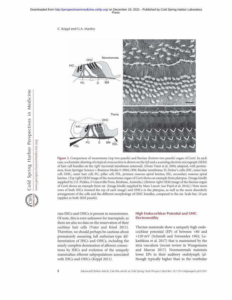
rian IHCs and OHCs is present in monotremes.Of note, this is even unknown for marsupials, asthere are also no data on the innervation of theircochlear hair cells (Vater and Kössl 2011).Therefore, we should perhaps be cautious aboutprematurely assuming full eutherian-type dif-ferentiation of IHCs and OHCs, including thenearly complete domination of afferent connec-tions by IHCs and evolution of the uniquelymammalian efferent subpopulations associatedwith IHCs and OHCs (Köppl 2011).
High Endocochlear Potential and OHCElectromotility
Therian mammals show a uniquely high endo-cochlear potential (EP) of between +80 and+120 mV (Schmidt and Fernandez 1962; Lu-kashkina et al. 2017) that is maintained by thestria vascularis (recent review in Wangemannand Marcus 2017). Nonmammals maintainlower EPs in their auditory endolymph (al-though typically higher than in the vestibular
OHC
BM
IHC
PC
Monotremata
Theria
D
OHC
BM
IHC
PSLSSLPC D
Figure 3. Comparison of monotreme (top two panels) and therian (bottom two panels) organ of Corti. In eachcase, a schematic drawing of a typical cross section is shown on the left and a scanning electronmicrograph (SEM)of hair-cell bundles on the right (tectorial membrane removed). (From Vater et al. 2004; adapted, with permis-sion, from Springer Science + Business Media © 2004.) BM, Basilar membrane; D, Deiter’s cells; IHC, inner haircell; OHC, outer hair cell; PC, pillar cell; PSL, primary osseous spiral lamina; SSL, secondary osseous spirallamina. (Top right) SEM image of the monotreme organ of Corti shows an example from platypus. (Image kindlysupplied by J.O. Pickles, © Graceville Press, Brisbane, Australia.) (Bottom right) SEM image of the therian organof Corti shows an example from rat. (Image kindly supplied by Marc Lenoir [see Pujol et al. 2016].) Note morerows of both IHCs (toward the top of each image) and OHCs in the platypus, as well as the more disorderlyarrangement of the cells and the different morphology of OHC bundles, compared to the rat. Scale bar, 10 µm(applies to both SEM panels).
C. Köppl and G.A. Manley
8 Advanced Online Article. Cite this article as Cold Spring Harb Perspect Med doi: 10.1101/cshperspect.a033241
ww
w.p
ersp
ecti
vesi
nm
edic
ine.
org
Press on December 19, 2021 - Published by Cold Spring Harbor Laboratoryhttp://perspectivesinmedicine.cshlp.org/Downloaded from

endolymph) and the tissue responsible for itsproduction lacks the multilayered structureand salient cellular specializations of the euthe-rian stria vascularis (recent review in Wilmset al. 2016). A stria vascularis has been identifiedat the gross morphological level in both mono-tremes (Pritchard 1881; Smith and Takasaka1971) and marsupials (Aitkin et al. 1979), sug-gesting an early origin in the mammalian line.Marsupials also show the uniquely high EP(Schmidt and Fernandez 1962), so it is safe toassume that they also share the cellular special-izations. The EP in monotremes has not beenmeasured, leaving it unknown just how far anystrial specializations have evolved in this group.
The ultimate cause (i.e., the selective pres-sure toward the uniquely high EP in mammals)is again somewhat speculative. Auditory sensi-tivity is compromised when the EP is experi-mentally lowered. Therefore, a gain in sensitivityis commonly cited as the reason for a high EP(e.g., Hibino et al. 2010). However, mammalsare not more sensitive than modern nonmam-mals (Wilms et al. 2016;Manley 2017a). Instead,it may be more specifically an improved sensi-tivity to higher frequencies that drove selectionfor an increased EP in both mammals and non-mammals (Wilms et al. 2016). Inmammals, thismay be additionally favored by providing an in-creased drive to OHC electromotility, their nov-el amplifying mechanism. So, when did electro-motility arise?
In auditory hair cells, two active, motile sys-tems have been identified that work to overcomeviscous damping in the fluid environment ofthe inner ear: (1) an ancestral mechanism inte-gral to the gating of transduction channels of thehair bundle, and (2) an electromotile responseunique to theOHCs ofmammals, based on largenumbers of the protein prestin in the lateral hair-cell membrane (Hudspeth 2008). The ancestryof the prestin gene has been traced back to ananion transporter, and crucial intermediatesteps for acquiring voltage sensitivity and theability to produce a motile response have beenidentified (reviews in He et al. 2014; Russell2014). Importantly, this includes the prestin var-iant based on genomic sequence data from theplatypus and expressed in cultured cells (Tan
et al. 2011). It may thus be assumed that mono-treme OHCs possess a degree of electromotility,although comparativemeasurements on the cul-tured expression system suggested that the mo-tion effected by platypus prestin is only about60% of that of eutherian prestin under compa-rable voltage stimulation (Tan et al. 2011). Fur-thermore, and important for in vivo function,prestins from both platypus and a marsupial(opossum) species show their peak sensitivityat significantly more positive membrane poten-tials than a typical eutherian prestin from gerbil(Fig. 4) (Tan et al. 2011; Liu et al. 2012). Indeed,only eutherian prestin has undergone a final,crucial modification that shifted its peak sensi-tivity closer to the natural resting potential ofOHC and abolished most of the original aniontransporter function (Liu et al. 2012). Taken to-gether, a parsimonious interpretation is that
–150 –100 –50
Gerbil
Opossum
Platypus
Non
linea
r ca
paci
tanc
e (r
elat
ive)
Membrane potential (mV)
500
Figure 4. Schematic illustration of nonlinear capaci-tance measured in cultured cells transfected withprestin derived from genomic sequence data of amonotreme (platypus), a marsupial (opossum), anda placental mammal (gerbil); after data shown in Tanet al. (2011) and Liu et al. (2012). Nonlinear capaci-tance reflects the voltage sensitivity of prestin and, forplatypus and gerbil prestin, has been shown to corre-latewith the extent of themotile response. The shadedregion indicates the approximate range of in vivo out-er hair cell (OHC) membrane potential fluctuations.Note that both platypus and opossumprestin have thepeak of their sensitivity outside this range.
Evolution of the Cochlea
Advanced Online Article. Cite this article as Cold Spring Harb Perspect Med doi: 10.1101/cshperspect.a033241 9
ww
w.p
ersp
ecti
vesi
nm
edic
ine.
org
Press on December 19, 2021 - Published by Cold Spring Harbor Laboratoryhttp://perspectivesinmedicine.cshlp.org/Downloaded from

prestin-based electromotility arose early inmammalian evolution, concurrently with thedifferentiation of IHCs and OHCs. However,its effectiveness would initially have been lowand probably still is in modern monotremesand marsupials, because of its nonoptimal volt-age activation range and lowermotile force. Thisis also consistent with the low upper frequencylimit of monotreme hearing, around 15 kHz(Gates et al. 1974; Mills and Shepherd 2001).We suggest that a gradual shift of importancefrom the plesiomorphic transduction-channel-based mechanism to the novel electromotility,simultaneous with a gradual expansion of sensi-tivity to higher frequencies, is most likely.
Loss of the Lagenar Macula and ItsConsequences for Endolymphatic Calcium
Like nonmammals, monotremes still have thevestibular lagenar macula situated at the apexof their cochlear duct (Pritchard 1881). In the-rian mammals, the lagena is lost. When exactlythis happened during mammalian evolution iscurrently difficult to pinpoint from fossil evi-dence (recent review in Schultz et al. 2017)and it may well have happened multiple timesin extinct, early branch-offs of the therian line(Fig. 1) (Hoffmann et al. 2014). What favored aloss of the lagena remains equally unclear (Man-ley 2017b). In the therian lineage, it is likely tohave happened around the time of the first fullcoiling of the therian cochlea (Luo et al. 2011). Ithas been suggested that the two events may belinked and the lagenar macula was not lost buttransformed and formed the low-frequency re-gion at the apex of the organ of Corti (Fritzschet al. 2013). This scenario seems unlikely, how-ever, as mammalian ancestors with a lagena al-most certainly already possessed low-frequencyhearing organs. In any case, the disappearanceof the vestibular lagenar macula and its associ-ated otolith must have had a profound effect onthe calcium metabolism of the auditory organ(Manley 2017b).
An otolithic membrane is typical for vestib-ular maculae that respond to linear accelera-tions. The otoliths consist of calcium salts whosesurface is in ionic exchange with the surround-
ing fluid. If the calcium concentration in thesurrounding fluids falls too low, the otoliths’surfaces erode and continue to do so until thecrystals have dissolved (Payan et al. 2002). Con-sistent with this, calcium levels in vestibular en-dolymph and also in the scalae mediae of non-mammals are at least 100 µM and often higher(Ferrary et al. 1988; Manley et al. 2004; Ghanemet al. 2008), whereas typical values for the scalamedia of therian mammals are 20–30 µM (i.e.,about one order of magnitude lower [Wange-mann and Marcus 2017]). Calcium in endo-lymph influences many aspects of the hearingprocess, from the integrity of the tectorial mem-brane (Kronester-Frei 1979) to the mechano-electrical transduction of the hair cells (Fetti-place and Ricci 2006). The final loss of otolithsbordering on the auditory scala media presum-ably caused at least a minor crisis in theriancochlear function during the Cretaceous period(Manley 2017b). Such a crisis would have drivenchanges in the mechanisms of ion homeostasis,modifications to the structure of the tectorialmembrane, and alterations to the mechanoelec-trical transduction channels. Among the uniquefeatures of the eutherian cochlea that may haveresulted from that is the collagen matrix of itstectorial membrane (Goodyear and Richardson2002). Furthermore, in eutherianmammals, cal-cium is actively pumped into scala media andthere is evidence that even partial impairment ofthis activity leads to a drop in endolymphatic[Ca2+] below a critical threshold, where trans-duction fails (Wood et al. 2004). Finally, a re-duced Ca dependence of the transduction chan-nels on eutherian cochlear hair cells has beenproposed (Peng et al. 2013, 2016). None of thesefeatures have been characterized for any mono-treme or marsupial, so it remains speculativewhich changes are truly related to the loss ofthe vestibular otolith and how long these pro-cesses may have taken.
Loss of Regenerative Capacity
The loss of regenerative capacity in themamma-lian cochlea will be briefly mentioned here, as itis having a major impact on concepts to amelio-rate human hearing loss by attempting to regen-
C. Köppl and G.A. Manley
10 Advanced Online Article. Cite this article as Cold Spring Harb Perspect Med doi: 10.1101/cshperspect.a033241
ww
w.p
ersp
ecti
vesi
nm
edic
ine.
org
Press on December 19, 2021 - Published by Cold Spring Harbor Laboratoryhttp://perspectivesinmedicine.cshlp.org/Downloaded from
erate lost hair cells. One unexpected finding ofcomparative auditory research is the remarkableability of bird cochleae to regenerate hair cellsand restore auditory function after damage byloud sounds or other insults (Cotanche 1999;Smolders 1999; Rubel et al. 2013; Ryals et al.2013). This is apparently an ongoing processthat leads to themaintenance of pristine hearingthresholds into exceptionally old age (up to 13years in European starlings [Langemann et al.1999] and up to 23 years in barn owls [Krummet al. 2017]). The ability to regenerate hair cells,inherited from ancestors and still shown in, forexample, mammalian vestibular organs (Walsheet al. 2003; Warchol 2011), has been lost in themammalian cochlea. This loss appears to belinked to the characteristic structure of the organof Corti, where also the supporting cells special-ize to an extreme degree and are integrated intothe transmission of acoustic energy—from ac-tive hair cells, for example, into the movementsof the organ of Corti. The crucial difference tobirds thus lies in the more “primitive” state ofavian cochlear support cells that are only onestep away from dividing and forming a newhair cell and a supporting cell (Brigande andHeller 2009). In mammals, the much greaterspecialization of the supporting cells (e.g., pillarcells, Deiters’s cells) probably precludes dedif-ferentiation and reentry into the cell cycle. Itseems that a gradual loss of hair cells and dete-rioration in auditory health throughout life is aprice mammals pay for the evolution of the or-gan of Corti. Current research is exploring dif-ferent ways of inducing dedifferentiation or in-troducing stem cells into the cochlea, with thehope of inserting new hair cells and thus initi-ating therapies for human hearing loss (Mittalet al. 2017).
HUMAN COCHLEAE
Amongmammals, the human cochlea shows anaverage length of ∼34 mm; the length variesamong therian mammals from 7 to 70 mm (Va-ter et al. 2004). Recently, it has become routine toexamine cochlear length before implanting co-chlear devices, which has revealed a surprisinglylarge variation in its dimensions in humans.
There are small average differences betweenthe sexes, but maximal reported differences be-tween individuals are up to 18% (Erixon et al.2008) or evenmore than one-third (36%) (Wür-fel et al. 2014). Differences in the numbers ofauditory nerve fibers can be even larger (re-viewed in Miller 1985). It is not known whethersuch length variations have an effect on the co-chlear map of frequencies (are some frequenciesmissing in short cochleae or is the space constantsmaller?).
Humans are, of course, primates that areclosely related to chimpanzees. Information onthe form and length of primate cochleae hasbeen reviewed (Coleman and Colbert 2010;Wannaprasert and Jeffery 2015), describing in-teresting changes observed in human ancestors.In primates, cochlear length correlates negative-ly with the upper frequency limits (Kirk andGosselin-Ildari 2009). The earliest primateshad shorter cochleae and good high-frequencybut probably poor low-frequency hearing. Per-haps because of a general increase in body size inthe ancestors of modern monkeys and apes, co-chlear size increased, leading to improved low-frequency hearing (Coleman and Boyer 2012).In the lineage leading to humans and chimpan-zees, cochlear size again increased significantly,probably enabling even better low-frequencyhearing (Braga et al. 2015). It is difficult toknow to what extent these changes were simplya result of increasing animal size, and to whatextent they may have been driven by selectionfor cochleaemore appropriate for the processingof low-frequency communication signals. Al-though there is some evidence for improvedlow-frequency—relative to high-frequency—se-lectivity in human cochleae (Manley and vanDijk 2016), the basic question of the frequencyselectivity of the human cochlea is still a highlycontroversial issue.
CONCLUDING REMARKS
The mammalian cochlea evolved many of itscharacteristic features early, and these sharedtraits were inherited by all living representatives.Later features, such as the spiral shape, loss of thelagena macula, and integration into a bony en-
Evolution of the Cochlea
Advanced Online Article. Cite this article as Cold Spring Harb Perspect Med doi: 10.1101/cshperspect.a033241 11
ww
w.p
ersp
ecti
vesi
nm
edic
ine.
org
Press on December 19, 2021 - Published by Cold Spring Harbor Laboratoryhttp://perspectivesinmedicine.cshlp.org/Downloaded from

closure evolved only in therians, showed greaterspecialization toward higher frequencies and theuse of prestin as an active hair-cell motor. Overeons of time, these specializations led to the lossof the ability to regenerate hair cells, leading toprogressive hearing loss in long-lived speciessuch as humans.
ACKNOWLEDGMENTS
The authors gratefully acknowledge support bythe Deutsche Forschungsgemeinschaft (DFG)over many years, currently through the Clusterof Excellence “Hearing4All” DFG-EXI 1077;and DFG KO 1143/14-1 and 1143/15-1 to C.K.Further support by the Bundesministerium fürBildung und Forschung (BMBF) “US-GermanCollaboration in Computational Neuroscience”program, FEPAS 01GQ1505B to C.K.
REFERENCES
Aitkin L, Johnstone BM. 1972. Middle-ear function in amonotreme: The echidna (Tachyglossus aculeatus). JExp Zool 180: 245–250.
Aitkin LM, Gates GR, Kenyon E. 1979. Some peripheralauditory characteristics of the marsupial brush-tailedpossum, Trichosurus vulpecula. J Exp Zool A Comp ExpBiol 209: 317–322.
Alexander G. 1904. Entwicklung und Bau des inneren Ge-hörorgans von Echidna aculeata [Development andmor-phology of the auditory inner ear of Echidna aculeata].Denkschr Med-naturwiss Ges Jena 6: 3–118.
Allin EF, Hopson JA. 1992. Evolution of the auditorysystem in Synapsida (“mammal-like reptiles”) as seen inthe fossil record. In The evolutionary biology of hear-ing (ed. Webster DB, et al.), pp. 587–614. Springer,New York.
Braga J, Loubess JM, Descouens D, Dumoncel J, ThackerayJH, Kahn JL, de Beer F, Riberon A, Hoffman K, Bala-resque P, et al. 2015. Disproportionate cochlear lengthin genus Homo shows a high phylogenetic signal duringapes’ hearing evolution. PLoS ONE 10: e0127780.
Brigande JV, Heller S. 2009. Quo vadis, hair cell regenera-tion? Nat Neurosci 12: 679–685.
Brown CH, May BJ. 2005. Comparative mammalian soundlocalization. In Sound source localization (ed. Popper AN,Fay RR), pp. 124–178. Springer, New York.
Coleman MN, Boyer DM. 2012. Inner ear evolution in pri-mates through the Cenozoic: Implications for the evolu-tion of hearing. Anat Rec 295: 615–631.
Coleman MN, Colbert MW. 2010. Correlations between au-ditory structures and hearing sensitivity in non-humanprimates. J Morphol 271: 511–532.
Cotanche DA. 1999. Structural recovery from sound andaminoglycoside damage in the avian cochlea.Audiol Neu-rootol 4: 271–285.
Ekdale EG. 2016. The ear of mammals: From monotremesto humans. In Evolution of the vertebrate ear: Evidencefrom the fossil record (ed. Clack JA, et al.), pp. 175–206.Springer, Basel, Switzerland.
Erixon E, Högstorp H, Wadin K, Rask-Andersen H. 2008.Variational anatomy of the human cochlea: Implicationsfor cochlear implantation. Otol Neurotol 30: 14–22.
Ferrary E, Tran Ba Huy P, Roinel N, Bernard C, Amiel C.1988. Calcium and the inner ear fluids. Acta OtolaryngolSuppl 460: 13–17.
Fettiplace R, Ricci AJ. 2006. Mechanoelectrical transductionin auditory hair cells. In Vertebrate hair cells (ed. EatockRA, et al.), pp. 154–203. Springer, New York.
Fritzsch B, Pan N, Jahan I, Duncan JS, Kopecky BJ, ElliottKL, Kersigo J, Yang T. 2013. Evolution and developmentof the tetrapod auditory system: An organ of Corti-centricperspective. Evol Dev 15: 63–79.
Gates GR, Saunders J, Bock GR, Aitkin L, Elliott MA. 1974.Peripheral auditory function in the platypus. Ornitho-rhynchus anatinus. J Acoust Soc Am 56: 152–156.
Ghanem TA, Breneman KD, Rabbitt RD, Brown M. 2008.Ionic composition of endolymph and perilymph in theinner ear of the oyster toadfish, Opsanus tau. Biol Bull214: 83–90.
Goodyear RJ, Richardson GP. 2002. Extracellular matricesassociated with the apical surfaces of sensory epithelia inthe inner ear: Molecular and structural diversity. J Neuro-biol 53: 212–227.
Grothe B, Pecka M. 2014. The natural history of sound lo-calization in mammals—A story of neuronal inhibition.Front Neural Circuits 8: 116.
HanG,Mao F, Bi S,Wang Y,Meng J. 2017. A Jurassic glidingeuharamiyidan mammal with an ear of five auditorybones. Nature 551: 451–456.
He DZ, Lovas S, Ai Y, Li Y, Beisel KW. 2014. Prestin at year14: Progress and prospect. Hear Res 311: 25–35.
Heffner RS, Heffner HE. 1982. Hearing in the elephant (El-ephas maximus): Absolute sensitivity, frequency discrim-ination, and sound localization. J Comp Physiol Psychol96: 926–944.
Heffner RS, Koay G, Heffner HE. 2001. Audiograms of fivespecies of rodents: Implications for the evolution of hear-ing and the perception of pitch. Hearing Res 157: 138–152.
Hibino H, Nin F, Tsuzuki C, Kurachi Y. 2010. How is thehighly positive endocochlear potential formed? The spe-cific architecture of the stria vascularis and the roles of theion-transport apparatus. Pflugers Arch 459: 521–533.
Hoffmann S, O’Connor PM, Kirk EC,Wible JR, Krause DW.2014. Endocranial and inner ear morphology of Vintanasertichi (Mammalia, Gondwanatheria) from the Late Cre-taceous of Madagascar. J Vert Paleontol 34: 110–137.
Hudspeth AJ. 2008. Making an effort to listen: Mechanicalamplification in the ear. Neuron 59: 530–545.
Kirk EC, Gosselin-Ildari AD. 2009. Cochlear labyrinth vol-ume and hearing abilities in primates.Anat Rec 292: 765–776.
C. Köppl and G.A. Manley
12 Advanced Online Article. Cite this article as Cold Spring Harb Perspect Med doi: 10.1101/cshperspect.a033241
ww
w.p
ersp
ecti
vesi
nm
edic
ine.
org
Press on December 19, 2021 - Published by Cold Spring Harbor Laboratoryhttp://perspectivesinmedicine.cshlp.org/Downloaded from

Kitazawa T, Takechi M, Hirasawa T, Adachi N, Narboux-Neme N, Kume H, Maeda K, Hirai T, Miyagawa-TomitaS, Kurihara Y, et al. 2015. Developmental genetic basesbehind the independent origin of the tympanic mem-brane in mammals and diapsids. Nat Commun 6: 6853.
Köppl C. 2011. Evolution of the octavolateral efferent system.In Auditory and vestibular efferents (ed. Ryugo D, et al.),pp. 217–259. Springer, New York.
Kronester-Frei A. 1979. The effect of changes in endolym-phatic ion concentrations on the tectorial membrane.Hearing Res 1: 81–94.
Krumm B, Klump G, Köppl C, Langemann U. 2017. Barnowls have ageless ears. Proc Biol Sci 284: 20171584.
Ladhams A, Pickles JO. 1996. Morphology of the mono-treme organ of Corti and macula lagena. J Comp Neurol366: 335–347.
Langemann U, Hamann I, Friebe A. 1999. A behavioral testof presbycusis in the bird auditory system. Hearing Res137: 68–76.
Liu Z, Li GH, Huang JF, Murphy RW, Shi P. 2012. Hearingaid for vertebrates via multiple episodic adaptive eventson prestin genes. Mol Biol Evol 29: 2187–2198.
Lukashkina VA, Levic S, Lukashkin AN, Strenzke N, RussellIJ. 2017. A connexin30 mutation rescues hearing andreveals roles for gap junctions in cochlear amplificationand micromechanics. Nat Commun 8: 14530.
Luo ZX. 2007. Transformation and diversification in earlymammal evolution. Nature 450: 1011–1019.
Luo ZX, Ruf I, Schultz JA, Martin TA. 2011. Fossil evidenceon evolution of inner ear cochlea in Jurassic mammals.Proc R Soc B 278: 28–34.
Luo ZX, Schultz JA, Ekdale EG. 2016. Evolution of the mid-dle and inner ears of mammaliaforms: The approach tomammals. In Evolution of the vertebrate ear: Evidencefrom the fossil record (ed. Clack JA, et al.), pp. 139–174.Springer, Basel, Switzerland.
Manley GA. 1972. Frequency response of the middle ear ofgeckos. J Comp Physiol 81: 251–258.
Manley GA. 2010. An evolutionary perspective on middleears. Hearing Res 263: 3–8.
Manley GA. 2012. Evolutionary paths to mammalian co-chleae. J Assoc Res Otolaryngol 13: 733–743.
Manley GA. 2013. Mosaic evolution of the mammalian au-ditory periphery. In Basic aspects of hearing (ed.Moore B,et al.), pp. 3–9. Springer, New York.
Manley GA. 2016. The foundations of high-frequency hear-ing in earlymammals. JMammEvol doi: 10.1007/s10914-016-9379-0.
ManleyGA. 2017a. Comparative auditory neuroscience:Un-derstanding the evolution and function of ears. J Assoc ResOtolaryngol 18: 1–24.
Manley GA. 2017b. The mammalian Cretaceous cochlearrevolution. Hearing Res 352: 23–29.
ManleyGA, Johnstone BM. 1974.Middle-ear function in theguinea pig. J Acoust Soc Am 56: 571–576.
Manley GA, van Dijk P. 2016. Frequency selectivity of thehuman cochlea: Suppression tuning of spontaneous oto-acoustic emissions. Hear Res 336: 53–62.
Manley GA, Irvine DRF, Johnston BM. 1972. Frequencyresponse of bat tympanic membrane. Nature 237: 112–113.
Manley GA, Sienknecht UJ, Köppl C. 2004. Calcium mod-ulates the frequency and amplitude of spontaneous oto-acoustic emissions in the bobtail skink. J Neurophysiol 92:2685–2693.
Manley GA, Lukashkin AN, Simoes P, Burwood GWS, Rus-sell IJ. 2018. The mammalian ear: Physics and the prin-ciples of evolution. Acoust Today 14: 8–16.
Martin T, Marugán-Lobón J, Vullo R, Martin-Abad H, LuoZX, Buscalioni AD. 2015. A Cretaceous eutriconodontand integument evolution in early mammals. Nature526: 380–384.
MasonMJ. 2013.Ofmice,moles and guinea pigs: Functionalmorphology of the middle ear in living mammals. HearRes 301: 4–18.
Miller MR. 1985. Quantitative studies of auditory hair cellsand nerves in lizards. J Comp Neurol 232: 1–24.
Mills DM, ShepherdK. 2001. Distortion product otoacousticemission and auditory brainstem responses in the echid-na (Tachyglossus aculeatus). J Assoc Res Otolaryngol 2:130–146.
Mittal R, Nguyen D, Patel AP, Debs LH, Mittal J, YanD, Eshraghi AA, Van De Water TR, Liu XZ. 2017.Recent advancements in the regeneration of auditoryhair cells and hearing restoration. Front Mol Neurosci10: 236.
Narins PM, Willi UB. 2012. The golden mole middle ear: Asensor for airborne and substrate-borne vibrations. InFrontiers in sensing (ed. Barth FG, et al.), pp. 275–286.Springer, Vienna.
Neugebauer DC, Thurm U. 1984. Chemical dissection ofstereovilli from fish inner ear reveals differences fromintestinal microvilli. J Neurocytol 13: 797–808.
Novacek MJ. 1977. Aspects of the problem of variation, or-igin and evolution of the eutherian auditory bulla.MammRev 7: 131–149.
Payan P, Borelli G, Priouzeau F, De Pontual H, Boeuf G,Mayer-Gostan N. 2002. Otolith growth in trout Onco-rhynchus mykiss: Supply of Ca2+ and Sr2+ to the saccularendolymph. J Exp Biol 205: 2687–2695.
Peng AW, Effertz T, Ricci AJ. 2013. Adaptation of mamma-lian auditory hair cell mechanotransduction is indepen-dent of calcium entry. Neuron 80: 960–972.
Peng AW, Gnanasambandam R, Sachs F, Ricci AJ. 2016.Adaptation independent modulation of auditory haircell mechanotransduction channel open probability im-plicates a role for the lipid bilayer. J Neurosci 36: 2945–2956.
Pietsch M, Aguirre Dávila L, Erfurt P, Avci E, Lenarz T, KralA. 2017. Spiral form of the human cochlea results fromspatial constraints. Sci Rep 7: 7500.
Pritchard U. 1881. The cochlea of the Ornithorhynchusplatypus compared with that of ordinary mammals andof birds. Proc R Soc Lond 31: 206–211.
Pujol R, et al. 2016. Cochlea: Overview. In Journey into theworld of hearing. NeurOreille, Montpellier, France, www.cochlea.eu/en/cochlea.
Evolution of the Cochlea
Advanced Online Article. Cite this article as Cold Spring Harb Perspect Med doi: 10.1101/cshperspect.a033241 13
ww
w.p
ersp
ecti
vesi
nm
edic
ine.
org
Press on December 19, 2021 - Published by Cold Spring Harbor Laboratoryhttp://perspectivesinmedicine.cshlp.org/Downloaded from

Rubel EW, Furrer SA, Stone JS. 2013. A brief history of haircell regeneration research and speculations on the future.Hearing Res 297: 42–51.
Ruggero MA, Temchin AN. 2002. The roles of the external,middle, and inner ears in determining the bandwidth ofhearing. Proc Natl Acad Sci 99: 13206–13210.
Russell IJ. 2014. Roles for prestin in harnessing the basilarmembrane to the organ of Corti. In Insights from com-parative hearing research (ed. Köppl C, et al.), pp. 37–67.Springer, New York.
Ryals BM, Dent ML, Dooling RJ. 2013. Return of functionafter hair cell regeneration. Hearing Res 297: 113–120.
Schmidt RS, Fernandez C. 1962. Labyrinthine DC potentialsin representative vertebrates. J Cell Comp Physiol 59: 311–322.
Schultz JA, Zeller U, Luo ZX. 2017. Inner ear labyrinth anat-omy of monotremes and implications for mammalianinner ear evolution. J Morphol 278: 236–263.
Smith CA, Takasaka T. 1971. Auditory receptor organs ofreptiles, birds, and mammals. Contr Sens Physiol 5: 129–178.
Smolders JWT. 1999. Functional recovery in the avian earafter hair cell regeneration. Audiol Neurootol 4: 286–302.
Takechi M, Kuratani S. 2010. History of studies on mamma-lian middle ear evolution: A comparative morphologicaland developmental biology perspective. J Exp Zool B MolDev Evol 314B: 417–433.
Tan X, Pecka JL, Tang J, Okoruwa OE, Zhang Q, Beisel KW,He DZZ. 2011. From zebrafish to mammal: Functionalevolution of prestin, the motor protein of cochlear outerhair cells. J Neurophysiol 105: 36–44.
Vater M, Kössl M. 2011. Comparative aspects of cochlearfunctional organization in mammals. Hearing Res 273:89–99.
Vater M, Meng J, Fox RC. 2004. Hearing organ evolutionand specialization: Early and later mammals. In Evolutionof the vertebrate auditory system (ed. Manley GA, et al.),pp. 256–288. Springer, New York.
Walshe P, Walsh M, McConn Walsh R. 2003. Hair cell re-generation in the inner ear: A review.Clin Otolaryngol 28:5–13.
Wangemann P,Marcus DC. 2017. Ion and fluid homeostasisin the cochlea. In Understanding the cochlea (ed. ManleyGA, et al.), pp. 253–286. Springer, New York.
Wannaprasert T, Jeffery N. 2015. Variations of mammaliancochlear shape in relation to hearing frequency and skullsize. Trop Nat Hist 15: 41–54.
Warchol ME. 2011. Sensory regeneration in the vertebrateinner ear: Differences at the levels of cells and species.Hearing Res 273: 72–79.
Wilms V, Köppl C, Söffgen C, Hartmann AM, NothwangHG. 2016. Molecular bases of K+ secretory cells in theinner ear: Shared and distinct features between birdsand mammals. Sci Rep 6: 34203.
Wood JD, Muchinsky SJ, Filoteo AG, Penniston JT, TempelBL. 2004. Low endolymph calcium concentrations indeafwaddler2J mice suggest that PMCA2 contributes toendolymph calciummaintenance. J Assoc Res Otolaryngol5: 99–110.
Würfel W, Lanfermann H, Lenarz T, Majdani O. 2014. Co-chlear length determination using cone beam computedtomography in a clinical setting. Hear Res 316: 65–72.
C. Köppl and G.A. Manley
14 Advanced Online Article. Cite this article as Cold Spring Harb Perspect Med doi: 10.1101/cshperspect.a033241
ww
w.p
ersp
ecti
vesi
nm
edic
ine.
org
Press on December 19, 2021 - Published by Cold Spring Harbor Laboratoryhttp://perspectivesinmedicine.cshlp.org/Downloaded from

published online September 4, 2018Cold Spring Harb Perspect Med Christine Köppl and Geoffrey A. Manley A Functional Perspective on the Evolution of the Cochlea
Subject Collection Function and Dysfunction of the Cochlea
Etiologies and MechanismsHidden Hearing Loss: A Disorder with Multiple
et al.David C. Kohrman, Guoqiang Wan, Luis Cassinotti,
Convergent Extension to Planar PolarityDevelopment and Patterning of the Cochlea: From
Mireille Montcouquiol and Matthew W. Kelley
DysfunctionHair Cell Afferent Synapses: Function and
Mustapha, et al.Stuart L. Johnson, Saaid Safieddine, Mirna
FunctionHair-Bundle Links: Genetics as the Gateway to
Guy P. Richardson and Christine Petit
Active Biomechanics of Sensory Hair BundlesDolores Bozovic
StrategiesOtotoxicity: Mechanisms and Otoprotective Aminoglycoside- and Cisplatin-Induced
Corné J. Kros and Peter S. Steyger
and FunctionsThe Tectorial Membrane: Mechanical Properties
M. FreemanJonathan B. Sellon, Roozbeh Ghaffari and Dennis
Inner Ear Hair CellsFunction and Dysfunction of TMC Channels in
David P. Corey, Nurunisa Akyuz and Jeffrey R. Holt
The Epidemiology of DeafnessAbraham M. Sheffield and Richard J.H. Smith
Cochlear Gene TherapyLawrence Lustig and Omar Akil
Toward the Optical Cochlear Implant
MoserTobias Dombrowski, Vladan Rankovic and Tobias
Age-Related Hearing LossMichael R. Bowl and Sally J. Dawson
Outer Hair Cells and ElectromotilityJonathan Ashmore
FunctionDevelopment and Maintenance of Cochlear Inner Ear Connexin Channels: Roles in
Fabio Mammano
Sensory Cells of the Inner EarInteractions between Macrophages and the
Mark E. WarcholCochleaA Functional Perspective on the Evolution of the
Christine Köppl and Geoffrey A. Manley
http://perspectivesinmedicine.cshlp.org/cgi/collection/ For additional articles in this collection, see
Copyright © 2018 Cold Spring Harbor Laboratory Press; all rights reserved
Press on December 19, 2021 - Published by Cold Spring Harbor Laboratoryhttp://perspectivesinmedicine.cshlp.org/Downloaded from