a model for membrane potential and intracellular ion ... et al. membrane... · 1 a model for...
TRANSCRIPT
1
A model for membrane potential and intracellular ion distribution
A.K. Khitrin1, K.A. Khitrin2, and M.A. Model3*
1Department of Chemistry and Biochemistry, Kent State University, Kent, OH 44242, USA
2Department of Physics, Kent State University, Kent, OH 44242, USA
3Department of Biological Sciences, Kent State University, Kent, OH 44242, USA,
tel. 330-672-0774, [email protected]
*Corresponding author
Keywords: membrane potential, surface potential, Donnan potential, Gouy-Chapman theory
Published in Chemistry and Physics of Lipids 184 (2014) 76–81
Abstract
Most cells carry a negative electric charge. It produces a potential difference across the membrane,
which regulates voltage-sensitive ion transport and ATP synthesis in mitochondria. The negative
charge comes partly from an excess of negative ions in the cell interior (Donnan potential) and
partly from ionized groups on the membrane (surface potential). In this work we propose some
important modifications to the existing theory of membrane potential. First, we calculate the
concentration profile of intracellular positive ions and derive a simple equation to assess the
submembrane depletion of positive ions that gives rise to the Donnan potential. The extent of
depletion varies with potential, which may provide a regulatory mechanism for ion pumps and
channels. Next we consider the surface component of the potential and note that the standard Gouy-
Chapman theory has been developed for planar membranes, whereas real cell membranes have a
closed geometry. In this case, charges on the membrane surface are not expected to generate fields
extending into the cell interior. This fact calls for reinterpretation of some theoretical points as
well as experimental data. In particular, the experimentally demonstrated electrostatic attraction
between cationic proteins and the negative membrane must now be explained without invoking
intracellular fields, and we suggest a new mechanism that can account for this interaction.

2
INTRODUCTION
Most healthy cells and mitochondria are characterized by a negative resting potential. The process
mainly responsible for its generation is intracellular accumulation of K+ (by the Na+,K+ pump)
combined with high permeability for K+; this allows some ions to exit the cell down the
concentration gradient, leaving behind an unbalanced negative charge. This is the classic Donnan
mechanism, which is considered in detail in physiology textbooks (6, 48). Additionally, the
stoichiometry of the Na+,K+ pump is such that bringing two K+ ions into the cell is coupled with
extrusion of three ions of Na+. Direct electrogenic contribution of the pump to the membrane
potential is usually small (47, 45) but can be significant or even dominant in some cell types (2,
18, 25). The large (150-200 mV) negative potential of mitochondria is generated exclusively by
pumping out protons by the respiratory chain (35).
Regardless of the exact mechanism of potential generation, a deficit of positive ions is created
inside a cell or an organelle. In a typical animal cell, the total concentration of charges is on the
order of 300 mM. Negative charges are mostly associated with proteins and nucleic acids; the
contribution of Cl- can range from 4 to 60 mM, depending on the cell origin (7). Positive charges
are mostly represented by freely diffusible K+ and, sometimes, by Na+; the ion composition of
mitochondrial matrix is similar to that of the cytosol (1, 44). It is estimated that an excess of
negative charges by about 3 M should be sufficient to create a 90 mV difference across the
membrane (6).
The other contribution to the total potential comes from fixed charges on the membrane. These
charges are associated with anionic phospholipids that are present both on the inner and outer sides
of the membrane (9). Surface charges are efficiently screened by the ions present in the aqueous
media but can produce local fields extending about a Debye distance (on the order of a nanometer)
into the aqueous phase. Quantitatively, this field is described by the Gouy-Chapman theory (17,
31); more detailed models include dipolar fields from zwitterionic lipids and explicit polarization
of water molecules (7). Applications of the surface potential theory to biological membranes have
been extensive (23, 30, 31, 34, 36, 45, 50); in particular, the field originating from surface charges
has been implicated in binding of positively charged cytoplasmic proteins to the inner leaflet of
the plasma membrane (19, 37).
In the present paper we propose some modifications to the existing theory of membrane potential.
We note that whether the cell potential has a Donnan or a surface origin, the entire charge that
generates the transmembrane potential difference is localized to a nanometer-deep strip under the
membrane (in the case of Donnan potential, this layer has a reduced concentration of positive ions).
This effect has been recognized before (26), but here we present a simple theoretical model to
describe the concentration profile of intracellular charges. Next, we reexamine the origin of the
surface potential. The standard treatment of surface charges is based on the assumption that the
entire membrane is a plane sheet. In reality, biological membranes have a closed geometry, and
we show that this fact is expected to have important consequences for electric phenomena: surface
charges, on the average, are not supposed to generate internal fields. This calls for reassessment of
a large body of experimental data; in particular, the new model must be reconciled with the
evidence of electrostatic attraction between positively charged cytoplasmic proteins and the
negative membrane. We thus suggest an alternative general mechanism of protein-membrane
binding based on minimization of electric energy of the system.

3
THEORY AND DISCUSSION
1. Concentration profiles of intracellular charges. We assume for now that negative charges (e.g.,
anionic proteins) are fixed and uniformly distributed throughout the bulk of the cell. Monovalent
positive charges, on the other hand, are mobile and present at a slightly lower net concentration.
As with any conductor, an unbalanced charge should be confined to a thin surface layer (which, in
our case, is the space immediately under the membrane), so that the potential deep inside the cell
would be constant. The concentration profile for positive ions can be found from the balance
between the diffusion flux and the flux created by electric field (Fig. 1):
Figure 1. The balance between diffusion and electromotive fluxes in a sub-membrane layer.
Membrane is shown by the shaded area and the ion concentration profile by the solid line.
Nonuniform distribution of ions creates two opposite fluxes, electrostatic and diffusional; the
balance between these fluxes at any point (indicated by the dashed line) determines the equilibrium
concentration profile.
-D (dn/dr) + nµeE = 0 (1)
Here D is the diffusion coefficient, n is the concentration of positive ions (number per m3), r is the
distance along a normal to the membrane, µ is the mobility, e is the electron charge, and E is the
electric field created by nonuniform distribution of charges. If we assume that a cell has a spherical
or a flat shape, the electric field E can be calculated by applying the Gauss theorem:
E(r) = σ/(εε0) = (εε0)-1𝑒 ∫ (𝑛 − 𝑛0 )d𝑟′
𝑟
−∞, (2)

4
where σ(Cm-2) is the charge density per membrane area, ε and ε0 are the relative and absolute
dielectric permittivity, and n0 is the concentration of negative ions. Only the total charge on the
left of the dashed line in Fig. 1 contributes to the electric field at the position of the line. By
combining Eq. 1, Eq. 2, and the Einstein-Smoluchowski relation: D = µkT (where k is the
Boltzmann constant and T is the absolute temperature), one obtains the equation
dn/dr = {e2/(εε0kT)}n ∫ (𝑛 − 𝑛0 )d𝑟′𝑟
−∞. (3)
By introducing the relative concentration c = n/n0 and the characteristic Debye-Hückel length
λ = {εε0kT/(e2n0)}1/2, (4)
Eq. 3 can be rewritten as
dc/dr = λ-2 c ∫ (𝑐 − 1)d𝑟′𝑟
−∞. (5)
For T = 300K, ε = 60, and n0 corresponding to 0.1M, the Debye-Hückel length is about 1 nm.
Finally, in terms of the dimensionless coordinate x = r/λ, Eq. 5 assumes the form
dc/dx = c ∫ (𝑐 − 1)d𝑥′𝑥
−∞. (6)
This integral-differential equation describes the concentration profile resulting from the balance
between the diffusion flux and the flux created by electric field. Because direct numerical solution
of Eq. 6 is computationally inefficient, it was solved by converting it to a second-order differential
equation:
d2(lnc)/dx2 = c – 1. (7)
The soliton-like solution describes a transition from c = 1 (at x → -∞) to c = 0 at large x (Fig. 2a).
The solution can be shifted by an arbitrary constant along the x-axis; this shift represents one of
the integration constants. In Fig. 2a, zero value of x was arbitrarily chosen at a point where c =
0.5. For practical calculations, the following empirical function was found to give an accurate
approximation to the solution:
c(x) = 0.5 tanh (0.0021236 - 0.632263 x - 0.0642222 x2 - 0.0108275 x3) + 0.5 (8)
5
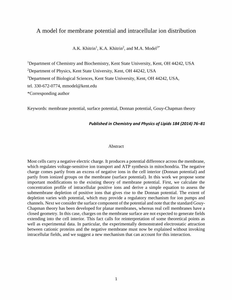
Figure 2. (a) Solution of Eq. 6 for the concentration profile. The membrane location at different x0
results in different membrane potentials. (b) The membrane potential as a function of x0.
To find the position of the membrane boundary x0 we relate our solution to the cell potential Δφ.
In doing so we assume that most of the potential drop Δφ occurs within the membrane; the rationale
for this will be given later in the text. In this case we can estimate Δφ as the product of E(r0) found
from Eq. (2) and the membrane thickness l. Relative dielectric permittivity ε of the electrolyte
should now be replaced by that of the membrane εm.
Δφ = l (εmε0)-1𝑒 ∫ (𝑛 − 𝑛0 )𝑑𝑟
𝑟0
−∞. (9)
After conversion to dimensionless units, as in Eq. 6, we have
Δφ = φ0 ∫ (𝑐 − 1)𝑑𝑥𝑥0
−∞, φ0 = γ (ε/εm) (kT/e), (10)
where γ = l/λ is the dimensionless thickness of the membrane, x0 is the position of the membrane
boundary and c is the solution of Eq. 6 (Fig. 2a). Estimates of the parameters in Eq. 9 give kT/e =
26 mV at T = 300K, and φ0 ≈ 1 V for γ = 3 and ε/εm = 15. Fig. 2b shows the potential inside the
cell in units of φ0. Once x0 is determined from the data in Fig. 2b for a given Δφ, the concentration
of positive ions at the membrane boundary cm can be found from Fig. 2a. The dependence of cm
on Δφ is shown in Fig. 3. Note that cm(Δφ) describes the concentration at the inner membrane
surface for any monovalent positive ion or their sum. Concentration profile c(x) can also be viewed
as an equilibrium Boltzmann distribution in a self-consistent potential. Therefore, if a divalent ion
is present at a concentration small enough not to affect the potential, its relative concentration c2
can be estimated as c2 = c2. The concentration of a divalent positive ion at the membrane boundary
is shown in Fig. 3 by a dashed line.

6
Figure 3. Concentration of monovalent (solid) and divalent (dashed line) ions at the membrane as
a function of the cell potential.
The described model predicts a relatively small degree of depletion. Its magnitude can be estimated
at ≈ 5% for a typical cell membrane potential of -60 mV and ≈ 15% for a three times larger
mitochondrial potential. The effect for divalent cations, such as calcium, will be approximately
twice as large (for small depletions). It needs to be recognized though that our model does not take
into account the discreet nature of fixed charges. Indeed, discreet negative charges are likely to
sequester a fraction of mobile positive ions, thus reducing their effective concentration. As a result,
a greater relative amount of sub-membrane depletion will be needed to achieve the same potential.
Quantitative assessment of this effect would require detailed knowledge of the distribution of
negative charges, including dipolar effects (7), but, qualitatively, one can say that calculations
based on the continuous model are likely to underestimate the effect. Since the extent of depletion
is potential-dependent, it would be tempting to speculate that it might provide a stabilizing
feedback, especially for mitochondria, where large changes in the potential are observed during
cell growth, differentiation, motility, cancerous transformation, calcium signaling, excitotoxicity
and apoptosis (12, 43).
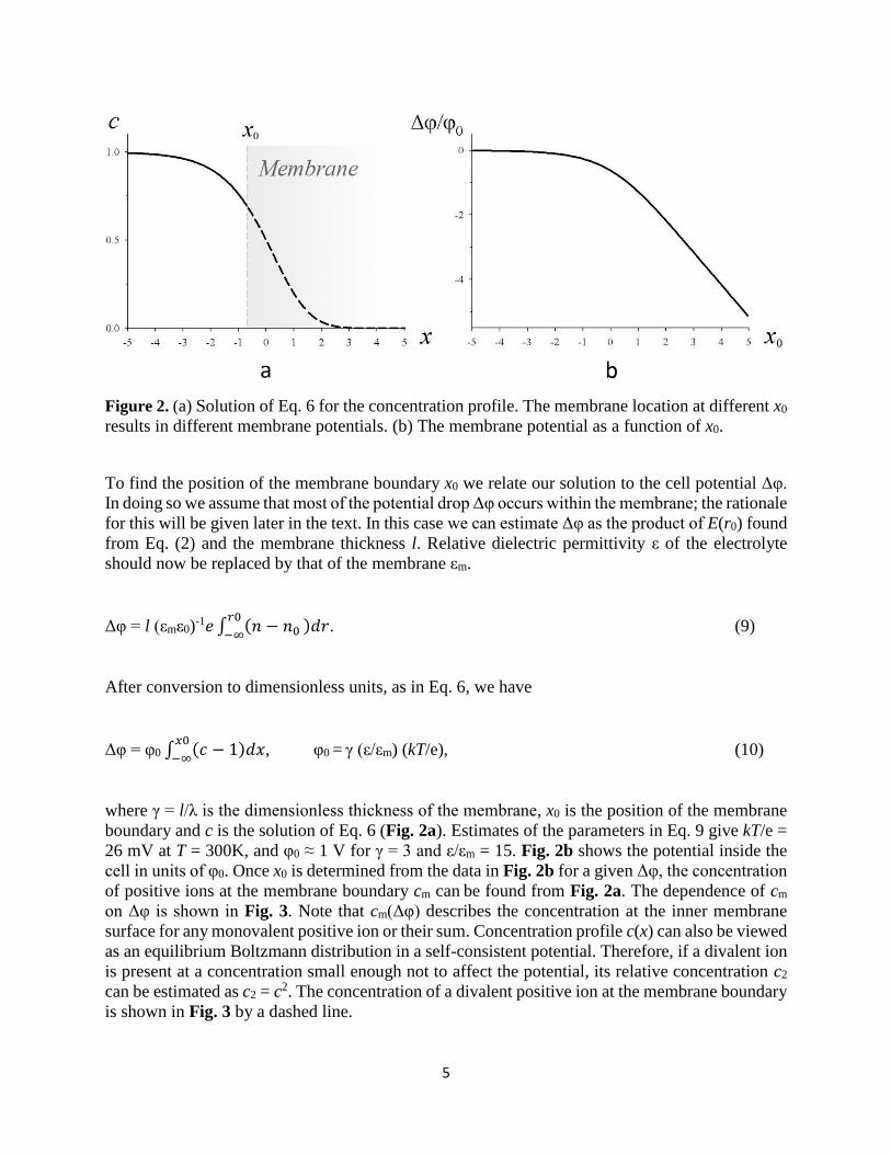
2. Qualitative features of the potential profile. The total potential difference between the cell
interior and the outside solution (the one that can be measured with electrodes) comprises three
components: the voltage drop inside the membrane φm and across the two regions on both sides
of the membrane, φin and φout (Fig. 4). First, consider the potential created by fixed membrane
charges (Fig. 4a). In the literature, the inner potential difference φinis often shown positive
(dashed line in Fig. 4a), as resulting from negative charges on the inner side of the membrane.
Such conclusion is derived from the Gouy-Chapman model for a flat charged sheet immersed in a
neutral electrolyte. Since the electric field is negligible at distances larger than Debye length, it
must have been assumed that closing the membrane would not affect the result. We believe,
however, that this is not so. Consider a spherical shell carrying uniformly distributed surface
charges but without any electrolyte inside. According to the Gauss theorem, the electric field inside
is zero. Now add an electrically neutral electrolyte. From the requirement of minimum energy of
the system it follows that the electrolyte must remain unpolarized. Indeed, suppose that a
spherically symmetric perturbation of the charge density inside the shell has developed. It will not

7
change the electrostatic energy density in the membrane and outside the cell since that depends
only on the total charge inside the cell. However, the region with non-zero electric field will
increase the electrostatic energy within the cell. Additionally, a non-uniform distribution of ions
will increase the free energy associated with mixing. For these reasons we conclude that
membrane-bound charges cannot polarize intracellular electrolyte and the potential profile would
be more accurately represented by the solid line in Fig. 4a.
The above reasoning does not apply to a flat membrane, in which case it is possible to show that
formation of double electric layers on both sides of the membrane actually reduces energy. The
ultimate reason for the difference is that, for a closed geometry, the condition of electrolyte
neutrality (charge conservation) becomes more restrictive.
Because the spherical membrane is positioned outside its inner surface but inside its outer surface,
only inner charges will create the potential within the membrane. This again is different from the
standard treatment, which assumes that the surface-related part of φm is determined by the
difference between surface charge densities inside and outside. The reasoning based on the Gauss
theorem compels us to conclude that outside charges play no role in the generation of φm.
This, however, brings up an interesting question. It is known that certain treatments can eliminate
the membrane potential; ionophores, metabolic inhibitors or the opening of the mitochondrial
permeability transition pore dissipate the potential across the inner mitochondrial membrane (12,
35, 38, 42), and high extracellular potassium does the same to the plasma membrane (12, 28, 39).
This fact presents no difficulty within the standard model because only the difference in surface
charges would be responsible for any residual potential; thus, if the charge densities on both
surfaces are approximately equal, then complete depolarization is possible. But for a spherical cell,
dissipation of the Donnan potential is expected to leave the contribution from interior surface
charges intact (the above-mentioned treatments are not expected to change the surface potential
(16)). One possible explanation to this fact is that the actual inner surface charge density is smaller
than is commonly thought. The cytosol is an extremely crowded space (41), and the dissociation
constants of phosphate groups of lipids facing the cell interior may be different from those in a
dilute buffer.
So far, we have only been considering the potential created by surface charges. A complete
description of the membrane potential must include the effects of unbalanced negative charges in
the bulk of the cell. As shown earlier, these uncompensated bulk charges are expected to be limited
to a thin sub-membrane layer and to produce a potential that is shown qualitatively in Fig. 4b. The
depletion-related φin is at least an order of magnitude smaller than φm for the following two
reasons. First, submembrane electric fields extend only over the distance of about the Debye
length, which is smaller than the 3-5 nm width of the membrane (14, 45). Second, the relative
dielectric permittivity of the aqueous solution ε is much larger than that of the membrane εm ≈ 2-
5 (14, 31, 45), making the electric field inside the membrane correspondingly stronger (see also
(26)). These considerations apply only to the inner potential. The outer potential φout is reported
to reach 15-30 mV on the plasma membrane (10, 30).
8
Figure 4. Potential profiles created by (a) immobile negative charges attached to the membrane
surfaces; the dashed line shows the profile according to the open membrane model (b) sub-
membrane depletion of positive mobile ions, and (c) the sum of the contributions in (a) and (b).
See text for additional explanations.
3. Hypothesis for the mechanism of protein-membrane interactions. We have argued that the
membrane surface creates no electric field in the cell interior. At the same time, there is substantial
experimental evidence in favor of nonspecific electrostatic attraction between acidic phospholipids
and positively charged intracellular proteins (19, 29, 49). This apparent paradox needs to be
addressed. First, we note that the statement about the surface potential being zero inside requires
some refinement: it applies only to average fields. For spherical or flat cells, zero average would
also imply zero local fields, but deviations would be expected within small membrane protrusions
or invaginations. Likewise, nonuniform distribution of charges within the membrane (15, 24) may
give rise to local internal fields. Such effects, for example, may play a role in highly convoluted
mitochondrial crystae. Future work may help evaluate the magnitude of this effect.
In addition to that, we hypothesize that there might be yet another, and presumably more universal,
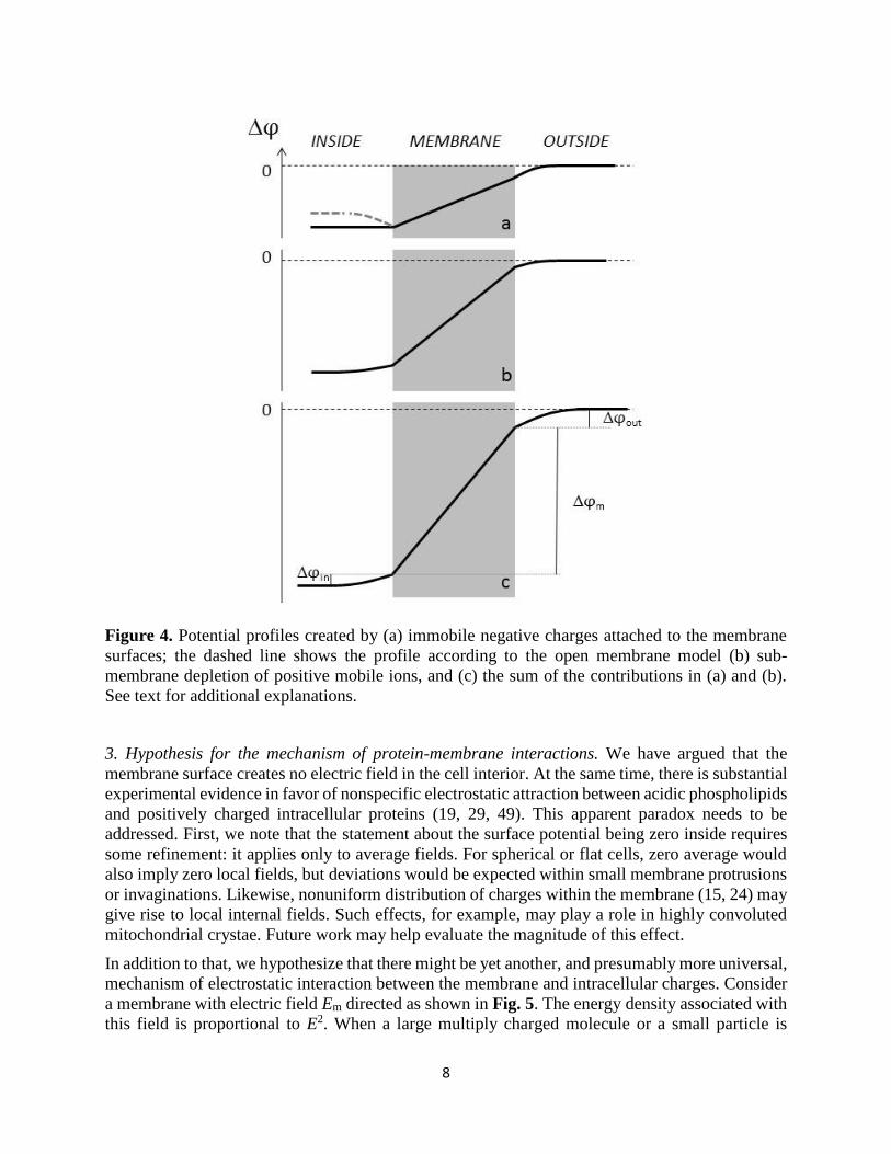
mechanism of electrostatic interaction between the membrane and intracellular charges. Consider
a membrane with electric field Em directed as shown in Fig. 5. The energy density associated with
this field is proportional to E2. When a large multiply charged molecule or a small particle is

9
located in a conducting liquid far from the membrane, its electric field Ep1 decays at the Debye
distance. However, if the molecule closely approaches the membrane, its electric field begins to
penetrate the membrane without decay. Now a much stronger field Ep2, directed opposite to Em,
emerges within the membrane. The total field in the membrane becomes reduced, and so does the
electrostatic energy of the system (Fig. 5). Therefore, close placement of a large positively charged
molecule on the membrane would be thermodynamically favorable.
A simple estimate of the binding energy G can be made by assuming that: (1) the protein is large
compared to the membrane thickness l; (2) by neglecting the energy of the Debye layer; (3) by
neglecting the entropic contribution from the ionic layer surrounding the protein:
ΔG = (Sl/2εmε0) (σp2 + 2σpσm), (11)
where S is the protein/membrane contact area, σm is the surface density of membrane charges, and
σp is the surface density of protein charges. One can see that, for a given σm, the strongest binding
(minimum G) is experienced by particles with σp = -σm, but for low membrane potentials |σm| <
0.5|σp| repulsion is expected.
This mechanism may operate either on planar or spherical membranes. An interesting situation
might arise for a flat membrane when the potential is generated by positive charges adsorbed on
the right side of the membrane (in the configuration depicted in Fig. 5). The expected attraction of
positive molecules from the opposite side of the membrane would be equivalent to attraction of
like charges! Needless to say, this hypothesis needs to be tested experimentally.
The other line of evidence for electrostatic interactions between phospholipids and positively
charge molecules comes from the work on artificial lipid vesicles (3-5, 13, 21, 22, 27, 32, 33, 46,
51) or plane lipid layers (11, 20, 22, 40). However, flat membranes or the outer surface of vesicles
represent a different electrical environment, and there is no contradiction between those results
and our hypothesis.

10
Figure 5. The proposed mechanism of binding of a positively charged large molecule or particle
to the interior of the membrane surface. (A). When the particle is in a conducting liquid, the electric
field created by ionized groups extends only a short distance into the liquid. (B). When the particle
comes in apposition with the membrane, its field penetrates into the membrane in the direction
opposite to the field already present in the membrane. (C) Superposition of the field from the
particle with the field in the membrane results in a decrease in the total field in the area indicated
by the dotted line. The energy density within that area decreases to create an energetically
favorable condition for particle binding.
4. Conclusions. Near-membrane depletion of positive ions could in principle be verified by using
appropriate membrane-linked ion probes. The loss of a negative potential would be expected to
raise the subcellular concentration of positive ions and stimulate their efflux, which, indeed, is a
common cellular response to depolarization (35). Of course, cells have others ways to regulate ion
traffic, so the biological significance of the hypothesized depletion cannot be claimed at this point.
Nevertheless, as a little-appreciated consequence of the Donnan potential, this effect may be
interesting at least from the theoretical perspective.
With regard to the surface potential theory, we have shown that simple coulombic interactions
between cytoplasmic proteins and the inner membrane are incompatible with the model of a
spherical, uniform and continuously charged membrane. Since real membranes are not such,
additional theoretical work is necessary to evaluate the importance of deviations from symmetry
and uniformity; one might find, for example, that electrostatic interactions should be limited to
certain areas of the membrane. The alternative (or complementary) mechanism of attraction
between an electrically polarized membrane and charges immersed in a conducting liquid should
be testable on model systems.
ACKNOWLEDGEMENTS
The authors thank Dr. G. Cevc for advice. KAK acknowledges a support from NSF Grant No.
PHY-1206187.
REFERENCES
1. Arrebola F, Fernández-Segura E, Campos A, Crespo PV, Skepper JN, Warley A
Changes in intracellular electrolyte concentrations during apoptosis induced by UV
irradiation of human myeloblastic cells. Am J Physiol Cell Physiol 290: C638-C649,
2006.
2. Bashford CL, Pasternak CA. Plasma membrane potential of some animal cells is
generated by ion pumping, not by ion gradients. Trends Biochem Sci 11: 113-116, 1986.
3. Benfenati F, Greengard P, Brunner J, Bähler M. Electrostatic and hydrophobic
interactions of synapsin I and synapsin I fragments with phospholipid bilayers. J. Cell
Biol 108: 1851-1862, 1989.

11
4. Ben-Tal N, Honig B, Miller C, McLaughlin S. Electrostatic binding of proteins to
membranes. Theoretical predictions and experimental results with charybdotoxin and
phospholipid vesicles. Biophys J 73: 1717-1727, 1997.
5. Ben-Tal N, Honig B, Peitzsch RM, Denisov G, McLaughlin S. Binding of small basic
peptides to membranes containing acidic lipids: theoretical models and experimental
results. Biophys J 71:561-575, 1996.
6. Blaustein MP, Kao JPY, Matteson DP. Cellular Physiology. Philadelphia: Elsevier
Mosby, 2004.
7. Bohinc K, Giner-Casares JJ, May S. Analytic model for the dipole potential of a lipid
layer. J Phys Chem B. 118:7568–7576, 2014.
8. Bregestovski P, Waseem T, Mukhtarov M. Genetically encoded optical sensors for
monitoring of intracellular chloride and chloride-selective channel activity. Front Mol
Neurosci 2: 15, 2009.
9. Burry RW, Wood JG. Contributions of lipids and proteins to the surface charge of
membranes. An electron microscopy study with cationized and anionized ferritin. J Cell
Biol 82: 726-741, 1979.
10. Cevc G. Membrane electrostatics. Biochim Biophys Acta 1031: 311-382, 1990.
11. Cevc G, Marsh D. Properties of the electrical double layer near the interface between a
charged bilayer membrane and electrolyte solution: experiment vs. theory. J Phys Chem
87: 376-379, 1983.
12. Chen LB. Mitochondrial membrane potential in living cells. Annu Rev Cell Biol 4: 155-
181, 1988.
13. Chen Y, Ludescher RD, Montville TJ. Electrostatic interactions, but not the YGNGV
consensus motif, govern the binding of pediocin PA-1 and its fragments to phospholipid
vesicles. Appl Environ Microbiol 63: 4770-4777, 1997.
14. Coster HGL. The physics of cell membranes. J Biol Phys 29: 363-399, 2003.
15. Eagles PA, Johnson LN, Van Horn C. The distribution of anionic sites on the surface of
the chromaffin granule membrane. J Ultrastruct Res 55: 87-95, 1976.
16. Elul R. Fixed charge in the cell membrane. J Physiol 189: 351-365, 1967.
17. Glaser R. Biophysics. An Introduction, 2nd ed. Heidelberg: Springer, 2012.
18. Glitsch HG. Electrophysiology of the sodium-potassium-ATPase in cardiac cells.
Physiol Rev 81: 1791-1826, 2001.
19. Goldenberg NM, Steinberg BE. Surface charge: a key determinant of protein
localization and function. Cancer Res 70: 1277-1280, 2010.
20. Hall K, Lee TH, Aguilar MI. The role of electrostatic interactions in the membrane
binding of melittin. J Mol Recognit 24: 108-118, 2011.
21. Higgins DL, Mann KG. The interaction of bovine factor V and factor V-derived
peptides with phospholipid vesicles. J Biol Chem 258: 6503-6508, 1983.
22. Hoernke M, Schwieger C, Kerth A, Blume A. Binding of cationic pentapeptides with
modified side chain lengths to negatively charged lipid membranes: Complex interplay of
electrostatic and hydrophobic interactions. Biochim Biophys Acta 1818: 1663-1672, 2012.
23. Honig BH, Hubbell WL, Flewelling RF. Electrostatic interactions in membranes and
proteins. Annu Rev Biophys Biophys Chem 15: 163-93, 1986.
24. Howell SL, Tyhurst M. Distribution of anionic sites on surface of B cell granule and
plasma membranes: a study using cationic ferritin. J Cell Sci 27: 289-301, 1977.

12
25. Ishida Y, Chused TM. Lack of voltage sensitive potassium channels and generation of
membrane potential by sodium potassium ATPase in murine T lymphocytes. J Immunol
151: 610-620, 1993.
26. Jäckle J. The causal theory of the resting potential of cells. J Theor Biol 249: 445-463,
2007.
27. Kimelberg HK, Papahadjopoulos D. Interactions of basic proteins with phospholipid
membranes. Binding and changes in the sodium permeability of phosphatidylserine
vesicles. J Biol Chem 246: 1142-1148, 1971.
28. Krasznai Z, Márián T, Balkay L, Emri M, Trón L. Flow cytometric determination of
absolute membrane potential of cells. J Photochem Photobiol B. 28: 93-99, 1995.
29. Leventis PA, Grinstein S. The distribution and function of phosphatidylserine in cellular
membranes. Annu Rev Biophys 39: 407-27, 2010.
30. Loew LM. Electrical properties of biomembranes. In: Biomembranes. Physical aspects,
edited by M. Shinitzky M. Weinheim, Germany:VCH, 1993. p. 341-371.
31. McLaughlin S. The electrostatic properties of membranes. Annu Rev Biophys Biophys
Chem 18: 113-136, 1989.
32. Mertins O, Dimova R. Binding of chitosan to phospholipid vesicles studied with
isothermal titration calorimetry. Langmuir 27: 5506-5515, 2011.
33. Murray D, Arbuzova A, Hangyás-Mihályné G, Gambhir A, Ben-Tal N, Honig B,
McLaughlin S. Electrostatic properties of membranes containing acidic lipids and
adsorbed basic peptides: theory and experiment. Biophys J 77: 3176-3188, 1999.
34. Neumcke B. Surface charges on biological membranes. In: Membranes and intercellular
communication, edited by Balian R, Chabre M, Devaus PF. Amsterdam: North Holland
Publishing Company, 1981, p. 471-483.
35. Nicholls DG, Ward MW. Mitochondrial membrane potential and neuronal glutamate
excitotoxicity: mortality and millivolts. Trends Neurosci 23: 166-174, 2000.
36. Ohki S. The origin of electrical potential in biological systems. In: Comprehensive
Treatise of Electrochemistry, vol. 10, edited by Srinivasan S, Chimadzhev YA, Bockris
JO, Conway BE, Yeager E. New York: Plenum, 1985, p. 2-130.
37. Olivotto M, Arcangeli A, Carlà M, Wanke E. Electric fields at the plasma membrane
level: a neglected element in the mechanisms of cell signalling. Bioessays 18: 495-504,
1996.
38. Perry SW, Norman JP, Barbieri J, Brown EB, Gelbard HA. Mitochondrial membrane
potential probes and the proton gradient: a practical usage guide. Biotechniques 50: 98-
115, 2011.
39. Rink TJ, Montecucco C, Hesketh TR, Tsien RY. Lymphocyte membrane potential
assessed with fluorescent probes. Biochim Biophys Acta 595:15-30, 1980.
40. Salay LC, Ferreira M, Oliveira ON Jr, Nakaie CR, Schreier S. Headgroup specificity
for the interaction of the antimicrobial peptide tritrpticin with phospholipid Langmuir
monolayers. Colloids Surf B Biointerfaces 100: 95-102, 2012.
41. Shepherd VA. The cytomatrix as a cooperative system of macromolecular and water
networks. Curr Top Dev Biol 75: 171–223, 2006.
42. Skulachev VP. Why are mitochondria involved in apoptosis? Permeability transition
pores and apoptosis as selective mechanisms to eliminate superoxide-producing
mitochondria and cell. FEBS Lett 397:7-10, 1996.

13
43. Smaili SS, Hsu YT, Youle RJ, Russell JT. Mitochondria in Ca2+ signaling and
apoptosis. J Bioenerg Biomembr 32: 35-46, 2000.
44. Somlyo AP, Somlyo AV, Shuman H. Electron probe analysis of vascular smooth
muscle. Composition of mitochondria, nuclei, and cytoplasm. J Cell Biol 81: 316-335,
1979.
45. Sperelakis N. Origin of resting membrane potentials. In: Cell Physiology Sourcebook, 4th
ed., edited by Sperelakis N. Amsterdam: Elsevier, p. 121-145, 2012
46. Terzi E, Hölzemann G, Seelig J. Alzheimer beta-amyloid peptide 25-35: electrostatic
interactions with phospholipid membranes. Biochemistry 33: 7434-7441, 1994.
47. Thomas RC. Electrogenic sodium pump in nerve and muscle cells. Physiol Rev 52: 563-
594, 1972.
48. Wright SH. Generation of resting membrane potential. Adv Physiol Educ 28: 139-142,
2004.
49. Yeung T, Gilbert GE, Shi J, Silvius J, Kapus A, Grinstein S. Membrane
phosphatidylserine regulates surface charge and protein localization. Science 319: 210-
213, 2008.
50. Yeung T, Grinstein S. Lipid signaling and the modulation of surface charge during
phagocytosis. Immunol Rev 219: 17-36, 2007.
51. Zhang X, Rizo J, Südhof TC. Mechanism of phospholipid binding by the C2A-domain
of synaptotagmin I. Biochemistry 37:12395-12403, 1998.