a new era of uveitis: impact of polymerase chain … a new era of uveitis: impact of polymerase...
TRANSCRIPT

REVIEW
A new era of uveitis: impact of polymerase chain reactionin intraocular inflammatory diseases
Manabu Mochizuki1,3 • Sunao Sugita2 • Koju Kamoi1 • Hiroshi Takase1
Received: 13 June 2016 / Accepted: 28 July 2016 / Published online: 27 October 2016
� Japanese Ophthalmological Society 2016
Abstract Uveitis is a sight-threatening intraocular
inflammatory disorder which may occur from both infec-
tious and non-infectious or autoimmune causes. The fre-
quency of infectious uveitis and autoimmune uveitis varies
depending on countries and regions. According to a
nationwide survey conducted by the Japanese Ocular
Inflammation Society, infectious and non-infectious uveitis
accounted for 16.4 and 50.1% of new patients, respectively
while the remaining 33.5% of new uveitis cases were not
classified or were idiopathic uveitis. Infectious uveitis is
particularly important because it causes tissue damage to
the eye and may result in blindness unless treated. How-
ever, it can be treated if the pathogenic microorganisms are
identified promptly and accurately. Remarkable advance-
ments in molecular and immunological technologies have
been made in the last decade, and the diagnosis of infec-
tious uveitis has been greatly improved by the application
of molecular and immunological investigations, particu-
larly polymerase chain reaction (PCR). PCR performed on
a small amount of ocular samples provides a prompt,
sensitive, and specific molecular diagnosis of pathogenic
microorganisms in the eye. This technology has opened a
new era in the diagnosis and treatment of uveitis, enabling
physicians to establish new clinical entities of uveitis
caused by infectious microorganisms, identify pathogens in
the eyes of many patients with uveitis, and determine
prompt diagnosis and appropriate therapy. Here we review
the PCR process, new PCR tests specialized for ocular
diseases, microorganisms detected by the PCR tests, dis-
eases in the eye caused by these microorganisms, and the
clinical characteristics, diagnosis, and therapy of uveitis.
Keywords Polymerase chain reaction (PCR) � Multiplex
PCR � Real-time PCR � Comprehensive PCR � Infectiousuveitis
Introduction
Uveitis is a sight-threatening intraocular inflammatory
disorder caused by autoimmune mechanisms or infection.
The proportion of autoimmune uveitis and infectious
uveitis varies depending on countries and regions [1, 2].
Two nation-wide epidemiological surveys of uveitis have
been conducted in Japan by the Japanese Ocular Inflam-
mation Society in 2002 [3] and 2009 [4], respectively. The
2009 prospective survey enrolled a total of 3830 patients
who were newly diagnosed with uveitis over a 1-year
period at 36 university hospitals. Of these patients, 33.5%
had unclassified or idiopathic uveitis and 66.5% were
identified with specific etiologies consisting of non-infec-
tious diseases (50.1%) and infectious diseases (16.4%).
The leading etiology of uveitis was sarcoidosis (10.6%)
followed by Vogt–Koyanagi–Harada disease (7.0%), acute
anterior uveitis (AAU) (6.5%) including HLA-B27-posi-
tive AAU (1.9%), scleritis with or without uveitis (6.1%),
herpetic iritis (4.2%), Behcet’s disease (3.9%), bacterial
endophthalmitis (2.5%), and masquerade syndrome (2.5%)
including intraocular lymphoma (1.3%).
& Manabu Mochizuki
1 Department of Ophthalmology and Visual Science, Graduate
School of Medical and Dental Sciences, Tokyo Medical and
Dental University, 1-5-45 Yushima,
Bunkyo-ku, Tokyo 113-8519, Japan
2 Laboratory for Retinal Regeneration, Center for
Developmental Biology, RIKEN, Kobe, Japan
3 Miyata Eye Hospital, Miyakonojo, Miyazaki, Japan
123
Jpn J Ophthalmol (2017) 61:1–20
DOI 10.1007/s10384-016-0474-9
Remarkable advancements in molecular and immuno-
logical technologies have been made in the last decade. The
diagnosis of uveitis in clinical practice has been greatly
changed by the application of molecular and immunological
investigations, particularly polymerase chain reaction
(PCR). These tests have resulted in the identification of new
clinical entities of uveitis based on the detection of patho-
genic microorganisms in the eye [5–7]. More importantly,
these new diagnostic tests are highly sensitive, specific, and
quick, and they require only a small sample.
Infectious intraocular inflammation causes tissue dam-
age to the eye and may result in blindness unless the
pathogenic microorganisms are identified promptly and
accurately and consequently managed with the appropriate
medical therapy, such as antibiotics, antifungal agents, or
antiviral agents. PCR testing has opened a new era in the
diagnosis and treatment of uveitis.
Here we review the PCR process, new PCR tests spe-
cialized for ocular diseases, microorganisms detected by
the PCR tests, diseases in the eye caused by these
microorganisms, and their clinical characteristics, diagno-
sis, and therapy.
PCR as a new diagnostic tool
The PCR is a molecular biology technique used to amplify a
specific region (the target) of DNA by many orders of
magnitude within a short period of time. It was first descri-
bed by Saiki et al. in 1985 [8]. The amount of the amplified
product is determined by the availability various substrates
in the reaction which become limiting as the reaction pro-
gresses. A basic PCR set-up requires several components
and reagents, including (1) a DNA template that contains the
target DNA region; (2) at least two primer pairs (the sense
and antisense strands of the DNA target); (3) Taq poly-
merase (or another DNA polymerase); (4) various agents,
such as nucleotides (e.g., dNTPs) and buffers.
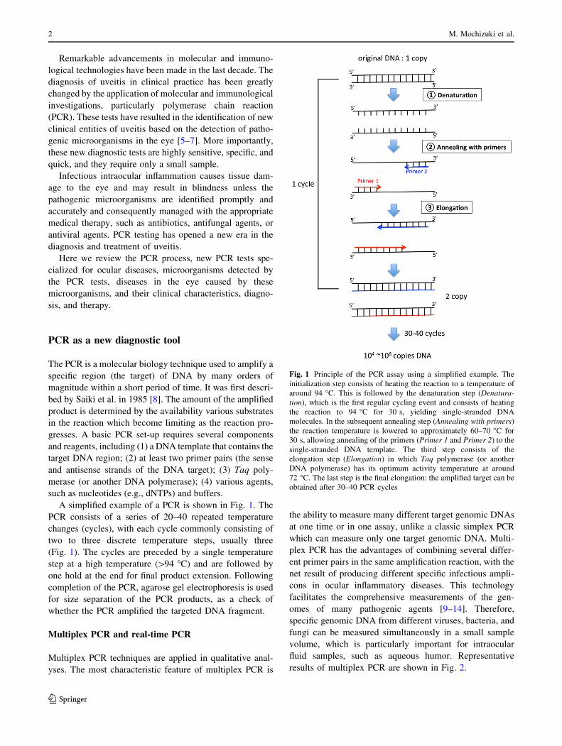
A simplified example of a PCR is shown in Fig. 1. The
PCR consists of a series of 20–40 repeated temperature
changes (cycles), with each cycle commonly consisting of
two to three discrete temperature steps, usually three
(Fig. 1). The cycles are preceded by a single temperature
step at a high temperature ([94 �C) and are followed by
one hold at the end for final product extension. Following
completion of the PCR, agarose gel electrophoresis is used
for size separation of the PCR products, as a check of
whether the PCR amplified the targeted DNA fragment.
Multiplex PCR and real-time PCR
Multiplex PCR techniques are applied in qualitative anal-
yses. The most characteristic feature of multiplex PCR is
the ability to measure many different target genomic DNAs
at one time or in one assay, unlike a classic simplex PCR
which can measure only one target genomic DNA. Multi-
plex PCR has the advantages of combining several differ-
ent primer pairs in the same amplification reaction, with the
net result of producing different specific infectious ampli-
cons in ocular inflammatory diseases. This technology
facilitates the comprehensive measurements of the gen-
omes of many pathogenic agents [9–14]. Therefore,
specific genomic DNA from different viruses, bacteria, and
fungi can be measured simultaneously in a small sample
volume, which is particularly important for intraocular
fluid samples, such as aqueous humor. Representative
results of multiplex PCR are shown in Fig. 2.
Fig. 1 Principle of the PCR assay using a simplified example. The
initialization step consists of heating the reaction to a temperature of
around 94 �C. This is followed by the denaturation step (Denatura-
tion), which is the first regular cycling event and consists of heating
the reaction to 94 �C for 30 s, yielding single-stranded DNA
molecules. In the subsequent annealing step (Annealing with primers)
the reaction temperature is lowered to approximately 60–70 �C for
30 s, allowing annealing of the primers (Primer 1 and Primer 2) to the
single-stranded DNA template. The third step consists of the
elongation step (Elongation) in which Taq polymerase (or another
DNA polymerase) has its optimum activity temperature at around
72 �C. The last step is the final elongation: the amplified target can be
obtained after 30–40 PCR cycles
2 M. Mochizuki et al.
123

However, multiplex PCR or any other qualitative PCR
techniques cannot quantitatively measure copy number of
genomic DNA. Given the extremely high sensitivity of
PCR, positive results from qualitative PCR techniques can
be false positives due to contamination. Therefore, it is
essential to quantitatively measure the copy number of
genomic DNA by real-time PCR to evaluate the pathogenic
role of pathogens detected by qualitative PCR techniques.
Representative results from real-time PCR are shown in
Fig. 3 where the real-time PCR is performed with Ampli-
Taq Gold and the Real-Time PCR 7300 system (Applied
Biosystems, Foster City, CA, USA) or the LightCycler 480
II System (Roche Life Sciences, Penzberg, Germany), as
examples.
Broad-range real-time PCR
Broad-range PCR techniques can be used to detect the
presence of bacterial or fungal genomic DNA. Bacterial
ribosomal DNA genes (16S rDNA) or fungal ribosomal
DNA genes (18S or 28S rDNA) were previously used for
qualitative broad-range PCR performed with ocular fluids
of patients with infectious endophthalmitis and uveitis
[15–18]. Broad-range PCR techniques use primers and
probes for conserved regions in the genomes of bacteria
and fungi of many species. As such, they can provide
evidence of the presence of bacterial or fungal infection
in the eye, but are not able to identify the species and
strains of bacteria or fungi. In clinical settings, it is
common practice to treat patients with endophthalmitis or
infectious uveitis with antibiotics before samples are
collected for culture, smear, and/or PCR tests. In these
clinical situations, PCR is more effective than cultures
and smears in detecting bacterial and fungal genomes in
the ocular fluids [18] because PCR may still be able to
detect the infectious DNA of living or dead bacteria and
fungi. However, previous broad-range PCR techniques for
bacteria and fungi were not able to determine quantitative
measurements of these genomes in ocular samples.
Therefore, we designed real-time PCR primers and probes
for bacterial 16S rDNA [19, 20] and fungal 18S/28S
rDNA amplifications [21, 22], which made it possible to
perform quantitative measurements. Representative results
for this PCR technique performed using the LightCycler
480 II System (Roche Life Sciences) are shown in Fig. 4.
In these patients, who were suspected to have infectious
endophthalmitis based on clinical evidence, high copy
numbers of the bacterial DNA (Fig. 4a) or fungal DNA
(Fig. 4b) were detected, indicating the presence of the
infection. The reliability of the broad-range real-time PCR
assay for bacterial 16S rDNA was examined by detecting
16 different strains (12 Gram-positive strains and 4 Gram-
negative strains) of common pathogens of endophthalmi-
tis, and the PCR system detected genomic DNA of target
bacteria at levels as low as 1–10 colony-forming units in
diluted vitreous samples [20].
Fig. 2 Results of a multiplex PCR assay. After DNA extraction from
the sample, multiplex PCR was performed to screen for all 8 human
herpes viruses (HHVs; HHV1–HHV8) by using LightCycler (Roche
Life Sciences, Penzberg, Germany) capillaries. a At a melting
temperature (Tm) of 57 �C, a significant positive curve was detected,
indicating the detection of herpes simplex virus 1 DNA (HSV1-DNA;
HHV1) in the sample; the sample was confirmed to be negative for
HSV2, varicella–zoster virus (HHV3), Epstein–Barr virus (HHV4),
cytomegalovirus (CMV; HHV5), HHV6, HHV7, and HHV8. b At a
Tm of 62 �C, a significant positive curve was detected, indicating the
detection of CMV DNA (upper graph). In the case of CMV-negative
infections, there was flat line or no significant positive curve, such as
shown in the lower panel
A new era of uveitis: impact of polymerase chain reaction in intraocular inflammatory diseases 3
123

The bacterial 16S rDNA broad-range real-time PCR was
positive in the ocular fluids from 18 of the 19 (95%)
patients with bacterial endophthalmitis with high copy
numbers ranging from 1.7 9 103 to 1.7 9 109 copies/ml.
The PCR was positive in samples from three of the 50 (6%)
non-infectious control uveitis patients and in none of the
samples from the 40 control non-uveitis patients [19].
The fungal 28S rDNA broad-range real-time PCR assay
was tested using ocular fluids from 40 patients without
ocular inflammation and 497 patients with ocular inflam-
mation, including 76 patients with clinically suspected
bacterial and fungal endophthalmitis and 421 patients with
infectious or non-infectious uveitis. The PCR assay
detected fungal 28S rDNA in 11 patients with high copy
numbers ranging from 1.7 9 103 to 7.9 9 106 copies/ml,
and ten of these 11 patients were diagnosed with fungal
endophthalmitis by other investigations and showed a good
response to anti-fungal therapy; there were two false-neg-
ative cases and one false-positive case [22].
False-negative results in PCR assays for bacterial
infection directly relate to the sensitivity of the PCR assay.
In our multicenter prospective PCR study [14], false-neg-
ative results were obtained for 12 of the 38 samples from
clinically suspected infectious bacterial endophthalmitis.
The majority of the samples with the false-negative results
were obtained when PCR was performed using the aqueous
humor, suggesting that the manner in which the samples
are collected is important. In our samples, bacterial 16S
rDNA was not detected in some patients with endogenous
bacterial endophthalmitis [14, 19]. Endogenous endoph-
thalmitis pathogens are disseminated in the eye through
hematogenous routes; consequently, it is possible that
although we did not detect the bacterial DNA in the
aqueous samples, we may have detected some bacterial
DNA if we had used samples from vitreous fluid or the
Fig. 3 Results of a quantitative real-time PCR assay. Quantitative
real-time PCR of the ocular sample is shown. The genomic DNA
copy numbers of Varicella–Zoster Virus (VZV) (a) and Toxoplasma
(Toxoplasma gondii) (b) in the sample are shown. Control DNA (106,
104, 102, and 101 copies/ml) by real-time PCR was used to establish
the standard curve, and the DNA concentration in the samples was
calculated by using the standard curve. The final copy number of
DNA in the sample (copies/ml) is calculated based on the obtained
sample volume and final dilution volume. Values of[10 copies/ml
are considered to be significant
Fig. 4 Results of a broad-range PCR assay. Bacterial 16S rDNA in
the vitreous fluid sample (a) and fungal 28S rDNA in the vitreous
fluid (b) are shown. Specific primers and probes to bacterial 16S
rDNA and fungal 18S/28S rDNA were designed, and the PCR assay
was performed with a LightCycler 480 II System (Roche Life
Sciences). Control DNA (106, 104, 102, and 101 copies/ml) amplified
by the broad-range PCR is also shown
4 M. Mochizuki et al.
123

retina. Correct sampling appears to be important to avoid
false-negative results in bacterial 16S broad-range PCR
techniques.
A false-positive result for fungal 28S rDNA was
obtained in one patient with non-infectious ocular disease
by the broad-range PCR assay [22]. A few false-positive
results of bacterial 16S PCR were also found in our mul-
ticenter prospective PCR study [14]. False-positive results
for bacterial 16S rDNA were cases of idiopathic uveitis,
but the copy numbers of bacterial 16S in these patients
were not high [14]. Such false-positive results can also be
due to contamination caused by technical errors
[14, 19, 22]. Therefore, we set the cut-off value in these
broad-range PCR assays so that bacterial or fungal copy
number values of [100 copies/ml in the sample were
considered to be significant [19, 22]. However, at the
present time it is difficult to avoid the false-positive results
due to limitations of this technology. If the results of broad-
range real-time PCR exhibit low copy numbers of bacterial
16S or fungal 28S in the sample, other approaches, such as
conventional culture and smears, may help avoid misdi-
agnosis in these cases.
A comprehensive PCR system
A comprehensive PCR system consisting of a combination
of multiplex PCR and real-time PCR has recently been
developed for intraocular fluids with the aim to diagnose
infectious uveitis [14]. The multiplex PCR was designed to
detect genomic DNA of all eight human herpes viruses
(HHVs): HHV1 (herpes simplex virus type 1; HSV1),
HHV2, HHV3 (varicella–zoster virus; VZV), HHV4 (Ep-
stein–Barr virus; EBV), HHV5 (cytomegalovirus; CMV),
HHV6, HHV7, and HHV8 [11, 12]. The PCR assay was
also designed to qualitatively detect genomic DNA of
toxoplasma (Toxoplasma gondii B1 gene) [13], parvovirus
B19 (Parvo B19), BK virus (BKV), and JC virus (JCV).
Parvo B19, BKV, and JCV genomic DNA are often
detected in intraocular fluid samples from patients with
ocular inflammatory diseases, especially immunodeficient
patients (unpublished observation). In our laboratory, this
PCR assay is performed with a LightCycler PCR System
(Roche Life Sciences). The multiplex PCR assay screens
for the infectious genomes of all human herpes viruses
(HHV1–8) [11, 12] and ocular toxoplasma [13]. When
positive results are generated by the multiplex PCR, then
quantitative real-time PCR is performed to measure the
copy number of the genome in the sample and to confirm
its pathogenic role. A high copy number obtained in the
real-time PCR assay indicates active viral replication in the
eye. In addition, we use different primer pairs for multiplex
PCR and real-time PCR in order to avoid false-positive
results as much as possible.
The sensitivity, specificity, positive predictive values,
and negative predictive values of the comprehensive PCR
system for the diagnosis of infectious ocular diseases were
91.3, 98.8, 98.5, and 92.4%, respectively [14]. The com-
prehensive PCR system has an additional clinically
important advantage in that a negative result can support
the exclusion of many common infectious etiologies of
intraocular inflammation, thus facilitating the decision-
making process of whether to use corticosteroids and/or
immunosuppressive agents as necessary.
Multiplex solid-phase PCR strip kit
To improve the previously developed comprehensive PCR
assay [14], a new comprehensive PCR strip kit (a multi-
plex solid-phase PCR strip kit) was established to diag-
nose infectious ocular diseases [23]. The PCR strip
consists of a 12-tube multiplex PCR strip coated with
primers (forward and reverse) and probes (6FAM, HEX,
and Cy5) targeting the following ocular infectious disease
pathogens: HSV1, HSV2, VZV, EBV, CMV, HHV6,
HHV7, HHV8, human T-lymphotropic virus/human T-cell
leukemia virus type 1 (HTLV-1), adenovirus, Mycobac-
terium tuberculosis, Propionibacterium acnes, Treponema
pallidum, Candida albicans, C. glabrata, C. krusei,
Fusarium, Aspergillus, Toxocara, Toxoplasma gondii,
Chlamydia trachomatis, Acanthamoeba, bacterial 16S
rDNA, fungal 28S rDNA, as well as internal controls such
as glyceraldehyde 3-phosphate dehydrogenase and TATA-
binding protein (Fig. 5a).
This new multiplex PCR strip has at least two advan-
tages: (1) the genomes for 24 common pathogens of vari-
ous infectious eye diseases can be detected in one assay
that takes only a few hours (Fig. 5), and (2) the PCR
process becomes simpler and can be performed with
commonly used PCR instruments. A representative result
measured by the system is shown in Fig. 5c. In this case of
infective endophthalmitis, a significant positive curve was
detected for only Candida glabrata and fungal 28S rDNA.
A prospective multicenter clinical study is necessary to
evaluate the usefulness of the multiplex PCR strip.
Viruses
Human herpes viruses
In the past decade, PCR has revealed the significant
involvement of HHVs in the pathogenesis of various types
of uveitis, and this technology has greatly contributed to
the diagnosis of many ocular diseases. Table 1 summarizes
HHV-related ocular inflammatory diseases and pathogenic
HHVs detected by PCR assays.
A new era of uveitis: impact of polymerase chain reaction in intraocular inflammatory diseases 5
123

Fig. 5 Multiplex PCR strip. a,b The PCR strip consists of a
12-tube multiplex PCR strip
coated with primers (forward
and reverse) and probes (6FAM,
HEX, and Cy5) targeting the 24
target genomic DNA of
common pathogens. c A
representative result of fungal
endophthalmitis. GAPDH
Glyceraldehyde 3-phosphate
dehydrogenase, HTLV-1 human
T-lymphotropic virus/human
T-cell leukemia virus type 1, TB
tuberculosis caused by
Mycobacterium tuberculosis,
TBP TATA-binding protein,
ADV adenovirus
Table 1 Summary of ocular inflammatory diseases associated with human herpes viruses
Herpes virus Ocular disease References (order in year of publication)a
HHV1 (herpes simplex virus type 1; HSV1) Keratitis [26], [9]
Corneal endotheliitis and iritis [26]
Acute retinal necrosis [27], [11]
HHV2 (HSV2) Acute retinal necrosis [43], [27], [11]
HHV3 (varicella–zoster virus; VZV) Herpes zoster ophthalmicus/zoster sine herpete [31], [40]
Acute retinal necrosis [28], [27], [11]
HHV4 (Epstein–Barr virus; EBV) Anterior uveitis/pan-uveitis [55], [54], [53]
Acute retinal necrosis [11]
Intraocular lymphoma [56]
HHV5 (cytomegalovirus; CMV Corneal endotheliitis [36], [35], [7], [37]
Iritis (uveitis) [33], [6], [34]
Cytomegalovirus retinitis [32]
HHV6 Iritis (uveitis) [59], [61], [60]
Endophthalmitis [60]
Keratitis [61], [63], [60], [62]
AIDS-associated retinitis [64]
HHV7 Corneal endotheliitis and iritis [66]
HHV8 Corneal endotheliitis and iritis [67]
Kaposi’s sarcoma (conjunctiva, ocular adnexa) [70]
HHV1–8 Eight types of human herpes virus, AIDS acquired immune deficiency syndromea These are representative references by PCR-associated papers
6 M. Mochizuki et al.
123
The eight HHVs affect various ocular tissues and cause
anterior and/or posterior uveitis. Cell-free HHV DNA has
been detected by PCR testing of ocular fluids (aqueous
humor, vitreous fluids) from patients with various types of
uveitis, including herpetic keratouveitis, herpes zoster
ophthalmicus, zoster sine herpete, acute retinal necrosis,
and CMV retinitis. The combined use of multiplex PCR
and quantitative real-time PCR can detect genomic DNA of
HHVs rapidly and accurately from small amounts of ocular
fluid, which facilitates the diagnosis [11, 12]. Conse-
quently, uveitis caused by HHVs can be identified in
conjunction with the pathogenic virus detected by the PCR
analysis, such as HSV1 anterior uveitis (AU), VZV-AU,
CMV-AU, CMV retinitis, HSV1 acute retinal necrosis
(ARN), HSV2-ARN, and VZV-ARN.
HSV1, HSV2, VZV, and CMV
Genomic DNA of HSV [24–27], VZV [25, 28–31], and
CMV [6, 7, 25, 32–38] has been detected by PCR in the
aqueous humor of certain types of AU, corneal endotheli-
itis, and posterior uveitis with necrotizing retinitis (ARN
and CMV retinitis).
Although CMV retinitis is a well-known opportunistic
infection in immunodeficient individuals, such as human
immunodeficiency virus (HIV)-infected patients [32],
CMV had not been recognized as a cause of AU and/or
corneal endotheliitis in immunocompetent individuals until
PCR was introduced into clinical practice. Genomic DNA
of CMV has been detected in patients with corneal
endotheliitis with whitish keratic precipitates (KPs) and
corneal edema and in patients with AU. The pathogenic
role of CMV in these new diseases and clinical features has
been investigated [6, 7, 34–39].
CMV-associated corneal endotheliitis is unilateral in
most cases and characterized by corneal endothelial edema
with small whitish KPs, which typically form a ring shape
and are described as ‘‘coin-shaped lesions’’ [7, 38] with few
or no cells in the anterior chamber. Corneal endothelial cells
are significantly affected by CMV, resulting in corneal
endothelial cell loss and eventually in bullous keratopathy.
CMV-AU is also a unilateral AU characterized by small
whitish KPs, typically coin-shaped lesions. However,
CMV-AU always has a small proportion of cells in the
anterior chamber with or without corneal endotheliitis;
other features of CMV-AU are diffuse iris atrophy, epi-
sodes of high intraocular pressure (IOP), and no involve-
ment of the posterior segment of the eye [6,33–35]. These
ocular manifestations of CMV-AU are similar to those of
Posner–Schlossman syndrome and Fuchs heterochromic
iridocyclitis. In fact, CMV DNA was detected by PCR in
some, but not all, patients previously diagnosed as having
Posner–Schlossman syndrome or Fuchs heterochromic
iridocyclitis, suggesting that CMV is a causative agent of
Posner–Schlossman syndrome and Fuchs heterochromic
iridocyclitis. Although corneal endotheliitis was not evi-
dent in CMV-AU, corneal endothelial cell density in the
affected eye was significantly lower than in the unaffected
fellow eye in CMV-AU patients, and the intensity of cor-
neal endothelial cell loss was significantly correlated with
CMV viral load in the anterior aqueous humor [37].
PCR technology makes it possible to distinguish the
clinical features of HSV1-AU, VZV-AU, and CMV-AU.
According to various authors [6, 25], HSV1-AU, VZV-AU,
and CMV-AU share the following clinical features: (1)
occurrence in immunocompetent patients; (2) unilateral
involvement; (3) relatively acute process; (4) presence of
large KPs; (5) episodes of high IOP. However, there are a
number of differences in their clinical presentations. VZV-
AU has the most severe inflammation by means of its
severe aqueous flare, highest viral load in the aqueous
humor, and presence of segmental iris atrophy, which
occurs in the chronic stage of the disease. Segmental iris
atrophy in VZV-AU is associated with the viral load of
VZV in the aqueous humor [40]. Patients with CMV-AU
have the mildest intraocular inflammation, but the highest
elevation in IOP in comparison to these other two types of
uveitis, and the corneal endothelial cell density is signifi-
cantly lower in the affected eye than in the fellow eye [37].
Patients with CMV-AU and CMV corneal endotheliitis
have the worst prognoses due to corneal endothelial cell
damage resulting in bullous keratopathy.
HSV, VZV, and CMV also cause completely different
types of ocular inflammatory diseases affecting the poste-
rior segment of the eye with necrotizing retinitis, such as
ARN in immunocompetent patients and CMV retinitis in
immunocompromised patients.
ARN is a well-known sight-threatening disorder with
progressive retinal necrosis resulting in poor visual prog-
nosis. Therefore, a rapid diagnosis and initiation of the
appropriate treatment with sensitive antiviral agents from
the early stage of the disease are essential to save the
patient’s sight. In earlier days, the diagnosis of ARN was
based solely on clinical ocular signs [41]. The development
of an immunological test, the Goldmann–Witmer coeffi-
cient (GWC), which measures the ratio of specific antibody
in the eye and serum [42] revealed HSV and VZV to be
causative agents of ARN. The GWC is calculated by using
the total amount of immunoglobulin G (IgG) in the
intraocular fluid and serum collected on the same day and
the amount of pathogen-specific IgG (specific IgG)
according to the following formula: GWC = (specific IgG
in intraocular fluids/total IgG in intraocular fluids)/(serum-
specific IgG/serum total IgG). A GWC value of[1 and\6
evokes suspicion, and a value of C6 is significant. The
subsequent use of PCR provides a more rapid diagnosis
A new era of uveitis: impact of polymerase chain reaction in intraocular inflammatory diseases 7
123

with higher sensitivity and smaller samples [28]. It is
currently accepted that three HHVs, namely, HHV1
(HSV1), HHV2 (HSV2), and HHV3 (VZV), are causative
agents of ARN. The majority of ARN is caused by VZV,
followed by HSV1 and HSV2. However, even today it is
difficult to diagnose pathogenic viruses solely based on the
clinical presentation. Although ocular manifestations of
ARN caused by the three viruses are very similar, VZV-
ARN is more aggressive and more frequently seen in
elderly individuals than HSV1-ARN and HSV2-ARN.
HSV2-ARN is predominantly found in young patients
(mean age 21.2 years), whereas VZV-ARN is more com-
monly seen in older patients ([40 years) [43].
A new criteria for the diagnosis of ARNwas established by
a Japanese group in 2015 [44]. The diagnostic criteria take
both the results of PCR testing and the Goldmann–Witmer
coefficient measurement into consideration (Table 2).
Figure 6 depicts a representative case of HSV2-ARN.
Multiplex qualitative PCR and real-time quantitative PCR
assays detected HSV2 genomic DNA in the aqueous humor
sample, but no genomes of other herpes viruses were
detected. Treatment with intravenous acyclovir followed
by oral valaciclovir (prodrug of acyclovir) was effective
therapy for this patient with ARN.
Regarding the treatment of AU caused by HHVs, a
recent review [45] states that aciclovir remains the
mainstay of therapy for patients with HSV/VZV-AU and
that CMV-AU is to be treated with ganciclovir ointment
first, although prolonged therapy is required in many
cases. For the treatment of CMV corneal endotheliitis,
long-term topical ganciclovir (0.5%) and corticosteroids
have been reported to preserve corneal endothelial cell
function [46]. Topical 0.15% ganciclovir gel is also
effective in CMV corneal endotheliitis [47]. The dosage
Table 2 Diagnostic criteria for acute retinal necrosis
I. Basic concepts
1. Diagnosis is made on the basis of the combination of the ocular findings in the early stage, clinical courses, and the virologic testing of
intraocular fluids
2. When early-stage ocular findings 1a and 1b are positive, acute retinal necrosis is strongly suspected, and virologic testing of the
intraocular fluids and antiviral therapy are highly recommended
3. The final diagnosis is determined on the basis of the subsequent clinical course and the virologic test results
4. Acute retinal necrosis usually occurs in immunocompetent individuals. In immunodeficient patients, it should be noted that in addition to
the ocular symptoms or clinical courses described below, the ocular symtoms vary
II. Diagnostic criteria
1. Ocular findings in the early stage
1a. Anterior chamber cells or mutton-fat keratic precipitates
1b. Yellow-white lesion(s) in the peripheral retina (granular or patchy in the early stage, then gradually merging)
1c. Retinal arteritis
1d. Hyperemia of the optic disc
1e. Inflammatory vitreous opacities
1f. Elevated intraocular pressure
2. Clinical courses
2a. Rapid expansion of retinal lesion(s) circumferentially
2b. Development of retinal break or retinal detachment
2c. Retinal vascular occlusion
2d. Optic atrophy
2e. Response to antiviral agents
3. Virologic testing of intraocular fluids
Positive by either PCR or Goldmann–Witmer coefficient for HSV-1, HSV-2, or VZV
III. Classification
1. Virus-confirmed acute retinal necrosis
Presence of ocular findings 1a and 1b, presence of any 1 of the 5 clinical courses, and a positive virologic test result
2. Virus-unconfirmed acute retinal necrosis
Presence of 4 of the 6 ocular findings including 1a and 1b, presence of any 2 of the 5 clinical courses, and a negative virologic test result or
when virologic testing has not been performed
This table was reproduced from Takase et al. [44] with permission
8 M. Mochizuki et al.
123

and duration of systemic medications depend on the
intensity of inflammation in the anterior chamber and the
viral load in the aqueous humor. For HSV/VZV-AU, we
usually use oral valganciclovir, a prodrug of aciclovir, at
a dose of 1500 mg/day for HSV-AU and 3000 mg/day for
VZV-AU for the first 2 weeks, followed by a 50%
reduction in the dose for another 4–8 weeks, with the
medication discontinued after confirmation that the
inflammation has disappeared. Although the inflammatory
reaction in the anterior chamber in CMV-AU is mild, it
eventually causes damage to the corneal endothelial cells.
Therefore, we aggressively treat CMV-AU with valgan-
ciclovir (1800 mg/day) for the initial 2 weeks of treat-
ment, followed by 900 mg for 2–3 months, and then
450 mg for a further 1–2 months. CMV-AU often recurs
with a high IOP during tapering or termination of the
medication, so careful attention must be paid to the
patient during tapering of the dose, and timing of drug
cessation must be chosen with care. Intravitreal injection
of valganciclovir (600–1000 lg/0.1 ml) can be used to
avoid systemic adverse effects of oral valganciclovir
therapy. Bullous keratopathy in CMV-AU is treated by
penetrating keratoplasty or Descemet’s stripping auto-
mated endothelial keratoplasty (DSAEK). The surgery
must be performed after the CMV in the eye is thor-
oughly treated by anti-viral medications. Anshu et al. [48]
reported on four patients with DSAEK who had an
aqueous tap that was positive for CMV DNA, of whom
three developed a sudden decrease in endothelial cell
count and one developed retinitis.
The German Ophthalmologic Society distributed a
questionnaire on ARN therapy to 35 eye hospitals in
Germany [49]. Based on the data compiled from the
responding hospitals, all institutes commenced therapy
with aciclovir, followed by ganciclovir, foscarnet and, in
some cases, brivudine. Intravitreal injections were per-
formed in 46% of ARN patients and 80% of patients also
received steroids. Oral antiviral treatment was performed in
94% of patients. In Japan we usually start with intravenous
drip infusion of aciclovir (10–15 mg/kg, three times a day)
together with oral corticosteroid (30 mg/day of pred-
nisolone for the initial dose and tapering off in 1 month)
and acetylsalicylic acid (80–100 mg/day). If the yellow–
white retinal lesions disappear, treatment with intravenous
aciclovir is converted to 3000 mg/day of oral valaciclovir
for the next 4 weeks, followed by a 50% reduction in dose
for an additional 4 weeks. Once retinal detachment with
multiple retinal breaks occurs, vitreoretinal surgery is
necessary. The efficacy of prophylactic early vitrectomy in
ARN to prevent the development of retinal detachment is
controversial [50–52], but it has not been found to improve
final visual acuity as compared with patients not receiving
surgery [50].
Epstein–Bar virus
In a previous prospective study [14], PCR analysis of
ocular samples from 500 patients with infectious uveitis
detected EBV DNA in 17 patients (3.4%). Although the
case of a patient with AU and a high viral load of EBV was
Fig. 6 PCR assay for acute
retinal necrosis (ARN) (HSV2-
ARN). A slit lamp microscopic
photograph (a) and wide-view
fundus photograph (b) of theright eye of a patient with ARN.
Small keratic precipitates and
anterior chamber cells with
conjunctiva injection, and
yellowish retinal exudates
(white arrow) and disc swelling
(yellow arrow) together with
vitreous opacities are seen. cMultiplex PCR at 71 �C shows
a significant positive curve,
indicating the detection of
HSV2-DNA in the sample;
results for all other HHVs,
including HSV1 and VZV, were
negative. d Quantitative real-
time PCR revealed a high copy
number of HSV2-DNA
(2.78 9 104 copies/ml)
A new era of uveitis: impact of polymerase chain reaction in intraocular inflammatory diseases 9
123
reported [53], the viral load of most patients in whom EBV
DNA was detected was not very high, suggesting that EBV
replication does not occur in the eye. Ongkosuwito et al.
[54] observed that with respect to the pathogenesis of
ocular inflammation the EBV infection acts as a secondary
factor. EBV infects B-lymphocytes and retinal pigment
epithelial (RPE) cells [55], leading to the speculation that
the intraocular infiltrating B-lymphocytes and RPE cells
release the genome within the eye during intraocular
inflammation. In addition, EBV DNA is often detected in
ocular samples from patients with severe intraocular
inflammation or tissue damage, such as ARN [11, 56] and
intraocular lymphoma [56].
Human herpes virus 6
Human herpes virus 6 (HHV6) is a causative agent of
exanthema subitum in children [57]. More than a decade
ago, there were no reports of HHV6-associated ocular
infection; however, after PCR testing was introduced in
ocular diseases, the genomic DNA of HHV6 was detected
in patients with various ocular diseases that included uni-
lateral severe uveitis (ocular toxocariasis, bacterial
endophthalmitis, and idiopathic uveitis) [58–62] and ker-
atitis [62, 63]. None of these patients were immunocom-
promised. However, Fillet et al. [64] detected the HHV6
genome in acquired immunodeficiency syndrome (AIDS)-
associated retinitis. Two variants of HHV6 have been
identified, HHV6 type A and HHV6 type B [57]. In a study
of the variant types of HHV6 in uveitis patients, Sugita
et al. detected variant A in two of the eight patients tested
and variant B in six of these eight patients [60].
In our previous study [60], only a few patients with
infectious uveitis (n = 7/350, 2%) and keratitis (1/65,
1.5%) were PCR-positive for HHV6 DNA, with real-time
PCR revealing high copy numbers of HHV6 DNA, i.e., a
high viral load, in these patients. More importantly, HHV6
mRNA was detected in the ocular samples. These results
suggest that viral replication of HHV6 may occur in the
eye, thereby implicating HHV6 infection in ocular
inflammatory diseases. However, HHV6 infection (or
reactivation) in the eye might not be clinically relevant.
HHV6 can latently reside in cells of the lymphoid and
myeloid lineage, and it may enter the inflamed eye via
immune cells, such as T cells, monocytes, and leukocytes,
especially CD4? T cells, as a result of destruction of the
blood–retina barrier. In addition, similar to the results
reported for EBV infection [55], Arao et al. [65] demon-
strated that HHV6 (HHV6 type A) can infect human RPE
cells. Thus, the majority of the HHV6 genome in inflamed
eyes may be a consequence of the release of HHV6 DNA
and/or mRNA from resident ocular cells due to intraocular
inflammation. Although the actual role of HHV6 remains
undetermined, we believe that it plays a role as a secondary
factor in the pathogenesis of ocular inflammation.
HHV7 and HHV8
In our previous studies of PCR analysis of intraocular fluids
in patients with various types of uveitis, HHV7 and HHV8
were not detected in any patients [14]. However, there are
two reports of the genomic DNA of HHV7 and HHV8
being detected in ocular inflammatory diseases [66, 67].
HHV7 is transmitted early in childhood, and the virus
isolated from the peripheral blood mononuclear cells of a
healthy individual were found to be able to replicate and
produce progeny viruses in CD4? T cells [68]. Inoue et al.
[66] first reported patients with HHV7-related inflamma-
tion in the anterior segment of the eye that was charac-
terized by anterior chamber cells, keratic precipitates,
ocular hypertension, and/or corneal edema that initiates
from the corneal periphery and gradually progresses
throughout the cornea. HHV7 genomic DNA was detected
by PCR analysis of aqueous humor samples [66].
The HHV8 DNA sequence is closely related to that of
EBV and a member of the gamma-herpes virus subfamily,
respectively [69]. These viruses infect lymphocytes and are
associated with cell immortalization and transformation,
which could lead to HHV8-related neoplastic diseases,
such as Kaposi’s sarcoma [70]. In addition, Inoue reports
that patients with HHV8-related ocular inflammatory dis-
ease have active inflammation in the anterior segment of
the eye and/or corneal edema [67]. Since these patients had
etiology-unknown AU and the viral genome in the
inflamed eye, HHV7 and HHV8 infection may be causa-
tively related to ocular inflammation.
Rubella virus
Recent studies suggest that the rubella virus is associated
with Fuchs heterochromic iridocyclitis (FHI), which is a
chronic, unilateral iridocyclitis that is characterized by iris
heterochromia [71]. The ocular inflammation typically
occurs in the lighter colored eye of a young adult with
minimal ocular symptoms and typically presents with no
redness of the external eye. As a unilateral condition,
gradual progression of the disease can be associated with
cataract, glaucoma, and vitreous cellular infiltrates.
Quentin and Reiber [72] first detected the rubella gen-
ome by nested PCR in the aqueous humor (5/28, 18%). In
that same study, 52 patients (52 eyes) with FHI had
intraocular synthesis of rubella antibodies (antibody index
C1.5). De Groot-Mijnes et al. also showed intraocular
antibody production against rubella virus (GWC[3) in 13
of 14 patients (93%) with FHI but did not detect intraocular
antibody production against HSV, VZV, or T. gondii [73].
10 M. Mochizuki et al.
123

Suzuki et al. [74] demonstrated significant intraocular
synthesis of rubella virus antibodies (GWC[ 3) in all ten
of the Japanese patients with FHI which were tested, but
not in the control group (n = 8); moreover, the rubella
genome was detected by nested reverse transcription (RT)-
PCR in two of nine patients (22%) tested. Taken together,
these laboratory data strongly suggest a relationship
between FHI and the rubella virus.
Human T cell leukemia virus type 1
Human T-cell leukemia virus type 1 or human T-lym-
photropic virus type 1 is a retrovirus that causes adult
T-cell leukemia/lymphoma (ATL) [75] and tropical spastic
paraparesis/HTLV-1–associated myelopathy (TSP/HAM)
[76]. HTLV-1 is prevalent in Melanesia, the Caribbean
Islands, Central and South America, Central Africa, and
southwestern Japan [77]. In the 1990s, epidemiological,
clinical, and molecular studies involving the application of
PCR techniques demonstrated that HTLV-1 is also a cau-
sative pathogen for a certain type of uveitis referred to as
HTLV-1-associated uveitis or HTLV-1 uveitis [78–80].
HTLV-1 uveitis has been recognized as the third clinical
entity of HTLV-1 infection, following ATL and HAM/TSP
[81]. A recent survey revealed that the most common
clinical entity of uveitis in endemic areas in Japan is still
HTLV-1 uveitis [82]. HTLV-1 uveitis is a classic example
of investigations involving PCR analysis of intraocular
fluid samples and sero-epidemiological studies contributing
to the identification of a new clinical entity from etiology-
unknown cases of uveitis. In one study, the sero-prevalence
of HTLV-1 in idiopathic uveitis was significantly higher in
HTLV-1 endemic areas than in control groups of etiology-
defined uveitis and non-uveitic ocular diseases [78, 79].
Proviral DNA of HTLV-1 was detected by RT-PCR assay
in intraocular infiltrating cells of HTLV-1-seropositive
patients with idiopathic uveitis [79], but not in those of
HTLV-1-seropositive patients with etiology-defined uveitis
(Behcet’s disease and sarcoidosis) [83]. In the latter study,
Ono et al. reported that the viral load of HTLV-1 in the eye
was significantly higher than that in peripheral circulation
[83] and was correlated with intensity of intraocular
inflammation [84]. Sagawa et al. established T-cell clones
from the ocular infiltrating cells of the patients, and these
T-cell clones subsequently detected viral particles [85].
Furthermore, HTLV-1-infected T-cell clones established
from the eyes of HTLV-1 uveitis patients produced large
amounts of various inflammatory cytokines, and this pro-
duction was significantly suppressed by corticosteroids
[85]. These data indicate that HTLV-1 is a causative agent
of uveitis.
Major ocular symptoms at the initial presentation of
HTLV-1 uveitis are the sudden onset of floaters, foggy
vision, and blurred vision [86]. Other symptoms are pain/
burning, itching, and foreign body sensation [87]. Most
patients have an intermediate uveitis with moderate or
heavy vitreous opacities in the form of membranous,
lacework-like, or dense opacities [86, 88] (Fig. 7). An
association between HTLV-1 uveitis and Graves’ disease is
a characteristic systemic feature of HTLV-1 infection.
HTLV-1 uveitis invariably occurs after the onset of
Graves’ disease [89].
The diagnosis of HTLV-1 uveitis is based on a sero-
logical test and exclusion of other uveitis entities with
defined causes. Testing for anti-HTLV-1 antibodies should
be positive by using the particle agglutination method or
chemiluminescent enzyme immunoassay. In indeterminate
serological cases, further investigation with western blot-
ting and PCR is needed [87].
Corticosteroids are effective in treating the ocular
inflammation of the disease [80, 86, 90, 91]. AU is usually
cured with topical corticosteroids together with mydriatics.
Mild AU might be treated with topical non-steroidal anti-
inflammatory drugs. In cases of severe vitreous opacity, the
sub-Tenon injection of triamcinolone acetonide is useful; if
this is not effective, oral prednisolone is given at an initial
dosage of 0.5 mg/kg for a couple of months
[76, 82, 86, 91]. Visual prognosis is generally good.
However, approximately 10% of patients have poor visual
outcome due to ocular complications, such as secondary
glaucoma, cystoid macular edema, and epiretinal
membrane [91].
In patients with ATL, opportunistic infection and
malignant cell infiltration of the eye are the main oph-
thalmic features, with CMV retinitis being the most fre-
quently observed opportunistic infection. Numerous case
reports demonstrate that ATL cells can infiltrate almost all
ocular tissues, such as the orbit, cornea, iris, lens, vitreous,
uvea, retina, sclera, and optic nerve [92]. Kamoi et al.
recently reported a distinctive characteristic of ATL cell
infiltration into the eye [93]: the ATL cells prefer to form
segmental multiple rounded nodules, especially at the
palpebral conjunctiva around the lacrimal punctum during
their infiltration into the ocular mucous membrane (Fig. 8).
Despite advances in novel treatment agents, the prognosis
for ATL remains poor. However, further development of
ATL treatments could improve the overall survival time in
the future. Therefore, ophthalmologists should be aware of
ocular manifestations of ATL.
Dengue virus
Dengue virus is classified as a member of the Flaviviridae
family of positive, single-stranded, enveloped RNA viruses
transmitted by mosquitoes, namely, Aedes albopictus or
Aedes aegypti, to humans. Dengue virus causes a life-
A new era of uveitis: impact of polymerase chain reaction in intraocular inflammatory diseases 11
123

threatening disease with high fever known as dengue fever.
The disease is endemic in many countries, mainly in
tropical regions [94], and it is estimated that more than 100
million people suffer from dengue infection [95]. Dengue
virus infection has been also reported sporadically in Japan
in the last few years, presumably transmitted by mosqui-
toes accidentally transferred by airplanes and/or ships from
endemic regions of the world or by infected travelers
coming or returning to Japan from endemic regions that
then get bitten by mosquitoes locally with subsequent
transmission to others.
Dengue fever is characterized by sudden high fever,
headaches, myalgia, vomiting, and diarrhea. Dengue
hemorrhagic fever, one of the most severe complications in
children, leads to bleeding diathesis and thrombocytopenia
and, in the worst case, may lead to dengue shock syndrome,
which is accompanied by hypotension [96–98].
Ocular involvement in the disease has been reported in
tropical areas such as India, Singapore, and Thailand
[99, 100]. The most common ocular involvement is pete-
chial subconjunctival hemorrhage associated with throm-
bocytopenia. Retinal disease caused by the viral infection
typically involves the macular area as a result of involve-
ment of the retinal and/or choroidal vessels. The patho-
logical findings are retinal hemorrhage, venular sheathing,
yellow subretinal dots, retinal pigment epithelium mottling,
round foveal swelling, disc hyperemia, disc edema, and
arteriolar sheathing [101].
Fluorescein angiography and indocyanine green
angiography are useful modalities to assess the severity of
dengue maculopathy and retinal/choroidal circulation [95].
Eyes with occlusive vascular involvement tend to have
residual scotomas, corresponding to depressed multifocal
electroretinogram recordings. Optical coherence tomogra-
phy is also useful for assessing the severity and monitoring
the progress of retinal conditions such as macula edema
and retinal thickening [95]. The pathophysiological
mechanism of dengue maculopathy is still unclear. The
delay of ocular symptoms from the onset of illness may be
explained by the immune-mediated mechanism rather than
direct viral infection of the eye.
The diagnosis of dengue fever is based on typical clin-
ical presentation as well as serological tests, such as
seropositivity for dengue IgM by enzyme-linked
Fig. 7 HTLV-1 uveitis. a An
ocular fundus picture showing
vitreous opacities in the left eye.
b Fluorescein angiography
showing dye leakage from the
optic disc and retinal blood
vessels
Fig. 8 Adult T-cell leukemia/lymphoma (ATL) cell infiltration. A
rounded nodule is present at the palpebral conjunctiva (arrow) around
the superior lacrimal punctum in the right eye of a patient with ATL
(a), and a rounded nodule was present at the inferior lacrimal
punctum (arrow) of the same patient (b). The PCR assay detected
HTLV-1 proviral DNA and monoclonal T-cell receptor gamma-chain
gene rearrangements in samples from both nodules around the
lacrimal punctum, which confirmed ATL infiltration in the
conjunctiva
12 M. Mochizuki et al.
123

immunosorbent assay (ELISA) and nucleic acid amplifi-
cation tests such as RT-PCR and real-time PCR [102]. The
diagnosis of dengue maculopathy is based on clinical fea-
tures as well as fundus imaging with fluorescein angiog-
raphy, indocyanine green angiography, and optical
coherence tomography [95].
Effective treatment for dengue fever-related ophthalmic
manifestations has not yet been determined. The efficacy of
treatment is not clear, as the patient is likely to recover with
a self-limiting course. To date, immunosuppression with
the administration of steroids and immunoglobulins has
been used as a treatment, but the efficacy of such therapy is
unclear [95].
Zika virus
The Zika virus (ZIKV), classified as a flavivirus, was dis-
covered in rhesus monkeys and subsequently isolated in
humans. The virus is mainly transmitted by Aedes mos-
quitoes, and ZIKV infection occurs in areas where Aedes
aegypti is endemic [103]. Sexual transmission of the virus
has also been reported, and mother-to-child transmission
through breastfeeding is also considered to be an infection
route [104].
The diagnosis of ZIKV infection is based on the results
of real-time PCR, but the detection of the virus is possible
only in the first few days of acute infection [105]. Real-time
PCR is not helpful for confirming infection in infants;
consequently, ZIKV-related microcephaly is diagnosed
clinically [106]. The frequency of cross-reactions with other
types of flaviviruses may complicate a correct diagnosis.
Although it is estimated that 80% of patients infected
with ZIKV are asymptomatic, an increase in symptomatic
patients has been reported in Brazil since 2015. An asso-
ciation between ZIKV and neurologic malformations, such
as microcephaly, has also been reported, as has an associ-
ation between ZIKV and ocular diseases in newborn infants
[107, 108]. In these studies, [30% of infants born with
ZIKV-associated microcephaly had congenital ocular
lesions. Focal pigment mottling and chorioretinal atrophy in
the posterior pole, especially the macular area, and optic
disc abnormalities are characteristic features of ZIKV
infection, but active uveitis or vasculitis has not been
observed. With respect to other ocular manifestations,
conjunctivitis has been frequently reported in patients
infected with ZIKV [109].
West Nile virus
The West Nile virus (WNV) is a single-stranded RNA
flavivirus which was first reported in the West Nile district.
This virus is a member of the Japanese encephalitis virus
serocomplex and has widely spread from Africa to other
continents [110]. It is mainly transmitted by Culex mos-
quitoes, but other transmission routes, such as through
blood transfusion and organ transplantation, transplacental
transmission, laboratory transmission, and breast feeding,
have recently been recognized [111].
Most WNV-infected individuals are asymptomatic, but
approximately 20% of infected individuals become symp-
tomatic with high fever, myalgia, arthralgia, malaise,
nausea, skin rash, and pharyngitis. Meningoencephalitis is
also seen in elderly patients and diabetic patients.
In the eye, multifocal chorioretinitis is the most common
presentation of WNV infection. Multifocal chorioretinitis
occurs in almost 80% of patients with acute WNV infection
associated with neurologic illness [111]. Diabetes mellitus
is a potential risk factor for developing multifocal chori-
oretinitis [112]. Active chorioretinitis forms circular lesions
with early hypofluorescence and late staining on fluorescein
angiography. Inactive chorioretinitis forms atrophic and
pigmented lesions, which is called a ‘‘target-like appear-
ance’’. Lesions with central hypofluorescence and periph-
eral hyperfluorescence can be identifed by fluorescein
angiography [113]. A linear pattern of chorioretinal lesions
is a typical feature of WNV that occurs in more than 80% of
eyes with chorioretinitis [114]. A radially oriented/curvi-
linear pattern in the temporal posterior fundus is typical and
seems to be related to the location of retinal nerve fibers.
Other ophthalmic manifestations have also been repor-
ted, including AU associated with vitritis without chori-
oretinitis [115] and WNV-associated optic nerve
involvement, such as optic neuritis [112].
Diagnosis of WNV infection is based on clinical
investigation and laboratory testing. The first laboratory
diagnostic method was the detection of WNV-specific IgM
antibodies in serum or cerebrospinal fluid using an ELISA.
The plaque-reduction neutralization test is also useful
because it can distinguish any false-positive results of
ELISA and serologic cross-reactions among the fla-
viviruses [110]. In addition to serology tests, molecular
techniques, such as WNV envelope gene-specific RT-PCR
and RT loop-mediated isothermal gene amplification
assays, can be performed to confirm WNV infection [116].
Although clinical studies of treatments, such as the
interferon a-2b trial, have been reported [117], no proven
treatment has yet been reported for WNV infection.
Patients with severe systemic diseases require intensive
supportive therapy. Depending on the ophthalmic compli-
cation that arises, treatment consists of topical steroids (for
AU), retinal photocoagulation and injection of anti-vascu-
lar endothelial growth factor (for neovascularization due to
occlusive vasculitis), and pars plana vitrectomy (for vitre-
ous hemorrhage). Basically, as ocular involvement has a
self-limited mechanism [118], only palliative care is stan-
dardly provided.
A new era of uveitis: impact of polymerase chain reaction in intraocular inflammatory diseases 13
123

Ebola virus
Ebola virus was first recognized in 1976 after a series of
individuals developed hemorrhagic fevers with a high
mortality rate near the Ebola River in Zaire [119]. The
Ebola virus is categorized as a member of the Filoviridae
family of single-stranded RNA viruses. An outbreak of
Ebola virus disease in western Africa in 2014 received a
large amount of public attention.
The first reported ophthalmic manifestation of Ebola
virus was uveitis [120], but the appearance of uveitis was
delayed by 1–2 months after the diagnosis of Ebola
infection. Patients were treated with topical corticosteroid
and cycloplegia. The uveitis in Ebola patients seems to
represent late-onset delayed hypersensitivity to Ebola viral
antigen.
The Ebola virus has recently been detected by PCR
techniques in the conjunctiva and aqueous humor even
when blood testing for viral antigens was negative [121].
This persistence of the Ebola virus in the eye is clinically
important. The persistent ocular infection may be
explained by the immune-privileged status of the eye, but
further investigations are needed.
Bacteria and fungi
Endophthalmitis is caused by either endogenous or
exogenous infection of various pathogens, mostly bacteria
or fungi. According to the results of a nationwide survey
conducted by the Japanese Ocular Inflammation Society in
2009, the proportions of bacterial and fungal endoph-
thalmitis in uveitis in Japan up to that time were 2.5 and
1.0%, respectively [4]. Bacterial endophthalmitis is initi-
ated by endogenous infection following the infection of
other systemic organs or by exogenous infection after
intraocular surgeries. Fungal endophthalmitis is mostly
caused by endogenous infection in patients with an intra-
venous catheter or mild or severe immunocompromised
status (elderly patients, uncontrolled diabetes, systemic
chemotherapy, or AIDS). Traumatic endophthalmitis can
be caused either by bacterial or fungal infection [122, 123].
The typical ocular manifestations of bacterial endoph-
thalmitis are ocular pain, conjunctival hyperemia, scleral
congestion, hypopyon, fibrin formation in the anterior
chamber, posterior iris synechiae, vitreous opacity (Fig. 9),
retinal arteritis, and cellular infiltrates in the retina that
eventually cause necrotic lesions in the retina. The severity
and rapidness of inflammation depend on the route of the
infection and the species of bacteria.
Fungal endophthalmitis usually occurs bilaterally. The
ocular manifestation is characterized by anterior chamber
cells, hypopyon, vitreous cells, and single or multiple white
retinal exudative lesions. The retinal lesions gradually
elevate and eventually form fungal balls. Old retinal
lesions sometimes cause choroidal neovascularization.
Endogenous bacterial and/or fungal endophthalmitis
should first be suspected based on the ocular findings and
systemic conditions, such as general malaise, high fever, or
a history of surgery or immunosuppression. Therefore, the
first steps in the diagnosis of endogenous endophthalmitis
are to check the patient’s vital signs and perform blood tests
to check for the presence of bacteremia, fungemia, and
elevated levels of C-reactive protein and/or beta-D-glucan.
The gold standard for the diagnosis of bacterial or fungal
endophthalmitis is the growth and isolation of the pathogen
from intraocular specimens by conventional culture tech-
niques. However, the detection of the pathogen is often
unsuccessful due to the small amount of intraocular sample
or the degradation of pathogens in the intraocular fluids.
Real-time broad-range PCR using intraocular fluids pro-
vides a quick and sensitive method to detect the commonly
preserved genomic sequences of bacteria or fungi, as well
acquire important information as to whether the pathogen
is present or absent in the ocular microenvironment
[14, 19, 21, 22]. When the result of the broad-range PCR
assay is positive, analysis of the gene sequence followed by
the BLAST analysis may identify the specific species [19].
Broad-range PCR is also useful to exclude possible infec-
tion and diagnose non-infectious endophthalmitis, such as
sterile endophthalmitis which may arise after the intravit-
real injection of triamcinolone (Fig. 10) or lens-induced
uveitis [124].
Parasites
Toxoplasma gondii
Toxoplasmosis is a zoonotic infection caused by an intra-
cellular infection of Toxoplasma gondii. Humans are the
intermediate hosts, whereas cats are the final hosts.
Transmission of T. gondii to humans is established by oral
ingestion of cysts in raw meat of the intermediate host,
such as cattle, pigs, and sheep or oocysts in soil or water
contaminated with cat feces.
If a mother is infected with T. gondii, fetal transmission
could occur via the placenta. Fetal infection in early
pregnancy can cause miscarriage or stillbirth. Infection in
the middle or late period of pregnancy can cause chori-
oretinitis, cerebral edema, brain calcification, or mental
retardation, in addition to low birth weight [125]. Chori-
oretinitis often exhibits bilateral presentation, and it is
often found as necrotic scar lesions of the macula. Relapses
are reported in approximately 30% of patients with con-
genital ocular toxoplasmosis [126].
14 M. Mochizuki et al.
123

Acquired ocular toxoplasmosis usually occurs unilater-
ally. Localized chorioretinal inflammatory white lesions
are the most common presentation of acquired ocular
toxoplasmosis. Retinal vasculitis, mainly arteritis, around
the chorioretinal lesions and vitreous opacities occur. At
the time of relapse, small patchy white lesions (daughter
lesions) appear around the original lesion. The elevated
white lesion that is adjacent to the optic nerve head,
referred to as the Edmund–Jensen type (Fig. 11), may
result in a fan-shaped visual field defect.
When ocular toxoplasmosis is suspected, the most
essential diagnostic examination to be undertaken is to
measure the serum antibody titer of anti-toxoplasma anti-
bodies. If a congenital infection is suspected, the antibodies
should be measured in sera from both the mother and the
child. In most cases, toxoplasma infection occurs subclin-
ically; therefore, it is difficult to diagnose ocular toxo-
plasmosis based only on the rise in serum antibody titers,
and one should be careful when interpreting the ocular
manifestations and the serum antibody titers. In addition to
the measurement of IgG antibody, IgM antibody titers have
high diagnostic value because they increase early in the
acquired infection.
The analysis of intraocular fluids, aqueous humor, or
vitreous fluid is becoming increasingly important for the
diagnosis of ocular toxoplasmosis. PCR for the detection of
a Toxoplasma-specific gene region is reported to be useful
[13, 127]. In our previous study [13], PCR analysis using a
combination of multiplex qualitative PCR and real-time
quantitative PCR assays of intraocular fluids, mostly the
aqueous humor, detected DNA of Toxoplasma gondii in all
patients with active uveitis, but not in patients with inactive
old lesions (Fig. 11). In addition, to prove that the antibody
production against T. gondii is taking place in the eye, it is
useful to calculate the GWC [127, 128]. The sensitivity of
diagnosing ocular toxoplasma can be further improved by
Fig. 9 Endogenous bacterial endophthalmitis. a Fibrin formation and
posterior iris synechia were seen in a 62-year-old man by slit lamp
examination after the carotid artery catheter had been positioned for
hemodialysis. Because the fundus was not visible, pars plana
vitrectomy was performed. b White exudates in the posterior pole,
retinal hemorrhage, and white vessels were observed during surgery.
Although bacteria were not detected by culture of vitreous specimen,
bacterial 16S rDNA was detected in the vitreous sample
(2.07 9 104 copies/ml) by real-time broad-range PCR
Fig. 10 Aseptic endophthalmitis. a An intravitreal injection of
triamcinolone acetonide was administered to a 59-year-old woman
with macular edema due to sarcoidosis uveitis; the next morning she
had developed hypopyon in the eye. Bacterial endophthalmitis was
suspected. An intravitreal injection of antibiotics was administered
and concurrently aqueous humor was aspirated for PCR assay and
culture, but no bacteria or fungi were detected by either culture or the
PCR assay. We then made a diagnosis of aseptic endophthalmitis, and
intravitreal injection of antibiotics was discontinued. Only antibiotic
eye drops were used. b The hypopyon dramatically decreased after
4 days of treatment
A new era of uveitis: impact of polymerase chain reaction in intraocular inflammatory diseases 15
123

analyzing the intraocular fluids by PCR and GWC in
addition to serological tests.
Toxocara canis and T. cati
Toxocariasis is caused by the larva migrans of the round-
worms Toxocara canis and T. cati and can take place in
any organ. Intraocular inflammation occurs when Toxocara
larva hatch in the body and migrate into the eye—a con-
dition referred to as ocular toxocariasis. T. canis and T. cati
lay eggs in the gut of its final host, i.e., the dog or the cat,
respectively, and the eggs are excreted in the feces. Oral
ingestion of these eggs either through oral contact with
contaminated fingers that have touched soil or sandboxes
polluted with such feces or by eating raw meat of livestock
infected with these roundworms through polluted soil may
result in the transmission of Toxocara larva to humans.
Subjective symptoms are blurred vision, floaters, and
decreased visual acuity. Ocular findings of toxocariasis are
elevated white retinal lesions in the posterior pole or the
peripheral retina (Fig. 12) and cellular infiltrates observed
around the lesions. Intense cellular infiltration in the vit-
reous is also a typical finding of ocular toxocariasis. Most
of the ocular findings are seen unilaterally, and the clinical
course is usually chronic and relapsing.
For the diagnosis of ocular toxocariasis, in addition to
the characteristic ocular findings, interviewing the patient
to determine consumption of raw meat or possession of
dogs or cats as pets is quite important. When ocular tox-
ocariasis is suspected, the definitive diagnosis should be
based on the detection of specific antibodies against the
larva of T. canis or T. cati by using an ELISA or the
antibody measurement kit (Toxocara CHECK�, EY Lab-
oratories Ltd., H.K., Hong Kong) in the serum or intraoc-
ular fluids of patients [129]. However, the serum antibody
may result in a negative result because of the small lesion
size of ocular toxocariasis in comparison with systemic
toxocariasis. In addition, there are some subclinical infec-
tions of toxocara larva and some asymptomatic healthy
carriers. Therefore, the serum antibody measurements of
Toxocara larva should be interpreted with care, while the
detection of the antibodies in intraocular fluids has greater
diagnostic value [130]. Although the molecular diagnosis
of Toxocara larva has been reported by using liver biopsy
specimens [131], experimental animal models of ocular
toxocariasis [132], or carnivore feces [133], PCR assays are
less frequently used to diagnose ocular toxocariasis using
Fig. 11 Ocular toxoplasmosis. A slit lamp (a) and fundus (b) pho-tograph of a patient with active ocular toxoplasmosis revealing
diffuse keratic precipitates and anterior chamber cells (a) and retinal
yellowish-white mass lesions (Edmund–Jensen type: black arrow) and
retinal-pigmented exudates (white arrows) together with vitreous
opacities (b). Serum anti-T. gondii immunoglobulin G was 9640;
results of the multiplex PCR using the aqueous humor of the eye were
positive for Toxoplasma gondii DNA, and real-time PCR revealed
1.1 9 106 copies/ml. Reproduced from Sugita et al. [13] with
permission
Fig. 12 Ocular toxocariasis. An elevated white lesion surrounded by
white exudates is seen in the temporal peripheral retina. An Epi-
retinal membrane in the posterior pole and hard exudates are also
seen. The result of the Toxocara CHECK� assay using the serum of
the patient to measure specific antibody specific to Toxocara canis
larva was positive, but multiplex PCR with the aqueous humor was
negative for T. canis
16 M. Mochizuki et al.
123

intraocular fluids from uveitis patients due to their low
sensitivity.
Conclusions
In conclusion, PCR assays performed with small amounts
of ocular samples provide a prompt, sensitive, and specific
molecular diagnosis of pathogenic microorganisms in the
eye. This new technology opens a new era of uveitis and
helps to establish new clinical entities of uveitis caused by
infectious microorganisms, identify pathogens in the eyes
of many patients with uveitis, and determine prompt
diagnosis and appropriate therapy.
Acknowledgements This work was supported by The Ministry of
Health, Labour and Welfare (2009–2011) and The Ministry of Edu-
cation, Culture, Sports, Science and Technology (B, 25293357).
Conflicts of interest M. Mochizuki, Lecture fees (NOVARTIS
Pharmaceutical, Santen, Senju, Tanabe Mitsubishi, Wakamoto); S.
Sugita, Lecture fees (Kurume Domon-kai, OZAK, Santen, Senju,
Tanabe-Mitsubishi); K. Kamoi, Moderator for the conference fees
(Senju); H. Takase, Lecture fees (Abbvie, Alcon, Santen, Senju,
Tanabe-Mitsubishi).
References
1. Nussenblatt RB, Whitcup SM, editors. Uveitis. Fundamentals
and clinical practice. Philadelphia: Mosby; 2004.
2. Gritz DC. Epidemiology. In: Zierhut M, Pavesio C, Ohno S,
Orefice F, Rao NA, editors. Intraocular inflammation. Berlin:
Springer; 2016. p. 83–92.
3. Goto H, Mochizuki M, Yamaki K, Kotake S, Usui M, Ohno S.
Epidemiological survey of intraocular inflammation in Japan.
Jpn J Ophthalmol. 2007;51:41–4.
4. Ohguro N, Sonoda KH, Takeuchi M, Matsumura M, Mochizuki
M. The 2009 prospective multi-center epidemiologic survey of
uveitis in Japan. Jpn J Ophthalmol. 2012;56:432–5.
5. Mochizuki M, Watanabe T, Yamaguchi K, Yoshimura K, Naka-
shima S, Shirao M, et al. Uveitis associated with human T-cell
lymphotropic virus type I. Am J Ophthalmol. 1992;114:123–9.
6. Chee SP, Bacsal K, Jap A, Se-Thoe SY, Cheng CL, Tan BH.
Clinical features of cytomegalovirus anterior uveitis in
immunocompetent patients. Am J Ophthalmol.
2008;145:834–40.
7. Koizumi N, Yamasaki K, Kawasaki S, Sotozono C, Inatomi T,
Mochida C, et al. Cytomegalovirus as an etiologic factor in
corneal endotheliitis. Ophthalmology. 2008;115:292–7.
8. Saiki RK, Scharf S, Faloona F, Mullis KB, Horn GT, Erlich HA,
et al. Enzymatic amplification of beta-globin genomic sequences
and restriction site analysis for diagnosis of sickle cell anemia.
Science. 1985;230:1350–4.
9. Robert PY, Traccard I, Adenis JP, Denis F, Ranger-Rogez S.
Multiplex detection of herpesviruses in tear fluid using the ‘‘stair
primers’’ PCR method: prospective study of 93 patients. J Med
Virol. 2002;66:506–11.
10. Chichili GR, Athmanathan S, Farhatullah S, Gangopadhyay N,
Jalali S, Pasricha G, et al. Multiplex polymerase chain reaction
for the detection of herpes simplex virus, varicella–zoster virus
and cytomegalovirus in ocular specimens. Curr Eye Res.
2003;27:85–90.
11. Sugita S, Iwanaga Y, Kawaguchi T, Futagami Y, Horie S, Usui
T, et al. Detection of herpesvirus genome by multiplex poly-
merase chain reaction (PCR) and real-time PCR in ocular fluids
of patients with acute retinal necrosis. Nippon Ganka Gakkai
Zasshi. 2008;112:30–8.
12. Sugita S, Shimizu N, Watanabe K, Mizukami M, Morio T,
Sugamoto Y, et al. Use of multiplex PCR and real-time PCR to
detect human herpes virus genome in ocular fluids of patients
with uveitis. Br J Ophthalmol. 2008;92:928–32.
13. Sugita S, Ogawa M, Inoue S, Shimizu N, Mochizuki M. Diag-
nosis of ocular toxoplasmosis by two polymerase chain reaction
(PCR) examinations: qualitative multiplex and quantitative real-
time. Jpn J Ophthalmol. 2011;55:495–501.
14. Sugita S, Ogawa M, Shimizu N, Morio T, Ohguro N, Nakai K,
et al. Use of a comprehensive polymerase chain reaction system
for diagnosis of ocular infectious diseases. Ophthalmology.
2013;120:1761–8.
15. Knox CM, Cevallos V, Margolis TP, Dean D. Identification of
bacterial pathogens in patients with endophthalmitis by 16S
ribosomal DNA typing. Am J Ophthalmol. 1999;128:511–2.
16. Jaeger EE, Carroll NM, Choudhury S, Dunlop AA, Towler HM,
Matheson MM, et al. Rapid detection and identification of
Candida, Aspergillus, and Fusarium species in ocular samples
using nested PCR. J Clin Microbiol. 2000;38:2902–8.
17. Okhravi N, Adamson P, Carroll N, Dunlop A, Matheson MM,
Towler HM, et al. PCR-based evidence of bacterial involvement
in eyes with suspected intraocular infection. Invest Ophthalmol
Vis Sci. 2000;41:3474–9.
18. Chiquet C, Cornut PL, Benito Y, Thuret G, Maurin M, Lafon-
taine PO, et al. Eubacterial PCR for bacterial detection and
identification in 100 acute postcataract surgery endophthalmitis.
Invest Ophthalmol Vis Sci. 2008;49:1971–8.
19. Sugita S, Shimizu N, Watanabe K, Katayama M, Horie S,
Ogawa M, et al. Diagnosis of bacterial endophthalmitis by
broad-range quantitative PCR. Br J Ophthalmol. 2011;95:345–9.
20. Ogawa M, Sugita S, Shimizu N, Watanabe K, Nakagawa I,
Mochizuki M. Broad-range real-time PCR assay for detection of
bacterial DNA in ocular samples from infectious endoph-
thalmitis. Jpn J Ophthalmol. 2012;56:529–35.
21. Sugita S, Kamoi K, Ogawa M, Watanabe K, Shimizu N,
Mochizuki M. Detection of Candida and Aspergillus species
DNA using broad-range real-time PCR for fungal endoph-
thalmitis. Graefes Arch Clin Exp Ophthalmol. 2012;250:391–8.
22. Ogawa M, Sugita S, Watanabe K, Shimizu N, Mochizuki M.
Novel diagnosis of fungal endophthalmitis by broad-range real-
time PCR detection of fungal 28S ribosomal DNA. Graefes
Arch Clin Exp Ophthalmol. 2012;250:1877–83.
23. Nakano S, Sugita S, Tomaru T, Nakamura H, Takase H, Shi-
mizu N, et al. Establishment of a new comprehensive poly-
merase chain reaction (PCR) strip kit for diagnose infectious eye
diseases. Invest Ophthalmol Vis Sci. 2016;57:4328 (ARVO
E-abstract).
24. Usui M, Usui N, Goto H, Minoda H, Rai T. Polymerase chain
reaction for diagnosis of herpetic intraocular inflammation. Ocul
Immunol Inflamm. 1993;1:105–12.
25. Takase H, Kubono R, Terada Y, Imai A, Fukuda S, Tomita M,
et al. Comparison of the ocular characteristics of anterior uveitis
caused by herpes simplex virus, varicella–zoster virus, and
cytomegalovirus. Jpn J Ophthalmol. 2015;59:14–20.
26. Rong BL, Pavan-Langston D, Weng QP, Martinez R, Cherry
JM, Dunkel EC. Detection of herpes simplex virus thymidine
kinase and latency-associated transcript gene sequences in
human herpetic corneas by polymerase chain reaction amplifi-
cation. Invest Ophthalmol Vis Sci. 1991;32:1808–15.
A new era of uveitis: impact of polymerase chain reaction in intraocular inflammatory diseases 17
123
27. Lau CH, Missotten T, Salzmann J, Lightman SL. Acute retinal
necrosis features, management, and outcomes. Ophthalmology.
2007;114:756–62.
28. Nishi M, Hanashiro R, Mori S, Masuda K, Mochizuki M, Hondo
R. Polymerase chain reaction for the detection of the varicella-
zoster genome in ocular samples from patients with acute retinal
necrosis. Am J Ophthalmol. 1992;114:603–9.
29. Yamamoto S, Tada R, Shimomura Y, Pavan-Langston D,
Dunkel EC, Tano Y. Detecting varicella-zoster virus DNA in
iridocyclitis using polymerase chain reaction: a case of zoster
sine herpete. Arch Ophthalmol. 1995;113:1358–9.
30. Nakamura M, Tanabe M, Yamada Y, Azumi A. Zoster sine
herpete with bilateral ocular involvement. Am J Ophthalmol.
2000;129:809–10.
31. Schwab IR. Herpes zoster sine herpete. A potential cause of
iridoplegic granulomatous iridocyclitis. Ophthalmology.
1997;104:1421–5.
32. Fox GM, Crouse CA, Chuang EL, Pflugfelder SC, Cleary TJ,
Nelson SJ, et al. Detection of herpesvirus DNA in vitreous and
aqueous specimens by the polymerase chain reaction. Arch
Ophthalmol. 1991;109:266–71.
33. de Schryver I, Rozenberg F, Cassoux N, Michelson S, Kestelyn
P, Lehoang P, et al. Diagnosis and treatment of cytomegalovirus
iridocyclitis without retinal necrosis. Br J Ophthalmol.
2006;90:852–5.
34. Kawaguchi T, Sugita S, Shimizu N, Mochizuki M. Kinetics of
aqueous flare, intraocular pressure and virus-DNA copies in a
patient with cytomegalovirus iridocyclitis without retinitis. Int
Ophthalmol. 2007;27:383–6.
35. Chee SP, Bacsal K, Jap A, Se-Thoe SY, Cheng CL, Tan BH.
Corneal endotheliitis associated with evidence of cytomegalo-
virus infection. Ophthalmology. 2007;114:798–803.
36. Koizumi N, Yamasaki K, Kawasaki S, Sotozono C, Inatomi T,
Mochida C, et al. Cytomegalovirus in aqueous humor from an eye
with corneal endotheliitis. Am J Ophthalmol. 2006;141:564–5.
37. Miyanaga M, Sugita S, Shimizu N, Morio T, Miyata K, Mar-
uyama K, et al. A significant association of viral loads with
corneal endothelial cell damage in cytomegalovirus anterior
uveitis. Br J Ophthalmol. 2010;94:336–40.
38. Koizumi N, Inatomi T, Suzuki T, Shiraishi A, Ohashi Y, Kan-
dori M, Japan Corneal Endotheliitis Study Group, et al. Clinical
features and management of cytomegalovirus corneal endothe-
liitis: analysis of 106 cases from the Japan corneal endotheliitis
study. Br J Ophthalmol. 2015;99:54–8.
39. Kandori M, Miyazaki D, Yakura K, Komatsu N, Touge C,
Ishikura R, et al. Relationship between the number of cytome-
galovirus in anterior chamber and severity of anterior segment
inflammation. Jpn J Ophthalmol. 2013;57:497–502.
40. Kido S, Sugita S, Horie S, Miyanaga M, Miyata K, Shimizu N,
et al. Association of varicella zoster virus load in the aqueous
humor with clinical manifestations of anterior uveitis in herpes
zoster ophthalmicus and zoster sine herpete. Br J Ophthalmol.
2008;92:505–8.
41. Holland GN, Executive Committee of the American Uveitis
Society. Standard diagnostic criteria for the acute retinal
necrosis syndrome. Am J Ophthalmol. 1994;117:663–7.
42. de Boer JH, Verhagen C, Bruinenberg M, Rothova A, de Jong
PT, Baarsma GS, et al. Serologic and polymerase chain reaction
analysis of intraocular fluids in the diagnosis of infectious
uveitis. Am J Ophthalmol. 1996;121:650–8.
43. Van Gelder RN, Willig JL, Holland GN, Kaplan HJ. Herpes
simplex virus type 2 as a cause of acute retinal necrosis syn-
drome in young patients. Ophthalmology. 2001;108:869–76.
44. Takase H, Okada AA, Goto H, Mizuki N, Namba K, Ohguro N,
et al. Development and validation of new diagnostic criteria for
acute retinal necrosis. Jpn J Ophthalmol. 2015;59:14–20.
45. Jap A, Chee SP. Viral anterior uveitis. Curr Opin Ophthalmol.
2011;22:483–8.
46. Fan NW, Chung YC, Liu YC, Liu CJ, Kuo YS, Lin PY. Long-
term topical ganciclovir and corticosteroids preserve corneal
endothelial function in cytomegalovirus corneal endotheliitis.
Cornea. 2016;35:596–601.
47. Koizumi N, Miyazaki D, Inoue T, Ohtani F, Kandori-Inoue M,
Inatomi T, et al. The effect of topical application of 0.15%
ganciclovir gel on cytomegalovirus corneal endotheliitis. Br J
Ophthalmol. 2016. doi:10.1136/bjophthalmol-2015-308238.
48. Anshu A, Chee SP, Mehta JS, Tan DT. Cytomegalovirus
endotheliitis in Descemet’s stripping endothelial keratoplasty.
Ophthalmology. 2009;116:624–30.
49. Winterhalter S, Stuebiger N, Maier AK, Pleyer U, Heiligenhaus
A, Mackensen F, et al. Acute retinal necrosis: diagnostic and
treatment strategies in Germany. Ocul Immunol Inflamm.
2015;23:1–7.
50. Hillenkamp J, Nolle B, Bruns C, Rautenberg P, Fickenscher H,
Roider J. Acute retinal necrosis: clinical features, early vitrec-
tomy, and outcomes. Ophthalmology. 2009;116:1971–5.
51. Ishida T, Sugamoto Y, Sugita S, Mochizuki M. Prophylactic
vitrectomy for acute retinal necrosis. Jpn J Ophthalmol.
2009;53:486–9.
52. Iwahashi-Shima C, Azumi A, Ohguro N, Okada AA, Kaburaki
T, Goto H, et al. Acute retinal necrosis: factors associated with
anatomic and visual outcomes. Jpn J Ophthalmol.
2013;57:98–103.
53. Takahashi H, Sugita S, Shimizu N, Mochizuki M. A high viral
load of Epstein–Barr virus DNA in ocular fluids in an HLA-
B27-negative acute anterior uveitis patient with psoriasis. Jpn J
Ophthalmol. 2008;52:136–8.
54. Ongkosuwito JV, Van der Lelij A, Bruinenberg M, Wienesen-
van Doorn M, Feron EJ, Hoyng CB, et al. Increased presence of
Epstein–Barr virus DNA in ocular fluid samples from HIV
negative immunocompromised patients with uveitis. Br J Oph-
thalmol. 1998;82:245–51.
55. Chodosh J, Gan YJ, Sixbey JW. Detection of Epstein–Barr virus
genome in ocular tissues. Ophthalmology. 1996;103:687–90.
56. Yamamoto S, Sugita S, Sugamoto Y, Shimizu N, Morio T,
Mochizuki M. Quantitative PCR for the detection of genomic
DNA of Epstein–Barr virus in ocular fluids of patients with
uveitis. Jpn J Ophthalmol. 2008;52:463–7.
57. Schirmer EC, Wyatt LS, Yamanishi K, Rodriguez WJ, Frenkel
N. Differentiation between two distinct classes of viruses now
classified as human herpesvirus 6. Proc Natl Acad Sci USA.
1991;88:5922–6.
58. Qavi HB, Green MT, Pearson G, Ablashi D. Possible role of
HHV-6 in the development of AIDS retinitis. In Vivo.
1994;8:527–32.
59. Sugita S, Shimizu N, Kawaguchi T, Akao N, Morio T, Mochizuki
M. Identification of human herpesvirus 6 in a patient with severe
unilateral panuveitis. Arch Ophthalmol. 2007;125:1426–7.
60. Sugita S, Shimizu N, Watanabe K, Ogawa M, Maruyama K,Usui N, et al. Virological analysis in patients with human herpes
virus 6-associated ocular inflammatory disorders. Invest Oph-
thalmol Vis Sci. 2012;53:4692–8.
61. Ogata N, Koike N, Yoshikawa T, Takahashi K. Human her-
pesvirus 6-associated uveitis with optic neuritis diagnosed by
multiplex PCR. Jpn J Ophthalmol. 2011;55:502–5.
62. Yokogawa H, Kobayashi A, Yamazaki N, Sugiyama K. Identi-
fication of cytomegalovirus and human herpesvirus-6 DNA in a
patient with corneal endotheliitis. Jpn J Ophthalmol.
2013;57:185–90.
63. Okuno T, Hooper LC, Ursea R, Smith J, Nussenblatt R, Hooks
JJ, et al. Role of human herpes virus 6 in corneal inflammation
alone or with human herpesviruses. Cornea. 2011;30:204–7.
18 M. Mochizuki et al.
123

64. Fillet AM, Reux I, Joberty C, Fournier JG, Hauw JJ, Le Hoang
P, et al. Detection of human herpes virus 6 in AIDS-associated
retinitis by means of in situ hybridization, polymerase chain
reaction and immunohistochemistry. J Med Virol.
1996;49:289–95.
65. Arao Y, Soushi S, Sato Y, Moriishi E, Ando Y, Yamada M,
et al. Infection of a human retinal pigment epithelial cell line
with human herpesvirus 6 variant A. J Med Virol.
1997;53:105–10.
66. Inoue T, Kandori M, Takamatsu F, Hori Y, Maeda N. Corneal
endotheliitis with quantitative polymerase chain reaction posi-
tive for human herpesvirus 7. Arch Ophthalmol.
2010;128:502–3.
67. Inoue T, Takamatsu F, Kubota A, Hori Y, Maeda N, Nishida K.
Human herpesvirus 8 in corneal endotheliitis resulting in graft
failure after penetrating keratoplasty refractory to allograft
rejection therapy. Arch Ophthalmol. 2011;129:1629–30.
68. Frenkel N, Schirmer EC, Wyatt LS, Katsafanas G, Roffman E,
Danovich RM, et al. Isolation of a new herpesvirus from human
CD4? T cells. Proc Natl Acad Sci USA. 1990;87:748–52.
69. Moore PS, Gao SJ, Dominguez G, Cesarman E, Lungu O,
Knowles DM, et al. Primary characterization of a herpesvirus
agent associated with Kaposi’s sarcomae. J Virol.
1996;70:549–58.
70. Kumar N, McLean K, Inoue N, Moles DR, Scully C, Porter SR,
et al. Human herpesvirus 8 genoprevalence in populations at
disparate risks of Kaposi’s sarcoma. J Med Virol. 2007;79:52–9.
71. Jones NP. Fuchs’ heterochromic uveitis: an update. Surv Oph-
thalmol. 1993;37:253–72.
72. Quentin CD, Reiber H. Fuchs heterochromic cyclitis: rubella
virus antibodies and genome in aqueous humor. Am J Oph-
thalmol. 2004;138:46–54.
73. De Groot-Mijnes JD, Rothova A, Van Loon AM, Schuller M,
Ten Dam-Van Loon NH, De Boer JH, et al. Polymerase chain
reaction and Goldmann–Witmer coefficient analysis are com-
plimentary for the diagnosis of infectious uveitis. Am J Oph-
thalmol. 2006;141:313–8.
74. Suzuki J, Goto H, Komase K, Abo H, Fujii K, Otsuki N, et al.
Rubella virus as a possible etiological agent of Fuchs hete-
rochromic iridocyclitis. Graefes Arch Clin Exp Ophthalmol.
2010;248:1487–91.
75. Yoshida M, Seiki M, Yamabguchi K, Takatsuki K. Monoclonal
integration of human T-cell leukemia provirus in all primary
tumors of adult T-cell leukemia suggests causative role of
human T-cell leukemia virus in the disease. Proc Natl Acad Sci.
1984;81:2534–7.
76. Osame M, Usuku K, Izumo S, Ihichi N, Amitani H, Igata A,
et al. HTLV-I associated myelopathy, a new clinical entity.
Lancet. 1986;1:1031–2.
77. Tajima K, Hinuma Y. Epidemiology of HTLV-I/II in Japan and
the world. In: Takatsuki K, Hinuma Y, Yoshida M, editors.
Advances in adult T-cell leukemia and HTLV-I research: Gann
monograph on cancer research. Tokyo: Japan Scientific Press;
1992. p. 129–49.
78. Nakao K, Matsumoto M, Ohba N. Seroprevalence of antibodies
to HTLV-I in patients with ocular disorders. Br J Ophthalmol.
1991;75:76–8.
79. Mochizuki M, Watanabe T, Yamaguchi K, Takatsuki K,
Yoshimura K, Shirao M, et al. HTLV-I uveitis: a distinct clinical
entity caused by HTLV-I. Jpn J Cancer Res. 1992;83:236–9.
80. Mochizuki M, Watanabe T, Yamaguchi K, Yoshimura K, Naka-
shima S, Shirao M, et al. Uveitis associated with human T-cell
lymphotropic virus type I. Am J Ophthalmol. 1992;114:123–9.
81. Watanabe T. Current status of HTLV-1 infection. Int J Hematol.
2011;94:430–4.
82. Miyanaga M, Shimizu K, Kawaguchi T, Miyata K, Mochizuki
M. A clinical survey of uveitis in HTLV-1 endemic region. Ocul
Immunol Inflamm. 2009;17:335–41.
83. Ono A, Mochizuki M, Yamaguchi K, Miyata N, Watanabe T.
Immunological and virological characterization of the primary
infiltrating cells in the aqueous humor of human T-cell leukemia
virus type-1 uveitis. Invest Ophthalmol Vis Sci.
1997;38:676–89.
84. Ono A, Mochizuki M, Yamaguchi K, Miyata N, Watanabe T.
Increased number of circulating HTLV-1 infected cells in
peripheral blood mononuclear cells of HTLV-1 uveitis patients:
a quantitative polymerase chain reaction study. Br J Ophthal-
mol. 1995;79:270–6.
85. Sagawa K, Mochizuki M, Masuoka K, Katagiri K, Katayama T,
Maeda T, et al. Immunopathological mechanisms of human
T-cell lymphotropic virus type 1 (HTLV-I) uveitis: detection of
HTLV-I-infected T cells in the eye and their constitutive cyto-
kine production. J Clin Invest. 1995;95:852–8.
86. Yoshimura K, Mochizuki M, Araki S, Miyata N, Yamaguchi K,
Tajima K, et al. Clinical and immunologic features of human
T-cell lymphotropic virus type I uveitis. Am J Ophthalmol.
1993;116:156–63.
87. Kamoi K, Mochizuki M. HTLV-1 uveitis. In: Zierhut M,
Pavesio C, Ohno S, Orefice F, Rao NA, editors. Intraocular
inflammation. Berlin: Springer; 2016. p. 1997–1201.
88. Kamoi K, Mochizuki M. HTLV infection and the eye. Curr Opin
Ophthalmol. 2012;23:557–61.
89. Yamaguchi K, Mochizuki M, Watanabe T, Yoshimura K, Shirao
M, Araki S, et al. Human T lymphotropic virus type 1 uveitis
after Graves’ disease. Br J Ophthalmol. 1994;78:163–6.
90. Kamoi K, Mochizuki M. HTLV-1 uveitis. Front Microbiol.
2012;3:270.
91. Takahashi T, Takase H, Urano T, Sugita S, Miyata K, Miyata N,
et al. Clinical features of human T-lymphotropic virus type 1
uveitis: a long-term follow-up. Ocul Immunol Inflamm.
2000;8:235–41.
92. Liu MM, Furusato E, Cao X, Shen D, Chan CC. Ocular mani-
festations and pathology of adult T-cell leukemia/lymphoma
associated with human T-lymphotropic virus type 1. Rare
Tumors. 2010;2:e63.
93. Kamoi K, Nagata Y, Mochizuki M, Kobayashi D, Ohno N,
Uchimaru K, et al. Formation of segmental rounded nodules
during infiltration of adult T-cell leukemia cells into the ocular
mucous membrane. Cornea. 2016;35:137–9.
94. Simmons CP, Farrar JJ, Nguyen VV, Wills B. Dengue. N Engl J
Med. 2012;366:1423–32.
95. Bacsal KE, Chee SP, Cheng CL, Flores JV. Dengue-associated
maculopathy. Arch Ophthalmol. 2007;125:501–10.
96. Nukui Y, Tajima S, Kotaki A, Ito M, Takasaki T, Koike K, et al.
Novel dengue virus type 1 from travelers to Yap State,
Micronesia. Emerg Infect Dis. 2006;12:343–6.
97. Ooi EE, Goh KT, Gubler DJ. Dengue prevention and 35 years
of vector control in Singapore. Emerg Infect Dis.2006;12:887–93.
98. Halstead SB. Global epidemiology of dengue hemorrhagic
fever. Southeast Asian J Trop Med Public Health.
1990;21:636–41.
99. Haritoglou C, Scholz F, Bialasiewicz A, Klauss V. Ocular
manifestation in dengue fever. Ophthalmologe. 2000;97:433–6.
100. Siqueira RC, Vitral NP, Campos WR, Orefice F, de Moraes
Figueiredo LT. Ocular manifestations in dengue fever. Ocul
Immunol Inflamm. 2004;12:323–7.
101. Khairallah M, Chee SP, Rathinam SR, Attia S, Nadella V. Novel
infectious agents causing uveitis. Int Ophthalmol.
2010;30:465–83.
A new era of uveitis: impact of polymerase chain reaction in intraocular inflammatory diseases 19
123

102. Guzman MG, Halstead SB, Artsob H, Buchy P, Farrar J, Gubler
DJ, et al. Dengue: a continuing global threat. Nat Rev Microbiol.
2010;8:S7–16.
103. Ioos S, Mallet HP, Leparc Goffart I, Gauthier V, Cardoso T,
Herida M. Current Zika virus epidemiology and recent epi-
demics. Med Mal Infect. 2014;44:302–7.
104. Lanciotti RS, Kosoy OL, Laven JJ, Velez JO, Lambert AJ,
Johnson AJ, et al. Genetic and serologic properties of Zika virus
associated with an epidemic, Yap State, Micronesia, 2007.
Emerg Infect Dis. 2008;14:1232–9.
105. Kuno G, Chang GJ. Full-length sequencing and genomic char-
acterization of Bagaza, Kedougou, and Zika viruses. Arch Virol.
2007;152:687–96.
106. Hayes EB. Zika virus outside Africa. Emerg Infect Dis.
2009;15:1347–50.
107. de Paula Freitas B, de Oliveira Dias JR, Prazeres J, Sacramento
GA, Ko AI, Maia M, et al. Ocular findings in infants with
microcephaly associated with presumed Zika virus congenital
infection in Salvador, Brazil. JAMA Ophthalmol.
2016;134:529–35.
108. Brasil P, Pereira JP, Jr., Raja Gabaglia C, Damasceno L,
Wakimoto M, Ribeiro Nogueira RM, et al. Zika virus infection
in pregnant women in Rio de Janeiro—preliminary report.
N Engl J Med. 2016. doi:10.1056/NEJMoa1602412.
109. Duffy MR, Chen TH, Hancock WT, Powers AM, Kool JL,
Lanciotti RS, et al. Zika virus outbreak on Yap Island, Federated
States of Micronesia. N Engl J Med. 2009;360:2536–43.
110. Petersen LR, Marfin AA. West Nile virus: a primer for the
clinician. Ann Intern Med. 2002;137:173–9.
111. Garg S, Jampol LM. Systemic and intraocular manifestations of
West Nile virus infection. Surv Ophthalmol. 2005;50:3–13.
112. Khairallah M, Ben Yahia S, Ladjimi A, Zeghidi H, Ben
Romdhane F, Besbes L, et al. Chorioretinal involvement in
patients with West Nile virus infection. Ophthalmology.
2004;111:2065–70.
113. Khairallah M, Kahloun R, Ben Yahia S, Jelliti B, Messaoud R.
New infectious etiologies for posterior uveitis. Ophthalmic Res.
2013;49:66–72.
114. Khairallah M, Ben Yahia S, Attia S, Zaouali S, Ladjimi A,
Messaoud R. Linear pattern of West Nile virus-associated
chorioretinitis is related to retinal nerve fibres organization. Eye
(Lond). 2007;21:952–5.
115. Myers JP, Leveque TK, Johnson MW. Extensive chorioretinitis
and severe vision loss associated with West Nile virus menin-
goencephalitis. Arch Ophthalmol. 2005;123:1754–6.
116. Sivakumar RR, Prajna L, Arya LK, Muraly P, Shukla J, Saxena
D, et al. Molecular diagnosis and ocular imaging of West Nile
virus retinitis and neuroretinitis. Ophthalmology.
2013;120:1820–6.
117. Gea-Banacloche J, Johnson RT, Bagic A, Butman JA, Murray
PR, Agrawal AG. West Nile virus: pathogenesis and therapeutic
options. Ann Intern Med. 2004;140:545–53.
118. Seth RK, Stoessel KM, Adelman RA. Choroidal neovascular-
ization associated with West Nile virus chorioretinitis. Semin
Ophthalmol. 2007;22:81–4.
119. Emond RT, Evans B, Bowen ET, Lloyd G. A case of Ebola virus
infection. Br Med J. 1977;2(6086):541–4.
120. Kibadi K, Mupapa K, Kuvula K, Massamba M, Ndaberey D,
Muyembe-Tamfum JJ, et al. Late ophthalmologic manifestations
in survivors of the 1995 Ebola virus epidemic in Kikwit,
Democratic Republic of the Congo. J Infect Dis.
1999;179(Suppl 1):S13–4.
121. Varkey JB, Shantha JG, Crozier I, Kraft CS, Lyon GM, Mehta
AK, et al. Persistence of Ebola virus in ocular fluid during
convalescence. N Engl J Med. 2015;372:2423–7.
122. Rishi E, Rishi P, Koundanya VV, Sahu C, Roy R, Bhende PS.
Post-traumatic endophthalmitis in 143 eyes of children and
adolescents from India. Eye (Lond). 2016;30:615–20.
123. Long C, Liu B, Xu C, Jing Y, Yuan Z, Lin X. Causative
organisms of post-traumatic endophthalmitis: a 20-year retro-
spective study. BMC Ophthalmol. 2014;14:34.
124. Otsuka H, Kawano H, Sonoda S, Nakamura M, Sakamoto T.
Particle-induced endophthalmitis: possible mechanisms of ster-
ile endophthalmitis after intravitreal triamcinolone. Invest
Ophthalmol Vis Sci. 2013;54:1758–66.
125. Galvan-Ramirez Mde L, Troyo R, Roman S, Calvillo-Sanchez
C, Bernal-Redondo R. A systematic review and meta-analysis of
Toxoplasma gondii infection among the Mexican population.
Parasites Vectors. 2012;5:271.
126. Hogan MJ. Ocular toxoplasmosis. Am J Ophthalmol.
1958;46:467–94.
127. Talabani H, Asseraf M, Yera H, Delair E, Ancelle T, Thulliez P,
et al. Contributions of immunoblotting, real-time PCR, and the
Goldmann-Witmer coefficient to diagnosis of atypical toxo-
plasmic retinochoroiditis. J Clin Microbiol. 2009;47:2131–5.
128. De Groot-Mijnes JD, Rothova A, Van Loon AM, Schuller M,
Ten Dam-Van Loon NH, De Boer JH, et al. Polymerase chain
reaction and Goldmann-Witmer coefficient analysis are com-
plimentary for the diagnosis of infectious uveitis. Am J Oph-
thalmol. 2006;141:313–8.
129. Dubinsky P, Akao N, Reiterova K, Konakova G. Comparison of
the sensitive screening kit with two ELISA sets for detection of
anti-Toxocara antibodies. Southeast Asian J Trop Med Public
Health. 2000;31:394–8.
130. de Visser L, Rothova A, de Boer JH, van Loon AM, Kerkhoff
FT, Canninga-van Dijk MR, et al. Diagnosis of ocular toxo-
cariasis by establishing intraocular antibody production. Am J
Ophthalmol. 2008;145:369–74.
131. Rai SK, Uga S, Wu Z, Takahashi Y, Matsumura T. Use of
polymerase chain reaction in the diagnosis of toxocariasis: an
experimental study. Southeast Asian J Trop Med Public Health.
1997;28:541–4.
132. Zibaei M, Sadjjadi SM, Karamian M, Uga S, Oryan A, Jahadi-
Hosseini SH. A comparative histopathology, serology and
molecular study, on experimental ocular toxocariasis by Toxo-
cara cati in Mongolian gerbils and Wistar rats. Biomed Res Int.
2013;2013:109580.
133. Knapp J, Umhang G, Poulle ML, Millon L. Development of a
real-time PCR for a sensitive one-step copro-diagnosis allowing
both the identification of carnivore feces and the detection of
Toxocara spp. and Echinococcus multilocularis. Appl Environ
Microbiol. 2016;82:2950–8.
20 M. Mochizuki et al.
123