a novel photic entrainment mechanism for the circadian
TRANSCRIPT

RESEARCH ARTICLE Open Access
A novel photic entrainment mechanism forthe circadian clock in an insect:involvement of c-fos and cryptochromesYuki Kutaragi1†, Atsushi Tokuoka1†, Yasuaki Tomiyama1, Motoki Nose1, Takayuki Watanabe2, Tetsuya Bando3,Yoshiyuki Moriyama4 and Kenji Tomioka1*
Abstract
Background : Entrainment to the environmental light cycle is an essential property of the circadian clock. Althoughthe compound eye is known to be the major photoreceptor necessary for entrainment in many insects, themolecular mechanisms of photic entrainment remain to be explored.
Results: We found that cryptochromes (crys) and c-fos mediate photic entrainment of the circadian clock in ahemimetabolous insect, the cricket Gryllus bimaculatus. We examined the effects of RNA interference (RNAi)-mediated knockdown of the cry genes, Gb’cry1 and Gb’cry2, on photic entrainment, and light-induced resettingof the circadian locomotor rhythm. Gb’cry2 RNAi accelerated entrainment for delay shifts, while Gb’cry1/ Gb’cry2 doubleRNAi resulted in significant lengthening of transient cycles in both advance and delay shifts, and even in entrainmentfailure in some crickets. Double RNAi also strongly suppressed light induced resetting. The Gb’cry-mediated phase shiftor resetting of the rhythm was preceded by light-induced Gb’c-fosB expression. We also found that Gb’c-fosB, Gb’cry2and Gb’period (Gb’per) were likely co-expressed in some optic lobe neurons.
Conclusion: Based on these results, we propose a novel model for photic entrainment of the insect circadian clock,which relies on the light information perceived by the compound eye.
Keywords: C-fos, Circadian clock, Clock gene, Cryptochrome, Insect, Photic entrainment
BackgroundThe circadian clock is an endogenous, highly conservedtiming mechanism in animals that is used to anticipateand adapt to daily environmental changes [1]. The oscil-latory mechanism of insect clocks consists of interlinkedtranscriptional and translational feedback loops [2–4].The major players in the loops are so called ‘clock genes’,including period (per), timeless (tim), Clock (Clk), andcycle (cyc). It is generally thought that the products ofClk and cyc genes heterodimerize to form a CLOCK(CLK)/CYCLE (CYC) complex, which activates tran-scription of per and tim in the late day to early night,and PERIOD (PER) and TIMELESS (TIM) proteins forma heterodimer that then inhibits CLK/CYC transcrip-tional activity later at night [2, 3]. This negative feedback
is thought to produce an approximately 24 h rhythm.There is an additional loop producing rhythmic expres-sion of either Clk or cyc [4]. This oscillatory mechanismincludes vrille (vri) and Par domain protein 1 (Pdp1) forClk [5, 6], and ecdysone induced protein 75 (E75) andhormone receptor 3 (HR3) for cyc [7].An essential property of the clock is the ability to
synchronize with daily environmental cycles, with sunlightas the most important time cue. The mechanism for thissynchronization, or entrainment, is best understood inDrosophila, which uses CRYPTOCHROME (dCRY orCRY1) as a photoreceptor molecule. CRY1 is a flavin-based blue light receptor, and is known to be expressed ina limited number of clock neurons [8], where it leads toTIM degradation in a light dependent manner [9, 10], andresets the clock [11, 12]. However, many insects possessanother type of CRY, CRY2 [13, 14], which is more similarto mammalian CRYs. The role of mammalian CRYs is notcompletely understood. Certain lines of evidence suggest
* Correspondence: [email protected]†Yuki Kutaragi and Atsushi Tokuoka contributed equally to this work.1Graduate School of Natural Science and Technology, Okayama University,Okayama 700-8530, JapanFull list of author information is available at the end of the article
© The Author(s). 2018 Open Access This article is distributed under the terms of the Creative Commons Attribution 4.0International License (http://creativecommons.org/licenses/by/4.0/), which permits unrestricted use, distribution, andreproduction in any medium, provided you give appropriate credit to the original author(s) and the source, provide a link tothe Creative Commons license, and indicate if changes were made. The Creative Commons Public Domain Dedication waiver(http://creativecommons.org/publicdomain/zero/1.0/) applies to the data made available in this article, unless otherwise stated.
Kutaragi et al. Zoological Letters (2018) 4:26 https://doi.org/10.1186/s40851-018-0109-8
they are involved in the core oscillatory mechanism, work-ing together with PER to repress transcriptional activity ofCLK and BRAIN AND MUSCLE ARNT LIKE 1 (BMAL1,the mammalian homologue of CYC) complex [15, 16].However, some studies have shown that mammalian CRYalso plays a role as a photoreceptor, and is involved inphotic entrainment of the suprachiasmatic nucleus (SCN)or peripheral clocks [17, 18]. In insects, CRY2 is believedto be involved in the core clock oscillatory mechanism[14], mainly based on assays using cultured cell systems.The role of CRY2 is yet to be explored in vivo.In the present study, we investigated the role of cry1
and cry2 genes in photic entrainment of the circadianclock in the cricket, Gryllus bimaculatus. In this cricket,photic entrainment solely depends on the compound eye[19, 20], and the major circadian photoreceptor mol-ecule in the compound eye is opsin-Long Wavelength(Gb’OpLW) [21, 22]. We have previously shown that re-setting of the clock by the extension of the light phaseduring the early subjective night includes transcriptionalregulation of clock genes, with Pdp1 as the first responderto light [22]. However, the reset mechanism in other situa-tions, e.g. in free-running conditions or during the night,remains unknown. We have recently shown that thecricket genome includes two cry genes, which are involvedin the oscillatory machinery of its internal clock [23]. Un-like other insects, Gb’cry2 has several transcriptional vari-ants that form a feedback loop in a specific combinationwith the isoforms and Gb’cry1 [23]. Our RNAi experi-ments reveal for the first time that re-entrainment toshifted light cycles was rather accelerated by reduced ex-pression of Gb’cry2, but severely disrupted by Gb’cry1 andGb’cry2 double knock-down. We also show that Gb’c-fosBis involved in the photic entrainment pathway. Based onthese results, we propose a novel model of photic entrain-ment of the insect circadian clock, which furthers our un-derstanding of the insect circadian system.
Materials and methodsExperimental animalsEighth instar nymphs and adult males of the cricket,Gryllus bimaculatus, were used. They were purchased orobtained from a laboratory colony maintained understandard environmental conditions, with a lighting regi-men of alternating 12 h light and 12 h darkness (LD12:12; light: 0600–1800; Japan standard time, JST) andat a constant temperature of 25 ± 0.5 °C. They were fedlaboratory chow and water.
Measurement of mRNA levelsThe mRNA levels of Gb’cry1 (GenBank/EMBL/DDBJ Ac-cession No. LC202047), Gb’cry2 (LC202053), Gb’c-fosA(fra-A, LC215243), Gb’c-fosB (fra-B, LC215244), andGb’Pdp1 were measured by quantitative real-time
polymerase chain reaction (qPCR) [22]. Total RNA wasextracted and purified from six adult male optic lobes withTRIzol Reagent (Invitrogen, Carlsbad, CA, USA), andtreated with DNase I to remove contaminating genomicDNA. About 500 ng total RNA from each sample was re-verse transcribed with random 6mers using PrimeScriptRT reagent kit (TaKaRa, Shiga, Japan). qPCR was per-formed in the Mx3000P real-time PCR system (Strata-gene, La Jolla, CA, USA) using FastStart Universal SYBRGreen Master (Roche, Tokyo, Japan) including SYBRGreen, with primers listed in Additional file 1: Table S1.We used Gb’rpl18a (GenBank/EMBL/DDBJ AccessionNo. DC448653) as an internal reference gene. Quantifica-tion was based on a standard curve obtained with knownamounts of template DNA. The results were analyzedusing the instrument vendor-associated software. Thevalues were normalized with those of Gb’rpl18a at eachtime point. Results of 3–8 independent experiments wereused to calculate the mean ± SEM.
RNAiDouble stranded RNA (dsRNA) for Gb’cry1, Gb’cry2,Gb’c-fosA, Gb’c-fos (for targeting both Gb’c-fosA andGb’c-fosB), Gb’opsin-long wavelength (Gb’opLW) (GenBank/EMBL/DDBJ accession No. LC004297), Gb’opsin-blue(Gb’opBlue) (LC004296), and DsRed2 derived from a coralspecies (Discosoma sp.), were synthesized using MEGA-script High Yield Transcription Kit (Ambion, Austin, TX,USA). For Gb’cry1, Gb’cry2, Gb’c-fos, Gb’c-fosA, Gb’opLW,and Gb’opBlue, template cDNA fragments for in vitro tran-scription were amplified by PCR from the cricket braincDNA library using ExTaq DNA polymerase (TaKaRa).Primers tagged with T7 or T3 promoter sequences wereused for PCR amplification (primer sequences are listed inAdditional file 1: Table S1). For DsRed2 dsRNA, a DsRed2cDNA fragment was amplified from pDsRed2-N1 (Clon-tech, Mountain View, CA, USA) with primers listed inAdditional file 1: Table S1. Amplified fragments werepurified with phenol/chloroform and precipitated withethanol. RNA was synthesized from each of thesecDNA fragments using T7 or T3 RNA polymerase.Synthesized RNA was extracted with phenol/chloro-form, and suspended in 50 μl TE buffer after isopro-panol precipitation. The yield and quality of RNA wasassessed by spectrophotometer (Genequant Pro,Amersham Bioscience, Piscataway, NJ, USA), andequal amounts of sense and antisense RNAs werethen mixed. The RNA mixture was denatured for5 min at 100 °C and annealed by a gradual cooling toroom temperature (25 °C). After ethanol precipitation,the obtained dsRNA was suspended in UltraPureDNase/RNase-Free Distilled Water (Invitrogen) andadjusted to a final concentration of 20 μM. ThedsRNA solution was stored at − 80 °C until use.
Kutaragi et al. Zoological Letters (2018) 4:26 Page 2 of 12
760 nl of dsRNA solution was injected with Nanoliter In-jector (WPI, Sarasota, FL, USA) into the abdomen of 8thinstar nymphs or adult crickets anesthetized with CO2.
In situ hybridizationThe adult male heads were collected at Zeitgeber time18 (ZT18: ZT 0 and ZT 12 correspond to light-on andlight-off, respectively), fixed for 24 h at 4 °C with PFAsolution (4% paraformaldehyde in phosphate bufferedsaline), dehydrated by a series of butyl alcohol and etha-nol, and embedded in paraffin. Tissues were sectioned at6 μm and mounted on MAS-GP type A coated slides(Matsunami Glass, Osaka, Japan). In situ hybridization(ISH) was performed using ViewRNA ISH Tissue Assay(Affymetrix, Santa Clara, CA) following the manufac-turer’s protocol. In brief, tissue sections were subjectedto xylene deparaffinization followed by ethanol dehydra-tion. To unmask the RNA targets, deparaffinized sec-tions were incubated in pretreatment buffer at 90–95 °Cfor 10 min and digested with protease (1:100 dilution) at40 °C for 10 min, followed by fixation with 10% neutralbuffered formalin at room temperature for 5 min.Unmasked tissue sections were subsequently hybridizedwith the ViewRNA probe set (1:50 dilution) for 2 h at40 °C, followed by series of post-hybridization washes.The ViewRNA probes used for detecting Gb’per (Acces-sion No. AB375516) and Gb’cry2 were designed andsynthesized by Affymetrix, covering 1027–2016 and1548–2669 base region, respectively. A non-probesample was utilized as a negative control. Signal amplifi-cation was achieved via a series of sequential hybridiza-tions and washes according to the manufacture’sprotocol. Signals for Gb’per and Gb’cry2 were detectedwith Fast Red or Fast Blue substrate, respectively. Slideswere post-fixed in 10% neutral buffered formalin,mounted in Dako Ultramount mounting medium (Dako,Carpinteria, CA), observed and photographed using lightmicroscopy (BZ-X700, KEYENCE, Osaka, Japan).
In situ RT-PCRHeads were collected at ZT21 from adult males thatwere exposed to light for 1 h from ZT20. They werefixed in 4% PFA solution for 24 h at 4 °C, dehydrated bya series of butyl alcohol and ethanol, and embedded inparaffin. Tissues were sectioned at 6 μm and mountedon MAS-GP type A coated slides. The sections werepretreated with 1 U/μl DNase I (TaKaRa) in DNase buf-fer containing 2 U/μl RNase inhibitor (TaKaRa) at 37 °Covernight. Following the DNase I treatment, the sectionswere washed with RNase-free PBS and RNase-free water.One-step in situ RT-PCR was performed using RT-PCRQuick Master Mix (TOYOBO, Osaka, Japan) in a ther-mal cycler (Mastercycler, Eppendorf ) with in situAdapter. Final concentration of the reaction mixture was
as follows: 1× RT-PCR Quick Master Mix, 2.5 mMMn(OAc)2, 0.2 μM of forward and reverse primers forGb’c-fosB (described in Additional file 1: Table S1), 40 U/μlRNase inhibitor (TaKaRa), 1.2 μl/100 μl of 1 mM digoxi-genin (DIG)-11-dUTP (Roche) and relevant amount ofRNase free water. The cDNA was synthesized at 60 °C for30 min. PCR amplification consisted of an initial denatur-ation step of 94 °C for 1 min, followed by 20 reaction cy-cles of denaturation (94 °C, 30 s), annealing (60 °C, 30 s),and extension (72 °C, 1 min), then by termination with afinal extension reaction at 72 °C for 7 min. The sectionswere fixed with 4% PFA solution for 10 min at 4 °C andwashed in 0.1× standard saline citrate and washing buffer.After a blocking step, the sections were incubated withalkaline-phosphatase-conjugated sheep anti-digoxigeninFab antibody (Roche). DIG-labeled PCR products were de-tected with 4-nitro blue tetrazolium chloride (Roche) and5-bromo-4-chloro-3-indolyl phosphate (Roche) by incu-bating the sections for the relevant length of time. Thesections were mounted, observed and photographed usinglight microscopy (BZ-X700, KEYENCE).
Behavioral analysisLocomotor activities were recorded according to Mor-iyama et al. [24]. Briefly, the final instar nymphs or adultcrickets were individually housed in a transparent plasticbox (18 × 9 × 4.5 cm) with a rocking substratum. Thenumber of substratum rocks was recorded every 6 minby a computerized system. Food and water were pro-vided ad libitum. The actographs were placed in an incu-bator (MIR-153, Sanyo Biomedica, Osaka, Japan) withconstant temperature at 25 ± 0.5 °C, and light was intro-duced by a cool white fluorescent lamp connected to anelectric timer. The light intensity was 600–1000 lx at theanimal’s level, varying with proximity to the lamp. Theraw data were displayed as conventional double-plottedactograms to judge activity patterns, and statistically an-alyzed by the chi-square periodogram [25] with Acto-gram J (http://actogramj.neurofly.de/) [26]. If a peak ofthe periodogram appeared above the 0.05 confidencelevel, the power value (height of the peak above theconfidence level) was greater than or equal to 10, andthe width of the peak was greater than or equal to 2,the period for the peak was designated as statisticallysignificant [27].The magnitude of phase shifts caused by a light pulse
or a light phase extension was estimated by fitting a re-gression line to daily activity onsets at steady-statefree-running in constant darkness (DD) after light treat-ments. The phase of the free-running rhythm was deter-mined on the day of light treatment by extrapolating theregression line. The same value was obtained for the con-trol crickets, which received the same treatments but weretransferred to DD without light treatment. The magnitude
Kutaragi et al. Zoological Letters (2018) 4:26 Page 3 of 12

of phase shift of an animal caused by light treatments wasestimated by subtracting the average value of the controlgroup from the value of each light-treated animal.
Statistical analysisOne-way analysis of variance (ANOVA) followed by apost-hoc Tukey-Kramer test was used to compare differ-ences in the mean mRNA levels between the differenttime points, or in the means of magnitudes of phaseshifts between groups with various treatments. To com-pare the means of two groups, t-test was used. In allstatistical tests, the significance level was set at α = 0.05.
ResultsGb’cry1 and Gb’cry2 play an important role in photicentrainmentWe examined the effects of Gb’cry1RNAi and Gb’cry2RNAi onlight entrainment of the locomotor rhythm (Fig. 1). We firstconfirmed that the crickets were entrained to LD12:12. Theonset of their nocturnal activity occurred slightly beforelight-off for both Gb’cry1RNAi and Gb’cry2RNAi, and their Ψvalues were similar to that of DsRed2RNAi control crickets(Table 1). When the light cycle was advanced or delayed by6 h, control crickets treated with dsDsRed2 re-synchronizedto the shifted LD, with transient cycles of approximatelyfour days for both shift directions (Table 2). AlthoughGb’cry1RNAi and Gb’cry2RNAi crickets also re-synchronizedto the shifted LD cycles, the transients tended to be a littleshorter than those of the control crickets; however, only thedelay shifts of Gb’cry2RNAi crickets were statisticallysignificant (Table 2). Although some of the Gb’cry2RNAi
crickets showed instantaneous entrainment to a delayed LD(Fig. 1c), we have to examine whether this is true entrain-ment or a result of strong negative masking of light. An in-tense bout of activity occurred at light-on during thetransient cycles in advance shifts (Fig. 1a-c). Treatment withdsRNA of Gb’cry2 significantly reduced the light-induced re-sponses (Fig. 1c, Additional file 2: Figure S1). The Ψ afterthe shifts was again close to that of the DsRed2RNAi controls(Table 1).We then tested the effect of Gb’cry1 and Gb’cry2
double knock-down on photic entrainment (Fig. 1d-i).For both advance and delay shifts, some fraction of thetreated crickets could not synchronize within 14 days, asexemplified in Fig. 1e, h, and i. The loss of entrainmentwas observed in 6% (1/16) and 11% (2/18) of crickets,for advance and delay shifts, respectively. Even in there-entrained crickets, the transient cycles were signifi-cantly greater than in control crickets, with cycles of 6.8± 1.52 days and 5.56 ± 1.59 days for advance and delayshifts, respectively (Table 2), and the time course anddirection of re-entrainment were variable, with somecrickets responding to a 6 h advance shift by synchroniz-ing with delay shifts (Fig. 1f ). This variability may be
related to the wide variation of the free-running period inthe double RNAi crickets [23]. Interestingly, somecrickets showed a rhythm splitting, where a compo-nent re-synchronized with a gradual advance whilethe other component did not respond and stayed al-most at the same phase (Fig. 1e). The establishedfinal phase-relationship with LD was often abnormalin the double RNAi crickets, leading to greater Ψvalues (Table 1), although the difference was signifi-cant only after advance shifts. Similar to Gb’cry2RNAi
crickets, the light-induced activity during advanceshifts was significantly reduced than that of the con-trol crickets (Additional file 2: Figure S1).
Effects of Gb’cry1RNAi and Gb’cry2RNAi on phase responsesto a light pulseWe examined the effects of Gb’cry1RNAi and Gb’cry2RNAi
treatment on phase-shifts of the locomotor rhythmcaused by a 3 h light pulse given at late night (ZT20) orearly subjective night (Circadian time (CT) 12: CT 0 andCT 12 correspond to subjective dawn and subjectivesunset, respectively) after a transfer to DD. A 3 h lightexposure given at ZT20 caused a phase advance by 2.44± 0.54 h (n = 7) in DsRed2RNAi crickets. The Gb’cry1RNAi
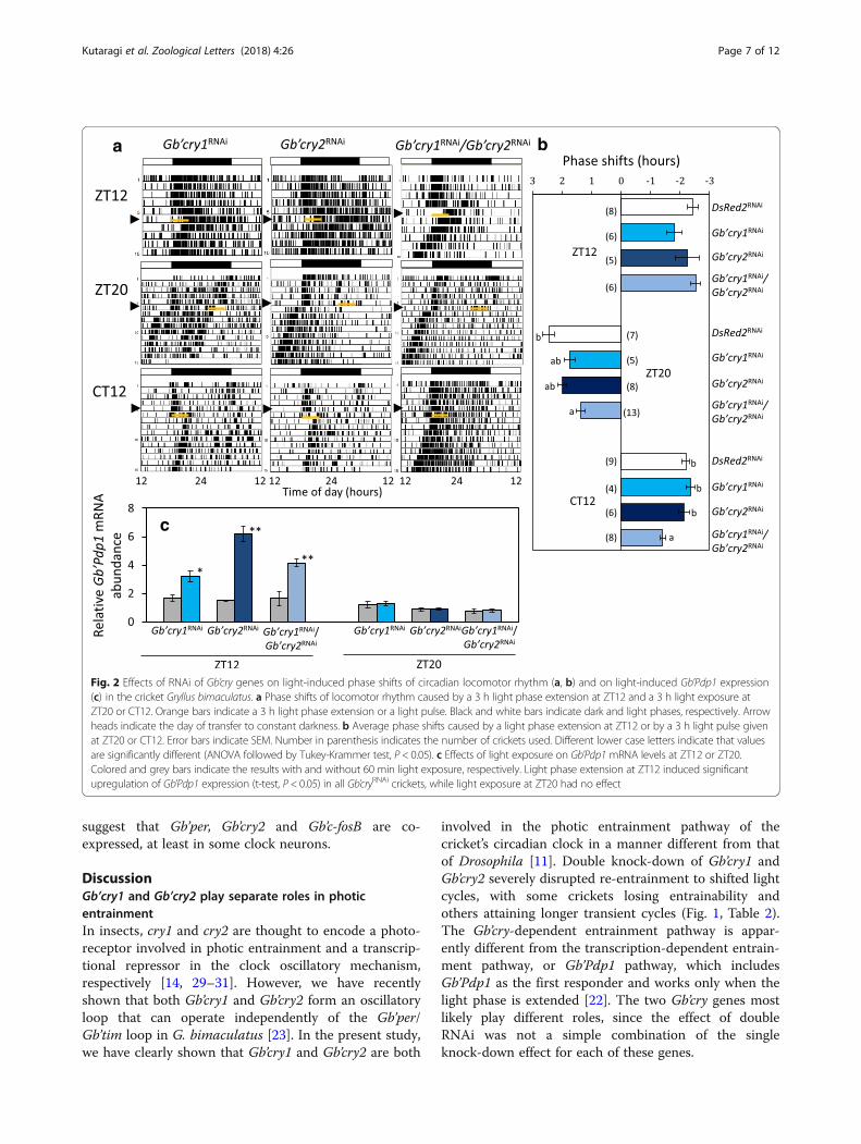
and Gb’cry2RNAi crickets showed advance shifts (1.75 ±0.47 h, n = 5; 1.99 ± 0.37 h, n = 8, respectively, Fig. 2)with the magnitude slightly less than that of the control,but the difference was not significant. In Gb’cry1/Gb’cry2double RNAi crickets, the shift was 1.37 ± 0.53 h (n =13), and was significantly smaller than that of the con-trol (Fig. 2). A 3 h light pulse given at CT12 induceddelay shifts in Gb’cry1RNAi and Gb’cry2RNAi crickets, withthe magnitude similar to that of DsRed2RNAi controlcrickets, while in Gb’cry1RNAi/Gb’cry2RNAi crickets, themagnitude was − 1.41 ± 0.26 h (n = 8), which was signifi-cantly smaller than that of the control (− 2.20 ± 0.37 h,n = 9) (Fig. 2a, b).We also examined the effects of a 3 h extension of light
phase (ZT12–15) on the phase of free-running locomotorrhythms in the ensuing DD. There were no significant ef-fects of single or double dsRNA treatment of Gb’cry geneson the magnitude of the phase shifts (Fig. 2a, b).
Effects of Gb’cry1 and Gb’cry2 knock-down on photicresponses of Gb’Pdp1We next examined the effects of Gb’cry1RNAi andGb’cry2RNAi treatment on light-induced Gb’Pdp1 upregu-lation, which is the first responder to light phase exten-sion at early subjective night [22]. We measured themRNA levels of Gb’Pdp1 1 h after light phase extensionstarting at ZT12, or 1 h after light exposure starting atZT20.In all RNAi crickets, Gb’Pdp1 was upregulated 1 h
after light-on at ZT12 in comparison to the control
Kutaragi et al. Zoological Letters (2018) 4:26 Page 4 of 12

a b c
d
g h i
e f
Fig. 1 Entrainment of the locomotor rhythm to LD12:12 in DsRed2RNAi (a), Gb’cry1RNAi (b), Gb’cry2RNAi (c), and Gb’cry1RNAi/Gb’cry2RNAi crickets (Gryllusbimaculatus) (d-i). Light cycles were advanced or delayed by 6 h on the day indicated by an arrowhead on the left side of the actograms. Whiteand black bars above the actograms indicate light (white) and dark (black) cycles. Yellow arrows indicate the light induced activity at light-onafter a 6 h phase advance of the light cycle. Transient cycles were shorter in Gb’cry1RNAi and Gb’cry2RNAi crickets than in the control (DsRed2RNAi)crickets, but were longer in Gb’cry1RNAi/Gb’cry2RNAi crickets. Some of the Gb’cry1RNAi/Gb’cry2RNAi crickets (e, h, i) apparently lost entrainability
Table 1 Effects of Gb’cry1 and Gb’cry2 RNAi on the phase relationship between light-off and activity onset. Different lower case lettersindicate that values are significantly different (ANOVA followed by Tukey-test, P < 0.05). Ψadvance and Ψdelay indicate reestablished phaserelationship after 6 h phase advance or delay of LD cycles, respectively. Numbers in parenthesis indicate number of animals used
Treatment Ψoriginal Ψadvance Ψdelay
dsDsRed2 0.51 ± 0.46 h (28) 0.68 ± 0.56a h (18) 0.92 ± 0.82ab h (13)
dsGb’cry1 0.60 ± 0.51 h (35) 0.69 ± 0.60a h (28) 0.73 ± 0.66ab h (21)
dsGb’cry2 0.52 ± 0.78 h (15) 0.56 ± 0.74a h (16) 0.30 ± 0.28a h (14)
dsGb’cry1/dsGb’cry2 0.87 ± 0.84 h (45) 1.69 ± 1.60b h (14) 1.02 ± 0.76b h (18)
Kutaragi et al. Zoological Letters (2018) 4:26 Page 5 of 12
without light exposure, while no significant changeswere observed in Gb’Pdp1 levels when a light pulse wasgiven at ZT20 (Fig. 2c). The results were similar to thoseobserved in untreated crickets [22], suggesting thatGb’crys are not upstream components of thetranscription-dependent entrainment pathway.
Gb’c-fosB is involved upstream of Gb’crysSince a bZip transcription factor gene, c-fos, is known tobe up-regulated by light exposure in mammalian circa-dian clocks [28], we examined whether it also respondedto light in crickets. G. bimaculatus has two isoforms ofc-fos: Gb’c-fosA (fra-A, LC215243) and Gb’c-fosB (fra-B,LC215244), which arise from alternative promoter usageor alternative splicing from a single locus. We thustested the effects of a 3 h light exposure at ZT20 andCT12 on their expression levels. Gb’c-fosB was signifi-cantly upregulated following light exposure, increasingfrom 30 min after light-exposure, but increasing signifi-cantly after 60 min (Fig. 3a), and then decreasing tobasal levels after 120 min.We then examined the effect of Gb’c-fosRNAi on the
light-induced resetting of the circadian locomotorrhythm. Gb’c-fosA and Gb’c-fosB were both targeted be-cause their nucleotide sequences are mostly identical,and we could not make a specific knock-down ofGb’c-fosB. The dsRNA treatment effectively knockeddown the Gb’c-fosB mRNA levels to approximately 25%of that of the DsRed2RNAi controls (Additional file 3:Figure S2). The Gb’c-fosRNAi had no significant effect onthe locomotor rhythm; the treated crickets showed arhythm synchronized to LD cycles and free-running inDD (Fig. 3b). There was no significant difference infree-running period in DD between DsRed2RNAi crickets(23.70 ± 0.38 [mean ± SD] h, n = 9) and Gb’c-fosRNAi
crickets (23.81 ± 0.30 h, n = 8) which were transferreddirectly to DD. A 3 h light pulse given at CT12 orZT20 delayed or advanced the rhythm in Gb’c-fosRNAi
by − 1.32 ± 0.86 h (n = 19) and 1.24 ± 0.59 h (n = 10),respectively, but the magnitude was significantly smallerthan that of DsRed2RNAi control crickets (Fig. 3b, c). Wealso used dsRNA specific for Gb’c-fosA, and found no sig-nificant effects on advance shifts caused by a 3 h lightpulse at ZT20 (Additional file 4: Figure S3). The suppres-sion of phase-shifts by Gb’c-fosRNAi was therefore appar-ently caused by knockdown of Gb’c-fosB. However, nosuppression was observed when light phase was extendedby 3 h at ZT12 (Fig. 3b, c).Following RNAi of Gb’opLW, light induced phase shifts
were significantly reduced in all light treatments (Fig. 3b, c),being consistent with previous observations [21, 22]. Wefurther examined the effects of Gb’c-fos knock-down onGb’Pdp1 levels in response to light phase extension. InGb’c-fosRNAi crickets, Gb’Pdp1 mRNA levels were up-regu-lated when the light phase was extended at ZT12, while nosignificant changes were observed when light was given atZT20 or CT12 (Fig. 3d), similar to control crickets [22],suggesting that the Pdp1-dependent entrainment pathwayis independent of the c-fos pathway.We then examined the effects of Gb’opLWRNAi on
Gb’c-fosB levels, since Gb’OpLW is the major photo-receptor for photic entrainment in this cricket [21, 22].We observed no significant changes in Gb’c-fosB levelsfollowing light exposure at CT12 and ZT20 inGb’opLWRNAi crickets (Fig. 4a), while Gb’c-fosB was sig-nificantly upregulated in Gb’opBlueRNAi crickets 1 h afterlight exposure at ZT20 (Fig.4b). This suggests that thesignal from Gb’OpLW is required for the Gb’c-fosBdependent entrainment pathway.We then examined the effects of Gb’cry knock-down
on Gb’c-fosB mRNA levels. We found that Gb’cry1 orGb’cry2 single knock-down or double knock-down didnot affect light-induced upregulation of Gb’c-fosB atCT12 or ZT20 (Fig. 4c, d), suggesting that Gb’crys aredownstream of Gb’c-fosB.
Gb’cry2, Gb’per, and Gb’c-fosB are expressed in the opticlobeTo determine whether Gb’cry2, Gb’per, and Gb’c-fosB areexpressed in the clock neurons, we examined their ex-pression in the optic lobe by in situ hybridization or insitu RT-PCR. Although Gb’per and Gb’cry2 wereexpressed in many cells in the optic lobe at ZT18, theyco-localized in some neurons that are located near theouter chiasma between the lamina and medulla and theouter limb of lamina (Fig. 5). By in situ RT-PCRGb’c-fosB was found to be expressed by 1 h light expos-ure at ZT20 in cells closely located near the area inwhich Gb’per and Gb’cry2 are expressed. These results
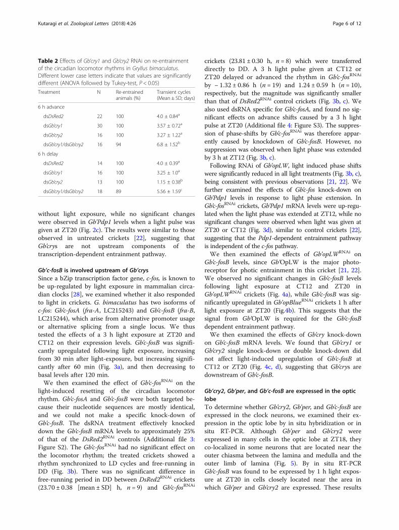
Table 2 Effects of Gb’cry1 and Gb’cry2 RNAi on re-entrainmentof the circadian locomotor rhythms in Gryllus bimaculatus.Different lower case letters indicate that values are significantlydifferent (ANOVA followed by Tukey-test, P < 0.05)
Treatment N Re-entrainedanimals (%)
Transient cycles(Mean ± SD; days)
6 h advance
dsDsRed2 22 100 4.0 ± 0.84a
dsGb’cry1 30 100 3.57 ± 0.72a
dsGb’cry2 16 100 3.27 ± 1.22a
dsGb’cry1/dsGb’cry2 16 94 6.8 ± 1.52b
6 h delay
dsDsRed2 14 100 4.0 ± 0.39a
dsGb’cry1 16 100 3.25 ± 1.0a
dsGb’cry2 13 100 1.15 ± 0.38b
dsGb’cry1/dsGb’cry2 18 89 5.56 ± 1.59c
Kutaragi et al. Zoological Letters (2018) 4:26 Page 6 of 12
suggest that Gb’per, Gb’cry2 and Gb’c-fosB are co-expressed, at least in some clock neurons.
DiscussionGb’cry1 and Gb’cry2 play separate roles in photicentrainmentIn insects, cry1 and cry2 are thought to encode a photo-receptor involved in photic entrainment and a transcrip-tional repressor in the clock oscillatory mechanism,respectively [14, 29–31]. However, we have recentlyshown that both Gb’cry1 and Gb’cry2 form an oscillatoryloop that can operate independently of the Gb’per/Gb’tim loop in G. bimaculatus [23]. In the present study,we have clearly shown that Gb’cry1 and Gb’cry2 are both
involved in the photic entrainment pathway of thecricket’s circadian clock in a manner different from thatof Drosophila [11]. Double knock-down of Gb’cry1 andGb’cry2 severely disrupted re-entrainment to shifted lightcycles, with some crickets losing entrainability andothers attaining longer transient cycles (Fig. 1, Table 2).The Gb’cry-dependent entrainment pathway is appar-ently different from the transcription-dependent entrain-ment pathway, or Gb’Pdp1 pathway, which includesGb’Pdp1 as the first responder and works only when thelight phase is extended [22]. The two Gb’cry genes mostlikely play different roles, since the effect of doubleRNAi was not a simple combination of the singleknock-down effect for each of these genes.
a
c
b
Fig. 2 Effects of RNAi of Gb’cry genes on light-induced phase shifts of circadian locomotor rhythm (a, b) and on light-induced Gb’Pdp1 expression(c) in the cricket Gryllus bimaculatus. a Phase shifts of locomotor rhythm caused by a 3 h light phase extension at ZT12 and a 3 h light exposure atZT20 or CT12. Orange bars indicate a 3 h light phase extension or a light pulse. Black and white bars indicate dark and light phases, respectively. Arrowheads indicate the day of transfer to constant darkness. b Average phase shifts caused by a light phase extension at ZT12 or by a 3 h light pulse givenat ZT20 or CT12. Error bars indicate SEM. Number in parenthesis indicates the number of crickets used. Different lower case letters indicate that valuesare significantly different (ANOVA followed by Tukey-Krammer test, P < 0.05). c Effects of light exposure on Gb’Pdp1 mRNA levels at ZT12 or ZT20.Colored and grey bars indicate the results with and without 60 min light exposure, respectively. Light phase extension at ZT12 induced significantupregulation of Gb’Pdp1 expression (t-test, P < 0.05) in all Gb’cryRNAi crickets, while light exposure at ZT20 had no effect
Kutaragi et al. Zoological Letters (2018) 4:26 Page 7 of 12

The Gb’cry2 knock-down accelerated re-synchronizationof the locomotor rhythm to delayed LDs (Fig. 1c). Al-though we still need to examine whether this is true en-trainment, this fact is reminiscent of the report that cryknock-out mice have significantly larger phase delays thanwild-type mice [32]. Because Gb’cry2 is an essential com-ponent of the cry-oscillatory loop [23], its knock-down re-sults in a severe impairment of the loop. The accelerationof light resetting may be caused by the Gb’Pdp1 pathway,which is still functional even in this condition. However,entrainment through the Gb’Pdp1 pathway apparently in-complete, because double RNAi of Gb’cry genes preventsphotic entrainment. This may also partly explain the large
Ψ of the rhythm after phase shifts in Gb’cry double RNAicrickets (Table 1), which is similar to cry-deficient mice[32]. In contrast, Gb’cry1RNAi only has a small effect on en-trainment (Fig. 1b), because the cry-loop can be operatedby Gb’cry2 variants [23].
The Gb’c-fosB pathway is involved in photic entrainmentAlthough the molecular mechanism of Gb’cry-dependentlight resetting of the cricket’s clock still remains to be ex-plored, Gb’c-fosB apparently plays a regulatory role up-stream of Gb’crys since Gb’c-fosB expression wasupregulated by light even after knocking-down of Gb’crys(Fig. 4). This hypothesis is strongly supported by the
a
b
d
c
Fig. 3 Changes of Gb’c-fosB mRNA levels caused by light exposure (a), effects of RNAi of Gb’c-fos and Gb’opLW genes on the light-induced phaseshifts of locomotor rhythms (b, c), and effects of Gb’c-fosRNAi on the light induced Gb’Pdp1 expression (d) in the cricket Gryllus bimaculatus.a Gb’c-fosB was upregulated by light exposure at ZT20 and CT12, and a statistically significant difference was evident after 60 min of exposure(**P < 0.01, t-test). The effect was greater in the late night (ZT20). Yellow and grey bars indicate the results with and without light exposure,respectively. b Phase shifts of locomotor rhythm caused by a 3 h light phase extension at ZT12 or 3 h light exposure at ZT20 or CT12 (orangebars). Black and white bars indicate dark and light phases, respectively. Arrow heads indicate the day of transfer to constant darkness. c Average phaseshifts caused by a light phase extension at ZT12 or by a 3 h light pulse given at ZT20 or CT12. Error bars indicate SEM. Different lower-case lettersindicate that values are significantly different (ANOVA followed by Tukey-Kramer test, P < 0.05). Gb’c-fosRNAi suppressed the light-induced phase shifts atZT20 and CT12 but not at ZT12, and Gb’opLWRNAi strongly suppressed them in all cases (ANOVA followed by Tukey-Kramer test, P < 0.01). d Effects oflight exposure on Gb’Pdp1 mRNA levels at ZT12, ZT20, and CT12 in Gb’c-fosRNAi crickets. Yellow and grey bars indicate the results with and without60 min light exposure, respectively. Light phase extension at ZT12 induced significant upregulation of Gb’Pdp1 expression (**P < 0.01,t-test) in Gb’c-fosRNAi crickets, while no apparent effect was observed following light exposure at ZT20 and CT12
Kutaragi et al. Zoological Letters (2018) 4:26 Page 8 of 12

a
b d
c
Fig. 4 Effects of RNAi of Gb’opLW (a), Gb’opBlue (b), and Gb’cry genes (c, d) on light induced Gb’c-fosB mRNA expression in the cricket Gryllusbimaculatus. Gray and yellow columns indicate samples kept in darkness or exposed to light for 60 min, respectively. DD, kept in darkness; LP,exposed to light. Bars indicate SEM. N = 3 or 4. a Gb’opLWRNAi suppressed light-induced upregulation of Gb’c-fosB. b-d RNAi of Gb’opBlue or Gb’crygenes had no effects on the light-induced upregulation of Gb’c-fosB at ZT20 and CT12. *P < 0.05, t-test
a d
b c
Fig. 5 Expression of Gb’per, Gb’cry2, and Gb’c-fosB in the optic lobe of the cricket Gryllus bimaculatus. a-c In situ hybridization of Gb’per (red) andGb’cry2 (blue) in the optic lobe sampled at ZT18. b and c shows magnification of areas near outer chiasma indicated in a. Short arrows indicatethe cells coexpressing Gb’per and Gb’cry2. d In situ PCR of Gb’c-fosB in the optic lobe after 1 h light exposure at ZT20. Gb’c-fosB was stronglyexpressed in the cells near outer chiasma and along the outer surface of lamina. For further explanations see text
Kutaragi et al. Zoological Letters (2018) 4:26 Page 9 of 12

finding that Gb’c-fosB expression detected by in situRT-PCR overlapped with that of cells co-expressing Gb’perand Gb’cry2. c-fos has been used as a marker for light in-put in vertebrate circadian clocks because of its rapid in-duction after light exposure [33]. Its product proteinforms a complex, activating protein 1 (AP-1), with othertranscription factor genes, such as jun gene family mem-bers, and activates transcription of target genes [34]. How-ever, its role in resetting of the circadian clock remainsmostly unclear. Our results revealed for the first time thatc-fos plays a major role in resetting the clock in insects,since Gb’c-fosRNAi strongly prevents light induced phaseshifts of the circadian rhythm. This also suggests thatGb’crys are downstream of Gb’c-fosB. At present, althoughthe connection between Gb’c-fosB and Gb’crys is unclear,F-box and leucin rich repeat proteins (FBXL) and Bromo-domain and WD repeat domain containing 3(BRWD3) may be mediating this connection, as theyare known to regulate CRY degradation by ubiquitinationof CRY [35–37]. Light input to the Gb’c-fosB pathway isthrough Gb’OpLW, because the light-induced upregula-tion of Gb’c-fosB is eliminated by Gb’opLWRNAi. Based onthe present findings and those of previous studies, themost likely hypothesis appears to be that light informationis supplied to the clock neurons by neurotransmissionthrough the OpLW pathway, causing induction ofGb’c-fosB, followed by a functional modulation of FBXL orBRWD3, which may ubiquitinate CRYs and reset the os-cillatory mechanism including CRYs (Fig. 6). This hypoth-esis should be tested in future studies.
Besides the Gb’c-fosB mediated pathway, Gb’crys mightalso be regulated by light through other mechanisms.Our previous study showed that light phase extension atearly night induced Gb’cry2 upregulation with an in-crease of Gb’Pdp1 and Gb’Clk in nymphal crickets [22],suggesting the E-box mediated transcription of Gb’cry2by Gb’Clk. Another possibility is through a D-box medi-ated regulation known for zebrafish clocks, in whichlight induces cry1a expression through a D-box medi-ated mechanism including PAR bZip factors, PAR andTEF-1 [38]. Because D-boxes are found in thecis-regulatory region of Gb’cry1 and Gb’cry2, a similarmechanism might be involved in the cricket clock. Theseissues should be addressed in future studies.
Heterogeneous nature of clock cellsIn the cricket’s clock, there must be at least two sets ofclock neurons since some fraction of the Gb’cry1/Gb’cry2double RNAi crickets showed a rhythm dissociation intotwo components when the light cycle was shifted by 6 h:one component still retained photic entrainability, whileit was lost in the other (Fig. 1e). The heterogenous cellu-lar organization of the clock is quite similar to that ob-served in Drosophila and mammals [39, 40], whereentrainability to light is different between the cerebral orSCN clock neurons [41–43].The heterogeneous cellular nature of the clock may
explain the effect of Gb’cry2RNAi on the free-runninglocomotor rhythm. We have previously shown thatGb’cry2RNAi crickets have a wide variety of free-running
Fig. 6 A model for the light entrainment mechanism of the cricket circadian clock. The clock includes two major oscillatory loops, one for Gb’perand Gb’tim, like in Drosophila, and the other for Gb’cry2. The latter is comprised of Gb’cry1 and two Gb’cry2 isoforms, Gb’cry2c and Gb’cry2f, and theirproduct proteins form a complex that suppresses the transcription mediated by Gb’CLK/Gb’CYC complex [23]. Light is perceived by the retinularcells in the compound eye expressing Gb’opLW, and the information is transmitted to the clock neuron in the optic lobe through neurotransmitters.This neurotransmission causes upregulation of Gb’c-fosB in the clock neurons, which finally affects the Gb’cry2 oscillatory loop. The change in the Gb’cry2loop may reset the whole clock system since the Gb’cry2 loop interact with the Gb’per/Gb’tim loop by influencing Gb’CLK/Gb’CYC. In addition to theGb’cry2 pathway, the Gb’Pdp1 pathway resets the clock by upregulating Gb’Clk expression when light off was delayed [22]
Kutaragi et al. Zoological Letters (2018) 4:26 Page 10 of 12

periods in DD [23], which may reflect the underlying os-cillatory neurons. The lack of Gb’CRY2 following RNAimay destroy the stable free-running of the clock neuron,hence causing dissociation among the cells. This is rem-iniscent of the mammalian SCN clock, where CRY playsa role as a coupling factor of clock cells that have vari-able free-running periods [39, 44–46].
Comparison with other insect clocksIn the present study, we found for the first time that bothGb’cry1 and Gb’cry2 play important roles in photic en-trainment of the circadian clock in crickets. In hemi-metabolous insects, the photoreceptor for entrainment isbelieved to reside in the compound eye [19, 47, 48], andthis assumption has been confirmed at the molecular levelin the cricket G. bimaculatus [21]. Therefore, Gb’cry1 andGb’cry2 are both active in the clock resetting mechanismdownstream of the neurotransmission from the retinalphotoreceptor. Although the exact role of Gb’cry genesshould be examined in future studies, the cricket’sGb’CRY1 likely works together with Gb’CRY2 to form anoscillatory feedback loop, which can operate independ-ently of the Gb’per/Gb’tim loop [23]. As for insect cry2, itis believed to be involved in the clock machinery as aclock component [14]. However, we have shown for thefirst time that Gb’cry2 also plays an important role in thephotic entrainment of the clock. Our results shed light onthe insect clock mechanism and provide deeper under-standing of its photic entrainment and diversification.
ConclusionsPhotic entrainment is an essential property of the animalcircadian clock that sets the appropriate timing of behav-ioral and physiological events in a 24 h cycle. Althoughmost insects use compound eyes as photoreceptors for en-trainment, the molecular mechanisms underlying entrain-ment remain largely unknown. Here we elucidated for thefirst time the molecular entrainment pathway using ahemimetabolous insect, the cricket Gryllus bimaculatus.Our results suggest that neural signals mediated bygreen-sensitive opsins in the compound eye firstup-regulate an isoform of the bZip transcription factorgene, Gb’c-fos, which subsequently resets the clockthrough Gb’cry1 and Gb’cry2. These findings contribute tounderstanding of the photic entrainment mechanism ofinsect clocks.
Additional files
Additional file 1: Table S1. PCR primers used for quantitative RT-PCRand dsRNA synthesis. The primers tagged with T7 or T3 promoter sequenceswere used for PCR amplification for dsRNA synthesis. T7 and T3 sequencesare underlined. (DOCX 18 kb)
Additional file 2: Figure S1. Effect of RNAi of cry genes on locomotoractivity during the first 3 h after light-on in the cricket Gryllus bimaculatus.The activity was measured on the first day after 6 h phase advance oflight-on. Error bars indicate SEM. Numbers in parenthesis indicate thenumber of animals used. Gb’cry2RNAi and Gb’cry1RNAi/Gb’cry2RNAi significantlyreduced the light-induced locomotor activity compared to DsRed2RNAi
treatment (*P < 0.05, **P < 0.01, Dunnett’s test). (PDF 58 kb)
Additional file 3: Figure S2. Gb’c-fosRNAi significantly down-regulatedboth Gb’c-fosA and Gb’c-fosB mRNA levels in the optic lobe of the cricketGryllus bimaculatus (**P < 0.01, t-test). The optic lobes were collected atZT20 seven days after dsRNA injection. mRNA levels were measured byqPCR and are shown relative to those of Gb’rpl18a. The values shown aremean ± SEM of six samples. (PDF 49 kb)
Additional file 4: Figure S3. A: Gb’c-fosARNAi had no significant effectson the light induced phase advance in the cricket Gryllus bimaculatus. A3 h light pulse was given at ZT20 on the day of transfer to DD, whichwas seven days after dsRNA injection. Numbers in the parenthesis indicatethe number of animals used. B and C: Gb’c-fosARNAi significantly knockeddown Gb’c-fosA mRNA levels (*P < 0.05, t-test), but had no significant effecton Gb’c-fosB mRNA levels. mRNA levels were measured by qPCR and areshown relative to those of Gb’rpl18a. The values shown are mean ± SEM offour samples. (PDF 69 kb)
AcknowledgmentsWe thank Taishi Yoshii for helpful discussions and Tsugumichi Shinohara forhis assistance in manuscript preparation.
Authors’ contributionsYK and KT designed the experiments. YK, AT, MN, YT, YM performed theexperiments. TB and TW performed promoter analysis of Gb’cry genes andmolecular analysis of Gb’c-fos gene, respectively. YK and KT analyzed the dataand wrote the manuscript. All authors read and approved the final manuscript.
FundingThis study was supported in part by JSPS KAKENHI Grant NumberJP15H0440017 and JP18H02480.
Availability of data and materialsThe datasets supporting the conclusions of this article are included withinthe article.
Ethics approval and consent to participateNot applicable.
Consent for publicationNot applicable.
Competing interestsThe authors declare they have no competing interests.
Publisher’s NoteSpringer Nature remains neutral with regard to jurisdictional claims inpublished maps and institutional affiliations.
Author details1Graduate School of Natural Science and Technology, Okayama University,Okayama 700-8530, Japan. 2Research Institute for Electronic Science,Hokkaido University, Sapporo 060-0811, Japan. 3Graduate School of Medicine,Dentistry and Pharmaceutical Sciences, Okayama University , Okayama700-8558, Japan. 4Department of Natural Sciences, Kawasaki Medical School,Matsushima 577, Kurashiki 701-0192, Japan.
Received: 2 July 2018 Accepted: 4 September 2018
References1. Saunders DS, Steel CGH, Vafopoulou X, Lewis RD. Insect Clocks, vol. 560.
Amsterdam: Elsevier 3rd ed. p. 2002.
Kutaragi et al. Zoological Letters (2018) 4:26 Page 11 of 12

2. Hardin P. Molecular mechanisms of circadian timekeeping in Drosophila.Sleep Biol Rhythms. 2009;7:235–42.
3. Tataroglu O, Emery P. The molecular ticks of the Drosophila circadian clock.Curr Opin Insect Sci. 2015;7:51–7.
4. Tomioka K, Matsumoto A. Circadian molecular clockworks in non-modelinsects. Curr Opin Insect Sci. 2015;7:58–64.
5. Blau J, Young MW. Cycling vrille expression is required for a functionalDrosophila clock. Cell. 1999;99:661–71.
6. Cyran SA, Buchsbaum AM, Reddy KL, Lin M-C, Glossop NRJ, Hardin PE, et al.vrille, Pdp1 and dClock form a second feedback loop in the Drosophilacircadian clock. Cell. 2003;112:329–41.
7. Kamae Y, Uryu O, Miki T, Tomioka K. The nuclear receptor genes HR3 andE75 are required for the circadian rhythm in a primitive insect. PLoS One.2014;9:e114899.
8. Yoshii T, Todo T, Wülbeck C, Stanewsky R, Helfrich-Förster C. Cryptochromeis present in the compound eyes and a subset of Drosophila's clockneurons. J Comp Neurol. 2008;508:952–66.
9. Ceriani MF, Darlington TK, Staknis D, Mas P, Petti AA, Weitz CJ, et al. Light-dependent sequentation of TIMELESS by CRYPTOCHROME. Science. 1999;285:553–6.
10. Lin F-J, Song W, Meyer-Bernstein E, Naidoo N, Sehgal A. Photic signaling bycryptochrome in the Drosophila circadian system. Mol Cell Biol. 2001;21:7287–94.
11. Emery P, So WV, Kaneko M, Hall JC, Rosbash M. CRY, a Drosophila clock andlight-regulated cryptochrome, is a major contributor to circadian rhythmresetting and photosensitivity. Cell. 1998;95:669–79.
12. Ishikawa T, Matsumoto A, Kato Jr. T, Togashi S, Ryo H, Ikenaga M, et al.DCRY is a Drosophila photoreceptor protein implicated in light entrainmentof circadian rhythm. Gene Cells 1999;4:57–65.
13. Ingram KK, Kutowoi A, Wurm Y, Shoemaker D, Meier R, Bloch G. The molecularclockwork of the fire ant Solenopsis invicta. PLoS One. 2012;7:e45715.
14. Yuan Q, Metterville D, Briscoe AD, Reppert SM. Insect cryptochromes: geneduplication and loss define diverse ways to construct insect circadianclocks. Mol Biol Evol. 2007;24:948–55.
15. Langmesser S, Tallone T, Bordon A, Rusconi S, Albrecht U. Interaction ofcircadian clock proteins PER2 and CRY with BMAL1 and CLOCK. BMC MolBiol. 2008;9:41.
16. Ye R, Selby CP, Chiou Y-Y, Ozkan-Dagliyan I, Gaddameedhi S, Sancar A. Dualmodes of CLOCK:BMAL1 inhibition mediated by Cryptochrome and periodproteins in the mammalian circadian clock. Genes Dev. 2016;28:1989–98.
17. Selby CP, Thompson C, Schmitz TM, Van Gelder RN, Sancar A. Functionalredundancy of cryptochromes and classical photoreceptors for nonvisualocular photoreception in mice. Proc Natl Acad Sci. 2000;97:14697–702.
18. Nathalie H, Schleicher E, Kacprzak S, Bouly J-P, Picot M, Wu W, et al. Humanand Drosophila cryptochromes are light activated by flavin photoreductionin living cells. PLoS Biol. 2008;6:e160.
19. Tomioka K, Chiba Y. Effects of nymphal stage optic nerve severance or opticlobe removal on the circadian locomotor rhythm of the cricket. Zool Sci.1984;1:375–82.
20. Yukizane M, Tomioka K. Neural pathways involved in mutual interactionsbetween optic lobe circadian pacemakers in the cricket Gryllus bimaculatus.J Comp Physiol A. 1995;176:601–10.
21. Komada S, Kamae Y, Koyanagi M, Tatewaki K, Hassaneen E, Saifullah A, et al.Green-sensitive opsin is the photoreceptor for photic entrainment of aninsect circadian clock. Zoological Letters. 2015;1:11.
22. Kutaragi Y, Miki T, Bando T, Tomioka K. Transcriptional and non-transcriptional events are involved in photic entrainment of the circadianclock in the cricket Gryllus bimaculatus. Physiol Entomol. 2016;41:358–68.
23. Tokuoka A, Itoh TQ, Hori S, Uryu O, Danbara Y, Nose M, et al. cryptochrome genesform an oscillatory loop independent of the per/tim loop in the circadianclockwork of the cricket Gryllus bimaculatus. Zoological Letters. 2017;3:5.
24. Moriyama Y, Sakamoto T, Karpova SG, Matsumoto A, Noji S, Tomioka K. RNAinterference of the clock gene period disrupts circadian rhythms in thecricket Gryllus bimaculatus. J Biol Rhythm. 2008;23:308–18.
25. Sokolove PG, Bushell WN. The chi square periodogram: its utility for analysisof circadian rhythm. J Theor Biol. 1978;72:131–60.
26. Schmid B, Helfrich-Förster C, Yoshii T. A new ImageJ plug-in “ActogramJ” forchronobiological analyses. J Biol Rhythm. 2011;26:464–7.
27. Kaneko M, Park JH, Cheng Y, Hardin PE, Hall JC. Disruption of synaptictransmission or clock-gene-product oscillations in circadian pacemaker cells ofDrosophila cause abnormal behavioral rhythms. J Neurobiol. 2000;43:207–33.
28. Kornhauser JM, Nelson DE, Mayo KE, Takahashi JS. Photic and circadianregulation of c-fos gene expression in hamster suprachiasmatic nucleus.Neuron. 1990;5:127–34.
29. Zhang L, Lear BC, Seluzicki A, Allada R. The CRYPTOCHROME photoreceptorgates PDF neuropeptide signaling to set circadian network hierarchy inDrosophila. Curr Biol. 2009;19:2050–5.
30. Zhu H, Yuan Q, Briscoe AD, Froy O, Casselman A, Reppert SM. The two CRYsof the butterfly. Curr Biol. 2005;15:R953–4.
31. Michae AK, Fribourgh JL, Gelder RNV, Partch CL. Animal cryptochromes:divergent roles in light perception, circadian timekeeping and beyond.Photochem Photobiol. 2017;93:128–40.
32. Spoelstra K, Albrecht U, GTJvd H, Brauer V, Daan S. Phase responses to lightpulses in mice lacking functional Per or Cry genes. J Biol Rhythm. 2004;19:518–29.
33. Moore HAM, Whitmore D. Circadian rhythmicity and light sensitivity of thezebrafish brain. PLoS One. 2014;9:e86176.
34. Guillaumond F, Sage D, Deprez P, Bosler O, Becquet D, François-Bellan AM.Circadian binding activity of AP-1, a regulator of the arylalkylamineN-acetyltransferase gene in the rat pineal gland, depends on circadian Fra-2,c-Jun, and Jun-D expression and is regulated by the clock's zeitgebers.J Neurochem. 2000;75:1398–407.
35. Yoo S-H, Mohawk JA, Siepka SM, Shan Y, Huh SK, Hong H-K, et al.Competing E3 ubiquitin ligase govern circadian periodicity by degradationof CRY in nucleus and cytoplasm. Cell. 2013;152:1091–105.
36. Hirano A, Yumimoto K, Tsunematsu R, Matsumoto M, Oyama M, Kozuka-Hata H, et al. FBXL21 regulates oscillation of the circadian clock throughubiquitination and stabilization of Cryptochromes. Cell. 2013;152:1106–18.
37. Ozturk N, VanVickle-Chavez SJ, Akileswaran L, Van Gelder RN, Sancar A.Ramshackle (Brwd3) promotes light-induced ubiquitylation of DrosophilaCryptochrome by DDB1-CUL4-ROC1 E3 ligase complex. Proc Natl Acad Sci.2013;110:4980–5.
38. Mracek P, Santoriello C, Idda ML, Pagano C, Ben-Moshe Z, Gothilf Y, et al.Regulation of per and cry genes reveals a central role for the D-boxenhancer in light-dependent gene expression. PLoS One. 2012;7:e51278.
39. Yamaguchi S, Isejima H, Matsuo T, Okura R, Yagita K, Kobayashi M, et al.Synchronization of cellular clocks in the suprachiasmatic nucleus. Science.2003;302:1408–12.
40. Kaneko M, Hall JC. Neuroanatomy of cells expressing clock genes inDrosophila: transgenic manipulation of the period and timeless genes tomark the perikarya of circadian pacemaker neurons and their projections.J Comp Neurol. 2000;422:66–94.
41. Miyasako Y, Umezaki Y, Tomioka K. Separate sets of cerebral clock neuronsare responsible for light and temperature entrainment of Drosophilacircadian locomotor rhythms. J Biol Rhythm. 2007;22:115–26.
42. Yoshii T, Hermann-Luibl C, Kistenpfennig C, Schmid B, Tomioka K, Helfrich-Förster C. Cryptochrome-dependent and -independent circadianentrainment circuits in Drosophila. J Neurosci. 2015;35:6131–41.
43. Golombek DA, Rosenstein RE. Physiology of circadian entrainment. PhysiolReviews. 2010;90:1063–102.
44. Ono D, Honma S, K-i H. Cryptochromes are critical for the development ofcoherent circadian rhythms in the mouse suprachiasmatic nucleus. NatCommun. 2013;4:1666.
45. Evans JA, Pan H, Liu AC, Welsh DK. Cry1−/− circadian rhythmicity dependson SCN intercellular coupling. J Biol Rhythm. 2012;27:443–52.
46. Inagaki N, Honma S, Ono D, Tanahashi Y, Honma K-i. Separate oscillatingcell groups in mouse suprachiasmatic nucleus couple photoperiodically tothe onset and end of daily activity. Proc Natl Acad Sci. 2007;104:7664–9.
47. Nishiitsutsuji-Uwo J, Pittendrigh CS. Central nervous system control ofcircadian rhythmicity in the cockroach. II. The pathway of light signals thatentrain the rhythm. Z vergl Physiol. 1968;58:1–13.
48. Page TL. Interaction between bilaterally paired components of thecockroach circadian system. J Comp Physiol. 1978;124:225–36.
Kutaragi et al. Zoological Letters (2018) 4:26 Page 12 of 12