a publication by retina - review of ophthalmology publication by page 18 page 22 page 25 page 30...
TRANSCRIPT

R E T I N AS P E C I A L I S T
M A R C H 2 0 1 5
A P U B L I C A T I O N B Y
PAGE 18
PAGE 22
PAGE 25
PAGE 30
What we can take from the study.
R E T I N A - S P E C I A L I S T . C O M
The RVO Workup: When It’s Necessary And What to Order
Real-Life Story of the‘Bionic Eye’: The ARGUS II
How to Manage a Migrating Dexamethasone Implant
LESSONS DRCR.NET
PROTOCOL T
Can Anti-VEGF Cause GA in Wet AMD? Literature Review
What’s the Cause of This Panuveitis? Retina Fellows ForumPage 12 Page 15
FROM
001_rs0315_fc_rk2.indd 1001_rs0315_fc_rk2.indd 1 3/17/15 11:56 AM3/17/15 11:56 AM

RP0215_Allergan Ozurdex.indd 2RP0215_Allergan Ozurdex.indd 2 1/22/15 1:29 PM1/22/15 1:29 PM

RP0215_Allergan Ozurdex.indd 3RP0215_Allergan Ozurdex.indd 3 1/22/15 1:29 PM1/22/15 1:29 PM
RP0215_Allergan Ozurdex PI.indd 1RP0215_Allergan Ozurdex PI.indd 1 1/22/15 1:44 PM1/22/15 1:44 PM

Go FurtherWithout Leaving
Home
Continue your professional development and sharpen your clinical skills through convenient CME programs online and on your schedule.
Review of Ophthalmology® offers continuing education for physicians and staff, covering the latest in disease diagnosis and treatment, surgical advances and other topics, available any time on our website at CME online.
Download a QR scanner app. Launch app and hold your mobile device over the code to view www.reviewofophthalmology.com/continuing_education/.
www.reviewofophthalmology.com/continuing_education/
005_rs0315_fracthousead.indd 1005_rs0315_fracthousead.indd 1 3/18/15 10:28 AM3/18/15 10:28 AM
Sunshine Act Review Period Set to OpenYou may have already received
a notice from the Centers for Medicare and Medicaid Ser-
vices (CMS) to register for the Open Payments system. Come April, indi-vidual physicians will have an oppor-tunity to view payment data CMS has collected from device makers and pharmaceutical companies before the information goes public.
The release is part of the Open Payments program, also known as the Physician Payment Sunshine Act, created by the Affordable Care Act. It will list consulting fees, research grants, travel reimbursements and other gifts medical device makers, drug companies and group purchas-ing organizations paid physicians and teaching hospitals for all of 2014. The initial release last year included only data for the last fi ve months of 2013.
The 2013 data listed 4.4 million payments totaling nearly $3.5 bil-lion. Some 1,419 manufacturers and group purchasing plans paid that out to 546,000 individual physicians and 1,360 teaching hospitals. The 2013 data omitted another $1.1 billion in payments because of data problems.
Last year only 26,000 physicians, or 4.8 percent, registered in the Open Payments system to review the pay-ments attributed to them.
The American Academy of Oph-
thalmology (AAO) is encouraging members to review the data before the June 30 release. “Members who might have erroneous data reported in their name ought to be proactive in identifying and correcting that,” says Michael X. Repka, MD, AAO medi-cal director for government affairs. “If I was going to tell an ophthalmologist what to do, I would say sign up, take advantage of the period of time in which data are there for review to identify disputed data, as CMS calls it, and request its withdrawal.”
Eli Y. Adashi, MD, MS, a professor at Brown University in Providence, R.I., explored the problems with the initial data release in a recent article in the Journal of the American Medi-cal Association.1 “Last year was differ-ent,” he says in an interview. “There really wasn’t all that much time. The website was clunky and, worse, it failed and had to be taken off line for close to two weeks.”
Dr. Adashi has advised physicians who receive significant commercial support to take advantage of the re-view and dispute period. “The key argument in looking at the data is to ensure accuracy, but not just in dollar amounts, but the breakdown of sup-port into categories to see that they’re appropriately characterized,” he says.
“This is not a once and done pro-
RETINA SPECIALIST | MARCH 20156
N E W S
IN BRIEF
• Genentech received Food and Drug Administration approval for Lucentis (ranibizumab injection) for treatment of diabetic retinopathy in diabetic macular edema. The FDA in February granted Lucentis Breakthrough Therapy Designation and Priority Review for this indication based on results from the RISE and RIDE Phase III clinical trials.
• RXi Pharmaceuticals Corp. has received a U.S. patent for the delivery of double-stranded siRNAs (21 to 23 nucleotides in length) across the blood-retina barrier for the treatment of wet age-related macular degeneration or diabetic retinopathy. The patent, part of RXi’s acquired OPKO estate, is scheduled to expire in 2023.
• Spark Therapeutics has initiated enrollment of a Phase I/II clinical trial of its product candidate, SPK-CHM, for patients with choroideremia.
• Allegro Ophthalmics received a $2 million grant from the Type 1 Diabetes Program of The Leona M. and Harry B. Helmsley Charitable Trust to support a Phase II study of Luminate, its integrin peptide therapy currently in multiple Phase II studies, for treatment of dia-betic macular edema.
• iCura Vision has licensed the intellec-tual property portfolio and associated research and development program for an oral medication intended to treat dry AMD. The company intends to place the fi rst of these candidates, ICR-14967, into clinical development in 2016. Under the terms of the licensing agreement, the National Institute of Health’s Blueprint
Neurotherapeutics Network, which has funded the discovery and early devel-opment of the medication, will continue to provide fi nancial support through Phase I studies.
006_rs0315_news_rk2.indd 6006_rs0315_news_rk2.indd 6 3/17/15 11:57 AM3/17/15 11:57 AM

DARPin Abicipar Pegol a ‘Free Call Option’ for Actavis
cess; it’s an ongoing process for our members,” Dr. Repka says. “Even though the public wasn’t much inter-ested last year in the data, it doesn’t mean they won’t be this time.”
Registration is available on the CMS website at cms.gov/OpenPay-
ments/Program-Participants/Physi-cians-and-Teaching-Hospitals/Regis-tration.html.
REFERENCE1. Santhakumar S, Adashi EY. Viewpoint: The Physician Payment Sunshine Act; Testing the value of transparency. JAMA. 2015;313:23-24.
RETINA SPECIALIST | MARCH 2015 7
BUSINESS OFFICES
11 CAMPUS BOULEVARD, SUITE 100
NEWTOWN SQUARE, PA 19073
SUBSCRIPTION INQUIRIES (877) 529-1746
(USA ONLY); OUTSIDE USA, CALL (847) 763-9630
BUSINESS STAFF
PUBLISHER
JAMES HENNE
(610) 492-1017 [email protected]
REGIONAL SALES MANAGER
MICHELE BARRETT
(610) 492-1014 [email protected]
REGIONAL SALES MANAGER
MICHAEL HOSTER
(610) 492-1028 [email protected]
CLASSIFIED ADVERTISING
(888)-498-1460
VICE PRESIDENT OF OPERATIONS
CASEY FOSTER
(610) 492-1007 [email protected]
PRODUCTION MANAGER
SCOTT TOBIN
(610) 492-1011 [email protected]
SUBSCRIPTIONS
$63 A YEAR, $99 (U.S.) IN CANADA,
$158 (U.S.) IN ALL OTHER COUNTRIES.
SUBSCRIPTIONS E-MAIL:
CIRCULATION
PO BOX 71, CONGERS, NY 10920-0071
(877) 529-1746
OUTSIDE USA: (845) 267-3065
SENIOR CIRCULATION MANAGER
HAMILTON MAHER
(212) 219-7870 [email protected]
CHIEF OPERATING OFFICER
JEFF MACDONALD
CEO, INFORMATION GROUP SERVICES
MARC FERRARA
SENIOR VICE PRESIDENT, HUMAN RESOURCES
LORRAINE ORLANDO
VICE PRESIDENT, CREATIVE SERVICES & PRODUCTION
MONICA TETTAMANZI
VICE PRESIDENT, CIRCULATION
EMELDA BAREA
100 Avenue of the Americas
New York, NY 10013
R E T I N AS P E C I A L I S T
Actavis PLC is awaiting results from a key clinical trial before it decides the next step with
Allergan’s most promising anti-age related macular degeneration (AMD) therapy, DARPin abicipar pegol.
Actavis CEO and President Brent Saunders told a Goldman Sachs con-ference in January that DARPin ab-icipar pegol “is not in our model.” He called the drug “kind of almost like a free call option,” and noted it has the potential generate billions in revenue. “And I think the question of how much we invest in it will really be a decision we take after we see the Japanese study readout in the second quarter,” Mr. Saunders said.
That was a reference to the BAM-BOO trial (NCT02101504), a Phase II study in Tokyo currently recruiting patients. The trial is comparing the safety and effi cacy of abicipar pegol to ranibizumab (Lucentis, Genentech) and a sham in patients with wet AMD over 20 weeks total. The study is due to be completed in August.
Allergan licensed the DARPin plat-form—it stands for designed ankyrin repeat proteins—from Molecular Partners AG in Switzerland.
Molecular Partners last year released results from the Aller-gan-sponsored, double-masked stage 3 Phase II study that demonstrated
that DARPin abicipar pegol provid-ed equal or potentially higher vision gains for wet AMD with fewer in-jections compared to existing an-ti-VEGF therapy. At the time, Al-lergan announced that full Phase III development would start in the sec-ond quarter this year.
Mr. Saunders said one issue with abicipar pegol has been “a little in-fl ammation, which is not a surprise,” and that other anti-VEGF drugs had similar issues until they were refor-mulated. A reformulation has shown promise in rabbit eyes, he said.
“The Japanese study will really con-fi rm that for us, but if we have a drug that can be dosed three, four times a year, well then we’ve got a game- changer,” he said. That would also give Actavis, soon to be renamed Al-lergan, a multi-billion-dollar drug that would “fuel a lot of future growth,” Mr. Saunders said.
But in the meantime, he said the company would concentrate on com-binations and other drugs.
Overall, the combined company announced it would invest more than $1 billion in brand product develop-ment this year. Actavis Senior Vice President C. David Nicholson, PhD, will become executive vice president for branded research and develop-ment and report to Mr. Saunders.
006_rs0315_news_rk2.indd 7006_rs0315_news_rk2.indd 7 3/17/15 11:57 AM3/17/15 11:57 AM

RETINA SPECIALIST | MARCH 20158
R E T I N AS P E C I A L I S T
A P U B L I C A T I O N B Y
T A B L E O F C O N T E N T S
18Lessons from DRCR.net Protocol TWhat we can take from the study
and apply in our clinics.
By Nathan C. Steinle, MD,Dante Pieramici, MD
22RVO Workup: When It’s Necesssary and What to OrderA review of the risk factors and
differential diagnosis.
By J. Michael Jumper, MD
25Real-Life Story of the ‘Bionic Eye,’ the Argus IIA real-life experience with the only
retinal prosthesis approved by the
FDA and Health Canada.
By Ramiro S. Maldonado, MD, Mark S. Humayun, MD, PhD, Paul Hahn, MD, PhD
30How to Manage a Migrating Intravitreal Dexamethasone ImplantWhat migration means and how it can
impact vision.
By Rahul N. Khurana, MD
M A R C H 2 0 1 5
F E AT U R E S
D E PA R T M E N T S
18
6 NewsSunshine Act Review To Open; DARPin
Abicipar Pegol a ‘Free Call Option’
11 Editor’s PageWelcome and Hang On!
By Charles C. Wykoff, MD, PhD
12 Literature ReviewCan Anti-VEGF Cause GA in Wet AMD?
By E. Radhika Ramenaden, MD, Philip J. Rosenfeld, MD, PhD
15 Retina Fellows ForumWhat’s the Cause of This Panuveitis?
Edited by Lisa Olmos de Koo, MD, MBA
34 Retina CEOPaying for Drugs When the Bill Is Due.
Edited by Warren Laurita
35 Coding ConsultOCT and FP: Why Can’t I Bill Both?
By Kirk A. Mack, COMT, COE, CPC, CPMA
37 Innovation InsightSelecting Patients for Iluvien Implant.
38 Clinical Trial Q&AA Quick Look at CHROMA
And SPECTRI trials.
With Neil B. Bressler, MD
Cover art by Mark Erickson jiredesign.com
008_rs0315_toc_ja_rk.indd 8008_rs0315_toc_ja_rk.indd 8 3/18/15 2:51 PM3/18/15 2:51 PM

IMPORTANT SAFETY INFORMATION FOR EYLEA® (aflibercept) INJECTION
EYLEA® (aflibercept) Injection is contraindicated in patients with ocular or periocular infections, active intraocular inflammation, or known hypersensitivity to aflibercept or to any of the excipients in EYLEA.
Intravitreal injections, including those with EYLEA, have been associated with endophthalmitis and retinal detachments. Proper aseptic injection technique must always be used when administering EYLEA. Patients should be instructed to report any symptoms suggestive of endophthalmitis or retinal detachment without delay and should be managed appropriately. Intraocular inflammation has been reported with the use of EYLEA.
Acute increases in intraocular pressure have been seen within 60 minutes of intravitreal injection, including with EYLEA. Sustained increases in intraocular pressure have also been reported after repeated intravitreal dosing with VEGF inhibitors. Intraocular pressure and the perfusion of the optic nerve head should be monitored and managed appropriately.
There is a potential risk of arterial thromboembolic events (ATEs) following use of intravitreal VEGF inhibitors, including EYLEA, defined as nonfatal stroke, nonfatal myocardial infarction, or vascular death (including deaths of unknown cause). The incidence of reported thromboembolic events in wet AMD studies during the first year was 1.8% (32 out of 1824) in the combined group of patients treated with EYLEA. The incidence in the DME studies during the first year was 3.3% (19 out of 578) in the combined group of patients treated with EYLEA compared with 2.8% (8 out of 287) in the control group. There were no reported thromboembolic events in the patients treated with EYLEA in the first six months of the RVO studies.
Serious adverse reactions related to the injection procedure have occurred in <0.1% of intravitreal injections with EYLEA including endophthalmitis and retinal detachment.
receiving EYLEA were conjunctival hemorrhage, eye pain, cataract, vitreous floaters, intraocular pressure increased, and vitreous detachment.
IMPORTANT PRESCRIBING INFORMATION FOR EYLEA® (aflibercept) INJECTIONEYLEA® (aflibercept) Injection is indicated for the treatment of patients with
Neovascular (Wet) Age-related Macular Degeneration (AMD): The recommended dose is 2 mg administered by intravitreal injection every 4 weeks (monthly) for the first 12 weeks (3 months), followed by 2 mg once every 8 weeks (2 months). Although EYLEA may be dosed as frequently as 2 mg every 4 weeks (monthly), additional efficacy was not demonstrated when EYLEA was dosed every 4 weeks compared to every 8 weeks.
Macular Edema following Retinal Vein Occlusion (RVO): The recommended dose is 2 mg administered by intravitreal injection every 4 weeks (monthly).
Diabetic Macular Edema (DME): The recommended dose is 2 mg administered by intravitreal injection every 4 weeks (monthly) for the first 5 injections, followed by 2 mg once every 8 weeks (2 months). Although EYLEA may be dosed as frequently as 2 mg every 4 weeks (monthly), additional efficacy was not demonstrated when EYLEA was dosed every 4 weeks compared to every 8 weeks.
Please see brief summary of full Prescribing Information on the following page.
For more information, visit www.EYLEA.com.
Reference: 1. EYLEA® (afl ibercept) Injection full U.S. Prescribing Information. Regeneron Pharmaceuticals, Inc. October 2014.
EYLEA is a registered trademark of Regeneron Pharmaceuticals, Inc.
© 2015, Regeneron Pharmaceuticals, Inc. All rights reserved 1/2015
777 Old Saw Mill River Road, Tarrytown, NY 10591 LEA-0687
* BCVA = best-corrected visual acuity, as measured by Early Treatment Diabetic Retinopathy Study (ETDRS) letters.
As Demonstrated in 2 Pivotal, Phase 3 Trials in Patients With DME Evaluating Mean Change in BCVA* at 52 Weeks vs Baseline1
Although EYLEA may be dosed as frequently as 2 mg every 4 weeks (monthly), additional effi cacy was not demonstrated when EYLEA was dosed every 4 weeks compared to every 8 weeks.
EYLEA® (afl ibercept) Injection Offers Extended Dosing in DME—2-mg Every 8 Weeks
Following 5 Initial Monthly Doses1
Initial Dosing Follow-Up Dosing5 Initial 2-mg Injections Monthly
(Every 4 Weeks)2-mg Every 2 Months
(Every 8 Weeks)
RS0315_Regeneron.indd 1RS0315_Regeneron.indd 1 3/9/15 12:47 PM3/9/15 12:47 PM

RS0315_Regeneron PI.indd 1RS0315_Regeneron PI.indd 1 3/9/15 12:49 PM3/9/15 12:49 PM

RETINA SPECIALIST | MARCH 2015 11
Welcome and Hang On!“The only thing that is constant is change.” — Heraclitus, 535 B.C.
When I was born, there were 4 billion people on Earth. Today, 75 percent more people are bustling
about our planet—over 7 billion, with a cresting gray tsunami and a life expectancy in the United States of 79 years. The majority of retinal diseases affl ict adults, increasing in prevalence with age; these exudative and degenerative disorders result in far more blindness in developed countries than all other eye diseases combined.
Fortunately, the developments of treatments for many of these affl ic-tions are shifting the epidemiology of visual impairment. Incident cases of age-related macular degenera-tion-derived blindness have fallen 50 percent in some developed countries since the dawn of the anti-VEGF revolution.
The management of exudative retinal diseases was initially fairly straightforward: To inject or not to inject? Maybe throw in, To add laser or not to add laser?
But, with accumulating compari-son data (DRCR.net Protocol T, page 18) coupled with an expanding list of pharmacologic agents (steroid implants, page 30), the decision tree for managing retinal diseases
is growing ever more complex. As of March 1, Clinicaltrials.gov
listed 2,395 “retina” studies, includ-ing 659 actively recruiting. Over the next decade, the debates of beva-cizumab versus aflibercept versus ranibizumab will be but one of many important conversations with clini-cal, economic and —May I add?— possibly ethical implications.
For the retina specialist, phar-macologic agents are but one of the issues to consider; choices of diag-nostic equipment, laser devices and surgical instrumentation are expand-ing even more rapidly.
Over the months and years to come, Retina Specialist will strive to be a unique, doctor-mediated forum for vitreoretinal specialists across the United States and around the globe to stay informed on the latest approaches, technologies and trials impacting the care of our patients as well as our evolving practice man-agement environment.
While the Greek philosopher Her-aclitus may have lived over 2, 500 years ago, when the human popu-lation was just 1 percent what is it today, his teachings are more rel-evant than ever: “All entities move and nothing remains still.” Hang on!
By Charles C. Wykoff, MD, PhD
R E T I N AS P E C I A L I S T
A P U B L I C A T I O N B Y
11 Campus Blvd., Suite 100Newtown Square, PA 19073Telephone (610) 492-1000Fax (610) 492-1049
Editorial inquiries (610) 492-1003Advertising inquiries (610) 492-1011E-mail [email protected]
EDITORIAL STAFFEDITOR-IN-CHIEFChristopher Glenn [email protected]
CHIEF MEDICAL EDITORCharles C. Wykoff, MD, PhD [email protected]
EDITORRichard Mark Kirkner [email protected]
MANAGING EDITORWalter C. Bethke [email protected]
SENIOR EDITORChristopher Kent [email protected]
ASSOCIATE EDITORKelly Hills [email protected]
SENIOR ART/PRODUCTION DIRECTORJoe Morris [email protected]
ART DIRECTORJared Araujo [email protected]
GRAPHIC DESIGNERMatt Egger [email protected]
AD PRODUCTION MANAGERScott Tobin [email protected]
BUSINESS STAFFPUBLISHERJames Henne [email protected] MANAGERS Michele Barrett [email protected] Hoster [email protected] PRESIDENT OPERATIONSCasey Foster [email protected]
EDITORIAL BOARDDavid Brown, MD, HoustonKevin Corcoran, COE, CPC, San Bernardino, Calif.Emmett T. Cunningham, MD, PhD, San FranciscoLisa Olmos de Koo, MD, MBA, Los AngelesKirk A. Mack, COMT, COE CPC, CPMA, Galveston, TexasJonathan L. Prenner, MD, Edison, N.J.Carl D. Regillo, MD, FACS, Philadelphia Philip J. Rosenfeld, MD, PhD, MiamiWarren Laurita, Beachwood, Ohio
EDITORIAL CONTRIBUTORSAndrew W. Browne, MD, PhD, Los AngelesJiun Do, MD, PhD, Los Angeles Paul Hahn, MD, PhD, Durham, N.C.Mark S. Humayun, MD, PhD, Los AngelesJ. Michael Jumper, MD, San FranciscoRahul N. Khurana, MD, San FranciscoPaul Lucas, Atlanta Ramiro S. Maldonado, MD, Durham, N.C.Dante Pieramici, MD, Santa Barbara, Calif. E. Radhika Ramenaden, MD, MiamiDamien C. Rodger, MD, PhD, Los AngelesNathan C. Steinle, MD, Santa Barbara, Calif.
Jobson Medical Information LLC
EDITOR’S PAGE
011_rs0315_editors_rk.indd 11011_rs0315_editors_rk.indd 11 3/23/15 10:55 AM3/23/15 10:55 AM

RETINA SPECIALIST | MARCH 201512
LITERATURE REVIEW
There is no doubt that anti-VEGF therapy is the gold standard for the treatment of macular neo-vascularization (MNV) in AMD.1-5 In the Phase III ANCHOR and MARINA trials, monthly intra-
vitreal injections of ranibizumab (Lucentis, Genentech) resulted in overall vision improvement with 90 percent of eyes losing fewer than 15 letters of vision at two years.1,2,6
When we analyzed the subjects in these two studies who lost or gained at least three lines of vision after two years, we found that the color fundus, fl uorescein angio-graphic (FA) and optical coherence tomography (OCT)
images from eyes with vision loss showed dry lesions with-out fl uorescein leakage characterized as atrophic scars. At that time, we proposed that vision loss after anti-VEGF was unlike vision loss associated with previous therapies used to treat wet AMD.
Before ranibizumab, vision loss resulted from fi brotic scars; but now with ranibizumab, vision loss results from atrophic scars.7 Possible explanations included a subset of patients in whom normal disease progression ensued once the macula was dried or who were exquisitely sensitive to anti-VEGF therapy and progressed to macular atrophy because of the therapy. Interestingly, in all subsequent an-ti-VEGF trials using monthly dosing, as in the ANCHOR and MARINA trials, about 10 percent of patients lost at least three lines of vision after two years.
Geographic Atrophy and the CATT Trial In the CATT trial, which compared intravitreal bevaci-
zumab (Avastin, Genentech) with ranibizumab, both an-ti-VEGF drugs were benefi cial, but eyes that had monthly injections exhibited more geographic atrophy (GA) than as-needed (PRN) injections regardless of the drugs. Moreover, ranibizumab was associated with a signifi cantly higher risk of forming GA than bevacizumab regardless of treatment regimen.4
Juan E. Grunwald, MD, and colleagues evaluated risk factors for development of GA using color fundus, FA and OCT images from trial patients. They found that a higher rate of residual fl uid on OCT corresponded to a lower the rate of GA, which suggested that excessive drying of the macula might promote development of GA.8 Additional
By E. Radhika Ramenaden, MD, and Philip J. Rosenfeld, MD, PhD »
Can Anti-VEGF Cause GA in Wet AMD?A host of trials are trying to answer this question. Here’s what they’ve found so far.
Figure. Development of geographic atrophy in eyes with neo-vascular age-related macular degeneration after two years of anti-vascular endothelial growth factor therapy. A1, Choroidal neovascularization at baseline on color fundus photography. A2, Classic CNV (white arrow) at baseline on fl uorescein angiogra-phy. A3, Optical coherence tomography showing at baseline the CNV lesion with prominent intraretinal fl uid (white arrow). A4, At two years of follow-up, CFP depicts a large GA lesion in the area of the previously active CNV (black arrow) and an additional small area of GA superior to the baseline CNV (white arrow). A5, At two years of follow-up, FA shows areas of hyperfl uorescence with well demarcated margins corresponding with the areas of GA in A4. A6, An OCT scan showing at two-year follow-up increased choroidal signal penetration. (Used with permission of Elsevier)
012_rs0315_Literature Review_rk3 copy.indd 12012_rs0315_Literature Review_rk3 copy.indd 12 3/17/15 3:15 PM3/17/15 3:15 PM

R E T I N AS P E C I A L I S T
A P U B L I C A T I O N B Y
Retina Specialist focuses on the latest advances in the diagnosis, medical management and surgical treatment of diseases of the retina, along with practical, real-world advice from leading clinicians and other experts on managing the successful retina practice.
The new addition to the Review family serves the entire Retina Specialist marketplace. Look for your issue in the mail:
» MARCH
» JUNE
» SEPTEMBER
» NOVEMBER
NEW REVIEW PUBLICATION!
R E T I N AS P E C I A L I S T
M A R C H 2 0 1 5
A P U B L I C A T I O N B Y
PAGE 18
PAGE 22
PAGE 25
PAGE 30
What we can take from the study.
R E T I N A - S P E C I A L I S T . C O M
The RVO Workup: When It’s Necessary And What to Order
Real-Life Story of the‘Bionic Eye’: The ARGUS II
How to Manage a Migrating Dexamethasone Implant
LESSONS DRCR.NET
PROTOCOL T
Can Anti-VEGF Cause GA in Wet AMD? Literature Review
What’s the Cause of This Panuveitis? Retina Fellows ForumPage 12 Page 15
FROM
001_rs0315_fc_rk2.indd 1 3/17/15 11:56 AM
For inquiries contact [email protected]
Advertising opportunitiesJAMES HENNE • PUBLISHER
Email: [email protected] • Phone: (610) 492-1017
2015_retinaspecialist_housead.indd 12015_retinaspecialist_housead.indd 1 3/17/15 4:20 PM3/17/15 4:20 PM

RETINA SPECIALIST | MARCH 201514
evidence from CATT also support-ed the effect of dosing frequency on GA formation.
When subjects were switched from monthly to PRN injections af-ter year one, the switched group had lower rates of GA under PRN ther-apy. Moreover, this lower rate was similar to the second-year rate of GA in those who received PRN therapy for two years but lower than the sec-ond-year rate of GA in subjects who continued with monthly therapy in the second year.8
Notably, visual acuities in year two were similar in the ranibizumab and bevacizumab groups, presum-ably because most of the GA that developed did not involve the central macula. Multivariate analysis identi-fi ed baseline risk factors associated with GA development to be visual acuity <20/200, retinal angiomatous proliferation (RAP) lesions, GA in the fellow eye and intraretinal fl uid.8
A Closer Look at GAA later analysis by Dr. Grunwald
and co-authors used color and FA images to evaluate the growth rate and location of GA in subjects, whether or not they had GA at base-line.9 Like the previous cohort study, this analysis found the GA growth rate was significantly greater with ranibizumab than bevacizumab with no signifi cant genetic associations.8,9 In this study, the GA growth rate did not differ between dosing regimens.
GA growth rates were higher with each millimeter the GA margin was from the foveal center, or with great-er proximity of GA to the MNV, a his-tory of GA in the fellow eye and the presence of predominantly classic lesions rather than minimally classic and occult lesions.9 A retrospective study of GA in patients who received
bevacizumab, ranibizumab and/or aflibercept (Eylea, Regeneron) by Luna Xu, MD, and colleagues, also found that the risk for GA was significantly higher in RAP lesions (type 3 MNV) and lower in occult lesions. They also reported a trend toward GA with greater number of injections, but it was not statistically signifi cant.10
Gui-Shuang Ying, PhD, and col-leagues performed another retro-spective cohort analysis of the CATT data looking at patient factors that led to sustained vision loss at two years. Bevacizumab treatment was among the baseline factors independently associated with a signifi cantly higher incidence of sustained vision loss. However, they found foveal scarring tended to cause vision loss more often in the bevacizumab-treated eyes while foveal GA tended to be the cause more often in the ranibi-zumab-treated eyes. However, these fi ndings were not statistically signif-icant. This study did not find any statistical difference between the monthly and PRN dosing groups.11
IVAN was another large prospec-tive clinical trial comparing contin-uous and discontinuous regimens with bevacizumab and ranibizumab. Unlike the CATT cohort analysis, the percentage of participants with incident GA in IVAN did not differ significantly between drug groups. However, the IVAN analysis did show signifi cantly higher rates of GA with continuous treatment compared with discontinuous treatment, which was similar to the CATT fi ndings.5
A small retrospective study by Er-ika Tanaka, MD, and colleagues fol-lowed eyes with AMD and MNV for 3.5 years, longer than CATT or IVAN. They reported that GA did not de-velop outside the original boundaries
of MNV during anti-VEGF treat-ment unless the eye had GA outside these boundaries at baseline. They also showed that eyes demonstrating enlargement of GA outside the orig-inal MNV boundaries appeared to manifest enlargement of preexisting GA, just as fellow eyes did when they observed the natural evolution of GA in the absence of MNV.12
Similarly, in a retrospective case series analyzing FA and OCT imag-es, Roomasa Channa, MD, and col-leagues reported that a majority of eyes developing GA after anti-VEGF therapy did so in areas occupied by MNV while in other eyes GA was present before treatment started. They found that no eyes developed atrophy outside of or adjacent to ar-eas of prior MNV.13 Whether autofl u-orescence would be better imaging for studying GA progression in eyes undergoing anti-VEGF, as Nishant Kumar, MBBS, and colleagues sug-gested, remains to be seen.14
Why Does GA Form?Despite existing prospective and
retrospective studies, it remains
Dr. Rosenfeld is professor at Bascom Palmer Eye Insti-tute, University of Miami Mill-er School of Medicine. He has been the principal investiga-tor and study chair for several clinical trials involving wet and dry AMD. Dr. Ramenaden is a medical retina fellow at Bas-com Palmer.
(Continued on page 36)
012_rs0315_Literature Review_rk3 copy.indd 14012_rs0315_Literature Review_rk3 copy.indd 14 3/17/15 12:04 PM3/17/15 12:04 PM

RETINA SPECIALIST | MARCH 2015 15
A 37-year-old Hispanic wom-an came to the Los Angeles County Hospital/University of Southern California emer-
gency department complaining of a painful, burning sensation and red-ness in her right eye with blurry vi-sion. The symptoms began two weeks earlier and worsened until she devel-oped a pressure-like headache.
She complained of sensitivity to light and was experiencing night sweats, fevers and cough. She denied recent weight loss, changes in bowel habits, chest pain, diffi culty breath-ing, rashes or changes in sensorium. She said that she had normal vision in both eyes since childhood until three years earlier, when she had a similar episode of painful vision loss in her left eye. She vaguely recalled using an eye drop at the time and had eventual resolution of the irritation and pain, but resultant poor vision in that eye.
HistoryIn addition to the right eye pain,
the patient reported pain along her entire left leg and lower back for about one year. She reported a his-tory of hypothyroidism, dyslipidemia and depression, and also noted par-ticularly heavy menstrual periods. She had no systemic or current oph-thalmic medication use. Her family history was negative. She denied to-bacco, alcohol or illicit drug use. She was allergic to ibuprofen.
ExaminationThe patient was slightly overweight
and in no apparent distress. BCVA was 20/200+1 OD and 20/200+1 OS. Intraocular pressure was with-in normal limits OU. Pupils were 4
mm OD and 2.5 mm OS, and the amount of anisocoria was equal in both light and dark. She had a slug-gishly reactive pupil and a relative afferent pupillary defect (APD) OD, and a normally round and reactive pupil OS. Brightness sense OD was 40 percent of OS, and she reported red desaturation OD. She could not see the Ishihara color plate OD, but she correctly identified six of eight plates OS. She had no pain with eye movement.
The slit lamp examination OD was signifi cant for diffuse conjunctival in-jection accompanied by diffuse, fi ne keratic precipitates and 3+ anterior chamber cell. The iris had developed a fibrinous strand along the pupil-lary margin, but the lens was clear. The anterior segment OS was unre-markable without evidence of prior infl ammation.
A dilated exam OD revealed a hazy view secondary to heavy vitreous cell, sometimes in clumps, but the op-tic nerve appeared pink, sharp and well-perfused. The macula appeared
grossly fl at, the vessels appeared per-fused, but we could not adequately visualize the periphery. The left eye had no vitreous cell, a normally de-marcated optic nerve head with a fi -brous tuft extending into the vitreous and a full-thickness macular hole that appeared chronic. The vessels OS were mildly attenuated, and far in the periphery contiguous bands of retinal fi brosis were visible (Figure 1).
Diagnosis, Workup, Treatment This panuveitis involved a broad
differential diagnosis and collection of autoimmune and infectious serol-ogies (Box, page 17). B-scan ultra-sonography of the right eye demon-strated vitreous opacities and a fl at retinal profi le in all quadrants. Flu-orescein angiogram, while of poor quality in the right eye due to media opacity, showed peripheral staining without progressive leakage from the inferotemporal venous arcade, diminished far temporal and nasal capillary perfusion and progressive staining of fi brotic ridgeline tempo-
RETINA FELLOWS FORUM Edited by Lisa C. Olmos de Koo, MD, MBA »
Figure 1. Bilateral widefi eld fundus photo demonstrated signifi cant vitreous cell OD (left) and a sharply demarcated optic nerve with vitreal fi brous tuft, full thickness macular hole, attenuated vessels and peripheral retinal fi brosis OS (right). Fundus aut-ofl uorescence OS showed a hyperfl uorescent ring (inset I) while fl uorescein angiogram showed peripheral staining without leakage along the inferotemporal venous arcades, reduced temporal and nasal capillary perfusion, and progressive staining of fi brotic ridgeline temporally (II, III). OCT demonstrated the full thickness macular hole (IV).
What’s the Cause of This Panuveitis?Sensitivity to light, pain and blurry vision persisted for two weeks. By Andrew W. Browne, MD, PhD, Jiun Do, MD, PhD, and Damien C. Rodger, MD, PhD
I II III
IV
015_rs0315_retinaff_ja3_rk.indd 15015_rs0315_retinaff_ja3_rk.indd 15 3/18/15 11:11 AM3/18/15 11:11 AM

RETINA SPECIALIST | MARCH 201516
rally in the left eye. Opti-cal coherence tomography (OCT) could not capture images in the affected eye due to the media opaci-ty, but confirmed a stage 4 full-thickness macular hole with an epiretinal membrane in the left eye (Figure 2).
We started her on topi-cal prednisolone q1h and atropine BID to prevent synechia and treat photo-phobia. She agreed to re-turn the following morn-ing, but instead returned two days later.
At the second visit, her vision remained stable, but the anterior cell had decreased to 1+ with per-sistent, diffuse fi ne keratic precipitates. The dilated exam was clear enough to reveal small peripheral white patches in the infe-rior and temporal periph-eries, suggestive of toxo-plasmosis or sarcoidosis as a possible granulomatous etiology for her panuveitis. The previously collected complete blood count with differential was normal except for a mild microcytic anemia (later worked up and thought to be due to menor-rhagia). Tuberculin PPD, serum RPR and treponemal antibodies were neg-ative, and a chest X-ray normal.
After we discussed the possible viral etiology and the risks of treat-ment with steroid therapy only, we prescribed acyclovir 800 mg PO fi ve times daily, started oral prednisone (1 mg/kg) daily after 24 hours, and followed closely while awaiting the remaining lab results. On follow-up three days later, the ANA (1:80 speck-
led) and HSV-2 IgG were signifi cant-ly elevated while all other serologies, including Lyme, VZV IgM, HSV IgM and HSV-1 IgG, were normal.
We ordered VZV serologies, but the results were delayed. Our sus-picion of a herpetic etiology based on the lab results was growing. The improved fundus view demonstrated more obvious retinal whitening and a history of a likely similar but resolved event in the fellow eye that resulted in the permanently decreased vision. We admitted her for intravenous acy-clovir and continued oral prednisone 40 mg daily. We also administered an intravitreal injection of foscarnet.
Polymerase chain reaction (PCR)
of an anterior chamber paracentesis performed on the day of admission was positive for herpes simplex virus-2 (HSV-2). We diag-nosed panuveitis second-ary to HSV-2 in the right eye and a compatible his-tory in the left eye result-ing in the chronic macular hole secondary to vitreous degeneration.
The patient was kept in the hospital for one week of IV acyclovir and received an additional foscarnet injec-tion on day 12. Follow-up included regular 50° and widefi eld fundus photogra-phy as well as OCT imaging (Figure 2) for monitoring. She continues follow-up so we can monitor her for de-velopment of retinal necro-sis and retinal detachment.
DiscussionPanuveitis has many
potential etiologies. In the absence of medical risk
factors for immune system compro-mise, one must maintain a broad differential throughout the workup. Viral panuveitis and associated ret-initis can be devastating, especially when left untreated. This presenta-tion is unique given the patient’s his-tory of a similar episode in the fellow eye that resolved without interven-tions but resulted in a macular hole and peripheral retinal fi brosis.
Because our patient presented about two weeks after the onset of symptoms, she had developed an impressive granulomatous panu-veitis. Here, we prefer to describe this as a viral panuveitis rather than acute retinal necrosis (ARN).
Figure 2. Widefi eld fundus photography OD demonstrated improve-ment in vitreous cells on day of admission and visible white lesions peripherally (day 5). Further resolution of vitreous opacity allowed for more detailed examination and identifi cation of arteriolar sheathing. VA continued to improve in correlation with vitreous clearing.
015_rs0315_retinaff_ja3_rk.indd 16015_rs0315_retinaff_ja3_rk.indd 16 3/18/15 10:59 AM3/18/15 10:59 AM
RETINA SPECIALIST | MARCH 2015 17
However, these entities likely exist on a spectrum.
The standard diagnostic criteria for ARN include multiple foci of periph-eral retinal necrosis, rapid progres-sion without therapy, circumferential spread, occlusive arterial vasculop-athy with prominent inflammation and, possibly, optic neuropathy and painful scleritis.1
Our patient demonstrated several discrete foci of retinal whitening with likely prior circumferential spread that we could not fully appreciate because of signifi cant infl ammation in the anterior chamber and vitreous. Once the vitritis cleared, arteriolar sheathing was obvious.
This patient also demonstrated other fi ndings characteristic of ARN: an APD, reduced brightness sense and red desaturation. Although she had symptoms for two weeks before her visit, she had no rapid progression of any retinochoroidopathy, possibly thanks to the initiation of therapy.
PCR evaluation of intraocular fl u-ids for HSV, VZV and CMV enhanced our ability to diagnose viral panuveitis and, indeed, ARN, although the lack of infl ammation may also have helped differentiate the latter. Historically, ARN has been associated with VZV infections. However, HSV-2 occurs with a higher incidence in younger patients like ours, while VZV is more prominent in older patients.2
Uveitic processes require targeted treatment. In infectious etiologies, premature systemic immunosup-pressive therapy can accelerate the infection, especially in tuberculosis, syphilis or viral entities. A safe ap-proach involves starting topical cor-ticosteroids for anterior chamber infl ammation while initiating labora-tory tests with close follow-up.
When clinical suspicion is high and
a viral etiology has been confi rmed, expeditious, targeted treatment consisting of systemic and, usually, intravitreal antivirals should occur. Systemic steroids may follow soon afterward. The treatment strategy of HSV-2 panuveitis has been adopted from ARN.
Anti-herpetic drugs include older agents like acyclovir and newer drugs like valacyclovir. Oral acyclovir has lower systemic bioavailability than valacyclovir, so dosing of acyclovir for ARN is frequently fi ve-10 days IV be-fore transitioning to oral therapy fi ve times daily for up to three months, a practice established well before high-bioavailability oral agents like valacyclovir were available.3 Com-bined intravitreal and systemic antivi-ral therapy has been associated with better visual outcomes than systemic therapy alone.4,5 Even after a patient starts antiviral therapy, continued close follow-up is important because multidrug resistance has been known to exist among herpes viruses.6
ConclusionEffective management of panu-
veitis requires prompt and accurate identifi cation of the etiology, because the wrong treatment can allow the
disease to progress. The differential diagnosis must be broad and include infectious and autoimmune etiolo-gies, especially in immunocompetent patients. Intraocular fluid analysis greatly facilitates diagnosis but may require some time to yield defi nitive results. In the early phases of eval-uation, the clinical context and ex-amination, with particular attention to immune status and risk factors, should guide management.
References1. Holland GN. Standard diagnostic criteria for the acute retinal necrosis syndrome. Executive Committee of the American Uveitis Society. Am J Ophthalmol. 1994;117: 663–667.2. Van Gelder RN, Willig JL, Holland JN, Kaplan HJ. Herpes simplex virus type 2 as a cause of acute retinal necrosis syndrome in young patients. Ophthalmology. 2001;108:869–876.3. Taylor SR Hamilton R, Cooper CY, et al. Valacyclovir in the treatment of acute retinal necrosis. BMC Ophthalmol. 2012;12:48.4. Flaxel CJ, Yeh S, Lauer AJ. Combination systemic and intravitreal antiviral therapy in the management of acute retinal necrosis syndrome (an American Ophthalmological Society thesis). Trans Am Ophthalmol Soc. 2013;111:133-144.5. Yeh S, Suhler EP, Smith JR, et al. Combination systemic and intravitreal antiviral therapy in the management of acute retinal necrosis syndrome. Ophthalm Surg Lasers Imaging Retina. 2014;45:399-407.6. Dokey AT, Haug SJ, McDonald HR, et al. Acute retinal necrosis secondary to multidrug-resistant herpes simplex virus 2 in an immunocompetent adolescent. Retin Cases Brief Rep, 2014;8:260-264.
Differential Diagnosis Of Panuveitis
InfectiousViral
• HSV-1, HSV-2, CMV, VZVBacterial
• M. tuberculosis, B. hensleae, T. palladium
Parasitic• Toxoplasmosis
Autoimmune/Infl ammatorySystemic lupus erythematosusGranulomatous polyangitisVogt-Koyanagi-Harada
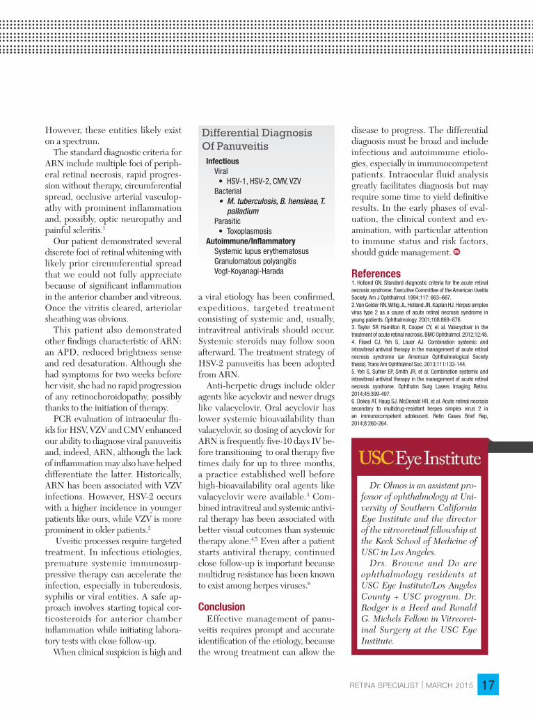
Dr. Olmos is an assistant pro-fessor of ophthalmology at Uni-versity of Southern California Eye Institute and the director of the vitreoretinal fellowship at the Keck School of Medicine of USC in Los Angeles.
Drs. Browne and Do are ophthalmology residents at USC Eye Institute/Los Angeles County + USC program. Dr. Rodger is a Heed and Ronald G. Michels Fellow in Vitreoret-inal Surgery at the USC Eye Institute.
015_rs0315_retinaff_ja3_rk.indd 17015_rs0315_retinaff_ja3_rk.indd 17 3/18/15 10:59 AM3/18/15 10:59 AM

RETINA SPECIALIST | MARCH 201518
DRCR.NET PROTOCOL T
LESSONS FROM
The National Institutes of Heath-sponsored Diabetic Retinopathy Clinical Research Network (DRCR.net) should be commended for conducting and recently publishing the much-anticipated results of a landmark study compar-ing three intravitreal anti-VEGF agents in the treatment of diabetic macular edema (DME).1 Known as DRCR.net Protocol T, these trial results will have a major impact on how we manage our patients with DME.
RETINA SPECIALIST | MARCH 201518
FEATURE DRCR.net Protocol T
Protocol T is the fi rst trial to com-pare the efficacy and safety of the commercially available anti-VEGF drugs used in the treatment of DME: ranibizumab (Lucentis, Genentech), bevacizumab (Avastin, Genentech) and afl ibercept (Eylea, Regeneron). Ranibizumab and aflibercept have been approved for DME by the Food and Drug Administration (FDA), whereas bevacizumab is widely used off-label for DME. (In fact, bevaci-zumab is not FDA-approved for any ocular condition.)
DRCR.net Protocol 1 confi rmed the significant cost differences be-tween these agents—Medicare al-lowable charges range from $1,961 for 2 mg afl ibercept to $1,189 for 0.3 mg ranibizumab to $67 for 1.25 mg bevacizumab—but did not answer to any differences in efficacy or safety.
Impetus for Protocol TDetermining a difference between
the agents was the impetus for the DRCR.net Protocol T comparative trial. In the retina community, Pro-tocol T has been dubbed the “CATT study for DME.” However, Protocol T compared three drugs for DME whereas CATT—the Comparison of Age-Related Macular Degeneration Treatments Trials (CATT)—focused on two drugs and dosing strategies (modifi ed PRN versus monthly ther-apy).2 Here, we review the fi ndings of DRCR.net Protocol T and explore how they can be applied in the clinic.
Eighty-nine clinical sites in the United States participated in Proto-col T, enrolling 660 adults with either type 1 or type 2 diabetes and DME involving the central macula on op-tical coherence tomography (OCT).
Participants were 18 years or older, with vision of 20/32 to 20/320.
Patients did not have to be na-ive to treatment, but they could not have received anti-VEGF therapy within the last year. Masked partic-ipants were treated with 0.05 mL of either 2 mg aflibercept, 1.25 mg bevacizumab or 0.3 mg ranibi-
ABOUT THE AUTHORS
Illustration by Mark Erikson, jirehdesign.com
Drs. Steinle (top) and Pieramici are with California Retina Consultants and Research Foundation, Santa Barbara. Dr. Pieramici is a member of the DRCR.net Protocol T writing committee.
What we can take from the study and apply in our clinics.By Nathan C. Steinle, MD, and Dante Pieramici, MD
018_rs0315_fx_drcrt_rk3 copy.indd 18018_rs0315_fx_drcrt_rk3 copy.indd 18 3/17/15 12:20 PM3/17/15 12:20 PM

RETINA SPECIALIST | MARCH 2015 19RETINA SPECIALIST | MARCH 2015 19
zumab, with a primary endpoint of change in visual acuity at one year, with follow-up planned through two years. One eye of each subject was assigned randomly with equal proba-bility to one of the treatment groups.3
The treatment regimen was a modified PRN protocol based on vision and OCT findings. During the fi rst six months, participants re-ceived monthly injections unless vi-sual acuity was 20/20 or better and OCT central thickness was better than the eligibility threshold. Start-ing at week 24, irrespective of vision or OCT thickness, an injection was withheld if the patient showed no improvement or worsening after two consecutive injections, but treatment was restarted if vision or OCT thick-ness worsened. The threshold for improvement/worsening was >4 let-ter score (one Snellen line) change in visual acuity or a >9 percent change in central OCT thickness. Focal/grid laser was started at or after 24 weeks if DME persisted based on proto-col-specifi c criteria.
Vision Outcomes
The topline DRCR.net Protocol T results revealed improvement in vision from baseline to one year with all three drugs (Tables 1 and 2, pages 20 and 21). Improvement was great-est with afl ibercept (+13 letters) than ranibizumab (+11 letters) or beva-cizumab (+10 letters), a statistically significant mean difference of 2-3 letters at one year.
This difference appeared to be driven by baseline vision. Half of the study participants had VA of ap-proximately 20/40 or 20/32. In these patients, the mean letter score im-provement was +8.3 with ranibizum-ab, +8.0 with aflibercept and +7.5 with bevacizumab (each pairwise comparison p>0.5).
However, when initial visual acuity was 20/50 or worse, the mean letter improvement was +18.9 with afl iber-cept, +14.2 with ranibizumab and +11.8 with bevacizumab (p values: aflibercept-bevacizumab <0.001, aflibercept-ranibizumab = 0.003, ranibizumab-bevacizumab = 0.21). OCT central thickness and vision outcomes showed a similar effect favoring aflibercept when baseline retinal thickness was >400 µm.
More injections or other treat-ments did not drive this relative benefit of aflibercept. In fact, the aflibercept group had one fewer median number of injections9 than the ranibizumab or bevacizumab groups (p=0.045).10 Laser was also performed less often with afl ibercept (37 percent) than ranibizumab (46 percent) or bevacizumab (56 per-cent) (p<0.001).
Anatomic OutcomesThe anatomic results paralleled
the vision results, with the greatest benefi t on reduction in retinal ede-ma seen in the patients treated with afl ibercept. At one year, central OCT thickness decreased on average by 169 µm (afl ibercept), 147 µm (ranibi-zumab) and 101 µm (bevacizumab).
Overall, aflibercept reduced retinal edema better than the other two agents (p<0.001) and ranibizumab outperformed beva-cizumab (p<0.001). This held true regardless of the presenting vision.
A few key points are worth empha-sizing based on the trial results:
• First, all three agents, on aver-age, improved vision and reduced retinal thickness in patients with cen-ter-involved DME. The benefi ts of treatment could be seen as early as four weeks in some patients, but not
The Population Burden of Diabetes And Genesis of anti-VEGF TherapyDiabetes continues to be a growing concern for the American health-care system as its prevalence continues to rise. From 1980 through 2011, the crude prevalence of diagnosed diabetes increased 176 percent in the United States (from 2.5 to 6.9 percent of the popu-lation).11 Some statistical models predict that one-third of Americans will have diabetes by 2050.12
Because diabetes is often a disease of working-age adults, it is a tremendous burden on not only the health-care system but also the labor force. In 2011, 63 percent of the adult cases diagnosed within the past year were in individuals ages 40–64 years; and 16% were diagnosed in those ages 18–39 years.13 Approximately half of these diabetic patients will develop retinopathy, with diabetic macular edema being the number one reason for vision loss.14
Thirty years have passed since the landmark Early Treatment Diabetic Retinopathy Study (ETDRS) Report Number 1 was published in 1985, which provided longstanding guidelines for laser photocoagulation use in patients with clinically signifi cant DME.10 Corticosteroids have also been investigated in the treatment of DME, as triamcinolone,15 fl uocinolone16 and dexamethasone17 have all been used as intravitreal therapies for DME.
Recent years have ushered in the era of anti-VEGF drugs for treatment of DME. Well-designed, multicenter clinical trials have shown the benefi ts of these drugs in the treatment of DME: the RISE/RIDE studies6 of ranibizumab; the BOLT study18 of bevacizumab; and the VISTA/VIVID Studies19 of afl ibercept.
The DRCR.net Protocol I study demonstrated the effi cacy and superiority of ranibizumab with immediate or deferred laser versus laser alone in the treatment of center-involved DME. Protocol I also revealed that similar visual and anatomic outcomes could be achieved with a PRN (as needed) protocol that reduced the burden of treatment compared to monthly therapy.20,21 It became very apparent to investigators and clinicians that anti-VEGF therapy was superior to the then “standard of care” for DME—laser photocoagulation.
018_rs0315_fx_drcrt_rk3 copy.indd 19018_rs0315_fx_drcrt_rk3 copy.indd 19 3/17/15 12:21 PM3/17/15 12:21 PM

RETINA SPECIALIST | MARCH 201520 RETINA SPECIALIST | MARCH 201520
all patients derived a benefi t. • Second, when patients presented
with relatively good vision (20/40 or better), all three drugs resulted in similar vision outcomes at one year, although the anatomic results were better with aflibercept and ranibi-zumab. Studies have reported that 75 percent of patients with DME pres-ent with visual acuity of 20/40 or bet-ter.4,5 Since VAs reported in the study were standardized vision measure-ments, it is not exactly clear how this translates into the vision measured in our clinics. Standardized refracted visions can be several lines of vision better than typical Snellen visions.
In Protocol T, however, when the vision was 20/50 or less, the bene-fi t for vision favored afl ibercept over the other anti-VEGF agents on aver-age. Some may argue that the clinical relevance of a few letters is not very meaningful. Indeed, to one individ-ual such a difference may not be that meaningful. However, the differences between the drugs in this study are in the group averages, and they were statistically signifi cant to a very high level in many comparisons.
Viewed in another way, the chance of three lines of vision improvement in patients with VA of 20/50 or less at baseline was 63 percent greater with afl ibercept compared to bevaci-zumab, and 34 percent greater with afl ibercept compared to ranibizumab. Finally, it is important to realize that these represent one-year results to date (and DME is a chronic condi-tion). A planned secondary outcome measure in Protocol T will include change in visual acuity at two years.3 The second-year data will provide insight into the efficacy and safety of extended therapy and perhaps a better understanding of the rela-tive long-term treatment burden of these agents.
Questions About DosingSome may wonder whether the
0.5 mg dose of ranibizumab might have performed better than the 0.3 mg dose that was used in Protocol T. We will never know for sure, but data from the RISE and RIDE stud-ies6 found no signifi cant difference between the 0.3 and 0.5 mg doses in terms of effi cacy. On the other hand, some smaller studies have demon-strated additional benefi t on vision and anatomy in some DME patients when switched to higher dosages of ranibizumab.7
A second dosing issue concerns the preparation of bevacizumab. The bevacizumab used in Protocol T was repackaged at a central pharma-cy into single-use vials. These vials underwent independent testing for sterility, purity and potency prior to use.1 Although somewhat unlikely to fully account for differences seen with bevacizumab in this study, sig-nificant variability of bevacizumab
obtained from compounding phar-macies has been reported and needs to be considered in our “real world patients”.8
The Safety IssueWith regards to safety, no differ-
ences in intraocular inflammation were noted among the three groups, and endophthalmitis was rare (oc-curring in 0.02 percent of injections). Rates of serious adverse events, deaths, hospitalizations and major cardiovascular events were similar in the three treatment groups. Howev-er, a post hoc analysis did fi nd a small but statistically signifi cant increase in reported cardiac and/or vascular adverse events in the ranibizumab group, although it is possible that this fi nding was a chance event.
The relatively small number of pa-tients in each treatment group (af-libercept, 224; bevacizumab, 218; and ranibizumab, 218) is unlikely to manifest differences in rare serious
DRCR.net Protocol TFEATURE
TABLE 1: DRCR.net Protocol T Outcomes for VA >20/50 At Baseline (Letter Score <69)
Visual Acuity Outcomes Afl ibercept Bevacizumab RanibizumabNo. of eyes 102 102 101VA at baseline Mean letter score 56.2±11.1 56.6±10.6 56.5±9.9 Approximate Snellen equivalent 20/80 20/80 20/80VA at 1 yr Mean letter score 75.2±10.9 68.5±13.6 70.7±12.0 Approximate Snellen equivalent 20/32 20/40 20/40Change from baseline in letter score Mean improvement (p=<.0.001) 18.9±11.5 11.8±12.0 14.2±10.6
Central Subfi eld Thickness (measured with OCT)No. of eyes 101 100 99CST at baseline—µm 452±145 467±155 431±138CST at 1 yr—µm 238±81 328±154 252±92Mean change in CST from baseline—µm (p=<.0.001) −210±151 −135±152 −176±151
CST <250 µm at 1 yr —no. (%) 71 (70) 39 (39) 55 (56)
Adopted from: Diabetic Retinopathy Clinical Research Network. Afl ibercept, bevacizumab, or ranibizumab for diabetic macular edema. N Engl J Med. 2015; Feb 18. [Epub ahead of print].
018_rs0315_fx_drcrt_rk3 copy.indd 20018_rs0315_fx_drcrt_rk3 copy.indd 20 3/17/15 12:21 PM3/17/15 12:21 PM
RETINA SPECIALIST | MARCH 2015 21RETINA SPECIALIST | MARCH 2015 21
adverse events. These three agents have known differences in vivo in regards to their systemic pharma-cokinetic profi les,9 so the continued monitoring of patients for systemic adverse events is prudent, especially in more at-risk DME patients, many of whom, such as those on dialysis, were ineligible for this and other Phase III clinical trials for DME.
The results of clinical trials should serve as guidelines for clinical care. The study results refl ect population average outcomes; much individual variability underlies these fi ndings. When considering the individual pa-tient, these data can be instructive, but many additional issues, includ-ing cost, factor in the decision of which agent or method of treatment to utilize initially. In addition, this study did not address the benefits of a treatment strategy that begins with one anti-VEGF agent and then switches to another if the result is less than desirable.
The DRCR.net should be com-mended for the tedious work in-volved in seeing Protocol T through to completion. It will remain an im-portant study for many years. As ret-ina specialists, it is an exciting time for our ever-growing diabetic pa-tient population. As opposed to the ETDRS era of reducing the rate of vision loss in DME,10 it is thrilling to offer our patients three agents that offer the chance for signifi cant vision gains. The debate as to how to best use these three agents in DME from the standpoints of treatment burden, cost, effi cacy and safety will certainly provide hours of discussion at the upcoming meetings this year.
Disclosures: Dr. Pieramici dis-closed he has been a consultant and adviser to Genentech and has re-ceived research funding from Genen-tech and Regeneron. Dr. Steinle is a speaker for Regeneron and Throm-boGenics and receives research funding from Genentech.
References1. The Diabetic Retinopathy Clinical Research Network. Afl ibercept, bevacizumab, or ranibizumab for diabetic macular edema. N Engl J Med. 2015; Feb 18. [Epub ahead of print].2. Martin DF, Maguire MG, Ying GS, et al, CATT Research Group. Ranibizumab and bevacizumab for neovascular age-related macular degeneration. N Engl J Med. 2011;364:1897-1908.3. ClinicalTrials.gov. Comparative Effectiveness Study of Intravitreal Afl ibercept, Bevacizumab, and Ranibizumab for DME (Protocol T). Available at: https://clinicaltrials.gov/ct2/show/study/NCT01627249. Accessed February 10, 2015.4. Early Treatment Diabetic Retinopathy Study Research Group. Focal photocoagulation treatment of diabetic macular edema. Relationship of treatment effect to fl uorescein angiographic and other retinal characteristics at baseline: ETDRS report no. 19. Arch Ophthalmol. 1995;113:1144-1155.5. Martin DR, Maguire MG. Treatment choice for diabetic macular edema. N Engl J Med. 2015. Feb 18. [Epub ahead of print]6. Brown DM, Nguyen QD, Marcus DM. Long-term outcomes of ranibizumab therapy for diabetic macular edema: the 36-month results from two phase III trials: RISE and RIDE. Ophthalmology. 2013;120:2013-2022.7. Dhoot DS, Pieramici DJ, Nasir M, et al. Residual edema evaluation with ranibizumab 0.5 mg and 2.0 mg formulations for diabetic macular edema (REEF study). Eye (Lond). 2015; Jan. 30 [Epub ahead of print]8. Yannuzzi NA, Klufas MA, Quach L, et al. Evaluation of compounded bevacizumab prepared for intravitreal injection. JAMA Ophthalmol. 2015; 133:32-39.9. Avery RL, Castellarin AA, Steinle NC, et al. Systemic pharmacokinetics following intravitreal injections of ranibizumab, bevacizumab or afl ibercept in patients with neovascular AMD. Br J Ophthalmol. 2014; 98:1636-1641.10. Early Treatment Diabetic Retinopathy Study Research group. Photocoagulation for diabetic macular edema. Early Treatment Diabetic Retinopathy Study report number 1. Arch Ophthalmol. 1985;103:1796-1806.11. Centers for Disease Control and Prevention. Crude and Age-Adjusted Rate per 100 of Civilian, Noninstitutionalized Population with Diagnosed Diabetes, United States, 1980–2011. Available at: http://www.cdc.gov/diabetes/statistics/prev/national/fi gage.htm. Accessed February 26, 2015. 12. Boyle JP, Thompson TJ, Gregg EW, Barker LE, Williamson DF. Projection of the year 2050 burden of diabetes in the US adult population: dynamic modeling of incidence, mortality, and prediabetes prevalence. Popul Health Metr. 2010;8: 29.13. Centers for Disease Control and Prevention. Distribution of Age at Diagnosis of Diabetes Among Adult Incident Cases Aged 18–79 Years, United States, 2011. Available at: http://www.cdc.gov/diabetes/statistics/age/fi g1.htm. Accessed February 15, 2015.14. Varma R Bressler N, Doan QV, et al. Prevalence of and risk factors for diabetic macular edema in the United States. JAMA Ophthalmol. 2014;132: 1334-1340.15. Beck RW, Ewards AR, Aiello LP, et al. Diabetic Retinopathy Clinical Research Network (DRCR.net) Three-year follow-up of a randomized trial comparing focal/grid photocoagulation and intravitreal triamcinolone for diabetic macular edema. Arch Ophthalmol. 2009;127:245-251.16. Campochiaro PA, Hafi z G, Shah SM, et al. Sustained ocular delivery of fl uocinolone acetonide by an intravitreal insert. Ophthalmology. 2010; 117: 1393-1399 e3.17. Boyer DS, Yoon YH Belfort R Jr, et al. Three-year, randomized, sham-controlled trial of dexamethasone intravitreal implant in patients with diabetic macular edema. Ophthalmology. 2014;121:1904-1914.18. Rajendram R, Fraser-Bell S, Kaines A. A 2-year prospective randomized controlled trial of intravitreal bevacizumab or laser therapy (BOLT) in the management of diabetic macular edema: 24-month data: report 3. Arch Ophthalmol. 2012; 130:972-979.19 Korobelnik JF, Do DV, Schmidt-Erfurth U, et al. Intravitreal afl ibercept for diabetic macular edema. Ophthalmology. 2014;121:2247-2254.20. Elman, MJ, Ayala A, Bressler NM, et al. Randomized trial evaluating ranibizumab plus prompt or deferred laser or triamcinolone plus prompt laser for diabetic macular edema. Ophthalmology. 2010;117:1064-1077.e35.21. Elman MJ, Ayala A, Bressler NM, et al. Intravitreal ranibizumab for diabetic macular edema with prompt versus deferred laser treatment: 5-year randomized trial results. Ophthalmology. 2015;122:375-381.
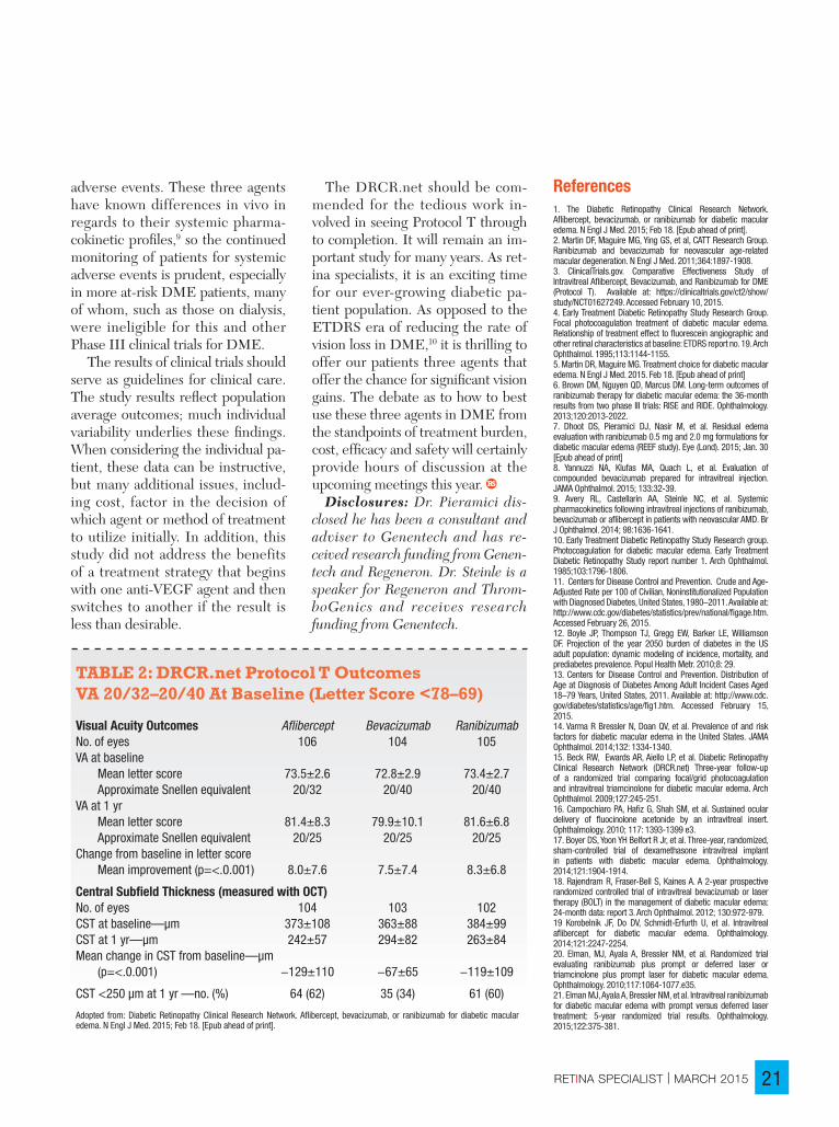
TABLE 2: DRCR.net Protocol T Outcomes VA 20/32–20/40 At Baseline (Letter Score <78–69)
Visual Acuity Outcomes Afl ibercept Bevacizumab RanibizumabNo. of eyes 106 104 105VA at baseline Mean letter score 73.5±2.6 72.8±2.9 73.4±2.7 Approximate Snellen equivalent 20/32 20/40 20/40VA at 1 yr Mean letter score 81.4±8.3 79.9±10.1 81.6±6.8 Approximate Snellen equivalent 20/25 20/25 20/25Change from baseline in letter score Mean improvement (p=<.0.001) 8.0±7.6 7.5±7.4 8.3±6.8
Central Subfi eld Thickness (measured with OCT)No. of eyes 104 103 102CST at baseline—µm 373±108 363±88 384±99CST at 1 yr—µm 242±57 294±82 263±84Mean change in CST from baseline—µm (p=<.0.001) −129±110 −67±65 −119±109
CST <250 µm at 1 yr —no. (%) 64 (62) 35 (34) 61 (60)
Adopted from: Diabetic Retinopathy Clinical Research Network. Afl ibercept, bevacizumab, or ranibizumab for diabetic macular edema. N Engl J Med. 2015; Feb 18. [Epub ahead of print].
018_rs0315_fx_drcrt_rk3 copy.indd 21018_rs0315_fx_drcrt_rk3 copy.indd 21 3/17/15 3:19 PM3/17/15 3:19 PM
RETINA SPECIALIST | MARCH 201522
Risk FactorsThe main risk factors for RVO in-
clude age and systemic vascular dis-orders. A study in Israel found that the rate of RVO increased exponen-tially with age—from 0.93 per 1,000 in people age 64 years and younger to 5.36 per 1,000 for those 65 and older.2 That said, RVO can occur at any age. Sohan Singh Hayreh, MD, PhD, DSc, and colleagues study re-ported that 49 percent of RVO pa-tients were younger than 65 years old. Also, a significant percentage were younger than 45; 15 percent for central RVO (CRVO), 10 percent for hemi-CRVO and 5 percent for BRVO.3
Hypertension, hyperlipidemia and diabetes, all atherosclerotic risk factors, are strongly associated with
RVO development. Paul R.A. O’Ma-honey, MD, and co-authors calcu-lated the population-attributable risk percentage of these factors and found that 46 percent of RVO cases are due to hypertension, 20 percent due to hyperlipidemia and 5 percent due to diabetes.4 This emphasizes that, while RVO is indeed an occlu-sion of a retinal venule, the condition is primarily a consequence of athero-sclerosis of the adjacent arteriole.
History and ExaminationWith this in mind, a systemic eval-
uation of RVO should include a thor-ough medical history that investi-gates atherosclerotic risk factors such as hypertension, hyperlipidemia, di-abetes, smoking, obesity and family history of coronary artery disease. An
assessment for hypercoagulability risk factors should also take into ac-count estrogen exposure, malignan-cy, pregnancy or a family history of thrombophilia before age 50 years.
Glaucoma and ocular hyperten-sion are risk factors for CRVO. This underscores the importance of in-quiring about a family history of glaucoma and evaluating both eyes for glaucoma, keeping in mind that intraocular pressure (IOP) may be lower in an eye with acute CRVO.5
THE RVO WORKUP When It’s Necessary and What to Order
Retinal vein occlusion (RVO), the second most common retinal vascular disease after diabetic retinopathy, affects more than 16 million people worldwide.1 While the pathophysiology of RVO is not completely understood, the concept of venous thrombosis attributed to Rudolph Virchow 150 years ago remains relevant. He proposed that the triad of vessel wall injury, stasis and coagulopathy were important in the creation of a venous clot. This article
reviews the risk factors for RVO and the workup and differential diagnosis of the various types of vein occlusion.
Retinal Vein Occlusion
Dr. Jumper is in private practice with West Coast Retina in San Francisco, CA. He is co-director of the vitreoretinal fellowship program at Califor-
nia Pacifi c Medical Center.
ABOUT THE AUTHOR
By J. Michael Jumper, MD
FEATURE
022_rs0315_RVO_rk3.indd 22022_rs0315_RVO_rk3.indd 22 3/17/15 12:25 PM3/17/15 12:25 PM

RETINA SPECIALIST | MARCH 2015 23
Blood pressure should be mea-sured not only in the offi ce but also serially to rule out masked hyper-tension. Many patients with elevated blood pressure measured in the of-fi ce will manifest “white-coat hyper-tension,” which carries a strong risk of conversion to sustained hyperten-sion over time.6
Any patient suspected of RVO should have a complete ophthalmo-logical examination that includes pu-pillary assessment and gonioscopy. Dr. Hayreh, and co-authors empha-sized four functional vision tests as most helpful in differentiating isch-emic and nonischemic CRVO: vi-sual acuity; assessment of a relative afferent pupillary defect measured in log units using neutral density fi l-ters; visual field testing; and elec-troretinography. They stated that the combined data from these tests allows one to determine ischemic status with a high specifi city and sen-sitivity much greater than fl uorescein angiography or ophthalmoscopy.7
In the acute phase, hemorrhage can create blockage of fl uorescence, making it diffi cult to evaluate vascu-lar perfusion. In RVO with severe hemorrhage, one should consider obtaining color fundus images and deferring angiography until the blood clears and more information can be obtained (Figure 1). If a pa-tient with RVO has a fl uorescein an-giogram, late images of the iris of both eyes can help identify rubeosis.
With advances in imaging technol-ogy, the benefi ts of widefi eld fl uores-cein angiography have been shown in detecting peripheral ischemia not otherwise visible.8 Spectral domain or swept-source optical coherence tomography (OCT) is also important in assessing macular edema in RVO and should be done initially and to follow therapy response.
Controversy of Lab TestingThe most controversial issue in
the RVO workup involves the utility of laboratory testing. A reasonable approach is to consider a directed assessment of atherosclerotic risk factors for any patient with an RVO. These include a lipid profi le, blood glucose measurement and complete blood count. Atypical features such as intraocular infl ammation should lead to further testing for conditions such as sarcoidosis, syphilis and sys-temic lupus erythematosus.
Over the past two decades, a number of mostly small series and
case reports have explored the role of thrombophilic abnormalities in RVO development. Inherited thrombophilic defects include de-ficiencies of antithrombin, protein C and protein S, factor V Leiden and the prothrombin G20210A mutation.
Acquired risk factors for throm-bosis include the antiphospholipid antibody syndrome, myeloprolifer-ative disorders, immobilization, ma-jor surgery, malignancy, estrogens and heparin-induced thrombocy-topenia. Other risk factors include hyperhomocysteinemia and elevated
Figure 1. Acute branch retinal vein occlusion in a 61-year-old man shows dense hemorrhage. Fluorescein angiography was deferred because venous block-age would limit its utility.
Hyperhomoscysteinemia and the antiphospholipid antibody syndromes are the two coagulation defects that cause both arterial and venous thrombosis. This may explain why these defects have been found to be risk factors for retinal vein occlusion.
Table: Coagulation Defects That Cause Arterial and Venous Thrombosis
Abnormality Arterial Venous
Factor V Leiden - +
Prothrombin G20210A - +
Antithrombin defi ciency - +
Protein C defi ciency - +
Protein S defi ciency - +
Hyperhomocysteinemia + +
Antiphospholipid antibody syndromes + +
022_rs0315_RVO_rk3.indd 23022_rs0315_RVO_rk3.indd 23 3/17/15 12:25 PM3/17/15 12:25 PM
RETINA SPECIALIST | MARCH 201524
factors VIII, IX and XI. Of all of these abnormalities, only hyperhomocyste-inemia and the antiphospholipid an-tibody syndrome are associated with both arterial and venous thrombosis. All other defects are classical venous thrombophilias (Table, page 23).
A young patient with a RVO and no identifi able risk factors will typ-ically undergo an extensive battery of tests looking for some or all of the thrombophilia risk factors. Research indicates that this approach is rare-ly helpful. Danish researcher Prof. Janne Ingerslev said, “Most well characterized risk factors for gener-al venous thrombosis occur sporad-ically in RVO, and have no major importance in the pathophysiology of RVO.”9
Italian investigators found no dif-ference between RVO patients and age/sex matched controls in levels of antithrombin, protein C, protein S and homocysteine, lupus anticoag-ulant, anticardiolipin antibodies or prothrombin polymorphisms.10 In this same study, arterial hypertension and diabetes were the only factors more common to the RVO patients compared to controls (Figure 2).
Dutch investigators found the two risk factors for arterial thrombosis—hyperhomocysteinemia and the anti-phospholipid antibody syndromes—were associated with RVO.11 J. Michael Lahey, MD, and colleagues studied young patients with CRVO and found at least one thrombophilia lab abnormality in 27 percent of sub-jects, most commonly elevated homo-cysteine levels and antiphospholipid antibody titers,12 which supports the Dutch study. In Dr. Lahey’s study, the one patient with a systemic disease (protein S deficiency) had bilateral CRVO.
When ordering medical tests, con-sider how the result will alter treat-
ment. RVO has only two treatment options when the thrombophil-ia workup is positive. For a patient with hyperhomocysteinemia, one can consider B-complex vitamin supple-mentation. Randomized studies have failed to prove that reducing homo-cysteine levels is effective in reducing the risk of stroke, heart attack or other thromboembolic events.13
The only other treatment option for a patient with a thrombophilia is long-term anticoagulation. RVO is unlike a deep venous thrombosis where anticoagulation is used to prevent a downstream pulmonary embolism. Little evidence exists that anticoag-ulation prevents further ocular vas-cular occlusion. Anticoagulation has no known benefi t on the active clot. Also, in the acute phase of an RVO, anticoagulation may be harmful to the neural retinal tissue by increasing in-traretinal hemorrhage, as Dr. Hayreh pointed out.14
ConclusionRVO can happen at any age but is
most common in patients over age 65. Vein occlusion shares risk fac-tors of atherosclerosis. The clinician should direct the workup toward evaluation of hypertension, hyperlip-
idemia and diabetes. Thrombophilia testing is rarely
needed, but can be considered in patients who have a family history of clotting under the age of 50, a personal history of clotting or an un-usual presentation such as bilateral simultaneous occlusion.
Consider testing for clotting defects associated with arterial dis-ease, including the antiphospholipid antibody syndrome and hyperho-mocysteinemia. Even if a clotting abnormality is discovered, it may or may not be related to the ocular condition. Furthermore, treatment in the acute phase, especially with anticoagulation, may do more harm than good.
Disclosure: Dr. Jumper has no relevant confl icts to disclose.
References1. Rogers S, McIntosh RL, Cheung N, et al. The prevalence of retinal vein occlusion: pooled data from population studies from the United States, Europe, Asia, and Australia. Ophthalmology. 2010; 117:313.e1–319.e1.2. David R, Zangwill L, Badarna M, Yassur Y. Epidemiology of retinal vein occlusion and its association with glaucoma and increased intraocular pressure. Ophthalmologica. 1988;197;69–74.3. Hayreh SS, Zimmerman MB, PodhajskyP. Incidence of various types of retinal vein occlusion and their recurrence and demographic characteristics. Am. J. Ophthalmol. 1994;117:429–441.4. O’Mahoney PR, Wong DT, Ray JG. Retinal vein occlusion and traditional risk factors for 4therosclerosis. Arch Ophthalmol. 2008;126:692-699.5. Hayreh SS, March W, Phelps CD. Ocular hypotony following retinal vein occlusion. Arch. Ophthalmol. 1978;96:827–833.6. Mancia G, Bombelli M, Facchetti R, et al. Long-term risk of sustained hypertension in white-coat or masked hypertension. Hypertension, 2009;54:226-232.7. Hayreh SS, Klugman MR, Beri M, Kimura AE, Podhajsky P. Differentiation of ischemic from non-ischemic central retinal vein occlusion during the early acute phase. Graefe’s Arch Clin Exp Ophthalmol. 1999;228:201-217. 8. Spaide RF. Peripheral areas of nonperfusion in treated central retinal vein occlusion as imaged by wide-fi eld fl uorescein angiography. Retina. 2011;31:829-837.9. Ingerslev J. Thrombophilia: a feature of importance in retinal vein thrombosis? Acta Ophthalmol Scand. 1999;77:619–621.10. Di Capua M, Coppola A, Albisinni R, et al. Cardiovascular risk factors and outcome in patients with retinal vein occlusion. J Thromb Thrombolysis. 2010; 30:16-22.11. Janssen MC, den Heijer M, Cruysberg JR, et al. Retinal vein occlusion: a form of venous thrombosis or a complication of atherosclerosis? A meta-analysis of thrombophilic factors. Thromb Haemost. 2005; 93:1021-1026.12. Lahey JM, Tunc M, Kearney J, et al. Laboratory evaluation of hypercoagulable states in patients with central retinal vein occlusion who are less than 56 years of age. Ophthalmology. 2002;109:126–13113. The VITATOPS Trial Study Group. B vitamins in patients with recent transient ischaemic attack or stroke in the VITAmins TO Prevent Stroke (VITATOPS) trial: a randomised, double-blind, parallel, placebo-controlled trial. Lancet Neurol. 2010;9:855–865. 14. Hayreh SS. Prevalent misconceptions about acute retinal vascular occlusive disorders. Prog Retinal Eye Res. 2005;24:493–519.
Retinal Vein Occlusion
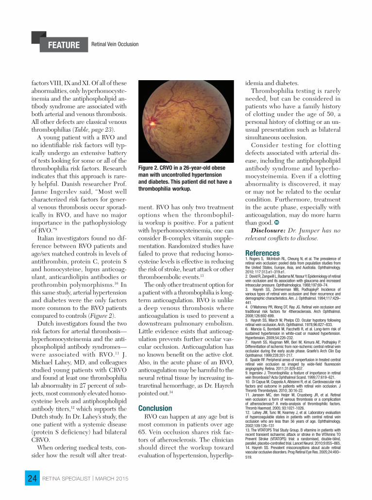
Figure 2. CRVO in a 26-year-old obese man with uncontrolled hypertension and diabetes. This patient did not have a thrombophilia workup.
FEATURE
022_rs0315_RVO_rk3.indd 24022_rs0315_RVO_rk3.indd 24 3/17/15 12:25 PM3/17/15 12:25 PM

RETINA SPECIALIST | MARCH 2015 25
‘BI NICEYE,’ THE
REAL-LIFE STORY OF THE
he journey of artifi cial restoration of vision began in 1929 when Otfrid Foerster reported that electrical stimu-lation of the occipital cortex caused a subject to see a phosphene (a spot of light produced by direct stimulation of the visual system).1 Years later, in 1956, Graham Tassicker implanted a light sensitive selenium cell behind the retina of a blind patient, transiently restoring the patient’s ability to perceive light.2,3 These fi rst steps laid the foundation for modern artifi cial retinal implants that are restoring patients’ vision today.
Retinal implants can be classifi ed according to their location as epiretinal (tacked to the retinal surface) or
‘Bionic Eye’
subretinal (between photoreceptors and RPE). The Argus II epiretinal prosthesis (Second Sight Medical Products) is currently the only reti-nal prosthesis approved by the Food and Drug Administration and Health Canada. In Europe, the Argus II, as well as the Alpha IMS (Retinal Im-plant AG), a light-sensitive subretinal implant, have received the CE mark.
Other retinal implants still in de-velopment or clinical trials include the Boston Retinal Implant (Boston Retinal Implant Project), the Epi- Ret-3 (RWTH Aachen University, Germany) and Intelligent Retinal Im-plant System (IRIS) (IMI Intelligent Medical Implants GmbH, Germany) (Table 1, page 26).
Several alternative implant-based
approaches that aim to restore vision are also in early development phases. These include cortical prostheses that directly stimulate the occipital cortex, neurotransmitter-based retinal pros-theses and photovoltaic cellular ap-proaches.4-6 In addition sensory-sub-stitution devices, such as auditory, tactile and tongue-stimulating devic-es, are also undergoing investigation.7
The Argus II Retinal ProsthesisThe Argus II prosthesis is current-
ly the only retinal prosthesis with both FDA and CE approval.9 This 60-electrode implant is a second-gen-eration device following the Argus I, a 16-electrode implant that was placed in six subjects on an investigative basis at the University of Southern
ABOUT THE AUTHORS
Dr. Maldonado is a resident at Duke University Eye Center, Durham, N.C.
Dr. Humayun is the Cornelius J. Pings Chair in Biomedi-cal Sciences; professor of ophthalmology, biomedical engineering, and cell and neu-robiology; director, Institute for
Biomedical Therapeutics; and co-director, University of South-ern California Eye Institute, Los Angeles.
Dr. Hahn is assistant professor of ophthalmology for vitreoret-inal surgery and diseases at Duke University Eye Center.
By Ramiro S. Maldonado, MD, Mark S. Humayun, MD, PhD, and Paul Hahn, MD, PhD
FEATURE
025_rs0315_ARGUS_rk2_ja_rk.indd 25025_rs0315_ARGUS_rk2_ja_rk.indd 25 3/18/15 11:02 AM3/18/15 11:02 AM

RETINA SPECIALIST | MARCH 201526
‘Bionic Eye’
California starting in 2002 (Table 2).Argus II consists of an external
(wearable) and internal (surgically implanted) component. The external equipment consists of glasses, a video processing unit (VPU) and a wired cable (Figure 1A). The glasses hold a miniature video camera in the nasal bridge that transmits images to the wired VPU for image processing. The VPU transforms the images to data that are then transmitted wirelessly to the internal implant in the eye.
The implanted component consists of a receiving coil and electronics case secured to the eye in a scleral buckle fashion and the 60-electrode array se-cured to the retina with a retinal tack (Figure 1B).
The data the VPU sends wirelessly to the electronics package stimulates the array to emit small pulses of elec-tricity that excite the remaining viable inner retina cells, including gangli-on cells. These artifi cially stimulated retinal ganglion cells transmit signals through their axons to the lateral ge-niculate nucleus and then the occipi-tal cortex, which perceives patterns of light. The fi nal step is for patients to
piece together these patterns of light into form vision.
Clinical Trials In 2009, the Argus II was FDA-
approved as a Humanitarian Use Device intended for treatment of a small patient populations (fewer than 4,000 individuals per year in the
United States). To attain this desig-nation, the Argus II was required to demonstrate safety and, on a reason-able basis, probable benefi t to health that outweighs the risk of injury or illness.
In the largest prospective, single arm, unmasked, multicenter Argus II study, 30 subjects from 10 medical centers (six in the United State and four in Europe) were implanted with the Argus II on a research basis prior to FDA-approval.8 Mean age was 58 years (range of 28-77 years). Nine women and 21 men participated. Twenty-nine had a diagnosis of reti-nitis pigmentosa, and one had a diag-nosis of choroideremia. Twenty-nine had bare light perception vision, and one had no light perception. Sub-jects served as their own control (sys-tem on versus off).
Because the profound vision loss in this patient population precluded ac-curate assessment of visual acuities by conventional methods, such as visual acuity charts, custom end-points were carefully designed in this trial.
Figure 1. Argus II retinal prosthesis components: Diagram of external components including glasses with micro camera, transmitter (radio frequency coil) and video processing unit (A). Diagram of surgically implanted component including implant coil, electronics case and 60-electrode array (B). Multicolor scanning laser ophthalmoscope view of the array optimally centered on the macula and attached to the retina by a retinal tack (C). (A and B courtesy Second Sight Medical Products; C courtesy of Paul Hahn, MD, PhD)
Taable 1. Charactteeristics of f curreent reetinal impllants
Argus II8,9 Alpha IMS9,10Boston RetinalImplant11
Epi-Ret-312, 13Intelligent Medical Implants9,14
FDA-approved Yes No No No No
CE Mark Yes Yes No No No
Method of image capture
External camera
Multiphotodiode array
External camera
External camera External camera
Array location Epiretinal Subretinal Subretinal Epiretinal Epiretinal
Number of electrodes 60 1,500 100 25 49
Array dimension 3 x 5 mm 3.1 x 3 mm 5 x 5 mm Information
not availableInformation not available
Electrode size 200 µm 15 x 30 µm 400 µm 100 µm 100-360 µm
FEATURE
025_rs0315_ARGUS_rk2_ja_rk.indd 26025_rs0315_ARGUS_rk2_ja_rk.indd 26 3/17/15 3:21 PM3/17/15 3:21 PM
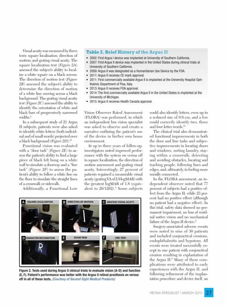
RETINA SPECIALIST | MARCH 2015 27
Visual acuity was measured by three tests: square localization; direction of motion; and grating visual acuity. The square localization test (Figure 2A) assessed the subject’s ability to local-ize a white square on a black screen. The direction of motion test (Figure 2B) assessed the subject’s ability to determine the direction of motion of a white line moving across a black background. The grating visual acuity test (Figure 2C) assessed the ability to identify the orientation of white and black bars of progressively narrowed widths.8
In a subsequent study of 21 Argus II subjects, patients were also asked to identify white letters (both individ-ual and of small words) projected over a black background (Figure 2D).15
Functional vision was evaluated with a “door task” (Figure 2E) to as-sess the patient’s ability to fi nd a large piece of black felt hung on a white wall to simulate a doorway and a “line task” (Figure 2F) to assess the pa-tient’s ability to follow a white line on the fl oor to simulate the straight lines of a crosswalk or sidewalk.
Additionally, a Functional Low
Vision Observer Rated Assessment (FLORA) was performed, in which an independent low vision specialist was asked to observe and create a narrative outlining the patient’s use of the device in his/her own home environment.
At up to three years of follow-up, investigators noted improved perfor-mance with the system on versus off in square localization, the direction of motion assessment and grating visual acuity. Interestingly, 27 percent of patients regained a measurable visual acuity (grating VA of 2.9 logMAR) with the greatest logMAR of 1.8 (equiv-alent to 20/1262).8 Some subjects
could also identify letters, even up to a reduced size of 0.9 cm, and a few could correctly identify two, three and four letter words.15
The clinical trial also demonstrat-ed functional improvements in both the door and line tasks and subjec-tive improvements in locating doors and windows, sorting laundry, stay-ing within a crosswalk, detecting and avoiding obstacles, locating and tracking people, following lines and edges, and, ultimately, in feeling more socially connected.
In the FLORA assessment, an in-dependent observer noted that 77 percent of subjects had a positive ef-fect from the Argus II, while 23 per-cent had no positive effect (although no patient had a negative effect). In this trial, safety data showed no per-manent impairment, no loss of resid-ual native vision and no mechanical failure of the Argus II device.8
Surgery-associated adverse events were noted in nine of 30 patients and included conjunctival erosions, endophthalmitis and hypotony. All events were treated successfully ex-cept in one patient with conjunctival erosion resulting in explantation of the Argus II.8 Many of these com-plications were attributed to early experiences with the Argus II, and following refi nement of the implan-tation procedure and device itself, an
Figure 2. Tests used during Argus II clinical trials to evaluate vision (A-D) and function (E, F). Patient’s performance was better with the Argus II retinal prosthesis on versus off in all of these tests. (Courtesy of Second Sight Medical Products)
Taablee 2. BBrief Hisstory of thhe Arggus III• 2002: First Argus I device was implanted at University of Southern California.• 2007: First Argus II device was implanted in the United States during clinical trials at
University of Southern California.• 2009: Argus II was designated as a Humanitarian Use Device by the FDA. • 2011: Argus II receives CE mark approval.• 2011: First commercially available Argus II is implanted at the University Hospital Oph-
thalmic Department of Pisa, Italy.• 2013: Argus II receives FDA approval.• 2014: The fi rst commercially available Argus II in the United States is implanted at the
University of Michigan.• 2015: Argus II receives Health Canada approval.
025_rs0315_ARGUS_rk2_ja_rk.indd 27025_rs0315_ARGUS_rk2_ja_rk.indd 27 3/18/15 11:03 AM3/18/15 11:03 AM

RETINA SPECIALIST | MARCH 201528
‘Bionic Eye’
improved adverse event profile has been reported.16
The ‘Right Patient’The Argus II aims to restore ru-
dimentary functional vision to pa-tients with profound vision loss. It is FDA-approved for adults (age 25 years or older) with a diagnosis of reti-nitis pigmentosa and a history of prior useful vision who have progressed to bare light perception or no light per-ception vision in both eyes (Table 3).
Contraindications include comor-bidities that would prevent the im-plant from functioning properly (e.g., optic nerve disease, cortical blindness, history of retinal detachment, retinal vascular occlusion, trauma or severe strabismus). Other contraindications include conditions that would prevent adequate implantation of the Argus II, such as conjunctival thinning, axial length <20.5 mm or >26 mm (given the fi xed intraocular cable length) and conditions that prevent implant visu-alization such as corneal opacities.
Finally, the implant is not recom-mended in patients with a tendency to eye rubbing or with a metallic or active implantable device in the head such as cochlear implant.
Surgical Technique and Postoperative Follow-up
The implantation procedure for the Argus II was refi ned to require a com-bination of skills, many of which are familiar to the trained vitreoretinal surgeon (Table 4). The implant is se-cured to the wall of the eye in a fash-ion similar to a scleral buckle. The ar-ray is then inserted through a 5.5-mm sclerotomy and secured over the mac-ula in a unique technique involving the use of a retinal tack to secure the implant to the retina-choroid-sclera. Care around the delicate electronics is paramount, and precisely measured
external fi xation of the implant is crit-ical to allow optimal centering of the array over the macula. A patch graft is used to cover the electronics case, and suture tabs and complete closure of the overlying conjunctiva and tenon’s are important.
Postoperatively, standard follow-up is indicated to monitor for adverse events, particularly hypotony, con-junctival erosion and endophthalmi-tis. Standard postoperative eye drops are administered without extended steroid or antibiotic therapy beyond the postoperative period.
Within a few weeks following sur-gical implantation, the patient’s video processing unit (VPU) is customized to optimize each electrode according to the patient’s residual thresholds of stimulation. The VPU can be pro-grammed with multiple, easily adjust-able settings to optimize edge detec-tion or maximize visualization of high contrast items, for example. This fi t-ting process occurs over several days, after which the external glasses are turned on to stimulate the implanted array for the fi rst time.
Following programming, the pa-tient undergoes ongoing low vision rehabilitation and orientation and mobility training in conjunction with a low vision specialist and oc-cupational therapist to learn how to maximize the use of the device.
A Patient’s PerspectiveLarry, a 66-year-old man, was the
seventh patient to receive a commer-cially available Argus II retinal pros-thesis in the United States. He started to lose vision in his early 30s, when he was diagnosed with retinitis pigmen-tosa by Dr. Robert Machemer at the Duke Eye Center in Durham, N.C. At that time, Larry was informed that no medical treatment would prevent his vision loss, which slowly pro-gressed toward profound blindness over the years.
For the past 10 years or so, Larry could not identify if ambient lights were on or off, although clinical as-sessment with a specialized photo-flash test confirmed residual bare light perception vision. Larry was well adapted to his vision loss, but he hoped for more.
One of his favorite yearly traditions was to experience the fi reworks live with his youngest granddaughter sitting on his lap, and he felt that if could “see” the fl ashes of fi reworks for the fi rst time with her, it would all be worthwhile. Larry was well aware of the limitations of the device and the need for rigorous postoperative train-ing, and he was eager for the prospect of any improvement.
Following successful implantation, Larry’s journey with the Argus II has been marked with excitement. Three weeks postoperatively, following pro-gramming of his VPU, his device was turned on, resulting in visualization of phosphenes (perception of seeing light fl ashes in response to controlled electrical stimulation of the retina) for the first time in years. (A video of his experience is available on You-Tube at https://www.youtube.com/watch?v=CiyGOUHD2nI).
His wife describes what happened one day later: “As we were driving home, Larry turned the device on.
TTaablle 33. Argguss III Curr-reennt Eliigibillityy CCriteria• Adults, age 25 years or older.• Bare light or no light perception in both
eyes due to retinitis pigmentosa.• Previous history of useful form vision• Aphakia or pseudophakia (if present,
the natural lens will be removed during the implant procedure).
The Argus II implant is intended to be implanted in a single eye, typically the worse-seeing eye.
FEATURE
025_rs0315_ARGUS_rk2_ja_rk.indd 28025_rs0315_ARGUS_rk2_ja_rk.indd 28 3/17/15 12:29 PM3/17/15 12:29 PM

RETINA SPECIALIST | MARCH 2015 29
He was able to distinguish where the streetlights were, a lit billboard and headlights as they came our way. It was truly amazing!”
One of the most gratifying early experiences for Larry was with lamps in his house. “Before the Argus II, I could not tell if a lamp in the house was on or off without burning my hands,” he said. “Now with the Argus II, I can go around and turn lamps off that are on. That is pretty functional for me!”
Several weeks into his device, he tried joining his granddaughter in playing Velcro darts (Figure 3). As his wife describes, “I told him he should put on his glasses and see if he could tell where the dart board was. He put on his glasses. Shocker! He found the dart board and started playing. He rarely missed. That’s about as much fun as he has had in a long time play-ing a game. He wanted to try it again tomorrow and the next day and the next day! To be truthful, I was kind of surprised that he could see it as well as he did. But the darts didn’t lie. He did well!”
With ongoing low vision rehabili-tation, Larry has increasingly learned how to use his device to maximize
his experiences with it. He is able to identify the location of doorways and windows, he can reach out to fi nd and touch his wife’s and grand children’s faces. He should certainly be able to see the flashes of fireworks. His exploration of a new world with visual stimulation has just begun.
A Future for Artifi cial VisionThe approval of the Argus II marks
an exciting era of enthusiasm and hope for physicians and patients in the treatment of vision previously considered permanently lost.
Although it provides far from nat-ural vision and is currently only ap-proved for patients with profound vision loss from retinitis pigmentosa, the Argus II and other retinal im-plants and their early successes en-courage development of future tech-nologies to provide improved safety and durability, image resolution and applications in other diseases.
Disclosures: Dr. Humayun is a consultant, equity owner and holds patents with Second Sight Medi-cal Products, from whom he also receives lecture fees and grant sup-port. Dr. Hahn is a consultant to Second Sight.
References 1. Foerster O. No Beitrage zur Pathophysiologie der Sehbahn und der SehsphareTitle. J Psychol Neurol. 1929;39:463-485.2. Tassicker GE, inventor. Retinal stimulator. US patent 2760483. 1956:2,760,483.3. Brindley GS, Lewin WS. The sensations produced by electrical stimulation of the visual cortex. J Physiol. 1968;196:479-493.4. Torab K, Davis TS, Warren DJ, House PA, Normann RA, Greger B. Multiple factors may infl uence the performance of a visual prosthesis based on intracortical microstimulation: nonhuman primate behavioural experimentation. J Neural Eng. 2011;8:35001. 5. Bi A, Cui J, Ma Y-P, et al. Ectopic expression of a microbial-type rhodopsin restores visual responses in mice with photoreceptor degeneration. Neuron. 2006;50:23-33. 6. Peterman MC, Mehenti NZ, Bilbao K V, et al. The Artifi cial Synapse Chip: a fl exible retinal interface based on directed retinal cell growth and neurotransmitter stimulation. Artif Organs. 2003;27:975-985.7. Chekhchoukh A, Goumidi M, Vuillerme N, Payan Y, Glade N. Electrotactile vision substitution for 3D trajectory following. Conf Proc Annu Int Conf IEEE Eng Med Biol Soc IEEE Eng Med Biol Soc Annu Conf. 2013;2013:6413-6416. 8. Humayun MS, Dorn JD, da Cruz L, et al. Interim results from the international trial of Second Sight’s visual prosthesis. Ophthalmology. 2012;119:779-788. 9. Chuang AT, Margo CE, Greenberg PB. Retinal implants: a systematic review. Br J Ophthalmol. 2014;98:852-856. 10. Stingl K, Bartz-Schmidt KU, Besch D, et al. Artifi cial vision with wirelessly powered subretinal electronic implant alpha-IMS. Proc Biol Sci. 2013;280:20130077. 11. Rizzo JF 3rd. Update on retinal prosthetic research: the Boston Retinal Implant Project. J Neuroophthalmol. 2011;31:160-168. 12. Roessler G, Laube T, Brockmann C, et al. Implantation and explantation of a wireless epiretinal retina implant device: observations during the EPIRET3 prospective clinical trial. Invest Ophthalmol Vis Sci. 2009;50:3003-3008. 13. Menzel-Severing J, Laube T, Brockmann C, et al. Implantation and explantation of an active epiretinal visual prosthesis: 2-year follow-up data from the EPIRET3 prospective clinical trial. Eye (Lond). 2012;26:501-509. 14. Matthaei M, Zeitz O, Keserü M, et al. Progress in the development of vision prostheses. Ophthalmologica. 2011;225:187-192. 15. Da Cruz L, Coley BF, Dorn J, et al. The Argus II epiretinal prosthesis system allows letter and word reading and long-term function in patients with profound vision loss. Br J Ophthalmol. 2013;97:632-636. 16. Rizzo S, Belting C, Cinelli L, et al. The Argus II Retinal Prosthesis: 12-month outcomes from a single-study center. Am J Ophthalmol. 2014;157:1282-1290.
Figure 3. Larry, the seventh U.S. patient to receive a commercially available Argus II retinal prosthesis, is wearing and using the Argus II and describing to Dr. Paul Hahn his visual experiences (A). At his home, Larry plays Velcro darts with the Argus II (B); as his wife explains: “He found the dart board and started playing. He rarely missed. That’s about as much fun as he has had in a long time playing a game.”
TTablle 44. Argguss IIISSurrggicaaal TecchnniqqueThe initial surgery to implant the Argus II may take 3-5 hours (4 hours on average). Surgical steps include:
• Crystalline lens removal, if applica-ble.
• Peritomy and muscle isolation.• Placement of extra-ocular portion of
device.• Anchoring of suture tabs.• Securing scleral band.• Vitrectomy.• Creation of sclerotomy to insert array. • Tacking of array over macula.• Application of patch graft and
closure.
025_rs0315_ARGUS_rk2_ja_rk.indd 29025_rs0315_ARGUS_rk2_ja_rk.indd 29 3/18/15 11:05 AM3/18/15 11:05 AM

RETINA SPECIALIST | MARCH 201530
However, these implants can occasionally migrate into the anterior chamber, causing vision-threatening complications that involve perma-nent corneal decompensation.5-7 The initial reports were in aphakic eyes, and authors speculated that aphakia is the main risk factor for anterior chamber complications.6
Risk Factors for MigrationThe largest series of DEX implant
anterior chamber migration re-viewed the risk factors, clinical out-comes and management strategies.7 The authors described 15 patients with 18 episodes of anterior cham-ber migration of the DEX implant. All eyes had been treated for cystoid macular edema from various caus-
es including central retinal vein oc-clusion (CRVO), branch retinal vein occlusion (BRVO), non-infectious uveitis and DME. All cases had a history of vitrectomy and either an absent or defective lens capsule. All 15 patients had prior pars plana vit-rectomy (PPV) and 14 (93 percent) had no lens capsule. Six eyes were aphakic, four had an anterior-cham-ber intraocular lens (ACIOL) (Fig-ure), two had a scleral-fi xated IOL, two had a posterior-chamber IOL (PCIOL) and one had an iris-fi xat-ed PCIOL. Five patients had prior uncomplicated dexamethasone implant injections with the same lens and capsule status.7
The average number of days from injection to migration was 13 (range,
fi ve to 44 days). Corneal edema was the most signifi cant vision-threaten-ing complication, which presented in all but two cases. In all the patients presenting with corneal edema, the time from injection to migration was 19 days or less.7
In one case without corneal ede-ma, the implant was lodged between the iris and the lens capsule, so it was not in proximity to the corneal endothelium. In the other case with-
How to Manage a MIGRATING
DEXAMETHASONEINTRAVITREAL
IMPLANTT
he dexamethasone intravitreal implant (DEX implant; Ozurdex, Allergan Inc.) has been approved for the treat-ment of macular edema secondary to retinal vein occlusion, noninfectious uveitis affecting the posterior segment and diabetic macular edema (DME).1-3 The DEX implant is a non-tethered biodegradable sustained release implant containing 0.7 mg dexamethasone in the Novadur (Allergan) solid polymer drug-delivery system. With
the approval for DME in 2014, retina specialists are using the DEX implant more frequently for complicated cases, such as those that do not respond to anti-VEGF therapy and for DME in vitrectomized eyes.4
Migrating Implant
Dr. Khurana is a partner with Northern California Retina Vit-reous Associates and a clinical assistant professor of oph-thalmology at the University of
California, San Francisco.
ABOUT THE AUTHOR
By Rahul N. Khurana, MD
FEATURE
030_rs0315_Ozurdex_ja1.indd 30030_rs0315_Ozurdex_ja1.indd 30 3/17/15 4:49 PM3/17/15 4:49 PM
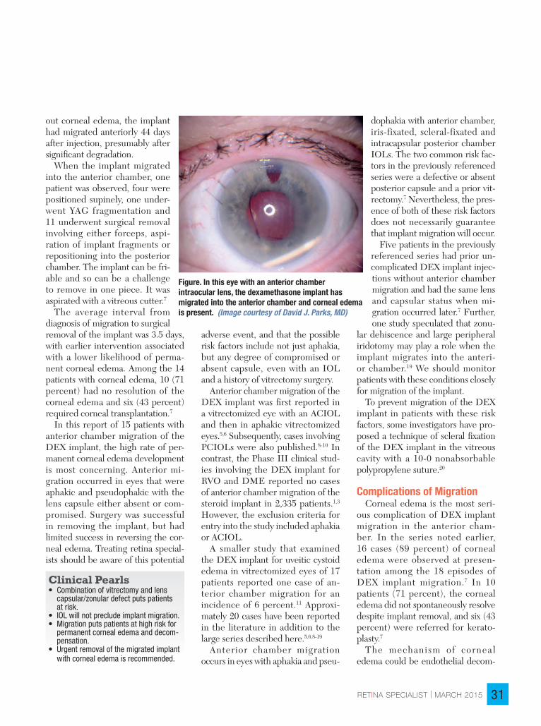
RETINA SPECIALIST | MARCH 2015 31
out corneal edema, the implant had migrated anteriorly 44 days after injection, presumably after signifi cant degradation.
When the implant migrated into the anterior chamber, one patient was observed, four were positioned supinely, one under-went YAG fragmentation and 11 underwent surgical removal involving either forceps, aspi-ration of implant fragments or repositioning into the posterior chamber. The implant can be fri-able and so can be a challenge to remove in one piece. It was aspirated with a vitreous cutter.7
The average interval from diagnosis of migration to surgical removal of the implant was 3.5 days, with earlier intervention associated with a lower likelihood of perma-nent corneal edema. Among the 14 patients with corneal edema, 10 (71 percent) had no resolution of the corneal edema and six (43 percent) required corneal transplantation.7
In this report of 15 patients with anterior chamber migration of the DEX implant, the high rate of per-manent corneal edema development is most concerning. Anterior mi-gration occurred in eyes that were aphakic and pseudophakic with the lens capsule either absent or com-promised. Surgery was successful in removing the implant, but had limited success in reversing the cor-neal edema. Treating retina special-ists should be aware of this potential
adverse event, and that the possible risk factors include not just aphakia, but any degree of compromised or absent capsule, even with an IOL and a history of vitrectomy surgery.
Anterior chamber migration of the DEX implant was fi rst reported in a vitrectomized eye with an ACIOL and then in aphakic vitrectomized eyes.5,6 Subsequently, cases involving PCIOLs were also published.8-10 In contrast, the Phase III clinical stud-ies involving the DEX implant for RVO and DME reported no cases of anterior chamber migration of the steroid implant in 2,335 patients.1,3 However, the exclusion criteria for entry into the study included aphakia or ACIOL.
A smaller study that examined the DEX implant for uveitic cystoid edema in vitrectomized eyes of 17 patients reported one case of an-terior chamber migration for an incidence of 6 percent.11 Approxi-mately 20 cases have been reported in the literature in addition to the large series described here.5,6,8-19
Anterior chamber migration occurs in eyes with aphakia and pseu-
dophakia with anterior chamber, iris-fixated, scleral-fixated and intracapsular posterior chamber IOLs. The two common risk fac-tors in the previously referenced series were a defective or absent posterior capsule and a prior vit-rectomy.7 Nevertheless, the pres-ence of both of these risk factors does not necessarily guarantee that implant migration will occur.
Five patients in the previously referenced series had prior un-complicated DEX implant injec-tions without anterior chamber migration and had the same lens and capsular status when mi-gration occurred later.7 Further, one study speculated that zonu-
lar dehiscence and large peripheral iridotomy may play a role when the implant migrates into the anteri-or chamber.19 We should monitor patients with these conditions closely for migration of the implant.
To prevent migration of the DEX implant in patients with these risk factors, some investigators have pro-posed a technique of scleral fi xation of the DEX implant in the vitreous cavity with a 10-0 nonabsorbable polypropylene suture.20
Complications of MigrationCorneal edema is the most seri-
ous complication of DEX implant migration in the anterior cham-ber. In the series noted earlier, 16 cases (89 percent) of corneal edema were observed at presen-tation among the 18 episodes of DEX implant migration.7 In 10 patients (71 percent), the corneal edema did not spontaneously resolve despite implant removal, and six (43 percent) were referred for kerato-plasty.7
The mechanism of cornea l edema could be endothelial decom-
Figure. In this eye with an anterior chamber intraocular lens, the dexamethasone implant has migrated into the anterior chamber and corneal edema is present. (Image courtesy of David J. Parks, MD)
Clinical Pearls• Combination of vitrectomy and lens
capsular/zonular defect puts patients at risk.
• IOL will not preclude implant migration.• Migration puts patients at high risk for
permanent corneal edema and decom-pensation.
• Urgent removal of the migrated implant with corneal edema is recommended.
030_rs0315_Ozurdex_ja1.indd 31030_rs0315_Ozurdex_ja1.indd 31 3/17/15 4:49 PM3/17/15 4:49 PM

RETINA SPECIALIST | MARCH 201532
pensation either due to chemical toxicity from any component of the DEX implant (dexamethasone, lactic acid or glycolic acid) or mechanical trauma from a rigid object. Specular microscopy has demonstrated cor-neal endothelial cell loss when the DEX implant migrates in the anteri-or chamber.10
High doses of dexamethasone are cytotoxic to corneal endothelial cells, inducing both apoptosis and necro-sis.21 Corneal edema occurs with ear-ly migration (within less than three weeks).7
Two previously reported cases in which the implant migrated at five weeks and three months after injection did not develop corneal edema.6,12 With late migration (oc-curring after three weeks), the im-plant may have degraded enough to cause minimal corneal toxicity from the dexamethasone, but further study is required.
Anterior chamber migration is not unique to the DEX implant. It was first reported after intravitreal in-jection of triamcinolone acetonide, resulting in a pseudohypopyon with the steroid crystals in the anterior chamber.22,23 Anterior chamber mi-gration is also possible with another intravitreal device involving the fl uo-cinolone acetonide implant (Iluvien, Alimera Sciences).
The FDA approved Iluvien in 2014 for treatment of DME in pa-tients previously treated with corti-costeroids who did not have a rise in intraocular pressure. It contains 0.19 mg of fluocinolone acetonide and is injected into the vitreous cavity. Because it is a free-fl oating implant (and non-tethered like the DEX im-plant), Iluvien can migrate into the anterior chamber in vitrectomized eyes. Cases of anterior chamber migration have been reported,
but none have been published. The package insert for Iluvien
carries a warning that notes that patients in whom the posterior cap-sule is either absent or torn are at risk of implant migration into the anterior chamber. The incidence of corneal issues with the fl uocinolone acetonide implant that migrates anteriorly is not known.
However, corneal issues have been reported with another fluo-cinolone acetonide intravitreal im-plant, Retisert (Bausch + Lomb),
which has been reported to migrate anteriorly and cause corneal ede-ma.24,25 Retisert does have a larger dose of fl uocinolone acetonide than Iluvien—0.59 mg vs. 0.19 mg—and both of these large doses of steroid may be toxic to the corneal endothelium.
Retina specialists should be aware of this potential complication with the fl uocinolone acetonide implants and exercise caution in the setting of anterior chamber migration and corneal edema.
Migrating Implant
Reports of Dexamethasone Implant Migration in the LiteratureType of analysis Episodes
reported (n)History Lens status
Case report5 1 Prior anterior vitrectomy ACIOLRetrospective review6
3 Post-lensectomy vitrectomy
Aphakia
Retrospective review7
18 (15 patients)
Absence of lens capsule and prior pars plana vitrectomy
Aphakia (6); ACIOL (4); scleral-fi xated PCIOL (2); PCIOL (2); iris-fi xated PCIOL (1)
Case report8 1 Prior vitrectomy AphakiaCase report9 1 Prior pars plana vitrectomy Scleral-fi xated PCIOLCase report10 3 Prior pars plana vitrectomy 2 ACIOL and 1 PCIOL with
zonular ruptureRetrospective review11
1 Uveitic CME and pars plana vitrectomy
Aphakia
Case report12 1 Peripheral iridotomy PCIOLCase report13 1 Prior pars plana vitrectomy Iris-claw ACIOL lens
implantationCase report14 1 Chronic CME secondary
to idiopathic intermediate uveitis
NA
Case report15 1 Pseudophakic CME; ruptured lens capsule
ACIOL
Case report16 1 Macular edema due to CRVO
Scleral-fi xated PCIOL
Case report17 1 Hemi-retinal vein occlu-sion, neovascular glauco-ma, hyphema, recurrent vitreous hemorrhage, pars plana vitrectomy, pan-retinal photocoagulation, Ahmed valve implant
Pseudophakia
Retrospective review18
4 Noninfectious posterior uveitis
Aphakia
Case report19 1 Noninfectious posterior uveitis
PCIOL with zonular com-promise
FEATURE
030_rs0315_Ozurdex_ja1.indd 32030_rs0315_Ozurdex_ja1.indd 32 3/17/15 4:50 PM3/17/15 4:50 PM

RETINA SPECIALIST | MARCH 2015 33
Management OptionsA few management options exist
in the setting of anterior chamber migration of the DEX implant. Ob-servation may be considered in the absence of corneal edema. Supine positioning of the patient and pupil dilation in aphakic eyes may allow the implant to fall back posteriorly.14
Pilocarpine may reduce pupil size either primarily or following repo-sitioning to minimize the chance of the implant moving anteriorly, but recurrent migration may still occur.6 A slit-lamp procedure using a needle to reposition the implant posteriorly is an option as well, but the implant may return.13 Typically, no corneal edema is present with late migration (after three weeks), so in these cases observation could be employed.
If the implant does migrate into the anterior chamber and corneal edema ensues, it should be removed as soon as possible to prevent corneal decompensation. Early removal of the DEX implant has been known to reduce the likelihood of perma-nent corneal endothelial damage.7 This suggests that removing the im-plant earlier is better for the corneal endothelium and preventing chronic corneal edema.
With regards to the surgical technique for removing the DEX implant, an anterior segment approach using viscoelastic to coax the implant toward a limbal incision can be used if the implant is not fri-able. Once it moves to the incision, the implant can be removed by gen-tly grasping it with a forceps.
An elegant surgical technique involving standard vitreoretinal instrumentation, viscoelastic, a mod-ified Sheets glide and angled for-ceps to avoid fragmentation of the implant and limit iatrogenic morbid-ity has also been described.26 A help-
ful surgical pearl involves using an angled McPherson forceps to grasp the implant parallel to its long axis and avoid dissolution of the implant into multiple particulate fragments.26
A friable implant can be diffi cult to grasp with forceps or other sur-gical instruments, and it could dis-integrate into numerous fragments with minimal manipulation. In these situations, aspiration with a vitreous cutter or similar instrument may be necessary to remove the implant. Be prepared for these scenarios and anticipate the possible need for vit-reoretinal instrumentation when at-tempting to remove the implant.7
The package insert for the dexa-methasone implant was modifi ed in September 2012 to refl ect that con-traindications include aphakia and an ACIOL with rupture of the posterior capsule. However, the combination of compromised zonular or posterior capsular integrity and a history of vit-rectomy makes anterior migration of the DEX implant more likely.
It is important to emphasize that the presence of an IOL alone will not prevent migration in the absence of an intact posterior lens capsule. When migration occurs, the patient is at risk for corneal edema and decompensation. If corneal edema does occur, I recommend urgent removal of the DEX implant.
Disclosures: Dr. Khurana dis-closed that he is a consultant and in-vestigator for Allergan; a consultant and speaker for Genentech; and a consultant, speaker and investigator for Regeneron.
References1. Haller JA, Bandello F, Belfort R Jr., et al. Randomized, sham-controlled trial of dexamethasone intravitreal implant in patients with macular edema due to retinal vein occlusion. Ophthalmology. 2010;117:1134-1146 e1133.2. Lowder C, Belfort R Jr., Lightman S, et al. Dexamethasone intravitreal implant for noninfectious intermediate or posterior uveitis. Arch Ophthalmol. 2011;129:545-553.3. Boyer DS, Yoon YH, Belfort R Jr., et al. Three-year, randomized,
sham-controlled trial of dexamethasone intravitreal implant in patients with diabetic macular edema. Ophthalmology. 2014;121:1904-1914.4. Boyer DS, Faber D, Gupta S, et al. Dexamethasone intravitreal implant for treatment of diabetic macular edema in vitrectomized patients. Retina. 2011;31:915-923.5. Pardo-Lopez D, Frances-Munoz E, Gallego-Pinazo R, Diaz-Llopis M. Anterior chamber migration of dexamethasone intravitreal implant (Ozurdex®). Graefes Arch Clin Exp Ophthalmol. 2011.6. Bansal R, Bansal P, Kulkarni P, Gupta V, Sharma A, Gupta A. Wandering Ozurdex® implant. J Ophthalmic Infl amm Infect. 2012;2:1-5.7. Khurana RN, Appa SN, McCannel CA, et al. Dexamethasone implant anterior chamber migration: risk factors, complications, and management strategies. Ophthalmology. 2014;121:67-71.8. Cronin KM, Govind K, Kurup SK. Late migration of dexamethasone implant into anterior chamber. Arch Ophthalmol. 2012;130:711.9. Voykov B, Bartz-Schmidt KU. Dislocation of dexamethasone intravitreous implant. Arch Ophthalmol. 2012;130:706.10. Malcles A, Janin-Manifi cat H, Yhuel Y, et al. [Anterior chamber migration of intravitreal dexamethasone implant (Ozurdex®) in pseudophakic eyes: Report of three cases.]. J Fr Ophtalmol. 2013;36:362-367.11. Adan A, Pelegrin L, Rey A, et al. Dexamethasone intravitreal implant for treatment of uveitic persistent cystoid macular edema in vitrectomized patients. Retina. 2013;33:1435-1440.12. Eadie JA LR, Capone, Jr A. Migration of Ozurdex implant into the anterior chamber. Retinal Cases and Brief Reports. 2012;6:269-270.13. Vela JI, Crespi J, Andreu D. Repositioning of dexamethasone intravitreal implant (Ozurdex®) migrated into the anterior chamber. Int Ophthalmol. 2012;32:583-584.14. Kishore SA, Schaal S. Management of anterior chamber dislocation of dexamethasone implant. Ocul Immunol Infl amm. 2013;21:70-71.15. Marin-Lambies C, Gallego-Pinazo R, Garcia-Delpech S, Diaz-Llopis M. [Ozurdex® and aphakia: a combination to avoid.] Arch Soc Esp Oftalmol. 2012;87:191-192.16. Laplace O, Rodallec T, Akesbi J, Sandali O. [Anterior chamber migration of a dexamethasone implant in a pseudophakic patient with a scleral-fi xated posterior chamber intraocular lens]. J Fr Ophtalmol. 2013;36:e59-61.17. Collet B. Management of Ozurdex in the anterior chamber. JAMA Ophthalmol. 2013;131:1651-1652.18. Bratton ML, He YG, Weakley DR. Dexamethasone intravitreal implant (Ozurdex) for the treatment of pediatric uveitis. JAAPOS. 2014;18:110-113.19. Kocak N, Ozturk T, Karahan E, Kaynak S. Anterior migration of dexamethasone implant in a pseudophakic patient with intact posterior capsule. Indian J Ophthalmol. 2014;62:1086-1088.20. Mateo C, Alkabes M, Bures-Jelstrup A. Scleral fi xation of dexamethasone intravitreal implant (Ozurdex®) in a case of angle-supported lens implantation. International ophthalmology. 2014;34:661-665.21. Chen WL, Lin CT, Yao CC, et al. In-vitro effects of dexamethasone on cellular proliferation, apoptosis, and Na+-K+-ATPase activity of bovine corneal endothelial cells. Ocul Immunol Infl amm. 2006;14:215-223.22. Ruiz-Moreno JM, Montero JA, Artola A, Barile S. Anterior chamber transit of triamcinolone after intravitreal injection. Arch Ophthalmol. 2005;123):129-130.23. Moshfeghi AA, Scott IU, Flynn HW, Jr., Puliafi to CA. Pseudohypopyon after intravitreal triamcinolone acetonide injection for cystoid macular edema. Am J Ophthalmol. 2004;138:489-492.24. Akduman L, Cetin EN, Levy J, Becker MD, Mackensen F, Lim LL. Spontaneous dissociation and dislocation of Retisert pellet. Ocul Immunol Infl amm. 2013;21:87-89.25. Chang PY, Kresch Z, Samson CM, Gentile RC. Spontaneous dissociation of fl uocinolone acetonide sustained release implant (Retisert) with dislocation into the anterior chamber. Ocul Immunol Infl amm. 2014:11:1-4.26. Stelton CR TJ, Peterson LT, Khurana RN, Yeh S. Surgical management of anterior chamber migration of a dexamethasone intravitreal implant. Ophthalm Surg Lasers Imaging Retina. 2015. Submitted for publication.
030_rs0315_Ozurdex_ja1.indd 33030_rs0315_Ozurdex_ja1.indd 33 3/17/15 4:50 PM3/17/15 4:50 PM

RETINA SPECIALIST | MARCH 201534
RETINA CEO Edited By Warren Laurita »
Paying for Drugs When the Bill Is DueCash forecasting can help manage those high-limit credit cards. By Paul Lucas
Cash is king! Never more than now has this oft-used business cliché been more appropriate for the med-
ical practice administrator—in particular, a vitreoretinal practice administrator. We all deal with stretching the ever-falling reim-bursement dollar and controlling overhead, but few medical spe-cialists have had to adapt as quick-ly to dramatic fi nancial changes as the retinal subspecialty.
With the advent of high cost FDA-approved injectable and implantable pharmaceuticals, and with more on the way, the protocol for managing cash and the related drug inventories must change to ensure adequate cash is available to meet ongoing oper-ating expenses and drug inventory purchases.
To handle the high volume of drug purchases, the typical practice uses a high-limit credit card. These cards have varying terms, of course, but most offer a grace period for pay-ment with no penalty or interest ranging anywhere from seven to 30 days. Online account access lets you readily see future payment obliga-tions and due dates.
A Sword With Two EdgesComplicating this process are the
various deferred payment terms the drug manufacturers offer. Depend-ing on the drug, these can range from 30 to more than 100 days. Like any credit scenario, this is a two-edged sword. The lengthy payment terms give the practice the opportunity to use most of its drug inventory before the payment due dates. On the other
hand, if the practice does not place controls on cash management, there might not be enough money on hand when the credit card is due.
To ensure this dilemma never aris-es, the retina practice needs some basic cash forecasting tools. Best practices suggest a weekly projection of cash inflows and outflows for at least three months, although a full-year projection is best. Historical patient volumes and related reim-bursements offer the best source for projecting future incomes.
Payroll and payables are easy to account for by reviewing historical fi nancial statements and by breaking down the monthly expenses by week based on invoice-due dates. Drug purchases or, more importantly, the charge date (assuming they’re cred-it card purchase), can be identifi ed by reviewing purchase reports from manufacturers and/or distributors.
Combining these three elements
—reimbursement infl ows, oper-ating expense outfl ows and drug purchase payment dates—gives the details you need to forecast cash. Of course, capital purchases and any related debt-service pay-ments must also be considered. However, these transactions are generally planned in advance and can be incorporated into the cash forecast as the group makes pur-chase approvals.
Bringing Pieces TogetherGiven the fragmented sourc-
es of this critical information, an Excel spreadsheet or something similar can bring all these pieces together so that you can know each week’s cash position and
plan for it well in advance. Like any forecast, the numbers are only as good as the data entered.
Accordingly, the forecast should be updated at least weekly to remain current as well as provide as much advance notice as possible of any potential “tight” weeks that may lie ahead. I provide an example of just a few weeks of such a spreadsheet or reference.
By updating forecast data to actual numbers over time, the fore-cast will incorporate prior cash fl ow into predicted cash flow. With a vigilant eye on the numbers, the practice should find itself in the position to reduce (and hopefully eliminate) any guesswork surround-ing cash management.
Mr. Laurita is administrator at Retina Associates of Cleveland Inc. in Ohio.
Mr. Lucas is CFO and administra-tor of Georgia Retina PC, Atlanta.
This example of a cash forecast spreadsheet shows three weeks of actual revenues and expenses and two weeks of projected.
034_rs0315_retinaceo_rk1.indd 34034_rs0315_retinaceo_rk1.indd 34 3/17/15 3:23 PM3/17/15 3:23 PM
RETINA SPECIALIST | MARCH 2015 35
CODING COMMENTARY
Fairly often, questions arise re-garding the appropriate coding and billing of fundus photogra-phy, CPT 92250, and scanning
computerized ophthalmic diagnostic imaging of the retina (SCODI-R), CPT 92134, also called retinal OCT. According to Medicare’s National Correct Coding Initiative (NCCI), these codes are considered mutual-ly exclusive when performed on the same date of service; bill just one.
We appreciate that some diagnostic technologies, such as scanning laser ophthalmoscopy (SLO), allow simul-taneous capture of a fundus photog-raphy (FP) and optical coherence tomography (OCT) image. Choosing the code to submit on a claim for re-imbursement requires some careful consideration. New instructions were published in CPT Assistant in De-cember 2014 to guide the selection.
Billing Both OCT and FPIn 2000, the NCCI edits bun-
dled the original SCODI code (CPT 92135) with fundus photography (FP) (CPT 92250). In 2011, a new SCO-DI code, 92134, was bundled with FP, 92250.1 A bundle means that just one service, usually the higher-reim-bursed one, will be reimbursed when both are done on the same day.
However, the NCCI edits are not absolute, and under some circum-stances they do allow reimbursement for both services performed on the same day. According to the NCCI edits, the modifi er indicator is “1”, so it is possible to unbundle SCODI-R and FP using modifi er 59, subject to certain limitations.
The General Correct Coding Pol-icies for NCCI Policy Manual for
Medicare Services2 discusses the col-umn 1 and column 2 edits in the Mu-tually Exclusive Edit Table. It states:
The CMS developed the NCCI to prevent inappropriate payment of services that should not be reported together … Each edit table contains edits which are pairs of HCPCS/CPT codes that in general should not be reported together. Each edit has a column one and column two HCPCS/CPT code. If a provider reports the two codes of an edit pair, the column two code is denied, and the column one code is eligi-ble for payment. However, if it is clinically appropriate to utilize an NCCI-associated modifi er, both the column one and column two codes are eligible for payment.Using modifier 59 to break the
NCCI edit must be “clinically appro-priate”.3 CMS published the Modifi -er 59 article providing further guid-ance on this issue. It states:
Modifi er 59 is used appropriately for different anatomic sites during the same encounter only when pro-cedures which are not ordinarily performed or encountered on the same day are performed on differ-ent organs, or different anatomic regions, or in limited situations on different, non-contiguous lesions in different anatomic regions of the same organ.It further states: “Treatment of
posterior segment structures in the eye constitutes treatment of a sin-gle anatomic site.” If FP and OCT are done in a single encounter, the use of modifier 59 precludes this guidance. Further, because the posterior segment of the eyeis considered a contiguous struc-
ture, the guidance rarely supports breaking the NCCI edit.
Coding for SLOIn April 1999, CPT Assistant an-
swered a question about proper cod-ing for SLO.4 It stated:
CPT 92135 Scanning computer-ized ophthalmic diagnostic imaging (eg, scanning laser) with interpreta-tion and report, unilateral, is intend-ed to report a method of objective measuring involving a quantitative determination of the thickness of the retinal nerve fiber layer and computer analysis of the data with the fi nal results of creation of a da-tabase fi le, saving data for further comparing in follow-up examina-tions. It is not appropriate to as-sign CPT code 92135 for scanning laser fundus photography. CPT code 92250 Fundus photography with interpretation and report, that describes generation of retinal im-age only, and not data generation, would be appropriately assigned for this procedure.Fifteen years later in 2014, in
the context of new technology that expanded the capabilities of SLO, the same question was posed to CPT As-sistant. The answer, in the November 2014 issue,5 was different in a subtle and nuanced manner:
Q: Is it appropriate to report CPT code 92135 (now codes 92133 and 92134) for this method of examina-tion of the fundus? A: If the scanner produces an image of the retina or optic nerve along with other data and imaging for quantitative anal-ysis, it would be appropriate to report a single service from
By Kirk A. Mack, COMT, COE, CPC, CPMA »
OCT and FP: Why can’t I bill both?New guidance from CMS describes why and the few times when you can.
035_rs0315_coding_rk3 copy.indd 35035_rs0315_coding_rk3 copy.indd 35 3/17/15 12:40 PM3/17/15 12:40 PM

RETINA SPECIALIST | MARCH 201536
the appropriate scanning computerized ophthal-mic diagnostic imaging code range (92133-92134). If only an image is obtained then code 92250 would be reported.It goes on to explain that medical necessity of the FP or
OCT service depends on what is needed rather than what is done.
It is important to note that if the only necessary service provided is generating a fundus photograph without the need to quantify the nerve fi ber layer thickness and to ana-lyze the data via a computer, then code 92250 is appropri-ate even if the photograph was taken with a scanning laser.
Pick Just One, But Which One?When FP and OCT are performed concurrently, some
billers routinely choose the test with the highest reimburse-ment, typically fundus photography, to submit on a claim. In light of the discussion here, that approach is too simplis-tic and ignores the medical necessity criterion.
The individual condition of the patient is a better starting point when selecting which code to bill; the retina specialist should make the choice rather than depend on the biller to pick the code that represents more dollars.
ConclusionAdvances in ophthalmic imaging, such as SLO, permit si-
multaneous capture of FP and OCT, but Medicare’s NCCI edits permit billing just one CPT code, 92250 or 92134, in most cases.
Old instructions in CPT Assistant stipulated that 92250 is the correct code to report SLO. New instructions in December 2014 say it depends on whether qualitative or quantitative information is required for the patient’s con-dition. No single answer suits every situation; one needs to exercise clinical judgment to make an appropriate code selection.
Mr. Mack is a senior consultant with the Corcoran Con-sulting Group. He is based in Galveston, Texas.
References1. CMS.gov. NCCI Coding Edits. Version 21.0. Effective January 1, 2015. Available at: http://www.cms.gov/Medicare/Coding/NationalCorrectCodInitEd/NCCI-Coding-Edits.html. Accessed January 20, 2015.2. CMS.gov. NCCI Policy Manual for Medicare Services. Effective January 1, 2015. Chapter 1. Available at: http://www.cms.gov/Medicare/Coding/NationalCorrectCodInitEd/index.html?redirect=/NationalCorrectCodInitEd/01_overview.asp. Accessed January 20, 2015. 3. CMS.gov. Modifi er 59 Article: Proper Usage Regarding Distinct Procedural Service. Updated 10/28/2014. Available at: http://www.cms.gov/Medicare/Coding/NationalCorrectCodInitEd/downloads/modifi er59.pdf. Accessed January 20, 2015. 4. Coding Clarifi cation. Medicine Special Ophthalmological Services. American Medical Association. CPT Assistant. April 1999; 10.5. Coding Clarifi cation. Special Ophthalmological Services (92133, 92134). American Medical Association CPT Assistant. November 2014;10.
Can Anti-VEGF Cause GA in Wet AMD?(Literature Review, continued from page 14)unclear whether the GA that develops in patients under-going anti-VEGF therapy secondary to neovascular AMD results from macular drying followed by normal disease progression, or whether VEGF inhibition has a neurotoxic effect on the macula, thus causing the more frequent ap-pearance and more rapid growth of GA.
We await the formal publication of the HARBOR Study, in which 2 mg ranibizumab and 0.5 mg ranibizumab were compared using both monthly and PRN regimens. While no data is available on afl ibercept and the incidence of GA in the VIEW trials, an ongoing study in Australia comparing ranibizumab and aflibercept to determine whether one causes more GA than the other in neovascular AMD should prove helpful in resolving this question.
As we debate whether anti-VEGF therapy causes GA, there is no question about the effi cacy of anti-VEGF ther-apy for the treatment of MNV in AMD, and we should not withhold treatment. The goal continues to be a dry macula. However, once the macula is dry and GA formation is con-fi rmed, the challenge we have is whether we should consid-er a less aggressive regimen. This question of anti-VEGF therapy and GA will have particular relevance in the future when sustained-release formulations reach our clinics.
Disclosure: Dr. Rosenfeld disclosed he is a consultant to Genentech.
References1. Brown DM, Kaiser PK, Michels M, Soubrane G, et al. Ranibizumab versus verteporfi n for neovascular age-related macular degeneration. N Engl J Med. 2006, 355:1432-1444.2. Rosenfeld PJ, Brown DM, Heier JS, et al. Ranibizumab for neovascular age-related macular degeneration. N Engl J Med. 2006;355:1419-1431.3. Heier JS, Brown DM, Chong V, et al. Intravitreal afl ibercept (VEGF trap-eye) in wet age-related macular degeneration. Ophthalmology. 2012;119:2537-2548.4. Martin DF, Maguire MG, Fine SL, et al. Ranibizumab and bevacizumab for treatment of neovascular age-related macular degeneration: two-year results. Ophthalmology. 2012;119:1388-1398.5. Chakravarthy U, Harding SP, Rogers CA, et al. Alternative treatments to inhibit VEGF in age-related choroidal neovascularisation: 2-year fi ndings of the IVAN randomised controlled trial. Lancet. 2013;382:1258-1267.6. Brown DM, Michels M, Kaiser PK, et al. Ranibizumab versus verteporfi n photodynamic therapy for neovascular age-related macular degeneration: Two-year results of the ANCHOR study. Ophthalmology. 2009;116:57-65 e55.7. Rosenfeld PJ, Shapiro H, Tuomi L, et al. Characteristics of patients losing vision after 2 years of monthly dosing in the phase III ranibizumab clinical trials. Ophthalmology. 2011;118:523-530.8. Grunwald JE, Daniel E, Huang J, et al. Risk of geographic atrophy in the Comparison of Age-related Macular Degeneration Treatments Trials. Ophthalmology. 2014;121:150-161.9. Grunwald JE, Pistilli M, Ying GS, et al. Growth of geographic atrophy in the Comparison of Age-related Macular Degeneration Treatments Trials. Ophthalmology. 2014. Dec. 24. [E pub ahead of print]10. Xu L, Mrejen S, Jung JJ, et al. Geographic atrophy in patients receiving anti-vascular endothelial growth factor for neovascular age-related macular degeneration. Retina. 2015;35:176-186.11. Ying GS, Kim BJ, Maguire MG, et al. Sustained visual acuity loss in the comparison of age-related macular degeneration treatments trials. JAMA Ophthalmology. 2014;132:915-921.12. Tanaka E, Chaikitmongkol V, Bressler SB, Bressler NM. Vision-threatening lesions developing with longer-term follow-up after treatment of neovascular age-related macular degeneration. Ophthalmology. 2015;122:153-161.13. Channa R, Sophie R, Bagheri S, et al. Regression of choroidal neovascularization results in macular atrophy in anti-vascular endothelial growth factor-treated eyes. Am J Ophthalmol. 2015;159:9-19.14. Kumar N, Mrejen S, Fung AT, et al. Retinal pigment epithelial cell loss assessed by fundus autofl uorescence imaging in neovascular age-related macular degeneration. Ophthalmology. 2013;120:334-341.
035_rs0315_coding_rk3 copy.indd 36035_rs0315_coding_rk3 copy.indd 36 3/17/15 12:40 PM3/17/15 12:40 PM

RETINA SPECIALIST | MARCH 2015 37
INNOVATION INSIGHT By Richard Mark Kirkner »
Selecting Patients for the Iluvien ImplantSustained-release device emerges after a decade-long journey.
With the shipments of the first supplies of the multi-year, sustained-re-lease implant Iluvien,
vitreoretinal surgeon Sanford Chen, MD, acquired another option for patients with diabetic macular ede-ma (DME) who do not completely respond to anti-VEGF therapy. “If you look at the numbers of patients on chronic care, 25 percent or more of them continue to have persistent swelling, and a significant number don’t achieve better than 20/40 visual acuity as well,” he says.
Iluvien releases submicrogram levels of fluocinolone acetonide to the retina for the treatment of DME. The Food and Drug Admin-istration (FDA) approved Iluvien in September 2014, and Alimera Sciences spent the intervening months building inventory and a sales force, says Dan Myers, Alim-era co-founder, president and CEO. Iluvien contains 0.19 mg of fl uocino-lone acetonide.
When Anti-VEGF Doesn’t WorkDr. Chen, who practices at Or-
ange County Retina in Southern California and is a member of the Fluocinolone Acetonide in Diabet-ic Macular Edema (FAME) Study Group, says the implant also meets a need for patients who cannot sustain the burden of monthly anti-VEGF therapy. “Patients with diabetes are usually younger and productive members of society,” Dr. Chen says.
“They also frequently have other comorbities (such as heart and kid-ney disease) that require follow-up at other doctors’ offi ces. This may make it diffi cult for them to come
into the offi ce every month for intravitreal anti-VEGF injections,” Dr. Chen says.
Another population well-suited to the implant may be non-responders or suboptimal responders to anti- VEGF agents, Dr. Chen says. “We know through research that DME is a multifactorial disease and there is probably more than just a VEGF component; there is likely an infl am-matory component to it as well,” he says.
Patients who have had previous vitrectomy or retina surgery repre-sent a lesser-known group that may benefit from the Iluvien implant. “Having the implant may help in their treatment, because once a patient has had a vitrectomy, the anti-VEGF agent literally can be cleared out in a week,” Dr. Chen says. “The pharmacokinetics of the Iluvien implant follows zero-order kinetics and allows for slow release up to 36 months.”
An Education ProcessFDA approval of Iluvien culmi-
nated a decade-long journey that in-cluded three prior negative FDA de-cisions. The journey started in 2004 when Alimera acquired the rights to the implant for delivering steroids to the back of the eye. At the time, Mr. Myers had recently started Alimera, having previously been with Novar-tis Ophthalmics, where he served as president from 1997 through 2002.
Mr. Myers chalks up the FDA ex-perience as a learning process. “It’s the same learning process about dia-betic macular edema that the retina community is going through right now,” he says. The breakthrough was
language in the fi nal label indication that required a patient to have had prior steroid treatment with no clin-ically signifi cant rise in IOP.
“We’ve learned, as the retina spe-cialists have learned, that DME is more of a multifactorial disease than wet age-related macular degener-ation,” Mr. Myers says. “Multiple cytokines are involved, and DME has more of an infl ammatory aspect than we realized a few years ago.”
In 2011, when the FDA issued a critical Complete Response Letter on Iluvien, clinical opinions about the use of steroids in the treatment of DME were just forming. “May-be the view questioning if a steroid would be necessary for treating DME in 2011 progressed to where we find ourselves today: where there’s no question there will be a large number of patients who will need both anti-VEGF therapy and treatment by steroids as well,” Mr. Myers says.
Now those patients with DME have a multi-year option.
The Iluvien implant.
037_rs0315_innovation_rk2.indd 37037_rs0315_innovation_rk2.indd 37 3/18/15 11:06 AM3/18/15 11:06 AM
RETINA SPECIALIST | MARCH 201538
CLINICAL TRIAL Q & A With Neil B. Bressler, MD»
A Quick Look at CHROMA, SPECTRILead investigator provides details on parallel trials of lampalizumab.
CHROMA and SPECTRI are parallel Phase III trials inves-tigating lampalizumab for the treatment of geographic atro-
phy (GA) in age-related macular de-generation (AMD). The identically designed, double-masked, random-ized studies will compare a 10-mg dose of lampalizumab administered every four or six weeks by intravitreal injection to sham injections. The tri-als are currently enrolling patients.
The MAHALO Phase IB/II trial preceded SPECTRI and CHRO-MA. MAHALO was a cohort study of 108 fellow untreated eyes of 143 patients with GA resulting from AMD. Comparing optical coher-ence tomography images at base-line and 18 months after the start of treatment with lampalizumab, the MAHALO study authors concluded that GA lesions seemed to enlarge at a slower rate in eyes with outer retinal tabulation (ORT) than in eyes without ORT.
Neil B. Bressler, MD, of Wilmer Eye Institute, Baltimore, and chair of the trials’ Executive Advisory Committee, answered questions via e-mail about the twin trials.
How do CHROMA and SPECTRI differ from the MAHALO phase II trial?
SPECTRI and CHROMA are Phase III studies being
done in parallel, whereas MAHA-LO was a Phase II study. The Phase II study was designed to determine if it was feasible to proceed with a Phase III trial. The Phase III trials are designed to identify, confi dently, if there is a benefi t, with
acceptable safety, of lampalizumab in the management of geographic atrophy from age-related macular degeneration.
What are the primary and secondary endpoints of the twin trials?
The primary outcome is change in geographic atrophy
area from baseline, as assessed by retinal imaging. The key second-ary outcomes include change in best-corrected visual acuity from baseline and change in additional measures of visual function.
How many centers will participate in the trials?
Approximately 275 centers are anticipated to enroll study
participants across more than 20 countries around the world between CHROMA and SPECTRI.
How many patients will ultimately be enrolled?
SPECTRI and the similarly designed CHROMA trials are
anticipated to enroll 936 patients in each study.
When will enrollment cease?
Enrollment likely will cease when the planned sample size
of 936 study participants has been attained in each study, assuming enrollment is being completed in a timely fashion and no substantive changes to the protocol are under-taken that might affect this plan.
Are the studies accepting any more centers/investigators?
At this point there are no plans to add new sites to CHRO-
MA-SPECTRI, but there are other studies being planned in GA. Sites can contact Roche (Genentech in the United States) if interested.
How would they contact the trial leaders?
Investigators interested in discussing possible participa-
tion in the study can contact Roche (www.roche.com/about_roche/roche_worldwide.htm) and refer-ence Study ID Number: GX29176.
When would the earliest Phase III results be available?
After the last patient enrolls, that last study participant will
reach the primary outcome one year later and complete the last study visit two years later. Additional time, is needed to analyze the data and pre-pare a report for regulatory author-ities. These processes dictate how soon the results would be available.
Q
Q
Q
Q
Q
Q
Q
A
A
A
A
A
A
A
A
Long Story Short: CHROMA and SPECTRIFormal study title: A Study Investigating the Safety and Effi cacy of Lampalizumab Intravitreal Injections in Patients With Geographic Atrophy Secondary to Age-Related Macular Degeneration. (SPECTRI and CHROMA share the same title.)
ClinicalTrials.gov Identifi ers: SPECTRI, NCT02247531; CHROMA, NCT02247479.
Q
038_rs0315_CTQA_ja_rk2.indd 38038_rs0315_CTQA_ja_rk2.indd 38 3/17/15 3:55 PM3/17/15 3:55 PM

Optos has the only imaging technology that can capture ultra-widefield images of
200° or 82% of the retina. We have expanded our already extensive variety of imaging
modalities with the addition of Indocyanine Green Angiography - optomap icg.
See more, discover more, and treat more effectively.
Find out more about what’s new from Optos. www.optosnextgen.com
optomap fa optomap icg
optomap optomap af
Building The Retina Company
Optos is Ultra-widefield Retinal Imaging
Images courtesy of SriniV
as Sadda, MD
- Doheny Eye Institute
Visit us at ASCRS - Booth #2708
RS0315_Optos.indd 1RS0315_Optos.indd 1 3/9/15 12:46 PM3/9/15 12:46 PM
The moment you discover visualization that delivers more for retinal assessment. New CIRRUS HD-OCT Smart Imaging.
Introducing the NEW CIRRUS™ HD-OCT scan patterns and en face clinical presets from ZEISS. Targeted visualization
of critical anatomy adds flexibility to your macular assestment while automatic centering improves clinic flow by
eliminating rescans due to missed fovea.
• SMART high-definition B-scan automatically centered on the fovea
• En Face clinical presets quickly localize pathology by retinal layer
Discover how the new SMART OCT imaging applications can deliver MORE for your practice.www.meditec.zeiss.com/cirrus
Carl Zeiss Meditec, Inc. 800.342.9821 www.meditec.zeiss.comCIR.6908 ©2015 Carl Zeiss Meditec, Inc. All rights reserved. Specification subject to change.
NEW!HD Scan Patterns
En Face Clinical
Presets
// SMART OCT MADE BY ZEISS
RS0315_Zeiss.indd 1RS0315_Zeiss.indd 1 3/4/15 3:51 PM3/4/15 3:51 PM